


Abstract
In Sudan, the widespread occurrence of fungal diseases, along with limited access to modern medicine, highlights the therapeutic significance of indigenous medicinal plants. Globally, both medicinal plants and their associated endophytic fungi constitute a major natural reservoir for novel antifungal compounds. This paper reviews the antifungal research undertaken on Sudanese medicinal plants and their endophytic fungi during the period 2000–2025. The literature survey was conducted using the databases Google Scholar, ScienceDirect, PubMed, Scopus, and Web of Science. In this review, a total of 88 plant species belonging to 51 families were investigated for antifungal activity, predominantly against species of Candida, Saccharomyces and Aspergillus. Among these, 16 species were specifically tested against Madurella mycetomatis, the primary causative agent of eumycetoma. The family Fabaceae was the most screened family. Analysis of the publications reviewed indicated that the majority of papers used the cup-plate diffusion method for assaying antifungal activity. The minimum inhibitory concentration was the most prominent assay used to detect anti-M. mycetomatis. Plant extracts of the Combretaceae family, Boswellia papyrifera and Pimpinella anisum essential oil, displayed promising antifungal activity. In addition, Alternaria sp., Curvularia sp. and Pleospora sp. endophytic fungi isolated from Vachellia nilotica and Cymbopogon schoenanthus also showed promising antifungal activity. Further investigation into fungal endophytes is essential to characterize species diversity and identify novel bioactive metabolites. Implementing a modernized research framework for Sudanese medicinal plants is critical for developing novel antifungal leads.
1. Introduction
The Republic of Sudan, located in Northeast Africa, is characterized by remarkable ecological diversity ranging from desert and semi-desert regions in the north to tropical savannah ecosystems with seasonal rainfall in the central and southern regions. Sudan features an exceptionally rich flora comprising 3137 species across 1280 genera and 170 families due to its diverse climates, with an estimated 15% of these plants being endemic [1]. Furthermore, this botanical diversity has made Sudan an important reservoir of medicinal plants that have long been used in traditional healthcare systems. Medicinal plants and their secondary metabolites are characterized by diverse chemical structures, low cost, high availability, strong antimicrobial activity, and minimal side effects. Consequently, these metabolites represent promising resources for the identification and development of novel antifungal drugs [2,3]. The traditional medicinal applications of these Sudanese plants have encouraged many pharmacological investigations. Several extracts and purified compounds have been assessed for their biological activities, especially their antibacterial, antifungal, antioxidant, antimalarial, anti-inflammatory, anticancer, and antidiabetic activities [4,5,6,7,8,9,10]. Nevertheless, systematic studies of the efficacy of these extracts and purified compounds on fungal infections remain relatively limited. Despite the therapeutic promise of Sudanese medicinal flora, phytochemical research remains largely limited to crude-extract screening. Advanced fractionation, and compound isolation are comparatively underrepresented in the literature, likely because of restricted access to analytical infrastructure, resource limitations, shortages of specialized personnel and the broader research constraints faced in Sudan [11].
In general, humans are relatively resistant to invasive fungal diseases. However, in immunocompromised patients and HIV-infected patients, the susceptibility toward opportunistic fungal infections is increased. Candida spp. are the most common isolates from immunocompromised patients, but Aspergillus or Cryptococcus may also be isolated from these patients [12,13]. Fungal infections are ubiquitous worldwide; however, their prevalence is more pronounced in sub-Saharan Africa. In Africa, studies indicate that approximately 8% of the Ethiopian population and 11.5% of the Eritrean population get severe fungal infections annually [14,15]. The substantial research on the prevalence of fungal diseases in Sudan revealed that approximately 5 million (10% of the total population) Sudanese individuals are afflicted by fungal infections. The predominant disease was scalp infection (tinea capitis), followed by recurring vulvovaginitis, fungal eye infections, and eumycetoma [16].
Mycetoma, “Madura foot,” is one of the most devastating fungal-associated diseases in Sudan. It is a chronic and destructive inflammatory skin infection caused by aerobic actinomycetes bacteria (actinomycetoma) or fungi (eumycetoma) [17,18,19]. It is classified as a poverty-related neglected tropical disease (NTD), as it predominantly impacts economically disadvantaged communities in endemic areas [20]. Mycetoma is abundant in tropical and subtropical regions, which are known as the mycetoma belt of Africa and encompasses Sudan, Chad, Ethiopia, Somalia, and Senegal [21]. Sudan has one of the highest rates of this disease globally, with more than 16,000 cases reported [16,21]. The primary culprit, Madurella mycetomatis, typically infects rural agricultural workers through minor skin wounds [22,23]. Treatment for this infection is notoriously difficult. Standard antifungal medications, which have low cure rates, are highly toxic and need years of adherence; they are also unaffordable for most patients [24]. Frequently, surgical amputation is the only viable option, leaving patients further destitute and disabled [25]. Consequently, there is an urgent need to find novel antifungal medicines that are accessible, safe, and effective.
Natural products continue to provide one of the most productive sources of antimicrobial drug leads, and increasing attention has focused on microorganisms associated with medicinal plants. The term “endophyte” refers to microbes (often fungi and bacteria) that can colonize the inner tissues of healthy plants without the manifestation of any disease symptoms [26]; however, they may become pathogenic when the host plant undergoes senescence [27]. Endophytes associated with medicinal plants can produce complex, highly active secondary metabolites [28]. Some of these microorganisms demonstrate enhanced biosynthetic capacities, likely due to genetic exchange with their host plants while living and proliferating inside healthy tissues [29]. There is some evidence which suggests that endophytic fungi provide a promising source of novel drugs. For example, sordaricin from Fusarium spp., are known to possess antifungal capabilities against a variety of infectious disease-causing organisms [30,31]. However, endophytic fungi associated with Sudanese medicinal plants may constitute a promising and largely unexplored source of novel antifungal compounds.
Fungal infections, particularly opportunistic mycoses (Candida, Aspergillus, and Cryptococcus) and NTD such as mycetoma, present a growing global health threat intensified by drug resistance and therapeutic limitations. Although Sudanese medicinal flora and associated fungal endophytes offer rich potential for novel drug discovery, existing research remains fragmented by inconsistent extraction protocols and unstandardized bioassays. The primary objectives of this review are to synthesize literature published between 2000 and 2025, consolidate scattered data on high-promise taxa, evaluate endophytes as sustainable alternatives to wild harvest, and identify current methodological bottlenecks in antifungal research. Ultimately, we highlight key priorities and methodological frameworks to assist future studies in antifungal drug discovery.
2. Methodology
Strategy for searching the literature: A literature review was conducted utilizing various electronic databases, including Google Scholar, ScienceDirect, PubMed, Scopus, and Web of Science. The search strategy was conducted using specific combinations of keywords and Boolean operators. The main search strings applied across databases included:
- (“antifungal” OR “antimicrobial “) AND (“Sudan” OR “Sudanese”) AND “medicinal plants”
- “mycetoma” AND (“Sudan” OR “Sudanese”) AND “medicinal plants”
- “endophytes” AND (“Sudan” OR “Sudanese”) AND “medicinal plants”
- These core search combinations were adapted as required for each database.
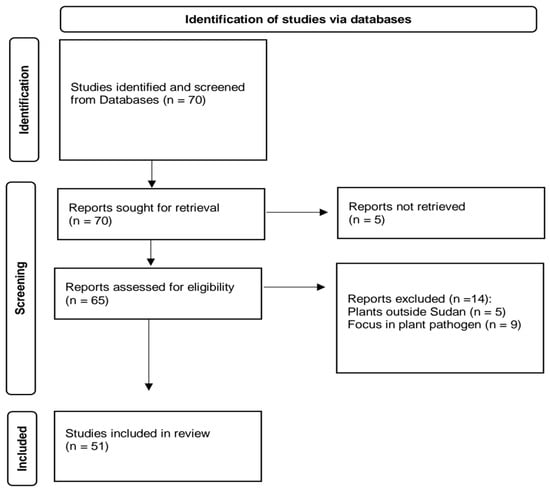
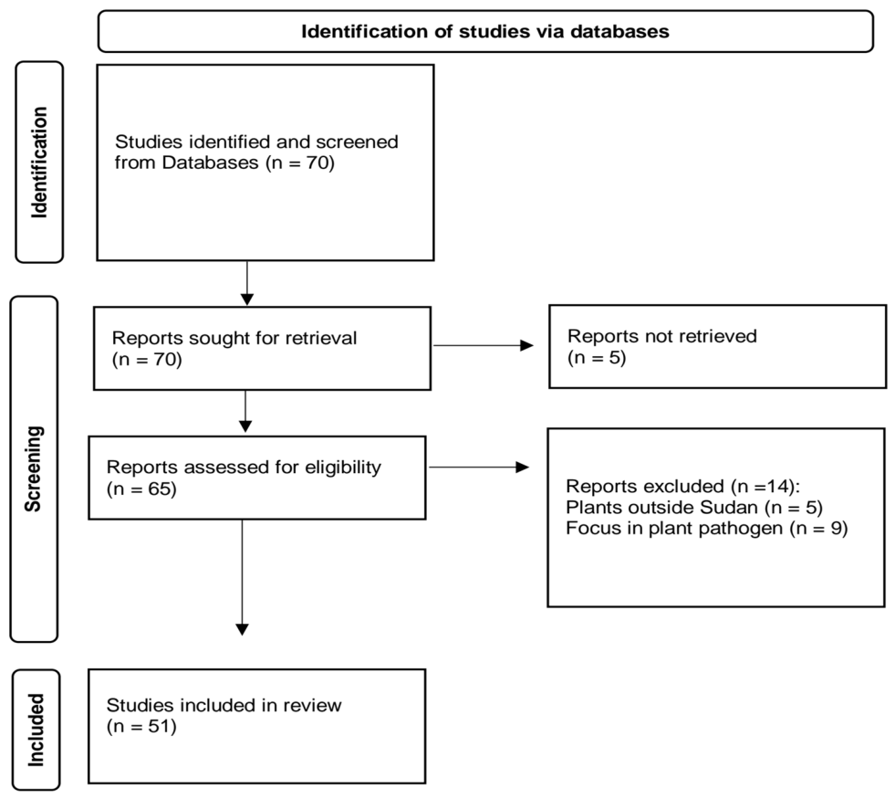
A literature search was conducted up to 29 December 2025, covering publications between 2000 and 2025. However, select relevant studies were cited in the discussion strictly to provide a broader comparative context. To minimize selection bias and ensure data accuracy, title and abstract screening and subsequent data extraction were performed independently by two reviewers in duplicate. The literature selection criteria and multi-stage screening process are illustrated in Figure 1. Out of the 70 identified studies, 65 were assessed and 51 met all inclusion criteria. These 51 studies evaluated a total of 88 plant species across 51 families, as several individual studies screened multiple species and families (for example, one study screened up to 17 plant species).
Figure 1.
Flow chart demonstrating the search strategy and study selection for studies on Sudanese medicinal plants and their endophytes for antifungal activity.
Studies were included if they presented original research published as full peer reviewed articles, full conference proceedings, or conference abstracts (addressing novel or emerging subjects) evaluating the antifungal properties of Sudanese medicinal plants or their associated fungal endophytes against human fungal pathogens, published in English and indexed in major international electronic databases. Conversely, studies were excluded if they: (i) evaluated plant species not grown in Sudan; (ii) targeted exclusively non-human or agricultural pathogens, as the scope of this review is restricted to medical mycology; (iii) were published in local journals inaccessible through international databases due to repository server limitations; or (iv) lacked accessible full text.
Data extraction: Relevant data were organized using Microsoft Word, documenting parameters such as; botanical name, family, plant part utilized, extraction solvent, bioassay methodology, antifungal potency, target fungal species, and references (Table 1).
3. Anti-Fungal Activity of Sudanese Medicinal Plants
3.1. Antifungal Assays and Solvent Extraction
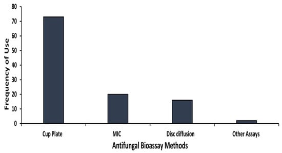
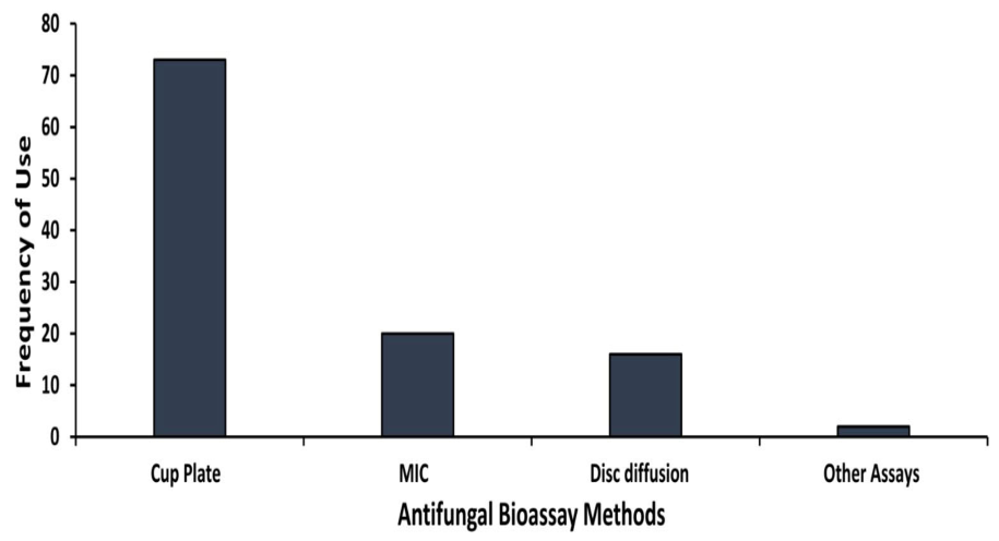
Antifungal studies on Sudanese medicinal plant extracts have been conducted using various methodologies. Figure 2 represents the proportion of studies and methods used to assess antifungal activity, mostly for the period 2000–2025. The most common methods used to investigate the antifungal activity of Sudanese medicinal plants are cup of agar diffusion, minimum inhibitory concentration (MIC) and disc diffusion. There are a few reports that mentioned mycelial fresh weight. Sasidharan et al. [32] reported that the main methods for in vitro antifungal screening are a diffusion method, MIC, minimum fungicidal concentration (MFC) and IC50 value on hyphal growth inhibition, as well as many other in vivo and in situ methods. The cup and disc diffusion studies were the first method of choice, possibly due to their simplicity and capacity to analyze a large number of test samples [33]. Even though disc or agar diffusion methodology is quick and a simple means of screening of antimicrobial activity, problems may arise when investigating oily samples [34,35]. Depending on the chemical nature of the particular sample, diffusion rates may produce inconsistent results [36]. Several papers reported results using both methods. The other antifungal method used in plant extract studies is the bio-autographic assay which was solely used to evaluate antifungal activity of isolated bioactive compounds from Terminalia brownii [37].
Figure 2.
Distribution of experimental bioassay methods used to evaluate antifungal activity of Sudanese medicinal plants.
A critical evaluation of the experimental methodologies compiled in Supplementary Table S1 reveals substantial reporting gaps across the reviewed literature. A significant proportion of the included studies omitted essential assay parameters, such as solvent controls, explicit incubation temperatures/durations, and the number of biological replicates. These parameters are crucial for ensuring experimental reproducibility and allowing readers to accurately interpret antifungal activity. Among the studies that included positive controls, Nystatin and Clotrimazole were the most frequently used reference drugs, with fewer studies incorporating Fluconazole or Amphotericin B. Moreover, positive controls were applied at widely varying concentrations and units (e.g., 5 µg/mL vs. 50 µg/mL, or mg/disc vs. µg/mL).
Additionally, incubation conditions varied widely even across the same fungal species. These inconsistent incubation settings directly impact inhibition zone values, making direct comparisons between different studies difficult. Where reported, most studies utilized two or three replicates, but several omitted this detail altogether. It is important to emphasize that performing and reporting a minimum of three independent replicates (n = 3) is vital for statistical validity. Overall, the observed inconsistency in reporting highlights an urgent need for standardized protocols and adherence to international guidelines when evaluating the antifungal efficacy of plant extracts. Extract concentrations reported in Table 1 varied considerably across studies, ranging from 2.5 to 100 mg/mL. However, the majority of studies employing the agar well (cup) diffusion method used a fixed concentration of 50 mg/mL. Notably, several studies failed to report the volume or concentration of extract applied to the cup or paper disc. This lack of standardization represents a major methodological limitation in the existing literature on Sudanese medicinal plant antifungal research. Furthermore, the majority of the reviewed literature focuses on the preliminary screening of crude extracts, a finding that aligns with the observations of Abdalla and Abdalla [38]. These authors noted that research often remains at the screening stage due to a lack of specialized facilities and a deficiency in research funding. This trend shows the important need for increased investment and infrastructure support for scientific research in Sudan. Even though the country has a vast botanical wealth and its potential as a source for novel medicinal compounds, these systemic challenges hinder the progression from crude extract analysis to the isolation and characterization of pure bioactive compounds [39]. Among the studies reviewed, methanol (28.3%) was identified as the most prevalent solvent for extraction, followed by chloroform (26.2%) and water (23.6%). Methanol is widely preferred in antifungal research due to its high extraction yield and strong organic polarity. This property enables it to dissolve a broad spectrum of bioactive phytochemicals, ranging from highly polar compounds to medium- and low-polarity constituents. Furthermore, methanol’s low boiling point allows for rapid evaporation via rotary evaporators. However, it is not always the best choice, since solvent selection depends on the chemical nature of the target compounds. Water remains important for safety and traditional relevance, while ethanol offers a useful compromise between efficiency and lower toxicity. Chloroform and ethyl acetate are useful for less polar compounds, but their toxicity limits direct application. Essential oils (EO) are also important antimicrobial extracts, although their volatility and compositional variability can complicate standardization [40,41].
3.2. Plant Species, Botanical Families, and Utilized Organs
Table 1 summarizes the collected reports, detailing 78 plant species belonging to 43 families investigated for antifungal activity. This represents 25.3% of the 170 plant families documented in Sudan [1]. The top five families based on the number of species tested for antifungal activity were Fabaceae (n = 12), Malvaceae (n = 6), Asteraceae (n = 5), Euphorbiaceae (n = 5), and Combretaceae (n = 5). This pattern partially aligns with the global dataset summarized by Shivangani and Hariprasad [42], who reported that the most investigated plant families for antifungal activity are Lamiaceae, Fabaceae, Asteraceae, Myrtaceae, Apiaceae, Lauraceae, Rutaceae, Solanaceae, Euphorbiaceae, and Meliaceae. Notably, although Combretaceae is not among the ten most frequently studied antifungal families in that global review, its prominence in African traditional medicine, particularly for dermatological and antifungal applications, explains its strong representation in our findings. The majority of the plant families have been found to have antifungal properties that belong to Angiosperms (flowering plants). The only exception is the Salviniaceae family, which represents the Pteridophytes (seedless vascular plants). This taxonomic distribution reflects both the historical importance and pharmacological value of flowering plants in Sudanese traditional medicine.
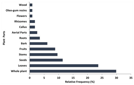
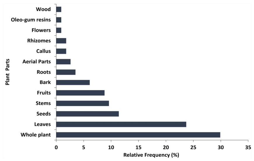
Figure 3 illustrates the distribution of various plant parts reported to possess antifungal activity. The analysis reveals that the whole plant represents the most frequently studied category, making up 29.9% of the literature. This is followed by leaves (23.7%) and seeds (11.4%). Other investigated components include stems (9.6%), fruits (8.8%), bark (6.1%), and roots (3.5%). While the whole plant ranks highest in this specific review, in general, antifungal research tends to focus more on leaves. This preference may be attributed to their environmental role; as leaves often produce a diverse array of antifungal secondary metabolites as a defense mechanism [42].
Figure 3.
Plant parts reported to contain antifungal activity of Sudanese medicinal plants.
3.3. Targeted Fungal Pathogens
The reviewed literature identified various fungal targets, the majority of which belong to the phylum Ascomycota (sac fungi). The most frequently investigated species were Candida albicans, Saccharomyces cerevisiae and Aspergillus niger. This finding agrees with the report by Abdalla and Abdalla [38]. They suggested that these species received more attention due to their ease of culturing and general availability. Furthermore, they were likely randomly selected for initial screening studies. Specifically, Aspergillus was chosen as a typical fungus with significant medical and commercial importance, while Candida was selected as a typical yeast responsible for many superficial human infections. Additionally, a few studies evaluated the activity against Aspergillus flavus and several dermatophytes, including; Epidermophyton floccosum, Microsporum audouinii, and various Trichophyton species such as T. rubrum, T. concentricum, and T. tonsurans. Although plant-derived antifungal extracts typically show stronger activity against Candida albicans than against A. niger or S. cerevisiae, due to the thicker cell walls and complex conidial structures of filamentous fungi [43,44], our data in Table 1 shows an inverse pattern, with A. niger and S. cerevisiae being more susceptible. This indicates that fungal susceptibility is strongly influenced by extract composition, solubility, assay conditions, and plant source. Strain-specific resistance in Candida, including efflux activity, cell wall remodeling, biofilm formation, and growth-stage differences, can further reduce its sensitivity [45]. These findings emphasize the need to screen multiple fungal species and strains under varied conditions to identify robust antifungal leads.
3.4. Bioactive Components of Medicinal Plants
Some reports have mentioned general phytochemical screening and the majority indicate the presence of flavonoids, polyphenols, tannins and triterpenes which accumulate in the polar extracts such as Portulaca oleracea and Combretum hartmannianum [46,47]. Lawsonia inermis, a commercially important cultivated medicinal plant, demonstrated strong antifungal activity. The phytochemical analysis revealed the presence of anthraquinones and tannic acid in high concentrations [47,48]. Also, three reports confirm the activity of Hydnora abyssinica rhizome extract as a good antifungal which showed the presence of condensed tannins, cardiac glycosides, phenols, flavonoids and reducing sugars [49,50,51]. Adansonia digitata pulp extract showed antifungal activity, the HPLC analysis demonstrated that polyphenol constituents contained 18 compounds, four of them were major compounds: ellagic acid (8.36%), coumaric acid (8.84%), rosmarinic acid (7.37%) and cinnamic acid (8.59%) [52]. Variable antifungal activity has been reported for Hyphaene thebaica. While Elegami et al. [47] observed significant antifungal activity (IZ = 16–19 mm), Abdalla [53] reported that methanolic extracts were inactive. This discrepancy likely reflects differences in geographic origin, climatic conditions, harvesting environment, and extraction protocols, all of which influence the qualitative and quantitative profile of secondary metabolites. Environmental factors such as temperature, precipitation, drought stress, soil fertility, and light intensity are well known to modulate the biosynthesis and accumulation of bioactive compounds [54]. Furthermore, variations in extraction methods, assay protocols, and the specific fungal strains tested can yield divergent antifungal outcomes. Consequently, the size of inhibition zones in diffusion assays is influenced by numerous technical factors; thus, values reported across independent studies should be interpreted cautiously and compared only within the same methodological framework [55,56].
Five of the plant species belong to the family Combretaceae (Combretum pentagonum, Combretum hartmannianum, Terminalia arjuna, Terminalia leiocarpa and T. brownii) exhibiting potent antifungal activity. The bark methanolic extract of T. arjuna gave the lowest MIC value (4.25 mg/mL) against Candida albicans, whereas the stem methanolic extracts of T. leiocarpa gave the lowest MIC value (0.18 mg/mL) against S. cerevisiae [50]. Previous phytochemical investigation on various species of the Combretaceae, revealed that they are particularly rich in tannins, flavonoids and saponins. These bioactive chemical compounds were previously correlated with this antifungal activity [57,58,59,60]. Latte and Kolodziej [61], Mickymaray [62] and Savarirajan et al. [63] showed that tannins, flavonoids and saponins respectively were responsible for this antifungal activity. Among these bioactive chemical compounds Fyhrquist et al. [60] highlighted that tannins could be attractive due to their low toxicity to human beings. Similarly, a study conducted by Salih et al. [37] showed that leaf extracts of T. brownii demonstrated significant anti-Candida activity, with MIC values ranging from 312 to 2500 µg/mL. Sixty compounds were identified from an ethyl acetate fraction, including several gallotannins (di-, tri-, and tetra-), chebulinic acid, and ellagitannins such as corilagin and terflavin B, flavonoids such as apigenin and luteolin-7-O-glucoside. Among these isolated constituents, apigenin and β-sitosterol demonstrated the most potent growth inhibition. Notably, to the best of our knowledge, T. brownii represents the only Sudanese medicinal plant species that has undergone detailed bioassay-guided fractionation and compound isolation for antifungal activity during the specific period of review.
Sudan has a wide range of aromatic plants. Species characterized by their EO content, such as Boswellia papyrifera and Pulicaria spp., have demonstrated notable antifungal properties (MIC: 5 µg/mL, 6.25 mg/mL respectively). Compositional analysis via GC-MS revealed that the primary constituents of P. crispa EO are 1,4-di-tert-butylbenzene (22.81%) and caryophyllene (13.19%). In contrast, P. undulata oil is dominated by camphor (44.48%) and thymyl acetate (10.31%) [64]. Conversely, fixed oils extracted from seeds derived from other Sudanese medicinal plant species show a broad spectrum of efficacy; while seeds from Acacia spp., Azadirachta indica, and Cannabis sativa were found to be inactive [65,66,67], Trigonella foenum-graecum and A. digitata exhibited antifungal activity [68,69].
Evaluating the antifungal potential of Sudanese medicinal plants requires a systematic approach, progressing from broad comparative screenings across multiple species and families to detailed bioassays on selected target species. Bioactivity can vary markedly depending on whether evaluations focus on single plant parts (e.g., leaves, roots or bark) or combinations of organs, because phytochemical profiles and metabolite concentrations differ among tissues. Although phytochemical isolation aims to identify individual active molecules, bioactivity often arises from synergistic interactions among multiple secondary metabolites. As noted by Van Vuuren [33], a single isolated compound may lack the efficacy of the crude extract, in which diverse constituents act synergistically to produce an enhanced biological response. To the best of our knowledge, no studies have yet been conducted to elucidate the precise antifungal mechanisms of action of these Sudanese medicinal plants. A major limitation identified across the reviewed literature globally, is that approximately 80% of studies are restricted to phenotypic MIC determination without investigating underlying mechanisms of action. This gap severely limits the translational potential of promising extracts, because mechanistic elucidation is essential to distinguish target-specific antifungal effects from non-specific cytotoxicity or general membrane perturbation. Shivangani et al. [42] indicate that the most frequently reported mechanisms for crude extracts are ergosterol biosynthesis inhibition (37.5%), cell wall damage (15%), and cell membrane disruption (12.3%), whereas purified compounds most often target chitin synthase and mitochondrial dysfunction. This overall lack of mechanistic clarity constitutes a substantial impediment to the progress of plant-derived antifungal pharmaceuticals.
Table 1.
In vitro antifungal assays of various Sudanese medicinal plant extracts.
4. Anti-Mycetoma Activity of Sudanese Medicinal Plants
A dedicated focus on mycetoma is warranted because Sudan represents a global hyper-endemic epicenter for eumycetoma (M. mycetomatis), a WHO-designated Neglected Tropical Disease characterized by severe tissue destruction and therapeutic resistance [24]. Given these distinct clinical challenges and the specialized bioassay protocols required for anti-mycetoma screenings compared to general opportunistic fungi, plant species evaluated against M. mycetomatis are synthesized in a separate standalone subsection.
The fungus M. mycetomatis is the most common causative agent world-wide [23]. For actinomycetoma success rates up to 90% are achieved when treated with antibiotics [93,94]. Treating eumycetoma necessitates a dual approach of surgical removal combined with an extended course of antifungal therapy, typically using Itraconazole. Despite this rigorous treatment regimen, recurrence rates are high, ranging from 25 to 50%. These relapses often lead to worse health complications and require limb amputation in up to 25% of cases [95,96]. Treatment with antifungal agents has given a mixed response, with only Itraconazole being effective. These techniques are relatively expensive and cannot be afforded by people with a weak economic background [97]. The limited documentation of African plants employed in the treatment of mycetoma may be attributed to either the efficacy of modern antibiotics or insufficient research conducted in endemic regions. Consequently, additional research is imperative to elucidate the African folklore related to the management of this disease. In light of this situation, it is essential to implement herbal remedies to mitigate the related health and economic burdens [98]. This section of the review, along with Table 2, concentrates on various medicinal plants from Sudan that demonstrate considerable effectiveness against mycetoma through antifungal properties. Ezaldeen et al. [99] reported a cross-sectional study of 311 eumycetoma patients at the Mycetoma Research Center (MRC), University of Khartoum, Sudan, which indicated that 42.4% of the patients utilized herbal medicines during their illness. Moringa oleifera, V. nilotica, Citrullus colocynthis, and Cuminum cyminum were the major species that were used. However, most users reported no therapeutic benefit, and 29.3% experienced complications. The high reliance on herbal remedies likely reflects dissatisfaction with current antifungal treatments. This shows that there is a need for more public health regulation and more research on the pharmacological effects of common plants. Kunna et al. [100] also stated that a cross-sectional study of 389 confirmed mycetoma patients at the MRC showed that all of the participants had used traditional medicine at some point during their treatment. Two-thirds initially consulted traditional healers, predominantly religious healers (fakis) and herbalists. Herbal medicine was the most common treatment that the specialist healers gave. Due to many reports of patients using traditional remedies, several Sudanese researchers, often in partnership with international research institutions have undertaken studies to empirically validate the efficacy of indigenous medicinal plants against mycetoma. Abd Algaffar and Khalid [101] investigated the roots of Tinospora bakis and the rhizomes of Curcuma longa, which exhibited potential efficacy against M. mycetomatis. The chloroform and methanol-acetone mixture extracts of T. bakis had MICs of 78.1 and 39.1 µg/mL, respectively. The 70% ethanolic extract of Curcuma longa had a MIC of 39.1 µg/mL. The observed activity is attributed to bioactive compounds such as berberine in T. bakis and curcumin in Curcuma longa, indicating their potential as natural antifungal agents for mycetoma treatment. In addition, the chloroform root extract of Haplophyllum tuberculatum demonstrated antifungal activity against M. mycetomatis in a resazurin microplate assay. HPLC-guided fractionation resulted in the isolation of two active lignans, nectandrin B and tetrahydrofuroguaiacin B, recognized as the principal antifungal components [102]. Ahmed et al. [103] found that ethanolic extracts of both fermented and non-fermented Senna obtusifolia leaves, as well as their solvent fractions, demonstrated considerable antifungal activity against M. mycetomatis (MIC = 39.1 µg/mL). Comparative chemical analysis (TLC, RP-HPLC-DAD, and LC–MS/MS) demonstrated that fermentation caused significant qualitative and quantitative alterations in polyphenolic composition, particularly converting flavones into phenolic acids. These chemical modifications may contribute to the observed antifungal efficacy and highlight the influence of fermentation on the bioactivity of S. obtusifolia.
The methanolic extract of T. brownii leaves and its fractions exhibited significant antifungal activity against M. mycetomatis. The methanolic crude extract had a MIC of 78.1 µg/mL, while the chloroform and ethyl acetate fractions exhibited stronger inhibition (MIC < 39.1 µg/mL). LC–MS/MS analysis identified various bioactive phenolics and flavonoids, including ellagic acid, catechin-3-O-gallate, isoorientin, myricetin-3-O-hexose gallate, and prenylated stilbenes, which are likely responsible for the observed antifungal activity [104]. Elfadil et al. [105] assessed the antifungal efficacy of methanolic extracts from seven Sudanese medicinal plants against M. mycetomatis, with B. papyrifera, Vachellia oerfota var. oerfota and N. sativa exhibiting notable effects. Boswellia papyrifera showed the strongest (MIC50 = 1 µg/mL), particularly in its methanol and ethyl acetate fractions. GC–MS analysis identified β-amyrin, β-amyrone, β-sitosterol, and stigmatriene, with the latter displaying the highest activity (MIC50 = 32 µg/mL). This MIC is much higher than the MIC of the whole crude extract (1 μg/mL), indicating that other compounds or a combination of compounds has a better ability to inhibit M. mycetomatis growth. The enhanced activity of the crude extracts suggests possible synergistic interactions. On the other hand, EO extracted from G. alata, Myristica fragrans, Pimpinella anisum, Syzygium aromaticum, and Thymus vulgaris exhibited potent in vitro antimycetomal activity against M. mycetomatis, with MICs ranging from 0.004–0.125% v/v. Among these, P. anisum EO showed the most promising efficacy, significantly enhancing survival in infected Galleria mellonella larvae without toxicity. GC-MS analysis revealed that the major component of the active P. anisum EO is trans-anethole (76.87%) [106].
Therefore, a paucity of research exists concerning the impact of Sudanese medicinal plants on eumycetoma, with the majority of studies emerging after the year 2000. This part of the review identifies 16 plant species spanning 14 families that have been evaluated for their anti-eumycetoma properties. The broth microdilution method was the primary technique employed to determine in vitro activity against M. mycetomatis. Various plant parts, including roots, rhizomes, bark, gum resins, and seeds, were extracted using organic solvents of differing polarities, and EO were obtained by hydrodistillation. In our dataset (Table 2), hydrodistilled EO and methanolic extracts generally showed the strongest antimycetoma activity, indicating that volatile terpenoids and semi-polar metabolites are particularly relevant in this context. Notably, B. papyrifera extracts and P. anisum EO exhibited potent and promising efficacy against the eumycetoma pathogen.
The reported activity of several Sudanese medicinal plants is promising given the limited efficacy of existing eumycetoma therapies. Thus, MIC values below 50 μg/mL for key Sudanese extracts and fractions are encouraging compared to other natural product leads. However, since crude extracts are chemically complex and may exert synergistic effects, their MIC values cannot be directly equated to pure antifungals like Itraconazole or Fluconazole. Rather, they serve as preliminary indicators of bioactive leads requiring further isolation and characterization [55,107]. In addition, comparing findings across regions remains challenging due to the limited number of studies in Sudan and neighboring areas, most of which lack standardized fractionation protocols. Ultimately, detailed investigations into their mechanisms of action are required before these plant extracts can be developed into viable therapeutic candidates [105]. Medicinal plants may exert anti-mycetoma effects through both pathogen directed and host directed mechanisms. In B. papyrifera, antifungal activity against M. mycetomatis has been attributed to triterpenoid-rich extracts and may reflect, at least in part, disruption of fungal viability, although the precise molecular target remains unresolved. Importantly, the same extract may also be valuable because mycetoma is a chronic inflammatory disease; thus, anti-inflammatory and wound-healing properties could help reduce the persistent inflammatory tissue response surrounding the grain. More generally, plant-derived metabolites may compromise fungal membrane integrity, interfere with cell wall biosynthesis, impair metabolic processes, and inhibit biofilm formation or other protective structures that support persistence within host tissue [105]. A more defensible mechanistic interpretation is that anise oil may act through broad antifungal stress, membrane disruption, and possible host-modulating effects rather than a single confirmed molecular target [106]. Other medicinal plants employ multifaceted strategies against mycetoma pathogens. Bioactive secondary metabolites such as alkaloids, terpenoids, and phenolics can disrupt plasma membrane integrity, induce metabolic dysfunction, and trigger apoptosis. Interestingly, certain natural compounds, such as synthetic cinnamon oil derivatives (CIN-102), may not reduce grain size or burden directly; instead, they potentiate host immune responses by stimulating the rapid encapsulation of fungal grains by host immune cells, thereby localizing and neutralizing the infection [108].
Table 2.
Antimycetomal activity of Sudanese medicinal plants extracts.
5. Antifungal Activity of Endophytic Fungi
Endophytic fungi serve as a prolific reservoir of antifungal agents, synthesizing metabolites with potent activity against fungal pathogens. Their antagonistic effects are mediated through the production of primary and secondary metabolites, together with volatile organic compounds, which suppress pathogen growth via multiple biochemical mechanisms [109,110]. The ability of endophytes to synthesize these metabolites is thought to have evolved through their long-term mutualistic association with host plants. By limiting the establishment of invading pathogens and competing microorganisms, endophytes enhance host survival while maintaining their own ecological niche within plant tissues. Continuous co-evolution with medicinal plants has likely contributed to the remarkable structural diversity and biological activity of endophyte-derived metabolites, as these microorganisms adapt to the host’s chemical composition and environmental pressures. Consequently, medicinal plants constitute an important reservoir of endophytic fungi capable of producing structurally diverse antifungal compounds with considerable pharmaceutical potential [111,112]. From an applied perspective, endophytic fungi provide several advantages over direct extraction of bioactive compounds from medicinal plants. Unlike plant tissues, endophytes can be cultured under controlled laboratory conditions and propagated through microbial fermentation, allowing a reliable and scalable production of valuable metabolites. This strategy reduces dependence on seasonal availability, geographical variation, and intensive harvesting of medicinal plants, thereby contributing to the conservation of natural plant populations. In addition, recent advances in fermentation technology, metabolic engineering, and genome-guided natural product discovery have created new opportunities to improve metabolite yields and accelerate the development of endophyte-derived antifungal agents for pharmaceutical applications [113,114].
In Sudan, only a limited number of medicinal plants have been investigated for endophytic microorganisms. Within this subset of reviewed literature, three out of the seven published studies evaluated the antifungal activity of isolated endophytes. Across these primary studies, four Sudanese medicinal plant species were collected for endophyte isolation, yielding a total of 25 endophytic fungal isolates, predominantly belonging to the genera Curvularia (5 isolates) and Aspergillus (4 isolates). Plant leaves and twigs constituted the main source of endophytes. Ethyl acetate was the most frequently used extraction solvent, followed by methanol. Ethyl acetate is widely used for extracting endophytic fungal metabolites in bioassay screening [115]. Its intermediate polarity allows efficient recovery of a broad range of semi-polar bioactive compounds, and it is easy to evaporate, forms clean phase separations with aqueous broths, and is less toxic than many chlorinated solvents, making it particularly suitable for natural products–based antifungal discovery. Across the reviewed literature, the referenced fungal species consistently utilized for antifungal assays were Candida albicans and A. niger. These were frequently employed as standard models to evaluate the antifungal efficacy of Sudanese plant extracts and their endophytes (Table 3). The trends of antifungal activity for endophyte derived extracts show higher growth inhibition against Candida albicans than against A. niger. This pattern aligns with broader antifungal screening trends in which yeasts are often more susceptible than filamentous fungi, although it is not universal and depends on the endophyte strain, host plant, extraction parameters, and assay conditions. In general, the antifungal activity varied depending on the endophyte isolate, plant host, and the tested pathogens.
More recently, Kheir et al. [116] isolated 10 endophytic fungi from V. nilotica, whose methanolic extracts demonstrated broad-spectrum antimicrobial activity, particularly against fungi. Some isolates, such as Curvularia sp. (isolated from twigs), Chaetomium sp. (isolated from leaves), and Alternaria sp. (isolated from fruits), exhibited stronger antifungal activity against A. niger (ZI = 20.6, 20.0, and 18.3 mm, respectively) than the extracts from their corresponding host organs (ZI = 13.3, 11.3, and 11.3 mm). The first and last of these isolates also inhibited Candida albicans (ZI = 20.6 and 22.7 mm) with an effect comparable to the host plant extracts (ZI = 20.7 and 23.3 mm). They also profiled the methanolic extracts of endophytes isolated from V. nilotica and found gallic acid and naringenin to be the major constituents. They observed that extracts rich in gallic acid lacked naringenin and vice versa. Leaf-derived endophytes were enriched in gallic acid; fruit-derived ones were rich in naringenin, and twig-derived isolates varied between the two. To enable a more rigorous evaluation of the reported antifungal efficacy of endophytic fungi, available methodological parameters were compiled in Supplementary Table S2. Where specified, Nystatin served as the positive control. However, a critical limitation highlighted by this Supplementary Table S2 is the frequent omission of essential experimental details in primary studies, including unstated solvent controls, missing replication parameters, and unspecified incubation conditions. This lack of standardized reporting underscores the need for consistent methodological frameworks in future antifungal evaluations of endophytes associated with Sudanese medicinal plants. Overall, this section of the review indicates that Sudanese medicinal plants harbor promising endophytic fungi that could serve as valuable sources for new antifungal compounds with potential industrial and biotechnological applications.
Nevertheless, the small number of studies available reflects the early stage of endophyte research in Sudan rather than the true diversity of its medicinal flora. Research on Sudanese medicinal plants has traditionally emphasized the phytochemistry and biological activities of plant tissues, whereas investigations of their associated microbiota have been relatively scarce, largely because endophyte isolation, taxonomic identification, and metabolite characterization require specialized methodologies and multidisciplinary expertise. Importantly, although only four medicinal plant species have been examined to date, every species investigated yielded cultivable fungal endophytes with measurable antifungal activity, suggesting that these microorganisms remain an under explored biological resource. Expanding endophyte research to include a broader range of Sudanese medicinal plants is therefore justified and may facilitate the discovery of novel antifungal metabolites. Moreover, the use of endophytic fungi as microbial producers provides a sustainable alternative to extensive harvesting of medicinal plants, enabling renewable production of valuable natural products through fermentation while contributing to biodiversity conservation and reducing pressure on wild plant populations [117,118].
Table 3.
Antifungal activity of endophytic extracts isolated from Sudanese medicinal plants.
6. Conclusions
Medicinal plants have emerged as important sources of antifungal agents, although many species of Sudanese medicinal plants have not been studied yet. In this review, a total of 88 plant species belonging to 51 families were investigated for antifungal activity, predominantly against species of Candida, Saccharomyces and Aspergillus. Among these, 16 species were specifically tested against M. mycetomatis. The family Fabaceae was the most screened family. Analysis of the publications reviewed indicated that the majority of papers used the cup-plate diffusion method for assaying antifungal activity. The MIC was the most prominent assay used to detect anti-M. mycetomatis. Our review particularly emphasizes that crude extracts from the Combretaceae family, B. papyrifera and P. anisum EO, and fungal endophytes (specifically Alternaria, Curvularia, and Pleospora spp.) isolated from V. nilotica and Cymbopogon schoenanthus exhibit significant antifungal activity. However, to translate these preliminary findings into therapeutic candidates, future research must address key scientific gaps rather than relying on routine screening. Priorities should focus on:
- Bioassay-guided isolation and chemical characterization of pure bioactive metabolites from these high-priority species.
- Evaluating mechanisms of action, systemic toxicity, and in vivo efficacy.
- Testing synergistic combinations between plant/endophytic metabolites and conventional antifungal drugs to lower dosage and reduce resistance.
- Transitioning to standardized high-throughput screening methods to ensure reproducibility across laboratories.
- Bridging the gap in Sudanese endophytic microflora research to move beyond conventional plant tissue extraction and uncover novel natural products.
Supplementary Materials
The following supporting information can be downloaded at: https://www.mdpi.com/article/10.3390/plants15203163/s1, Table S1: Methodological parameters for antifungal evaluations of Sudanese medicinal plants; Table S2: Methodological parameters for anti-fungal evaluations of endophytic fungi isolated from Sudanese medicinal plants.
Author Contributions
A.M.M.: Conceptualization, methodology, investigation, writing—original draft; S.Y. and I.B.: Validation, writing—review and editing; J.V.S.: Supervision, writing—review and editing. All authors have read and agreed to the published version of the manuscript.
Funding
This research received no external funding.
Data Availability Statement
No new data were created or analyzed in this study. Data sharing is not applicable to this article.
Acknowledgments
The first author (A. M. M.) expresses gratitude to the Institute of International Education’s Scholar Rescue Fund (IIE-SRF) for the fellowship support provided. The authors would like to acknowledge the use of Gemini 3.5 Flash (Google LLC, Mountain View, CA, USA) for language editing, grammar checking, and enhancing the academic tone of the manuscript. The core research, synthesis, and writing were generated solely by the authors.
Conflicts of Interest
The authors declare no conflicts of interest.
References
- Khalid, H.; Abdalla, W.E.; Abdelgadir, H. Gems from traditional North-African medicine: Medicinal and aromatic plants from Sudan. Nat. Prod. Bioprospect. 2012, 2, 92–103. [Google Scholar] [CrossRef] [Scilit]
- Higher Council for Environment and Natural Resources (HCENR). Sudan Fifth National Report to the Convention on Biological Diversity (CBD); Ministry of Environment, Forestry and Physical Development, Republic of Sudan: Khartoum, Sudan, 2014; Available online: https://www.cbd.int/doc/world/sd/sd-nr-05-en.pdf (accessed on 4 December 2025).
- Zhou, X.; Zeng, M.; Huang, F.; Qin, G.; Song, Z.; Liu, F. The potential role of plant secondary metabolites on antifungal and immunomodulatory effect. Appl. Microbiol. Biotechnol. 2023, 107, 4471–4492. [Google Scholar] [CrossRef] [Scilit] [PubMed]
- Saeed, M.E.; Abdelgadir, H.; Sugimoto, Y.; Khalid, H.E.; Efferth, T. Cytotoxicity of 35 medicinal plants from Sudan towards sensitive and multidrug-resistant cancer cells. J. Ethnopharmacol. 2015, 174, 644–658. [Google Scholar] [CrossRef] [Scilit] [PubMed]
- Mahmoud, A.B.; Mäser, P.; Kaiser, M.; Hamburger, M.; Khalid, S. Mining Sudanese medicinal plants for antiprotozoal agents. Front. Pharmacol. 2020, 11, 865. [Google Scholar] [CrossRef] [Scilit] [PubMed]
- Alamin, A.A.; Yagi, A.I.; Yagi, S.M. Evaluation of antidiabetic activity of plants used in Western Sudan. Asian Pac. J. Trop. Biomed. 2015, 5, 395–402. [Google Scholar] [CrossRef] [Scilit]
- Eldeen, I.M.S.; Van Staden, J. Cyclooxygenase inhibition and antimycobacterial effects of extracts from Sudanese medicinal plants. South Afr. J. Bot. 2008, 74, 225–229. [Google Scholar] [CrossRef] [Scilit][Green Version]
- Mohieldin, E.A.M.; Muddathir, A.M.; Yamauchi, K.; Mitsunaga, T. Anti-caries activity of selected Sudanese medicinal plants with emphasis on Terminalia laxiflora. Rev. Bras. Farmacogn. 2017, 27, 611–618. [Google Scholar] [CrossRef] [Scilit]
- Karar, M.G.E.; Kuhnert, N. Herbal drugs from Sudan: Traditional uses and phytoconstituents. Pharmacogn. Rev. 2017, 11, 83–103. [Google Scholar] [CrossRef] [Scilit] [PubMed]
- Muddathir, A.M.; Yamauchi, K.; Batubara, I.; Mohieldin, E.A.M.; Mitsunaga, T. Anti-tyrosinase, total phenolic content and antioxidant activity of selected Sudanese medicinal plants. South Afr. J. Bot. 2017, 109, 9–15. [Google Scholar] [CrossRef] [Scilit]
- Mohammed, A.M.A. Research advances in Sudanese traditional medicine: Opportunities, constrains and challenges. Altern. Integr. Med. 2013, 2, 226. [Google Scholar]
- Almeida, F.A.; Rodrigues, M.L.; Coelho, C. The still underestimated problem of fungal diseases worldwide. Front. Microbiol. 2019, 10, 214. [Google Scholar] [CrossRef] [Scilit] [PubMed]
- Kaur, R.; Dhakad, M.S.; Goyal, R.; Bhalla, P.; Dewan, R. Spectrum of opportunistic fungal infections in HIV/AIDS patients in tertiary care hospital in India. Can. J. Infect. Dis. Med. Microbiol. 2016, 2016, 2373424. [Google Scholar] [CrossRef] [Scilit] [PubMed]
- Tufa, T.B.; Denning, D.W. The burden of fungal infections in Ethiopia. J. Fungi 2019, 5, 109. [Google Scholar] [CrossRef] [Scilit] [PubMed]
- Werkneh, S.; Orefuwa, E.; Denning, D.W. Current situation of fungal diseases in Eritrea. Mycoses 2022, 65, 806–814. [Google Scholar] [CrossRef] [Scilit] [PubMed]
- Ahmed, S.A.; Ismail, M.; Albirair, M.; Nail, A.M.A.; Denning, D.W. Fungal infections in Sudan: An underestimated health problem. PLoS Negl. Trop. Dis. 2023, 17, e0011464. [Google Scholar] [CrossRef] [Scilit] [PubMed]
- Siddig, E.E.; Ahmed, A.; Ali, Y.; Bakhiet, M.S.; Mohamed, N.S.; Ahmed, E.S.; Fahal, A.H. Eumycetoma medical treatment: Past, current practice, latest advances and perspectives. Microbiol. Res. 2021, 12, 899–906. [Google Scholar] [CrossRef] [Scilit]
- Fahal, A.H. The Khartoum call for action Khartoum, Sudan—2019. Trans. R. Soc. Trop. Med. Hyg. 2021, 115, 295–296. [Google Scholar] [CrossRef] [Scilit]
- Fahal, A.H. Mycetoma: The journey from neglect to recognition as a neglected tropical disease. Trans. R. Soc. Trop. Med. Hyg. 2021, 115, 292–294. [Google Scholar] [CrossRef] [Scilit] [PubMed]
- Relhan, V.; Mahajan, K.; Agarwal, P.; Garg, V.K. Mycetoma: An update. Indian J. Dermatol. 2017, 62, 332–340. [Google Scholar] [CrossRef] [Scilit] [PubMed]
- Emery, D.; Denning, D.W. The global distribution of actinomycetoma and eumycetoma. PLoS Negl. Trop. Dis. 2020, 14, e0008397. [Google Scholar] [CrossRef] [Scilit] [PubMed]
- Fahal, A.H. Mycetoma. In Bailey and Love’s Short Practice of Surgery, 26th ed.; Williams, N.S., Bulstrode, C.J.K., O’Connell, P.R., Eds.; CRC Press/Taylor & Francis Group: Boca Raton, FL, USA, 2013; pp. 64–68. [Google Scholar]
- Van de Sande, W.W. Global burden of human mycetoma: A systematic review and meta-analysis. PLoS Negl. Trop. Dis. 2013, 7, e2550. [Google Scholar] [CrossRef] [Scilit] [PubMed]
- World Health Organization (WHO). Mycetoma. Available online: https://www.who.int/news-room/fact-sheets/detail/mycetoma (accessed on 9 December 2025).
- Badri, R.; Fahal, A.H. The impact of the Sudan armed conflict on mycetoma control. PLoS Negl. Trop. Dis. 2023, 17, e0011783. [Google Scholar] [CrossRef] [Scilit] [PubMed]
- Wani, Z.A.; Ashraf, N.; Mohiuddin, T.; Riyaz-Ul-Hassan, S. Plant-endophyte symbiosis, an ecological perspective. Appl. Microbiol. Biotechnol. 2015, 99, 2955–2965. [Google Scholar] [CrossRef] [Scilit] [PubMed]
- Rodriguez, R.; Redman, R. More than 400 million years of evolution and some plants still can’t make it on their own: Plant stress tolerance via fungal symbiosis. J. Exp. Bot. 2008, 59, 1109–1114. [Google Scholar] [CrossRef] [Scilit] [PubMed]
- Kothe, E. Special Issue: Plant growth promotion to biomass degradation. J. Basic Microbiol. 2023, 63, 3–4. [Google Scholar]
- Kim, J.W.; Kim, Y.J.; Eom, A.H. Diversity and community structure of endophytic fungi isolated from the brown seaweed Sargassum thunbergii in coastal regions of Korea. Mycobiology 2024, 52, 317–323. [Google Scholar] [CrossRef] [Scilit] [PubMed]
- Gouda, S.; Das, G.; Sen, S.K.; Shin, H.S.; Patra, J.K. Endophytes: A treasure house of bioactive compounds of medicinal importance. Front. Microbiol. 2016, 7, 1538. [Google Scholar] [CrossRef] [Scilit] [PubMed]
- Toghueo, R.M.K. Bioprospecting endophytic fungi from Fusarium genus as sources of bioactive metabolites. Mycology 2019, 11, 1–21. [Google Scholar] [CrossRef] [Scilit] [PubMed]
- Sasidharan, S.; Ping, K.Y.; Latha, L.Y.; Lachumy, S.J. Screening methods in the study of fungicidal property of medicinal plants. In Fungicides for Plant and Animal Diseases; Dhanasekaran, D., Thajuddin, N., Panneerselvam, A., Eds.; IntechOpen: London, UK, 2012; pp. 107–118. [Google Scholar]
- Van Vuuren, S.F. Antimicrobial activity of South African medicinal plants. J. Ethnopharmacol. 2008, 119, 462–472. [Google Scholar] [CrossRef] [Scilit] [PubMed]
- Hulankova, R. Methods for determination of antimicrobial activity of essential oils in vitro—A review. Plants 2024, 13, 2784. [Google Scholar] [CrossRef] [Scilit] [PubMed]
- Van de Vel, E.; Sampers, I.; Raes, K. A review on influencing factors on the minimum inhibitory concentration of essential oils. Crit. Rev. Food Sci. Nutr. 2019, 59, 357–378. [Google Scholar] [CrossRef] [Scilit] [PubMed]
- Hewitt, W.; Vincent, S. Theory and Application of Microbiological Assay; Academic Press (Inc.): London, UK, 2003. [Google Scholar]
- Salih, E.Y.A.; Julkunen-Tiitto, R.; Luukkanen, O.; Fyhrqvist, P. Anti-Candida activity of extracts containing ellagitannins, triterpenes and flavonoids of Terminalia brownii, a medicinal plant growing in semi-arid and savannah woodland in Sudan. Pharmaceutics 2022, 14, 2469. [Google Scholar] [CrossRef] [Scilit] [PubMed]
- Abdalla, W.E.; Abdallah, E.M. Review of Sudanese medicinal plants screened for antifungal activity. Int. J. Adv. Appl. Sci. 2016, 3, 85–101. [Google Scholar] [CrossRef] [Scilit][Green Version]
- Mayoka, G.; Cheuka, P.M.; Moyo, P.; Dziwornu, G.A.; Beukes, D. Value addition to African natural product-based drug discovery initiatives. J. Nat. Prod. 2025, 88, 2018–2028. [Google Scholar] [CrossRef] [Scilit] [PubMed]
- Pól, J.; Varadová Ostrá, E.; Karásek, P.; Roth, M.; Benešová, K.; Kotlaříková, P.; Čáslavský, J. Comparison of two different solvents employed for pressurised fluid extraction of stevioside from Stevia rebaudiana: Methanol versus water. Anal. Bioanal. Chem. 2007, 388, 1847–1857. [Google Scholar] [CrossRef] [Scilit] [PubMed]
- Altemimi, A.; Lakhssassi, N.; Baharlouei, A.; Watson, D.G.; Lightfoot, D.A. Phytochemicals: Extraction, isolation, and identification of bioactive compounds from plant extracts. Plants 2017, 6, 42. [Google Scholar] [CrossRef] [Scilit] [PubMed]
- Shivangani; Hariprasad, P. A systematic review to understand the current scenarios employing plants and their secondary metabolites as antifungal agents: Opportunities and challenges. South Afr. J. Bot. 2026, 189, 143–159. [Google Scholar] [CrossRef] [Scilit]
- Gizaw, A.; Marami, L.M.; Teshome, I.; Sarba, E.J.; Admasu, P.; Babele, D.A.; Dilba, G.M.; Bune, W.M.; Bayu, M.D.; Tadesse, M.; et al. Phytochemical screening and in vitro antifungal activity of selected medicinal plants against Candida albicans and Aspergillus niger in West Shewa Zone, Ethiopia. Adv. Pharmacol. Pharm. Sci. 2022, 2022, 3299146. [Google Scholar] [CrossRef] [Scilit] [PubMed]
- Oon, Y.N.; Chen, R.J.; Kuan, J.M.; Sit, N.W. Bioactivity of medicinal plant extracts against human fungal pathogens and evaluation of toxicity using Vero cells. Trop. Biomed. 2021, 38, 469–475. [Google Scholar] [CrossRef] [Scilit] [PubMed]
- Khan, S.; Imran, M.; Imran, M.; Pindari, N. Antimicrobial activity of various ethanolic plant extracts against pathogenic multi drug resistant Candida spp. Bioinformation 2017, 13, 67–72. [Google Scholar] [CrossRef] [Scilit] [PubMed]
- Omer, H.A.A.; Mustafa, A.A.; Kabbashi, S.A.; Taher, A.R. Antimicrobial, antioxidant activities and total phenolics contents of Portulaca oleracea crude extracts, Sudan. Res. Pharm. Health Sci. 2024, 10, 225–229. [Google Scholar]
- Elegami, A.A.; Muddathir, A.K.; Abdalla, W.E.; Elnima, I.E. Preliminary evaluation of the antifungal activity of seventeen Sudanese medicinal plants. Albuhuth Sci. J. 2006, 10, 33–46. [Google Scholar]
- Saadabi, A.M.A. Evaluation of Lawsonia inermis Linn. (Sudanese Henna) leaf extracts as an antimicrobial agent. Res. J. Biol. Sci. 2007, 2, 419–423. [Google Scholar]
- Saadabi, A.M.A.; Ayoub, S.M.H. Comparative bioactivity of Hydnora abyssinica A. Braun against different groups of fungi and bacteria. J. Med. Plants Res. 2009, 3, 262–265. [Google Scholar]
- Elegami, A.A.; Elnima, E.A.I.; El Ghazali, G.E.B. In vitro antifungal efficacy of some medicinal plants. Int. J. Sci. 2022, 11, 10–15. [Google Scholar] [CrossRef] [Scilit]
- Osman, M.E.; Justin, D.D.; Salih, H.B.; Omer, M.E.A.; Ayoub, S.M.H. Evaluation of phytochemical and antimicrobial activities of some Sudanese medicinal plants. World J. Pharm. Pharm. Sci. 2014, 3, 176–1776. [Google Scholar] [CrossRef] [Scilit][Green Version]
- Abbas, R.K. Reduce the risk of microbial activity and cytotoxicity by Adansonia digitata pulp extract grown under the semi-arid conditions of Sudan. Sci. Rep. 2025, 15, 30536. [Google Scholar] [CrossRef] [Scilit] [PubMed]
- Abdallah, E.M. Screening of methanolic extract for antimicrobial activity of Hyphaene thebaica L. fruit pulp from Sudanese folklore. South Asian J. Res. Microbiol. 2021, 9, 6–12. [Google Scholar] [CrossRef] [Scilit]
- Pant, P.; Pandey, S.; Dall’Acqua, S. The influence of environmental conditions on secondary metabolites in medicinal plants: A literature review. Chem. Biodivers. 2021, 18, e2100345. [Google Scholar] [CrossRef] [Scilit] [PubMed]
- Eloff, J.N. Avoiding pitfalls in determining antimicrobial activity of plant extracts and publishing the results. BMC Complement. Altern. Med. 2019, 19, 106. [Google Scholar] [CrossRef] [Scilit] [PubMed]
- Espinel-Ingroff, A. Standardized disk diffusion method for yeasts. Clin. Microbiol. Newsl. 2007, 29, 95–96. [Google Scholar] [CrossRef] [Scilit]
- Baba-Moussa, F.; Akpagana, K.; Bouchet, P. Antifungal activities of seven West African Combretaceae used in traditional medicine. J. Ethnopharmacol. 1999, 66, 335–338. [Google Scholar] [CrossRef] [Scilit] [PubMed]
- Batawila, K.; Kokou, K.; Koumaglo, K.; Gbeassor, M.; de Foucault, B.; Bouchet, P.; Akpagana, K. Antifungal activities of five Combretaceae used in Togolese traditional medicine. Fitoterapia 2005, 76, 264–268. [Google Scholar] [CrossRef] [Scilit] [PubMed]
- Masoko, P.; Picard, J.; Eloff, J.N. The antifungal activity of twenty-four Southern African Combretum species (Combretaceae). South Afr. J. Bot. 2007, 73, 173–183. [Google Scholar] [CrossRef] [Scilit]
- Fyhrqvist, P.; Mwasumbi, L.; Haeggstrom, C.A.; Vuorela, H.; Hiltunen, R.; Vuorela, P. Antifungal activity of selected species of Terminalia, Pteleopsis and Combretum (Combretaceae) collected in Tanzania. Pharm. Biol. 2008, 42, 308–317. [Google Scholar]
- Latte, P.M.; Kolodziej, H. Antifungal effects of hydrolysable tannins and related compounds on dermatophytes, mould fungi and yeasts. Z. Naturforsch. C J. Biosci. 2000, 55, 467–472. [Google Scholar] [CrossRef] [Scilit] [PubMed]
- Mickymaray, S. Efficacy and mechanism of traditional medicinal plants and bioactive compounds against clinically important pathogens. Antibiotics 2019, 8, 257. [Google Scholar] [CrossRef] [Scilit] [PubMed]
- Savarirajan, D.; Ramesh, V.; Muthaiyan, A. In vitro antidermatophytic activity of bioactive compounds from selected medicinal plants. J. Anal. Sci. Technol. 2021, 12, 53. [Google Scholar] [CrossRef] [Scilit] [PubMed]
- Mohamed, E.A.A.; Muddathir, A.M.; Osman, M.A. Antimicrobial activity, phytochemical screening of crude extracts, and essential oils constituents of two Pulicaria spp. growing in Sudan. Sci. Rep. 2020, 10, 17148. [Google Scholar] [CrossRef] [Scilit] [PubMed]
- Abdelghani, E.M.; Osman, M.E.; Abdalnabi, I.H. Extraction and biological activity of the fixed oils from some Sudanese Acacia species. Humanit. Nat. Sci. J. 2023, 4, 200–207. [Google Scholar]
- Abdel Karim, M.; AbdelAzim, I.; Um-Alahasan, T. Constituents and antimicrobial activity of Sudanese Azadirachta indica (Meliaceae) fixed oil. Excell. J. Med. Sci. 2019, 1. Available online: https://pdfs.semanticscholar.org/5d8d/7bf220402ec45fc1f7828c917d904cf4bd94.pdf (accessed on 1 October 2026).
- Ali, E.M.M.; Almagboul, A.Z.I.; Khogali, S.M.E.; Gergeir, U.M.A. Antimicrobial activity of Cannabis sativa L. Chin. Med. 2012, 3, 61–64. [Google Scholar]
- ElNour, M.E.M.; Ali, M.A.M.; Saeed, B.E.A.E. Antimicrobial activities and phytochemical screening of callus and seeds extracts of fenugreek (Trigonella foenum-graecum). Int. J. Curr. Microbiol. App. Sci. 2015, 4, 147–157. [Google Scholar]
- Hussein, M.B.; Hamad, M.N.M. GC-MS analysis, antimicrobial and antioxidant activity of Sudanese Adansonia digitata L. (Malvaceae) fixed oil. Saudi J. Med. Pharm. Sci. 2020, 6, 535–540. [Google Scholar] [CrossRef] [Scilit]
- Aburigal, Y.; Elmogtaba, E.; Hussein, I. Evaluation of phytochemical variation, antioxidant and antimicrobial potential of three Sudanese traditional medicinal plants. Bio. Nat. Resour. Eng. J. 2022, 6, 60–67. [Google Scholar] [CrossRef] [Scilit]
- Omar, M.M.A.; Abdealsiede, M.M.S.; Alrasheid, A.A.; Elbashir, A.A. Antioxidant, antimicrobial activities and HPLC quantitative analysis of some Sudanese medicinal plants. Int. J. Food Nutr. Sci. 2021, 8, 1–8. [Google Scholar]
- Zheleva-Dimitrova, D.; Gevrenova, R.; Zaharieva, M.M.; Najdenski, H.; Ruseva, S.; Lozanov, V.; Balabanova, V.; Yagi, S.; Momekov, G.; Mitev, V. HPLC-UV and LC–MS analyses of acylquinic acids in Geigeria alata (DC) Oliv. & Hiern. and their contribution to antioxidant and antimicrobial capacity. Phytochem. Anal. 2017, 28, 176–184. [Google Scholar] [CrossRef] [Scilit] [PubMed]
- Ahmed, I.F.; Elshikh, E.A. In vitro evaluation of antimicrobial activity and phytochemical investigation of crude extract from selected Sudanese medicinal plants. J. Complement. Med. Alt. Healthc. 2019, 10, 555795. [Google Scholar] [CrossRef] [Scilit]
- Mustafa, G.I.; Abdalla, A.A.; Ahmed, K.E.; Al-Magboul, A.Z.; Fadul, E. Antimicrobial activity of essential oils and extracts of oleo-gum resins from Boswellia papyrifera (Tarak Tarak) grown in some parts of the Sudan. Arab J. Med. Aromat. Plants 2020, 7, 22–35. [Google Scholar]
- Moglad, E.H.; Alhassan, M.S.; Koko, W.S.; Saadabi, A.M. In vitro antimicrobial activity of Sudanese medicinal plants. J. Med. Sci. 2012, 12, 219–223. [Google Scholar] [CrossRef] [Scilit]
- Moglad, E.; Abdalla, O.; Algadir, H.A.; Koko, W.S.; Saadabi, A.M. In vitro antimicrobial activity and cytotoxicity of Maerua oblongifolia. Int. J. Med. Med. Sci. 2014, 1, 32–37. [Google Scholar]
- Alhadi, E.A.; Khalid, H.S.; Alhassan, M.S.; Kabbashi, A.S.; Noor, M.O. Antimicrobial and phytochemical screening of Cordia africana in Sudan. World J. Pharm. Res. 2015, 4, 257–269. [Google Scholar]
- Hassan, L.E.A.; Sirat, H.M.; Yagi, S.M.A.; Koko, W.S.; Abdelwahab, S.I. In vitro antimicrobial activities of chloroformic, hexane and ethanolic extracts of Citrullus lanatus var. citroides (Wild Melon). J. Med. Plants Res. 2011, 5, 1338–1344. [Google Scholar]
- Kabbashi, A.S.; Koko, W.S.; Mohammed, S.E.A.; Musa, N.; Osman, E.E.; Dahab, M.M.; Fadul Allah, E.F.; Mohammed, K.A. In vitro amoebicidal, antimicrobial and antioxidant activities of the plants Adansonia digitata and Cucurbita maxima. Adv. Med. Plant Res. 2014, 2, 50–57. [Google Scholar]
- Kabbashi, A.S.; Mohammed, S.E.A.; Almagboul, A.Z.; Ahmed, I.F. Antimicrobial activity and cytotoxicity of ethanolic extract of Cyperus rotundus L. Am. J. Pharm. Pharm. Sci. 2015, 2, 1–13. [Google Scholar]
- Abd-Ulgadir, K.S.; Suliman, S.I.; Zakria, I.A.; Hassan, N.E.A. Antimicrobial potential of methanolic extracts of Hibiscus sabdariffa and Ricinus communis. Adv. Med. Plant Res. 2015, 3, 18–22. [Google Scholar]
- Kabbashi, A.S.; Garbi, M.I.; Osman, E.A.; Mahmoud, M.D.; Koko, W.S.; Abuzeid, N.; Salam, H.A.A.; Ahmed, I.F. In-vitro antimicrobial activity and cytotoxicity of ethanolic leaves extract of Acacia nilotica (L). Int. J. Multidiscip. Res. Dev. 2015, 2, 172–176. [Google Scholar]
- Magbool, F.F.; Ibrahim, M.A. Phytochemical screening, proximate analysis and antimicrobial activities of Dichrostachys cinerea (L). SunText Rev. Pharm. Sci. 2025, 6, 133. [Google Scholar] [CrossRef] [Scilit]
- Elegami, A.A.; Elnima, E.I.; Muddathir, A.K.; Omer, M.E. Antimicrobial activity of Plicosepalus acaciae. Fitoterapia 2001, 72, 431–434. [Google Scholar] [CrossRef] [Scilit] [PubMed]
- Mustafa, A.A.; Hamad, M.S.; Omer, A.A.H.; Taher, A.R. Evaluation of antimicrobial, antioxidant activities and total phenolic contents of Corchorus tridens, crude extracts. World J. Intern. Med. Surg. 2024, 1, 49–53. [Google Scholar]
- Mohamed, I.M.; Alrasheid, A.A.; Ayoub, S.M.H. Phytochemical screening, assessment of biological activity and nutritional value of Sudanese Hibiscus sabdariffa L. seeds. Plant Sci. Today 2024, 11, 340–346. [Google Scholar] [CrossRef] [Scilit]
- Saadabi, A.M.; Abu Zaid, I.E. An in vitro antimicrobial activity of Moringa oleifera L. seed extracts against different groups of microorganisms. Aust. J. Basic Appl. Sci. 2011, 5, 129–134. [Google Scholar]
- Omer, M.E.; Elnima, E.I. Antimicrobial activity of Ximenia americana. Fitoterapia 2003, 74, 122–126. [Google Scholar] [CrossRef] [Scilit] [PubMed]
- Kabbashi, A.S.; Garbi, M.I.; Osman, E.E.; Dahab, M.M.; Koko, W.S.; Abuzeid, N. In vitro antimicrobial activity of ethanolic seeds extract of Nigella sativa (Linn.) in Sudan. Afr. J. Microbiol. Res. 2015, 9, 788–792. [Google Scholar] [CrossRef] [Scilit]
- Elshiekh, Y.H. Phytochemical screening, antimicrobial and antioxidant activities of Ziziphus spina-christi (L.) leaves extracts. Napata Sci. J. 2022, 1, 17–29. [Google Scholar] [CrossRef] [Scilit]
- ElNour, M.E.M.; Mahmood, F.Z.A.R.; Yagoub, S.O. Callus induction and antimicrobial activities of callus and intact plant extracts of Datura stramonium L. Int. J. Sci. Res. 2014, 3, 1105–1109. [Google Scholar] [CrossRef] [Scilit]
- Salih, S.S.A.; Hammad, K.S.; Abdelgader, L.M.A.; Mahjaf, G.M. Detection of antifungal activity of turmeric (Curcuma longa L.) ethanolic extract on Candida species isolated from clinical specimens. Arch. Clin. Med. Microbiol. 2025, 4, 1–6. [Google Scholar]
- Zijlstra, E.E.; Van de Sande, W.W.J.; Welsh, O.; Mahgoub, E.S.; Goodfellow, M.; Fahal, A.H. Mycetoma: A unique neglected tropical disease. Lancet Infect. Dis. 2016, 16, 100–112. [Google Scholar] [CrossRef] [Scilit] [PubMed]
- Welsh, O.; Al-Abdely, H.M.; Salinas-Carmona, M.C.; Fahal, A.H. Mycetoma medical therapy. PLoS Negl. Trop. Dis. 2014, 8, e3218. [Google Scholar] [CrossRef] [Scilit] [PubMed]
- Wadal, A.; Elhassan, T.A.; Zein, H.A.; Abdel-Rahman, M.E.; Fahal, A.H. Predictors of post-operative mycetoma recurrence using machine-learning algorithms: The Mycetoma Research Center experience. PLoS Negl. Trop. Dis. 2016, 10, e0005007. [Google Scholar] [CrossRef] [Scilit] [PubMed]
- Zein, H.A.; Fahal, A.H.; Mahgoub, E.S.; El Hassan, T.A.; Abdel-Rahman, M.E. Predictors of cure, amputation and follow-up dropout among patients with mycetoma seen at the Mycetoma Research Centre, University of Khartoum, Sudan. Trans. R. Soc. Trop. Med. Hyg. 2012, 106, 639–644. [Google Scholar] [CrossRef] [Scilit] [PubMed]
- Patil, S.M.; Shirahatti, P.S.; Jayanthi, M.K.; Ramu, R. Role of medicinal plants in the treatment of eumycetoma: A review. J. Appl. Biol. Biotech. 2021, 9, 176–185. [Google Scholar] [CrossRef] [Scilit]
- Adam, G.O.; Kim, S.; Egbuna, C.; Oh, H. Ethnopharmacological properties of African medicinal plants for the treatment of neglected tropical diseases. Res. J. Pharmacogn. 2022, 9, 59–72. [Google Scholar]
- Ezaldeen, E.A.; Fahal, A.H.; Osman, A. Mycetoma herbal treatment: The Mycetoma Research Centre, Sudan experience. PLoS Negl. Trop. Dis. 2013, 7, e2400. [Google Scholar] [CrossRef] [Scilit] [PubMed]
- Kunna, E.; Yamamoto, T.; Fahal, A. The use of traditional medicines among mycetoma patients. Trans. R. Soc. Trop. Med. Hyg. 2021, 115, 297–306. [Google Scholar] [CrossRef] [Scilit] [PubMed]
- AbdAlgaffar, S.; Khalid, S.A. An in vitro evaluation of Tinospora bakis and Curcuma longa against Madurella mycetomatis. Planta Med. 2015, 81, PM_26. [Google Scholar] [CrossRef] [Scilit]
- Mahmoud, A.; Algaffar, S.A.; Mäser, P.; Khalid, S.; Hamburger, M. HPLC-based activity profiling of Haplophyllum tuberculatum in vitro activity against Madurella mycetomatis. Planta Med. 2019, 85, 1532. [Google Scholar] [CrossRef] [Scilit]
- Ahmed, S.; Ahmed, K.E.; Ali, H.; Omer, E.A.; Khalid, S.A. Polyphenols profiles of fermented and non-fermented Cassia obtusifolia leaves and their antifungal activity against Madurella mycetomatis. Planta Med. 2015, 81, PW_173. [Google Scholar] [CrossRef] [Scilit]
- Ali, H.A.; Omer, E.A.; Khalid, S.A. In vitro susceptibility of Madurella mycetomatis to the leaves extracts of Terminalia brownii. Planta Med. 2015, 81, PM_33. [Google Scholar] [CrossRef] [Scilit]
- Elfadil, H.; Fahal, A.; Kloezen, W.; Ahmed, E.M.; van de Sande, W. The in vitro antifungal activity of Sudanese medicinal plants against Madurella mycetomatis, the eumycetoma major causative agent. PLoS Negl. Trop. Dis. 2015, 9, e0003429. [Google Scholar] [CrossRef] [Scilit] [PubMed]
- Abd Algaffar, S.O.; Satyal, P.; Ashmawy, N.S.; Verbon, A.; van de Sande, W.W.J.; Khalid, S.A. In vitro and in vivo wide-spectrum dual antimycetomal activity of eight essential oils coupled with chemical composition and metabolomic profiling. Microbiol. Res. 2024, 15, 1280–1297. [Google Scholar] [CrossRef] [Scilit]
- Hemaiswarya, S.; Kruthiventi, A.K.; Doble, M. Synergism between natural products and antibiotics against infectious diseases. Phytomedicine 2008, 15, 639–652. [Google Scholar] [CrossRef] [Scilit] [PubMed]
- Konings, M.; Eadie, K.; Lim, W.; Fahal, A.H.; Mouton, J.; Tesse, N.; van de Sande, W.W. The synthetic synergistic cinnamon oil CIN-102 is active against Madurella mycetomatis, the most common causative agent of mycetoma. PLoS Negl. Trop. Dis. 2021, 15, e0009488. [Google Scholar] [CrossRef] [Scilit] [PubMed]
- Deshmukh, S.K.; Gupta, M.K.; Prakash, V.; Saxena, S. Endophytic fungi: A source of potential antifungal compounds. J. Fungi 2018, 4, 77. [Google Scholar] [CrossRef] [Scilit] [PubMed]
- Mendieta-Brito, S.; Sayed, M.; Hamza, F.A. Identification, characterization, antimicrobial activity and biocontrol potential of four endophytic fungi isolated from Amazonian plants. Sci. Rep. 2025, 15, 39361. [Google Scholar] [CrossRef] [Scilit] [PubMed]
- Xia, Y.; Liu, J.; Chen, C.; Mo, X.; Tan, Q.; He, Y.; Wang, Z.; Yin, J.; Zhou, G. The multifunctions and future prospects of endophytes and their metabolites in plant disease management. Microorganisms 2022, 10, 1072. [Google Scholar] [CrossRef] [Scilit] [PubMed]
- Alum, E.U.; Nwuruku, O.A.; Uti, D.E.; Echegu, D.A.; Ugwu, O.P.C.; Egba, S.I.; Agu, P.C.; Aja, P.M. Unlocking the potential of endophytes in enhancing plant secondary metabolite biosynthesis. Biochem. Biophys. Rep. 2026, 45, 102385. [Google Scholar] [CrossRef] [Scilit] [PubMed]
- Burragoni, S.G.; Jeon, J. Applications of endophytic microbes in agriculture, biotechnology, medicine, and beyond. Microbiol. Res. 2021, 245, 126691. [Google Scholar] [CrossRef] [Scilit] [PubMed]
- Cox, R.J. Engineered and total biosynthesis of fungal specialized metabolites. Nat. Rev. Chem. 2024, 8, 61–78. [Google Scholar] [CrossRef] [Scilit] [PubMed]
- Kumari, P.; Singh, A.; Singh, D.K.; Sharma, V.K.; Kumar, J.; Gupta, V.K.; Bhattacharya, S.; Kharwar, R.N. Isolation and purification of bioactive metabolites from an endophytic fungus Penicillium citrinum of Azadirachta indica. South Afr. J. Bot. 2021, 139, 449–457. [Google Scholar] [CrossRef] [Scilit]
- Kheir, E.A.M.; Elhussein, A.A.; El Hassan, G.O.M.; Yagi, S.M. Phenolic composition and biological activity of endophytic fungi isolates inhabited Acacia nilotica. Nat. Resour. Hum. Health 2023, 3, 364–369. [Google Scholar] [CrossRef] [Scilit] [PubMed]
- Mishra, S.; Srivastava, A.; Singh, A.; Pandey, G.C.; Srivastava, G. Symbiotic and pathogenic interactions at the fungi–plant interface under environmental constraints. Front. Fungal Biol. 2024, 5, 1363460. [Google Scholar] [CrossRef] [Scilit] [PubMed]
- Cui, C.; Liu, X.; Qiao, G.; Xia, W.; Ming, H. Advances in the study of endophytic microorganisms in medicinal plants and their secondary metabolites. J. Appl. Microbiol. 2026, 137, lxag065. [Google Scholar] [CrossRef] [Scilit]
- Mohamed, I.E.; Abd-Alati, T.A. Antimicrobial activity and cytotoxicity of endophytic fungi associated with four medicinal plants from Sudan. J. Phytopharm. 2020, 9, 348–355. [Google Scholar] [CrossRef] [Scilit]
- Sewaikit, A.M. Identification and antimicrobial activity of endophytic fungi associated with Ocimum basilicum (L.) from Sudan. Int. J. Curr. Microbiol. App. Sci. 2025, 14, 152–169. [Google Scholar] [CrossRef] [Scilit]
Disclaimer/Publisher’s Note: The statements, opinions and data contained in all publications are solely those of the individual author(s) and contributor(s) and not of MDPI and/or the editor(s). MDPI and/or the editor(s) disclaim responsibility for any injury to people or property resulting from any ideas, methods, instructions or products referred to in the content. |
© 2026 by the authors. Licensee MDPI, Basel, Switzerland. This article is an open access article distributed under the terms and conditions of the Creative Commons Attribution (CC BY) license.