


Abstract
MARK4 is a serine/threonine kinase implicated in neurodegenerative disorders and cancer, making it an attractive target for therapeutic intervention and drug repurposing. In this study, we investigated the inhibition and interaction of ibuprofen with MARK4 using an integrated experimental and computational approach employing steady-state fluorescence and UV-vis spectroscopy, circular dichroism, molecular docking, and molecular dynamics simulations. The ATPase assays showed that ibuprofen produces dose-dependent inhibition of MARK4 ATPase activity. The ATPase activity of MARK4 was inhibited by >93% with an IC50 of 4.02 ± 0.33 µM. The fluorescence titrations demonstrated quenching of MARK4 intrinsic fluorescence upon addition of ibuprofen. The thermodynamic analysis indicated spontaneous binding with favorable entropy and involvement of hydrophobic forces in the binding. The observed reduction in ellipticity suggests alterations in the secondary structure of MARK4 upon ibuprofen binding. The binding energy from docking was obtained as −7.0 kcal/mol. Ibuprofen was docked within the ATP or inhibitor binding site, with two hydrogen bonds. MD simulations confirmed the dynamic stability of the MARK4–ibuprofen complex over 100 ns. The complex retained overall compactness, with nominal increases in RMSD and RMSF relative to the apo protein. The findings demonstrate the potential of ibuprofen to inhibit MARK4 ATPase activity, which may further guide drug discovery.
1. Introduction
Protein kinases constitute one of the most versatile families of enzymes in eukaryotic cells, involved in diverse cellular processes through the reversible phosphorylation of serine, threonine, or tyrosine residues on target proteins [1]. Among these, the microtubule affinity-regulating kinases (MARKs) represent a subfamily of serine/threonine kinases that play important roles in cytoskeletal dynamics, neuronal polarity, and intracellular signaling [2]. Dysregulation of MARK activity has been implicated in a spectrum of pathological conditions, ranging from neurodegenerative diseases to cancer [3], signifying their importance as therapeutic targets. MARK4 is a critical regulator of microtubule stability and tau phosphorylation, linking it directly to the pathogenesis of Alzheimer’s disease (AD) [4] and other tauopathies. In addition, aberrant MARK4 expression has been associated with tumor progression [5], metabolic disorders [6], and inflammatory responses [7], highlighting its multifaceted role in disease biology. The structural organization of MARK4 includes a catalytic kinase domain (at the N-terminus), a ubiquitin-associated (UBA) domain, and a kinase-associated domain (KA1, at the C-terminus) [8]. This modular architecture enables MARK4 to integrate signals from multiple pathways and to interact with diverse substrates. The kinase domain is responsible for ATP binding and catalysis, while the UBA and KA1 domains contribute to regulatory functions and subcellular localization [9]. Given its role in cellular homeostasis, MARK4 has been the subject of increasing research interest, particularly in the context of drug discovery aimed at modulating its activity [3].
Ibuprofen, a widely used NSAID, has traditionally been employed to manage pain, fever, and inflammation by inhibiting cyclooxygenase (COX) enzymes. Ibuprofen has also been studied for potential neuroprotective and anticancer properties [10]. Epidemiological studies have suggested that long-term NSAID use may decrease the risk of Alzheimer’s disease [11], possibly by attenuating neuroinflammation. Previous studies on MARK4 inhibitors have identified several scaffolds, including flavonoids such as apigenin [12] and clinically approved drugs like galantamine [13]. These compounds exhibit varying degrees of affinity and selectivity, with some achieving low-micromolar inhibition. However, many suffer from limitations such as poor bioavailability, off-target effects, or lack of clinical validation. Ibuprofen, in contrast, is already widely used in clinical practice, with well-characterized pharmacokinetics and safety. Ibuprofen’s known pharmacology involves COX inhibition, whereas this study explores a different aspect: the direct interaction with MARK4 using biophysical and computational approaches.
In this study, we have investigated the inhibition and binding of ibuprofen to MARK4 using biophysical and computational techniques. UV-vis absorption spectroscopy was employed to monitor changes in protein chromophores upon ligand binding, while fluorescence quenching experiments quantified binding constants and elucidated the quenching mode. Circular dichroism spectroscopy provided insights into secondary-structure alterations induced by ibuprofen. Docking studies identified the key residues involved in ibuprofen recognition. MD simulations further validated the stability of the complex.
2. Materials and Methods
2.1. Chemicals and Reagents
MARK4 (SRP5046), ibuprofen (I4883), ATP (A6559), and malachite green phosphate assay kit (MAK307) were procured from Sigma-Aldrich, St. Louis, MO, USA. All other reagents used were of analytical grade and obtained from local commercial suppliers.
2.2. ATPase Inhibition Assay
The inhibition of MARK4 ATPase activity by ibuprofen was determined using the malachite green assay following a previously established protocol [14]. This assay relies on the colorimetric detection of free phosphate released upon ATP hydrolysis by the kinase. Briefly, MARK4 (5 µM) was pre-incubated with increasing concentrations of ibuprofen (0–100 µM) in assay buffer (pH 8.0, 20 mM Tris-HCl, 100 mM NaCl) at 25 °C for 60 min in a 96-well plate. Following pre-incubation, freshly prepared ATP (200 µM) and MgCl2 (10 mM) were added to initiate the enzymatic reaction in a 100 µL final reaction volume. The reaction mixture was incubated for 1 h at 25 °C to allow ATP hydrolysis. The reaction was subsequently quenched by the addition of 150 µL of malachite green reagent, and the plate was incubated at room temperature for color development. Finally, 100 µL of the reaction mixture was placed in a 96-well plate, and absorbance was taken at 620 nm using an ELISA reader, PR 4100, Bio-Rad (Hercules, CA, USA). Percentage inhibition was calculated relative to the control MARK4 ATPase activity.
2.3. UV-Vis Absorption Spectroscopy
All absorption measurements were performed on a double-beam UV-vis spectrophotometer. A fixed concentration of MARK4 (4 µM) was titrated with increasing concentrations of ibuprofen (2–10 µM). The absorption spectra of MARK4 and MARK4–ibuprofen mixtures were collected from 250 nm to 400 nm. Each titration point was allowed to equilibrate for 5 min before recording the spectrum. The absorption spectra of ibuprofen alone were also recorded in the same concentration range and subtracted from the spectra of the MARK4–ibuprofen complex. The change in absorbance (ΔA = A − A0, where A0 is the absorbance of MARK4 alone, and A is the absorbance in the presence of ibuprofen) was used to quantify complex formation. The binding parameters were derived under conditions where ligand concentration exceeded protein concentration; the potential free-ligand depletion effects were considered negligible. The dissociation constant Kd was obtained from the double-reciprocal form of the binding Equation (1) [15]:
where [S] is the total ligand concentration and ΔA∞ is the absorbance change at saturation. The association (binding) constant was calculated as Ka = 1/Kd. The degree of cooperativity (Hill coefficient, h) was estimated using the Hill Equation (2):
Gibbs free energy change was calculated from Equation (3):
where R is the gas constant, and T is the temperature.
2.4. Steady-State Fluorescence
Steady-state fluorescence measurements were recorded on a Shimadzu spectrofluorometer (Shimadzu Corporation, Kyoto, Japan) using 1.0 cm quartz cuvettes. MARK4 (5 µM) was titrated with increasing concentrations of ibuprofen (4–20 µM). MARK4 was excited at 280 nm, and emission spectra were collected from 300 to 390 nm. The fluorescence quenching experiments were performed at 298 K, 303 K, and 310 K to obtain the temperature dependence of quenching and binding. Fluorescence data were analyzed by standard equations. The fluorescence data were corrected for inner filter effects using the equation .
2.5. Circular Dichroism
Circular dichroism (CD) spectra were recorded to examine the changes in secondary structure in MARK4 upon binding to ibuprofen. Measurements were performed on a Jasco J-820 spectropolarimeter (JASCO Corporation, Tokyo, Japan) equipped with a sample holder maintained at 298 K. MARK4 was used at a final concentration of 4 µM, and spectra were collected for the protein alone (MARK4) and in the presence of ibuprofen at 10 µM. The CD spectrum of ibuprofen alone was also measured, and it was subtracted from the MARK4–ibuprofen complex to get the final spectra. All spectra were acquired in a 1.0 mm path-length quartz cuvette over the far-UV range (200–240 nm). The data were converted to mean residue ellipticity (MRE) for quantitative comparison.
2.6. Molecular Docking
Molecular docking was performed to study the preferred binding pose of ibuprofen within MARK4 and to identify the key protein–ligand contacts. The MARK4 crystal coordinates were retrieved from the Protein Data Bank (ID: 5ES1), and missing residues in the crystal structure were modeled with SWISS-MODEL. First, the docking method was validated by removing the pyrazolopyrimidine inhibitor and redocking it as the ligand. The inhibitor is [(1R,6R)-6-amino-2,2-difluorocyclohexyl]-5-ethyl-4-{6-(trifluoromethyl)pyrazolo[1,5-a]pyrimidin-3-yl}-2-thiophenecarboxamide and its structure is shown in Supplementary Figure S1. The inhibitor was found to be docked in the same cavity (active site) where it was originally present in the crystal structure. The redocked pose was found to have an RMSD of 0.40 Å, showing good reliability of the docking protocol. The protein structure was prepared by deleting the water molecules, adding the polar hydrogen atoms, and assigning the Kollman-type charges [16]. Ibuprofen coordinates were obtained from PubChem (CID: 3672), which corresponds to the racemic mixture. The default conformer provided in this entry encodes the pharmacologically active S-enantiomer, which was used in molecular docking. In AutoDock Tools-1.5.6, the ligand was used in the neutral carboxylic acid form as provided by PubChem, without manual protonation editing. The file was saved to the required docking format, which is PDBQT. The docking was performed using AutoDock Vina-1.1.2 [17] with a search space encompassing the entire protein molecule. The grid size was set to 52 × 64 × 70 Å with centers at x = −36.318, y = −16.609, and z = −0.78 to encompass the entire MARK4. The exhaustiveness was set to 200 to ensure thorough sampling, and 32 docked poses were generated. The top-scoring pose was analyzed visually and used for the interaction analysis. The protein–ligand contacts (hydrogen bonds, hydrophobic forces, and van der Waals bonds), interatomic distances, and interaction map were made using Discovery Studio.
2.7. Molecular Dynamics Simulations
Molecular dynamics (MD) simulations were done to study the stability and dynamic behavior of MARK4 in its apo form, in complex with ibuprofen, and in complex with a known inhibitor (a pyrazolopyrimidine inhibitor). The MD simulations were performed in GROMACS 2018.1 [18]. The protein residues were parameterized with the Amber99sb-ILDN force field [19], which assigns standard protonation states at neutral pH. The ligand topology was generated with Antechamber/GAFF [20] using AM1-BCC charges in AmberTools25 [21]. In this step, ibuprofen was modeled in its deprotonated form. Each protein–ligand complex was solvated in a cuboidal box with TIP3P water molecules, and a 1.0 nm minimum distance was kept between the box edges and the solute to correct for periodic boundary conditions (PBC). Six Cl− ions were added to neutralize MARK4, and 150 mM NaCl was also added to match the physiological salt levels. For energy minimization, the steepest descent algorithm was used. The equilibration was carried out in two stages. First, under the NVT ensemble for 1 ns at 310 K using the V-rescale thermostat [22]. The second stage of equilibration was done with the NPT ensemble at 310 K and at 1 bar for another 1 ns employing the Parrinello–Rahman barostat. All production runs were conducted for 100 ns, during which 1000 frames were saved in each trajectory. All MD simulations were performed in triplicate with different random seeds. The parameters such as RMSD, RMSF, Rg, SASA, and hydrogen bonds were calculated separately for each replicate and then averaged to obtain mean values with SD. For PCA and free-energy landscape analyses, the trajectory closer to the mean RMSD was selected as representative.
2.8. Statistical Analysis
All in vitro experiments were performed in three replicates. The data are presented as the average with standard deviations. Statistical significance was assessed by calculating p-values using a t-test, where treatment groups were compared to the control. * Indicates p < 0.05 compared to control. The molecular simulations were conducted in three runs, and the data are presented as the average with standard deviation.
3. Results and Discussion
3.1. Ibuprofen Inhibits the MARK4 ATPase Activity
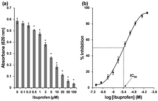
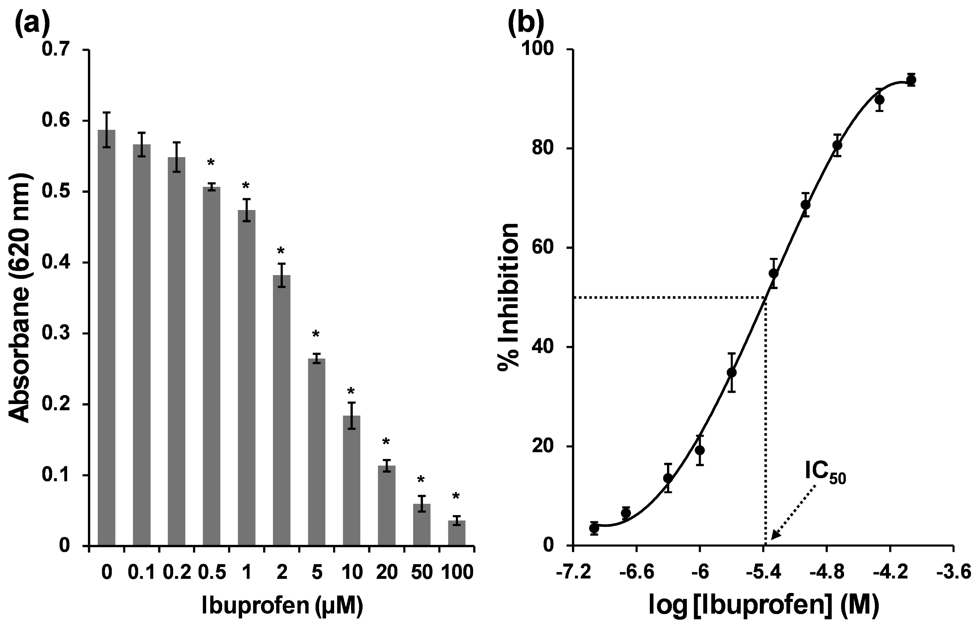
To evaluate the inhibitory effect of ibuprofen against MARK4, an ATPase inhibition assay was performed using the malachite green phosphate detection method. As illustrated in Figure 1a, ibuprofen exhibited dose-dependent inhibition of MARK4 ATPase activity. At the lowest tested concentration (0.1 µM), ibuprofen elicited a modest inhibition of 3.49 ± 1.24%, which gradually increased. At 1 µM, the inhibition reached 19.20 ± 2.95%, while at 2 µM, approximately 34.85 ± 3.87% of ATPase activity was suppressed. Notably, at 5 µM, ibuprofen inhibited more than half of MARK4 ATPase activity (54.83 ± 2.93%), and at 10 µM, inhibition rose to 68.69 ± 2.33%. At the higher concentrations tested (50 µM and 100 µM), ibuprofen demonstrated strong inhibitory effects of 89.79 ± 2.24% and 93.85 ± 1.17%, respectively, indicating near-complete suppression of the MARK4 ATPase activity. The IC50 values were determined by nonlinear regression using the PL4 dose-response model with the AAT Bio IC50 calculator. Each replicate dataset was fitted individually, giving IC50 values of 4.05 µM (95% CI: 3.58–4.57, R2 = 0.999), 3.74 µM (95% CI: 3.22–4.34, R2 = 0.999), and 4.38 µM (95% CI: 3.32–5.77, R2 = 0.996). The average IC50 value was calculated as 4.02 ± 0.33 µM (Figure 1b). It should be noted that the enzyme concentration used (5 µM) is comparable to the obtained IC50 (4.02 ± 0.33 µM); ligand depletion may influence the apparent value. Hence, to account for tight-binding conditions, inhibition data were also analyzed using the Morrison equation (Supplementary Figure S2). The analysis yielded a Ki value of 2.29 ± 0.31 µM. These results further show that ibuprofen binds MARK4 in the low micromolar range, consistent with tight-binding inhibition. This value places ibuprofen in the same potency range as several previously reported synthetic and natural MARK4 inhibitors, including donepezil (IC50 = 5.3 µM) and rivastigmine tartrate (IC50 = 6.74 µM) [14], as well as the pyrimidine-based N-heterarenes (IC50 = 5.35–16.53 µM) [23]. The IC50 of ibuprofen against MARK4 is within the plasma concentration range (>100 µM) achieved during conventional oral dosing [24]. This suggests that the inhibitory concentrations observed in vitro are pharmacologically relevant, although tissue distribution and target engagement in the brain remain to be established. The obtained IC50 (4.02 ± 0.33 µM) for ibuprofen also falls within the range reported for known MARK4 inhibitors using malachite-green ATPase assays, as described in earlier studies [25,26].
Figure 1.
(a) Inhibition of ATPase activity of MARK4 by ibuprofen. (b) Log plot of the inhibition of MARK4 ATPase activity by ibuprofen. The data are presented as the average of three replicates, with error bars as standard deviation. * indicates the p-value < 0.05 compared to control.
3.2. UV-Vis Spectroscopy
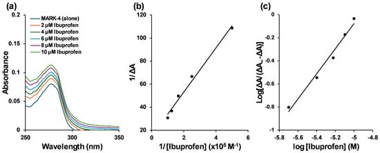
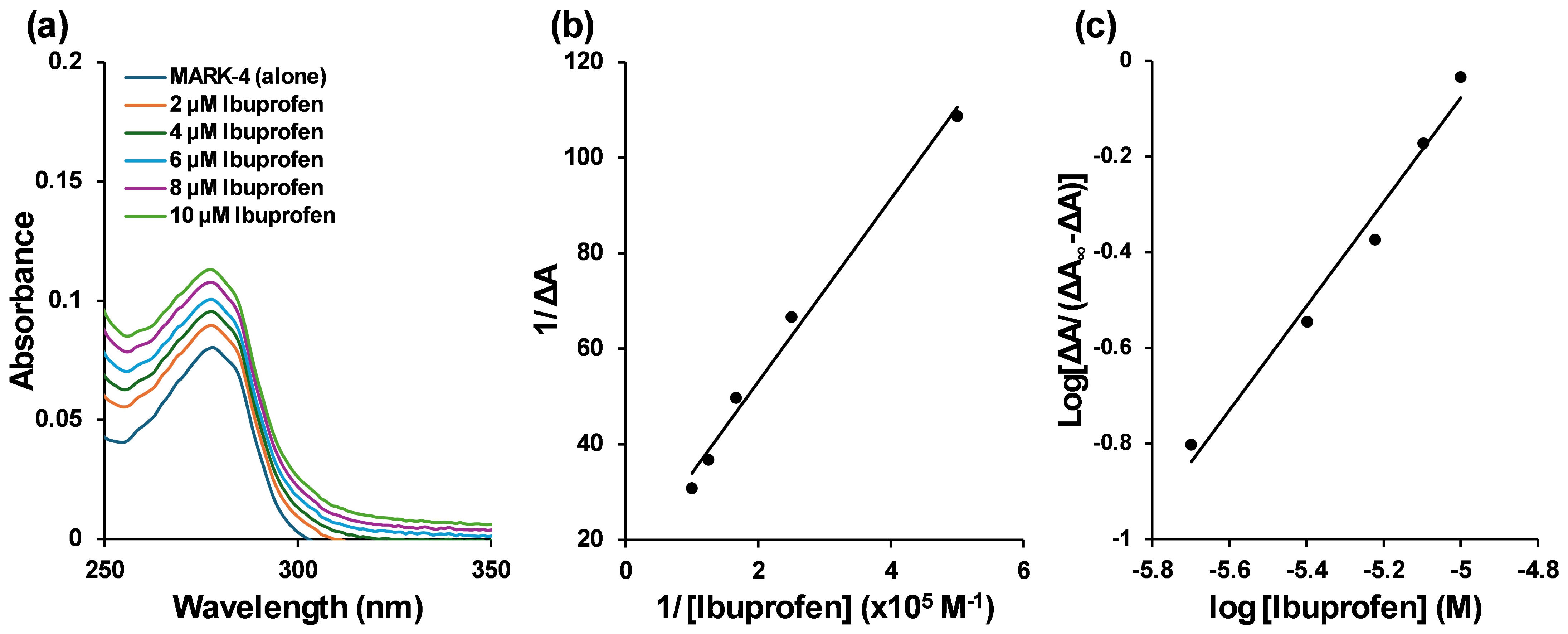
In UV-vis absorption spectroscopy of proteins, the near-UV region gives information on tertiary structure and aromatic side-chain environment, while the far-UV region reflects secondary-structure contributions. Changes in the protein absorption profile upon ligand addition therefore provide a brief idea about the ligand-induced alterations in MARK4 [27]. Titration of MARK4 with ibuprofen produced a noticeable increase in the protein absorption spectrum consistent with complex formation (Figure 2a). These spectral changes were quantified using standard equations.
Figure 2.
(a) UV-vis absorption spectra of MARK4 alone (4 µM) and in the presence of varying concentrations of ibuprofen (2–10 µM). (b) Plot of 1/ΔA versus 1/[Ibuprofen]. (c) Hill plot.
The binding parameters derived from the double-reciprocal and Hill plots are summarized in Table 1. The dissociation constant (Kd) for the MARK4–ibuprofen complex using the double-reciprocal plot (Figure 2b) was determined as 1.295 × 10⁻⁵ M, corresponding to an association constant (Kb) of 7.71 × 10⁴ M−1. It should be noted that the Kd value was derived from a double-reciprocal plot under non-saturating ligand conditions, which may lead to a poorly conditioned fit. The calculated Gibbs free energy change (ΔG) was −6.663 kcal/mol, indicating that complex formation is spontaneous under the experimental conditions. The Hill coefficient (h), obtained from the Hill plot (Figure 2c), is close to unity, indicating near-noncooperative binding with a slight tendency toward positive cooperativity [28]. The negative ΔG° confirms a thermodynamically favorable association driven by noncovalent interactions. The Hill coefficient value close to one suggests a single dominant binding site.
Table 1.
Values of various binding parameters for the interaction of ibuprofen with MARK4 obtained from UV-vis spectroscopy.
3.3. Steady-State Fluorescence
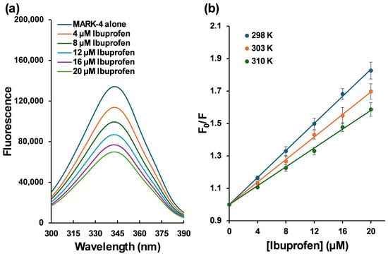
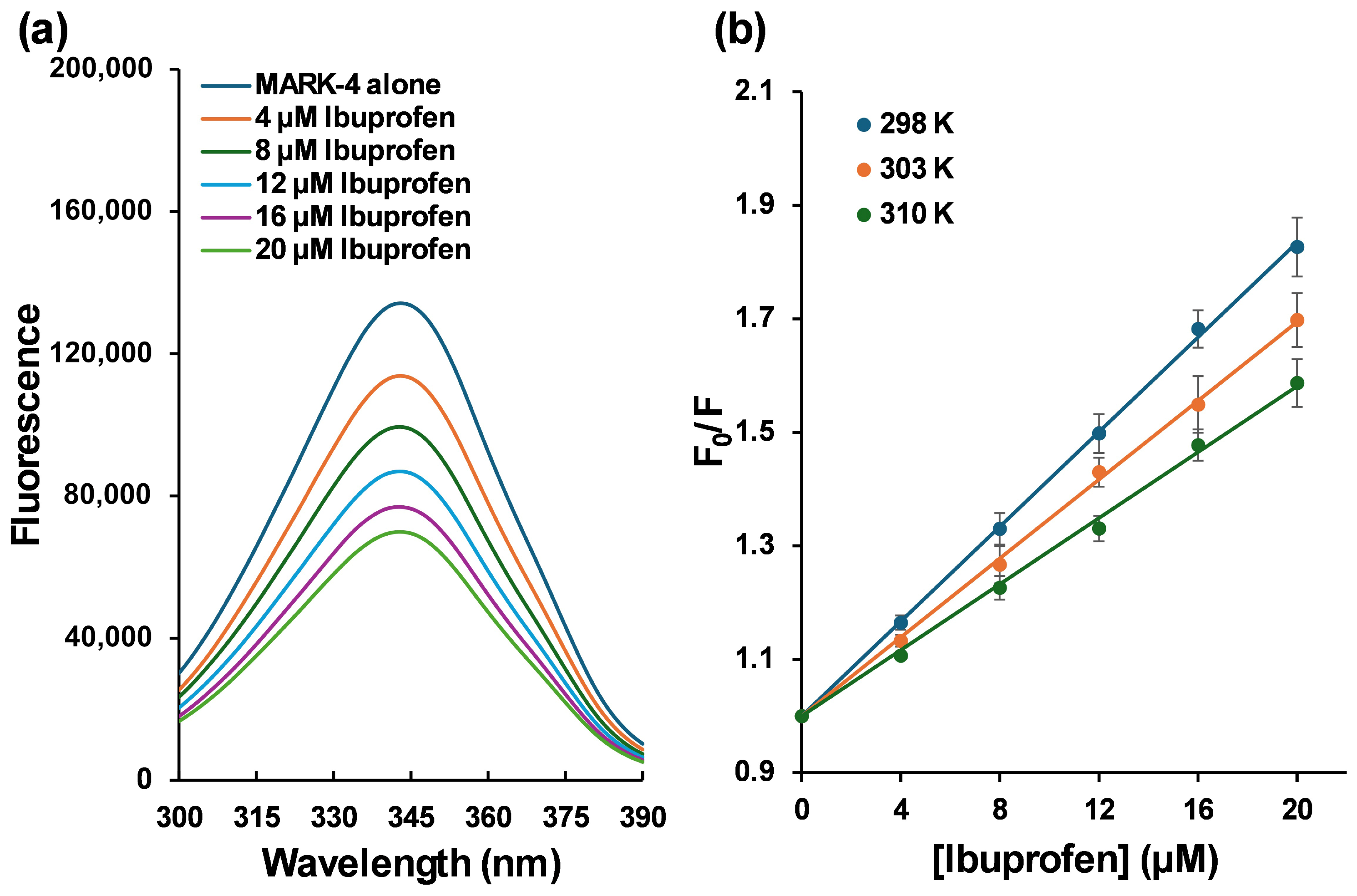
Fluorescence spectroscopy was employed to investigate the interaction between MARK4 and ibuprofen. The emission spectra of native MARK4 and MARK4–ibuprofen mixtures are presented in Figure 3a. Native MARK4 displayed a fluorescence maximum near 340 nm upon excitation at 280 nm. Incremental addition of ibuprofen produced a concentration-dependent decrease in intrinsic fluorescence intensity of MARK4. The quenching indicates that ibuprofen binds to MARK4 and alters the microenvironment, resulting in the formation of a MARK4–ibuprofen complex [29]. Before calculations, fluorescence data were corrected for inner filter effects. To quantify the quenching process, the emission data were analyzed using the Stern–Volmer approach with Equation (4).
Figure 3.
(a) Steady-state fluorescence of MARK4 (5 µM) in the presence of ibuprofen (4–20 µM). (b) Stern–Volmer plots of the MARK4–ibuprofen interaction. The data are presented as the average of three replicates; error bars are the standard deviation.
Stern–Volmer plot (Figure 3b) was constructed, and the slopes provided the Stern–Volmer constants (KSV) listed in Table 2. The KSV values were in the range of 2.90 ± 0.19 × 10⁴ M−1 to 4.17 ± 0.25 × 10⁴ M−1. The Stern–Volmer constant alone does not distinguish between static and dynamic quenching; the bimolecular quenching rate constant (Kq) was calculated from the lifetime of protein tryptophan fluorescence (τ0) using Equation (5).
Table 2.
Values of KSV, Kq, K, and n for the binding of MARK4 with ibuprofen at varying temperatures obtained from steady-state fluorescence spectroscopy.
In the above equation, τ0 = 2.7 × 10⁻⁹ s [14]. The resulting Kq values (1.08 ± 0.07 × 10¹³ M−1 s−1 to 1.55 ± 0.09 × 10¹³ M−1 s−1), as depicted in Table 2, were substantially higher than the diffusion-controlled limit for collisional quenching [30], indicating that the observed fluorescence decrease is consistent with formation of a nonfluorescent ground-state complex (static quenching) rather than from purely collisional encounters. This conclusion is further supported by the temperature dependence of KSV. The characteristic of static quenching is the decrease in KSV with rising temperature; hence, the quenching of MARK4’s fluorescence by ibuprofen is consistent with static quenching.
The binding parameters and stoichiometry were obtained from the double-logarithmic analysis of the fluorescence data using Equation (6).
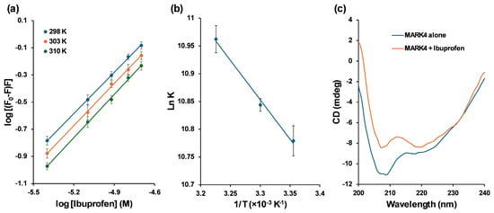
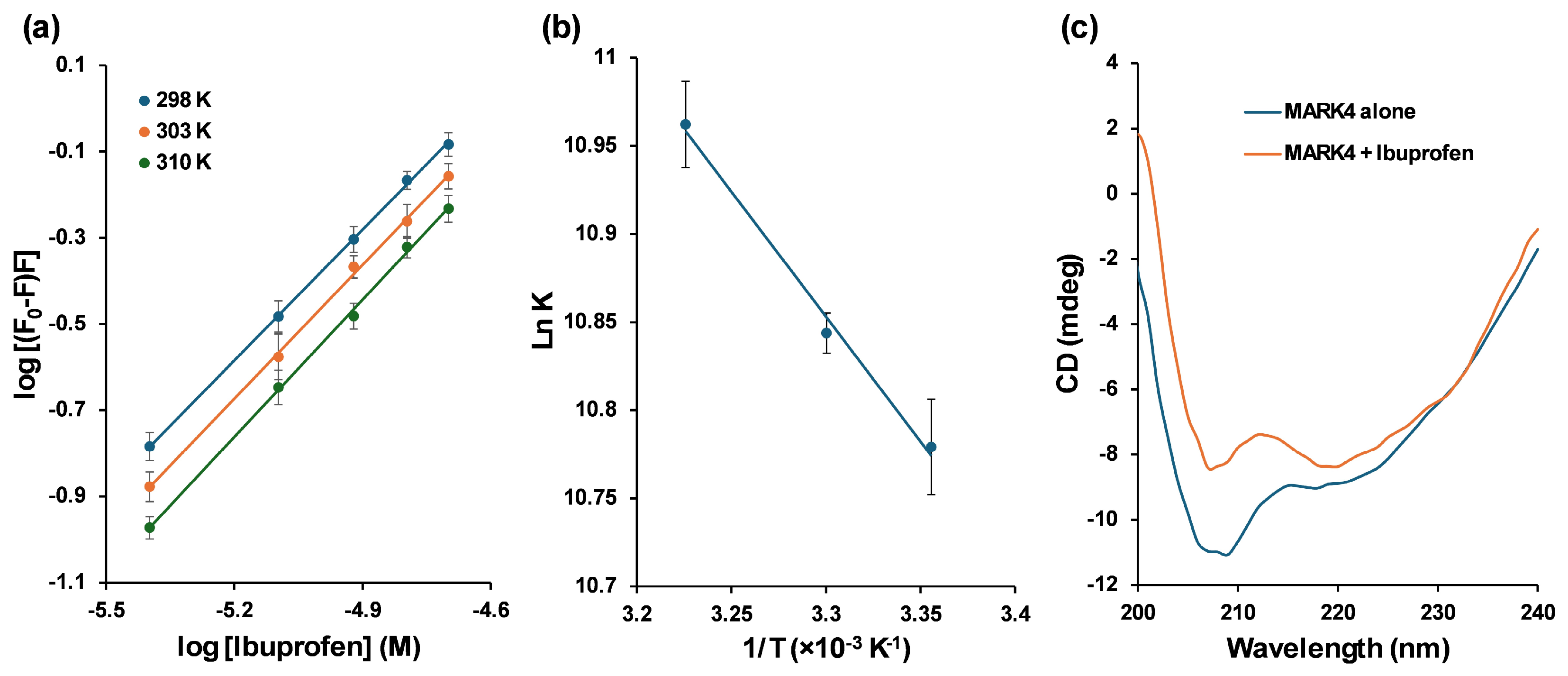
The slope of this double-log plot (Figure 4a) gives the number of binding sites (n), while the intercept provides the binding constant (K). Across the three temperatures examined (Table 2), K values increased with increasing temperature (4.80 ± 0.78 × 10⁴ M−1 to 5.76 ± 0.84 × 10⁴ M−1), indicating greater complex stability at physiological temperature compared to lower temperatures. The values of n (binding stoichiometry) close to 1 are consistent with one dominant binding site under the experimental conditions.
Figure 4.
(a) Double log plots of MARK4–ibuprofen interaction. (b) van’t Hoff plot of MARK4–ibuprofen interaction. (c) CD spectra of MARK4 alone and MARK4–ibuprofen complex. The data are presented as the average of three replicates, with error bars as standard deviation.
The thermodynamic parameters were derived from the temperature dependence of the binding constant. The values of entropy change and enthalpy change were calculated from the van’t Hoff plot (Figure 4b) with Equation (7):
The signs and magnitudes of the enthalpy (ΔH°) and entropy changes (ΔS°) indicate the dominant forces driving complex formation. In this case, the thermodynamic signature points to an interaction driven largely by hydrophobic effects and desolvation (positive ΔS°), consistent with displacement of ordered water and formation of van der Waals contacts at the binding interface [31]. Moreover, the positive ΔS° and ΔH° are typically attributed to the predominance of hydrophobic interactions during complex formation [32].
The Gibbs free energy change (ΔG°) was calculated with Equation (8):
The calculated Gibbs free energy values, as listed in Table 3, were negative at all temperatures (−6.75 kcal/mol to −6.38 kcal/mol), suggesting that the binding of ibuprofen with MARK4 is spontaneous in nature under experimental conditions.
Table 3.
Values of ΔG°, ΔH°, and ΔS° for the binding of MARK4 with ibuprofen at varying temperatures obtained from steady-state fluorescence spectroscopy.
3.4. Circular Dichroism
CD spectroscopy was used to study whether ibuprofen binding affects the secondary structure of MARK4. The far-UV CD spectrum of the native protein displayed a characteristic minimum near 208 nm (Figure 4c). The raw spectra of all samples are presented in Supplementary Figure S3. The addition of ibuprofen changed the CD signal. The intensity of the negative bands decreased, indicating changes in MARK4’s secondary structure. As the negative bands at 208 and 222 nm are associated with α-helical structures, the decrease in band intensity in this region suggests alterations in the secondary structure environment of MARK4 upon ligand binding. These changes indicate that ibuprofen induces alterations in the secondary structure of MARK4The spectrum retains helix-like features, but with substantially reduced intensity. Such helix loss can arise from direct ligand contacts that destabilize helix-forming segments, from ligand-induced shifts in tertiary packing that expose helical regions to solvent, or from a combination of both [33].
3.5. Molecular Docking
The in vitro assays found that ibuprofen binds to MARK4 and inhibits its ATPase activity with an IC50 in the low micromolar range. There were also noticeable changes in protein secondary structure. To further explore the molecular basis of MARK inhibition, we used computational approaches. Docking studies were performed to identify the interaction site of ibuprofen. The MD simulations provided insights into the stability and dynamic behavior of the MARK4–ibuprofen complex.
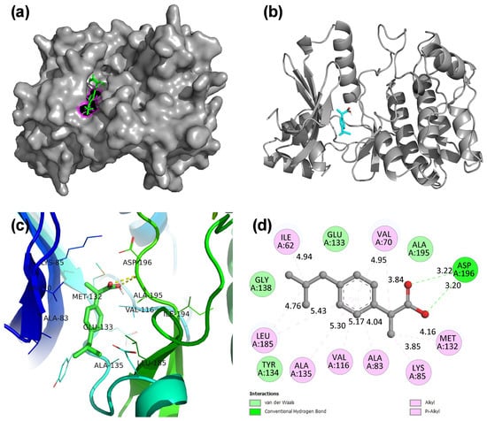
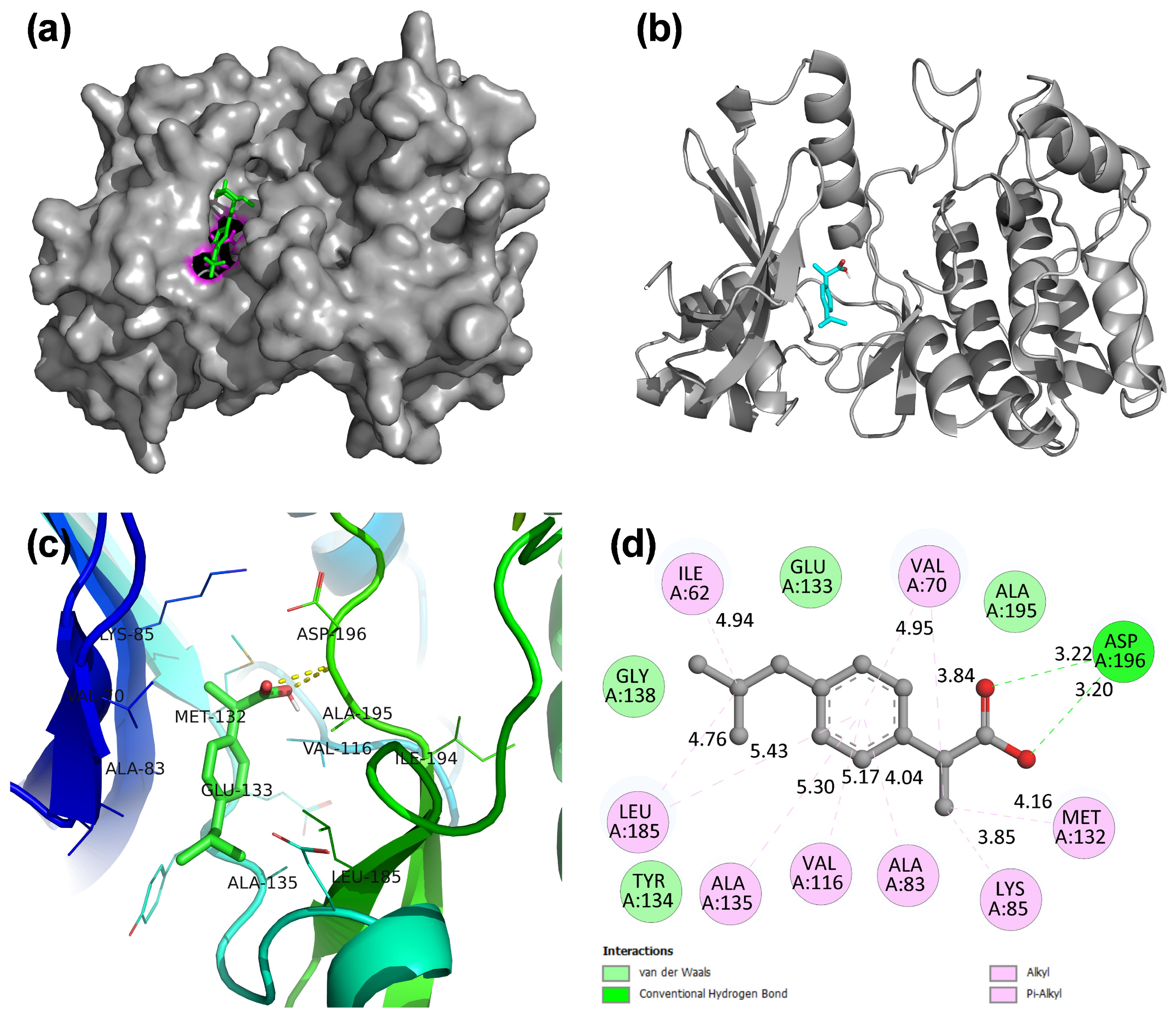
The molecular docking was validated by removing the inhibitor from the crystal structure and then redocking. After docking, the inhibitor occupied the same binding site with an almost overlapping binding pose, with an RMSD of 0.40 Å, validating the docking methodology. The docking of ibuprofen revealed that it was docked at the same active site and overlapped with the inhibitor (Figure 5a). The blind docking generated 20 binding poses, out of which 16 were localized to the ATP/inhibitor pocket, while only four were found at other sites, indicating clear enrichment of the ATP site. The top-scoring pose (−7.0 kcal/mol) was therefore selected for subsequent analysis. Two hydrogen bonds were predicted between ibuprofen and Asp196 in the top-scoring static docking pose (H-bond distances 3.22 Å and 3.20 Å). A network of van der Waals contacts with Tyr134, Gly138, Glu133, and Ala195 further stabilizes the complex (Figure 5b–d). A set of hydrophobic interactions with Leu185, Ala135, Val116, Ala83, Lys85, Met132, Val70, and Ile62 formed a nonpolar surface around the ligand, indicating that ibuprofen is comfortably accommodated by the hydrophobic sub-pocket. It should be noted that these interactions were predicted from the static docking pose in molecular docking. Subsequent MD simulations revealed a dynamic hydrogen bond network, with Glu133 showing the highest occupancy (22.5%), whereas the Asp196 contacts found in docking were not persistent in MD simulations. Since Lys85 is a catalytically important residue required for ATP binding in MARK4 [14], its involvement in hydrophobic interactions with ibuprofen suggests that the ligand may interfere with ATP recognition. This engagement, together with hydrogen bonding at Asp196 and stabilizing van der Waals contacts, provides a plausible structural basis for ibuprofen’s inhibition of MARK4 ATPase activity. The interaction pattern indicates a mixed polar/hydrophobic binding. The docking results support inhibition of MARK4 by ibuprofen, where it interacts at the active inhibitor binding site.
Figure 5.
(a) Overlap of docked ibuprofen with the crystal pyrazolopyrimidine inhibitor in MARK4. Ibuprofen is shown as magenta sticks, the crystal pyrazolopyrimidine inhibitor is shown as green sticks, and MARK4 is shown as gray surfaces. (b) Docked pose of the MARK4–ibuprofen complex. (c) Close view of the MARK4–ibuprofen complex. (d) 2D interaction diagram of MARK4–ibuprofen complex.
3.6. Molecular Dynamics Simulations
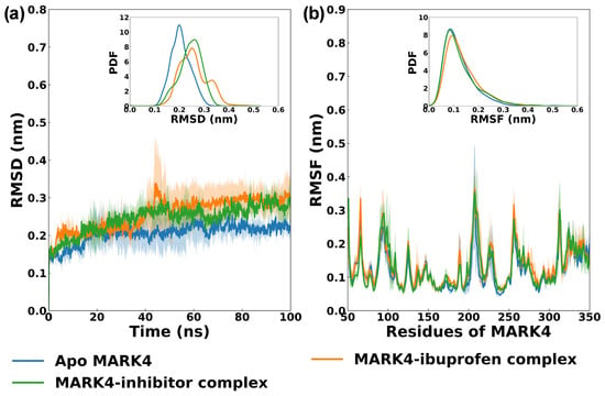
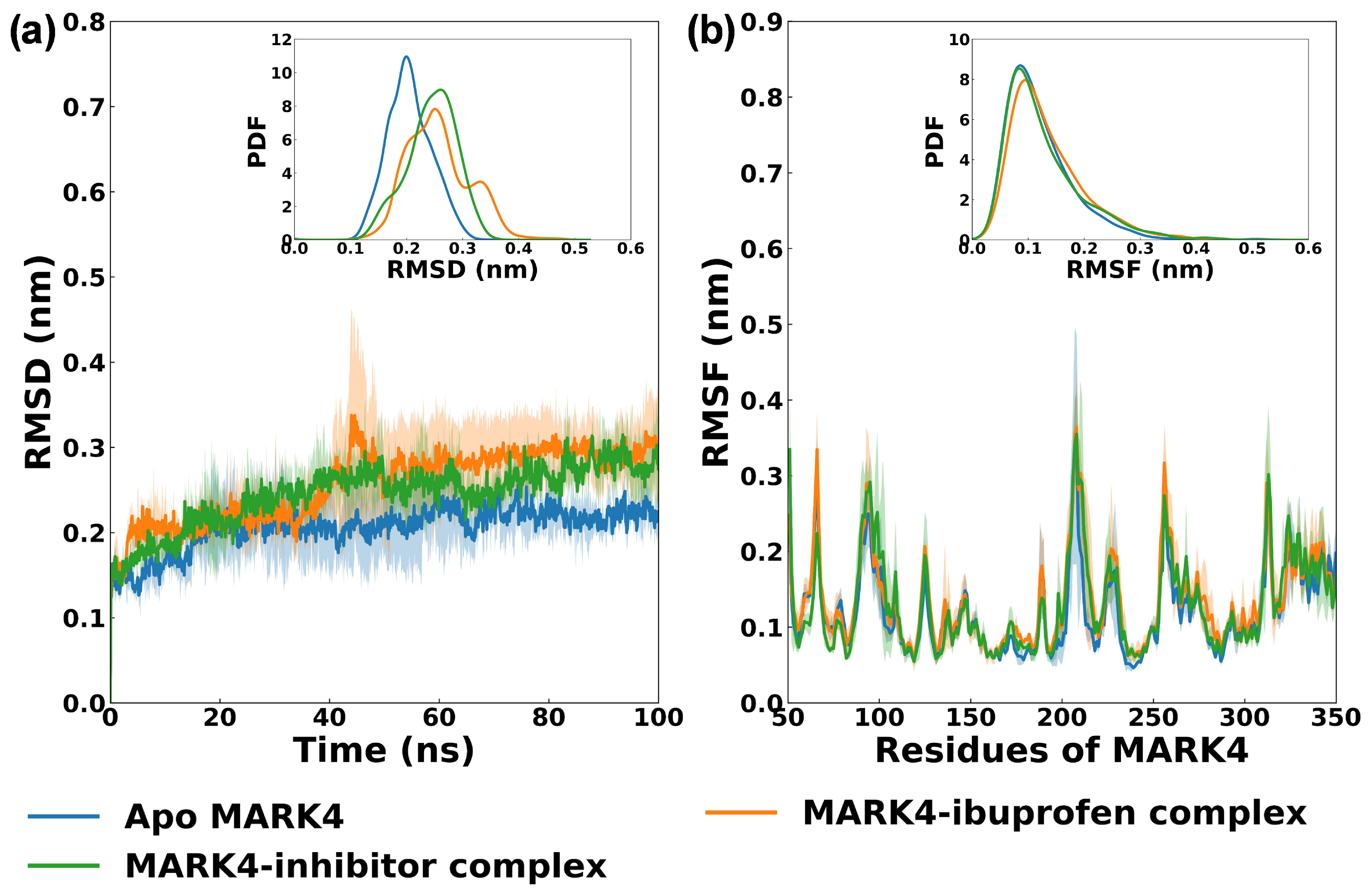
MD simulations were conducted to assess the stability and conformational dynamics of MARK4 in apo form, bound to ibuprofen, and bound to a known inhibitor. RMSD of the MD simulation trajectory provides a measure of structural deviations. Apo MARK4 exhibited an average RMSD of 0.205 ± 0.025 nm, indicating stable behavior (Figure 6a). Upon ibuprofen binding, RMSD increased slightly to 0.256 ± 0.042 nm, while the inhibitor complex showed a similar value of 0.244 ± 0.036 nm. The findings align with previous research where the RMSD of apo MARK4 and the MARK–apigenin complex was nearly 0.3 nm during a 300 ns MD simulation [12]. These results suggest that both ligands induce minor conformational adjustments but do not destabilize the protein, and the trajectories remained equilibrated throughout the simulation.
Figure 6.
(a) RMSD of backbone atoms of apo MARK4, MARK4–ibuprofen complex, and MARK4–inhibitor complex over a 100 ns MD simulation. The inset is the PDF of the RMSD plot. (b) RMSF of alpha carbon atoms (Cα) of MARK4 without and with ibuprofen or inhibitor. The inset is the PDF of the RMSF plot. The data are presented as the average of three simulation runs, with the shaded region representing the standard deviation.
The RMSF of Cα atoms was calculated to evaluate residue-level flexibility. The RMSF of almost all residues of MARK4 (Figure 6b) was below 0.2 nm in both the apo protein and in complex with the ligands (ibuprofen or a known inhibitor). Apo MARK4 displayed an average RMSF of 0.120 ± 0.053 nm that increased modestly to 0.134 ± 0.058 nm in the ibuprofen complex and 0.128 ± 0.060 nm in the inhibitor complex. The fluctuations were mainly found in the loop regions, consistent with their inherent flexibility in aqueous environments [34,35]. These findings indicate that ibuprofen binding negligibly alters the backbone mobility of MARK4.
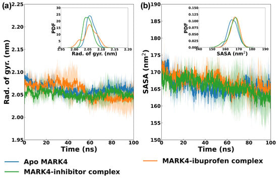
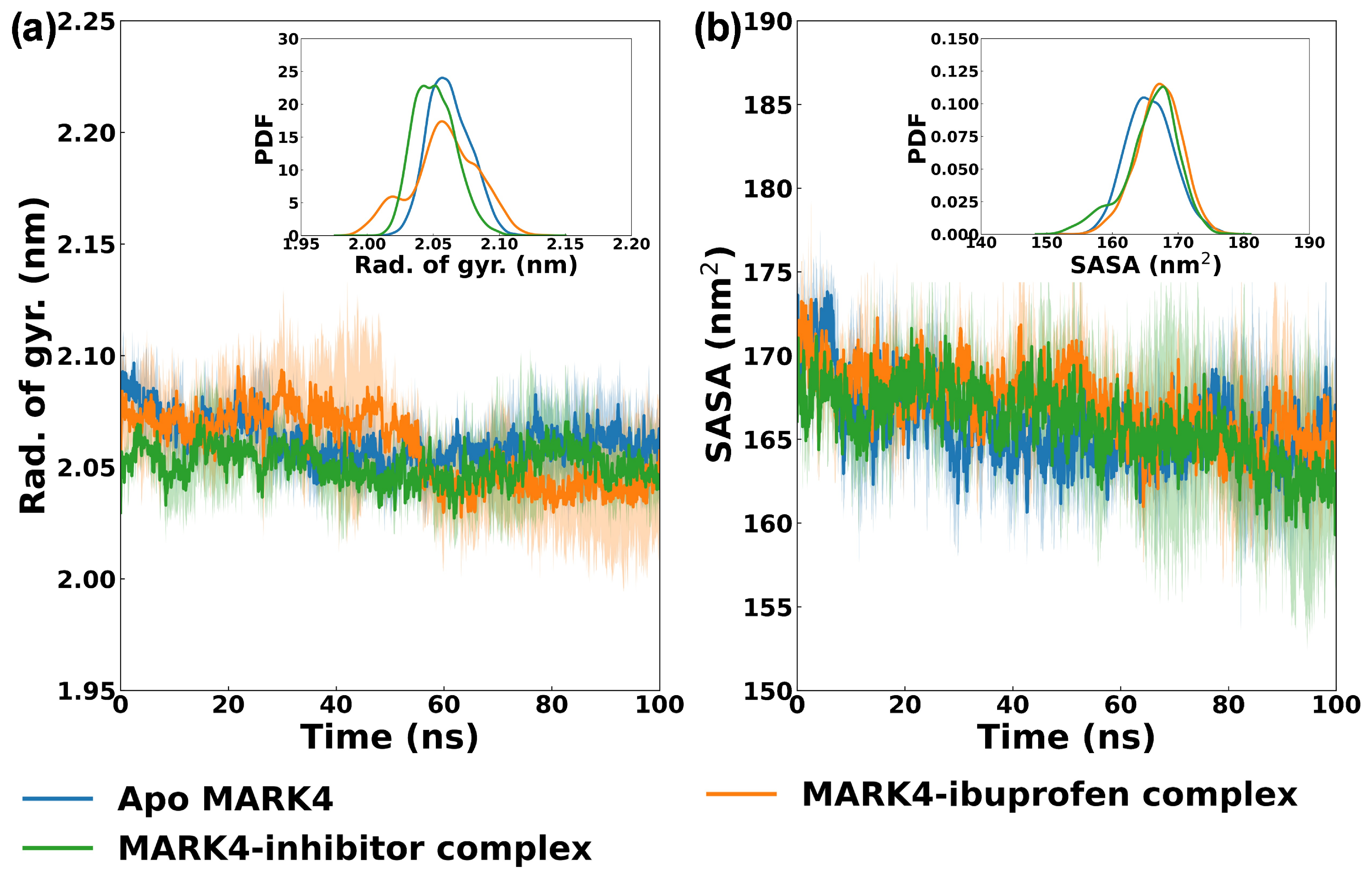
The radius of gyration (Rg) values reflect protein compactness. Apo MARK4 was found to have an average Rg of 2.062 ± 0.009 nm (Figure 7a), which remained nearly unchanged at 2.058 ± 0.016 nm in the ibuprofen complex and 2.051 ± 0.007 nm in the inhibitor complex. The minimal differences indicate that binding of ligands (either ibuprofen or inhibitor) does not affect the compactness of MARK4, and overall structural integrity was not changed. Similar research has been reported earlier, where the Rg of apo MARK4 and the MARK–apigenin complex was approximately 2.1 nm [12]. The solvent accessible surface area (SASA) values provide insights into surface exposure and conformational changes. Apo MARK4 exhibited a SASA of 165.748 ± 2.256 nm2 (Figure 7b), which increased slightly to 167.116 ± 2.143 nm2 upon ibuprofen binding, while the inhibitor complex showed 165.887 ± 2.208 nm2. The minor increase in SASA for the ibuprofen complex suggests subtle rearrangements at the protein surface, possibly due to hydrophobic pocket occupation, but without significant structural disruption. Overall, the analysis demonstrates that ibuprofen forms a stable complex with MARK4, maintaining conformational integrity comparable to the reference inhibitor. The slight increase in RMSD and SASA values indicates minor adjustments in protein flexibility and surface exposure, consistent with ligand accommodation. These findings also suggest that ibuprofen interacts stably with MARK4, inducing minor conformational changes without destabilizing the protein fold. The dynamic behavior of the ibuprofen complex closely resembles that of the inhibitor complex [33].
Figure 7.
(a) Radius of gyration (Rg) of backbone atoms of apo MARK4, MARK4–ibuprofen complex, and MARK4–inhibitor complex over a 100 ns MD simulation. The inset is the PDF of the Rg plot. (b) Solvent accessible surface area (SASA) of apo MARK4, MARK4–ibuprofen complex, and MARK4–inhibitor complex over a 100 ns MD simulation. The inset is the PDF of the SASA plot. The data are presented as the average of three simulation runs, with the shaded region representing the standard deviation.
To study the effect of ibuprofen on structural features of MARK4, secondary structures in the apo protein, ibuprofen-bound complex, and inhibitor-bound complex were analyzed. The apo MARK4 exhibited 23.95% coils, 14.50% β-sheets, 1.09% β-bridges, 9.83% bends, 14.14% turns, 33.64% α-helices, and 2.81% 3-helices. These values align with the literature [23]. With ibuprofen binding, the percentages of secondary structures shifted negligibly to 23.21% coils, 14.64% β-sheets, 1.04% β-bridges, 9.97% bends, 14.40% turns, 33.38% α-helices, and 3.33% 3-helices. In the presence of the inhibitor, the secondary structures in MARK4 also remained similar at 24.15% coils, 15.05% β-sheets, 0.68% β-bridges, 9.69% bends, 13.98% turns, 33.21% α-helices, and 3.20% 3-helices. The data show that binding of ibuprofen did not cause any major changes in the secondary structures, and the protein retained its stability during simulation. The slight increase in β-sheet and 3-helix content in the ibuprofen complex indicates minor structural rearrangements without any major change in protein folding.
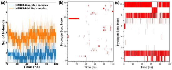
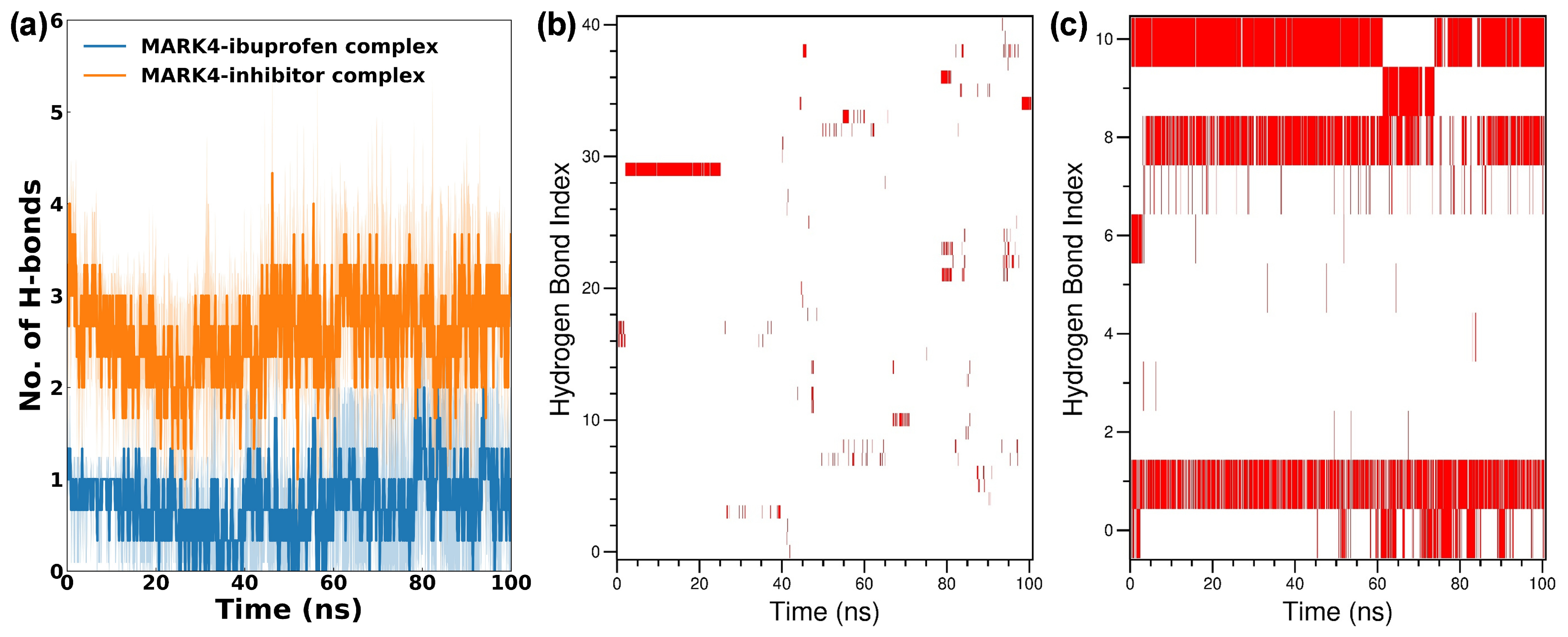
The hydrogen bonding pattern was also analyzed in MD simulations (Figure 8a). H-bonds are among the main stabilizing forces in a typical ligand–protein complex. The MARK4–ibuprofen complex formed an average of 0.73 ± 0.36 hydrogen bonds, whereas the MARK4–inhibitor complex established a significantly higher average of 2.59 ± 0.48 hydrogen bonds. This difference suggests that ibuprofen binding relies more on van der Waals and hydrophobic forces rather than hydrogen bonds, which is consistent with docking results showing ibuprofen’s predominantly nonpolar interactions. In contrast, the inhibitor complex benefits from stronger hydrogen bonding, contributing to its higher stability. The hydrogen bond existence map analysis revealed that Glu133 exhibited the highest hydrogen bond occupancy of 22.5% in the MARK4–ibuprofen complex (Figure 8b). In the MARK4–inhibitor complex, Asp196 (82.3%), Asn183 (69.1%), and Ala135 (78.9%) were the residues showing maximum hydrogen bond occupancy (Figure 8c).
Figure 8.
(a) Number of hydrogen bonds formed by ibuprofen or inhibitor over a 100 ns MD simulation. (b) Hydrogen bond existence map of the MARK4–ibuprofen complex. (c) Hydrogen bond existence map of the MARK4–inhibitor complex. The data are presented as the average of three simulation runs, with the shaded region representing standard deviations.
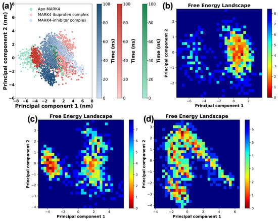
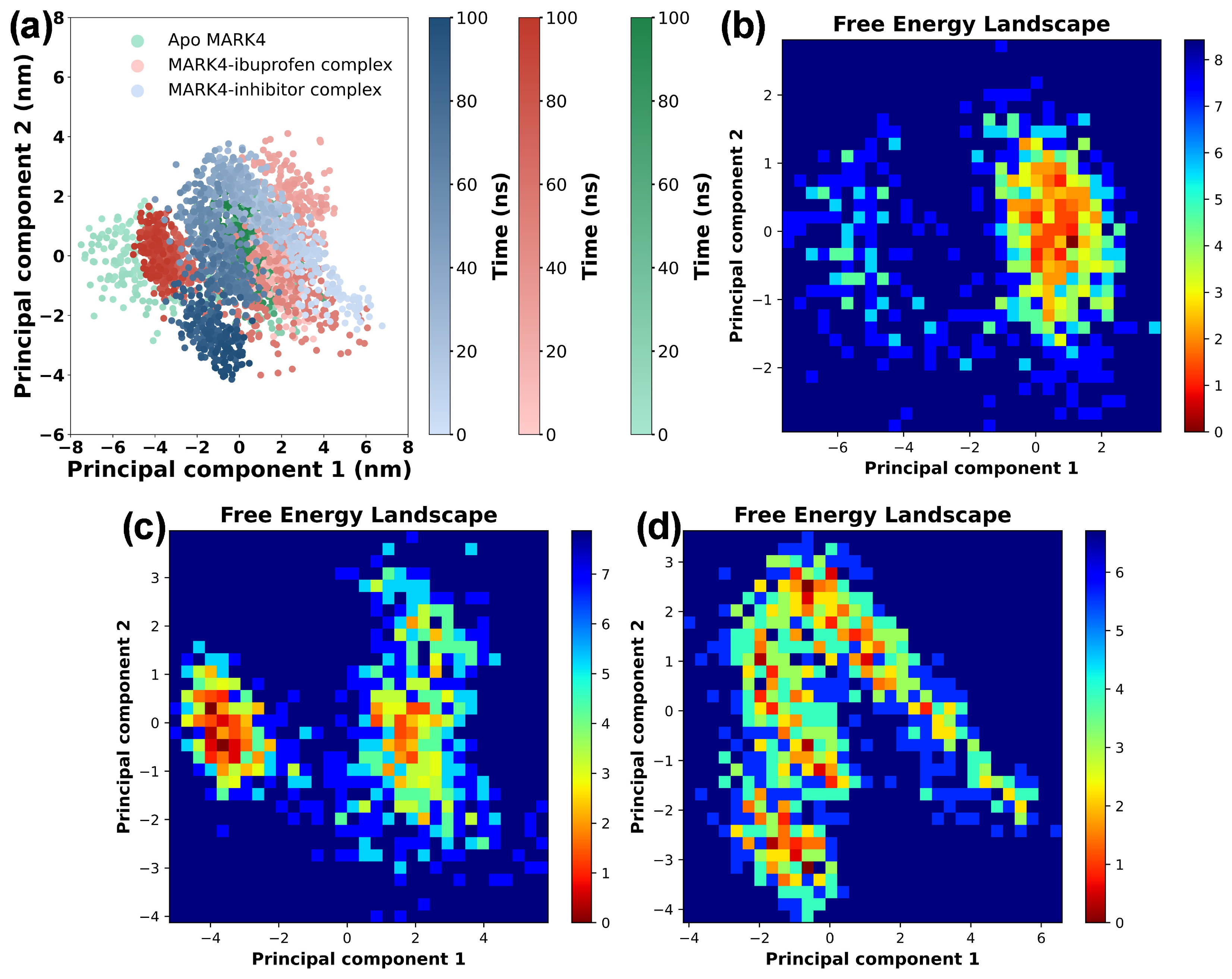
Principal component analysis (PCA) was performed to examine the flexibility of MARK4 in the absence and presence of ibuprofen. To get insight into overall structural variations in MARK4, two principal components (eigenvectors) were calculated. The projection of eigenvectors for apo MARK4 and its complexes is shown in Figure 9a. The distribution of eigenvectors of the MARK4–ibuprofen complex overlapped broadly with apo MARK4, indicating that binding of ibuprofen does not cause any major deviations in global motions. Likewise, the projection of the eigenvectors of the inhibitor complex was also comparable to the apo protein, with only minor differences. The eigenvector data were further utilized to make free energy landscapes (FEL), providing detailed information about conformational stability. The FEL of all systems is shown in Figure 9b–d. Apo MARK4 displayed one minimum at 74.2 ns, reflecting a relatively stable conformational state. In contrast, the MARK4–ibuprofen complex showed two minima at 73.1 ns and 91.9 ns, indicating slightly higher conformational heterogeneity compared to apo MARK4. The inhibitor complex also displayed two minima at 94.1 ns and 37.7 ns, suggesting enhanced conformational diversity relative to apo MARK4. The similarity in FEL profiles between the ibuprofen and inhibitor complexes indicates that ibuprofen stabilizes MARK4 in a manner comparable to the reference inhibitor, albeit with modestly increased heterogeneity. The PCA and FEL analyses confirm that MARK4 retains its native flexibility profile upon ibuprofen binding, and the protein maintains stable conformations across all complexes [12].
Figure 9.
(a) 2D projection of eigenvectors of apo MARK4, MARK4–ibuprofen complex, and MARK4–inhibitor complex over a 100 ns MD simulation. (b) Free energy landscape of apo MARK4. (c) Free energy landscape of MARK4–ibuprofen complex. (d) Free energy landscape of MARK4–inhibitor complex.
4. Conclusions
This study demonstrates that ibuprofen binds to MARK4 and inhibits its ATPase activity. The enzymatic assays established dose-dependent inhibition of MARK4 ATPase activity, with an IC50 of 4.02 ± 0.33 µM, which falls within the range reported in earlier studies. UV-vis and fluorescence quenching revealed and confirmed spontaneous complex formation with entropy-favored binding. The CD results showed decreased ellipticity, suggesting alterations in the secondary structure of MARK4 upon ibuprofen binding. The docking results support the binding of ibuprofen within the ATP/inhibitor-binding pocket of MARK4. inhibitor-binding. The MD simulations over 100 ns showed that the MARK4–ibuprofen complex is dynamically stable, with minor increases in backbone flexibility. The findings suggest that ibuprofen may inhibit MARK4. Future studies should employ orthogonal kinase assays and positive controls to confirm the effects of ibuprofen on MARK4 kinase activity. Further research is also needed to validate these findings in cellular models and assess the selectivity against related enzymes. In addition, structure-guided modifications should be explored to improve the activity and binding affinity of novel compounds. Further validation of MARK4 inhibition by ibuprofen in cellular models is warranted.
Supplementary Materials
The following supporting information can be downloaded at: https://www.mdpi.com/article/10.3390/biom16111575/s1, Supplementary Figure S1: Molecular structure of inhibitor which is [(1R,6R)-6-amino-2,2-difluorocyclohexyl]-5-ethyl-4-{6-(trifluoromethyl)pyrazolo[1,5-a]pyrimidin-3-yl}-2-thiophenecarboxamide. Supplementary Figure S2: Dose–response curve for inhibition of MARK4 by ibuprofen fitted with the Morrison equation. Supplementary Figure S3: (a) CD (mdeg) spectra of MARK alone (4 µM). (b) HT (V) spectra of MARK alone (4 µM). (c) CD (mdeg) spectra of MARK4 (4 µM) + Ibuprofen (10 µM). (d) HT (V) spectra of MARK4 (4 µM) + Ibuprofen (10 µM). (e) CD (mdeg) spectra of Ibuprofen alone (10 µM). (f) HT (V) spectra of Ibuprofen alone (10 µM).
Author Contributions
Conceptualization, T.S. and A.H.R.; methodology, T.S., H.O.A.A., A.A., and A.H.R.; software, T.S., S.A.A., and A.H.R.; investigation, T.S., H.O.A.A., and A.H.R.; data curation, T.S., A.A., and A.H.R.; writing—original draft preparation, T.S., H.O.A.A., S.A.A., A.A., and A.H.R.; writing—review and editing, T.S., H.O.A.A., S.A.A., A.A., and A.H.R. Funding acquisition, T.S., and A.H.R. All authors have read and agreed to the published version of the manuscript.
Funding
This research received no external funding.
Institutional Review Board Statement
Not applicable.
Informed Consent Statement
Not applicable.
Data Availability Statement
The original contributions presented in this study are included in the article/Supplementary Material. Further inquiries can be directed to the corresponding authors.
Acknowledgments
The researchers would like to thank the Deanship of Graduate Studies and Scientific Research at Qassim University (https://www.qu.edu.sa) for financial support (QU-APC-2026). During the preparation of this manuscript, Grammarly AI assistant (version: v1.2.302.1971) was used for grammar and language refinement. We have carefully reviewed and edited the output and taken full responsibility for the content.
Conflicts of Interest
The authors declare no conflicts of interest.
References
- Seok, S.-H. Structural Insights into Protein Regulation by Phosphorylation and Substrate Recognition of Protein Kinases/Phosphatases. Life 2021, 11, 957. [Google Scholar] [CrossRef] [Scilit]
- Zhang, Y.; Hua, S.; Jiang, K. Orchestrating Microtubules: A Review of Kinase–Dependent Regulatory Mechanisms. Cell Biol. Int. 2026, 50, e70133. [Google Scholar] [CrossRef] [Scilit]
- Alam, M.; Ahmed, S.; Abid, M.; Hasan, G.M.; Islam, A.; Hassan, M.I. Therapeutic Targeting of Microtubule Affinity-regulating Kinase 4 in Cancer and Neurodegenerative Diseases. J. Cell. Biochem. 2023, 124, 1223–1240. [Google Scholar] [CrossRef] [Scilit]
- Mumtaz; Ahmed, F.; Rabbani, S.A.; El-Tanani, M.; Najmi, A.K.; Ali, J.; Khan, M.A. Tauopathy in AD: Therapeutic Potential of MARK-4. Curr. Alzheimer Res. 2024, 21, 779–790. [Google Scholar] [CrossRef] [Scilit]
- Ma, F.; Yao, J.; Niu, X.; Zhang, J.; Shi, D.; Da, M. MARK4 Promotes the Malignant Phenotype of Gastric Cancer through the MAPK/ERK Signaling Pathway. Pathol. Res. Pract. 2024, 261, 155471. [Google Scholar] [CrossRef] [Scilit]
- Limlingan, S.J.M.; Krishnankutty, A.; Asada, A.; Ando, K.; Saito, T. MARK4 Regulates GLUT3 Surface Expression and Neuronal Glucose Uptake. J. Biochem. 2026. Online ahead of print. [Google Scholar] [CrossRef] [Scilit]
- Liu, Z.; Gan, L.; Chen, Y.; Luo, D.; Zhang, Z.; Cao, W.; Zhou, Z.; Lin, X.; Sun, C. Mark4 Promotes Oxidative Stress and Inflammation via Binding to PPARγ and Activating NF-ΚB Pathway in Mice Adipocytes. Sci. Rep. 2016, 6, 21382. [Google Scholar] [CrossRef] [Scilit]
- Mohammad, T.; Khan, F.I.; Lobb, K.A.; Islam, A.; Ahmad, F.; Hassan, M.I. Identification and Evaluation of Bioactive Natural Products as Potential Inhibitors of Human Microtubule Affinity-Regulating Kinase 4 (MARK4). J. Biomol. Struct. Dyn. 2019, 37, 1813–1829. [Google Scholar] [CrossRef] [Scilit]
- Ahrari, S.; Mogharrab, N.; Navapour, L. Structure and Dynamics of Inactive and Active MARK4: Conformational Switching through the Activation Process. J. Biomol. Struct. Dyn. 2020, 38, 2468–2481. [Google Scholar] [CrossRef] [Scilit]
- Upadhyay, A.; Amanullah, A.; Joshi, V.; Dhiman, R.; Prajapati, V.K.; Poluri, K.M.; Mishra, A. Ibuprofen-Based Advanced Therapeutics: Breaking the Inflammatory Link in Cancer, Neurodegeneration, and Diseases. Drug Metab. Rev. 2021, 53, 100–121. [Google Scholar] [CrossRef] [Scilit]
- Peng, K.-P.; Chang, K.-H. Long-Term Non-Steroidal Anti-Inflammatory Drug (NSAID) Use and Dementia or Cognitive Decline Risk in Adults over 50 Based on Epidemiological Studies: A Narrative Review. Tungs’ Med. J. 2026. Online ahead of print. [Google Scholar] [CrossRef] [Scilit]
- Hussain, A.; Jairajpuri, D.S.; Anwar, S.; Choudhury, A.; Hawwal, M.F.; Firdous, A.; Alajmi, M.F.; Hassan, M.I. Apigenin-Mediated MARK4 Inhibition: A Novel Approach in Advancing Alzheimer’s Disease Therapeutics. Mol. Divers. 2025, 29, 5745–5756. [Google Scholar] [CrossRef] [Scilit]
- Adnan, M.; DasGupta, D.; Anwar, S.; Shamsi, A.; Siddiqui, A.J.; Snoussi, M.; Bardakci, F.; Patel, M.; Hassan, M.I. Mechanistic Insights into MARK4 Inhibition by Galantamine toward Therapeutic Targeting of Alzheimer’s Disease. Front. Pharmacol. 2023, 14, 1276179. [Google Scholar] [CrossRef] [Scilit]
- Shamsi, A.; Anwar, S.; Mohammad, T.; Alajmi, M.F.; Hussain, A.; Rehman, M.T.; Hasan, G.M.; Islam, A.; Hassan, M.I. MARK4 Inhibited by AChE Inhibitors, Donepezil and Rivastigmine Tartrate: Insights into Alzheimer’s Disease Therapy. Biomolecules 2020, 10, 789. [Google Scholar] [CrossRef] [Scilit]
- Alam, M.M.; Qais, F.A.; Ahmad, I.; Alam, P.; Hasan Khan, R.; Naseem, I. Multi-Spectroscopic and Molecular Modelling Approach to Investigate the Interaction of Riboflavin with Human Serum Albumin. J. Biomol. Struct. Dyn. 2018, 36, 795–809. [Google Scholar] [CrossRef] [Scilit]
- Sarwar, T.; Rehman, A.A.; Arif, H.; Alwanian, W.M.; Alharbi, H.O.A.; Rahmani, A.H. Biophysical and Computational Insights into Alpha-1 Antitrypsin Aggregation and Its Inhibition by Natural Polyphenols. Biomedicines 2026, 14, 1310. [Google Scholar] [CrossRef] [Scilit]
- Trott, O.; Olson, A.J. AutoDock Vina: Improving the Speed and Accuracy of Docking with a New Scoring Function, Efficient Optimization, and Multithreading. J. Comput. Chem. 2009, 31, 455–461. [Google Scholar] [CrossRef] [Scilit]
- Van Der Spoel, D.; Lindahl, E.; Hess, B.; Groenhof, G.; Mark, A.E.; Berendsen, H.J.C. GROMACS: Fast, Flexible, and Free. J. Comput. Chem. 2005, 26, 1701–1718. [Google Scholar] [CrossRef] [Scilit]
- Lindorff-Larsen, K.; Piana, S.; Palmo, K.; Maragakis, P.; Klepeis, J.L.; Dror, R.O.; Shaw, D.E. Improved Side-chain Torsion Potentials for the Amber Ff99SB Protein Force Field. Proteins Struct. Funct. Bioinform. 2010, 78, 1950–1958. [Google Scholar] [CrossRef] [Scilit]
- Wang, J.; Wolf, R.M.; Caldwell, J.W.; Kollman, P.A.; Case, D.A. Development and Testing of a General Amber Force Field. J. Comput. Chem. 2004, 25, 1157–1174. [Google Scholar] [CrossRef] [Scilit]
- Jakalian, A.; Bush, B.L.; Jack, D.B.; Bayly, C.I. Fast, Efficient Generation of High-Quality Atomic Charges. AM1-BCC Model: I. Method. J. Comput. Chem. 2000, 21, 132–146. [Google Scholar] [CrossRef]
- Bussi, G.; Donadio, D.; Parrinello, M. Canonical Sampling through Velocity Rescaling. J. Chem. Phys. 2007, 126, 014101. [Google Scholar] [CrossRef] [Scilit]
- Haque, A.; Alenezi, K.M.; Abdul Rasheed, M.S.M.; Rahman, M.A.; Anwar, S.; Ahamad, S.; Gupta, D. Experimental and Theoretical Studies on Structural Changes in the Microtubule Affinity-Regulating Kinase 4 (MARK4) Protein Induced by N-Hetarenes: A New Class of Therapeutic Candidates for Alzheimer’s Disease. Front. Med. 2025, 12, 1529845. [Google Scholar] [CrossRef] [Scilit]
- Smrčka, D.; Ramesh, S.; Dumicic, A.; Beránek, J. Optimizing Ibuprofen Dosing: Insights from in Vivo and Virtual Pharmacokinetic Trials. Eur. J. Pharm. Sci. 2026, 220, 107474. [Google Scholar] [CrossRef] [Scilit]
- Anwar, S.; Shamsi, A.; Shahbaaz, M.; Queen, A.; Khan, P.; Hasan, G.M.; Islam, A.; Alajmi, M.F.; Hussain, A.; Ahmad, F.; et al. Rosmarinic Acid Exhibits Anticancer Effects via MARK4 Inhibition. Sci. Rep. 2020, 10, 10300. [Google Scholar] [CrossRef] [Scilit]
- Voura, M.; Khan, P.; Thysiadis, S.; Katsamakas, S.; Queen, A.; Hasan, G.M.; Ali, S.; Sarli, V.; Hassan, M.I. Probing the Inhibition of Microtubule Affinity Regulating Kinase 4 by N-Substituted Acridones. Sci. Rep. 2019, 9, 1676. [Google Scholar] [CrossRef] [Scilit]
- Singhal, A.; Saini, U.; Chopra, B.; Dhingra, A.K.; Jain, A.; Chaudhary, J. UV-Visible Spectroscopy: A Review on Its Pharmaceutical and Bio-Allied Sciences Applications. Curr. Pharm. Anal. 2024, 20, 161–177. [Google Scholar] [CrossRef] [Scilit]
- Liu, Y.; Raymond, O.; Hodgkiss, J.M. Exploring Fluorescence Spectral Shifts in Aptamer-Intercalating Cyanine Dye Complexes upon Binding to Specific Small Molecules. ACS Sens. 2025, 10, 2266–2275. [Google Scholar] [CrossRef] [Scilit]
- Amir, M.; Javed, S. Elucidation of Binding Dynamics of Tyrosine Kinase Inhibitor Tepotinib, to Human Serum Albumin, Using Spectroscopic and Computational Approach. Int. J. Biol. Macromol. 2023, 241, 124656. [Google Scholar] [CrossRef] [Scilit]
- Al-twaijry, N.; Khan, M.S.; Alenad, A.; Alokail, M.S.; Arshad, M.; Al Kheraif, A.A. Exploring the Interaction of Tartrazine (Food Additive) Dye with Catalase Using Biophysical and Bioinformatics Tools. Luminescence 2025, 40, e70361. [Google Scholar] [CrossRef] [Scilit]
- Qais, F.A.; Sarwar, T.; Ahmad, I.; Khan, R.A.; Shahzad, S.A.; Husain, F.M. Glyburide Inhibits Non-Enzymatic Glycation of HSA: An Approach for the Management of AGEs Associated Diabetic Complications. Int. J. Biol. Macromol. 2021, 169, 143–152. [Google Scholar] [CrossRef] [Scilit]
- Qais, F.A.; Ahmad, I. Mechanism of Non-Enzymatic Antiglycation Action by Coumarin: A Biophysical Study. New J. Chem. 2019, 43, 12823–12835. [Google Scholar] [CrossRef] [Scilit]
- Anwar, S.; Khan, S.; Shamsi, A.; Anjum, F.; Shafie, A.; Islam, A.; Ahmad, F.; Hassan, M.I. Structure-based Investigation of MARK4 Inhibitory Potential of Naringenin for Therapeutic Management of Cancer and Neurodegenerative Diseases. J. Cell. Biochem. 2021, 122, 1445–1459. [Google Scholar] [CrossRef] [Scilit]
- Qais, F.A.; Alomar, S.Y.; Imran, M.A.; Hashmi, M.A. In-Silico Analysis of Phytocompounds of Olea Europaea as Potential Anti-Cancer Agents to Target PKM2 Protein. Molecules 2022, 27, 5793. [Google Scholar] [CrossRef] [Scilit]
- Sarwar, T.; Almatroudi, A.; Almatroodi, S.A.; Alharbi, H.O.A.; Rahmani, A.H. In Silico Analysis of Bioactive Compounds of Nigella Sativa as Potential Inhibitors of NS5B RdRp Protein Involved in the Pathogenesis of Hepatitis C Virus. J. Biomol. Struct. Dyn. 2026, 44, 427–446. [Google Scholar] [CrossRef] [Scilit]

Disclaimer/Publisher’s Note: The statements, opinions and data contained in all publications are solely those of the individual author(s) and contributor(s) and not of MDPI and/or the editor(s). MDPI and/or the editor(s) disclaim responsibility for any injury to people or property resulting from any ideas, methods, instructions or products referred to in the content. |
© 2026 by the authors. Licensee MDPI, Basel, Switzerland. This article is an open access article distributed under the terms and conditions of the Creative Commons Attribution (CC BY) license.