


Abstract
High-molecular-weight kininogen (HMWK) is a multifunctional plasma glycoprotein that serves as both the precursor of bradykinin (BK) and an essential cofactor within the contact activation pathway. Through interactions with factor XII, factor XI, prekallikrein and a range of biological surfaces, HMWK coordinates contact system assembly and links coagulation, inflammation and vascular biology. A growing body of evidence indicates that many of these interactions are strongly regulated by Zn2+, particularly through the histidine-rich D5 domain, which mediates binding to cellular receptors, extracellular matrices and negatively charged activating surfaces. Zn2+ promotes localisation of HMWK and associated contact factors, facilitates contact activation and influences the biological activities of both intact HMWK and cleaved HMWK. Emerging studies also suggest that alterations in extracellular zinc speciation, including those driven by the albumin-fatty acid-zinc switch, may modulate HMWK function and contribute to disease processes associated with inflammation and thrombosis. Beyond its established role in BK generation, HMWK participates in diverse processes including angiogenesis, host defence and responses to pathological surfaces such as amyloid aggregates. Despite significant advances in understanding contact system biology, key questions remain regarding the molecular basis of Zn2+ binding, the structural organisation of HMWK and the mechanisms governing contact activation in vivo. This review summarises current knowledge of HMWK structure and function, with particular emphasis on Zn2+-dependent regulation and the potential importance of zinc speciation in health and disease.
1. Kininogens and the Contact System
Kininogens are multifunctional, non-enzymatic plasma glycoproteins that occupy a central position at the interface between coagulation and inflammation. In most mammals, two forms are present: low-molecular-weight kininogen (LMWK) and high-molecular-weight kininogen (HMWK), which arise through alternative splicing of the KNG1 gene [1]. Both proteins are synthesised predominantly by the liver and circulate in plasma; HMWK is present at concentrations of approximately 70–80 μg/mL (~0.6–0.7 μM [2]), whereas reported plasma concentrations of LMWK range from approximately 45 to 120 μg/mL (~0.7–1.8 μM [3]). Together, these proteins serve as precursors for the vasoactive kinins bradykinin (BK) and lys-bradykinin (kallidin) [4,5].
LMWK is cleaved by tissue kallikreins to release Lys-bradykinin, whereas HMWK is processed by plasma kallikrein to generate BK. Synthesised in the liver [6], HMWK circulates in plasma and associates with the surfaces of platelets [7], endothelial cells [8], and granulocytes [9], where it facilitates assembly and regulation of contact system components. In addition to acting as a precursor of BK, HMWK functions as an essential cofactor in contact activation.
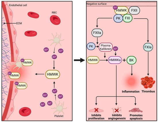
The contact system consists of factor XII (FXII), prekallikrein (PK) and HMWK [10,11]. HMWK promotes assembly of these components on negatively charged biological surfaces, thereby facilitating contact activation (Figure 1). Although factor XI (FXI) interacts closely with the contact system and can be activated by FXIIa, it is generally considered distinct from the core contact system and functions primarily within the intrinsic coagulation pathway. Classical contact activation occurs when FXII encounters activating surfaces such as glycosaminoglycans, polyphosphate (polyP), extracellular DNA, endotoxins or amyloid-like materials. Surface binding promotes conformational changes within FXII that facilitate its activation to FXIIa. FXIIa subsequently converts PK into plasma kallikrein, which further activates FXII in a positive-feedback loop that amplifies pathway activity while being regulated by C1 esterase inhibitor [12,13].
Figure 1.
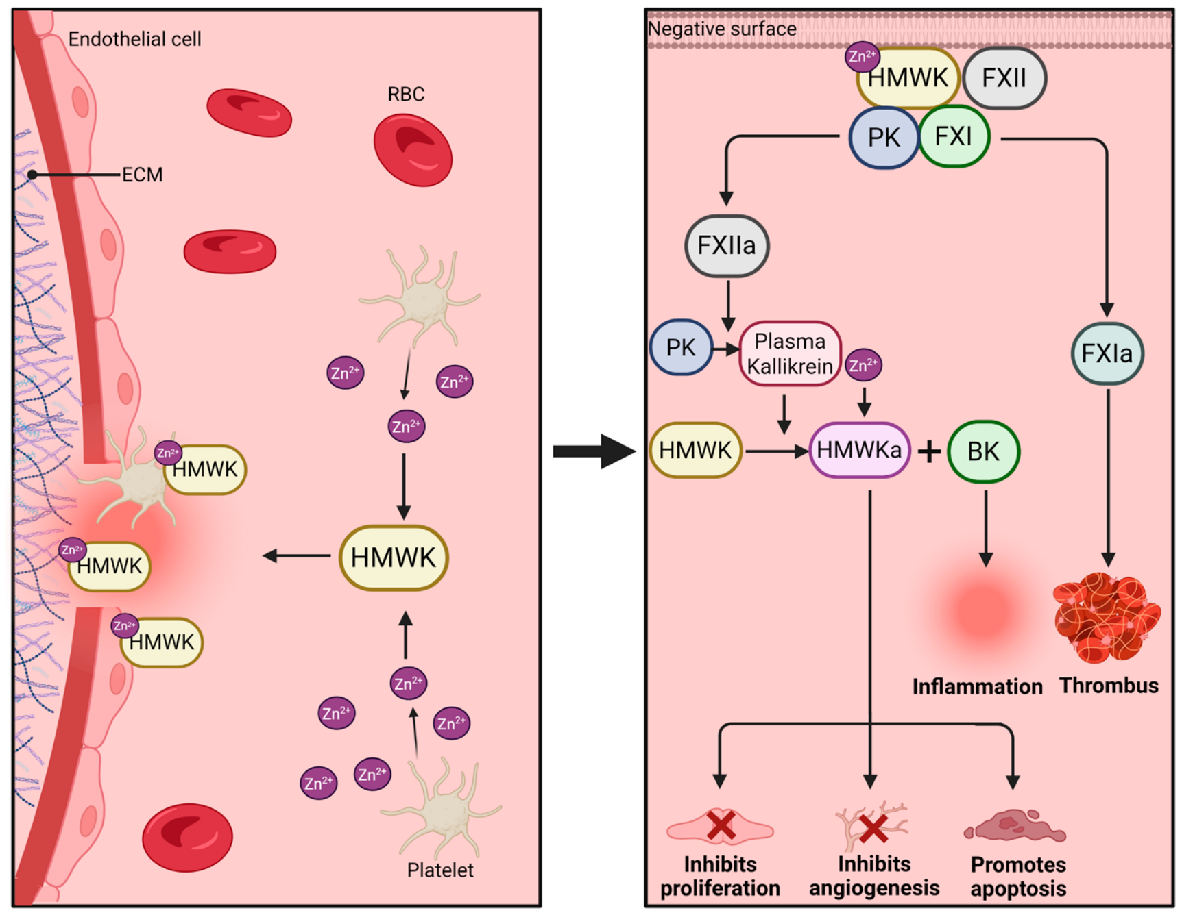
The role of Zn2+ in HMWK function and contact pathway activation. Binding of Zn2+, which may be released from activated platelets, induces a conformational change in HMWK that promotes its attachment to negatively charged surfaces. Physiological binding surfaces include exposed extracellular matrix (ECM) components and anionic proteins present on platelets and endothelial cells. Surface-bound HMWK facilitates the recruitment and activation of FXI and PK, promoting thrombus formation through the contact pathway and generation of plasma kallikrein. Plasma kallikrein cleaves HMWK to produce HMWKa and BK. BK promotes inflammatory responses, whereas HMWKa exerts anti-angiogenic effects and modulates endothelial cell function by inhibiting proliferation and inducing apoptosis. Created in BioRender. Marsh, J. (2026) https://BioRender.com/3835243.
Efficient assembly of the contact system is facilitated by Zn2+. Binding of Zn2+ increases the affinity of HMWK for negatively charged cellular surfaces and promotes recruitment of contact factors to sites of activation (Figure 1). On endothelial cells, FXII and HMWK associate with Zn2+-dependent receptor complexes involving globular C1q receptor (gC1qR), cytokeratin-1 (CK) and urokinase plasminogen activator receptor (uPAR) [13,14,15]. These interactions facilitate assembly of contact system components at the endothelial surface and promote local contact activation. In addition to endothelial cells and leukocytes, platelets represent an important source of extracellular Zn2+. Upon activation, platelet α-granules and dense granules release Zn2+ together with polyphosphate and other procoagulant mediators [16,17], generating a local environment that favours HMWK surface binding and contact-system assembly.
2. Kinin Release and Inflammatory Signalling
One of the principal consequences of contact system activation is proteolytic processing of HMWK and release of the vasoactive peptide BK. Activation of PK by FXIIa generates plasma kallikrein, which cleaves HMWK at multiple sites to liberate BK from domain D4 while producing cleaved HMWK (HMWKa) [1,4]. This reaction forms part of a positive-feedback loop in which kallikrein promotes further FXII activation and sustained kinin generation, thereby linking coagulation and inflammatory signalling [18,19].
Bradykinin mediates its biological effects through two G protein-coupled receptors, the constitutively expressed bradykinin B2 receptor (B2R) and the inducible bradykinin B1 receptor (B1R). Activation of B2R is responsible for the rapid vascular actions of BK, including vasodilation, increased vascular permeability and regulation of blood pressure [20]. In contrast, B1R expression is upregulated during inflammation, infection and tissue injury and is activated predominantly by des-Arg-bradykinin metabolites generated through carboxypeptidase-mediated processing of BK [21]. Because B1R undergoes limited desensitisation following activation, signalling through this receptor contributes to more persistent inflammatory responses [19].
The generation of BK from HMWK occurs through a series of ordered proteolytic cleavage events. Plasma kallikrein first cleaves HMWK to separate the heavy and light chains before releasing BK from domain D4 [12,22,23]. Subsequent cleavage within the light chain at R437-K438 proceeds more slowly and produces a stable terminal degradation product of HMWK [23]. Although plasma kallikrein is the principal physiological enzyme responsible for kinin generation, PK has also been reported, under certain experimental conditions, to cleave HMWK directly in a stoichiometric manner that releases BK independently of its conversion to plasma kallikrein [24].
While plasma kallikrein is the dominant physiological kinin-generating enzyme, alternative proteolytic pathways can also process HMWK. Activation of the lectin complement pathway generates mannan-binding lectin-associated serine proteases 1 and 2 (MASP-1 and MASP-2), both of which cleave HMWK independently of plasma kallikrein. However, only MASP-1 produces BK, whereas MASP-2 cleaves HMWK without releasing the peptide, highlighting the importance of cleavage-site specificity in determining kinin generation [23]. Similarly, plasmin can directly process HMWK, and the hereditary angioedema-associated Met379Lys variant of HMWK markedly enhances plasmin-mediated BK release, providing a mechanistic link between fibrinolysis and dysregulated kallikrein-kinin system activation [25].
Importantly, HMWK cleavage generates not only BK but also HMWKa, a biologically active product with functions distinct from its precursor. Proteolytic processing induces structural rearrangements that alter ligand and surface-binding properties and expose previously cryptic functional regions. Consequently, HMWKa contributes to inflammation, angiogenesis and vascular regulation through mechanisms that extend beyond BK generation alone.
3. Structure of Kininogen
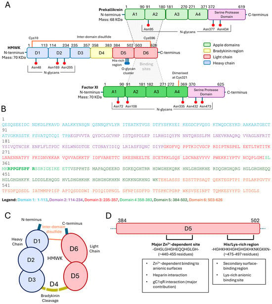
The mature HMWK polypeptide comprises 626 amino acids organised into six domains (D1–D6) (Figure 2). Domains D1–D3 form the N-terminal heavy chain, D4 contains the BK sequence, and D5–D6 constitute the C-terminal light chain [2,26]. HMWK circulates as a single-chain glycoprotein. During proteolytic processing by plasma kallikrein, release of BK generates HMWKa, in which the resulting heavy chain (D1–D3) and light chain (D5–D6) remain connected through an interchain disulfide bond [1,4,22,23]. Thus, the heavy- and light-chain nomenclature applies to cleaved HMWK rather than the native circulating protein. Domain D4 contains the BK sequence, whereas D5 and D6 provide functions that distinguish HMWK from LMWK [2,26]. Unlike LMWK, HMWK contains the additional D5 and D6 domains that underpin its role in contact activation. Domain D5 mediates interactions with negatively charged biological surfaces, whereas D6 contains binding sites for PK and FXI [18]. Consequently, circulating PK and FXI are predominantly found in complex with HMWK, facilitating localisation and assembly of contact system components at activating surfaces. Although FXI rather than HMWK is essential for normal haemostasis, the HMWK-FXI interaction contributes significantly to efficient surface-mediated clot initiation and thrombosis [18].
Figure 2.
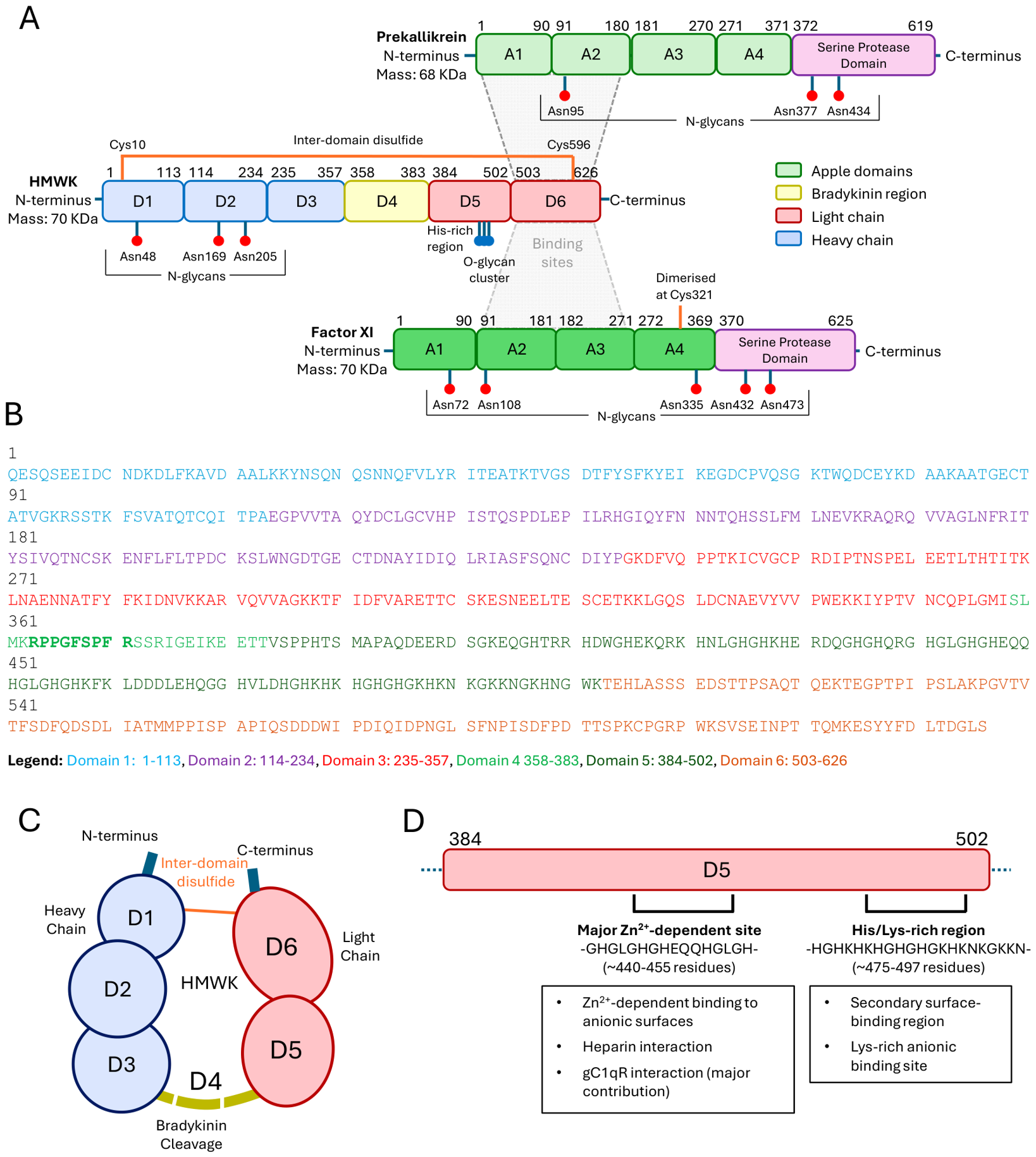
Structural organisation of high-molecular-weight kininogen (HMWK), prekallikrein (PK) and factor XI (FXI). (A) Schematic representation of HMWK, PK and FXI domain architecture, highlighting cleavage sites, glycosylation sites, and the major HMWK-PK and HMWK-FXI interaction regions. (B) Amino acid sequence of HMWK coloured by domain. The bradykinin sequence (within domain 4) is shown in bold. UniProt ID: P01042. (C) Schematic representation of HMWK following bradykinin release, illustrating the relative arrangement of domains D1–D3 and D5–D6 and the disulfide linkage connecting the heavy and light chains. (D) Schematic representation of the histidine-rich D5 domain of HWMK, which contributes to Zn2+-dependent interactions with contact system proteins and anionic surfaces. Note that domain boundaries, particularly within HMWK, should be considered approximate owing to the limited structural information available for the full-length protein.
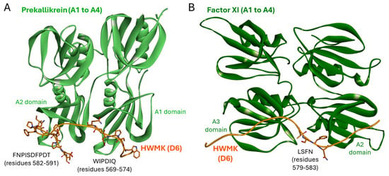
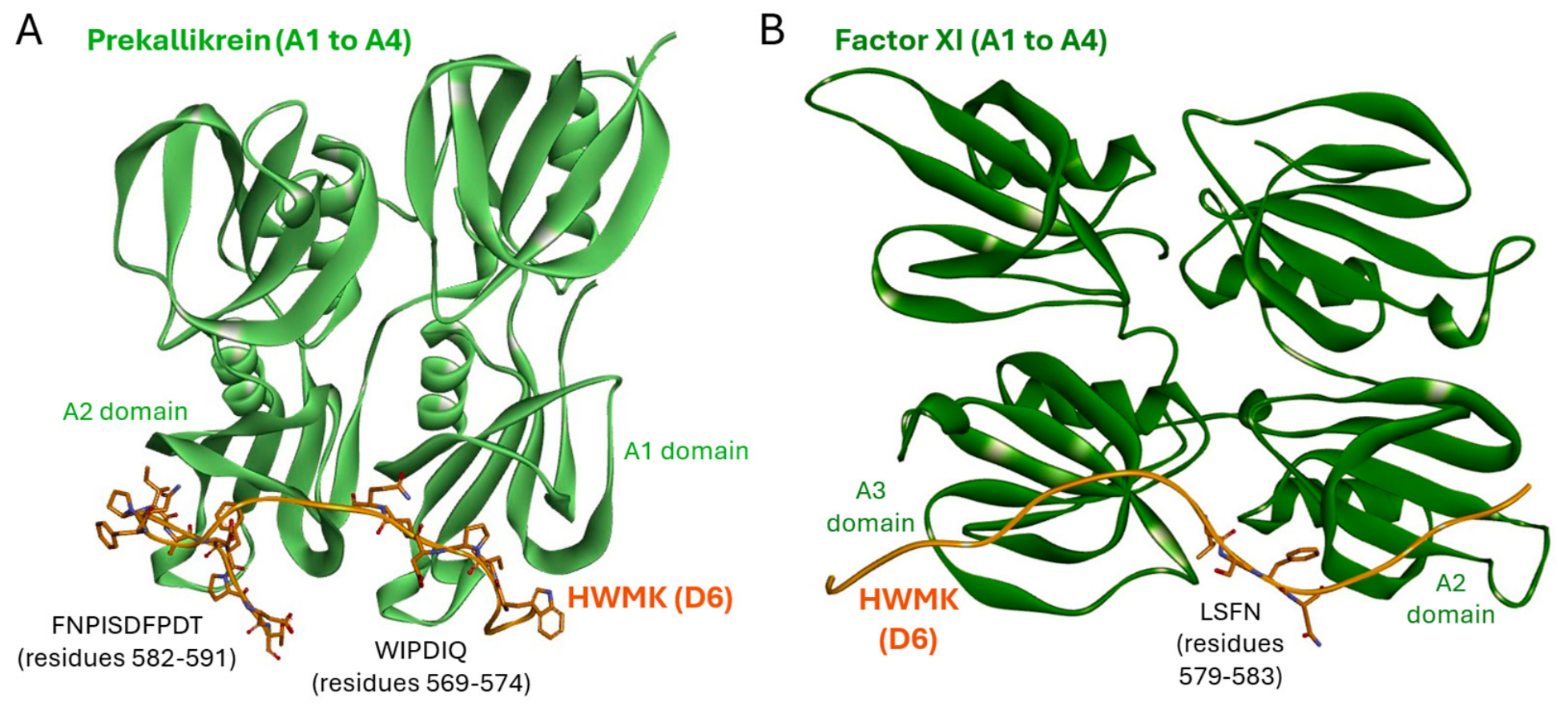
Binding of PK and FXI to HMWK is mediated through specialised protein interaction modules known as Apple domains. Both proteins contain four Apple domains (A1–A4) within their N-terminal heavy chains, preceding the catalytic serine protease domain. Structural studies demonstrated that PK and FXI recognise a common 31-residue segment within D6 but engage this region through distinct molecular mechanisms [1,27] (Figure 3). Crystal structures of recombinant PK and FXI heavy-chain fragments in complex with a synthetic HMWK D6 peptide revealed that the peptide adopts different conformations depending on the binding partner. In the PK complex, the HMWK peptide adopts a bent conformation and interacts with both the A1 and A2 domains. In contrast, FXI recognises the peptide through interactions involving the A2 and A3 domains [27]. These observations indicate that D6 possesses substantial conformational flexibility, enabling recognition of structurally related ligands through different binding modes. Importantly, these structures were determined using recombinant protein fragments and a synthetic HMWK D6 peptide rather than intact full-length HMWK [27].
Figure 3.
Structural basis of HMWK Domain 6 (D6) recognition by prekallikrein and factor XI. (A) Crystal structure of the prekallikrein heavy chain comprising Apple domains A1–A4 (light green) in complex with a peptide derived from HMWK D6 (orange) (PDB 7QOX) [27]. The HMWK peptide adopts a bent conformation and interacts with both the A1 and A2 domains through residues WIPDIQ (569–574) and FNPISDFPDT (582–591). (B) Crystal structure of the factor XI comprising Apple domains A1–A4 (dark green) bound to an HMWK D6 peptide (orange) (PDB 7QOT) [27]. In contrast to the PK complex, residues LSFN (579–583) occupy a binding pocket between the A2 and A3 domains. Structures were determined using recombinant PK and FXI heavy-chain fragments together with a synthetic 31-residue HMWK D6 peptide (Ser565–Lys595), rather than full-length HMWK.
A major functional feature of HMWK is its capacity to bind Zn2+. Domain D5 contains a histidine-rich sequence that constitutes the principal Zn2+-binding region of the molecule [28,29]. Zn2+ binding enhances the affinity of HMWK for endothelial cells and other negatively charged surfaces, thereby promoting assembly of PK, FXI and FXII at sites of contact activation [4,11,30]. Through these Zn2+-dependent interactions, HMWK functions as a molecular scaffold that coordinates assembly of the contact system.
Detailed mapping studies have identified at least two anionic surface-binding regions within D5, comprising a histidine-rich region and a more C-terminal histidine/lysine-rich region [31]. Both regions contribute to interactions with negatively charged ligands and surfaces, although the histidine-rich region appears to play a particularly important role in Zn2+-dependent binding [29,31]. Together, these subdomains create a flexible multivalent interaction platform that enables HMWK to engage a wide range of biological surfaces in a Zn2+-regulated manner [28,31].
Despite its central role in contact activation, the molecular basis of Zn2+ binding to HMWK remains poorly understood. No high-resolution structural information is currently available for the D5 domain, which is thought to adopt a highly flexible conformation owing to its histidine-rich, low-complexity sequence composition [1,4]. However, D5 shares structural and functional similarities with the histidine-rich region (HRR) of histidine-rich glycoprotein (HRG), another plasma protein whose ligand-binding properties are regulated by Zn2+ [32,33]. Both regions are compact histidine-rich domains enriched in Zn2+-binding residues, and mediate Zn2+-dependent interactions with heparin and other polyanionic ligands. However, the two domains differ substantially in sequence organisation. The HRG HRR is dominated by tandem GHHPH-like repeat motifs [34], whereas HMWK D5 contains a more heterogeneous arrangement of histidine-, lysine- and glycine-rich sequences [28]. These differences are likely to influence metal ion coordination and may contribute to the distinct ligand-binding properties reported for the two proteins.
Although direct structural information is lacking, studies of HRG may provide useful insight into how Zn2+ interacts with HMWK. Electron paramagnetic resonance-based analyses using Cu2+ as a spectroscopic surrogate for Zn2+ suggest that the HRG HRR does not contain a single, well-defined metal-binding site [35]. Instead, metal ions appear to be coordinated by multiple alternative clusters of histidine residues distributed throughout the flexible domain. Given the comparable size, histidine content and apparent disorder of HMWK D5, it is tempting to speculate that Zn2+ binding occurs through a similarly dynamic mechanism. Such a model would be consistent with the ability of Zn2+ to modulate conformational sampling and enhance interactions of HMWK with a diverse range of biological surfaces and ligands.
Increasing evidence suggests that D5 behaves as an intrinsically disordered protein region. The high content of histidine, glycine and lysine residues, together with the low sequence complexity of the domain, is predicted to impede formation of a stable hydrophobic core and instead favour a dynamic conformational ensemble [36]. Consistent with this view, biophysical studies of recombinant HK5 demonstrated spectroscopic properties characteristic of a predominantly random-coil structure and provided little evidence for stable secondary structure formation [36].
Interestingly, although D5 contains multiple Zn2+-binding histidine residues and Zn2+ is a critical regulator of HMWK function [28,29], available evidence suggests that Zn2+ binding does not induce a major disorder-to-order transition [36]. Circular dichroism analyses of HK5 in the presence of Zn2+ revealed no substantial increase in α-helical or β-sheet content, indicating that Zn2+ regulates ligand recognition without generating a stable folded structure. Instead, Zn2+ may alter local conformational preferences, electrostatic properties and interaction dynamics within the intrinsically disordered domain [36].
Intrinsic disorder may provide important functional advantages for D5. Unlike globular interaction domains that recognise a limited range of binding partners, D5 interacts with diverse ligands including negatively charged surfaces, glycosaminoglycans, ferritin, cellular receptors and extracellular matrix components [28,29,30,31].
However, relatively little is known about the structural consequences of these interactions. Ferritin binding has been shown to be metal-dependent, but no evidence for substantial folding of D5 upon complex formation has been reported [36]. Similarly, Zn2+-dependent interactions with heparin, glycosaminoglycans [28,31], gC1qR, endothelial cell receptors [13,15,30] and extracellular matrix components [37,38] are well documented, although it remains unclear whether these ligands induce local structural ordering or simply stabilise selected conformations within a largely disordered ensemble. Consequently, important questions remain regarding the structural basis of Zn2+-dependent ligand recognition and the extent to which partner-induced conformational selection contributes to HMWK function.
The available evidence suggests that D5 functions as a flexible, intrinsically disordered interaction platform capable of engaging a diverse range of ligands. However, for most interaction partners there is currently little information regarding the structural consequences of binding or the extent to which ligand association stabilises specific D5 conformations (Table 1).
Table 1.
Interaction partners of HMWK Domain 5 and current evidence regarding Zn2+ dependence and structural ordering.
Activation of the contact system results in proteolytic cleavage of HMWK by plasma kallikrein, releasing BK from domain D4 and generating HMWKa. Electron microscopy studies indicate that this conversion is accompanied by substantial conformational rearrangements [4]. These changes increase the accessibility of D5 and enhance HMWKa interactions with anionic surfaces [29,31], potentially contributing to its anti-adhesive properties. Simultaneously, liberated BK acts on endothelial cells to promote vasodilation and vascular permeability, linking structural remodelling of HMWK directly to its inflammatory functions [39].
4. Zn2+-Dependent Regulation of Contact Activation
Zn2+ is an important regulator of HMWK function and contact system assembly. Binding of Zn2+ enhances the interaction of HMWK with a wide range of anionic surfaces, including cell surfaces and extracellular glycosaminoglycans [8,13,14,15], extracellular matrix components [23,40], polyP [11], and protein aggregates such as β-amyloid (Aβ) [41]. Through these interactions, Zn2+ promotes localisation of HMWK and associated contact factors at sites where contact activation occurs.
PolyP is a particularly potent activator of the contact pathway and accelerates several contact activation reactions in a chain-length-dependent manner [42]. Because activated platelets release both polyP and Zn2+ [16,17], local concentrations of these molecules can substantially influence contact system activity during thrombus formation. Studies examining individual contact pathway reactions have demonstrated that the requirement for Zn2+ and HMWK varies according to the specific enzyme-substrate pair involved. Zn2+ is essential for reactions in which FXII acts as the substrate, whereas HMWK is most important in reactions mediated by plasma kallikrein [11]. Consistent with this model, plasma kallikrein requires HMWK for efficient binding to polyP-containing surfaces [43].
The contribution of HMWK therefore varies across individual contact pathway reactions. While HMWK is required for efficient plasma kallikrein-mediated reactions, it is dispensable for certain FXIIa-catalysed reactions, although it may still influence their kinetics and overall efficiency. Interestingly, at high concentrations (≥500 nM), HMWK inhibits polyP-driven contact activation, potentially through competition with FXII or FXIIa for polyP binding sites [11].
Many surfaces capable of activating the contact pathway, including collagen, polyP, misfolded proteins and kaolin, possess a high density of negative charge. Such surfaces electrostatically attract Zn2+, resulting in elevated local metal ion concentrations at the surface-plasma interface [32]. This phenomenon is likely to be physiologically important because HMWK exhibits relatively low affinity for many activating surfaces in the absence of Zn2+. Several Zn2+-binding sites have been identified within HMWK, particularly within the histidine-rich D5 region, and Zn2+ binding markedly enhances interactions between HMWK, activating surfaces and other contact system proteins [1,13,15]. Although the structural basis of this effect remains incompletely understood [1], Zn2+ binding is thought to modify the conformation and electrostatic properties of D5, thereby increasing the capacity of HMWK to associate with negatively charged surfaces and recruit contact factors [11,28,29].
Through these effects, Zn2+ promotes stable localisation of HMWK at activating surfaces while strengthening interactions with its principal binding partners, PK and FXI. HMWK therefore functions as a Zn2+-responsive molecular scaffold that concentrates contact system components at sites of activation. Recruitment of HMWK facilitates localisation of PK and FXI, enhancing assembly of the contact activation complex and promoting efficient initiation of the contact pathway [11].
Additional support for the importance of Zn2+ comes from metal chelation studies [11]. EDTA inhibits FXIIa-mediated activation of PK in the presence of polyP, indicating that metal-ion-dependent interactions contribute to efficient assembly of the activation complex. This inhibitory effect becomes progressively weaker at higher polyP concentrations, suggesting that increased surface availability can partially compensate for the loss of Zn2+-dependent interactions.
5. Cellular Anchorage, Multi-Protein Receptors, and Zinc Modulation
In addition to mediating interactions with activating surfaces, Zn2+ is required for efficient binding of HMWK to endothelial cells, platelets and leukocytes [7,8,9,15]. A widely accepted model proposes that HMWK associates with multiprotein receptor complexes containing gC1qR, CK1 and uPAR [13,14,15], which together may form receptor complexes that facilitate localisation of the contact system to cellular surfaces [14,15]. However, the extent to which these interactions occur in vivo, and their relative physiological importance remain incompletely understood [1,30].
Among these proteins, gC1qR is considered the principal high-affinity binding site for both HMWK and HMWKa [13,15], whereas CK1 appears to contribute to assembly and stabilisation of the cell-surface receptor complex [14]. In contrast, uPAR is thought to function primarily in downstream signalling and is particularly important for mediating the biological activities of HMWKa [2,15]. Notably, Zn2+ is required for efficient HMWKa binding to uPAR and for several of the anti-angiogenic effects attributed to HMWKa, highlighting an important role for Zn2+ in regulating HMWKa-dependent cellular responses [2,15,44].
More recently, the molecular basis of the HMWK-gC1qR interaction has been examined in greater detail [45]. Studies have demonstrated that D5 mediates high-affinity binding to gC1qR and contributes to the localisation of contact-system components at cellular surfaces [15,30,45]. The presence of multiple ligand-binding regions within D5 suggests that the domain functions as a flexible interaction platform capable of engaging gC1qR through multivalent contacts [31]. Structural and mutagenesis studies further support a role for gC1qR in clustering HMWK and other contact factors, thereby facilitating assembly of the contact system [45]. However, the precise structural basis of D5-gC1qR recognition and the contribution of individual D5 subdomains remain incompletely understood.
The overall affinity of HMWK and HMWKa for endothelial cells lies within the nanomolar range (Kd ≈ 7–52 nM), although the relative contribution of each form to total cellular binding remains unclear. Surface plasmon resonance studies using soluble extracellular forms of uPAR (suPAR), gC1qR and CK1 demonstrated that all interactions are strictly Zn2+ dependent [15]. Binding affinities followed the order gC1qR > CK1 > suPAR, supporting the view that gC1qR is the dominant receptor component. HMWK and HMWKa displayed similar affinities for gC1qR, whereas HMWKa exhibited a 2.6-fold higher affinity for CK1 and an approximately 53-fold higher affinity for suPAR [15].
Interestingly, receptor–receptor interactions were detected only between gC1qR and CK1, suggesting that a binary gC1qR-CK1 complex can assemble independently of kininogen [15]. Although suPAR displayed the weakest overall affinity, it was the only receptor capable of clearly discriminating between HMWK and HMWKa. This observation suggests that uPAR may play a central role in mediating differences in cellular recognition and signalling between the uncleaved and cleaved forms of HMWK. In contrast, gC1qR and CK1 are more likely to function primarily as anchoring molecules that recruit and concentrate HMWK at the cell surface prior to proteolytic conversion into HMWKa [15].
6. Clinical Context: Altered Zinc Homeostasis and Pathophysiology
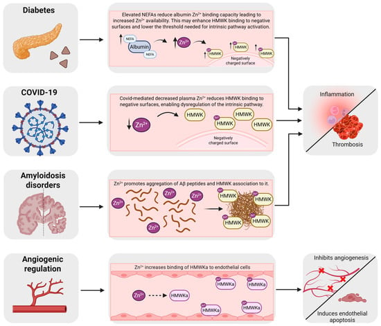
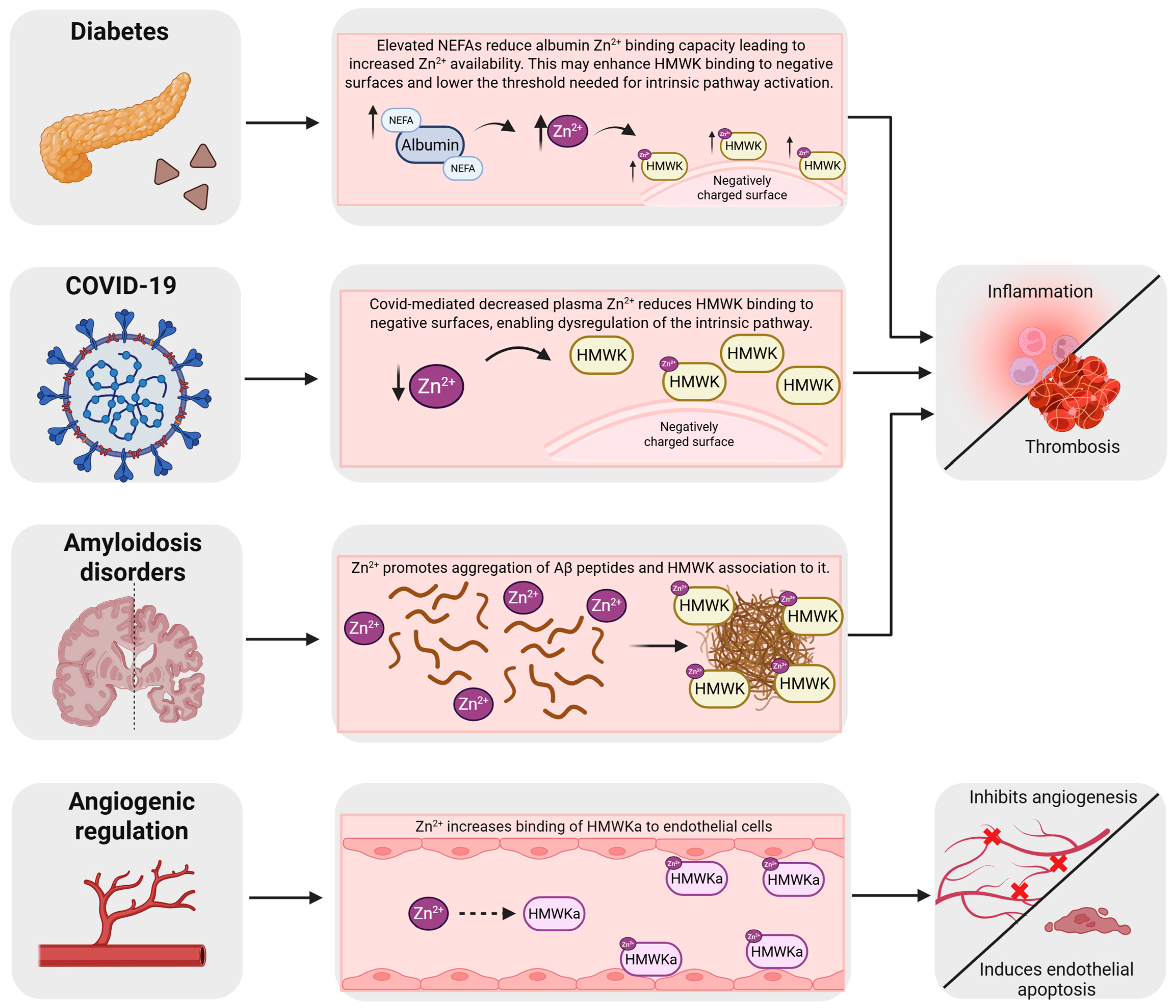
The biological activities of HMWK are strongly influenced by Zn2+ availability. Consequently, disturbances in zinc homeostasis have the potential to alter contact system activation, kinin generation, vascular permeability and inflammatory responses. Although the precise contribution of Zn2+ to HMWK function in human disease remains incompletely defined, several pathological settings illustrate potential links between zinc dysregulation and contact system biology (Figure 4).
Figure 4.
Potential consequences of altered Zn2+ homeostasis for HMWK function in different clinical settings. In diabetes, plasma NEFA levels are often elevated, reducing the ability of serum albumin to buffer Zn2+. The resulting increase in Zn2+ availability may enhance HMWK binding and lower the threshold for activation of the contact pathway. In COVID-19, systemic Zn2+ deficiency may impair HMWK interactions with negatively charged surfaces, contributing to dysregulation of contact activation. In amyloid disorders, Zn2+ facilitates Aβ aggregation and may enhance HMWK association with amyloid deposits. In each of these settings, altered HMWK function may contribute to a prothrombotic and pro-inflammatory state. Plasma kallikrein cleaves HMWK to generate HMWKa and BK. Zn2+ binding to HMWKa enhances its interaction with endothelial cells, promoting downstream effects including endothelial apoptosis and inhibition of angiogenesis. Created in BioRender. Marsh, J. (2026) https://BioRender.com/0w292hj.
6.1. Kininogen Deficiency and Naturally Occurring KNG1 Variants
Naturally occurring loss-of-function variants in the KNG1 gene provide important insight into the physiological functions of HMWK and the contact system. Individuals with severe HMWK deficiency typically exhibit markedly prolonged activated partial thromboplastin times, reflecting the important role of HMWK in contact-pathway activation and intrinsic coagulation assays. Despite these profound laboratory abnormalities, however, most reported individuals do not exhibit a significant bleeding tendency, indicating that HMWK is largely dispensable for normal haemostasis [46].
Recent studies have expanded the catalogue of pathogenic KNG1 variants and improved understanding of the molecular basis of HMWK deficiency. Adenaeuer et al. identified multiple deficiency-causing KNG1 variants associated with severe HMWK deficiency and provided a comprehensive analysis of the clinical and laboratory characteristics of affected individuals [46]. These studies demonstrated that severe HMWK deficiency is an exceptionally rare disorder characterised by profound abnormalities in contact-system activation despite an absence of major spontaneous bleeding manifestations [46]. Similarly, Wang et al. described a patient carrying the frameshift variant c.628_629delAA, which resulted in severe HMWK deficiency and markedly prolonged coagulation testing while producing only limited clinical symptoms [47].
The molecular consequences of these variants are expected to include impaired protein synthesis, defective secretion, reduced plasma stability, or loss of functional interactions required for efficient contact-system assembly [46,47]. Although the resulting defects substantially impair contact activation in vitro, the relatively mild clinical phenotype associated with severe HMWK deficiency suggests that HMWK-dependent contact activation is less important for normal haemostasis than might be inferred from its striking effects on coagulation assays. These observations are consistent with growing interest in therapeutic strategies that target the contact pathway while minimising bleeding risk.
As interest in therapeutic targeting of the contact system continues to grow, naturally occurring KNG1 variants provide valuable human genetic evidence regarding the physiological and pathological roles of HMWK. Further characterisation of these rare deficiencies may therefore help define the contribution of HMWK to thrombosis, inflammation and vascular dysfunction. While the effects of severe KNG1 deficiency on Zn2+-dependent HMWK interactions remain unknown, these observations highlight the broader importance of understanding how HMWK function is regulated under physiological and pathological conditions.
6.2. Diabetes, Zinc Speciation and Contact System Activation
Cardiometabolic disorders, including obesity and type 2 diabetes (T2D), are characterised by chronic low-grade inflammation, endothelial dysfunction and an increased risk of arterial and venous thrombosis [48,49]. A growing body of evidence suggests that dysregulation of plasma zinc handling may contribute to these pathologies by altering the activity of Zn2+-dependent proteins involved in coagulation and vascular biology [50].
Under normal conditions, approximately 75% of plasma Zn2+ is bound to serum albumin [51,52], which acts as the principal buffer and transporter of exchangeable zinc within the circulation. The capacity of albumin to bind Zn2+ is regulated by circulating non-esterified fatty acids (NEFAs) [52,53]. Binding of long-chain fatty acids to the high-affinity FA2 site on albumin induces an allosteric conformational change that disrupts the primary Zn2+-binding site (site A) [54], reducing the affinity of albumin for Zn2+ and increasing the availability of exchangeable metal ions within plasma [52,53,55]. This phenomenon, termed the albumin-fatty acid-zinc switch, provides a mechanistic link between lipid metabolism and extracellular zinc speciation.
Circulating NEFA concentrations are frequently elevated in obesity, insulin resistance and T2D [56,57], suggesting that perturbation of albumin-mediated zinc buffering may represent a common feature of these disorders [50,53]. Increased availability of exchangeable Zn2+ has been shown to alter the activity of several coagulation proteins, including HRG, fibrinogen and HMWK [34,41,58]. Although direct studies examining the consequences for HMWK are limited, the established role of Zn2+ in enhancing HMWK binding to anionic surfaces and promoting contact system assembly suggests that alterations in plasma zinc speciation could influence the threshold for contact activation.
This model is particularly attractive because HMWK functions as a Zn2+-responsive scaffold within the contact pathway [11,18]. Increased Zn2+ availability may facilitate interactions between HMWK and negatively charged biological surfaces [29,39], promote assembly of HMWK-PK-FXI complexes [18], and enhance localisation of contact factors at sites of vascular injury or inflammation. Consequently, the albumin-fatty acid-zinc switch may indirectly modulate kallikrein-kinin system activity by altering the Zn2+ environment in which HMWK operates [52,53].
In addition to effects on coagulation, excessive kallikrein-kinin signalling has been implicated in vascular dysfunction and chronic inflammation [10,19], both hallmarks of metabolic disease. BK regulates endothelial permeability, vascular tone and inflammatory cell recruitment [59], while activation of FXII and plasma kallikrein can amplify inflammatory signalling independently of kinin generation [10,19]. Perturbations in Zn2+ homeostasis may therefore influence both thrombotic and inflammatory pathways through effects on HMWK-dependent contact system activation.
Although direct evidence linking the albumin-fatty acid-zinc switch to enhanced HMWK activation in vivo is currently lacking, several observations support this possibility. Elevated plasma NEFA concentrations have been associated with prothrombotic alterations in haemostatic pathways [50,57], while altered zinc speciation redistributes Zn2+ among plasma proteins and increases zinc association with several candidate binding partners [53,55]. Together, these findings suggest a model in which metabolic dysregulation alters extracellular Zn2+ availability, thereby increasing the activity of Zn2+-responsive proteins such as HMWK and predisposing to contact-system-driven inflammation and thrombosis. Further studies are required to determine whether modulation of plasma zinc speciation contributes directly to contact pathway activation in obesity and T2D. More broadly, the albumin-fatty acid-zinc switch may influence multiple Zn2+-dependent activities of HMWK, including contact activation, cellular localisation, angiogenesis and interactions with pathological surfaces such as amyloid aggregates.
6.3. COVID-19 and Inflammation
Several studies have reported hypozincaemia in hospitalised COVID-19 patients, particularly in severe disease, with lower plasma zinc concentrations associated with poorer clinical outcomes and elevated inflammatory markers [60,61]. These observations suggest that disturbances in zinc homeostasis are a common feature of COVID-19 and may contribute to both immune dysregulation and altered vascular responses. Zn2+ is essential for both innate and adaptive immune function. Zinc deficiency impairs lymphocyte proliferation, differentiation and survival, particularly within T-cell populations, and can contribute to lymphopenia and impaired adaptive immune responses [62,63]. In parallel, Zn2+ serves as an important regulator of neutrophil activation and inflammatory signalling. Under conditions of zinc deficiency, neutrophil function can become dysregulated and has been associated with elevated neutrophil-to-lymphocyte ratios, a recognised marker of systemic inflammation and adverse clinical outcomes in COVID-19 [64,65]. The consequences of zinc deficiency are not restricted to leukocyte dysfunction. Zn2+ also regulates several plasma proteins involved in coagulation and inflammation, including HMWK and other components of the contact system.
Although most studies have focused on immune dysfunction rather than contact system activation, excessive kallikrein-kinin signalling and BK production have been proposed to contribute to vascular leakage, pulmonary inflammation and endothelial dysfunction in severe COVID-19 [66,67]. Given the established role of Zn2+ in regulating HMWK localisation and contact activation [11,13,14,15,28,29], disturbances in zinc homeostasis may influence inflammatory responses through both immune and vascular pathways.
6.4. Amyloidosis Disorders and Neuroinflammation
Protein aggregates associated with amyloid diseases can provide abnormal negatively charged surfaces capable of recruiting and activating components of the kallikrein-kinin system. Aggregated Aβ represents the best-characterised example and has been shown to activate the plasma kinin-forming cascade in a Zn2+-dependent manner [40,68]. D5 of HMWK mediates binding to these amyloid assemblies through Zn2+-dependent interactions [28,29,68], facilitating recruitment and organisation of contact system components at sites of protein aggregation. Zn2+ also promotes aggregation of Aβ peptides (Aβ1–38, Aβ1–40 and Aβ1–42), thereby increasing the availability of surfaces capable of supporting contact activation. Furthermore, Zn2+ enhances the association of HMWK and FXII with aggregated Aβ, promoting activation of the contact pathway and subsequent BK generation [68], consistent with current models of FXII activation on negatively charged activating surfaces [69].
The interaction between Zn2+ and amyloid pathology is, however, complex and context dependent. Soluble oligomeric Aβ species are widely considered to be important mediators of amyloid-associated toxicity and inflammation [70,71,72]. By binding to histidine residues within Aβ (His6, His13 and His14), Zn2+ influences the aggregation behaviour, structural organisation and biological activity of Aβ assemblies [73,74,75]. Consequently, changes in zinc availability may alter both the spectrum of Aβ species present within tissues and the capacity of amyloid deposits to support contact system activation.
Beyond Alzheimer’s disease, increasing evidence suggests that activation of the contact system may contribute to a broader range of neurological and cerebrovascular disorders [10,70]. Experimental and clinical studies have implicated FXII activation, plasma kallikrein and downstream contact-system signalling in ischaemic stroke [10,76,77], where contact-system activation may promote neuroinflammation, blood–brain barrier dysfunction and cerebral oedema. Similar mechanisms may contribute to vascular dementia and vascular cognitive impairment, conditions characterised by chronic cerebrovascular injury, endothelial dysfunction and persistent neurovascular inflammation. Although the specific contribution of HMWK remains poorly defined [1], its central role in contact-system assembly suggests that HMWK-mediated interactions may influence these processes. Given the established importance of Zn2+ in regulating HMWK localisation and contact-system activation [11,15], disturbances in zinc homeostasis could potentially modulate these pathways. However, direct evidence linking Zn2+-dependent HMWK function to vascular cognitive impairment, vascular dementia or other neurodegenerative disorders remains limited and further investigation is required.
Although most mechanistic evidence has been obtained using Aβ, similar principles may apply to other amyloidogenic proteins that form negatively charged aggregated structures capable of recruiting contact-system components. Further studies are required to determine whether Zn2+-dependent contact activation contributes to inflammation and vascular dysfunction in other protein-misfolding disorders.
6.5. Angiogenic Regulation
A distinctive feature of the kininogen system is its ability to generate both pro-angiogenic and anti-angiogenic mediators. BK promotes angiogenesis through activation of B1 and B2 receptors and the subsequent induction of angiogenic signalling pathways involving vascular endothelial growth factor and basic fibroblast growth factor [2,78].
In contrast, proteolytic conversion of HMWK to HMWKa generates a potent endogenous inhibitor of angiogenesis [2,79]. HMWKa suppresses angiogenesis through multiple complementary mechanisms, including disruption of endothelial adhesion, inhibition of cell-cycle progression, interference with pro-survival signalling pathways and induction of apoptosis. Histidine- and glycine-rich sequences within D5 (residues 441-457 and 475-502) selectively disrupt endothelial cell adhesion to vitronectin and gelatin, leading to detachment-induced apoptosis [79]. HMWKa suppresses endothelial proliferation and promotes apoptosis through multiple signalling mechanisms [37,38,80]. Additional anti-angiogenic activity arises through interactions between D5 and domains D2/D3 of uPAR. Binding of HMWKa to uPAR disrupts the vitronectin-uPAR-αvβ3-caveolin-Src-FAK-paxillin signalling axis, a focal-adhesion-associated pathway that regulates endothelial cell survival and migration [15].
Several observations suggest that these anti-angiogenic activities are influenced by Zn2+ and by interactions with HRG. HMWKa binds endothelial cell tropomyosin in a Zn2+-dependent manner [37], and studies using HRG-derived histidine-proline-rich peptides suggest that metal-ion binding and solution conditions can modulate these interactions [81]. Biophysical analyses indicate that metal ion binding alters interactions involving HRG-derived peptides and HMWKa-associated complexes [81], while acidic conditions may further influence interactions between histidine-rich domains and negatively charged tropomyosin. Local release of Zn2+ from activated platelets may augment these effects [59], potentially strengthening the anti-angiogenic properties of HMWKa.
7. Future Perspectives
Despite decades of research, important questions remain regarding the regulation of HMWK and the contact system by Zn2+. Considerable progress has been made in defining the biochemical organisation of the contact pathway and the interactions between HMWK, FXII, FXI and PK [11,18,27]. However, many aspects of the molecular mechanisms that govern contact activation remain incompletely understood.
A major unresolved question concerns the structural biology of HMWK itself. Although HMWK is recognised as a multifunctional six-domain protein that acts both as a cofactor and as a substrate for BK generation, no high-resolution three-dimensional structure of the intact protein is currently available [1]. Consequently, the structural basis of Zn2+ binding, contact factor recognition and surface association remains poorly defined. Little is known about the conformational organisation of the intrinsically disordered D5 region or how Zn2+ binding influences its interactions with anionic surfaces and cellular receptors [28,29,31]. Similarly, although electron microscopy studies suggest substantial structural rearrangement following conversion of HMWK to HMWKa, the molecular details of these changes remain unresolved [4].
A second major challenge is understanding how contact activation is initiated under physiological and pathological conditions. Classical models rely heavily on artificial activating surfaces such as kaolin; however, the identity and relative importance of endogenous activators in vivo remain uncertain. PolyP, misfolded proteins and amyloid aggregates have all been proposed as physiologically relevant activators [10,16,17,42,43,68,69]. Determining how these surfaces recruit Zn2+ and influence HMWK-dependent assembly of the contact pathway remain an important area for future investigation.
Emerging evidence suggests that zinc speciation, rather than total plasma zinc concentration, may be a critical determinant of contact system activity [50,53,55]. The discovery of the albumin-fatty acid-zinc switch [52,53,54] has highlighted how metabolic alterations can regulate the availability of exchangeable Zn2+ in plasma. Given the strong dependence of HMWK on Zn2+ for surface binding, receptor interactions and contact factor assembly, defining how changes in extracellular zinc speciation influence HMWK function is an important priority. Future studies should determine whether altered zinc buffering contributes directly to contact system activation in conditions such as obesity, diabetes and other cardiometabolic disorders [50,53,55,56,57].
Progress in this area will require advanced structural and analytical approaches. Advanced structural and biophysical approaches will be required to define the architecture of HMWK and its associated complexes [1,4,18,27]. Similarly, new approaches for measuring zinc speciation and labile Zn2+ pools in biological fluids may provide greater insight than conventional measurements of total plasma zinc concentration [55]. Such methods may help establish whether local fluctuations in Zn2+ availability occur at sites of thrombosis, inflammation or vascular injury, where contact activation is thought to take place.
From a translational perspective, targeting Zn2+-dependent contact system regulation represents an attractive therapeutic opportunity. Current therapeutic approaches largely focus on inhibition of individual pathway components, such as FXII, plasma kallikrein or BK receptors [10,19,20,59]. However, modulation of Zn2+ availability, disruption of Zn2+-dependent HMWK interactions, or manipulation of extracellular zinc speciation could potentially provide alternative strategies for controlling contact-system-driven inflammation and thrombosis. A more complete understanding of the molecular basis of Zn2+-dependent HMWK function will therefore be essential for translating these concepts into clinical interventions.
Ultimately, future research should move beyond viewing zinc as a simple micronutrient and instead consider its role as a dynamic regulator of extracellular protein function [34,50,58]. Defining how Zn2+ coordinates HMWK activity across inflammation, thrombosis, angiogenesis and metabolic disease may reveal new therapeutic opportunities and establish zinc speciation as an important determinant of vascular and inflammatory homeostasis.
8. Conclusions
HMWK occupies a unique position at the intersection of coagulation, inflammation and vascular biology. Beyond serving as the precursor of BK, it functions as a multifunctional scaffold that coordinates assembly of the contact system through interactions with FXII, FXI and plasma kallikrein. A growing body of evidence demonstrates that many of these interactions are strongly influenced by Zn2+, particularly through the histidine-rich D5 domain, which regulates binding to cellular receptors, activating surfaces and contact pathway proteins. Consequently, Zn2+ availability emerges as an important determinant of both contact activation and downstream inflammatory signalling.
Although substantial progress has been made in defining the biochemical and cellular functions of HMWK, many questions remain regarding the molecular basis of Zn2+ binding, the structural organisation of the intact protein and the mechanisms that govern contact activation in vivo. Emerging concepts, such as the albumin-fatty acid-zinc switch, suggest that zinc speciation, rather than total zinc concentration alone, may have important consequences for HMWK function in health and disease. Understanding how dynamic changes in extracellular Zn2+ availability influence contact system activation may therefore provide new insight into the pathogenesis of thrombosis, inflammation and vascular dysfunction, while identifying novel opportunities for therapeutic intervention.
Author Contributions
A.J.S. devised the overall concept. All authors contributed to the drafting and final review of the manuscript. R.K. and J.S.M. prepared the figures. All authors have read and agreed to the published version of the manuscript.
Funding
This research was funded by a Faculty Development Scholarship from the Higher Education Commission of Pakistan, grant number 121-FBS2-002 and Diabetes UK, grant number 23/0006528.
Institutional Review Board Statement
Not applicable.
Informed Consent Statement
Not applicable.
Data Availability Statement
No new data were created or analyzed in this study. Data sharing is not applicable to this article.
Acknowledgments
The authors are grateful to Miriam Veenhuizen for critically reviewing the manuscript. During the preparation of this manuscript, the author(s) acknowledge use of Microsoft 365 Copilot to improve text readability. The authors have reviewed and edited the output and take full responsibility for the content of this publication.
Conflicts of Interest
The authors declare no conflicts of interest.
Abbreviations
The following abbreviations are used in this manuscript:
| Aβ | β-amyloid |
| B1R | Bradykinin B1 receptor |
| B2R | Bradykinin B2 receptor |
| BK | Bradykinin |
| ECM | Extracellular matrix |
| FXI | Factor XI |
| FXII | Factor XII |
| gC1qR | Globular C1q receptor |
| HMWK | High molecular weight kininogen |
| HMWKa | Cleaved high molecular weight kininogen |
| HRG | Histidine-rich glycoprotein |
| HRR | Histidine-rich region |
| LMWK | Low molecular weight kininogen |
| MASP-1 | Mannan-binding lectin-associated serine protease 1 |
| MASP-2 | Mannan-binding lectin-associated serine protease 2 |
| NEFAs | Non-esterified fatty acids |
| PK | Prekallikrein |
| PolyP | Polyphosphate |
| suPAR | Soluble extracellular forms of urokinase plasminogen activator receptor |
| T2D | Type 2 diabetes |
| uPAR | Urokinase plasminogen activator receptor |
References
- Ponczek, M.B. High Molecular Weight Kininogen: A Review of the Structural Literature. Int. J. Mol. Sci. 2021, 22, 13370. [Google Scholar] [CrossRef] [Scilit] [PubMed]
- Guo, Y.L.; Colman, R.W. Two Faces of High-Molecular-Weight Kininogen (HK) in Angiogenesis: Bradykinin Turns It On and Cleaved HK (HKa) Turns It Off. J. Thromb. Haemost. 2005, 3, 667–676. [Google Scholar] [CrossRef] [Scilit] [PubMed]
- Cugno, M.; Scott, C.F.; Salerno, F.; Lorenzano, E.; Müller-Esterl, W.; Agostoni, A.; Colman, R.W. Parallel Reduction of Plasma Levels of High and Low Molecular Weight Kininogen in Patients with Cirrhosis. Thromb. Haemost. 1999, 82, 1428–1432. [Google Scholar] [CrossRef] [Scilit]
- Weisel, J.W.; Nagaswami, C.; Woodhead, J.L.; DeLa Cadena, R.A.; Page, J.D.; Colman, R.W. The Shape of High Molecular Weight Kininogen: Organization into Structural Domains, Changes with Activation, and Interactions with Prekallikrein, as Determined by Electron Microscopy. J. Biol. Chem. 1994, 269, 10100–10106. [Google Scholar] [CrossRef] [Scilit]
- Smith, S.A.; Travers, R.J.; Morrissey, J.H. How It All Starts: Initiation of the Clotting Cascade. Crit. Rev. Biochem. Mol. Biol. 2015, 50, 326–336. [Google Scholar] [CrossRef] [Scilit] [PubMed]
- Borges, D.R.; Gordon, A.H. Kininogen and Kininogenase Synthesis by the Liver of Normal and Injured Rats. J. Pharm. Pharmacol. 1976, 28, 44–48. [Google Scholar] [CrossRef] [Scilit] [PubMed]
- Schmaier, A.H.; Smith, P.M.; Purdon, A.D.; White, J.G.; Colman, R.W. High Molecular Weight Kininogen: Localization in the Unstimulated and Activated Platelet and Activation by a Platelet Calpain(s). Blood 1986, 67, 119–130. [Google Scholar] [CrossRef] [Scilit]
- Hasan, A.A.; Cines, D.B.; Ngaiza, J.R.; Jaffe, E.A.; Schmaier, A.H. High-Molecular-Weight Kininogen Is Exclusively Membrane Bound on Endothelial Cells to Influence Activation of Vascular Endothelium. Blood 1995, 85, 3134–3143. [Google Scholar] [CrossRef] [Scilit]
- Gustafson, E.J.; Schmaier, A.H.; Wachtfogel, Y.T.; Kaufman, N.; Kucich, U.; Colman, R.W. Human Neutrophils Contain and Bind High Molecular Weight Kininogen. J. Clin. Investig. 1989, 84, 28–35. [Google Scholar] [CrossRef] [Scilit] [PubMed]
- Mailer, R.K.; Rangaswamy, C.; Konrath, S.; Emsley, J.; Renné, T. An Update on Factor XII-Driven Vascular Inflammation. Biochim. Biophys. Acta Mol. Cell Res. 2022, 1869, 119166. [Google Scholar] [CrossRef] [Scilit] [PubMed]
- Wang, Y.; Ivanov, I.; Smith, S.A.; Gailani, D.; Morrissey, J.H. Polyphosphate, Zn2+ and High Molecular Weight Kininogen Modulate Individual Reactions of the Contact Pathway of Blood Clotting. J. Thromb. Haemost. 2019, 17, 2131–2140. [Google Scholar] [CrossRef] [Scilit] [PubMed]
- Shamanaev, A.; Ivanov, I.; Sun, M.F.; Litvak, M.; Srivastava, P.; Mohammed, B.M.; Shaban, R.; Maddur, A.; Verhamme, I.M.; McCarty, O.J.T.; et al. Model for Surface-Dependent Factor XII Activation: The Roles of Factor XII Heavy Chain Domains. Blood Adv. 2022, 6, 3142–3154. [Google Scholar] [CrossRef] [Scilit] [PubMed]
- Joseph, K.; Ghebrehiwet, B.; Kaplan, A.P. Cytokeratin 1 and gC1qR Mediate High Molecular Weight Kininogen Binding to Endothelial Cells. Clin. Immunol. 1999, 92, 246–255. [Google Scholar] [CrossRef] [Scilit] [PubMed]
- Joseph, K.; Tholanikunnel, B.G.; Ghebrehiwet, B.; Kaplan, A.P. Interaction of High Molecular Weight Kininogen Binding Proteins on Endothelial Cells. Thromb. Haemost. 2004, 91, 61–70. [Google Scholar] [CrossRef] [PubMed]
- Pixley, R.A.; Espinola, R.G.; Ghebrehiwet, B.; Joseph, K.; Kao, A.; Bdeir, K.; Cines, D.B.; Colman, R.W. Interaction of High-Molecular-Weight Kininogen with Endothelial Cell Binding Proteins suPAR, gC1qR and Cytokeratin 1 Determined by Surface Plasmon Resonance (BiaCore). Thromb. Haemost. 2011, 105, 1053–1059. [Google Scholar] [CrossRef] [Scilit] [PubMed]
- Ruiz, F.A.; Lea, C.R.; Oldfield, E.; Docampo, R. Human Platelet Dense Granules Contain Polyphosphate and Are Similar to Acidocalcisomes of Bacteria and Unicellular Eukaryotes. J. Biol. Chem. 2004, 279, 44250–44257. [Google Scholar] [CrossRef] [Scilit] [PubMed]
- Müller, F.; Mutch, N.J.; Schenk, W.A.; Smith, S.A.; Esterl, L.; Spronk, H.M.; Schmidbauer, S.; Gahl, W.A.; Morrissey, J.H.; Renné, T. Platelet Polyphosphates Are Proinflammatory and Procoagulant Mediators In Vivo. Cell 2009, 139, 1143–1156. [Google Scholar] [CrossRef] [Scilit] [PubMed]
- Mohammed, B.M.; Sun, M.F.; Cheng, Q.; Litvak, M.; McCrae, K.R.; Emsley, J.; McCarty, O.J.T.; Gailani, D. High Molecular Weight Kininogen Interactions with the Homologs Prekallikrein and Factor XI: Importance to Surface-Induced Coagulation. J. Thromb. Haemost. 2024, 22, 225–237. [Google Scholar] [CrossRef] [Scilit] [PubMed]
- Wu, Y. Contact Pathway of Coagulation and Inflammation. Thromb. J. 2015, 13, 17. [Google Scholar] [CrossRef] [Scilit] [PubMed]
- Jean, M.; Gera, L.; Charest-Morin, X.; Marceau, F.; Bachelard, H. In Vivo Effects of Bradykinin B2 Receptor Agonists with Varying Susceptibility to Peptidases. Front. Pharmacol. 2016, 6, 306. [Google Scholar] [CrossRef] [Scilit] [PubMed]
- Mendes, G.M.M.; do Nascimento, I.J.B.; Marazzi-Diniz, P.H.; da Silveira, I.B.; Itaborahy, M.F.; Viana, L.E.; Silva, F.A.; Santana, M.F.; Pinto, R.A.; Dutra, B.G.; et al. The des-Arg9-Bradykinin/B1R Axis: Hepatic Damage in COVID-19. Front. Physiol. 2022, 13, 1080837. [Google Scholar] [CrossRef] [Scilit] [PubMed]
- Favier, B.; Bicout, D.J.; Baroso, R.; Paclet, M.H.; Drouet, C. In Vitro Reconstitution of Kallikrein-Kinin System and Progress Curve Analysis. Biosci. Rep. 2022, 42, BSR20221081. [Google Scholar] [CrossRef] [Scilit] [PubMed]
- Dobó, J.; Major, B.; Kékesi, K.A.; Szabó, I.; Megyeri, M.; Hajela, K.; Juhász, G.; Závodszky, P.; Gál, P. Cleavage of Kininogen and Subsequent Bradykinin Release by the Complement Component: Mannose-Binding Lectin-Associated Serine Protease (MASP)-1. PLoS ONE 2011, 6, e20036. [Google Scholar] [CrossRef] [Scilit] [PubMed]
- Joseph, K.; Tholanikunnel, B.G.; Kaplan, A.P. Factor XII-Independent Cleavage of High-Molecular-Weight Kininogen by Prekallikrein and Inhibition by C1 Inhibitor. J. Allergy Clin. Immunol. 2009, 124, 143–149. [Google Scholar] [CrossRef] [Scilit] [PubMed]
- Dickeson, S.K.; Kumar, S.; Sun, M.F.; Litvak, M.; He, T.Z.; Phillips, D.R.; Roberts, E.T.; Feener, E.P.; Law, R.H.P.; Gailani, D. A Mechanism for Hereditary Angioedema Caused by a Methionine-379-to-Lysine Substitution in Kininogens. Blood 2024, 143, 684–696. [Google Scholar] [CrossRef] [Scilit] [PubMed]
- Colman, R.W.; Schmaier, A.H. Contact System: A Vascular Biology Modulator with Anticoagulant, Profibrinolytic, Antiadhesive, and Proinflammatory Attributes. Blood 1997, 90, 3819–3843. [Google Scholar] [CrossRef] [Scilit]
- Li, C.; Barroeta, A.B.; Wong, S.S.; Kim, H.J.; Pathak, M.; Dreveny, I.; Meijers, J.C.M.; Emsley, J. Structures of Factor XI and Prekallikrein Bound to Domain 6 of High-Molecular Weight Kininogen Reveal Alternate Domain 6 Conformations and Exosites. J. Thromb. Haemost. 2023, 21, 2378–2389. [Google Scholar] [CrossRef] [Scilit] [PubMed]
- Pixley, R.A.; Lin, Y.; Isordia-Salas, I.; Colman, R.W. Fine Mapping of the Sequences in Domain 5 of High Molecular Weight Kininogen (HK) Interacting with Heparin and Zinc. J. Thromb. Haemost. 2003, 1, 1791–1798. [Google Scholar] [CrossRef] [Scilit] [PubMed]
- De la Cadena, R.A.; Colman, R.W. The Sequence HGLGHGHEQQHGLGHGH in the Light Chain of High Molecular Weight Kininogen Serves as a Primary Structural Feature for Zinc-Dependent Binding to an Anionic Surface. Protein Sci. 1992, 1, 67–74. [Google Scholar] [CrossRef] [Scilit] [PubMed]
- Herwald, H.; Dedio, J.; Kellner, R.; Loos, M.; Müller-Esterl, W. Isolation and Characterization of the Kininogen-Binding Protein p33 from Endothelial Cells. Identity with the gC1q Receptor. J. Biol. Chem. 1996, 271, 13040–13047. [Google Scholar] [PubMed]
- Kunapuli, S.P.; DeLa Cadena, R.A.; Colman, R.W. Deletion Mutagenesis of High Molecular Weight Kininogen Light Chain: Identification of Two Anionic Surface Binding Subdomains. J. Biol. Chem. 1993, 268, 2486–2492. [Google Scholar] [CrossRef] [Scilit]
- Poon, I.K.; Patel, K.K.; Davis, D.S.; Parish, C.R.; Hulett, M.D. Histidine-Rich Glycoprotein: The Swiss Army Knife of Mammalian Plasma. Blood 2011, 117, 2093–2101. [Google Scholar] [CrossRef] [Scilit] [PubMed]
- Schmaier, A.H. Histidine-Rich Glycoprotein: Antithrombosis Without Bleeding. Blood Adv. 2023, 7, 5649–5650. [Google Scholar] [CrossRef] [Scilit] [PubMed]
- Stewart, A.J.; Blindauer, C.A.; Sadler, P.J. Plasma Fatty Acid Levels May Regulate the Zn2+-Dependent Activities of Histidine-Rich Glycoprotein. Biochimie 2009, 91, 1518–1522. [Google Scholar] [CrossRef] [Scilit] [PubMed]
- Ackermann, K.; Khazaipoul, S.; Wort, J.L.; Sobczak, A.I.S.; Mkami, H.E.; Stewart, A.J.; Bode, B.E. Investigating Native Metal Ion Binding Sites in Mammalian Histidine-Rich Glycoprotein. J. Am. Chem. Soc. 2023, 145, 8064–8072. [Google Scholar] [CrossRef] [Scilit] [PubMed]
- Huhn, A.J.; Parsonage, D.; Horita, D.A.; Torti, F.M.; Torti, S.V.; Hollis, T. The High-Molecular-Weight Kininogen Domain 5 Is an Intrinsically Unstructured Protein and Its Interaction with Ferritin Is Metal Mediated. Protein Sci. 2014, 23, 1013–1022. [Google Scholar] [CrossRef] [Scilit] [PubMed]
- Zhang, J.C.; Donate, F.; Qi, X.; Ziats, N.P.; Juarez, J.C.; Mazar, A.P.; Pang, Y.P.; McCrae, K.R. The Antiangiogenic Activity of Cleaved High Molecular Weight Kininogen Is Mediated through Binding to Endothelial Cell Tropomyosin. Proc. Natl. Acad. Sci. USA 2002, 99, 12224–12229. [Google Scholar] [CrossRef] [Scilit] [PubMed]
- Guo, Y.L.; Wang, S.; Cao, D.J.; Colman, R.W. Apoptotic Effect of Cleaved High Molecular Weight Kininogen Is Regulated by Extracellular Matrix Proteins. J. Cell Biochem. 2003, 89, 573–586. [Google Scholar] [CrossRef] [Scilit] [PubMed]
- Colman, R. Biologic Activities of the Contact Factors In Vivo. Thromb. Haemost. 1999, 82, 1568–1577. [Google Scholar] [CrossRef] [Scilit][Green Version]
- Sobczak, A.I.S.; Pitt, S.J.; Stewart, A.J. Influence of Zinc on Glycosaminoglycan Neutralisation During Coagulation. Metallomics 2018, 10, 1180–1190. [Google Scholar] [CrossRef] [Scilit] [PubMed]
- Yamamoto-Imoto, H.; Zamolodchikov, D.; Chen, Z.L.; Bourne, S.L.; Rizvi, S.; Singh, P.; Norris, E.H.; Weis-Garcia, F.; Strickland, S. A Novel Detection Method of Cleaved Plasma High-Molecular-Weight Kininogen Reveals Its Correlation with Alzheimer’s Pathology and Cognitive Impairment. Alzheimer’s Dement. Diagn. Assess. Dis. Monit. 2018, 10, 480–489. [Google Scholar] [CrossRef] [Scilit] [PubMed]
- Smith, S.A.; Mutch, N.J.; Baskar, D.; Rohloff, P.; Docampo, R.; Morrissey, J.H. Polyphosphate Modulates Blood Coagulation and Fibrinolysis. Proc. Natl. Acad. Sci. USA 2006, 103, 903–908. [Google Scholar] [CrossRef] [Scilit] [PubMed]
- Choi, S.H.; Smith, S.A.; Morrissey, J.H. Polyphosphate Is a Cofactor for the Activation of Factor XI by Thrombin. Blood 2011, 118, 6963–6970. [Google Scholar] [CrossRef] [Scilit] [PubMed]
- Zhang, J.C.; Claffey, K.; Sakthivel, R.; Darzynkiewicz, Z.; Shaw, D.E.; Leal, J.; Wang, Y.C.; Lu, F.M.; McCrae, K.R. Two-Chain High Molecular Weight Kininogen Induces Endothelial Cell Apoptosis and Inhibits Angiogenesis: Partial Activity within Domain 5. FASEB J. 2000, 14, 2589–2600. [Google Scholar] [CrossRef] [Scilit] [PubMed]
- Kaira, B.G.; Slater, A.; McCrae, K.R.; Dreveny, I.; Sumya, U.; Mutch, N.J.; Searle, M.; Emsley, J. Factor XII and Kininogen Asymmetric Assembly with gC1qR/C1QBP/P32 is Governed by Allostery. Blood 2020, 136, 1685–1697. [Google Scholar] [CrossRef] [Scilit] [PubMed]
- Adenaeuer, A.; Barco, S.; Trinchero, A.; Krutmann, S.; Nazir, H.F.; Ambaglio, C.; Rocco, V.; Pancione, Y.; Tomao, L.; Ruiz-Sáez, A.; et al. Severe High-Molecular-Weight Kininogen Deficiency: Clinical Characteristics, Deficiency-Causing KNG1 Variants, and Estimated Prevalence. J. Thromb. Haemost. 2023, 21, 237–254. [Google Scholar] [CrossRef] [Scilit] [PubMed]
- Wang, F.; Xie, H.; Zhou, X.; You, L.; Wang, M.; Yang, L. Genetic and Clinical Phenotype Analysis of a Case of High-Molecular-Weight Kininogen Deficiency Caused by a Frameshift Mutation c.628_629delAA in the KNG1 Gene. Ann. Haematol. 2026. [Google Scholar] [CrossRef] [Scilit] [PubMed]
- Gregor, M.F.; Hotamisligil, G.S. Inflammatory Mechanisms in Obesity. Annu. Rev. Immunol. 2011, 29, 415–445. [Google Scholar] [CrossRef] [Scilit] [PubMed]
- Kaur, R.; Kaur, M.; Singh, J. Endothelial Dysfunction and Platelet Hyperactivity in Type 2 Diabetes Mellitus: Molecular Insights and Therapeutic Strategies. Cardiovasc. Diabetol. 2018, 17, 121. [Google Scholar] [CrossRef] [Scilit] [PubMed]
- Hierons, S.J.; Marsh, J.S.; Wu, D.; Blindauer, C.A.; Stewart, A.J. The Interplay between Non-Esterified Fatty Acids and Plasma Zinc and Its Influence on Thrombotic Risk in Obesity and Type 2 Diabetes. Int. J. Mol. Sci. 2021, 22, 10140. [Google Scholar] [CrossRef] [Scilit] [PubMed]
- Foote, J.W.; Delves, H.T. Albumin Bound and Alpha-2-Macroglobulin Bound Zinc Concentrations in the Sera of Healthy Adults. J. Clin. Pathol. 1984, 37, 1050–1054. [Google Scholar] [CrossRef] [Scilit] [PubMed]
- Lu, J.; Stewart, A.J.; Sleep, D.; Sadler, P.J.; Pinheiro, T.J.T.; Blindauer, C.A. A Molecular Mechanism for Modulating Plasma Zn Speciation by Fatty Acids. J. Am. Chem. Soc. 2012, 134, 1454–1457. [Google Scholar] [CrossRef] [Scilit] [PubMed]
- Sobczak, A.I.S.; Katundu, K.G.H.; Phoenix, F.A.; Khazaipoul, S.; Yu, R.; Lampiao, F.; Stefanowicz, F.; Blindauer, C.A.; Pitt, S.J.; Smith, T.K.; et al. Albumin-Mediated Alteration of Plasma Zinc Speciation by Fatty Acids Modulates Blood Clotting in Type-2 Diabetes. Chem. Sci. 2021, 12, 4079–4093. [Google Scholar] [CrossRef] [Scilit] [PubMed]
- Wu, D.; Hierons, S.J.; Polepalli, S.; Gucwa, M.; Fritzen, R.; Markiewicz, M.; Sabín, J.; Minor, W.; Murzyn, K.; Blindauer, C.A.; et al. Targeted Removal of the FA2 Site on Human Albumin Prevents Fatty Acid-Mediated Inhibition of Zn2+ Binding. J. Lipid Res. 2024, 65, 100560. [Google Scholar] [CrossRef] [Scilit] [PubMed]
- Coverdale, J.P.C.; Barnett, J.P.; Adamu, A.H.; Griffiths, E.J.; Stewart, A.J.; Blindauer, C.A. A Metalloproteomic Analysis of Interactions between Plasma Proteins and Zinc: Elevated Fatty Acid Levels Affect Zinc Distribution. Metallomics 2019, 11, 1805–1819. [Google Scholar] [CrossRef] [Scilit] [PubMed]
- Boden, G. Obesity, Insulin Resistance and Free Fatty Acids. Curr. Opin. Endocrinol. Diabetes Obes. 2011, 18, 139–143. [Google Scholar] [CrossRef] [Scilit] [PubMed]
- Karpe, F.; Dickmann, J.R.; Frayn, K.N. Fatty Acids, Obesity, and Insulin Resistance: Time for a Reevaluation. Diabetes 2011, 60, 2441–2449. [Google Scholar] [CrossRef] [Scilit] [PubMed]
- Vu, T.T.; Fredenburgh, J.C.; Weitz, J.I. Zinc: An Important Cofactor in Haemostasis and Thrombosis. Thromb. Haemost. 2013, 109, 421–430. [Google Scholar] [CrossRef] [Scilit] [PubMed]
- Leeb-Lundberg, L.M.F.; Marceau, F.; Müller-Esterl, W.; Pettibone, D.J.; Zuraw, B.L. International Union of Pharmacology. XLV. Classification of the Kinin Receptor Family: From Molecular Mechanisms to Pathophysiological Consequences. Pharmacol. Rev. 2005, 57, 27–77. [Google Scholar] [CrossRef] [Scilit] [PubMed]
- Asl, S.H.; Nikfarjam, S.; Majidi Zolbanin, N.; Nassiri, R.; Jafari, R. Immunopharmacological Perspective on Zinc in SARS-CoV-2 Infection. Int. Immunopharmacol. 2021, 96, 107630. [Google Scholar] [CrossRef] [Scilit] [PubMed]
- Ekemen Keleş, Y.; Yılmaz Çiftdoğan, D.; Çolak, A.; Kara Aksay, A.; Üstündağ, G.; Şahin, A.; Yılmaz, N. Serum Zinc Levels in Pediatric Patients with COVID-19. Eur. J. Pediatr. 2022, 181, 1575–1584. [Google Scholar] [CrossRef] [Scilit] [PubMed]
- Prasad, A.S. Zinc: Role in Immunity, Oxidative Stress and Chronic Inflammation. Curr. Opin. Clin. Nutr. Metab. Care. 2009, 12, 646–652. [Google Scholar] [CrossRef] [Scilit] [PubMed]
- Bonaventura, P.; Benedetti, G.; Albarède, F.; Miossec, P. Zinc and Its Role in Immunity and Inflammation. Autoimmun. Rev. 2015, 14, 277–285. [Google Scholar] [CrossRef] [Scilit] [PubMed]
- Perestiuk, V.; Kosovska, T.; Volianska, L.; Boyarchuk, O. Association of Zinc Deficiency and Clinical Symptoms, Inflammatory Markers, Severity of COVID-19 in Hospitalized Children. Front. Nutr. 2025, 12, 1566505. [Google Scholar] [CrossRef] [Scilit] [PubMed]
- Henry, B.M.; de Oliveira, M.H.S.; Benoit, S.; Plebani, M.; Lippi, G. Hematologic, Biochemical and Immune Biomarker Abnormalities Associated with Severe Illness and Mortality in Coronavirus Disease 2019 (COVID-19): A Meta-Analysis. Clin. Chem. Lab. Med. 2020, 58, 1021–1028. [Google Scholar] [CrossRef] [Scilit] [PubMed]
- Garvin, M.R.; Alvarez, C.; Miller, J.I.; Prates, E.T.; Walker, A.M.; Amos, B.K.; Mast, A.E.; Justice, A.; Aronow, B.; Jacobson, D. A Mechanistic Model and Therapeutic Interventions for COVID-19 Involving a RAS-Mediated Bradykinin Storm. eLife 2020, 9, e59177. [Google Scholar] [CrossRef] [Scilit] [PubMed]
- van de Veerdonk, F.L.; Netea, M.G.; van Deuren, M.; van der Meer, J.W.M.; de Mast, Q.; Brüggemann, R.J.; van der Hoeven, H. Kallikrein-Kinin Blockade in Patients with COVID-19 to Prevent Acute Respiratory Distress Syndrome. eLife 2020, 9, e57555. [Google Scholar] [CrossRef] [Scilit] [PubMed]
- Shibayama, Y.; Joseph, K.; Nakazawa, Y.; Ghebreihiwet, B.; Peerschke, E.I.B.; Kaplan, A.P. Zinc-Dependent Activation of the Plasma Kinin-Forming Cascade by Aggregated β-Amyloid Protein. Clin. Immunol. 1999, 90, 89–99. [Google Scholar] [CrossRef] [Scilit] [PubMed]
- Konrath, S.; Mailer, R.K.; Renné, T. Mechanism, Functions, and Diagnostic Relevance of FXII Activation by Foreign Surfaces. Hamostaseologie 2021, 41, 347–361. [Google Scholar] [CrossRef] [Scilit] [PubMed]
- Walsh, D.M.; Selkoe, D.J. Aβ Oligomers: A Decade of Discovery. J. Neurochem. 2007, 101, 1172–1184. [Google Scholar] [CrossRef] [Scilit] [PubMed]
- Benilova, I.; Karran, E.; De Strooper, B. The Toxic Aβ Oligomer and Alzheimer’s Disease: An Emperor in Need of Clothes. Nat. Neurosci. 2012, 15, 349–357. [Google Scholar] [CrossRef] [Scilit] [PubMed]
- Cuajungco, M.P.; Goldstein, L.E.; Nunomura, A.; Smith, M.A.; Lim, J.T.; Atwood, C.S.; Huang, X.; Farrag, Y.W.; Perry, G.; Bush, A.I. Evidence That the Beta-Amyloid Plaques of Alzheimer’s Disease Represent the Redox-Silencing and Entombment of Aβ by Zinc. J. Biol. Chem. 2000, 275, 19439–19442. [Google Scholar] [CrossRef] [Scilit] [PubMed]
- Curtain, C.C.; Ali, F.; Volitakis, I.; Cherny, R.A.; Norton, R.S.; Beyreuther, K.; Barrow, C.J.; Masters, C.L.; Bush, A.I.; Barnham, K.J. Alzheimer’s Disease Amyloid-Beta Binds Copper and Zinc to Generate an Allosterically Ordered Membrane-Penetrating Structure Containing Superoxide Dismutase-Like Subunits. J. Biol. Chem. 2001, 276, 20466–20473. [Google Scholar] [CrossRef] [Scilit] [PubMed]
- Faller, P. Copper and Zinc Binding to Amyloid-Beta: Coordination, Dynamics, Aggregation, Reactivity and Metal-Ion Transfer. ChemBioChem 2009, 10, 2837–2845. [Google Scholar] [CrossRef] [Scilit] [PubMed]
- Atrián-Blasco, E.; Santoro, A.; Pountney, D.L.; Meloni, G.; Hureau, C.; Faller, P. Chemistry of Mammalian Metallothioneins and Their Interaction with Amyloidogenic Peptides and Proteins. Chem. Soc. Rev. 2017, 46, 7683–7693. [Google Scholar] [CrossRef] [Scilit] [PubMed]
- Kleinschnitz, C.; Stoll, G.; Bendszus, M.; Schuh, K.; Pauer, H.U.; Burfeind, P.; Renné, C.; Gailani, D.; Nieswandt, B.; Renné, T. Targeting Coagulation Factor XII Provides Protection from Pathological Thrombosis in Cerebral Ischemia without Interfering with Hemostasis. J. Exp. Med. 2006, 203, 513–518. [Google Scholar] [CrossRef] [Scilit] [PubMed]
- Göb, E.; Reymann, S.; Langhauser, F.; Schuhmann, M.K.; Kraft, P.; Thielmann, I.; Göbel, K.; Asaridou, C.M.; Bittner, S.; Merkulova-Rainon, T.; et al. Blocking of Plasma Kallikrein Ameliorates Stroke by Reducing Thromboinflammation. Ann. Neurol. 2015, 77, 784–803. [Google Scholar] [CrossRef] [Scilit] [PubMed]
- Ignjatovic, T.; Stanisavljevic, S.; Brovkovych, V.; Skidgel, R.A.; Erdös, E.G. Kinin B1 Receptors Stimulate Nitric Oxide Production in Endothelial Cells: Signaling Pathways Activated by Angiotensin I-Converting Enzyme Inhibitors and Peptide Ligands. Mol. Pharmacol. 2004, 66, 1310–1316. [Google Scholar] [CrossRef] [Scilit] [PubMed]
- Colman, R.W.; Jameson, B.A.; Lin, Y.; Johnson, D.; Mousa, S.A. Domain 5 of High Molecular Weight Kininogen (Kininostatin) Down-Regulates Endothelial Cell Proliferation and Migration and Inhibits Angiogenesis. Blood 2000, 95, 543–550. [Google Scholar] [CrossRef] [Scilit]
- Guo, Y.L.; Wang, S.; Colman, R.W. Kininostatin, an Angiogenic Inhibitor, Inhibits Proliferation and Induces Apoptosis of Human Endothelial Cells. Arterioscler. Thromb. Vasc. Biol. 2001, 21, 1427–1433. [Google Scholar] [CrossRef] [Scilit] [PubMed]
- Santoro, A.M.; Zimbone, S.; Magrì, A.; La Mendola, D.; Grasso, G. The Role of Copper(II) on Kininogen Binding to Tropomyosin in the Presence of a Histidine-Proline-Rich Peptide. Int. J. Mol. Sci. 2020, 21, 9343. [Google Scholar] [CrossRef] [Scilit] [PubMed]

Disclaimer/Publisher’s Note: The statements, opinions and data contained in all publications are solely those of the individual author(s) and contributor(s) and not of MDPI and/or the editor(s). MDPI and/or the editor(s) disclaim responsibility for any injury to people or property resulting from any ideas, methods, instructions or products referred to in the content. |
© 2026 by the authors. Licensee MDPI, Basel, Switzerland. This article is an open access article distributed under the terms and conditions of the Creative Commons Attribution (CC BY) license.