


Abstract
The A3 adenosine receptor (A3AR) is a stress-inducible G-protein-coupled receptor that is selectively upregulated in inflamed, hypoxic, and fibrotic tissues as well as in many malignancies, while remaining weakly expressed in most normal organs. This distinctive expression pattern provides a strong biological basis for pathology-selective pharmacology. Activation of A3AR by highly selective agonists, including piclidenoson (IB-MECA) and namodenoson (Cl-IB-MECA), initiates signaling through Gi proteins and phospholipase C (PLC), which in turn regulate a coordinated network of downstream intracellular pathways, including PI3K/Akt, NF-κB, MAPKs, and Wnt/β-catenin, resulting in suppression of inflammation, inhibition of pathological cell survival, and protection of metabolically stressed tissues. Over the three decades, extensive preclinical studies have demonstrated that A3AR agonism exerts anti-cancer, anti-fibrotic, immunomodulatory, neuroprotective, and organ-protective effects across diverse disease models, including hepatocellular carcinoma, pancreatic cancer, psoriasis, osteoarthritis, metabolic dysfunction-associated steatohepatitis, ischemic stroke, neurodegeneration, ophthalmic disorders, and inherited metabolic syndromes. Importantly, these mechanistic insights have been translated into clinical programs, with piclidenoson and namodenoson demonstrating favorable safety profiles and disease-modifying activity in inflammatory, fibrotic, and oncologic indications. This review integrates molecular, cellular, and translational evidence to highlight A3AR activation as a unifying therapeutic principle for diseases driven by inflammation, oxidative stress, hypoxia, and dysregulated cell survival, positioning selective A3AR agonists as first-in-class agents targeting the A3AR, with broad clinical applicability across multiple disease domains.
Keywords:
A3 adenosine receptor; piclidenoson; namodenoson; purinergic signaling; inflammation; cancer; fibrosis; metabolic dysfunction-associated steatohepatitis; neuroprotection; ischemic stroke; osteoarthritis; psoriasis; rheumatoid arthritis; Lowe syndrome; immunomodulation; disease-modifying therapy 1. Introduction
Adenosine is an evolutionarily conserved purine nucleoside that functions as a local metabolic and stress-response signal in virtually every tissue [1,2]. Adenosine is continuously generated under physiological conditions as part of normal cellular metabolism, primarily through intracellular hydrolysis of S-adenosylhomocysteine and the basal turnover of ATP, as well as extracellularly via ectonucleotidases (CD39 and CD73) that convert released ATP to adenosine. Under normoxic conditions, adenosine levels are tightly regulated by rapid phosphorylation via intracellular adenosine kinase and degradation by adenosine deaminase, maintaining low extracellular concentrations that contribute to tissue homeostasis. During hypoxia, ischemia, inflammation, or mechanical stress, ATP breakdown is markedly accelerated and adenosine clearance is reduced, leading to substantial extracellular accumulation and enhanced activation of four G-protein-coupled adenosine receptors: A1, A2A, A2B, and A3 [1,2,3]. These receptor subtypes differ in tissue distribution, G-protein coupling, and downstream second-messenger pathways, forming a tightly regulated system that fine-tunes cellular responses to metabolic and inflammatory stress [2,3]. Among the four adenosine receptor subtypes, A1 and A2A receptors have been extensively studied and are well-established modulators of neuronal activity, cardiovascular function, and inflammation. However, the A3AR (A3AR) has emerged as an especially attractive therapeutic target in the context of chronic inflammatory, oncologic, and neurodegenerative diseases, primarily due to its disease-selective overexpression and unique signaling profile. In contrast to A1 and A2A receptors, which are widely expressed in normal tissues and associated with dose-limiting cardiovascular and central nervous system effects, A3AR is expressed at low levels under physiological conditions but is markedly upregulated in pathological environments. This differential expression enables selective targeting of diseased tissues while minimizing systemic toxicity. Furthermore, A3AR activation exerts context-dependent effects, promoting apoptosis in malignant and inflammatory cells while conferring cytoprotection in normal or stressed tissues, a feature that distinguishes it from other adenosine receptor subtypes and supports its development as a disease-modifying therapeutic target [4,5].
A3AR was cloned in the early 1990s from rat and human tissues as the last member of the adenosine receptor family to be identified [6,7]. It couples predominantly to inhibitory Gi proteins, leading to suppression of adenylyl cyclase and a reduction in intracellular cyclic AMP (cAMP) [8]. In contrast to A1 and A2A receptors, A3AR is expressed at low levels in most normal tissues but is markedly upregulated in pathological environments, including inflamed tissues, hypoxic tumors, and fibrotic organs [5,9]. This disease-associated expression is driven in part by transcription factors such as NF-κB, AP-1, GATA-1, CREB and HIF-1α, which bind regulatory elements in the A3AR promoter under inflammatory and hypoxic conditions [5,10,11]. As a result, A3AR agonists can preferentially act in diseased tissues while largely sparing normal organs, a highly desirable property for chronic pharmacological intervention.
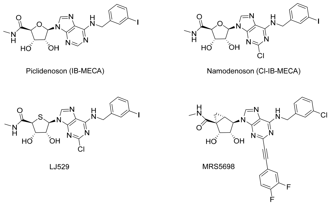
A3AR is a seven-transmembrane G-protein-coupled receptor with a well-defined orthosteric binding pocket that accommodates adenosine analogs bearing halogen or alkyl substitutions at the N6 position [12,13,14]. Structure–activity studies pioneered by Jacobson and colleagues led to the development of highly selective A3AR agonists, including IB-MECA, Cl-IB-MECA, LJ529 and MRS5698 [12,13,15]; Figure 1.
Figure 1.
Chemical structures of representative A3 adenosine receptor (A3AR) agonists, including piclidenoson (IB-MECA), namodenoson (Cl-IB-MECA), LJ529, and MRS5698. These compounds share a nucleoside scaffold with structural modifications that confer high affinity and selectivity for A3AR and have been investigated across oncology, inflammatory, and neurodegenerative disease models.
These compounds were subsequently in-licensed by Can-Fite BioPharma and developed under the names piclidenoson (IB-MECA) and namodenoson (Cl-IB-MECA), respectively. These molecules display nanomolar affinity and high selectivity for A3AR over A1 and A2A receptors [12,15].
Crystallographic and molecular-modeling studies have shown that subtle conformational features of the A3AR, particularly within the third intracellular loop and C-terminal tail, facilitate preferential coupling to Gi/Go proteins and activation of multiple downstream signaling pathways, which are described in detail in the following section [16,17,18]. Through these pathways, A3AR regulates a broad spectrum of cellular functions, including inhibition of inflammatory cytokine production, induction of apoptosis in malignant cells, attenuation of oxidative stress, and protection from ischemic injury [4,5,19,20,21]. In the nervous system, A3AR signaling limits microglial activation and oxidative damage, contributing to neuroprotection [22,23,24].
Over the past two decades, selective A3AR agonists have progressed from preclinical validation to late-stage clinical evaluation. Piclidenoson and namodenoson represent first-in-class orally bioavailable agents that exploit the disease-selective biology of this receptor [25,26,27]. In parallel, numerous academic groups have established A3AR as a mediator of tissue protection in cardiovascular, neurological, renal, and ophthalmic disease models [22,23,24,27,28,29]. Collectively, these studies extend the relevance of A3AR from oncology and autoimmune disorders to fibrosis, ischemia–reperfusion injury, neuropathic pain, vascular dementia, and Lowe syndrome.
This review synthesizes the expanding mechanistic and translational evidence base surrounding A3AR pharmacology, focusing on preclinical studies that elucidate receptor signaling and therapeutic potential across multiple organ systems, together with an overview of ongoing clinical development. By integrating molecular, pharmacologic, and clinical perspectives, this review highlights A3AR activation as a unifying therapeutic principle for diseases driven by inflammation, hypoxia, oxidative stress, and dysregulated cell survival.
1.1. Molecular and Cellular Mechanisms of A3AR Signaling
Ligand binding to A3AR initiates coupling to Gi/Go proteins, resulting in inhibition of adenylyl cyclase and a consequent decrease in intracellular cAMP levels. Reduced cAMP suppresses protein kinase A (PKA) activity, thereby altering the phosphorylation status of multiple transcriptional regulators and signaling intermediates [30,31,32]. This primary signaling event diverges into several intracellular cascades that account for the pleiotropic biological actions of A3AR.
In parallel with Gi-mediated signaling, A3AR activation recruits phospholipase C-β (PLCβ) via Gβγ subunits [31,32]. Activated PLCβ hydrolyzes phosphatidylinositol-4,5-bisphosphate (PI(4,5)P2) to generate inositol-1,4,5-trisphosphate (IP3) and diacylglycerol (DAG), leading to intracellular Ca2+ release and activation of protein kinase C (PKC) isoforms, respectively [33,34,35]. The combined reduction in cAMP and increase in Ca2+/PKC signaling creates a distinct intracellular fingerprint that differentiates A3AR from other adenosine receptor subtypes [31,34]. A3AR signaling intersects with multiple canonical kinase networks. In tumor and immune cells, A3AR agonists suppress the PI3K/Akt/mTOR pathway, resulting in downregulation of pro-survival and proliferative signals [12,17,36]. Reduced Akt phosphorylation leads to inhibition of mTOR, S6 kinase, and GSK-3β, thereby shifting the cellular balance toward apoptosis and growth arrest [12,36]. In parallel, A3AR activation inhibits NF-κB signaling by stabilizing the inhibitory protein IκB and preventing its degradation, leading to reduced NF-κB nuclear translocation and transcriptional activity [34,35,36,37,38]. This results in decreased expression of inflammatory mediators, including TNF-α, IL-1β, IL-6, IL-8, IL-23, and IL-17 [19,20,21].
A3AR also modulates the Wnt/β-catenin pathway, a mechanism of particular relevance in oncology, inflammation and fibrosis. A3AR agonists downregulate β-catenin expression and inhibit its nuclear translocation, thereby reducing transcription of genes that promote proliferation. In hepatocellular and pancreatic tumors, inhibition of the Wnt/β-catenin signaling pathway sensitizes malignant cells to apoptosis [39,40,41].
In Chinese Hamster Ovary (CHO) cells transfected with the human adenosine A3 receptor and in N13 microglia cells which endogenously express A3 receptors, A3AR activation modulates mitogen-activated protein kinases (MAPKs), including ERK1/2, JNK, and p38, which regulate stress responses, cytokine production, and neuronal survival [41].
Beyond cytosolic signaling, A3AR exerts profound effects on mitochondrial function. In cancer cells and activated fibroblasts, A3AR activation promotes expression of the pro-apoptotic protein Bax while downregulating anti-apoptotic Bcl-2, leading to cytochrome-c release and caspase activation [25,41]. Conversely, in non-malignant metabolically stressed tissues such as myocardium and brain, A3AR stimulation stabilizes mitochondrial membrane potential, reduces reactive oxygen species (ROS) generation, and preserves ATP synthesis, thereby conferring cytoprotection [34,42]. These divergent effects are explained by differential receptor density and cellular context, with A3AR activation selectively inducing apoptosis in overexpressing pathological cells while protecting healthy or hypoxic tissues [4,42].
Like many G-protein-coupled receptors, A3AR undergoes phosphorylation by GPCR kinases (GRKs) and by β-arrestin recruitment, internalization and desensitization [43]. This is followed by receptor resensitization and with prolonged (up to 24 h) agonist exposure, contributing to sustained receptor signaling during chronic disease [44,45].
Collectively, A3AR signaling constitutes a multi-axis regulatory network comprising a membranal arm (Gi–cAMP–PKA), an inflammatory arm (NF-κB and inflammatory cytokine inhibition), a growth-control arm (PI3K/Akt/mTOR and Wnt/β-catenin suppression), and a cytoprotective arm (mitochondrial stabilization and antioxidant defense). Through these integrative mechanisms, A3AR activation restores cellular homeostasis in tissues exposed to metabolic and inflammatory stress, providing a molecular framework for its broad efficacy across models of cancer, inflammatory disease, fibrosis, neurodegeneration, and organ ischemia.
The principal signaling axes and integrated biological effects of A3AR activation are summarized in Table 1.
Table 1.
Major A3AR signaling axes and integrated biological effects.
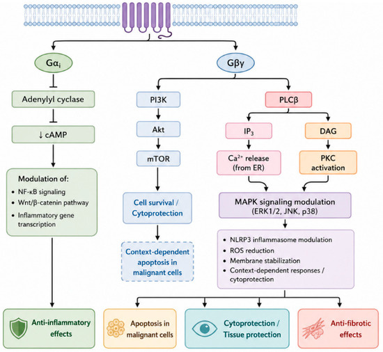
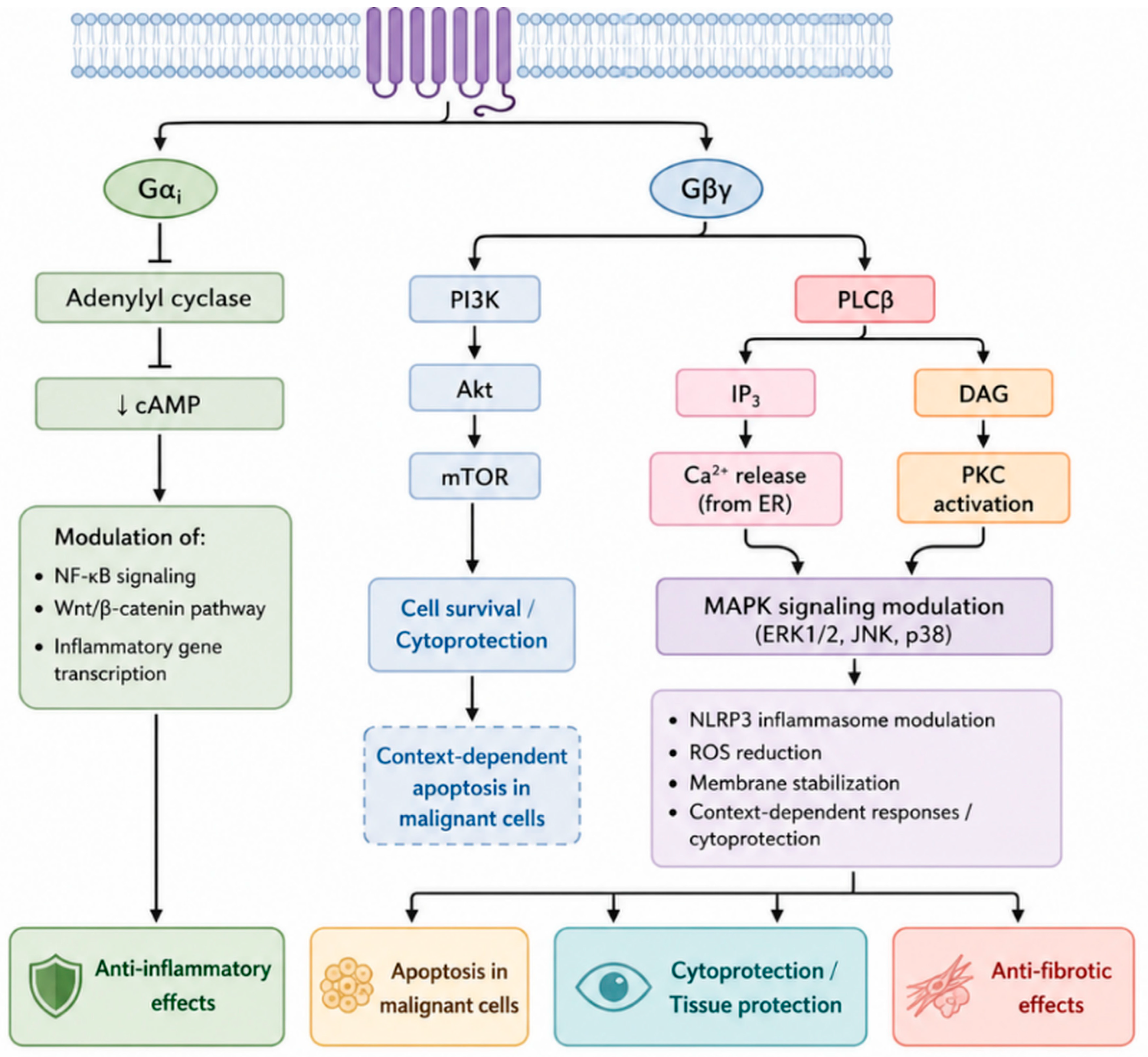
The major signaling architecture of A3AR activation is schematically summarized in Figure 2.
Figure 2.
Major signaling axes and integrated biological effects of A3 adenosine receptor (A3AR) activation. Binding of adenosine or selective agonists to A3AR primarily engages Gi proteins and Gβγ-mediated phospholipase C (PLC) signaling, resulting in inhibition of adenylyl cyclase, reduced cAMP levels, and activation of PLCβ-dependent second messengers. These proximal events regulate downstream pathways including PI3K/Akt/mTOR, NF-κB, Wnt/β-catenin, MAPKs (ERK, JNK, p38), the NLRP3 inflammasome, and mitochondrial stress responses. Collectively, these signaling networks mediate anti-inflammatory, anti-proliferative, anti-fibrotic, neuroprotective, and context-dependent cytotoxic or cytoprotective effects.
1.2. Preclinical Pharmacology and Disease Applications
Recent comprehensive reviews have summarized the expanding biology and therapeutic potential of A3AR agonists across oncology, inflammation, and organ-protective indications, highlighting their unique disease-selective pharmacology and broad translational relevance.
Representative preclinical disease models and mechanistic outcomes across organ systems are summarized in Table 2.
Table 2.
Preclinical disease models and mechanistic outcomes of A3AR agonists.
2. Anti-Cancer Activity
A3AR agonists exert anti-cancer activity across multiple malignancies through a dual mechanism that combines direct tumor cell cytotoxicity with modulation of the tumor microenvironment (TME) [41,45]. The most advanced body of evidence exists in hepatocellular carcinoma (HCC), with expanding preclinical and translational data in pancreatic ductal adenocarcinoma (PDAC) and at the same time, cytoprotective effects towards normal tissues [39,40,51].
In HCC, A3AR is frequently overexpressed in tumor tissue compared with adjacent normal liver, enabling selective pharmacologic targeting. Activation of A3AR suppresses tumor cell proliferation and induces apoptosis through inhibition of PI3K/Akt and NF-κB signaling, together with downregulation of Wnt/β-catenin-dependent transcription. These coordinated effects impair cell-cycle progression and reduce survival signaling, resulting in marked inhibition of tumor growth in xenograft and orthotopic HCC models [39].
In PDAC, A3AR agonists inhibit tumor growth in vitro and in vivo at nanomolar concentrations in an A3AR-dependent manner, promoting apoptosis and suppressing pro-survival pathways including PI3K/Akt, NF-κB, RAS, and Wnt/β-catenin signaling [40]. Importantly, a combination of A3AR agonists with gemcitabine or other cytotoxic agents produces additive or synergistic anti-tumor activity in both PDAC and HCC, supporting their use in combination regimens for these highly aggressive malignancies [65].
Beyond the liver and pancreas, anti-tumor effects of A3AR agonists have been demonstrated in experimental models of melanoma, colorectal carcinoma, breast cancer, and prostate cancer, indicating a broad anti-neoplastic profile driven by receptor overexpression in diverse tumor types [66,67,68].
Notably, the role of A3AR in tumor biology appears to be context-dependent and influenced by the tumor microenvironment. While the majority of studies support an anti-tumor effect of A3AR activation, earlier reports and selected experimental systems have suggested that A3AR signaling may, under certain conditions, support tumor cell survival or proliferation [52]. These seemingly divergent findings likely reflect differences in receptor expression levels, ligand concentration, and cellular context, highlighting the complexity of A3AR-mediated signaling in cancer. Accordingly, the regulatory role of A3AR overexpression in some tumor types remains incompletely understood and warrants further investigation.
A particularly important interaction exists between A3AR signaling and immune checkpoint regulation. Aberrant Wnt/β-catenin activation is a major driver of resistance to PD-1/PD-L1 immune checkpoint inhibitors (ICIs) in tumor cells [69,70,71]. A3AR agonists downregulate β-catenin and are therefore expected to reduce PD-L1 expression on tumor cells, potentially reversing immune exclusion and enhancing susceptibility to immune-mediated killing. Consistent with this mechanistic framework, exploratory in vitro studies have evaluated the effect of namodenoson in combination with PD-L1-targeting strategies on tumor immune signaling. In Hep3B hepatocellular carcinoma cells, namodenoson (10 nM, 48 h incubation) was associated with a reduction in PD-L1 protein expression (~51%), accompanied by β-catenin downregulation and inhibition of cell proliferation (~65%), supporting a potential role in modulating tumor immune evasion pathways.
Clinical development of namodenoson began with a Phase I study in healthy volunteers, followed by a Phase I/II study in HCC (NCT00790218) and a Phase II study in Child–Pugh B cirrhosis patients with advanced HCC (NCT02128958) [72,73]. A pivotal Phase III trial in this high-risk population is currently ongoing (NCT05201404) under FDA and EMA guidance. In PDAC, a Phase IIa study of namodenoson monotherapy is ongoing, with all patients enrolled, and combination studies with chemotherapy and ICIs under consideration. Across all clinical studies, namodenoson has demonstrated a placebo-like safety profile [74]. The compound received FDA Fast Track designation in 2015 and Orphan Drug designation for HCC in 2012, underscoring its potential to address a major unmet medical need.
The current clinical development landscape of A3AR agonists is summarized in Table 3.
Table 3.
Clinical development landscape of A3AR agonists.
3. Inflammation and Autoimmune Disorders
A3AR agonists exert potent anti-inflammatory activity by suppressing key cytokines that drive chronic immune-mediated diseases, including TNF-α, IL-6, IL-8, MIP-1α, IL-17, IL-23, and matrix metalloproteinases (MMPs). These effects are mediated primarily through inhibition of NF-κB signaling and stabilization of IκB [80,81,82,83,84].
3.1. Osteoarthritis
Shkhyan and colleagues first demonstrated that A3AR plays a protective role in cartilage homeostasis. Genetic deletion of A3AR in mice leads to spontaneous cartilage degeneration and osteoarthritis-like structural changes, providing strong evidence that basal A3AR activity protects against joint destruction [53]. Mechanistic analyses demonstrated that loss of A3 adenosine receptor (A3AR) signaling promotes chondrocyte hypertrophy and extracellular matrix degradation through activation of CaMKII and RUNX2 pathways, both strongly implicated in osteoarthritis progression, while A3AR activation suppresses these signaling axes and preserves cartilage integrity, consistent with the broader role of GPCR-mediated signaling in bone and cartilage homeostasis [54,55].
Subsequent studies demonstrated that A3AR activation suppresses NF-κB and PI3K/Akt signaling in inflammatory cells and chondrocytes, reducing production of catabolic enzymes and inflammatory mediators. Treatment with piclidenoson significantly attenuated cartilage destruction and improved pain-related behaviors in rodent OA models [56]. More recently, A3AR agonism was shown to suppress activation of the NLRP3 inflammasome and downstream caspase-1/GSDMD-mediated pyroptosis, linking purinergic signaling to emerging mechanisms of inflammatory cell death in osteoarthritic cartilage [47,48].
Veterinary clinical data further support translational relevance. In a multicenter trial in dogs with naturally occurring osteoarthritis, the A3AR agonist piclidenoson significantly improved Liverpool Osteoarthritis in Dogs (LOAD) and Visual Analog Scale (VAS) pain scores in a dose- and time-dependent manner, consistent with disease-modifying activity [57].
3.2. Rheumatoid Arthritis
Rheumatoid arthritis (RA) is characterized by synovial hyperplasia, immune-cell infiltration, and progressive joint destruction driven by pro-inflammatory cytokines and dysregulated intracellular signaling. A3AR is selectively overexpressed in peripheral blood mononuclear cells and synovial tissue of RA patients, making it an attractive therapeutic target [5,58]. Activation of A3AR suppresses PI3K/Akt and NF-κB signaling, resulting in reduced production of TNF-α, IL-6, IL-1β, IL-8, IL-17 and IL-23 [56,81].
In animal models of inflammatory arthritis, oral piclidenoson significantly reduced clinical arthritis scores, joint swelling, cartilage damage, and bone erosion, with efficacy comparable to standard disease-modifying antirheumatic drugs (DMARDs) and without evidence of global immunosuppression [59].
Clinical studies confirmed an excellent safety profile. A Phase 2a monotherapy study demonstrated significant ACR20/50/70 responses, with optimal efficacy observed at an oral dose of 1 mg administered twice daily [85]. Phase 2b combination studies with methotrexate (MTX) failed, likely due to MTX-induced downregulation of A3AR expression [74]. In contrast, a placebo-controlled Phase 2b monotherapy study met its primary endpoint, with particularly strong responses in treatment-naïve patients [86]. A Phase 3 trial confirmed safety and superiority over placebo but did not achieve non-inferiority to MTX, leading to discontinuation of RA development while validating A3AR biology in human disease [87].
3.3. Psoriasis
Psoriasis is driven by dysregulated interactions between keratinocytes and immune cells, particularly Th17 and Th1 lymphocytes, leading to overproduction of TNF-α, IL-17, IL-23, and IL-22. A3AR is selectively overexpressed in PBMCs and lesional skin of psoriasis patients, supporting its relevance as a biomarker-linked therapeutic target [5].
In vitro studies demonstrated that piclidenoson directly inhibits keratinocyte proliferation and suppresses NF-κB and PI3K/Akt signaling in human keratinocytes, confirming receptor-specific anti-inflammatory and anti-proliferative activity [80]. In vivo, selective A3AR agonists (MRS5698 and photocaged MRS7344) suppressed IL-23-driven psoriasiform inflammation in mouse models, reducing epidermal hyperplasia and immune-cell infiltration [60].
Can-Fite also demonstrated efficacy of piclidenoson in the imiquimod-induced psoriasis model, showing significant reductions in clinical skin scores demonstrating the topical effect of the drug (unpublished data). These findings translated clinically: Phase 2 and Phase 2/3 trials demonstrated safety and dose-dependent PASI improvements [88,89], and the Phase 3 COMFORT-1 trial showed statistically significant PASI-75 responses and superior tolerability compared with apremilast [90]. The COMFORT-2 Phase 3 study is currently enrolling patients.
4. Metabolic Dysfunction-Associated Steatohepatitis (MASH)
MASH, formerly non-alcoholic steatohepatitis (NASH), is characterized by hepatic steatosis, inflammation, hepatocyte injury, and progressive fibrosis driven by metabolic stress and innate immune activation.
In several rodent models including diet- and toxin-induced steatohepatitis, oral administration of the selective A3AR agonist namodenoson significantly reduced hepatic steatosis, lobular inflammation, hepatocyte ballooning, and fibrosis. These histological improvements were associated with downregulation of pro-inflammatory and pro-fibrotic mediators, including TNF-α and α-smooth muscle actin (α-SMA), reflecting inhibition of hepatic stellate cell activation and upregulation of adiponectin [61].
At the molecular level, A3AR agonists inhibit PI3K/Akt and NF-κB signaling in hepatocytes, Kupffer cells, and stellate cells, leading to suppression of inflammatory cytokine production and induction of apoptosis in activated fibrogenic cells. This integrated immune–metabolic and anti-fibrotic mechanism distinguishes A3AR agonism from therapies that target only lipid metabolism or inflammation.
Clinical translation supports these findings. In a Phase 2 study in MASH patients, namodenoson showed a very favorable safety profile. Evidence of liver-targeted biological activity included the following parameters: improvements in ALT and AST levels which decreased consistently during the study in a dose-dependent manner, an increase in serum adiponectin, a significant decrease in liver fat volume and a significant decrease in Fib-4 score, suggesting an inhibitory effect of the drug on fibrosis progression [75]. These results support A3AR agonism as a first-in-class, orally administered, disease-modifying approach that integrates anti-inflammatory, metabolic, and anti-fibrotic pathways. A Phase 2b study in MASH patients with biopsy-proven disease is currently ongoing.
5. A3AR Agonists in Neuroinflammatory and Neurodegenerative Diseases
A3AR is functionally expressed in microglia (a major cellular target), astrocytes, endothelial cells of the blood–brain barrier, and injured neurons, and its activation orchestrates neuroprotection through combined immunomodulatory and neuron-intrinsic survival mechanisms, including regulation of glial metabolism and inflammatory signaling. Mechanistically, A3AR agonism suppresses neuroinflammatory transcriptional programs (notably NF-κB-driven cytokine networks), reduces inducible nitric oxide synthase (iNOS) and redox stress signaling, and rebalances kinase pathways such as ERK/JNK/p38 MAPKs in a context-dependent manner. In parallel, A3AR activation stabilizes mitochondrial function and limits oxidative damage, including suppression of NADPH oxidase-dependent ROS generation, thereby preserving neuronal integrity in settings of hypoxia–ischemia and inflammatory injury. Transcriptomic and mechanistic studies further support the concept that A3AR agonists can shift activated microglia away from damaging inflammatory phenotypes, providing a rational foundation for disease modification in disorders where microglial activation drives progression [76,77,78,91].
5.1. Neuropathic Pain
A compelling body of work (particularly from Salvemini’s group and collaborators) supports A3AR agonists as non-opioid analgesics that target neuroinflammation and oxidative stress in the spinal cord. In a seminal paclitaxel-induced neuropathic pain model, the selective A3AR agonist IB-MECA (piclidenoson) prevented development of neuropathic pain by inhibiting spinal NADPH oxidase activation and downstream redox-dependent signaling, consistent with disease-relevant suppression of glial-driven sensitization [63]. Complementary medicinal-chemistry and preclinical studies identified highly selective A3AR agonists such as MRS5698, a highly selective A3AR agonist developed by academic groups, which has demonstrated robust anti-allodynic activity in chronic neuropathic pain models, strengthening the translational case for this target class [50,63,92]. More recently, mechanistic work has extended these findings by implicating mitochondrial A3AR-dependent pathways in chemotherapy-induced neuropathy and by confirming that A3AR agonists can prevent pain development without opioid-like tolerance or reward behavior. Additional studies show that A3AR agonism can act as a distinct analgesic strategy that may complement or replace opioid approaches in selected neuropathic pain contexts [46,49,64,92,93,94].
5.2. Ischemic Stroke and Brain Ischemia
Preclinical studies demonstrate that A3AR agonists can reduce ischemic brain injury in models of focal and global cerebral ischemia. Early work showed that A3AR activation reduces infarct burden and mitigates downstream inflammatory and apoptotic cascades in rodent cerebral ischemia. In a well-cited mechanistic study, the selective A3AR agonist LJ529 reduced brain ischemic injury through anti-inflammatory and cytoprotective mechanisms consistent with stabilization of cellular stress responses and limitation of cell death pathways. Additional experimental data support that sustained A3AR agonism can preserve ischemia-sensitive neuronal markers (e.g., MAP-2), reduce nitric oxide synthase induction, and modulate glial reactivity following ischemic insults, aligning with both anti-inflammatory and neuroprotective effects [62,95,96].
5.3. Alzheimer’s Disease and Neuroinflammation
While A3AR agonists remain less clinically developed in Alzheimer’s disease than A2A receptor antagonists, accumulating evidence supports a role for A3AR signaling in amyloidogenic processing and microglial phenotype control. In cultured neurons, caffeine was shown to suppress amyloid-β protein precursor (AβPP) internalization and reduce amyloid-β generation at least partly via A3AR-mediated mechanisms, providing a mechanistic link between A3AR signaling and amyloidogenic processing [97]. Additional study on A3AR signaling in Alzheimer’s disease highlights that A3AR can influence microglial activation states and inflammatory outputs, supporting continued exploration of A3AR-directed strategies in amyloid-associated neuroinflammation [97].
5.4. Vascular Dementia and Chronic Hypoperfusion
A major advance in the field emerged from work integrating mouse vascular dementia (VaD) transcriptomics with human single-nucleus RNA sequencing, which identified disruption of an intercellular CD39–A3AR signaling system in the neurovascular niche. In these studies, pharmacologic restoration of CD39–A3AR signaling using an A3AR-specific agonist (noted as clinically relevant) improved tissue integrity and behavioral outcomes in a VaD model, supporting A3AR activation as a potential repair-promoting pathway in chronic hypoperfusion-linked cognitive impairment [63,98]. These findings align with the broader concept that A3AR agonists can support neurovascular repair by dampening damaging microglial activation while promoting recovery programs in the injured white matter environment as described above.
5.5. Cardiovascular Protection
A3ARs are expressed in cardiomyocytes and vascular endothelial cells, and are upregulated by ischemic and hypoxic stress, contributing to endogenous cardioprotective signaling, reducing infarct size and enhancing cellular survival in ischemia–reperfusion models. [99]. In experimental myocardial ischemia–reperfusion injury, A3AR activation limits infarct size, preserves left-ventricular function and improves post-ischemic recovery [50]. Namodenoson and other A3AR agonists stabilize mitochondrial membrane potential, inhibit cytochrome-c release, and prevent caspase-3-mediated cardiomyocyte apoptosis, thereby preserving ATP generation and contractile function [100,101,102,103].
In parallel, A3AR activation suppresses NADPH-oxidase-dependent ROS generation and reduces inflammatory leukocyte adhesion to ischemic endothelium, limiting microvascular obstruction and reperfusion injury. Beyond ischemia–reperfusion injury, A3AR activation has been shown to attenuate doxorubicin-induced cardiomyocyte injury by reducing mitochondrial dysfunction and apoptotic signaling, supporting its potential role as a cardioprotective adjuvant during chemotherapy [103,104].
6. Ophthalmology and Retinal Protection
A3AR is expressed in retinal pigment epithelial cells, retinal ganglion cells, and microglia, and its expression is increased under conditions of ocular inflammation and ischemic stress. Activation of adenosine A3 receptor protects retinal ganglion cells from degeneration induced by ocular hypertension [30,96]. In models of uveitis and glaucoma, A3AR agonists suppress NF-κB-dependent inflammatory cytokines, inhibit leukocyte infiltration, and preserve retinal ganglion-cell survival [105,106].
Although oral piclidenoson showed limited efficacy in late-stage glaucoma trials, the strong preclinical evidence for direct retinal A3AR-mediated neuroprotection supports continued development via topical ocular formulations that bypass systemic dilution and maximize retinal exposure [107].
7. Lowe Syndrome (OCRL Deficiency)
Lowe syndrome is an X-linked multisystem disorder caused by mutations in the OCRL gene encoding phosphatidylinositol-5-phosphatase, leading to abnormal PI(4,5)P2 accumulation, defective endosomal trafficking, and cytoskeletal instability in kidney, brain, and eye tissues [108]. A seminal discovery by the TIGEM–Federico II Naples group demonstrated that A3AR activation corrects these defects at their biochemical root. In OCRL-deficient cells and mouse models, piclidenoson normalized PI(4,5)P2 levels through A3AR-PLC-dependent hydrolysis, restored endosomal morphology, reduced inflammatory signaling, and improved renal tubular function, including reduced proteinuria and improved reabsorption capacity. This unique mechanism represents a true disease-modifying rescue, rather than symptomatic management. These findings led to a translational partnership between Fondazione Telethon and Can-Fite BioPharma to advance piclidenoson into a Phase 2 clinical study at Bambino Gesù Children’s Hospital in Rome. This marks the first application of A3AR modulation in a rare inherited metabolic disease. A patent has been filed as of the use of A3AR agonist as a treatment for Lowe syndrome (WO 2025/011999 A1).
8. Safety Profile of Piclidenoson and Namodenoson
Across all clinical studies, namodenoson and piclidenoson have demonstrated a favorable safety profile, with most adverse events being mild. Reported side effects have included headache, gastrointestinal discomfort, and fatigue, and overlapped with those of placebo-treated patients. Importantly, no signal of immunosuppression or organ-specific toxicity has been consistently observed across studies. This safety profile is consistent with the relatively low expression of A3AR in normal tissues and its selective upregulation in pathological conditions. Nevertheless, as with other G-protein-coupled receptor-targeted therapies, long-term effects and potential context-dependent responses warrant continued evaluation in larger clinical trials [74].
9. Challenges and Limitations of A3AR-Targeted Therapies
Despite the largely consistent therapeutic profile of A3AR agonists, some earlier and seemingly contradictory findings have been reported in the literature. It should be noted that earlier studies explored A3AR antagonists as potential therapeutic agents, particularly in asthma and certain cancer models, based on observations in rodent systems where A3AR activation could promote bronchoconstriction or modulate hypoxia-related pathways. In addition, A3AR antagonists have been reported to reduce intraocular pressure, whereas A3AR agonists demonstrate neuroprotective effects in retinal tissues. These seemingly contradictory findings likely reflect species differences, tissue-specific receptor expression, and context-dependent signaling mechanisms. Current evidence in human systems and clinical studies predominantly supports a protective and anti-inflammatory role of A3AR activation. Like other G-protein-coupled receptors, A3AR is subject to desensitization, internalization, and context-dependent signaling, which may influence long-term efficacy in chronic indications. In addition, variability in receptor expression across patient populations may contribute to heterogeneous clinical responses and may partly explain why some clinical trials did not meet primary endpoints compared to standard-of-care therapies. Consistent with this, despite more than two decades of clinical development, A3AR agonists have not yet achieved regulatory approval, reflecting the challenges of translating receptor biology into consistent clinical benefit across heterogeneous patient populations.
Furthermore, while A3AR is generally overexpressed in pathological tissues, its role in tumor biology is complex and may be influenced by the tumor microenvironment. Some studies have suggested that under certain conditions, A3AR signaling may support tumor survival or proliferation, highlighting the need for a more nuanced understanding of receptor function in different cellular contexts.
From a clinical development perspective, the use of A3AR agonists may require careful patient stratification based on receptor expression levels, as well as optimization of combination strategies with established therapies such as chemotherapy or immune checkpoint inhibitors.
Finally, although a substantial body of preclinical and clinical data supports the therapeutic potential of A3AR agonists, further independent studies and large-scale clinical trials are needed to fully establish their efficacy across diverse disease indications.
10. Conclusions
Taken together, the accumulated body of evidence reviewed here clearly establishes the A3AR as a central regulator of tissue responses to metabolic, inflammatory, and hypoxic stress. Unlike other adenosine receptor subtypes, A3AR displays a disease-inducible expression profile, being markedly upregulated in malignant, inflamed, ischemic, and fibrotic tissues while remaining minimally expressed in most healthy organs. This feature creates a pharmacologic window that enables selective targeting of pathological cells with minimal off-target toxicity, a property that is uncommon among G-protein-coupled receptor targets and highly advantageous for chronic systemic therapy.
At the mechanistic level, A3AR activation integrates several interconnected signaling axes that converge on restoration of cellular homeostasis. Suppression of the PI3K/Akt/mTOR and Wnt/β-catenin pathways limits aberrant proliferation, fibrosis, and immune evasion, while inhibition of NF-κB and STAT3 reduces pro-inflammatory cytokine networks that sustain chronic disease. At the same time, A3AR signaling stabilizes mitochondrial function, reduces oxidative stress, and preserves bioenergetic capacity in neurons, cardiomyocytes, renal tubular cells, and endothelial cells exposed to ischemic or metabolic injury. The ability of a single receptor system to simultaneously regulate inflammation, cell survival, fibrosis, and tissue repair provides a coherent explanation for the remarkably broad therapeutic profile of A3AR agonists across oncology, immunology, neurology, cardiology, hepatology, and rare genetic diseases.
Importantly, these mechanistic principles have translated into consistent in vivo efficacy. In cancer models, A3AR agonists directly induce tumor cell apoptosis while reprogramming the tumor microenvironment toward immune permissiveness, including suppression of PD-L1 expression and β-catenin signaling. In autoimmune and inflammatory diseases such as rheumatoid arthritis, psoriasis, and osteoarthritis, A3AR agonism attenuates cytokine-driven tissue destruction without causing global immunosuppression. In metabolic and fibrotic liver disease, A3AR activation integrates anti-inflammatory, metabolic, and anti-fibrotic pathways, leading to histological improvement in experimental MASH. In the nervous system, the receptor functions as a master regulator of neuroinflammation, oxidative injury, and neuronal survival, conferring protection in stroke, neuropathic pain, Alzheimer’s disease, and vascular dementia. The extension of A3AR biology into rare inherited disorders such as Lowe syndrome further illustrates the receptor’s role in regulating phosphoinositide homeostasis and endosomal function, expanding its relevance beyond classical inflammation and cancer.
Clinical experience accumulated with piclidenoson and namodenoson confirms that these mechanistic advantages translate into an excellent safety profile, even with long-term oral administration. Across multiple indications, A3AR agonists have shown a placebo-like tolerability profile, consistent with their selective activity in diseased tissues. Moreover, emerging data indicate that baseline A3AR expression serves as a predictive biomarker for therapeutic response, providing a rational framework for patient stratification and precision medicine. This biomarker-driven approach may explain variable responses observed in earlier clinical trials and offers a path toward maximizing efficacy in future studies.
Taken together, the data support A3AR activation as a unifying, disease-modifying therapeutic strategy rather than a symptomatic intervention. By targeting the convergent molecular pathways that drive inflammation, oxidative stress, fibrosis, and pathological cell survival, A3AR agonists offer a platform capable of addressing complex, multi-factorial diseases that remain poorly served by current therapies. Continued integration of mechanistic insight, biomarker-guided clinical development, and rational combination strategies, particularly with immunotherapies and metabolic modulators, positions A3AR agonism as a powerful and versatile paradigm in modern translational medicine.
Funding
This research was funded by Can-Fite BioPharma Ltd.
Institutional Review Board Statement
Not applicable.
Informed Consent Statement
Not applicable.
Data Availability Statement
No new data were created or analyzed in this study. Data sharing is not applicable to this article.
Acknowledgments
During the preparation of this manuscript, the author used ChatGPT (OpenAI) to assist with language editing, formatting, and structuring of the text. The author reviewed and edited all content and takes full responsibility for the final manuscript.
Conflicts of Interest
The author is affiliated with Can-Fite BioPharma Ltd., a company developing A3 adenosine receptor agonists. This affiliation has been disclosed in the manuscript, which has been prepared to provide a balanced and comprehensive review of the field. The research was funded by Can-Fite BioPharma Ltd., and the funder had no role in the study design, data collection, analysis, interpretation, manuscript writing, or the decision to publish the results.
Abbreviations
A3AR, A3 adenosine receptor; AC, adenylyl cyclase; ADA, adenosine deaminase; Akt, protein kinase B; ATP, adenosine triphosphate; cAMP, cyclic adenosine monophosphate; DAG, diacylglycerol; ERK, extracellular signal-regulated kinase; GPCR, G-protein-coupled receptor; HCC, hepatocellular carcinoma; HIF-1α, hypoxia-inducible factor 1 alpha; IP3, inositol 1,4,5-trisphosphate; JNK, c-Jun N-terminal kinase; MAPKs, mitogen-activated protein kinases; mTOR, mechanistic target of rapamycin; NF-κB, nuclear factor kappa B; NLRP3, NOD-like receptor family pyrin domain containing 3; PD-L1, programmed death-ligand 1; PI3K, phosphoinositide 3-kinase; PKA, protein kinase A; PKC, protein kinase C; PLC, phospholipase C; ROS, reactive oxygen species; Wnt, wingless-related integration site; β-catenin, beta-catenin.
References
- Fredholm, B.B.; IJzerman, A.P.; Jacobson, K.A.; Klotz, K.-N.; Linden, J. International Union of Pharmacology. XXV. Nomenclature and classification of adenosine receptors. Pharmacol. Rev. 2001, 53, 527–552. [Google Scholar] [CrossRef] [Scilit]
- Haskó, G.; Cronstein, B.N. Adenosine: An endogenous regulator of innate immunity. Trends Immunol. 2004, 25, 33–39. [Google Scholar] [CrossRef] [Scilit]
- Ohta, A.; Sitkovsky, M. Role of G-protein–coupled adenosine receptors in downregulation of inflammation and protection from tissue damage. Nature 2001, 414, 916–920. [Google Scholar] [CrossRef] [Scilit]
- Abbracchio, M.P.; Burnstock, G. Purinergic signalling: Pathophysiological roles. Nat. Rev. Drug Discov. 2006, 5, 198–211. [Google Scholar]
- Ochaion, A.; Bar-Yehuda, S.; Cohen, S.; Barer, F.; Patoka, R.; Amital, H.; Reitblat, T.; Reitblat, A.; Ophir, J.; Konfino, I.; et al. The anti-inflammatory target A3 adenosine receptor is over-expressed in rheumatoid arthritis, psoriasis and Crohn’s disease. Cell. Immunol. 2009, 258, 115–122. [Google Scholar] [CrossRef] [Scilit]
- Salvatore, C.A.; Jacobson, M.A.; Taylor, H.E.; Linden, J.; Johnson, R.G. Molecular cloning and characterization of the human A3 adenosine receptor. Proc. Natl. Acad. Sci. USA 1993, 90, 10365–10369. [Google Scholar] [CrossRef] [Scilit]
- Zhou, Q.-Y.; Li, C.; Olah, M.E.; Johnson, R.A.; Stiles, G.L.; Civelli, O. Molecular cloning and characterization of an adenosine receptor: The A3 adenosine receptor. Proc. Natl. Acad. Sci. USA 1992, 89, 7432–7436. [Google Scholar] [CrossRef] [Scilit] [PubMed]
- Jacobson, K.A.; Park, K.S.; Jiang, J.L.; Kim, Y.C.; Olah, M.E.; Stiles, G.L.; Ji, X.D. Pharmacological characterization of novel A3 adenosine receptor-selective antagonists. Neuropharmacology 1997, 36, 1157–1165. [Google Scholar] [CrossRef] [Scilit] [PubMed]
- Gao, Z.-G.; Jacobson, K.A. A3 adenosine receptor ligands: Structure–activity relationships. Curr. Pharm. Des. 2005, 11, 3561–3578. [Google Scholar]
- Yaar, R.; Cataldo, L.M.; Tzatsos, A.; Francis, C.E.; Zhao, Z.; Ravid, K. Regulation of the A3 adenosine receptor gene in vascular smooth muscle cells: Role of a cAMP and GATA element. Mol. Pharmacol. 2002, 62, 1167–1176. [Google Scholar] [CrossRef] [Scilit]
- Merighi, S.; Benini, A.; Mirandola, P.; Gessi, S.; Varani, K.; Leung, E.; MacLennan, S.; Baraldi, P.G.; Borea, P.A. adenosine receptors modulate hypoxia-inducible factor-1α expression in human A375 melanoma cells. Neoplasia 2005, 7, 894–903. [Google Scholar] [CrossRef] [Scilit]
- Jacobson, K.A.; Gao, Z.-G. Adenosine receptors as therapeutic targets. Nat. Rev. Drug Discov. 2006, 5, 247–264. [Google Scholar] [CrossRef] [Scilit] [PubMed]
- Jacobson, K.A.; Müller, C.E.; IJzerman, A.P. Medicinal Chemistry of the A3 Adenosine Receptor: Agonists, Antagonists, and Receptor Engineering. Curr. Top. Med. Chem. 2009, 9, 1030–1047. [Google Scholar]
- Borea, P.A.; Gessi, S.; Merighi, S.; Varani, K. Adenosine as a Multi-Signalling Guardian Angel in Human Diseases: When, Where and How Does it Exert Its Protective Effects? Trends Pharmacol Sci. 2016, 37, 419–434. [Google Scholar] [CrossRef] [Scilit]
- Xia, L.; Heitman, L.H.; IJzerman, A.P. A binding kinetics study of human adenosine A3 receptor agonists. Biochem. Pharmacol. 2018, 153, 248–259. [Google Scholar] [CrossRef] [Scilit] [PubMed]
- Suresh, R.R.; Jain, S.; Chen, Z.; Tosh, D.K.; Ma, Y.; Podszun, M.C.; Rotman, Y.; Salvemini, D.; Jacobson, K.A. Design and In Vivo Activity of A3 Adenosine Receptor Agonist Prodrugs. Purinergic Signal. 2020, 16, 367–377. [Google Scholar] [CrossRef] [Scilit]
- Abbracchio, M.P.; Brambilla, R.; Ceruti, S.; Kim, H.O.; von Lubitz, D.K.; Jacobson, K.A.; Cattabeni, F. G protein–dependent activation of phospholipase C by adenosine A3 receptors in rat brain. Mol. Pharmacol. 1995, 48, 1038–1045. [Google Scholar] [CrossRef] [Scilit]
- Borea, P.A.; Varani, K.; Vincenzi, F.; Baraldi, P.G.; Aghazadeh Tabrizi, M.; Merighi, S.; Gessi, S. The A3 adenosine receptor: History and perspectives. Pharmacol. Rev. 2015, 67, 74–102. [Google Scholar] [CrossRef] [Scilit]
- Jacobson, K.A.; Merighi, S.; Varani, K.; Borea, P.A.; Baraldi, S.; Aghazadeh Tabrizi, M.; Romagnoli, R.; Baraldi, P.G.; Ciancetta, A.; Tosh, D.K.; et al. A3 adenosine receptors as modulators of inflammation: From medicinal chemistry to therapy. Med. Res. Rev. 2018, 38, 1031–1072. [Google Scholar] [CrossRef] [Scilit]
- Zhou, Z.; Gao, Z.; Yan, W.; Zhang, Y.; Huang, J.; Xiong, K. Adenosine A3 receptor activated in H2O2 oxidative stress of primary open-angle glaucoma. Ann. Transl. Med. 2021, 9, 526. [Google Scholar] [CrossRef] [Scilit]
- Gessi, S.; Merighi, S.; Sacchetto, V.; Simioni, C.; Borea, P.A. Adenosine receptors in health and disease. Adv. Pharmacol. 2011, 61, 41–87. [Google Scholar]
- Choi, I.Y.; Lee, J.C.; Ju, C.; Hwang, S.; Cho, G.S.; Lee, H.W.; Choi, W.J.; Jeong, L.S.; Kim, W.K. A3 adenosine receptor agonist reduces brain ischemic injury and inhibits inflammatory cell migration in rats. Am. J. Pathol. 2011, 179, 2042–2052. [Google Scholar] [CrossRef] [Scilit]
- Pietrowski, M.J.; Gabriele, M.; Paolicelli, R.C.; Maccarrone, M.; Molinari, C.; Limatola, C. Glial purinergic signaling in neurodegeneration. Front. Neurol. 2021, 12, 654850. [Google Scholar] [CrossRef] [Scilit]
- Li, P.; Li, X.; Deng, P.; Wang, D.; Bai, X.; Li, Y.; Luo, C.; Belguise, K.; Wang, X.; Wei, X.; et al. Activation of adenosine A3 receptor reduces early brain injury by alleviating neuroinflammation after subarachnoid hemorrhage in elderly rats. Aging 2020, 13, 694–713. [Google Scholar] [CrossRef] [Scilit]
- Fishman, P.; Bar-Yehuda, S.; Varani, K.; Gessi, S.; Merighi, S.; Borea, P.A. Agonists and antagonists: Molecular mechanisms and therapeutic applications. In A3 Adenosine Receptors: From Cell Biology to Pharmacology and Therapeutics; Borea, P.A., Ed.; Springer: Dordrecht, The Netherlands, 2010. [Google Scholar]
- Baraldi, P.G.; Cacciari, B.; Romagnoli, R.; Merighi, S.; Varani, K.; Borea, P.A.; Spalluto, G. A3 adenosine receptor ligands: History and perspectives. Med. Res. Rev. 2000, 20, 103–128. [Google Scholar] [CrossRef] [Scilit]
- Liang, B.T.; Jacobson, K.A. A physiological role of the adenosine A3 receptor: Sustained cardio protection. Proc. Natl. Acad. Sci. USA 1998, 95, 6995–6999. [Google Scholar] [CrossRef] [Scilit] [PubMed]
- Boia, R.; Salinas-Navarro, M.; Gallego-Ortega, A.; Galindo-Romero, C.; Aires, I.D.; Agudo-Barriuso, M.; Ambrósio, A.F.; Vidal-Sanz, M.; Santiago, A.R. Activation of adenosine A3 receptor protects retinal ganglion cells from degeneration induced by ocular hypertension. Cell Death Dis. 2020, 11, 401. [Google Scholar] [CrossRef] [Scilit] [PubMed]
- Lu, Y.; Zhang, R.; Ge, Y.; Carlström, M.; Wang, S.; Fu, Y.; Cheng, L.; Wei, J.; Roman, R.J.; Wang, L.; et al. Identification and function of adenosine A3 receptor in afferent arterioles. Am. J. Physiol. Ren. Physiol. 2015, 308, F1020–F1025. [Google Scholar] [CrossRef] [Scilit]
- Fishman, P.; Jacobson, K.A.; Ochaion, A.; Cohen, S.; Bar-Yehuda, S. The anti-cancer effect of A3 adenosine receptor agonists: A novel targeted therapy. Immunol. Endocr. Metab. Agents Med. Chem. 2007, 7, 298–303. [Google Scholar] [CrossRef] [Scilit]
- Koscsó, B.; Csóka, B.; Pacher, P.; Haskó, G. Investigational A3 adenosine receptor targeting agents. Expert Opin. Investig. Drugs 2011, 20, 757–768. [Google Scholar] [CrossRef] [Scilit]
- Ciancetta, A.; Rubio, P.; Lieberman, D.I.; Jacobson, K.A. A3 adenosine receptor activation mechanisms: Molecular dynamics analysis of inactive, active, and fully active states. J. Chem. Inf. Model. 2020, 60, 2896–2908. [Google Scholar] [CrossRef] [Scilit]
- Reshkin, S.J.; Guerra, L.; Bagorda, A.; Debellis, L.; Cardone, R.; Li, A.H.; Jacobson, K.A.; Casavola, V. Activation of A3 adenosine receptor induces calcium entry and chloride secretion in A6 cells. J. Membr. Biol. 2000, 178, 103–113. [Google Scholar] [CrossRef] [Scilit]
- Nishat, S.; Khan, L.A.; Ansari, Z.M.; Basir, S.F. Adenosine A3 receptor: A promising therapeutic target in cardiovascular disease. Curr. Cardiol. Rev. 2016, 12, 18–26. [Google Scholar] [CrossRef] [Scilit]
- Pierce, K.L.; Premont, R.T.; Lefkowitz, R.J. Seven-transmembrane receptors. Nat. Rev. Mol. Cell Biol. 2002, 3, 639–650. [Google Scholar] [CrossRef] [Scilit]
- Otsuki, T.; Kanno, T.; Fujita, Y.; Tabata, C.; Fukuoka, K.; Nakano, T.; Gotoh, A.; Nishizaki, T. A3 adenosine receptor-mediated p53-dependent apoptosis in Lu-65 human lung cancer cells. Cell Physiol. Biochem. 2012, 30, 210–220. [Google Scholar] [CrossRef] [Scilit] [PubMed]
- Fishman, P.; Bar-Yehuda, S.; Rath-Wolfson, L.; Ardon, E.; Barer, F.; Ochaion, A.; Madi, L. Targeting the A3 adenosine receptor for cancer therapy: Inhibition of prostate carcinoma cell growth by an A3AR agonist. Anticancer Res. 2003, 23, 2077–2083. [Google Scholar] [PubMed]
- Fishman, P.; Madi, L.; Bar-Yehuda, S.; Barer, F.; Del Valle, L.; Khalili, K. Evidence for involvement of Wnt signaling pathway in IB-MECA–mediated suppression of melanoma cells. Oncogene 2002, 21, 4060–4064. [Google Scholar] [CrossRef] [Scilit]
- Bar-Yehuda, S.; Stemmer, S.M.; Madi, L.; Castel, D.; Ochaion, A.; Cohn, S.; Barer, F.; Zabutti, A.; Perez-Liz, G.; Del Valle, L.; et al. The A3 adenosine receptor agonist CF102 induces apoptosis of hepatocellular carcinoma via de-regulation of the Wnt and NF-κB signal transduction pathways. Int. J. Oncol. 2008, 33, 287–295. [Google Scholar]
- Itzhak, I.; Bareket-Samish, A.; Fishman, P. Namodenoson inhibits the growth of pancreatic carcinoma via deregulation of the Wnt/β-catenin, NF-κB, and Ras signaling pathways. Biomolecules 2023, 13, 1584. [Google Scholar] [CrossRef] [Scilit]
- Hammarberg, C.; Fredholm, B.B.; Schulte, G. Adenosine A3 receptor-mediated regulation of p38 and ERK1/2 via phosphatidylinositol-3′-kinase. Biochem. Pharmacol. 2004, 67, 129–135. [Google Scholar] [CrossRef] [Scilit]
- Mazziotta, C.; Rotondo, J.C.; Lanzillotti, C.; Campione, G.; Martini, F.; Tognon, M. Cancer biology and molecular genetics of A3 adenosine receptor. Oncogene 2022, 41, 301–308. [Google Scholar] [CrossRef] [Scilit] [PubMed]
- Trincavelli, M.L.; Tuscano, D.; Marroni, M.; Klotz, K.-N.; Lucacchini, A.; Martini, C. Involvement of the mitogen–activated protein kinase cascade in agonist-mediated phosphorylation and regulation of A3 adenosine receptors. Biochim. Biophys. Acta 2002, 1591, 55–62. [Google Scholar] [CrossRef] [Scilit] [PubMed]
- Klaasse, E.C.; Ijzerman, A.P.; de Grip, W.J.; Beukers, M.W. Internalization and desensitization of adenosine receptors. Purinergic Signal. 2008, 4, 21–37. [Google Scholar] [CrossRef] [Scilit] [PubMed]
- Pergolizzi, J.V.; LeQuang, J.A.K.; Coleman, M.H. Adenosine A3 receptor and its potential role in cancer treatment: A narrative review. Cureus 2025, 17, e93950. [Google Scholar] [CrossRef] [Scilit]
- Singh, A.K.; Mahalingam, R.; Squillace, S.; Jacobson, K.A.; Tosh, D.K.; Dharmaraj, S.; Farr, S.A.; Kavelaars, A.; Salvemini, D.; Heijnen, C.J. Targeting the A3 adenosine receptor to prevent and reverse chemotherapy-induced neurotoxicities in mice. Acta Neuropathol. Commun. 2022, 10, 11. [Google Scholar] [CrossRef] [Scilit]
- Weaver, N.C.; Habusta, S.F.; Raymond, R.M. The role of extra-cellular adenosine receptors in modulating pain progression during osteoarthritis (OA): A systematic review of the literature. Rheumatol. Orthop. Med. 2018, 3, 1–5. [Google Scholar] [CrossRef] [Scilit]
- Bai, H.; Zhang, Z.; Liu, L.; Wang, X.; Song, X.; Gao, L. Activation of adenosine A3 receptor attenuates progression of osteoarthritis through inhibiting the NLRP3/caspase-1/GSDMD induced signalling. J. Cell. Mol. Med. 2022, 26, 4230–4243. [Google Scholar] [CrossRef] [Scilit]
- Doyle, T.M.; Janes, K.; Xiao, W.H.; Kolar, G.R.; Luecke, H.F.; Gratton, M.A.; Tosh, D.K.; Jacobson, K.A.; Bennett, G.J.; Salvemini, D. Mitochondrial A3 adenosine receptor as a mechanism for the protective effects of A3AR agonists on chemotherapy-induced neuropathic pain. J. Neurosci. 2025, 45, e1268242024. [Google Scholar] [CrossRef] [Scilit]
- Duangrat, R.; Chunnu, P.; Thongnum, A.; Govitrapong, P. Adenosine A3 receptor: From molecular signaling to therapeutic target in pathological heart conditions. Int. J. Mol. Sci. 2024, 25, 5763. [Google Scholar]
- Fishman, P.; Stemmer, S.M.; Bareket-Samish, A.; Silverman, M.H.; Kerns, W.D. Targeting the A3 adenosine receptor to treat hepatocellular carcinoma: Anti-cancer and hepatoprotective effects. Purinergic Signal. 2023, 19, 513–522. [Google Scholar] [CrossRef] [Scilit]
- Gessi, S.; Merighi, S.; Varani, K.; Cattabriga, E.; Benini, A.; Mirandola, P.; Leung, E.; MacLennan, S.; Feo, C.; Baraldi, S.; et al. Adenosine receptors in colon carcinoma tissues and colon tumoral cell lines: Focus on the A3 adenosine subtype. J. Cell Physiol. 2007, 211, 826–836. [Google Scholar] [CrossRef] [Scilit]
- Shkhyan, R.; Lee, S.; Gullo, F.; Li, L.; Peleli, M.; Carlström, M.; Chagin, A.S.; Banks, N.W.; Limfat, S.; Liu, N.Q.; et al. Genetic ablation of adenosine receptor A3 results in articular cartilage degeneration. J. Mol. Med. 2018, 96, 1049–1060. [Google Scholar] [CrossRef] [Scilit]
- Corciulo, C.; Lendhey, M.; Wilder, T.; Schoen, H.; Cornelissen, A.S.; Chang, G.; Kennedy, O.D.; Cronstein, B.N. Purines and Adenosine Receptors in Osteoarthritis. Biomolecules 2023, 13, 1760. [Google Scholar] [CrossRef] [Scilit]
- Zhang, Y.; Liu, X.; Wang, J.; Li, H.; Chen, Z.; Xu, L. The function of GPCRs in different bone cells. Int. J. Biol. Sci. 2025, 21, 4736–4761. [Google Scholar] [CrossRef] [Scilit]
- Bar-Yehuda, S.; Rath-Wolfson, L.; Del Valle, L.; Ochaion, A.; Cohen, S.; Patoka, R.; Zozulya, G.; Barer, F.; Atar, E.; Piña-Oviedo, S.; et al. Induction of an anti-inflammatory effect and prevention of cartilage damage in rat knee osteoarthritis by CF101 treatment. Arthritis Rheumatol. 2009, 60, 3061–3071. [Google Scholar] [CrossRef] [Scilit]
- Hanf, R.; Bareket-Samish, A.; Moulin, P.; Manias, F.; Seguela, J.; Baulez, B.; Sutre, V.; Koch, J.B.; Salindre, J.; Dubruque, M.; et al. Piclidenoson, an A3 Adenosine Receptor Agonist, Demonstrates Clinical Benefit in Canine Osteoarthritis: A Pilot Study. Front. Vet. Sci. 2026, 13, 1847566. [Google Scholar] [CrossRef] [Scilit]
- Merighi, S.; Varani, K.; Borea, P.A.; Gessi, S. Adenosine Receptors in Blood Cells: Biomarkers and Beyond. Pharmacol. Res. 2026, 230, 108274. [Google Scholar]
- Bar-Yehuda, S.; Silverman, M.H.; Kerns, W.D.; Ochaion, A.; Cohen, S.; Fishman, P. The anti-inflammatory effect of A3 adenosine receptor agonists: A novel targeted therapy for rheumatoid arthritis. Expert Opin. Investig. Drugs 2007, 16, 1601–1613. [Google Scholar] [CrossRef] [Scilit]
- López-Cano, M.; Filgaira, I.; Nolen, E.G.; Cabré, G.; Hernando, J.; Tosh, D.K.; Jacobson, K.A.; Soler, C.; Ciruela, F. Optical control of adenosine A3 receptor function in psoriasis. Pharmacol. Res. 2021, 170, 105731. [Google Scholar] [CrossRef] [Scilit]
- Fishman, P.; Cohen, S.; Itzhak, I.; Amer, J.; Salhab, A.; Barer, F.; Safadi, R. The he A3 adenosine receptor (A3AR) agonist namodenoson ameliorates non-alcoholic steatohepatitis in mice. Int. J. Mol. Med. 2019, 44, 2256–2264. [Google Scholar]
- von Lubitz, D.K.J.E.; Lin, R.C.-S.; Boyd, M.; Bischofberger, N.; Jacobson, K.A. Chronic administration of adenosine A3 receptor agonist and cerebral ischemia: Neuronal and glial effects. Eur. J. Pharmacol. 1999, 367, 157–163. [Google Scholar] [CrossRef] [Scilit]
- Tian, M.; Kawaguchi, R.; Shen, Y.; Machnicki, M.; Villegas, N.G.; Cooper, D.R.; Montgomery, N.; Cai, Y.; Haring, J.; Lan, R.; et al. Deconstructing the intercellular interactome in vascular dementia with focal ischemia for therapeutic applications. Cell 2025, 188, 5157–5174. [Google Scholar] [CrossRef] [Scilit]
- Janes, K.; Esposito, E.; Doyle, T.; Cuzzocrea, S.; Tosh, D.K.; Jacobson, K.A.; Salvemini, D. A3 adenosine receptor agonist prevents the development of paclitaxel-induced neuropathic pain by modulating spinal glial-restricted redox-dependent signaling pathways. Pain 2014, 155, 2560–2567.99. [Google Scholar] [CrossRef] [Scilit]
- Kotulova, J.; Lonova, K.; Kubickova, A.; Vrbkova, J.; Kourilova, P.; Hajduch, M.; Dzubak, P. 2-Cl-IB-MECA regulates the proliferative and drug resistance pathways and facilitates chemosensitivity in pancreatic and liver cancer cell lines. Int. J. Mol. Med. 2022, 49, 31. [Google Scholar] [CrossRef] [Scilit]
- Fishman, P.; Bar-Yehuda, S.; Ohana, G.; Pathak, S.; Wasserman, L.; Barer, F.; Multani, A.F. Adenosine acts as an inhibitor of lymphoma cell growth: A major role for the A3 adenosine receptor. Eur. J. Cancer 2000, 36, 1452–1458. [Google Scholar] [CrossRef] [Scilit]
- Fishman, P.; Bar-Yehuda, S.; Madi, L.; Cohn, I. A3 adenosine receptor as a target for cancer therapy. Anti-Cancer Drugs 2002, 13, 437–443. [Google Scholar] [CrossRef] [Scilit]
- Jafari, S.M.; Panjehpour, M.; Aghaei, M.; Joshaghani, H.R.; Enderami, S.E. A3 adenosine receptor agonist inhibited survival of breast cancer stem cells via GLI-1 and ERK1/2 pathway. J. Cell. Biochem. 2017, 118, 2909–2920. [Google Scholar] [CrossRef] [Scilit]
- Mortezaee, K. WNT/β-catenin regulatory roles on PD-(L)1 and immunotherapy responses. Clin. Exp. Med. 2024, 24, 15. [Google Scholar] [CrossRef] [Scilit]
- Spranger, S.; Bao, R.; Gajewski, T.F. Melanoma-intrinsic β-catenin signalling prevents anti-tumour immunity. Nature 2015, 523, 231–235. [Google Scholar] [CrossRef] [Scilit]
- Luke, J.J.; Bao, R.; Sweis, R.F.; Spranger, S.; Gajewski, T.F. WNT/β-catenin pathway activation correlates with immune exclusion across human cancers. Clin. Cancer Res. 2019, 25, 3074–3083. [Google Scholar] [CrossRef] [Scilit]
- Stemmer, M.S.; Benjaminov, F.; Medalia, G.; Ciuraru, N.B.; Silverman, M.H.; Bar-Yehuda, S.; Fishman, S.; Harpaz, Z.; Farbstein, M.; Cohen, S.; et al. CF102 for the treatment of hepatocellular carcinoma: A phase I/II, open-label, dose-escalation study. Oncologist 2013, 18, 25–26. [Google Scholar] [CrossRef] [Scilit]
- Stemmer, S.M.; Manojlovic, N.S.; Marinca, M.V.; Petrov, P.; Cherciu, N.; Ganea, D.; Ciuleanu, T.E.; Pusca, I.A.; Beg, M.S.; Purcell, W.T.; et al. Namodenoson in advanced hepatocellular carcinoma and Child–Pugh B cirrhosis: Randomized placebo-controlled clinical trial. Cancers 2021, 13, 187. [Google Scholar] [CrossRef] [Scilit]
- Fishman, P. Drugs targeting the A3 adenosine receptor: Human clinical study data. Molecules 2022, 27, 3680. [Google Scholar] [CrossRef] [Scilit]
- Safadi, R.; Braun, M.; Francis, A.; Milgrom, Y.; Massarwa, M.; Hakimian, D.; Hazou, W.; Issachar, A.; Harpaz, Z.; Farbstein, M.; et al. Randomised clinical trial: A phase II double-blind study of namodenoson in non-alcoholic fatty liver disease and steatohepatitis. Aliment. Pharmacol. Ther. 2021, 54, 1405–1415. [Google Scholar] [CrossRef] [Scilit]
- Farr, S.A.; Cuzzocrea, S.; Esposito, E.; Campolo, M.; Niehoff, M.L.; Doyle, T.M.; Salvemini, D. Adenosine A3 receptor as a novel therapeutic target to reduce secondary events and improve neurocognitive functions following traumatic brain injury. J. Neuroinflamm. 2020, 17, 339. [Google Scholar] [CrossRef] [Scilit]
- El-Naggar, A.E.; Helmy, M.M.; El-Gowilly, S.M.; El-Mas, M.M. Suppression by central adenosine A3 receptors of the cholinergic defense against cardiovascular aberrations of sepsis: Role of PI3K/MAPKs/NF-κB signaling. Front. Pharmacol. 2024, 15, 1418981. [Google Scholar] [CrossRef] [Scilit]
- Ferreira-Silva, J.; Aires, I.D.; Boia, R.; Ambrósio, A.F.; Santiago, A.R. Activation of adenosine A3 receptor inhibits microglia reactivity elicited by elevated pressure. Int. J. Mol. Sci. 2020, 21, 7218. [Google Scholar] [CrossRef] [Scilit]
- Lillo, A.; Serrano-Marín, J.; Lillo, J.; Raïch, I.; Navarro, G.; Franco, R. Gene regulation in activated microglia by adenosine A3 receptor agonists: A transcriptomics study. Neurochem. Int. 2024, 20, 237–245. [Google Scholar] [CrossRef] [Scilit]
- Cohen, S.; Barer, F.; Itzhak, I.; Silverman, M.H.; Fishman, P. Inhibition of IL-17 and IL-23 in human keratinocytes by the A3 adenosine receptor agonist piclidenoson. J. Immunol. Res. 2018, 2018, 2310970. [Google Scholar] [CrossRef] [Scilit] [PubMed]
- Cohen, S.; Fishman, P. Targeting the A3 adenosine receptor to treat cytokine release syndrome in cancer immunotherapy. Drug Des. Devel. Ther. 2019, 13, 491–497. [Google Scholar] [CrossRef] [Scilit]
- Kozić, L.; Mališ, V.; Drakul, M.; Rakočević, S.; Bokonjić, D.; Mihajlović, D. Anti-inflammatory properties of an A3 adenosine receptor agonist, piclidenoson, in a model of human peripheral blood mononuclear cell culture. Vojnosanit. Pregl. 2025, 82, 170–178. [Google Scholar] [CrossRef] [Scilit]
- Varani, K.; Padovan, M.; Vincenzi, F.; Targa, M.; Trotta, F.; Govoni, M.; Borea, P.A. A2A and A3 adenosine receptor expression in rheumatoid arthritis: Upregulation, inverse correlation with disease activity score and suppression of inflammatory cytokine and metalloproteinase release. Arthritis Res. Ther. 2011, 13, R197. [Google Scholar] [CrossRef] [Scilit] [PubMed]
- Ren, T.; Qiu, Y.; Wu, W.; Feng, X.; Ye, S.; Wang, Z.; Tian, T.; He, X.; Yu, C. Activation of adenosine A3 receptor alleviates TNF-α-induced inflammation through inhibition of the NF-κB signaling pathway in human colonic epithelial cells. Mediat. Inflamm. 2014, 2014, 818251. [Google Scholar] [CrossRef] [Scilit]
- Silverman, M.H.; Strand, V.; Markovits, D.; Nahir, M.; Reitblat, T.; Molad, Y.; Rosner, I.; Rozenbaum, M.; Mader, R.; Adawi, M.; et al. Clinical evidence for utilization of the A3 adenosine receptor as a target to treat rheumatoid arthritis: Data from a phase II clinical trial. J. Rheumatol. 2008, 35, 41–48. [Google Scholar]
- Bagaria, B.R.; Fishman, P.; Silverman, M.H.; Bristol, D.; Cohen, S.; Farbstein, M.; Harpaz, Z.; Fishman, S.; Oparanov, B.A.; Stoilov, R.M.; et al. Therapeutic Effect of Oral CF101 in Patients with Rheumatoid Arthritis: A Randomized, Double-blind, Placebo-controlled Phase II Study. Immunome Res. 2015, 11, 2–6. [Google Scholar]
- Reitblat, T.; Gurman-Balbir, A.; Harpaz, Z.; Farbstein, M.; Silverman, M.; Kerns, W.; Fishman, P. The efficacy and safety of piclidenoson vs. methotrexate in early rheumatoid arthritis: Phase III randomized, double-blind, placebo-controlled study. Arthritis Rheumatol. 2021, 73, 1708. [Google Scholar]
- David, M.; Akerman, L.; Ziv, M.; Kadurina, M.; Gospodinov, D.; Pavlotsky, F.; Yankova, R.; Kouzeva, V.; Ramon, M.; Silverman, M.H.; et al. Treatment of plaque-type psoriasis with oral CF101: Data from an exploratory randomized phase II clinical trial. J. Eur. Acad. Dermatol. Venereol. 2011, 26, 361–367. [Google Scholar] [CrossRef] [Scilit]
- David, M.; Gospodinov, D.K.; Gheorghe, N.; Mateev, G.S.; Rusinova, M.V.; Hristakieva, E.H.; Olovastru, L.G.; Patel, R.V.; Giurcaneanu, C.; Hitova, M.C.; et al. Treatment of plaque-type psoriasis with oral CF101: Data from a phase II/III multicenter, randomized, controlled trial. J. Drugs Dermatol. 2016, 15, 931–938. [Google Scholar]
- Papp, K.A.; Rizova-Byeska, S.; Dimova, L.; Simeonova, E.S.; Brezoev, P.; Celic, M.; Groppa, L.; Blicharski, T.; Selmanagic, A.; Kalicka-Dudzik, M.; et al. Efficacy and safety of piclidenoson in plaque psoriasis: Results from a randomized phase III clinical trial (COMFORT-1). J. Eur. Acad. Dermatol. Venereol. 2024, 38, 1112–1121. [Google Scholar] [CrossRef] [Scilit]
- Salmaso, V.; Menin, S.; Moro, S.; Spalluto, G.; Federico, S. Adenosine receptors in neuroinflammation and neurodegeneration. Cells 2025, 14, 1585. [Google Scholar] [CrossRef] [Scilit]
- Sapio, M.R.; Staedtler, E.S.; King, D.M.; Maric, D.; Jahanipour, J.; Ghetti, A.; Jacobson, K.A.; Mannes, A.J.; Iadarola, M.J. Analgesic candidate adenosine A3 receptors are expressed by perineuronal peripheral macrophages in human dorsal root ganglion and spinal cord microglia. Pain 2024, 165, 2323–2343. [Google Scholar] [CrossRef] [Scilit]
- Little, J.W.; Ford, A.; Symons-Liguori, A.M.; Chen, Z.; Janes, K.; Doyle, T.; Xie, J.; Luongo, L.; Tosh, D.K.; Maione, S.; et al. Endogenous adenosine A3 receptor activation selectively alleviates persistent pain states. Brain 2015, 138, 28–35. [Google Scholar] [CrossRef] [Scilit]
- Jung, S.-M.; Peyton, L.; Essa, H.; Choi, D.-S. Adenosine receptors: Emerging non-opioid targets for pain medications. Neurobiol. Pain 2022, 11, 100087. [Google Scholar] [CrossRef] [Scilit]
- Cheng, P.; Zhang, J.; Chu, Z.; Liu, W.; Lin, H.; Wu, Y.; Zhu, J. A3 Adenosine Receptor Agonist IB-MECA Reverses Chronic Cerebral Ischemia-Induced Inhibitory Avoidance Memory Deficit. Eur. J. Pharmacol. 2022, 921, 174874. [Google Scholar]
- Vincenzi, F.; Pasquini, S.; Contri, C.; Cappello, M.; Nigro, M.; Travagli, A.; Merighi, S.; Gessi, S.; Borea, P.A.; Varani, K. Pharmacology of adenosine receptors: Recent advancements. Biomolecules 2023, 13, 1387. [Google Scholar] [CrossRef] [Scilit]
- Li, S.; Geiger, N.H.; Soliman, M.L.; Hui, L.; Geiger, J.D.; Chen, X. Caffeine, through adenosine A3 receptor-mediated actions, suppresses amyloid-β protein precursor internalization and amyloid-β generation. J. Alzheimer’s Dis. 2015, 47, 73–83. [Google Scholar]
- Mitroi, D.N.; Tian, M.; Kawaguchi, R.; Lowry, W.E.; Carmichael, S.T. Single-nucleus transcriptome analysis reveals disease- and regeneration-associated endothelial cells in white matter vascular dementia. J. Cell. Mol. Med. 2022, 26, 3183–3195. [Google Scholar]
- Rothermel, B.A.; Hill, J.A. Adenosine A3 receptor and cardioprotection: Enticing, enigmatic, elusive. Circulation 2008, 118, 1708–1710. [Google Scholar]
- Thourani, V.H.; Nakamura, M.; Ronson, R.S.; Jordan, J.E.; Zhao, Z.-Q.; Levy, J.H.; Szlam, F.; Guyton, R.A.; Vinten-Johansen, J. Adenosine A3-receptor stimulation attenuates postischemic dysfunction through KATP channels. Am. J. Physiol. Heart Circ. Physiol. 1999, 277, H228–H235. [Google Scholar] [CrossRef] [Scilit]
- Auchampach, J.A.; Ge, Z.-D.; Wan, T.-C.; Moore, J.; Gross, G.J. A3 adenosine receptor agonist IB-MECA reduces myocardial ischemia–reperfusion injury in dogs. Am. J. Physiol. Heart Circ. Physiol. 2003, 285, H607–H613. [Google Scholar] [PubMed]
- McIntosh, V.J.; Lasley, R.D. Adenosine receptor-mediated cardioprotection: Are all 4 subtypes required or redundant? J. Cardiovasc. Pharmacol. Ther. 2012, 17, 21–33. [Google Scholar]
- Safran, N.; Shneyvays, V.; Balas, N.; Jacobson, K.A.; Nawrath, H.; Shainberg, A. Cardioprotective effects of adenosine A1 and A3 receptor activation during hypoxia in isolated rat cardiac myocytes. Mol. Cell. Biochem. 2001, 217, 143–152. [Google Scholar] [PubMed]
- Shneyvays, V.; Mamedova, L.; Zinman, T.; Jacobson, K.; Shainberg, A. Activation of A3 adenosine receptor protects against doxorubicin-induced cardiotoxicity. J. Mol. Cell. Cardiol. 2001, 33, 1249–1261. [Google Scholar]
- Zhang, M.; Budak, M.T.; Lu, W.; Khurana, T.S.; Zhang, X.; Laties, A.M.; Mitchell, C.H. Identification of the A3 adenosine receptor in rat retinal ganglion cells. Mol. Vis. 2006, 12, 937–948. [Google Scholar]
- Bar-Yehuda, S.; Luger, D.; Ochaion, A.; Cohen, S.; Patoka, R.; Zozulia, G.; Silver, P.B.; Garcia Ruiz De Morales, J.M.; Caspi, R.R. Inhibition of experimental autoimmune uveitis by the A3 adenosine receptor agonist CF101. Int. J. Mol. Med. 2011, 28, 727–731. [Google Scholar] [CrossRef] [Scilit]
- Fishman, P.; Cohen, S.; Bar-Yehuda, S. Targeting the A3 adenosine receptor for glaucoma treatment (Review). Int. J. Mol. Med. 2012, 30, 491–496. [Google Scholar]
- Brewer, E.D.; Loi, M.; Olivos-García, A.; Erdmann, K.S. Clinical variation in Lowe syndrome: What and how? Front. Cell Dev. Biol. 2025, 13, 1720452. [Google Scholar]

Disclaimer/Publisher’s Note: The statements, opinions and data contained in all publications are solely those of the individual author(s) and contributor(s) and not of MDPI and/or the editor(s). MDPI and/or the editor(s) disclaim responsibility for any injury to people or property resulting from any ideas, methods, instructions or products referred to in the content. |
© 2026 by the author. Licensee MDPI, Basel, Switzerland. This article is an open access article distributed under the terms and conditions of the Creative Commons Attribution (CC BY) license.