


Abstract
The Apelinergic system (AS) is a crucial endocrine system that plays a vital role in renal physiology and disease. All components of the AS are expressed throughout the kidneys in both humans and rodents. A multitude of studies have consistently shown that the AS exerts a protective effect on the kidneys across a wide spectrum of renal pathologies, encompassing acute kidney injury, chronic kidney disease, diabetic nephropathy, hypertensive kidney injury, cardiorenal syndrome, and renal cell carcinoma. Considering its pivotal role, it shows immense potential as a viable therapeutic target for renal disorders. A deeper mechanistic understanding of the AS will accelerate the rational development of novel therapeutic agents for kidney diseases. In this review, we offer a concise overview of the physiological and pathological roles of the AS in the kidney. Our focus lies on its diuretic effect and its renoprotective action against kidney injury. Enhancing the levels of peripheral Apelin or ELABELA peptides to a certain degree could potentially serve as a promising pharmacological therapeutic strategy for kidney diseases.
1. Introduction
The kidney is a vital organ that not only plays a crucial role in maintaining water and salt balance but also significantly contributes to cardiovascular homeostasis. Kidney diseases are major global non-communicable diseases, with a global prevalence exceeding 10%, affecting approximately 850 million people when including acute kidney injury (AKI), kidney failure, and dialysis/transplant recipients [1]. It ranks as the third fastest-growing cause of death globally, unique among non-communicable diseases in that its mortality rate increases with age [2]. Projections from global health models indicate that this condition will become the fifth leading cause of the life-years lost worldwide by 2040 [2]. Despite significant advances in understanding the pathological mechanisms underlying kidney diseases, a critical gap remains in the clinical availability of safe, effective, and disease-specific therapeutic agents or strategies. Hence, it is of utmost urgency to develop novel and potent therapeutic agents and strategies to optimize the management of kidney injury and mitigate the worldwide mortality associated with it.
The Apelinergic system (AS), an important endocrine system in the body, consists of two essential endogenous bioactive ligands, Apelin (a 77-amino-acid precursor peptide encoded by the Apln gene), ELABELA (ELA, also known as Toddler, a 54-amino-acid precursor peptide encoded by the Apela gene), and the G-protein-coupled receptor, Apelin receptor (Aplnr) [Apelin peptide jejunum (APJ), encoded by the Aplnr gene, was named prior to the discovery of its endogenous ligand apelin. Apelin was initially isolated from the bovine stomach by Tatemoto et al. in 1998 [3]. The designation “peptide jejunum” in APJ likely stems from the receptor’s notable expression within the jejunum.], exerting multiple biological activities in human physiologies and pathologies [4]. A variety of mature, shorter bioactive Apelin peptides (Table 1), primarily Apelin-36, Apelin-17, Apelin-13, and Apelin-12, are generated through the tissue-specific sequential proteolytic cleavage of Apelin-77, catalyzed by enzymes including aminopeptidases, serine proteases, and carboxypeptidases [5]. Specifically, the process begins with the removal of the signal peptide from Apelin-77 by signal peptidases in the endoplasmic reticulum (ER), yielding intermediates such as Apelin-51 or structurally related variants. These intermediates then undergo further cleavage in the trans-Golgi network or secretory vesicles, primarily catalyzed by proprotein convertases (PCs) such as PC1/3 and PC2. For example, PC1/3 cleaves Apelin-51 to produce Apelin-36, a major circulating isoform of apelin. Subsequent processing involves the removal of C-terminal basic residues by carboxypeptidase E. Additionally, exopeptidases such as carboxypeptidase N trim the C-terminus to generate shorter, highly active isoforms, including Apelin-17 and Apelin-13. Among them, Apelin-13 can be directly produced through the hydrolysis of Apelin-77 by proprotein convertase 3 or furin [5]. Similarly, the ELA-54 undergoes cleavage, giving rise to a mature ELA-32 peptide (a 32-amino-acid peptide). The ELA-32 peptide features two conserved di-arginine motifs, namely R31/R32 and R42/R43, which are recognized by a furin-like endopeptidase, leading to the generation of ELA-21 or ELA-11 [6]. Accumulating evidence has demonstrated that all the aforementioned mature apelin and ELA isoforms exhibit binding affinity for the APJ receptor, which couples with heterotrimeric G-proteins (Gαi, Gαq) and recruits β-arrestin to activate multiple intracellular signaling cascades, thereby regulating diverse cellular processes [7].
Table 1.
The active fragments of human Apelins and ELAs.
Although several studies have shown that Apela mRNA expression is more widespread during the developmental stage, with high-level expression in the hearts and kidneys of embryonic rodents and limited expression only in the kidneys of adult rats and mice [17,20] and humans [21], mounting evidence indicates that the AS has a broad distribution (Figure 1) and plays a pleiotropic yet essential role in various physiological and pathological processes [22] (Table 2). Moreover, while both apelins and ELAs are detectable in circulating plasma using antibody-based enzyme-linked immunosorbent assay, their primary cellular origins have not yet been fully characterized. Concurrently, a growing body of evidence from cohort and observational studies indicates that circulating apelin and ELA levels undergo dynamic alterations across diverse pathological conditions, these alterations closely correlating with disease severity. For instance, circulating ELA concentrations are markedly diminished in patients diagnosed with heart failure [23,24,25,26], atherosclerosis [18], hypertension [27,28,29], and diabetic nephropathy (DN) [30,31], suggesting ELA may serve as a promising biomarker to facilitate early diagnosis and predict clinical outcomes of these diseases. However, due to inherent limitations in current detection methodologies, it remains challenging to accurately quantify the specific concentrations of various biologically active fragments of ELAs and apelins in the circulation.
Figure 1.
An overview of the distribution of components of the human apelinergic system.
The general landscape of the AS, including its therapeutic potential, clinical implications, and potential crosstalk with the renin–angiotensin system (RAS), has been summarized in numerous reviews [7,32,33,34,35]. The primary objective of this article is to provide a comprehensive and systematic overview of recent advances in the evolving role of the AS in renal physiology and pathophysiology, with a specific focus on its intrarenal distribution, the regulatory function of water homeostasis, and protective effects against diverse forms of kidney injury.
Table 2.
Biological effects of the apelinergic system in multiple extrarenal organs/tissues.
2. Renal Distribution of the Apelinergic System
The human APLNR gene, located on chromosome 11 and encoding the APJ, was identified and cloned in 1993 [87]. The APJ exhibits approximately 50% homology with the type 1 angiotensin II (AngII) receptor (AT1R). However, it counteracts the stimulatory effect of AngII on the AT1R [88]. Subsequently, Apelin [3,89] and ELA [90,91], which are encoded by distinct genes (human APLN in chromosome X and human APELA in chromosome 4) and exhibit comparable biological effects, were successively identified as the endogenous ligand of the APJ. Notably, mice with Apelin [92,93] or APJ [94] deficiencies survive normally. In contrast, mice with ELA deficiency exhibited low-penetrance embryonic lethality and cardiovascular malformations [49,95]. These results indicate that maintaining normal growth and development requires ELA, but not Apelin or APJ. Evidently, despite differences among studies stemming from the limitations and detection sensitivities of selected methods, variations in species, and differences in antibodies used, accumulating evidence has demonstrated the presence of all components of the AS in the kidneys of humans and rodents (Table 3).
Table 3.
The cellular distribution of components of the renal apelinergic system.
2.1. Aplnr/APJ
RT-PCR analysis demonstrated that Aplnr mRNA is predominantly expressed in rat glomeruli, with relatively low expression detected in renal tubules [100,107,108]. In situ hybridization histochemistry (ISHH) and RNA-Seq further confirmed that renal Aplnr mRNA is highly concentrated in medullary non-tubular interstitial cells, as well as endothelial and vascular smooth muscle cells of glomerular arterioles, while lower levels were detected in proximal tubules (PTs), thick ascending limbs (TALs), distal convoluted tubules (DCTs), and collecting ducts (CDs) in rodents [96,100]. Regarding the renal APJ protein, immunofluorescence staining with a rabbit monoclonal anti-APJ antibody (ab214369, Abcam, Waltham, MA, USA) demonstrated that its expression in mice is confined to the TALs in the outer medulla [16]. In contrast, a recent study by Gaydarski et al. using immunohistochemical staining with a mouse monoclonal anti-APJ immunoglobulin G (IgG) antibody (sc-517300, Santa Cruz Biotechnology, Heidelberg, Germany) demonstrated its localization in tubular epithelial cells and glomerular endothelial cells in rats [101]. In the human kidney, immunofluorescence staining with a rabbit monoclonal anti-APJ antibody (ab84296, Abcam, Cambridge, UK; or PA5-21285, ThermoFisher Scientific, Berlin, Germany) revealed that APJ protein is found in endothelial cells of intrarenal arteries and renal arterioles, podocytes in the glomerulus, as well as in the PTs, the loop of Henle, the DCTs, the juxtaglomerular apparatus, and the CDs [97,98].
2.2. Apln/Apelin
In rodents, ISHH and RNA-Seq analyses demonstrated that Apln mRNA is predominantly localized to medullary Aplnr-positive cells and is undetectable in renal tubules, vascular endothelial cells, or glomeruli [96]. Regarding the renal Apelin protein, immunohistochemical staining initially indicated the presence of Apelin protein in vascular and tubular epithelial cells, as well as in glomeruli [102]. However, through immunofluorescence staining with a rabbit monoclonal anti-Apelin antibody (ab59469, Abcam), we detected Apelin protein on the apical membrane of the principal cells in the medullary CDs in mice [16], while Nyimanu et al. reported the ubiquitously expression of APELIN protein in the human kidney, including in the endothelial cells of intrarenal arteries and renal arterioles, the podocytes within the glomerulus, the PTs, the loop of Henle, and the CDs [97].
2.3. Apela/ELA
In rodents, ISHH and RNAscope analysis revealed that Apela mRNA is consistently expressed in the renal tubular epithelial cells, with the expression strongest in the loops of Henle and CDs and very weak or absent in the glomerular [16,96]. A single-cell RNA sequencing study further confirmed the selective localization of Apela mRNA in the principal cells of the CDs in mice [105]. Immunofluorescence staining provided additional validation for the aforementioned findings. ELA protein is mainly expressed in renal tubules, namely the PTs, DCTs, and CDs, while only weak signals are discernible in the glomeruli in rodents [16,17,104,106]. In the human kidney, there is controversy regarding the localization of ELA protein in endothelial cells, glomeruli, and hematopoietic cells. However, immunohistochemical and immunofluorescence staining using either a rabbit monoclonal anti-[pGlu1]ELA-32 antibody (H-007-19, Phoenix Pharmaceuticals, Mannheim, Germany) or a custom-made antibody has shown that the ELA protein is predominantly presented in tubular epithelial cells in the PTs, the loop of Henle, DCTs, and the CDs [97,99,103].
3. Regulatory Potential of the Apelinergic System on Water–Salt Balance
In disorders of water metabolism, such as hyponatremia and polyuria-polydipsia syndrome, systemic apelin and arginine vasopressin (AVP) secretion exhibit significant alterations in parallel with changes in plasma osmolality [109]. Specifically, Apelin and AVP were conversely regulated by osmotic stimuli. Water deprivation (WD) or salt loading elevated plasma AVP levels and reduced plasma Apelin levels, parallel with increased plasma osmolality [10,110]. In contrast, water loading (WL) decreased plasma AVP levels and increased plasma Apelin levels, parallel with reduced plasma osmolality [110]. Interestingly, the expression of renal AS components was also significantly altered in mice subjected to WD or chronic WL treatment. Specifically, renal mRNA levels of Apela, Apln, and Aplnr, as well as urinary apelin excretion, were markedly reduced in WD mice but significantly elevated in WL mice [111]. Collectively, these findings demonstrate that both systemic apelin secretion and locally produced renal apelin, induced in response to osmotic stress, may contribute to the regulation of body fluid homeostasis.
Accumulating evidence has demonstrated that apelin has an antagonistic effect on the antidiuretic activity of AVP (Table 4). Apelin-13 incubation significantly antagonized AVP-stimulated aquaporin-2 (AQP2) expression by inhibiting the cAMP/PKA signaling in cultured mouse cortical CD cells [112] and primary cultured rat inner medullary CD cells [111]. Systemic administration of apelin-13 partially attenuated the increase in urine osmolality and further elevated plasma osmolality, which was associated with reduced renal AQP2 mRNA and protein expression levels [111]. Actually, the response of Apelin to WD is contrary to that of AVP [10,113] (Figure 2). Furthermore, in lactating rats, characterized by increases in both synthesis and release of AVP, intracerebroventricular administration of Apelin-17 increased diuresis, accompanied by inhibited AVP neuron activity and AVP release [10]. Intravenous injection of Apelin-17 also enhanced diuresis, accompanied by a significant reduction in urine osmolality and apical AQP2 expression in CDs [100,114]. Apelin-17 inhibited AVP/V2R-induced cAMP production and Ca2+ influx but further enhanced AVP/V1R-stimulated Ca2+ influx in the CDs [114]. Therefore, the diuretic effect of Apelin is due not just to a central action that inhibits AVP release, but also to its direct blocking of the AVP/V2R/AQP2 signaling in the CDs. Furthermore, similar to apelin, systemic administration of ELA-32 demonstrated superior efficacy in promoting diuresis and enhancing water intake in adult rats, as well as in stimulating ERK1/2 phosphorylation via activation of the APJ-mediated Gi signaling pathway [20]. This implies the potential of peripheral ELA in the adult kidney in regulating fluid homeostasis via activating the APJ, awaiting further validation. Of note, in contrast to Apelin, intracerebroventricular injection of ELA-32 stimulated AVP and corticotropin-releasing hormone neurons, leading to an elevation of [Ca2+] in the paraventricular nucleus [40]. Thus, the diuretic effect of ELA may only occur when administered systemically rather than centrally.
Table 4.
Effects of exogenous Apelins or ELAs on water–salt balance.
Figure 2.
AVP and Apelin/ELA in response to osmotic stimuli. ↓ represents inhibition or downregulation, ↑ represents activation or upregulation.
Notably, the clinical application of apelin peptides is limited by their short in vivo half-life [116,119]. To prolong Apelins’ in vivo half-life, Gerbier et al. generated a metabolically stable Apelin-17 analog, named LIT01-196, by conjugating a fluorocarbon chain to the N-terminal of Apelin-17 [116]. LIT01-196 not only increased the plasma half-life but also had a subnanomolar affinity for the APJ, similar to that of Apelin-17, but was superior to Apelin-17 in promoting diuresis and protecting the cardiovascular system [116]. For instance, systemic administration of LIT01-196 via intravenous or subcutaneous injection lowered blood pressure in deoxycorticosterone acetate (DOCA)-salt-induced hypertensive rats by stimulating nitric oxide (NO) synthase through the APJ [120]. Furthermore, LIT01-196 subcutaneous injection abolished the antidiuretic effect of AVP by increasing urine output, reducing urine osmolality, moderately enhancing water intake, and progressively improving hyponatremia in rats with AVP-induced hyponatremia [115]. This is probably due to the inhibition of the V2R/AQP2 signaling pathway through the activation of the APJ in the collecting ducts. Additionally, FC-Apelin-13, an Apelin fusion protein by conjugating the human IgG FC fragment with Apelin-13 [119], may also exhibit effects on diuresis similar to Apelin-13 and LIT01-196, which could be further clarified. Overall, activation of the APJ by Apelin peptides or their modified analogues holds promise as a potential therapeutic approach for water retention/hyponatremia.
However, the above diuretic actions of Apelin and ELA peptides may be challenged by the phenomena observed in APJ-deficient mice [94,121]. Compared with wild-type mice, although there was no difference in urine volume and osmolality, APJ-deficient mice exhibited lower water intake [94]. Although plasma AVP concentration increased comparably in both genotype mice following WD, WD or AVP-induced reduction in urine volume and increase in urine osmolality were significantly abolished in APJ-deficient mice [94,121]. In contrast, compared to wild-type mice, salt loading significantly elevated urine volume and reduced plasma AVP levels in APJ-deficient mice [121]. These results suggest that APJ-null mice lose their urinary concentrating abilities during osmotic stimulation, implying an anti-diuretic effect of endogenous Apelin and ELA peptides in vivo. This may occur through alterations in central neuroendocrine expression, dysregulation of the central AVP response to osmotic stimuli, and impairment of AVP receptor-mediated signaling pathways in the kidney [94,121]. However, the abnormal fluid homeostasis observed in APJ knockout mice also implies that APJ might not be the sole receptor for Apelin and ELA. It is plausible that additional non-APJ receptors contribute to the diuretic effects of apelin and ELA peptides, and these receptors remain to be identified. Notably, the APJ receptor exhibits high sequence homology with the AT1R and forms heterodimers with it, resulting in potent inhibition of AngII/AT1R signaling [60,122]. Although apelin was reported to induce heterodimerization between APJ and AT1R, causing negative allosteric regulation of AT1R function [122], Sun et al. demonstrated that non-activated APJ suppressed the AngII/AT1R signaling, an effect which was diminished by apelin-activated APJ [88]. This controversial phenomenon demonstrates the potential dual roles of the apelin/APJ signaling pathway. The abnormal fluid homeostasis observed in APJ knockout mice may be attributed to the loss of inhibition of the AngII/AT1R signaling.
Although intravenous injection of Apelin-17 did not influence urinary Na+ and K+ excretion in lactating rats [10,114], it significantly increased plasma Na+ concentrations in AVP-infused rats [115]. Several other studies imply the potential of Apelin in regulating Na+ balance. First, in cultured mpkCCD cells, Apelin-13 counteracts the effects of aldosterone [117]. Apelin-13 significantly reduces the amiloride-sensitive transepithelial Na+ current and the expression levels of epithelial sodium channel α subunit by stimulating the ERK pathway and inhibiting the serum/glucocorticoid-regulated kinase 1 and Nedd4-2 pathways [117]. Second, in WD mice, Apelin significantly suppresses renal expression of Na+/K+/2Cl− cotransporter (NKCC2) [111], which is responsible for Na+ reabsorption in the thick ascending limb [123]. Third, in cultured murine proximal tubular cells, Apelin-13 inhibited Na+/H+ exchanger isoform 3 (NHE3) activity [118], which contributes to the reabsorption of 60–70% of the filtered Na+ [124]. Consequently, these findings imply that the AS may directly inhibit Na+ reabsorption in the kidneys. Nevertheless, additional research is required to validate this hypothesis and elucidate the underlying mechanisms.
4. Potential Roles of the Apelinergic System in Renal Pathology
The impacts of the AS on renal pathology have been extensively investigated, yielding critical insights into its multifaceted roles in the pathogenesis of acute kidney injury, chronic kidney disease, diabetic nephropathy, hypertensive kidney injury, cardiorenal syndrome, and renal cell carcinoma. Comprehensive analyses of diverse experimental models, therapeutic intervention strategies, underlying molecular mechanisms, and clinical outcomes associated with AS modulation have unveiled its substantial therapeutic potential in mitigating these renal injury conditions. These discoveries carry far-reaching implications, opening novel avenues for targeted therapeutic interventions and advancing our understanding of the intricate molecular underpinnings of kidney injuries.
4.1. Acute Kidney Injury
In a clinical setting, ischemia–reperfusion (I/R) injury is the most frequent cause of AKI [125], which serves as a crucial factor influencing morbidity and mortality during the perioperative period [126]. Patients with I/R-induced AKI are predisposed to a heightened risk of developing chronic kidney disease (CKD) [127]. In I/R-induced AKI mice, the renal levels of APJ were higher, but the renal levels of Apelin were lower than in sham mice [118], indicating a possible Apelin-APJ feedback loop in the tubules of I/R mice, in which the reduced Apelin levels may be a consequence of kidney injury, to limit the renal Apelin/APJ signaling. A series of reports suggests that Apelin-13 can effectively alleviate I/R-induced AKI in rodents [128,129,130,131,132,133], potentially providing promising therapeutic options for the clinical management of I/R-induced AKI. Specificity, Apelin-13 significantly mitigated renal I/R injury and inhibited apoptosis by restoring lysosomal membrane permeability, augmenting lysosomal biogenesis, and enhancing autophagy in mice [129,130]. In rats with I/R-induced AKI, the exogenous administration of Apelin-13 effectively attenuated the deleterious effects on renal function and histological architecture by ameliorating oxidative stress through increased antioxidant enzyme activity and Nrf2 expression [132,133]. Furthermore, subsequent investigations have confirmed that Apelin-13 exerts a renoprotective effect against I/R-induced AKI by activating the APJ receptor pathway, which inhibits renal cell apoptosis, oxidative stress, inflammation, and interstitial fibrosis in rats [128,131,134]. Thus, these reports unanimously indicate the renoprotective effect of Apelin-13 in a dose of 5–30 μg/kg/day against I/R-induced AKI. However, a recent study by Lopes-Gonçalves et al. demonstrated that a high dose of Apelin-13 (200 μg/kg/day) did not improve renal I/R injury in mice. Notably, this high-dose treatment also suppressed tubular epithelial cell proliferation and impaired the adaptive repair response to renal I/R injury [118]. Therefore, the alleviating effect of Apelin-13 on I/R-induced AKI may have a dose-dependent biphasic effect (Table 5).
Table 5.
Effects of exogenous Apelins or ELAs on acute kidney injury and chronic kidney disease.
AKI can also be triggered by nephrotoxicity of several toxic substances and specific drugs, along with sepsis, such as contrast media, cisplatin, cyclosporine, and lipopolysaccharide [146]. Interestingly, Apelin-13 has a similar antagonistic effect on AKI caused by these mentioned drugs (Figure 3). For example, emerging hypotheses suggest that Apelin-13 potently attenuates contrast-induced AKI through its dual antioxidant and cytoprotective actions [147]. In fact, contrast media significantly decreased the expression levels of renal Apelin protein. The application of exogenous Apelin-13 can improve contrast-induced AKI by reducing oxidative stress and apoptosis, at least in part by alleviating ER stress in renal tubular epithelial cells [135]. Apelin-13 administration significantly mitigates cisplatin-induced nephrotoxicity by reducing oxidative stress, inhibiting NF-κB p65-mediated inflammation [136], and protecting renal tubular mitochondria through the activation of the PGC-1α/ERRα/SIRT3 signaling [137]. Apelin-13 also antagonized cyclosporine-induced AKI in rats by activating NO and the nuclear factor of activated T-cell cytoplasmic 1 (NFATc1) pathway [138]. Moreover, Apelin-13 significantly ameliorated lipopolysaccharide-induced AKI in rats by improving mitochondrial quality and inhibiting the intrarenal RAS [139].
Figure 3.
The cellular antitoxic effects of the renal apelinergic system. The red ↓ represents inhibition or downregulation, the red ↑ represents activation or upregulation, the black ↓ represents signal transmission direction.
In I/R-induced AKI mice, renal Apela mRNA levels were also significantly reduced [17]. Studies have demonstrated that both ELA-32 and ELA-11 exert significant renoprotective effects in a murine model of renal I/R injury, as evidenced by their ability to alleviate renal fibrosis, suppress inflammatory responses, inhibit tubular epithelial cell apoptosis, and attenuate DNA damage responses. Notably, these peptides also mitigate renal tubular lesions and improve markers of renal dysfunction [17]. Contrary to initial expectations that these peptides would confer significant therapeutic benefits against I/R-induced AKI, both ELA-32 and ELA-11 exhibit a short in vivo half-life [119,148] with relatively low binding affinity for the APJ receptor, which substantially limits their translational potential for clinical applications. To overcome these limitations, researchers have explored structural modifications of these peptides using various strategies, including conjugation with palmitic acid (Pal), polyethylene glycol (PEG) [140], or the Fc fragment of immunoglobulin G (IgG) [141,142]. Notably, PEGylated and acylated ELA-11 analogues exhibit enhanced in vivo stability and APJ-binding affinity, and demonstrate superior renoprotective efficacy compared to native ELA-11 in a murine model of renal I/R injury [140]. Similarly, Fc-fused ELA-21 (Fc-ELA-21) significantly prolongs in vivo half-life in mice following administration, and functionally outperforms native ELA-21 in mitigating LPS- or I/R-induced AKI through activation of the PI3K/Akt signaling pathway [141,142]. These results consistently indicate that exogenous ELA peptides, including ELA-32, ELA-21, and ELA-11, could serve as effective therapeutic candidates for the treatment of AKI. Furthermore, Xiong and colleagues recently reported that renal tubule-specific ELA deletion aggravated pathological renal injury and the subsequent AKI-CKD transition in mice with I/R-induced AKI [104]. This exacerbation is driven by the aggravation of renal microvascular injury, which is mediated by APJ receptor-dependent activation of the arginine-metabolizing enzyme arginase 2 and the inhibition of prostaglandin E2 synthesis [104]. Thus, ELA peptides may act as antagonists against AKI via the APJ. However, this notion necessitates further validation, particularly through the use of a renal tubular-specific APJ deletion model.
Nevertheless, Apelin and ELA peptides could serve as effective agents for the therapy of AKI, thereby significantly impeding the progression of CKD.
4.2. Chronic Kidney Disease
Renal fibrosis, which serves as the shared pathological feature of diverse CKDs, including autosomal dominant polycystic kidney disease (ADPKD), is marked by the excessive deposition of extracellular matrix (ECM) in kidney tissues and progressively develops to end-stage renal failure [149]. In response to unilateral ureteral obstruction (UUO), although plasma Apelin levels were reduced in mice [143], the renal Apelin and APJ expression showed a compensatory significant increase [143,144]. Apelin-13 significantly ameliorated renal interstitial fibrosis in the UUO mice [143,144] by antagonizing the TGF-β1/SMAD-stimulated ECM [150,151] and activating the Akt/eNOS pathway [144]. Similarly, ELA-32 also attenuated ECM by inhibiting the TGF-β/SMAD, ERK1/2, and AKT pathways [152], implying the potential of ELA to antagonize renal interstitial fibrosis. This finding is supported by experimental observations that ELA-13 potently abrogates UUO-induced renal fibrosis in mice through inhibiting the TGF-β/SMAD and ERK1/2 signaling pathways [145]. Collectively, these studies highlight the therapeutic potential of Apelin and ELA peptides for CKD by targeting renal fibrosis, as summarized in Table 5.
Some studies have clinically investigated the association between Apelin/Elabela ELA and CKD. Nyimanu et al. demonstrated that plasma Apelin levels (but not ELA levels) were elevated in patients with CKD and were negatively correlated with kidney function [97]. In contrast, other studies have reported conflicting results. Lu et al. found that serum ELA levels (but not apelin levels) gradually and significantly decreased with declining estimated glomerular filtration rate (eGFR) in CKD patients [153]. Rahmati et al. [154] and Coskun et al. [155] observed no significant difference in serum Apelin levels between CKD patients and healthy controls, suggesting that plasma Apelin levels may not serve as a reliable biomarker for CKD. The underlying reasons for discrepancies in circulating Apelin or ELA levels among patients with CKD remain elusive, but they may be attributed to differences in patient cohorts (e.g., variable CKD stages, comorbid conditions) or methodological variations across studies (e.g., sample types, detection assays employed). Another plausible explanation is that complex interactions between comorbidities and disease progression may obscure the potential association between serum Apelin levels and the progression of CKD. Chapman et al. conducted a randomized, double-blind, placebo-controlled, crossover study to explore the therapeutic potential of Apelin-13 in CKD patients (NCT03956576). The study preliminarily demonstrated that [Pyr1]Apelin-13 confers short-term acute cardiovascular and renal benefits in CKD patients, including reduced mean arterial pressure and systemic vascular resistance, increased cardiac index, renal blood flow, natriuresis, and free water clearance, and reduced GFR, filtration fraction, and proteinuria [15]. While the chronic effects of [Pyr1]Apelin-13 were not evaluated in this study, their potential to improve patient outcomes appears promising. Although it is theoretically speculated that ELA peptides may exert similar beneficial effects in CKD patients as Apelin-13, there is currently a dearth of research in this field. Therefore, clinical studies to explore the therapeutic potential of ELA peptides in CKD, along with clinical trials regarding the long-acting application of Apelin or ELA peptides, are still needed.
In particular, when compared with healthy controls, serum Apelin levels were significantly lower in patients with ADPKD [156,157,158]. These levels exhibited an inverse association with TGF-β1 and a positive correlation with eGFR [157]. Furthermore, serum Apelin levels exhibit robust diagnostic performance in identifying disease progression in ADPKD patients [158]. Notably, there is still a lack of relevant reports on the impact of Apelin or ELA on ADPKD pathogenesis. However, considering the well-established pathogenic role of AVP/V2R signaling in driving ADPKD progression [159,160], we hypothesize that Apelin or ELA may exert renoprotective effects in ADPKD. This speculation is supported by their previously documented antagonistic actions on the AVP/V2R signaling pathway, as discussed earlier in this review. This is of greater interest to clarify in the future.
4.3. Diabetic Nephropathy
DN is a severe microvascular complication that may develop into severe end-stage renal failure [161]. The primary pathological feature of this complication is TGF-β1-driven renal fibrosis [162]. Serum Apelin-13 levels were notably elevated in DN patients and negatively correlated with serum TGF-β1 levels [163]. In patients with type 2 diabetes, serum Apelin levels were significantly increased [164] and positively associated with urinary albumin excretion [165]. These suggest the potential of serum Apelin-13 as an indicator for renal fibrosis or destruction in DN patients. Notably, controversy remains regarding the effects of exogenous Apelin-13 on diabetic nephropathy (DN). A series of studies by the Zeng group has demonstrated that elevated Apelin-13 levels promote DN progression through well-characterized cellular and molecular mechanisms [165,166,167]. Specifically, Apelin-13 induced podocyte apoptosis, renal functional impairment, and foot process injuries by suppressing podocyte autophagy in spontaneously type 2 diabetic KKAy mice [166]. It caused podocyte dysfunction by initiating ER stress through the reduction in proteasome activities [167]. Furthermore, in glomerular endothelial cells, Apelin-13 enhanced permeability by upregulating the expression of VEGFR2 and Tie2 [165]. The above effects thereby facilitate the advancement of DN. However, subsequent reports from the same group showed that Apelin-13 plays a protective role in streptozotocin-induced diabetic mice [168,169] and spontaneously type 2 diabetic KKAy mice [170] via distinct mechanisms. In podocytes, Apelin-13 inhibited EMT to reduce glomerular fibrosis by suppressing the β5i/TGF-β1/SMAD pathway [170]. In addition, in glomerular endothelial cells, Apelin-13 reduced glomerular basement membrane thickening in diabetic mice by preventing extracellular matrix synthesis via stimulating the SIRT3/KLF15 pathway [168] or alleviating endothelial-to-mesenchymal transition via increasing large conductance calcium-activated potassium channels to inhibit the Wnt/β-Catenin pathway [169]. In fact, the protective effect of Apelin-13 against DN has been confirmed by multiple research groups. Day et al. found that the administration of Apelin-13 had distinct effects depending on the treatment duration in mice with type 1 diabetes. A short-term treatment with Apelin-13 significantly reduced kidney and glomerular hypertrophy as well as renal inflammation. Meanwhile, a long-term treatment improved albuminuria. These beneficial effects were achieved by stimulating the antioxidant catalase [171]. Gao et al. reported that Apelin-13 mitigated DN by enhancing the biosynthesis of NO and suppressing renal fibrosis in streptozocin-injected rats [172]. Moreover, Chen et al. conducted experiments in spontaneous type 1 diabetic Akita mice, in which Apelin-13 attenuated a wide range of diabetes-induced renal manifestations, including a decrease in the glomerular filtration rate, the presence of proteinuria, glomerular enlargement, mesangial proliferation, and renal inflammatory responses, by inhibiting histone acetylation [173]. All these reports uniformly imply that Apelin-13 may have an impact on the progression of DN. Yet, it is of great importance that the exact therapeutic potential of Apelin-13 for DN requires further validation through well-designed clinical studies.
Compared to Apelin, the impact of ELA on DN is more evident. In patients with type 2 diabetes mellitus, serum ELA levels progressively decrease with the progression from normoalbuminuria to microalbuminuria and macroalbuminuria, and are positively correlated with eGFR [30,31]. These findings suggest that ELA may hold diagnostic and therapeutic potential for patients with DN. Renal ELA mRNA and protein levels were significantly reduced in db/db DN mice [66,174], which may be involved in the development of DN. Administration of ELA-21 attenuated renal inflammation and fibrosis, and ameliorated podocyte apoptosis by activating the PI3K/Akt/mTOR signaling pathway, thereby improving renal pathological changes and kidney dysfunction in streptozotocin-induced type 1 diabetic mice [175]. Additionally, ELA-21 exerted renoprotective effects in db/db mice by activating renal tubular autophagy, which attenuated renal pathological damage and interstitial fibrosis [174]. These pharmacological studies preliminarily indicate the antagonistic effect of ELA peptides on DN, which is further confirmed in transgenic mice. ELA deficiency in mice notably expedited the diabetic glomerular injury induced by streptozotocin, which was manifested by aggravated glomerular morphological damage, an increase in serum creatinine and blood urea nitrogen levels, and a rise in urinary albumin excretion [66]. Conversely, intrarenal transfection of ELA lentivirus to achieve ELA overexpression in db/db mice remarkably lowered serum creatinine and blood urea nitrogen levels, diminished urinary albumin excretion, and mitigated glomerular endothelial injury [66]. The beneficial effects of ELA overexpression were mediated by the activation of the APJ receptor, which subsequently modulated the AMPK/NLRP3 signaling pathway to exert renoprotective effects [66]. Therefore, pharmacological targeting of ELA may be considered as a novel and potentially effective therapeutic strategy for DN (Table 6).
Table 6.
Effects of exogenous Apelins or ELAs on diabetic nephropathy.
4.4. Hypertensive Kidney Injury
In patients with hypertension, the serum Apelin levels were significantly decreased and showed a negative correlation with the risk of hypertension [176,177,178]. The expression of Apelin and APJ in kidney tissue was significantly reduced in L-NAME-induced hypertensive rats [179]. However, in spontaneously hypertensive rats, the expression of the APJ was significantly upregulated in both glomeruli and renal tubules compared to normotensive control rats. Moreover, it was positively correlated with the glomerular sclerosis index and the tubulointerstitial damage index [101,180], respectively. Thus, the level of Apelin and Apelin receptor may be related to specific hypertension models. Indeed, the effect of Apelin on blood pressure also exhibits a dependence on hypertension models. For example, in hypertension induced by L-NAME or asymmetric dimethylarginine, which is associated with endothelial damage, intraperitoneal injection of Apelin-13 exacerbated the hypertension [181,182]. However, in rats with two-kidney-one-clip-induced chronic renovascular hypertension [183,184,185,186] or spontaneously hypertensive rats [187], intraperitoneal injection of Apelin-13 exhibited antihypertensive effects. Moreover, systemic administration of LIT01-196, a metabolically stable Apelin-17 analog, via intravenous or subcutaneous injection, decreased blood pressure in DOCA-salt-induced hypertensive rats [120]. More importantly, microinjection of apelin-13 into the paraventricular nucleus of spontaneously hypertensive rats resulted in elevated blood pressure, which was mediated by increased sympathetic activity and stimulated release of AVP [11,12]. Thus, as concluded by Mohammadi et al., the impact of Apelin on hypertension is closely related to the route of administration. Specifically, peripheral administration of Apelin reduces blood pressure, whereas central administration of Apelin exacerbates hypertension [188]. Moreover, subcutaneous injection of apelin-13 significantly improved blood pressure, renal function, and renal structural damage in a rat model of L-NAME-induced preeclampsia. These beneficial effects were attributed to increased nitric oxide (NO) production, reduced endothelin-1 (ET-1) bioavailability, and inhibition of reactive oxygen species (ROS) generation, lipid peroxidation, and inflammatory responses in both the maternal circulation and renal tissues [189]. Therefore, systemic administration of Apelin may exert a certain antagonistic effect against hypertensive kidney injury.
Interestingly, similar to Apelin, the effect of ELA on blood pressure also exhibits a dependence on the route of administration [6,41]. Specifically, peripheral administration of ELA exhibits antihypertensive and renoprotective effects [16,59,106,190]. In contrast, central administration of ELA raises blood pressure [39]. In hypertensive patients, serum ELA levels showed a progressive decline as blood pressure continued to rise [27,28,29]. These levels were negatively correlated with serum creatinine and blood urea nitrogen, while being positively correlated with the eGFR, suggesting that ELA has the potential to serve as a marker for hypertension-related kidney damage [27]. Moreover, the expression of renal Apela, Apln, and Aplnr mRNA in high-salt-fed Dahl salt-sensitive (DSS) rats [16], as well as renal Apela mRNA and ELA protein levels in DOCA/salt-treated rats [190], were significantly reduced. These results suggest that the decrease in renal ELA may promote hypertensive kidney injury. To support this view, in high-salt-fed DSS rats, either subcutaneous infusion of exogenous ELA-32 peptide [16] or intravenous injection of adeno-associated virus serotype 9 (AAV9) for Apela gene delivery [106], and in DOCA/salt-treated rats, intrarenal ELA-32 overexpression by intrarenal transfecting the Apela overexpression plasmid pcDNA3.1-Apela [190], were found to significantly inhibit renal inflammation, fibrosis, and kidney injury. This inhibition may be achieved by suppressing the intrarenal RAS [16] and the NADPH oxidase/ROS/NLRP3 pathway [190]. Moreover, in spontaneously hypertensive rats fed a high-salt diet, ELA-32 infusion was superior to Apelin-13 in reducing cardiovascular and renal dysfunctions, fibrosis, and hypertrophy. This superiority is likely attributed to ELA-32′s higher affinity for the APJ, its better resistance to degradation by RAS enzymes, and its ability to attenuate the overactivation of the cardiac and intrarenal RAS [191]. Therefore, these results suggest that peripheral ELA administration has nephroprotective effects against hypertensive kidney injury (Table 7).
Table 7.
Effects of exogenous Apelins or ELAs on hypertensive kidney injury and cardiorenal syndrome.
4.5. Cardiorenal Syndrome
Accumulating evidence has established a close relationship between Apelin/ELA and various heart diseases, highlighting the therapeutic potential of peripheral Apelin/ELA administration for these conditions [22,41]. Notably, due to the heterogeneity in disease severity, clinical complications, and patient demographics across different cohorts, inconsistent changes in circulating apelin or ELA levels have been reported in patients with cardiovascular diseases. For example, some studies have reported elevated plasma Apelin levels in heart failure patients with preserved or mid-range ejection fraction (HFpEF or HFmrEF) [26,198], while other reports have demonstrated a significant decrease in plasma Apelin levels in heart failure patients with reduced ejection fraction (HFrEF) [199,200,201,202] and atrial fibrillation patients [203]. For another instance, plasma ELA levels were elevated in patients with coronary heart disease [204,205,206] and HFrEF patients combined with well-preserved renal function [207], but reduced in patients with congenital heart disease [23], atrial fibrillation patients [24], hypertensive patients with HFmrEF [25], and HFpEF patients [26]. Nonetheless, the application of Apelin [192,193,194] or ELA [41] peripherally exhibited a significant cardio-protective action against pathological myocardial injury.
Once patients with heart failure are diagnosed with renal dysfunction, this condition is referred to as cardiorenal syndrome (CRS) [208]. Notably, in patients with CRS secondary to HFrEF, who exhibit concurrent deterioration of both renal and cardiac function, serum ELA levels were found to positively correlate with eGFR [207]. This finding underscores the potential role of ELA in maintaining renal function in patients with CRS. Studies by the Wang group have demonstrated that, in mice with CRS triggered by the ligation of the left anterior descending coronary artery, subcutaneous infusion of ELA-32 can significantly improve cardiac and renal function by inhibiting the NF-κB signalling pathway via the APJ [195,196]. Peripheral infusion of exogenous ELA-32 or Apelin-13 has also shown beneficial effects on cardio-renal function in other animal models. In septic rats induced by cecal ligation and puncture, ELA-32 infusion was superior to Apelin-13 in alleviating myocardial and renal dysfunction by distinctively inhibiting the release of pituitary vasopressin [197]. Thus, ELA peptides may be ideal therapeutic agents for CRS (Table 7).
4.6. Renal Cell Carcinoma
Accumulating evidence has established the critical role of the AS in both cancer diagnosis and therapeutic response [209,210]. Indeed, the APJ has been acknowledged as an indispensable factor for efficient cancer immunotherapy by interacting with Janus Kinase 1, modulating interferon-γ responses in tumours, and influencing the effector function of CD8+ T cells. Evidently, the absence of APJ is correlated with the inefficacy of cancer immunotherapy [211]. While the GSE6344 dataset suggested upregulation of APLN mRNA levels in clear cell renal cell carcinoma (ccRCC) [212], subsequent RT-qPCR analyses revealed no statistically significant difference in APLN mRNA expression between ccRCC tissues and adjacent normal controls [98,212]. The expression levels of APLN mRNA showed only a weak correlation with tumor histological grade [98]. Conversely, the levels of APJ protein were negatively correlated with the aggressiveness of ccRCC and the levels of programmed cell death ligand 1 by tumour cells in ccRCC [98]. Furthermore, ELA expression differs significantly between benign and malignant renal tumors [103]. Specifically, relative to control kidney tissue, ELA protein levels were markedly elevated in renal oncocytoma (a benign tumor) yet reduced in chromophobe RCC, papillary RCC, and ccRCC, the three most common malignant subtypes. Notably, within ccRCC, ELA levels were higher in Fuhrman Grade 4 (the most aggressive histologic grade) compared to Grades 1–3. Collectively, these findings imply the critical roles of the Apelin-ELA/APJ signaling pathway in the pathogenesis and progression of RCC. Notably, while Apelin is well-documented to drive cancer initiation and maintenance [210,213], the effects of ELA on tumor biology appear to be tumor-type dependent. For example, exogenous ELA-32 promoted the progression of ovarian clear cell carcinoma (OCCC) through APJ-independent inhibition of p53 expression, whereas silencing ELA expression attenuated OCCC tumorigenesis [214]. In contrast, ELA showed a tumor suppressor function in RCC through activating the mammalian target of rapamycin complex 1 (mTORC1) [99]. Overall, while the precise role of Apelin in RCC remains to be fully elucidated, the protective effect of ELA against renal malignancies has been experimentally validated. Due to the similar biological effects of Apelin and ELA, Apelin may have an antagonistic effect on RCC similar to that of ELA, which awaits further verification.
5. Future Prospective
Despite significant progress in elucidating the role of the AS in various disease states, direct evidence linking the AS modulation to patient survival outcomes or the long-term therapeutic efficacy of chronic apelin or ELA peptides administration remains elusive. Furthermore, several critical knowledge gaps persist in this field. First, clinical investigations into the renal physiological and pathological effects of apelin or ELA peptides are still relatively limited. Current evidence suggests that the therapeutic application of apelin or ELA peptides in kidney diseases requires systemic rather than central administration, an approach that may elicit unintended off-target effects. A notable example is the promotion of pathological angiogenesis [41,215], a critical consideration particularly for patients with comorbidities. Furthermore, the dosage of these peptides must be carefully optimized, as high doses have been associated with adverse effects [118,216].
Second, the short in vivo half-life of Apelinergic peptides [119,148,217] has posed significant challenges in accurately quantifying their levels in vivo and implementing them in clinical practice. In light of these circumstances, continuous efforts are being made to explore numerous small-molecule, peptide-based agonists, antagonists, and allosteric modulators of APJ [218], along with more stable peptides and non-peptide derivatives of Apelin and ELA. For instance, to enhance pharmacological stability, several strategies have been employed. These include N-PEGylation and N-palmitoylation of native peptides [140], amino acid substitution and fatty acid modification of native peptides [219,220], the fusion of Apelin-13 or ELA-21 to an IgG FC fragment [119,148] or a fluorocarbon chain [116], and the encapsulation of [Pyr1]Apelin-13 in PEGylated liposomal nanocarriers [221]. Furthermore, small-molecule APJ agonists such as MM07 [217], CMF-019 [222], and AMG986 [223], as well as antibody agonists against APJ [224], were developed successively. All these aforementioned efforts would increase the potential for the pharmacological utility of Apelinergic peptides.
Third, many theoretical issues still need clarification. For example, it is essential to determine the renal distributions, as well as the physiological and pathological roles, of Apelinergic peptide isoforms. For another example, the principal signaling pathways within the renal AS that mediate its renal effects remain to be fully elucidated, highlighting a critical gap in our understanding of AS’s physiological and pathophysiological roles in renal function. Moreover, further clarification is also needed on whether Apelin and ELA peptides exert their renal effects through renal APJ. Indeed, the regulatory impact of APJ deficiency on water balance is comparable to that exerted by Apelin and ELA peptides [94,121]. When Aplnr expression was knocked down using small interfering RNA in cultured NRK-52E renal tubular epithelial cells, this intervention did not attenuate the cytoprotective effects of ELA-11 and ELA-32 against hypoxia–reperfusion injury [17]. Collectively, these findings suggest the potential existence of non-APJ receptors in the kidney that mediate ELA peptide signaling, which requires further experimental validation through complementary approaches such as receptor binding assays or CRISPR-Cas9-mediated gene knockout studies.
6. Conclusions
In summary, targeting the AS represents a highly promising therapeutic strategy, primarily due to its potential to confer beneficial effects on renal function. These effects encompass the regulation of water–salt homeostasis and the amelioration of diverse renal pathologies, including AKI, CKD, DN, hypertensive kidney injury, CRS, and RCC (Figure 4). The AS undoubtedly warrants in-depth investigation, as it holds significant promise as a therapeutic target capable of providing renal protection and potentially improving the prognosis of patients with kidney diseases.
Figure 4.
Specific functions of the Apelinergic system in various renal pathologies and the potential underlying molecular mechanisms. The Apelinergic system exerts a potential renoprotective action antagonizing acute kidney injury, chronic kidney disease, diabetic nephropathy, hypertensive kidney injury, cardiorenal syndrome, and renal cell carcinoma. ↓ represents inhibition or downregulation, ↑ represents activation or upregulation.
Author Contributions
Conceptualization, C.X.; methodology, C.X. and M.W.; validation, C.X. and M.W.; formal analysis and interpretation, C.X. and M.W.; investigation, C.X. and M.W.; writing—original draft preparation, M.W.; writing—review and editing, C.X. and J.Y.; supervision, C.X.; project administration, C.X.; funding acquisition, C.X. All authors have read and agreed to the published version of the manuscript.
Funding
This work was supported by grants from the Jiangxi Provincial Natural Science Foundation (No. 20232BAB206018), Graduate Innovation Special Fund Project of Jiangxi Province (YC2024-B222), the National Natural Science Foundation of China (No. 82570536, 82160051, and 32100908), the science and technology research project in the Education Department of Jiangxi Province (No. GJJ2200904), Jiangxi “Double Thousand Plan” (No. jxsq2020101074), the Ph.D. Start-up Research Fund in Jiangxi University of Chinese Medicine (No. 2020BSZR009).
Institutional Review Board Statement
Not applicable.
Informed Consent Statement
Not applicable.
Data availability statement
Data sharing not applicable to this article as no datasets were generated or analyzed during the current study.
Conflicts of Interest
The authors have no conflict of interest to disclose.
References
- Smith, P.A.; Sarris, I.; Clark, K.; Wiles, K.; Bramham, K. Kidney disease and reproductive health. Nat. Rev. Nephrol. 2025, 21, 127–143. [Google Scholar] [CrossRef] [Scilit]
- Group KDIGO. KDIGO 2024 Clinical Practice Guideline for the Evaluation and Management of Chronic Kidney Disease. Kidney Int. 2024, 105, S117–S314. [Google Scholar] [CrossRef] [Scilit]
- Tatemoto, K.; Hosoya, M.; Habata, Y.; Fujii, R.; Kakegawa, T.; Zou, M.X.; Kawamata, Y.; Fukusumi, S.; Hinuma, S.; Kitada, C.; et al. Isolation and characterization of a novel endogenous peptide ligand for the human APJ receptor. Biochem. Biophys. Res. Commun. 1998, 251, 471–476. [Google Scholar] [CrossRef] [Scilit] [PubMed]
- Shin, K.; Kenward, C.; Rainey, J.K. Apelinergic System Structure and Function. Compr. Physiol. 2018, 8, 407–450. [Google Scholar] [CrossRef] [Scilit]
- Yamaleyeva, L.M.; Shaltout, H.A.; Varagic, J. Apelin-13 in blood pressure regulation and cardiovascular disease. Curr. Opin. Nephrol. Hypertens. 2016, 25, 396–403. [Google Scholar] [CrossRef] [Scilit] [PubMed]
- Xu, C. The Elabela in hypertension, cardiovascular disease, renal disease, and preeclampsia: An update. J. Hypertens. 2021, 39, 12–22. [Google Scholar] [CrossRef] [Scilit]
- Janssens, P.; Decuypere, J.-P.; Bammens, B.; Llorens-Cortes, C.; Vennekens, R.; Mekahli, D. The emerging role of the apelinergic system in kidney physiology and disease. Nephrol. Dial. Transpl. 2022, 37, 2314–2326. [Google Scholar] [CrossRef] [Scilit]
- Xu, R.; Liu, Y.; Hao, M.; Cao, G. Apelin-36 alleviates LPS-induced trophoblast cell injury by inhibiting GRP78/ASK1/JNK signaling. Tissue Cell 2023, 82, 102057. [Google Scholar] [CrossRef] [Scilit]
- He, Q.; Wang, Y.; Yang, H.; Wang, J.; Zhang, J.; Liu, D. Apelin-36 protects against lipopolysaccharide-induced acute lung injury by inhibiting the ASK1/MAPK signaling pathway. Mol. Med. Rep. 2021, 23, 6. [Google Scholar] [CrossRef] [Scilit]
- De Mota, N.; Goazigo, A.R.-L.; El Messari, S.; Chartrel, N.; Roesch, D.; Dujardin, C.; Kordon, C.; Vaudry, H.; Moos, F.; Llorens-Cortes, C. Apelin, a potent diuretic neuropeptide counteracting vasopressin actions through inhibition of vasopressin neuron activity and vasopressin release. Proc. Natl. Acad. Sci. USA 2004, 101, 10464–10469. [Google Scholar] [CrossRef] [Scilit] [PubMed]
- Zhang, F.; Sun, H.J.; Xiong, X.Q.; Chen, Q.; Li, Y.H.; Kang, Y.M.; Wang, J.J.; Gao, X.Y.; Zhu, G.Q. Apelin-13 and APJ in paraventricular nucleus contribute to hypertension via sympathetic activation and vasopressin release in spontaneously hypertensive rats. Acta Physiol. 2014, 212, 17–27. [Google Scholar] [CrossRef] [Scilit]
- Zhao, Y.; Li, Y.; Li, Z.; Xu, B.; Chen, P.; Yang, X. Superoxide anions modulate the performance of apelin in the paraventricular nucleus on sympathetic activity and blood pressure in spontaneously hypertensive rats. Peptides 2019, 121, 170051. [Google Scholar] [CrossRef] [Scilit]
- Xu, Q.; Li, P.; Gao, J.; Wang, W. Therapeutic targeting of oxidative-inflammatory crosstalk by Apelin-12 in neonatal hyperoxia-induced lung injury. Tissue Cell 2025, 99, 103285. [Google Scholar] [CrossRef] [Scilit]
- Zhang, L.; Luo, D.; Bai, L. Apelin-12 Attenuates LPS-Induced Cellular Senescence in Human Dental Pulp Cells via SIRT6-Mediated Pathways: Implications for Gingivitis Management. Mol. Oral. Microbiol. 2026, 41, 48–56. [Google Scholar] [CrossRef] [Scilit]
- Chapman, F.A.; Melville, V.; Godden, E.; Morrison, B.; Bruce, L.; Maguire, J.J.; Davenport, A.P.; Newby, D.E.; Dhaun, N. Cardiovascular and renal effects of apelin in chronic kidney disease: A randomised, double-blind, placebo-controlled, crossover study. Nat. Commun. 2024, 15, 8387. [Google Scholar] [CrossRef] [Scilit] [PubMed]
- Xu, C.; Wang, F.; Chen, Y.; Xie, S.; Sng, D.; Reversade, B.; Yang, T. ELABELA antagonizes intrarenal renin-angiotensin system to lower blood pressure and protects against renal injury. Am. J. Physiol. Ren. Physiol. 2020, 318, F1122–F1135. [Google Scholar] [CrossRef] [Scilit] [PubMed]
- Chen, H.; Wang, L.; Wang, W.; Cheng, C.; Zhang, Y.; Zhou, Y.; Wang, C.; Miao, X.; Wang, J.; Wang, C.; et al. ELABELA and an ELABELA Fragment Protect against AKI. J. Am. Soc. Nephrol. 2017, 28, 2694–2707. [Google Scholar] [CrossRef] [Scilit]
- Tang, L.; Yi, X.; Tan, W.; Yang, H.; Song, S.; Xiong, J.; Liu, C.; Zhang, Y.; Wang, M.; Zhu, M.; et al. ELABELA Ameliorates Atherosclerosis Through Restoring the M1/M2 Macrophage Balance in ApoE-/- Mice. J. Am. Heart Assoc. 2025, 14, e041261. [Google Scholar] [CrossRef] [Scilit] [PubMed]
- Ren, W.; Xi, Y.; Ma, Y.; Duan, X.; Wang, T.; Yu, M.; Qin, S.; Gong, D.; Tian, Z. Aerobic Exercise Upregulates Renal-Derived ELABELA to Improve Myocardial Fibrosis by Activating APJ-AMPK-Sirt1 and Inhibiting TGFβ1-Smad2/3 Signaling Pathways in Myocardial Infarction Mice. FASEB J. 2025, 39, e70734. [Google Scholar] [CrossRef] [Scilit]
- Deng, C.; Chen, H.; Yang, N.; Feng, Y.; Hsueh, A.J. Apela Regulates Fluid Homeostasis by Binding to the APJ Receptor to Activate Gi Signaling. J. Biol. Chem. 2015, 290, 18261–18268. [Google Scholar] [CrossRef] [Scilit]
- Wang, Z.; Yu, D.; Wang, M.; Wang, Q.; Kouznetsova, J.; Yang, R.; Qian, K.; Wu, W.; Shuldiner, A.; Sztalryd, C.; et al. Elabela-apelin receptor signaling pathway is functional in mammalian systems. Sci. Rep. 2015, 5, 8170. [Google Scholar] [CrossRef] [Scilit]
- Chapman, F.A.; Maguire, J.J.; Newby, D.E.; Davenport, A.P.; Dhaun, N. Targeting the apelin system for the treatment of cardiovascular diseases. Cardiovasc. Res. 2023, 119, 2683–2696. [Google Scholar] [CrossRef] [Scilit]
- Wang, J.; Zhou, Y.; Wang, Q.; Du, B.; Wu, Y.; Chen, Q.; Zhang, X.; Lu, Y.; Chen, S.; Sun, K. Elabela: A Novel Biomarker for Right Ventricular Pressure Overload in Children with Pulmonary Stenosis or Pulmonary Atresia with Intact Ventricular Septum. Front. Cardiovasc. Med. 2020, 7, 581848. [Google Scholar] [CrossRef] [Scilit]
- Cui, C.; Zhou, H.; Xu, J. ELABELA acts as a protective biomarker in patients with atrial fibrillation. J. Thorac. Dis. 2021, 13, 6876–6884. [Google Scholar] [CrossRef] [Scilit]
- Ma, Z.; Zhao, L.; Martin, S.; Zhang, Y.; Dong, Y.; Zhong, J.-C.; Yang, X.-C. Lower Plasma Elabela Levels in Hypertensive Patients With Heart Failure Predict the Occurrence of Major Adverse Cardiac Events: A Preliminary Study. Front. Cardiovasc. Med. 2021, 8, 638468. [Google Scholar] [CrossRef] [Scilit]
- Liu, C.; Xiong, J.; Yi, X.; Song, S.; Yang, H.; Tan, W.; Yang, X.; Zheng, L.; Yu, J.; Xu, C. Decreased plasma ELABELA level as a novel screening indicator for heart failure: A cohort and observational study. Sci. Rep. 2024, 14, 11333. [Google Scholar] [CrossRef] [Scilit] [PubMed]
- Tian, G.; Zheng, Q.; Zhang, Q.; Liu, X.; Lu, X. Serum Elabela expression is decreased in hypertensive patients and could be associated with the progression of hypertensive renal damage. Eur. J. Med. Res. 2024, 29, 94. [Google Scholar] [CrossRef] [Scilit] [PubMed]
- Li, Y.; Yang, X.; Ouyang, S.; He, J.; Yu, B.; Lin, X.; Zhang, Q.; Tao, J. Declined circulating Elabela levels in patients with essential hypertension and its association with impaired vascular function: A preliminary study. Clin. Exp. Hypertens. 2020, 42, 239–243. [Google Scholar] [CrossRef] [Scilit]
- Ma, Z.; Zhao, L.; Zhang, Y.-P.; Zhong, J.-C.; Yang, X.-C. Declined ELABELA plasma levels in hypertension patients with atrial fibrillation: A case control study. BMC Cardiovasc. Disord. 2021, 21, 390. [Google Scholar] [CrossRef] [Scilit]
- Onalan, E.; Doğan, Y.; Onalan, E.; Gozel, N.; Buran, I.; Donder, E. Elabela levels in patients with type 2 diabetes: Can it be a marker for diabetic nephropathy? Afr. Health Sci. 2020, 20, 833–840. [Google Scholar] [CrossRef] [Scilit] [PubMed]
- Zhang, H.; Gong, D.; Ni, L.; Shi, L.; Xu, W.; Shi, M.; Chen, J.; Ai, Y.; Zhang, X. Serum Elabela/Toddler Levels Are Associated with Albuminuria in Patients with Type 2 Diabetes. Cell. Physiol. Biochem. 2018, 48, 1347–1354. [Google Scholar] [CrossRef] [Scilit] [PubMed]
- Wagenaar, G.T.M.; Moll, G.N. Clinical significance of intervention in the renin-angiotensin-aldosterone-apelinergic system. Eur. J. Pharmacol. 2025, 1003, 177866. [Google Scholar] [CrossRef] [Scilit]
- Wagenaar, G.T.M.; Moll, G.N. Advances in the therapeutic potentials of ligands of the apelin receptor APJ. Eur. J. Pharmacol. 2025, 991, 177302. [Google Scholar] [CrossRef] [Scilit] [PubMed]
- Chapman, F.A.; Nyimanu, D.; Maguire, J.J.; Davenport, A.P.; Newby, D.E.; Dhaun, N. The therapeutic potential of apelin in kidney disease. Nat. Rev. Nephrol. 2021, 17, 840–853. [Google Scholar] [CrossRef] [Scilit]
- Patil, N.S.; Shelke, V.; Gaikwad, A.B. Apelinergic system in acute kidney injury: Mechanistic insights and therapeutic potential. Life Sci. 2024, 356, 123032. [Google Scholar] [CrossRef] [Scilit]
- Kamińska, K.; Borzuta, H.; Buczma, K.; Cudnoch-Jędrzejewska, A. Neuroprotective effect of apelin-13 and other apelin forms—A review. Pharmacol. Rep. 2024, 76, 439–451. [Google Scholar] [CrossRef] [Scilit]
- Li, W.; Xu, P.; Kong, L.; Feng, S.; Shen, N.; Huang, H.; Wang, W.; Xu, X.; Wang, X.; Wang, G.; et al. Elabela-APJ axis mediates angiogenesis via YAP/TAZ pathway in cerebral ischemia/reperfusion injury. Transl. Res. 2023, 257, 78–92. [Google Scholar] [CrossRef] [Scilit]
- Xu, P.; Kong, L.; Tao, C.; Zhu, Y.; Cheng, J.; Li, W.; Shen, N.; Li, R.; Zhang, C.; Wang, L.; et al. Elabela-APJ axis attenuates cerebral ischemia/reperfusion injury by inhibiting neuronal ferroptosis. Free Radic. Biol. Med. 2023, 196, 171–186. [Google Scholar] [CrossRef] [Scilit]
- Geng, Z.; Ye, C.; Tong, Y.; Zhang, F.; Zhou, Y.-B.; Xiong, X.-Q. Exacerbated pressor and sympathoexcitatory effects of central Elabela in spontaneously hypertensive rats. J. Physiol. Heart Circ. Physiol. 2020, 318, H124–H134. [Google Scholar] [CrossRef] [Scilit]
- Santoso, P.; Maejima, Y.; Kumamoto, K.; Takenoshita, S.; Shimomura, K. Central action of ELABELA reduces food intake and activates arginine vasopressin and corticotropin-releasing hormone neurons in the hypothalamic paraventricular nucleus. Neuroreport 2015, 26, 820–826. [Google Scholar] [CrossRef] [Scilit]
- Xu, C. Cardiovascular aspects of ELABELA: A potential diagnostic biomarker and therapeutic target. Vasc. Pharmacol. 2023, 151, 107193. [Google Scholar] [CrossRef] [Scilit]
- Yan, J.; Wang, A.; Cao, J.; Chen, L. Apelin/APJ system: An emerging therapeutic target for respiratory diseases. Cell. Mol. Life Sci. 2020, 77, 2919–2930. [Google Scholar] [CrossRef] [Scilit]
- Masoumi, J.; Zainodini, N.; Basirjafar, P.; Tavakoli, T.; Zandvakili, R.; Nemati, M.; Ramezani, M.; Rezayati, M.-T.; Ayoobi, F.; Khademalhosseini, M.; et al. Apelin receptor antagonist boosts dendritic cell vaccine efficacy in controlling angiogenic, metastatic and apoptotic-related factors in 4T1 breast tumor-bearing mice. Med. Oncol. 2023, 40, 179. [Google Scholar] [CrossRef] [Scilit]
- Masoumi, J.; Jafarzadeh, A.; Tavakoli, T.; Basirjafar, P.; Zandvakili, R.; Javan, M.R.; Taghipour, Z.; Moazzeni, S.M. Inhibition of apelin/APJ axis enhances the potential of dendritic cell-based vaccination to modulate TH1 and TH2 cell-related immune responses in an animal model of metastatic breast cancer. Adv. Med. Sci. 2022, 67, 170–178. [Google Scholar] [CrossRef] [Scilit]
- Birsen, I.; Erdogan, K.; Ozsoy, O.; Aksu, S.; Unlu, O.O.; Korkmaz, G.; Izgut-Uysal, V.N. Dual actions of apelin-13 in TNBS-induced colitis: Colonic protection via the gut-liver axis but intrinsic hepatotoxic potential following systemic administration. Tissue Cell 2025, 99, 103276. [Google Scholar] [CrossRef] [Scilit]
- Han, S.; Wang, G.; Qi, X.; Lee, H.M.; Englander, E.W.; Greeley, G.H., Jr. A possible role for hypoxia-induced apelin expression in enteric cell proliferation. Am. J. Physiol. Regul. Integr. Comp. Physiol. 2008, 294, R1832–R1839. [Google Scholar] [CrossRef] [Scilit]
- Bülbül, M.; Sinen, O.; Bayramoğlu, O.; Akkoyunlu, G. Enteric apelin enhances the stress-induced stimulation of colonic motor functions. Stress 2020, 23, 201–212. [Google Scholar] [CrossRef] [Scilit]
- Kurowska, P.; Barbe, A.; Różycka, M.; Chmielińska, J.; Dupont, J.; Rak, A. Apelin in Reproductive Physiology and Pathology of Different Species: A Critical Review. Int. J. Endocrinol. 2018, 2018, 9170480. [Google Scholar] [CrossRef] [Scilit]
- Ho, L.; van Dijk, M.; Chye, S.T.J.; Messerschmidt, D.M.; Chng, S.C.; Ong, S.; Yi, L.K.; Boussata, S.; Goh, G.H.-Y.; Afink, G.B.; et al. ELABELA deficiency promotes preeclampsia and cardiovascular malformations in mice. Science 2017, 357, 707–713. [Google Scholar] [CrossRef] [Scilit]
- Wang, L.; Wang, X.; He, N.; Yang, L.; Zou, Y.; Weng, Z.; Guo, L. ELABELA targets the MEK/ERK axis to enhance trophoblast invasion in early-onset preeclampsia. Sci. Rep. 2025, 15, 29357. [Google Scholar] [CrossRef] [Scilit]
- Georgiadou, D.; Boussata, S.; Ranzijn, W.H.M.; Root, L.E.A.; Hillenius, S.; de Weg, J.M.B.; Abheiden, C.N.H.; de Boer, M.A.; de Vries, J.I.P.; Vrijkotte, T.G.M.; et al. Peptide hormone ELABELA enhances extravillous trophoblast differentiation, but placenta is not the major source of circulating ELABELA in pregnancy. Sci. Rep. 2019, 9, 19077. [Google Scholar] [CrossRef] [Scilit]
- Ma, J.; Hu, H.; Lin, M.; Chen, L.; Liu, M.; Li, H.; Quan, S. ELABELA alleviates syncytiotrophoblast hypoxia/reoxygenation injury and preeclampsia-like symptoms in mice by reducing apoptosis. Placenta 2021, 106, 30–39. [Google Scholar] [CrossRef] [Scilit]
- Than, A.; Cheng, Y.; Foh, L.-C.; Leow, M.K.-S.; Lim, S.C.; Chuah, Y.J.; Kang, Y.; Chen, P. Apelin inhibits adipogenesis and lipolysis through distinct molecular pathways. Mol. Cell. Endocrinol. 2012, 362, 227–241. [Google Scholar] [CrossRef] [Scilit] [PubMed]
- Zhu, S.; Sun, F.; Li, W.; Cao, Y.; Wang, C.; Wang, Y.; Liang, D.; Zhang, R.; Zhang, S.; Wang, H.; et al. Apelin stimulates glucose uptake through the PI3K/Akt pathway and improves insulin resistance in 3T3-L1 adipocytes. Mol. Cell. Biochem. 2011, 352, 305–313. [Google Scholar] [CrossRef] [Scilit]
- Than, A.; He, H.L.; Chua, S.H.; Xu, D.; Sun, L.; Leow, M.K.-S.; Chen, P. Apelin Enhances Brown Adipogenesis and Browning of White Adipocytes. Apelin Enhanc. Brown Adipogenesis Browning White Adipocytes 2015, 290, 14679–14691. [Google Scholar] [CrossRef] [Scilit]
- Badavi, M.; Grootveld, M.; Jafari, F.; Dianat, M.; Shahrivar, F.F. Supplement therapy with apelin for improving the TSH level and lipid disorders in PTU-induced hypothyroid rats. Biotechnol. Appl. Biochem. 2022, 69, 668–675. [Google Scholar] [CrossRef] [Scilit]
- Shan, P.-F.; Lu, Y.; Cui, R.-R.; Jiang, Y.; Yuan, L.-Q.; Liao, E.-Y. Apelin attenuates the osteoblastic differentiation of vascular smooth muscle cells. PLoS ONE 2011, 6, e17938. [Google Scholar] [CrossRef] [Scilit]
- Han, X.; Wang, L.-Y.; Diao, Z.-L.; Liu, W.-H. Apelin: A novel inhibitor of vascular calcification in chronic kidney disease. Atherosclerosis 2016, 244, 1–8. [Google Scholar] [CrossRef] [Scilit]
- Ye, C.; Geng, Z.; Zhang, L.-L.; Zheng, F.; Zhou, Y.-B.; Zhu, G.-Q.; Xiong, X.-Q. Chronic infusion of ELABELA alleviates vascular remodeling in spontaneously hypertensive rats via anti-inflammatory, anti-oxidative and anti-proliferative effects. Acta Pharmacol. Sin. 2022, 43, 2573–2584. [Google Scholar] [CrossRef] [Scilit]
- Chun, H.J.; Ali, Z.A.; Kojima, Y.; Kundu, R.K.; Sheikh, A.Y.; Agrawal, R.; Zheng, L.; Leeper, N.J.; Pearl, N.E.; Patterson, A.J.; et al. Apelin signaling antagonizes Ang II effects in mouse models of atherosclerosis. J. Clin. Investig. 2008, 118, 3343–3354. [Google Scholar] [CrossRef] [Scilit]
- Fraga-Silva, R.A.; Seeman, H.; Montecucco, F.; da Silva, A.R.; Burger, F.; Costa-Fraga, F.P.; Anguenot, L.; Mach, F.; dos Santos, R.A.S.; Stergiopulos, N.; et al. Apelin-13 treatment enhances the stability of atherosclerotic plaques. Eur. J. Clin. Investig. 2018, 48, e12891. [Google Scholar] [CrossRef] [Scilit]
- Arababadi, M.K.; Asadikaram, P.; Asadikaram, G. APLN/APJ pathway: The key regulator of macrophage functions. Life Sci. 2019, 232, 116645. [Google Scholar] [CrossRef] [Scilit]
- Li, X.; Dou, N.; Zhong, L.; Wu, Y.; Cai, Z.; Zhao, Z.; Qu, L.; Jiang, Q. mPEG@ELA-11 Alleviates Atherosclerosis via AKT-ER Stress-Mediated Macrophage Modulation. BME Front. 2025, 6, 0203. [Google Scholar] [CrossRef] [Scilit] [PubMed]
- Peng, J.-Y.; Fu, X.; Luo, X.-Y.; Liu, F.; Zhang, B.; Zhou, B.; Sun, K.; Chen, A.F. Endothelial ELABELA improves post-ischemic angiogenesis by upregulating VEGFR2 expression. Transl. Res. 2024, 270, 13–23. [Google Scholar] [CrossRef] [Scilit]
- Helker, C.S.; Schuermann, A.; Pollmann, C.; Chng, S.C.; Kiefer, F.; Reversade, B.; Herzog, W. The hormonal peptide Elabela guides angioblasts to the midline during vasculogenesis. Elife 2015, 4, e06726. [Google Scholar] [CrossRef] [Scilit]
- Chen, Z.; Wang, Z.; Hu, Y.; Lin, H.; Yin, L.; Kong, J.; Zhang, Y.; Hu, B.; Li, T.; Zheng, X.; et al. ELABELA/APJ Axis Prevents Diabetic Glomerular Endothelial Injury by Regulating AMPK/NLRP3 Pathway. Inflammation 2023, 46, 2343–2358. [Google Scholar] [CrossRef] [Scilit]
- Hong, Y.; Li, J.; Zhong, Y.; Yang, S.; Pei, L.; Huang, Z.; Chen, X.; Wu, H.; Zheng, G.; Zeng, C.; et al. Elabela inhibits TRAF1/NF-kappaB induced oxidative DNA damage to promote diabetic foot ulcer wound healing. iScience 2023, 26, 107601. [Google Scholar] [CrossRef] [Scilit]
- Lv, S.-Y.; Cui, B.; Chen, W.-D.; Wang, Y.-D. Apelin/APJ system: A key therapeutic target for liver disease. Oncotarget 2017, 8, 112145–112151. [Google Scholar] [CrossRef] [Scilit]
- Özocak, A.B.; Şen, L.S.; Arıtürk, L.A.; Özkeçeci, N.; Yüksel, M.; Eyüboğlu, I.P.; Erzik, C.; Yenal, N.Ö.; Ercan, F.; Atıcı, A.E.; et al. Elabela alleviates ischemia/reperfusion-induced hepatic and remote organ injury by inhibiting oxidative stress in rats. Pflug. Arch. 2025, 477, 1103–1118. [Google Scholar] [CrossRef] [Scilit]
- Leng, Y.; Xie, T.; Tao, Y.; Zeng, Y.; Zhu, Q.; Wang, L. Roles of an endogenous peptide ELABELA in ameliorating hyperuricemia by inhibiting uric acid production via CYP27A1-modulated bile acid metabolism in mice. Biochem. Pharmacol. 2025, 242, 117408. [Google Scholar] [CrossRef] [Scilit]
- Wang, G.; Anini, Y.; Wei, W.; Qi, X.; O’carroll, A.-M.; Mochizuki, T.; Wang, H.-Q.; Hellmich, M.R.; Englander, E.W.; Greeley, G.H., Jr. Apelin, a new enteric peptide: Localization in the gastrointestinal tract, ontogeny, and stimulation of gastric cell proliferation and of cholecystokinin secretion. Endocrinology 2004, 145, 1342–1348. [Google Scholar] [CrossRef] [Scilit]
- Ohno, S.; Yakabi, K.; Ro, S.; Ochiai, M.; Onouchi, T.; Sakurada, T.; Takabayashi, H.; Ishida, S.; Takayama, K. Apelin-12 stimulates acid secretion through an increase of histamine release in rat stomachs. Regul. Pept. 2012, 174, 71–78. [Google Scholar] [CrossRef] [Scilit]
- Izgüt-Uysal, V.N.; Gemici, B.; Birsen, I.; Acar, N.; Üstünel, I. The protective effect of apelin against water-immersion and restraint stress-induced gastric damage. J. Physiol. Sci. 2014, 64, 279–289. [Google Scholar] [CrossRef] [Scilit] [PubMed]
- Antushevich, H.; Bierła, J.; Pawlina, B.; Kapica, M.; Krawczyńska, A.; Herman, A.P.; Kato, I.; Kuwahara, A.; Zabielski, R. Apelin’s effects on young rat gastrointestinal tract maturation. Peptides 2015, 65, 1–5. [Google Scholar] [CrossRef] [Scilit] [PubMed]
- Bülbül, M.; Sinen, O.; Gök, M.; Travagli, R.A. Apelin-13 inhibits gastric motility through vagal cholinergic pathway in rats. Am. J. Physiol. Gastrointest. Liver Physiol. 2018, 314, G201–G210. [Google Scholar] [CrossRef] [Scilit]
- Birsen, I.; İzgüt-Uysal, V.N.; Soylu, H.; Üstünel, I. The effect of apelin-13 on gastric ischemia/reperfusion injury: The roles of sensory nerves and vagus nerve. Can. J. Physiol. Pharmacol. 2020, 98, 282–295. [Google Scholar] [CrossRef] [Scilit]
- Guo, L.; Li, Q.; Wang, W.; Yu, P.; Pan, H.; Li, P.; Sun, Y.; Zhang, J. Apelin inhibits insulin secretion in pancreatic beta-cells by activation of PI3-kinase-phosphodiesterase 3B. Endocr. Res. 2009, 34, 142–154. [Google Scholar] [CrossRef] [Scilit]
- Chen, H.; Zheng, C.; Zhang, X.; Li, J.; Li, J.; Zheng, L.; Huang, K. Apelin alleviates diabetes-associated endoplasmic reticulum stress in the pancreas of Akita mice. Peptides 2011, 32, 1634–1639. [Google Scholar] [CrossRef] [Scilit]
- Han, S.; Englander, E.W.; Gomez, G.A.; Greeley, G.H., Jr. Apelin Regulates Nuclear Factor-kappaB’s Involvement in the Inflammatory Response of Pancreatitis. Pancreas 2017, 46, 64–70. [Google Scholar] [CrossRef] [Scilit]
- Gao, L.R.; Zhang, N.K.; Zhang, Y.; Chen, Y.; Wang, L.; Zhu, Y.; Tang, H.H. Overexpression of apelin in Wharton’ jelly mesenchymal stem cell reverses insulin resistance and promotes pancreatic β cell proliferation in type 2 diabetic rats. Stem Cell Res. Ther. 2018, 9, 339. [Google Scholar] [CrossRef] [Scilit]
- Ciftel, S.; Tumkaya, L.; Saral, S.; Mercantepe, T.; Akyildiz, K.; Yilmaz, A.; Mercantepe, F. The impact of apelin-13 on cisplatin-induced endocrine pancreas damage in rats: An in vivo study. Histochem. Cell Biol. 2024, 161, 391–408. [Google Scholar] [CrossRef] [Scilit]
- Wang, Y.-H.; Wu, Y.-Y.; Tsai, C.-H.; Fong, Y.-C.; Ko, C.-Y.; Chen, H.-T.; Law, Y.-Y.; Tang, C.-H. Apelin promotes RANKL-mediated osteoclastogenesis by activating MAPK and NF-κB pathways. Mol. Med. Rep. 2026, 33, 41. [Google Scholar] [CrossRef] [Scilit]
- Meng, X.; Meng, D.; Xiang, C.; Pu, L.; Yin, X.; Yang, J. Apelin-13 treats postmenopausal osteoporosis by reducing mitochondrial oxidative phosphorylation and modulating the OPG/RANK/RANKL/IGF-1 pathway. Biochem. Biophys. Res. Commun. 2025, 786, 152738. [Google Scholar] [CrossRef] [Scilit]
- Chen, L.; Shi, X.; Xie, J.; Weng, S.-J.; Xie, Z.-J.; Tang, J.-H.; Yan, D.-Y.; Wang, B.-Z.; Fang, K.-H.; Hong, C.-X.; et al. Apelin-13 induces mitophagy in bone marrow mesenchymal stem cells to suppress intracellular oxidative stress and ameliorate osteoporosis by activation of AMPK signaling pathway. Free Radic. Biol. Med. 2021, 163, 356–368. [Google Scholar] [CrossRef] [Scilit]
- Xie, H.; Yuan, L.-Q.; Luo, X.-H.; Huang, J.; Cui, R.-R.; Guo, L.-J.; Zhou, H.-D.; Wu, X.-P.; Liao, E.-Y. Apelin suppresses apoptosis of human osteoblasts. Apoptosis 2007, 12, 247–254. [Google Scholar] [CrossRef] [Scilit]
- Ma, R.-X.; Guo, Y.-F.; Feng, S.-X.; Huang, Y.-X.; Pan, L.-B.; Bu, Y.-T.; Wang, Y.-K.; Luo, J.; Lin, B.-H.; Yang, L. Elabela APJ Axis attenuates type II diabetes osteoporosis by enhancing the coupling of H-type angiogenesis and osteogenesis. Int. Immunopharmacol. 2026, 170, 116068. [Google Scholar] [CrossRef] [Scilit]
- O’Dowd, B.F.; Heiber, M.; Chan, A.; Heng, H.H.; Tsui, L.-C.; Kennedy, J.L.; Shi, X.; Petronis, A.; George, S.R.; Nguyen, T. A human gene that shows identity with the gene encoding the angiotensin receptor is located on chromosome 11. Gene 1993, 136, 355–360. [Google Scholar] [CrossRef] [Scilit] [PubMed]
- Sun, X.; Iida, S.; Yoshikawa, A.; Senbonmatsu, R.; Imanaka, K.; Maruyama, K.; Nishimura, S.; Inagami, T.; Senbonmatsu, T. Non-activated APJ suppresses the angiotensin II type 1 receptor, whereas apelin-activated APJ acts conversely. Hypertens. Res. 2011, 34, 701–706. [Google Scholar] [CrossRef] [Scilit] [PubMed]
- Lee, D.K.; Cheng, R.; Nguyen, T.; Fan, T.; Kariyawasam, A.P.; Liu, Y.; Osmond, D.H.; George, S.R.; O’Dowd, B.F. Characterization of apelin, the ligand for the APJ receptor. J. Neurochem. 2000, 74, 34–41. [Google Scholar] [CrossRef] [Scilit] [PubMed]
- Chng, S.C.; Ho, L.; Tian, J.; Reversade, B. ELABELA: A hormone essential for heart development signals via the apelin receptor. Dev. Cell 2013, 27, 672–680. [Google Scholar] [CrossRef] [Scilit]
- Pauli, A.; Norris, M.L.; Valen, E.; Chew, G.-L.; Gagnon, J.A.; Zimmerman, S.; Mitchell, A.; Ma, J.; Dubrulle, J.; Reyon, D.; et al. Toddler: An embryonic signal that promotes cell movement via Apelin receptors. Science 2014, 343, 1248636. [Google Scholar] [CrossRef] [Scilit]
- Papangeli, I.; Kim, J.; Maier, I.; Park, S.; Lee, A.; Kang, Y.; Tanaka, K.; Khan, O.F.; Ju, H.; Kojima, Y.; et al. MicroRNA 139-5p coordinates APLNR-CXCR4 crosstalk during vascular maturation. Nat. Commun. 2016, 7, 11268. [Google Scholar] [CrossRef] [Scilit] [PubMed]
- Sato, T.; Suzuki, T.; Watanabe, H.; Kadowaki, A.; Fukamizu, A.; Liu, P.P.; Kimura, A.; Ito, H.; Penninger, J.M.; Imai, Y.; et al. Apelin is a positive regulator of ACE2 in failing hearts. J. Clin. Investig. 2013, 123, 5203–5211. [Google Scholar] [CrossRef] [Scilit] [PubMed]
- Roberts, E.M.; Newson, M.J.F.; Pope, G.R.; Landgraf, R.; Lolait, S.J.; O’Carroll, A.-M. Abnormal fluid homeostasis in apelin receptor knockout mice. J. Endocrinol. 2009, 202, 453–462. [Google Scholar] [CrossRef] [Scilit]
- Freyer, L.; Hsu, C.-W.; Nowotschin, S.; Pauli, A.; Ishida, J.; Kuba, K.; Fukamizu, A.; Schier, A.F.; Hoodless, P.A.; Dickinson, M.E.; et al. Loss of Apela Peptide in Mice Causes Low Penetrance Embryonic Lethality and Defects in Early Mesodermal Derivatives. Cell Rep. 2017, 20, 2116–2130. [Google Scholar] [CrossRef] [Scilit]
- O’Carroll, A.-M.; Salih, S.; Griffiths, P.R.; Bijabhai, A.; Knepper, M.A.; Lolait, S.J. Expression and functional implications of the renal apelinergic system in rodents. PLoS ONE 2017, 12, e0183094. [Google Scholar] [CrossRef] [Scilit]
- Nyimanu, D.; Chapman, F.A.; Gallacher, P.J.; Kuc, R.E.; Williams, T.L.; Newby, D.E.; Maguire, J.J.; Davenport, A.P.; Dhaun, N. Apelin is expressed throughout the human kidney, is elevated in chronic kidney disease & associates independently with decline in kidney function. Br. J. Clin. Pharmacol. 2022, 88, 5295–5306. [Google Scholar] [CrossRef] [Scilit]
- Tolkach, Y.; Ellinger, J.; Kremer, A.; Esser, L.; Müller, S.C.; Stephan, C.; Jung, K.; Toma, M.; Kristiansen, G.; Hauser, S. Apelin and apelin receptor expression in renal cell carcinoma. Br. J. Cancer 2019, 120, 633–639. [Google Scholar] [CrossRef] [Scilit]
- Soulet, F.; Bodineau, C.; Hooks, K.B.; Descarpentrie, J.; Alves, I.; Dubreuil, M.; Mouchard, A.; Eugenie, M.; Hoepffner, J.-L.; López, J.J.; et al. ELA/APELA precursor cleaved by furin displays tumor suppressor function in renal cell carcinoma through mTORC1 activation. JCI Insight 2020, 5, e129070. [Google Scholar] [CrossRef] [Scilit]
- Hus-Citharel, A.; Bouby, N.; Frugière, A.; Bodineau, L.; Gasc, J.-M.; Llorens-Cortes, C. Effect of apelin on glomerular hemodynamic function in the rat kidney. Kidney Int. 2008, 74, 486–494. [Google Scholar] [CrossRef] [Scilit]
- Gaydarski, L.; Dimitrova, I.N.; Stanchev, S.; Iliev, A.; Kotov, G.; Kirkov, V.; Stamenov, N.; Dikov, T.; Georgiev, G.P.; Landzhov, B. Unraveling the Complex Molecular Interplay and Vascular Adaptive Changes in Hypertension-Induced Kidney Disease. Biomedicines 2024, 12, 1723. [Google Scholar] [CrossRef] [Scilit]
- Ripoll, E.; Pluvinet, R.; Torras, J.; Olivar, R.; Vidal, A.; Franquesa, M.; Cassis, L.; Cruzado, J.M.; Bestard, O.; Grinyó, J.M.; et al. In vivo therapeutic efficacy of intra-renal CD40 silencing in a model of humoral acute rejection. Gene Ther. 2011, 18, 945–952. [Google Scholar] [CrossRef] [Scilit]
- Artas, G.; Kuloglu, T.; Dagli, A.; Ugur, K.; Yardim, M.; Aydin, S.; Artas, H.; Kocdor, H. A promising biomarker to distinguish benign and malignant renal tumors: ELABELA. Niger. J. Clin. Pr. 2019, 22, 386–392. [Google Scholar] [CrossRef] [Scilit]
- Xiong, M.; Chen, H.; Fan, Y.; Jin, M.; Yang, D.; Chen, Y.; Zhang, Y.; Petersen, R.B.; Su, H.; Peng, A.; et al. Tubular Elabela-APJ axis attenuates ischemia-reperfusion induced acute kidney injury and the following AKI-CKD transition by protecting renal microcirculation. Theranostics 2023, 13, 3387–3401. [Google Scholar] [CrossRef] [Scilit]
- Chen, L.; Lee, J.W.; Chou, C.-L.; Nair, A.V.; Battistone, M.A.; Păunescu, T.G.; Merkulova, M.; Breton, S.; Verlander, J.W.; Wall, S.M.; et al. Transcriptomes of major renal collecting duct cell types in mouse identified by single-cell RNA-seq. Proc. Natl. Acad. Sci. USA 2017, 114, E9989–E9998. [Google Scholar] [CrossRef] [Scilit] [PubMed]
- Schreiber, C.; Holditch, S.; Generous, A.; Ikeda, Y. Sustained ELABELA Gene Therapy in High-salt Diet-induced Hypertensive Rats. Curr. Gene Ther. 2017, 16, 349–360. [Google Scholar] [CrossRef] [Scilit]
- O’Carroll, A.-M.; Selby, T.L.; Palkovits, M.; Lolait, S.J. Distribution of mRNA encoding B78/apj, the rat homologue of the human APJ receptor, and its endogenous ligand apelin in brain and peripheral tissues. Biochim. Biophys. Acta 2000, 492, 72–80. [Google Scholar] [CrossRef] [Scilit] [PubMed]
- Pope, G.R.; Roberts, E.M.; Lolait, S.J.; O’Carroll, A.-M. Central and peripheral apelin receptor distribution in the mouse: Species differences with rat. Peptides 2012, 33, 139–148. [Google Scholar] [CrossRef] [Scilit] [PubMed]
- Flahault, A.; Couvineau, P.; Alvear-Perez, R.; Iturrioz, X.; Llorens-Cortes, C. Role of the Vasopressin/Apelin Balance and Potential Use of Metabolically Stable Apelin Analogs in Water Metabolism Disorders. Front. Endocrinol. 2017, 8, 120. [Google Scholar] [CrossRef] [Scilit]
- Azizi, M.; Iturrioz, X.; Blanchard, A.; Peyrard, S.A.A.; De Mota, N.; Chartrel, N.; Vaudry, H.; Corvol, P.; Llorens-Cortes, C. Reciprocal regulation of plasma apelin and vasopressin by osmotic stimuli. J. Am. Soc. Nephrol. 2008, 19, 1015–1024. [Google Scholar] [CrossRef] [Scilit]
- Chen, Y.; Xu, C.; Hu, J.; Deng, M.; Qiu, Q.; Mo, S.; Du, Y.; Yang, T. Diuretic Action of Apelin-13 Mediated by Inhibiting cAMP/PKA/sPRR Pathway. Front. Physiol. 2021, 12, 642274. [Google Scholar] [CrossRef] [Scilit]
- Boulkeroua, C.; Ayari, H.; Khalfaoui, T.; Lafrance, M.; Besserer-Offroy, É.; Ekindi, N.; Sabbagh, R.; Dumaine, R.; Lesusr, O.; Sarret, P.; et al. Apelin-13 Regulates Vasopressin-Induced Aquaporin-2 Expression and Trafficking in Kidney Collecting Duct Cells. Cell Physiol. Biochem. 2019, 53, 687–700. [Google Scholar] [CrossRef] [Scilit]
- Goazigo, A.R.-L.; Morinville, A.; Burlet, A.; Llorens-Cortes, C.; Beaudet, A. Dehydration-induced cross-regulation of apelin and vasopressin immunoreactivity levels in magnocellular hypothalamic neurons. Endocrinology 2004, 145, 4392–4400. [Google Scholar] [CrossRef] [Scilit] [PubMed]
- Hus-Citharel, A.; Bodineau, L.; Frugière, A.; Joubert, F.; Bouby, N.; Llorens-Cortes, C. Apelin counteracts vasopressin-induced water reabsorption via cross talk between apelin and vasopressin receptor signaling pathways in the rat collecting duct. Endocrinology 2014, 155, 4483–4493. [Google Scholar] [CrossRef] [Scilit] [PubMed]
- Flahault, A.; Girault-Sotias, P.-E.; Keck, M.; Alvear-Perez, R.; De Mota, N.; Estéoulle, L.; Ramanoudjame, S.M.; Iturrioz, X.; Bonnet, D.; Llorens-Cortes, C. A metabolically stable apelin-17 analog decreases AVP-induced antidiuresis and improves hyponatremia. Nat. Commun. 2021, 305, 305. [Google Scholar] [CrossRef] [Scilit] [PubMed]
- Gerbier, R.; Alvear-Perez, R.; Margathe, J.; Flahault, A.; Couvineau, P.; Gao, J.; De Mota, N.; Dabire, H.; Li, B.; Ceraudo, E.; et al. Development of original metabolically stable apelin-17 analogs with diuretic and cardiovascular effects. FASEB J. 2017, 31, 687–700. [Google Scholar] [CrossRef] [Scilit]
- Ayari, H.; Chraibi, A. Apelin-13 Decreases Epithelial Sodium Channel (ENaC) Expression and Activity in Kidney Collecting Duct Cells. Cell Physiol. Biochem. 2022, 56, 1–12. [Google Scholar] [CrossRef] [Scilit]
- Lopes-Gonçalves, G.; Costa-Pessoa, J.M.; de Ponte, M.C.; Braz, H.M.; Oliveira-Souza, M. Insights into the effects of apelin-13 on renal function and NHE3 activity following ischemia/reperfusion-induced acute kidney injury. Front. Physiol. 2025, 16, 1544274. [Google Scholar] [CrossRef] [Scilit]
- Wang, W.; Zhang, D.; Yang, R.; Xia, W.; Qian, K.; Shi, Z.; Brown, R.; Zhou, H.; Xi, Y.; Shi, L.; et al. Hepatic and cardiac beneficial effects of a long-acting Fc-apelin fusion protein in diet-induced obese mice. Diabetes Metab. Res. Rev. 2018, 34, e2997. [Google Scholar] [CrossRef] [Scilit]
- Flahault, A.; Keck, M.; Girault-Sotias, P.-E.; Esteoulle, L.; De Mota, N.; Bonnet, D.; Llorens-Cortes, C. LIT01-196, a Metabolically Stable Apelin-17 Analog, Normalizes Blood Pressure in Hypertensive DOCA-Salt Rats via a NO Synthase-dependent Mechanism. Front. Pharmacol. 2021, 12, 715095. [Google Scholar] [CrossRef] [Scilit]
- Roberts, E.M.; Pope, G.R.; Newson, M.J.F.; Landgraf, R.; Lolait, S.J.; O’Carroll, A. Stimulus-specific neuroendocrine responses to osmotic challenges in apelin receptor knockout mice. J. Neuroendocr. 2010, 301, 301–308. [Google Scholar] [CrossRef] [Scilit] [PubMed]
- Siddiquee, K.; Hampton, J.; McAnally, D.; May, L.; Smith, L. The apelin receptor inhibits the angiotensin II type 1 receptor via allosteric trans-inhibition. Br. J. Pharmacol. 2013, 168, 1104–1117. [Google Scholar] [CrossRef] [Scilit]
- Jung, J.; Basile, D.P.; Pratt, J.H. Sodium reabsorption in the thick ascending limb in relation to blood pressure: A clinical perspective. Hypertension 2011, 57, 873–879. [Google Scholar] [CrossRef] [Scilit] [PubMed]
- Lapointe, M.S.; Sodhi, C.; Sahai, A.; Batlle, D. Na+/H+ exchange activity and NHE-3 expression in renal tubules from the spontaneously hypertensive rat. Kidney Int. 2002, 62, 157–165. [Google Scholar] [CrossRef] [Scilit] [PubMed]
- Basile, D.P.; Anderson, M.D.; Sutton, T.A. Pathophysiology of acute kidney injury. Compr. Physiol. 2012, 2, 1303–1353. [Google Scholar] [CrossRef] [Scilit]
- Han, S.J.; Lee, H.T. Mechanisms and therapeutic targets of ischemic acute kidney injury. Kidney Res. Clin. Pr. 2019, 38, 427–440. [Google Scholar] [CrossRef] [Scilit]
- Guo, R.; Duan, J.; Pan, S.; Cheng, F.; Qiao, Y.; Feng, Q.; Liu, D.; Liu, Z. The Road from AKI to CKD: Molecular Mechanisms and Therapeutic Targets of Ferroptosis. Cell Death Dis. 2023, 14, 426. [Google Scholar] [CrossRef] [Scilit]
- Wang, Y.; Wang, Y.; Xue, K.; Gao, F.; Li, C.; Fang, H. Elevated reactivity of Apelin inhibited renal fibrosis induced by chronic intermittent hypoxia. Arch. Biochem. Biophys. 2021, 711, 109021. [Google Scholar] [CrossRef] [Scilit]
- Zheng, X.; Chen, D.; Wu, J.; Gao, Z.; Huang, M.; Fan, C.; Chang, J.; Liu, Y.; Zeng, X.; Wang, W. Apelin-13 inhibits ischemia-reperfusion mediated podocyte apoptosis by reducing m-TOR phosphorylation to enhance autophagy. FASEB J. 2025, 39, e70319. [Google Scholar] [CrossRef] [Scilit]
- Pan, H.; Li, S.; Ning, Y.; Hu, Z. Apelin-13 exerts protective effects against acute kidney injury by lysosomal function regulation. Ren. Fail. 2025, 47, 2480243. [Google Scholar] [CrossRef] [Scilit]
- Chen, H.; Wan, D.; Wang, L.; Peng, A.; Xiao, H.; Petersen, R.B.; Liu, C.; Zheng, L.; Huang, K. Apelin protects against acute renal injury by inhibiting TGF-β1. Biochim. Biophys. Acta 2015, 1852, 1278–1287. [Google Scholar] [CrossRef] [Scilit]
- Zhang, X.; Zhu, Y.; Zhou, Y.; Fei, B. Activation of Nrf2 Signaling by Apelin Attenuates Renal Ischemia Reperfusion Injury in Diabetic Rats. Diabetes Metab. Syndr. Obes. 2020, 13, 2169–2177. [Google Scholar] [CrossRef] [Scilit] [PubMed]
- Bircan, B.; Çakır, M.; Kırbağ, S.; Gül, H.F. Effect of apelin hormone on renal ischemia/reperfusion induced oxidative damage in rats. Ren. Fail. 2016, 38, 1122–1128. [Google Scholar] [CrossRef] [Scilit]
- Zhang, J.; Yu, C.; Wang, H.; Xiao, H.; Wen, S.; Hua, Y.; Wei, G. Apelin-13 alleviated ischemia reperfusion injury via KLF2/NF-κB signaling pathway after kidney transplantation. Int. Immunopharmacol. 2026, 168, 115802. [Google Scholar] [CrossRef] [Scilit]
- Liu, Q.; Duan, S.-B.; Wang, L.; Luo, X.-Q.; Wang, H.-S.; Deng, Y.-H.; Wu, X.; Wu, T.; Yan, P.; Kang, Y.-X. Apelin-13 alleviates contrast-induced acute kidney injury by inhibiting endoplasmic reticulum stress. Ren. Fail. 2023, 45, 2179852. [Google Scholar] [CrossRef] [Scilit]
- Topcu, A.; Saral, S.; Mercantepe, T.; Akyildiz, K.; Tumkaya, L.; Yilmaz, A. The effects of apelin-13 against cisplatin-induced nephrotoxicity in rats. Drug Chem. Toxicol. 2023, 46, 77–87. [Google Scholar] [CrossRef] [Scilit] [PubMed]
- Guan, Y.-M.; Diao, Z.-L.; Huang, H.-D.; Zheng, J.-F.; Zhang, Q.-D.; Wang, L.-Y.; Liu, W.-H. Bioactive peptide apelin rescues acute kidney injury by protecting the function of renal tubular mitochondria. Amino Acids 2021, 53, 1229–1240. [Google Scholar] [CrossRef] [Scilit]
- Kim, J.; Yang, J.; Han, B.; Kwon, H.; Kim, J.; Choi, S. Protective Role of Apelin Against Cyclosporine-Induced Renal Tubular Injury in Rats. Transpl. Proc. 2017, 49, 1499–1509. [Google Scholar] [CrossRef] [Scilit]
- Sabry, M.M.; Ahmed, M.M.; Maksoud, O.M.A.; Rashed, L.; Morcos, M.A.; El-Maaty, A.A.; Galal, A.M.; Sharawy, N. Carnitine, apelin and resveratrol regulate mitochondrial quality control (QC) related proteins and ameliorate acute kidney injury: Role of hydrogen peroxide. Arch. Physiol. Biochem. 2022, 128, 1391–1400. [Google Scholar] [CrossRef] [Scilit]
- Wang, C.; Xiong, M.; Yang, C.; Yang, D.; Zheng, J.; Fan, Y.; Wang, S.; Gai, Y.; Lan, X.; Chen, H.; et al. PEGylated and Acylated Elabela Analogues Show Enhanced Receptor Binding, Prolonged Stability, and Remedy of Acute Kidney Injury. J. Med. Chem. 2020, 63, 16028–16042. [Google Scholar] [CrossRef] [Scilit]
- Xu, F.; Zhou, H.; Wu, M.; Zhang, H.; Zhang, Y.; Zhao, Q.; Brown, R.; Gong, D.-W.; Miao, L. Fc-Elabela fusion protein attenuates lipopolysaccharide-induced kidney injury in mice. Biosci. Rep. 2020, 40, BSR20192397. [Google Scholar] [CrossRef] [Scilit]
- Xu, F.; Wu, M.; Lu, X.; Zhang, H.; Shi, L.; Xi, Y.; Zhou, H.; Wang, J.; Miao, L.; Gong, D.-W.; et al. Effect of Fc-Elabela-21 on renal ischemia/reperfusion injury in mice: Mediation of anti-apoptotic effect via Akt phosphorylation. Peptides 2022, 147, 170682. [Google Scholar] [CrossRef] [Scilit] [PubMed]
- Wang, L.-Y.; Diao, Z.-L.; Zhang, D.-L.; Zheng, J.-F.; Zhang, Q.-D.; Ding, J.-X.; Liu, W.-H. The regulatory peptide apelin: A novel inhibitor of renal interstitial fibrosis. Amino Acids 2014, 46, 2693–2704. [Google Scholar] [CrossRef] [Scilit]
- Nishida, M.; Okumura, Y.; Oka, T.; Toiyama, K.; Ozawa, S.; Itoi, T.; Hamaoka, K. The role of apelin on the alleviative effect of Angiotensin receptor blocker in unilateral ureteral obstruction-induced renal fibrosis. Nephron Extra 2012, 2, 39–47. [Google Scholar] [CrossRef] [Scilit]
- Yan, Z.; Shi, Y.; Yang, R.; Xue, J.; Fu, C. ELABELA-derived peptide ELA13 attenuates kidney fibrosis by inhibiting the Smad and ERK signaling pathways. J. Zhejiang Univ. Sci. B 2024, 25, 341–353. [Google Scholar] [CrossRef] [Scilit]
- Masset, C.; Le Turnier, P.; Bressollette-Bodin, C.; Renaudin, K.; Raffi, F.; Dantal, J. Virus-Associated Nephropathies: A Narrative Review. Int. J. Mol. Sci. 2022, 23, 12014. [Google Scholar] [CrossRef] [Scilit]
- Seifirad, S.; Masoudkabir, F. Apelin could reduce risk of contrast-induced nephropathy in patients with congestive heart failure. Med. Hypotheses 2013, 81, 898–900. [Google Scholar] [CrossRef] [Scilit]
- Xi, Y.; Yu, D.; Yang, R.; Zhao, Q.; Wang, J.; Zhang, H.; Qian, K.; Shi, Z.; Wang, W.; Brown, R.; et al. Recombinant Fc-Elabela fusion protein has extended plasma half-life andmitigates post-infarct heart dysfunction in rats. Int. J. Cardiol. 2019, 292, 180–187. [Google Scholar] [CrossRef] [Scilit] [PubMed]
- Zeisberg, M.; Neilson, E.G. Mechanisms of tubulointerstitial fibrosis. J. Am. Soc. Nephrol. 2010, 21, 1819–1834. [Google Scholar] [CrossRef] [Scilit]
- Wang, L.-Y.; Diao, Z.-L.; Zheng, J.-F.; Wu, Y.-R.; Zhang, Q.-D.; Liu, W.-H. Apelin attenuates TGF-β1-induced epithelial to mesenchymal transition via activation of PKC-ε in human renal tubular epithelial cells. Peptides 2017, 96, 44–52. [Google Scholar] [CrossRef] [Scilit] [PubMed]
- Gao, R.; Wu, Y.; Yang, Q.; Chen, L.; Chen, J.; Wang, B.; Liu, Z.; Jin, J.; Li, J.; Wu, G. The Interaction of Apelin and FGFR1 Ameliorated the Kidney Fibrosis through Suppression of TGFβ-Induced Endothelial-to-Mesenchymal Transition. Oxid. Med. Cell. Longev. 2023, 2023, 5012474. [Google Scholar] [CrossRef] [Scilit]
- Xie, S.; Xu, F.; Lu, Y.; Zhang, Y.; Li, X.; Yu, M.; Cui, W. Elabela Attenuates the TGF-β1-Induced Epithelial-Mesenchymal Transition of Peritoneal Mesothelial Cells in Patients Receiving Peritoneal Dialysis. Front. Pharmacol. 2022, 13, 890881. [Google Scholar] [CrossRef] [Scilit] [PubMed]
- Lu, X.; Liu, S.; Luan, R.; Cui, W.; Chen, Y.; Zhang, Y.; Lu, Y.; Zhang, H.; Shi, L.; Miao, L.; et al. Serum elabela and apelin levels during different stages of chronic kidney disease. Ren. Fail. 2020, 42, 667–672. [Google Scholar] [CrossRef] [Scilit] [PubMed]
- Rahmati, S.; Sadeghi, S.; Mahalleh, M.; Behnoush, A.H.; Pourgholi, M.; Hosseini, K.; Moosazadeh, M. Circulating levels of visfatin and apelin as biomarkers in chronic kidney disease: A systematic review and meta-analysis. Int. Urol. Nephrol. 2025, 57, 2901–2915. [Google Scholar] [CrossRef] [Scilit] [PubMed]
- Yavuz, Y.C.; Sevinc, C.; Deniz, M.S.; Yavuz, S.; Altunoren, O.; Sayarlioglu, H.; Dogan, E. The Role of Apelin 13 in Progression of Chronic Kidney Disease. Iran. J. Kidney Dis. 2015, 9, 369–373. [Google Scholar]
- Janssens, P.; Cools, W.; de Mota, N.; Decuypere, J.-P.; Torres, V.; Wissing, K.M.; Vennekens, R.; Bammens, B.; Llorens-Cortes, C.; Mekahli, D. Apelin is altered in subjects with autosomal dominant polycystic kidney disease and preserved kidney function. Nephrol. Dial. Transpl. 2023, 38, 1907–1911. [Google Scholar] [CrossRef] [Scilit]
- Kocer, D.; Karakukcu, C.; Ozturk, F.; Eroglu, E.; Kocyigit, I. Evaluation of Fibrosis Markers: Apelin and Transforming Growth Factor-β1 in Autosomal Dominant Polycystic Kidney Disease Patients. Ther. Apher. Dial. 2016, 20, 517–522. [Google Scholar] [CrossRef] [Scilit]
- Lacquaniti, A.; Chirico, V.; Lupica, R.; Buemi, A.; Loddo, S.; Caccamo, C.; Salis, P.; Bertani, T.; Buemi, M. Apelin and copeptin: Two opposite biomarkers associated with kidney function decline and cyst growth in autosomal dominant polycystic kidney disease. Peptides 2013, 49, 1–8. [Google Scholar] [CrossRef] [Scilit]
- Wang, X.; Wu, Y.; Ward, C.J.; Harris, P.C.; Torres, V.E. Vasopressin directly regulates cyst growth in polycystic kidney disease. J. Am. Soc. Nephrol. 2008, 19, 102–108. [Google Scholar] [CrossRef] [Scilit]
- Wang, X.; Constans, M.M.; Chebib, F.T.; Torres, V.E.; Pellegrini, L. Effect of a Vasopressin V2 Receptor Antagonist on Polycystic Kidney Disease Development in a Rat Model. Am. J. Nephrol. 2019, 49, 487–493. [Google Scholar] [CrossRef] [Scilit]
- Xue, R.; Gui, D.; Zheng, L.; Zhai, R.; Wang, F.; Wang, N. Mechanistic Insight and Management of Diabetic Nephropathy: Recent Progress and Future Perspective. J. Diabetes Res. 2017, 2017, 1839809. [Google Scholar] [CrossRef] [Scilit]
- Tang, J.; Liu, F.; Cooper, M.E.; Chai, Z. Renal fibrosis as a hallmark of diabetic kidney disease: Potential role of targeting transforming growth factor-beta (TGF-β) and related molecules. Expert Opin. Ther. Targets 2022, 26, 721–738. [Google Scholar] [CrossRef] [Scilit]
- Wang, Q.; Liu, X.; Zhai, A.; Xu, H.; Ma, S.; Liu, Y. Expression of apelin-13 and its negative correlation with TGF-β1 in patients with diabetic kidney disease. Exp. Ther. Med. 2024, 27, 110. [Google Scholar] [CrossRef] [Scilit] [PubMed]
- Noori-Zadeh, A.; Bakhtiyari, S.; Khanjari, S.; Haghani, K.; Darabi, S. Elevated blood apelin levels in type 2 diabetes mellitus: A systematic review and meta-analysis. Diabetes Res. Clin. Pract. 2019, 148, 43–53. [Google Scholar] [CrossRef] [Scilit]
- Zhang, B.-H.; Wang, W.; Wang, H.; Yin, J.; Zeng, X.-J. Promoting effects of the adipokine, apelin, on diabetic nephropathy. PLoS ONE 2013, 8, e60457. [Google Scholar] [CrossRef] [Scilit] [PubMed]
- Liu, Y.; Zhang, J.; Wang, Y.; Zeng, X. Apelin involved in progression of diabetic nephropathy by inhibiting autophagy in podocytes. Cell Death Dis. 2017, 8, e3006. [Google Scholar] [CrossRef] [Scilit]
- Guo, C.; Liu, Y.; Zhao, W.; Wei, S.; Zhang, X.; Wang, W.; Zeng, X. Apelin promotes diabetic nephropathy by inducing podocyte dysfunction via inhibiting proteasome activities. J. Cell. Mol. Med. 2015, 19, 2273–2285. [Google Scholar] [CrossRef] [Scilit] [PubMed]
- Huang, M.; Chang, J.; Liu, Y.; Yin, J.; Zeng, X. Apelin/APJ alleviates diabetic nephropathy by improving glomerular endothelial cells dysfunction via SIRT3-KLF15. Mol. Med. Rep. 2025, 31, 122. [Google Scholar] [CrossRef] [Scilit]
- Fan, C.; Liu, Y.; Chang, J.; Huang, M.; Gao, Z.; Wang, H.; Zheng, X.; Li, M.; Xu, J.; Yin, J.; et al. Apelin Alleviated Endothelial-To-Mesenchymal Transition via Increasing BKCa in Diabetic Nephropathy. FASEB J. 2025, 39, e70747. [Google Scholar] [CrossRef] [Scilit]
- Yin, J.; Wang, Y.; Chang, J.; Li, B.; Zhang, J.; Liu, Y.; Lai, S.; Jiang, Y.; Li, H.; Zeng, X. Apelin inhibited epithelial-mesenchymal transition of podocytes in diabetic mice through downregulating immunoproteasome subunits β5i. Cell Death Dis. 2018, 9, 1031. [Google Scholar] [CrossRef] [Scilit]
- Day, R.T.; Cavaglieri, R.C.; Feliers, D. Apelin retards the progression of diabetic nephropathy. Am. J. Physiol. Ren. Physiol. 2013, 304, F788–F800. [Google Scholar] [CrossRef] [Scilit]
- Gao, Z.; Zhong, X.; Tan, Y.-X.; Liu, D. Apelin-13 alleviates diabetic nephropathy by enhancing nitric oxide production and suppressing kidney tissue fibrosis. Int. J. Mol. Med. 2021, 48, 175. [Google Scholar] [CrossRef] [Scilit] [PubMed]
- Chen, H.; Li, J.; Jiao, L.; Petersen, R.B.; Li, J.; Peng, A.; Zheng, L.; Huang, K. Apelin inhibits the development of diabetic nephropathy by regulating histone acetylation in Akita mouse. J. Physiol. 2014, 592, 505–521. [Google Scholar] [CrossRef] [Scilit]
- Zheng, X.; Yin, L.; Song, J.; Chen, J.; Gu, W.; Shi, M.; Zhang, H. ELABELA protects against diabetic kidney disease by activating high glucose-inhibited renal tubular autophagy. J. Biomed. Res. 2023, 37, 460–469. [Google Scholar] [CrossRef] [Scilit]
- Zhang, Y.; Wang, Y.; Luo, M.; Xu, F.; Lu, Y.; Zhou, X.; Cui, W.; Miao, L. Elabela protects against podocyte injury in mice with streptozocin-induced diabetes by associating with the PI3K/Akt/mTOR pathway. Peptides 2019, 114, 29–37. [Google Scholar] [CrossRef] [Scilit] [PubMed]
- Xie, H.; Luo, G.; Zheng, Y.; Hu, D.; Peng, F.; Xie, L. Lowered circulating apelin is significantly associated with an increased risk for hypertension: A meta-analysis. Clin. Exp. Hypertens. 2017, 39, 435–440. [Google Scholar] [CrossRef] [Scilit]
- Samareh Fekri, M.; Etminan, A.; Rashidnedjad, A.; Mojibian, A.; Masoomi, Y. Serum Apelin Peptide Level in Hemodialysis Patients With Pulmonary Arterial Hypertension. Iran. J. Kidney Dis. 2018, 12, 209–214. [Google Scholar] [PubMed]
- Wang, T.; Liu, C.; Jia, L.; Ding, J. The association between apelin polymorphisms and hypertension in China: A meta-analysis. J. Renin Angiotensin Aldosterone Syst. 2019, 20, 1470320319827204. [Google Scholar] [CrossRef] [Scilit]
- Sekerci, R.; Acar, N.; Tepekoy, F.; Ustunel, I.; Keles-Celik, N. Apelin/APJ expression in the heart and kidneys of hypertensive rats. Acta Histochem. 2018, 120, 196–204. [Google Scholar] [CrossRef] [Scilit]
- Stanchev, S.; Gaydarski, L.; Dimitrova, I.N.; Kotov, G.; Landzhov, B.; Kirkov, V.; Iliev, A. Renal structural changes and apelin receptor expression in spontaneously hypertensive rats: Implications for hypertension-induced kidney injury. Folia Morphol 2024, ahead of print. [Google Scholar] [CrossRef] [Scilit]
- Nagano, K.; Ishida, J.; Unno, M.; Matsukura, T.; Fukamizu, A. Apelin elevates blood pressure in ICR mice with L-NAME-induced endothelial dysfunction. Mol. Med. Rep. 2013, 7, 1371–1375. [Google Scholar] [CrossRef] [Scilit] [PubMed]
- Han, X.; Zhang, D.-L.; Yin, D.-X.; Zhang, Q.-D.; Liu, W.-H. Apelin-13 deteriorates hypertension in rats after damage of the vascular endothelium by ADMA. Can. J. Physiol. Pharmacol. 2013, 91, 708–714. [Google Scholar] [CrossRef] [Scilit]
- Hekmat, A.S.; Najafipour, H.; Nekooian, A.A.; Esmaeli-Mahani, S.; Javanmardi, K. Cardiovascular responses to apelin in two-kidney-one-clip hypertensive rats and its receptor expression in ischemic and non-ischemic kidneys. Regul. Pept. 2011, 172, 62–68. [Google Scholar] [CrossRef] [Scilit]
- Najafipour, H.; Hekmat, A.S.; Nekooian, A.A.; Esmaeili-Mahani, S. Apelin receptor expression in ischemic and non- ischemic kidneys and cardiovascular responses to apelin in chronic two-kidney-one-clip hypertension in rats. Regul. Pept. 2012, 718, 43–50. [Google Scholar] [CrossRef] [Scilit]
- Yeganeh-Hajahmadi, M.; Najafipour, H.; Rostamzadeh, F. The differential effects of low and high doses of apelin through opioid receptors on the blood pressure of rats with renovascular hypertension. Hypertens. Res. 2017, 40, 732–737. [Google Scholar] [CrossRef] [Scilit]
- Rostamzadeh, F.; Najafipour, H.; Yeganeh-Hajahmadi, M.; Joukar, S. Opioid receptors mediate inotropic and depressor effects of apelin in rats with 2K1C-induced chronic renovascular hypertension. Clin. Exp. Pharmacol. Physiol. 2018, 45, 187–197. [Google Scholar] [CrossRef] [Scilit]
- Ishida, J.; Hashimoto, T.; Hashimoto, Y.; Nishiwaki, S.; Iguchi, T.; Harada, S.; Sugaya, T.; Matsuzaki, H.; Yamamoto, R.; Shiota, N.; et al. Regulatory roles for APJ, a seven-transmembrane receptor related to angiotensin-type 1 receptor in blood pressure in vivo. J. Biol. Chem. 2004, 279, 26274–26279. [Google Scholar] [CrossRef] [Scilit]
- Mohammadi, M.; Mohamadi, M.; Moradi, A.; Ramawad, H.A.; Gharin, P.; Azizi, Y.; Yousefifard, M. Apelin as a Candidate for Hypertension Management; a Systematic Review and Meta-Analysis on Animal Studies. Arch. Acad. Emerg. Med. 2022, 10, e90. [Google Scholar] [CrossRef] [Scilit]
- Hamza, R.Z.; Diab, A.A.A.; Zahra, M.H.; Asalah, A.K.; Attia, M.S.; Moursi, S.M. Ameliorative effect of apelin-13 against renal complications in L-NAME-induced preeclampsia in rats. PeerJ 2021, 9, e11110. [Google Scholar] [CrossRef] [Scilit]
- Chen, Z.; Wu, C.; Liu, Y.; Li, H.; Zhu, Y.; Huang, C.; Lin, H.; Qiao, Q.; Huang, M.; Zhu, Q.; et al. ELABELA attenuates deoxycorticosterone acetate/salt-induced hypertension and renal injury by inhibition of NADPH oxidase/ROS/NLRP3 inflammasome pathway. Cell Death Dis. 2020, 11, 698. [Google Scholar] [CrossRef] [Scilit] [PubMed]
- Sainsily, X.; Coquerel, D.; Giguère, H.; Dumont, L.; Tran, K.; Noll, C.; Ionescu, A.L.; Côté, J.; Longpré, J.-M.; Carpentier, A.; et al. Elabela Protects Spontaneously Hypertensive Rats From Hypertension and Cardiorenal Dysfunctions Exacerbated by Dietary High-Salt Intake. Front. Pharmacol. 2021, 12, 709467. [Google Scholar] [CrossRef] [Scilit] [PubMed]
- El Mathari, B.; Briand, P.; Corbier, A.; Poirier, B.; Briand, V.; Raffenne-Devillers, A.; Harnist, M.-P.; Guillot, E.; Guilbert, F.; Janiak, P. Apelin improves cardiac function mainly through peripheral vasodilation in a mouse model of dilated cardiomyopathy. Peptides 2021, 142, 170568. [Google Scholar] [CrossRef] [Scilit] [PubMed]
- Zhong, S.; Guo, H.; Wang, H.; Xing, D.; Lu, T.; Yang, J.; Wang, C. Apelin-13 alleviated cardiac fibrosis via inhibiting the PI3K/Akt pathway to attenuate oxidative stress in rats with myocardial infarction-induced heart failure. Biosci. Rep. 2020, 40, BSR20200040. [Google Scholar] [CrossRef] [Scilit]
- Li, Y.; Lu, H.; Xu, W.; Shang, Y.; Zhao, C.; Wang, Y.; Yang, R.; Jin, S.; Wu, Y.; Wang, X.; et al. Apelin ameliorated acute heart failure via inhibiting endoplasmic reticulum stress in rabbits. Amino Acids 2021, 53, 417–427. [Google Scholar] [CrossRef] [Scilit]
- Jin, L.; Li, Q.; Li, J.; Pan, Y.; Zou, J.; Wu, X.; Wang, Z. Apela inhibits systemic and renal inflammatory reactions in mice with type I cardiorenal syndrome. FASEB J. 2021, 35, e21907. [Google Scholar] [CrossRef] [Scilit] [PubMed]
- Pan, Y.; Li, Q.; Yan, H.; Huang, J.; Wang, Z. Apela improves cardiac and renal function in mice with acute myocardial infarction. J. Cell. Mol. Med. 2020, 24, 10382–10390. [Google Scholar] [CrossRef] [Scilit]
- Coquerel, D.; Chagnon, F.; Sainsily, X.; Dumont, L.; Murza, A.; Côté, J.; Dumaine, R.; Sarret, P.; Marsault, É.; Salvail, D.; et al. ELABELA Improves Cardio-Renal Outcome in Fatal Experimental Septic Shock. Crit. Care Med. 2017, 45, e1139–e1148. [Google Scholar] [CrossRef] [Scilit]
- Han, L.; Jie, B.; Luo, J.; Chen, L.; Jia, Y.; Guo, L.; Zhao, Y.; Chen, X.; Zhu, X.; Teng, X.; et al. Increased plasma level of apelin with NYHA grade II and III but not IV. Amino Acids 2020, 52, 823–829. [Google Scholar] [CrossRef] [Scilit]
- Chong, K.S.; Gardner, R.S.; Morton, J.J.; Ashley, E.A.; McDonagh, T.A. Plasma concentrations of the novel peptide apelin are decreased in patients with chronic heart failure. Eur. J. Heart Fail. 2006, 8, 355–360. [Google Scholar] [CrossRef] [Scilit]
- Francia, P.; Salvati, A.; Balla, C.; De Paolis, P.; Pagannone, E.; Borro, M.; Gentile, G.; Simmaco, M.; De Biase, L.; Volpe, M. Cardiac resynchronization therapy increases plasma levels of the endogenous inotrope apelin. Eur. J. Heart Fail. 2007, 9, 306–309. [Google Scholar] [CrossRef] [Scilit]
- Chandrasekaran, B.; Kalra, P.R.; Donovan, J.; Hooper, J.; Clague, J.R.; McDonagh, T.A. Myocardial apelin production is reduced in humans with left ventricular systolic dysfunction. J. Card. Fail. 2010, 16, 556–561. [Google Scholar] [CrossRef] [Scilit]
- Földes, G.; Horkay, F.; Szokodi, I.; Vuolteenaho, O.; Ilves, M.; Lindstedt, K.A.; Mäyränpää, M.; Sármán, B.; Seres, L.; Skoumal, R.; et al. Circulating and cardiac levels of apelin, the novel ligand of the orphan receptor APJ, in patients with heart failure. Biochem. Biophys. Res. Commun. 2003, 308, 480–485. [Google Scholar] [CrossRef] [Scilit]
- Bezak, B.; Snopek, P.; Tothova, L.; Jajcay, N.; Mojto, V.; Farsky, S.; Gmitterova, K.; Krivosik, M.; Stankova, S.; Duranikova, O.; et al. Plasmatic apelin shows a promising potential as a screening biomarker for atrial fibrillation. Bratisl. Lek. Listy 2023, 124, 368–372. [Google Scholar] [CrossRef] [Scilit]
- Dönmez, Y.; Acele, A. Increased Elabela levels in the acute ST segment elevation myocardial infarction patients. Medicine 2019, 98, e17645. [Google Scholar] [CrossRef] [Scilit]
- Tian, Q.-P.; Liu, M.-L.; Zhang, Y.-R.; Ji, D.-R.; Liu, S.-M.; Zhao, J.; Qi, Y.-F. Plasma Level of Elabela in Patients with Coronary Heart Disease and Its Correlation with the Disease Classification. Int. Heart J. 2021, 62, 752–755. [Google Scholar] [CrossRef] [Scilit] [PubMed]
- Acele, A.; Bulut, A.; Donmez, Y.; Koc, M. Serum Elabela Level Significantly Increased in Patients with Complete Heart Block. Braz. J. Cardiovasc. Surg. 2020, 35, 683–688. [Google Scholar] [CrossRef] [Scilit] [PubMed]
- Zong, Y.; Wang, Y.; Hu, Y.; Wang, Z. Clinical Significance of Apela in Acute Cardiorenal Insuffiency of Chronic Heart Failure. Kidney Blood Press. Res. 2024, 49, 100–113. [Google Scholar] [CrossRef] [Scilit]
- Rangaswami, J.; Bhalla, V.; Blair, J.E.; Chang, T.I.; Costa, S.; Lentine, K.L.; Lerma, E.V.; Mezue, K.; Molitch, M.; Mullens, W.; et al. Cardiorenal Syndrome: Classification, Pathophysiology, Diagnosis, and Treatment Strategies: A Scientific Statement From the American Heart Association. Circulation 2019, 139, e840–e878. [Google Scholar] [CrossRef] [Scilit]
- Liet, B.; Nys, N.; Siegfried, G. Elabela/toddler: New peptide with a promising future in cancer diagnostic and therapy. Biochim. Biophys. Acta Mol. Cell Res. 2021, 1868, 119065. [Google Scholar] [CrossRef] [Scilit]
- Naldi, L.; Peri, A.; Fibbi, B. Apelin/APJ: Another Player in the Cancer Biology Network. Int. J. Mol. Sci. 2025, 26, 2986. [Google Scholar] [CrossRef] [Scilit]
- Patel, S.J.; Sanjana, N.E.; Kishton, R.J.; Eidizadeh, A.; Vodnala, S.K.; Cam, M.; Gartner, J.J.; Jia, L.; Steinberg, S.M.; Yamamoto, T.N.; et al. Identification of essential genes for cancer immunotherapy. Nature 2017, 548, 537–542. [Google Scholar] [CrossRef] [Scilit] [PubMed]
- Zhang, H.-P.; Zou, J.; Xu, Z.-Q.; Ruan, J.; Yang, S.-D.; Yin, Y.; Mu, H.-J. Association of leptin, visfatin, apelin, resistin and adiponectin with clear cell renal cell carcinoma. Oncol. Lett. 2017, 13, 463–468. [Google Scholar] [CrossRef] [Scilit]
- Demoures, B.; Soulet, F.; Descarpentrie, J.; Galeano-Otero, I.; Collado, J.S.; Casado, M.; Smani, T.; González, A.; Alves, I.; Lalloué, F.; et al. Repression of apelin Furin cleavage sites provides antimetastatic strategy in colorectal cancer. EMBO Mol. Med. 2025, 17, 504–534. [Google Scholar] [CrossRef] [Scilit]
- Yi, Y.; Tsai, S.-H.; Cheng, J.-C.; Wang, E.Y.; Anglesio, M.S.; Cochrane, D.R.; Fuller, M.; Gibb, E.A.; Wei, W.; Huntsman, D.G.; et al. APELA promotes tumour growth and cell migration in ovarian cancer in a p53-dependent manner. Gynecol. Oncol. 2017, 147, 663–671. [Google Scholar] [CrossRef] [Scilit]
- Gao, S.; Chen, H. Therapeutic potential of apelin and Elabela in cardiovascular disease. Biomed. Pharmacother. 2023, 166, 115268. [Google Scholar] [CrossRef] [Scilit]
- Liu, D.; Luo, D.; Ge, H.; Zhang, C.; Wei, S.; Liang, D.; Tang, D.; Li, J.; Lin, Y. Exposure to higher concentrations of exogenous ELABELA causes HTR-8/SVneo trophoblast cell dysfunction: A possible pathogenesis of pre-eclampsia. Pregnancy Hypertens. 2022, 30, 181–188. [Google Scholar] [CrossRef] [Scilit] [PubMed]
- Brame, A.L.; Maguire, J.J.; Yang, P.; Dyson, A.; Torella, R.; Cheriyan, J.; Singer, M.; Glen, R.C.; Wilkinson, I.B.; Davenport, A.P. Design, characterization, and first-in-human study of the vascular actions of a novel biased apelin receptor agonist. Hypertension 2015, 65, 834–840. [Google Scholar] [CrossRef] [Scilit] [PubMed]
- Fischer, C. A patent review of apelin receptor (APJR) modulators (2014-2019). Expert. Opin. Ther. Pat. 2020, 30, 251–261. [Google Scholar] [CrossRef] [Scilit]
- Juhl, C.; Els-Heindl, S.; Schönauer, R.; Redlich, G.; Haaf, E.; Wunder, F.; Riedl, B.; Burkhardt, N.; Beck-Sickinger, A.G.; Bierer, D. Development of Potent and Metabolically Stable APJ Ligands with High Therapeutic Potential. ChemMedChem 2016, 11, 2378–2384. [Google Scholar] [CrossRef] [Scilit]
- Fernandez, K.X.; Fischer, C.; Vu, J.; Gheblawi, M.; Wang, W.; Gottschalk, S.; Iturrioz, X.; Llorens-Cortés, C.; Oudit, G.Y.; Vederas, J.C. Metabolically stable apelin-analogues, incorporating cyclohexylalanine and homoarginine, as potent apelin receptor activators. RSC Med. Chem. 2021, 12, 1402–1413. [Google Scholar] [CrossRef] [Scilit] [PubMed]
- Besserer-Offroy, É.; Bérubé, P.; Côté, J.; Murza, A.; Longpré, J.-M.; Dumaine, R.; Lesur, O.; Auger-Messier, M.; Leduc, R.; Marsault, É.; et al. The hypotensive effect of activated apelin receptor is correlated with β-arrestin recruitment. Pharmacol. Res. 2018, 131, 7–16. [Google Scholar] [CrossRef] [Scilit] [PubMed]
- Read, C.; Fitzpatrick, C.M.; Yang, P.; Kuc, R.E.; Maguire, J.J.; Glen, R.C.; Foster, R.E.; Davenport, A.P. Cardiac action of the first G protein biased small molecule apelin agonist. Biochem. Pharmacol. 2016, 116, 63–72. [Google Scholar] [CrossRef] [Scilit] [PubMed]
- Ason, B.; Chen, Y.; Guo, Q.; Hoagland, K.M.; Chui, R.W.; Fielden, M.; Sutherland, W.; Chen, R.; Zhang, Y.; Mihardja, S.; et al. Cardiovascular response to small-molecule APJ activation. JCI Insight 2020, 5, e132898. [Google Scholar] [CrossRef] [Scilit]
- Ma, Y.; Ding, Y.; Song, X.; Ma, X.; Li, X.; Zhang, N.; Song, Y.; Sun, Y.; Shen, Y.; Zhong, W.; et al. Structure-guided discovery of a single-domain antibody agonist against human apelin receptor. Sci. Adv. 2020, 6, eaax7379. [Google Scholar] [CrossRef] [Scilit] [PubMed]
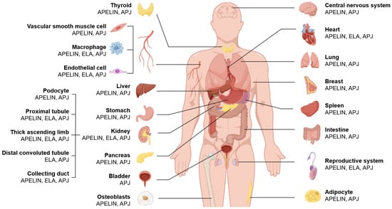
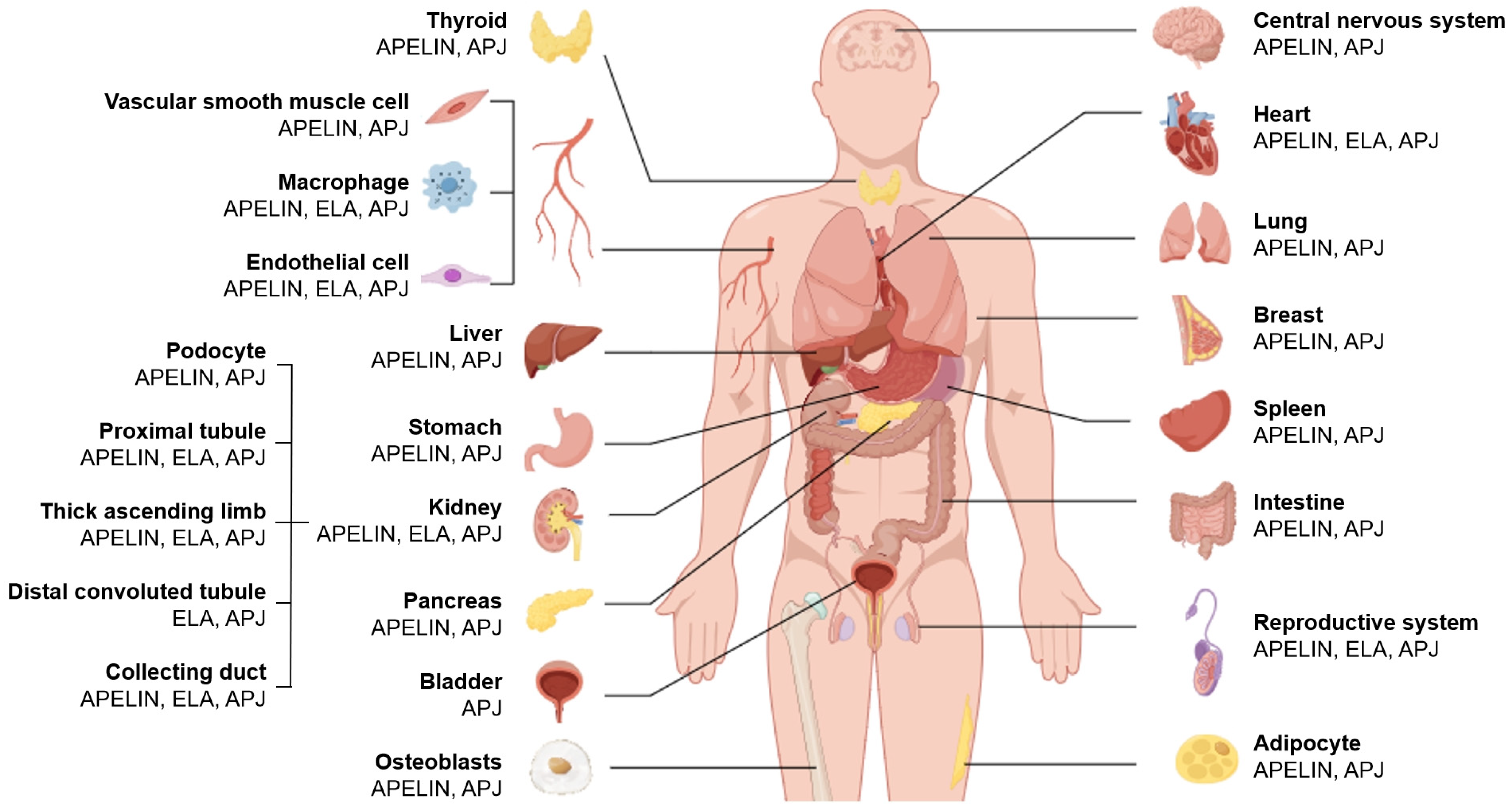
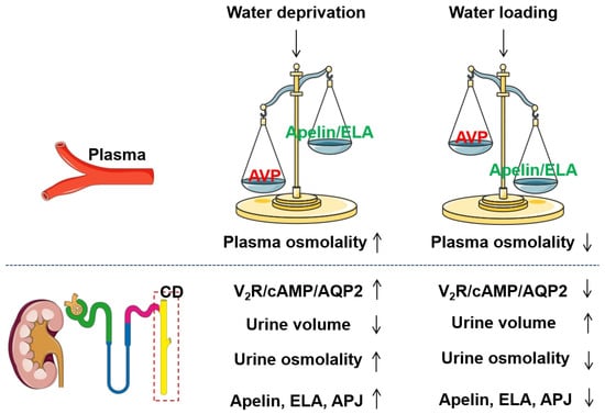
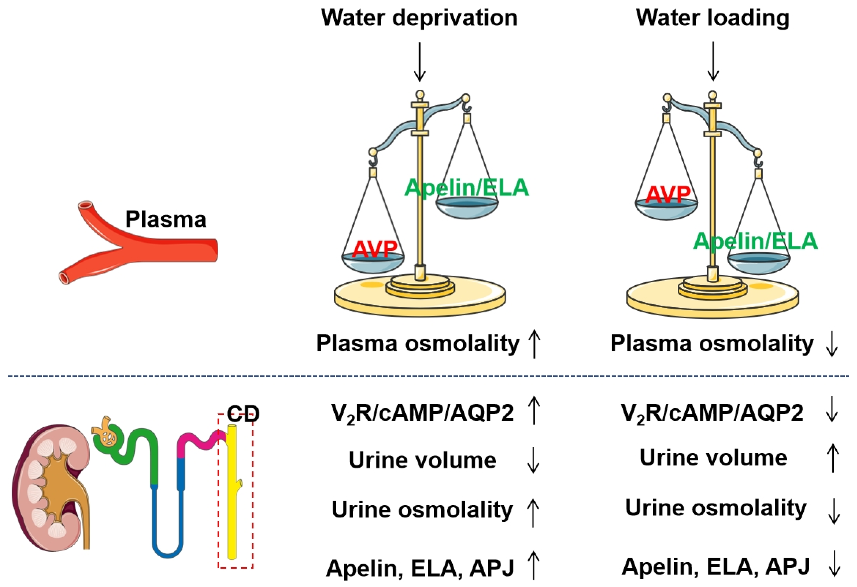
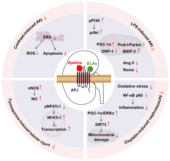
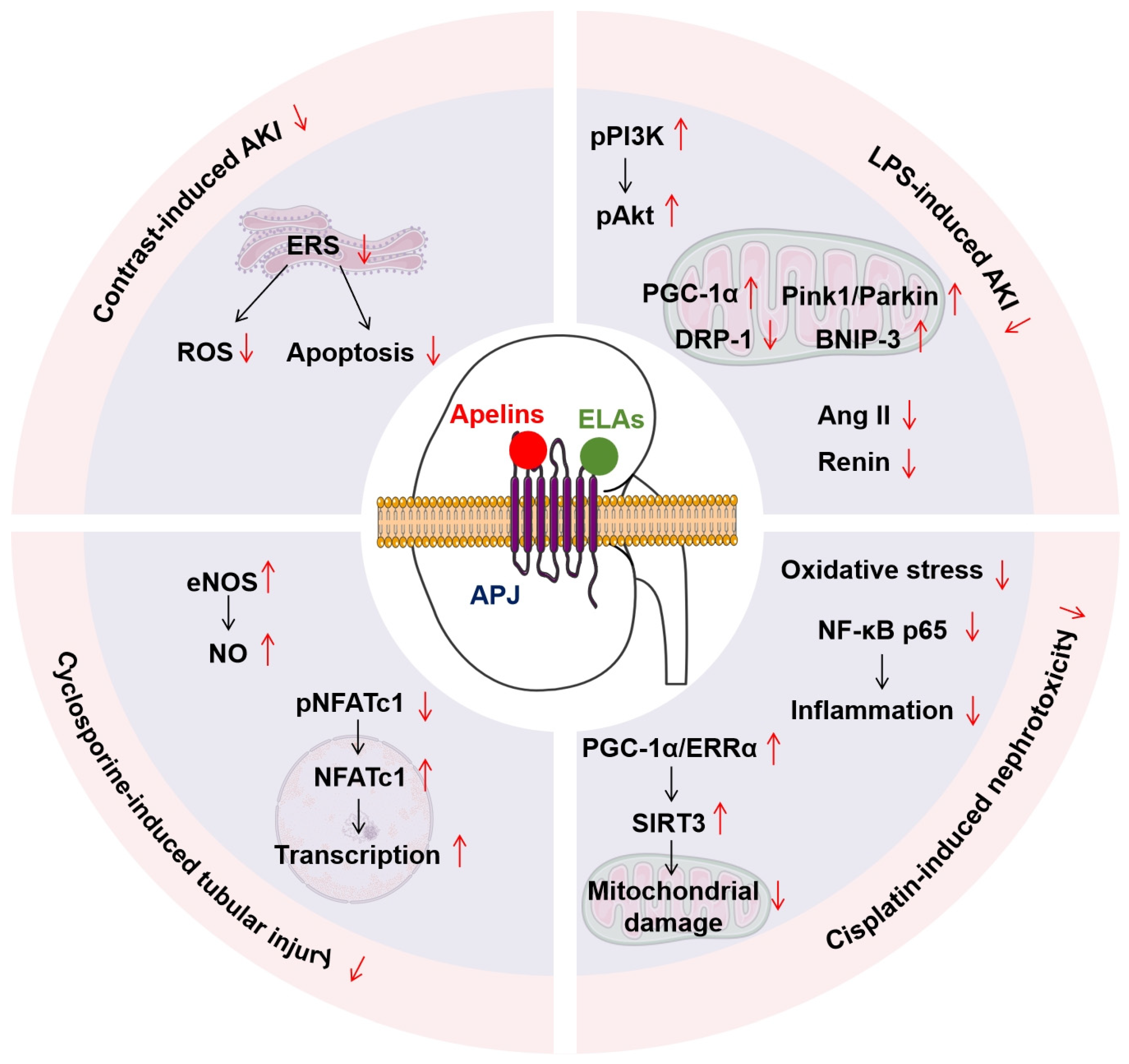
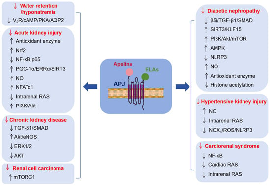
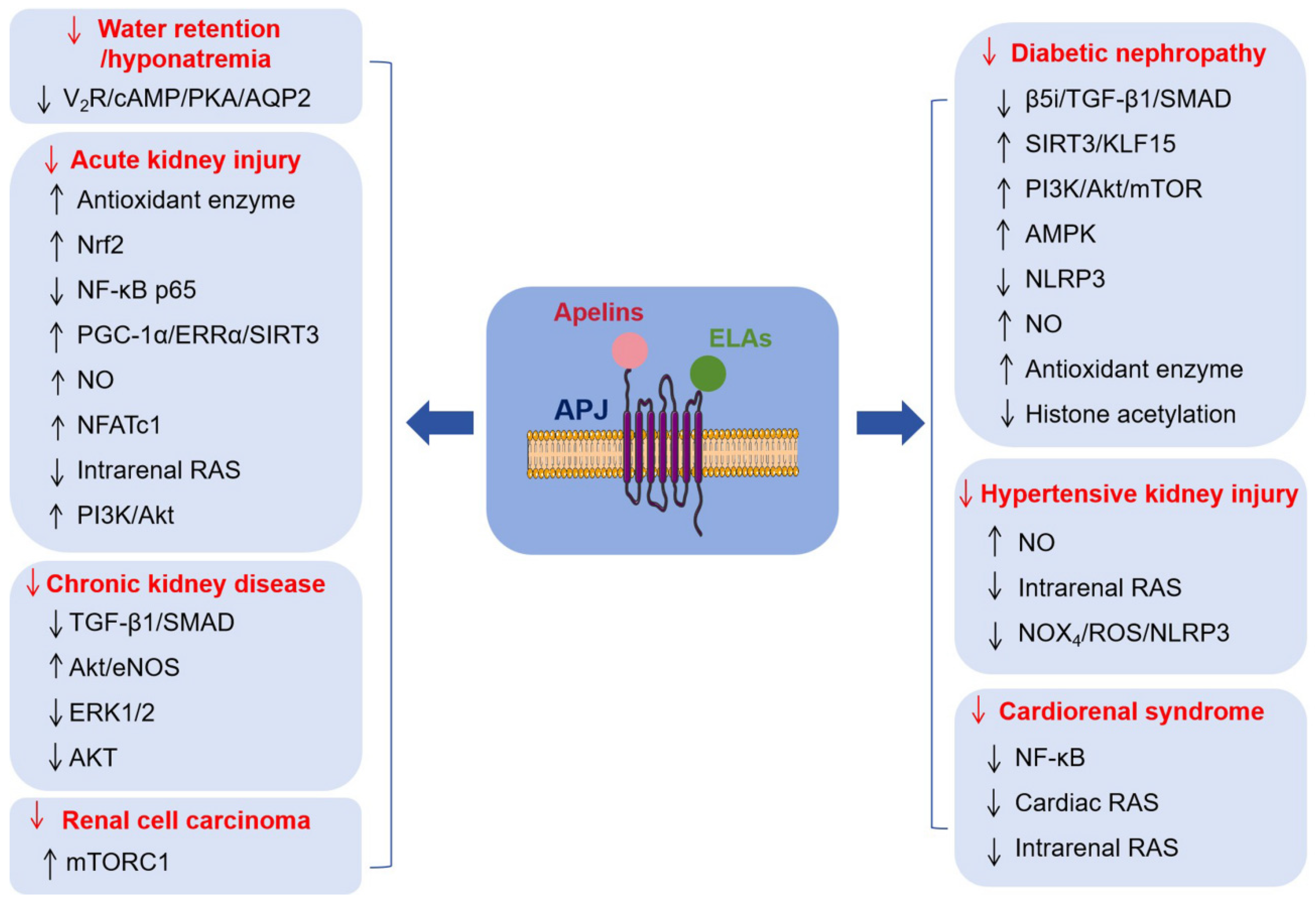

Disclaimer/Publisher’s Note: The statements, opinions and data contained in all publications are solely those of the individual author(s) and contributor(s) and not of MDPI and/or the editor(s). MDPI and/or the editor(s) disclaim responsibility for any injury to people or property resulting from any ideas, methods, instructions or products referred to in the content. |
© 2026 by the authors. Licensee MDPI, Basel, Switzerland. This article is an open access article distributed under the terms and conditions of the Creative Commons Attribution (CC BY) license.