


Abstract
Collagen membranes used for guided bone regeneration provide limited intrinsic osteogenic stimulation. This study evaluated whether L-serine incorporation enhances bone regeneration in ovariectomized rat calvarial defects. L-serine was loaded onto collagen membranes, characterized by X-ray diffraction and infrared spectroscopy, and its release was measured over 168 h. MC3T3-E1 cells were treated with L-serine (10–1000 µM); Runx2, osterix, and BMP-2/4 were assessed by Western blotting at 8 and 24 h, and matrix mineralization was evaluated by Alizarin Red S staining at days 7 and 17. Ovariectomized rats received bilateral calvarial defects treated with no graft, collagen membrane, or L-serine-incorporated membrane and were evaluated at 4 and 8 weeks. Each treatment group comprised 10 defects per time point (n = 10), and micro-CT outcomes were analyzed with an animal-block linear model followed by Holm-adjusted pairwise contrasts. Characterization indicated physical incorporation without covalent conjugation, and release was biphasic, reaching 72% at 8 h and 98.5% at 168 h. Runx2 and osterix responses were more prominent at 8 h than at 24 h. At 4 weeks, bone volume was greater with the L-serine membrane than with control or membrane alone, while BV/TV was greater than with membrane alone. At 8 weeks, bone volume remained greater than control and BMD was greater than membrane alone. L-serine incorporation enhanced early bone regeneration, consistent with a short-term osteogenic stimulus.
1. Introduction
Alveolar bone regeneration is a fundamental concern in oral and maxillofacial surgery, implant dentistry, and periodontal reconstruction, as adequate bone volume is essential for both functional rehabilitation and esthetic restoration [1]. Sufficient alveolar bone is required to provide structural support for dental implants, maintain facial contour, and achieve long-term stability of prosthetic and periodontal treatment outcomes [2]. However, alveolar bone defects frequently arise from oral and maxillofacial diseases, and these deficiencies often complicate surgical reconstruction and compromise treatment predictability [1]. In such situations, insufficient bone quantity or quality may limit implant placement, impair tissue support, and negatively affect both function and appearance. To overcome these limitations, guided bone regeneration (GBR) has been widely adopted as a well-established surgical approach for promoting selective bone healing [3]. By using a barrier membrane to exclude the rapid ingrowth of epithelial and connective tissues while preserving a secluded space for osteogenic cell migration and proliferation, GBR creates a favorable environment for new bone formation within the defect [4].
GBR membranes play both biological and mechanical roles in the regenerative process, and these properties are critical for successful bone healing [5,6]. Mechanically, the membrane helps maintain a protected space over the defect, stabilizes the blood clot, and supports the local environment required for new bone formation [7]. Based on their material characteristics, GBR membranes are generally classified into non-resorbable and resorbable types. Non-resorbable membranes, such as expanded polytetrafluoroethylene, provide excellent space-maintaining ability but often require a second surgery for removal and are associated with a higher risk of exposure-related complications [8]. In contrast, resorbable membranes eliminate the need for a second surgical procedure for membrane removal and are therefore more convenient in clinical practice [8,9,10]. Among them, collagen membranes have been widely used because of their favorable biocompatibility, biodegradability, low immunogenicity, and ease of handling [1,11,12].
Despite their widespread clinical use, conventional collagen membranes have several limitations that may restrict their regenerative potential when used alone. In most cases, collagen membranes primarily function as passive barriers that prevent soft tissue invasion, rather than actively stimulating osteogenesis within the defect [13]. Although this barrier function is essential for GBR, conventional collagen membranes generally provide only limited intrinsic osteogenic stimulation to surrounding cells and tissues [14]. In addition, the relatively rapid degradation of some collagen membranes may shorten the duration of barrier function before sufficient bone regeneration has occurred, particularly in defects that require prolonged healing [15]. Their limited mechanical stability and restricted space-maintaining capacity may further compromise regenerative outcomes, especially in large or structurally unfavorable defects.
L-serine is a biologically important amino acid that participates in a wide range of cellular processes, including protein synthesis, intermediary metabolism, cell proliferation, and the biosynthesis of membrane lipids and other matrix-related components [16]. Because these functions are closely associated with tissue repair and cellular activity during regeneration, L-serine has attracted interest as a potential bioactive component in regenerative biomaterials [15]. In the context of bone healing, L-serine may help support osteoblastic activity, extracellular matrix synthesis, and the establishment of a local microenvironment favorable for new bone formation [16]. In addition, previous studies on serine-containing or sericin-derived biomaterials have suggested that serine-rich materials may exert beneficial effects on bone regeneration and osteogenic responses [17]. Although such findings do not establish L-serine as an independent osteoinductive factor, they support the possibility that local delivery of L-serine could enhance the biological performance of a barrier membrane. Therefore, incorporation of L-serine into a collagen GBR membrane may convert the membrane from a primarily passive barrier into a more bioactive scaffold capable of not only isolating the defect site but also promoting a regenerative environment conducive to bone healing.
The choice of experimental model reflects the clinical setting in which a biofunctionalized membrane would be most useful. Postmenopausal estrogen deficiency reduces bone formation and accelerates bone resorption, and guided bone regeneration performed under these conditions yields less predictable outcomes than in healthy bone [18,19]. The ovariectomized rat is an established model of estrogen-deficient bone loss and therefore represents a compromised healing condition in which any additional osteogenic stimulation provided by the membrane should be most readily detected [20]. This model also permits direct comparison with a previous report in which an L-serine-incorporated gelatin sponge enhanced bone regeneration in ovariectomized rat calvarial defects [21], and with evidence linking serine metabolism to bone turnover under estrogen-deficient conditions [17,22].
Accordingly, the purpose of this study was to evaluate whether an L-serine-incorporated GBR membrane could enhance bone regeneration compared with an unfilled control and a collagen membrane alone. Using an experimental bone defect model, the regenerative efficacy of the modified membrane was assessed by comparing the extent of new bone formation and defect healing among the groups. We hypothesized that incorporation of L-serine would improve the biological performance of the GBR membrane and result in superior bone regeneration compared with both the unfilled control and the conventional collagen membrane.
2. Materials and Methods
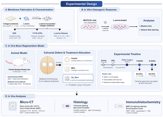
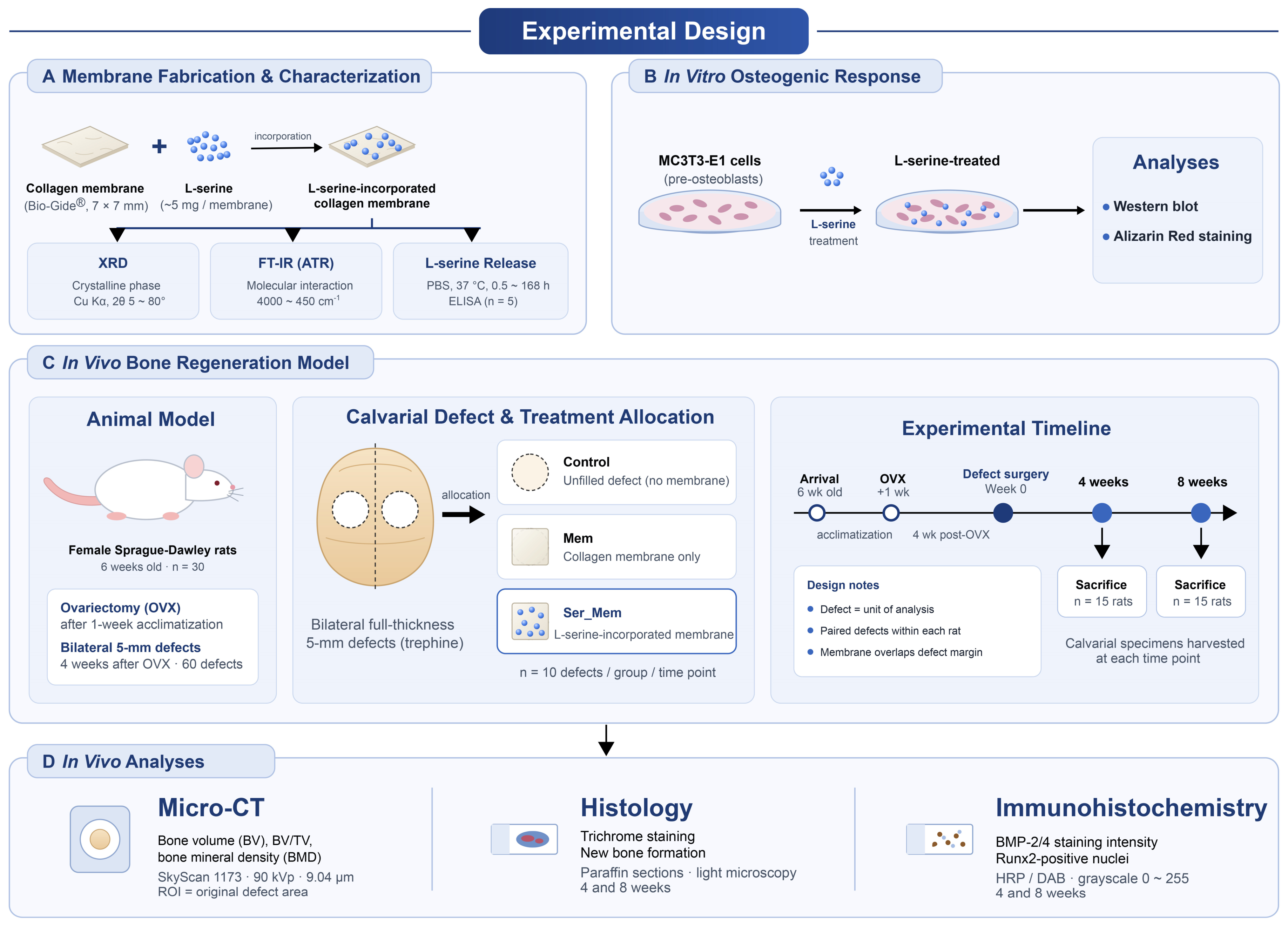
An overview of the overall study design—including membrane fabrication and characterization, the in vitro osteogenic experiments, and the in vivo ovariectomized rat calvarial defect model—is provided in Figure 1.
Figure 1.
Overview of the experimental design.
2.1. Preparation of the L-Serine-Incorporated Collagen Membrane
Bio-Gide® collagen membrane (Geistlich Pharma AG, Wolhusen, Switzerland) was trimmed into 7 mm × 7 mm rectangular pieces (0.49 cm2; approximate dry mass of 3 mg per piece). L-serine (cat. no. S4500; lot no. 0000270921; Sigma-Aldrich, St. Louis, MO, USA; ≥99% purity, crystalline powder) was dissolved in sterile distilled water at a concentration of 10 mg/mL and passed through a 0.22-µm sterile filter. Subsequently, 500 µL of the L-serine solution was applied dropwise onto the rough surface of each membrane piece, corresponding to a nominal loading amount of approximately 5 mg L-serine per membrane. The loaded membranes were dried at 25 °C for 8 h in a sterile laminar-flow cabinet and stored at 4 °C in a desiccator until use. Membranes used in the collagen membrane-only group were processed identically using an equal volume of sterile distilled water without L-serine, so that both groups underwent the same wetting and drying procedure. Specimens for the release experiment were prepared using the same procedure, with the L-serine solution volume adjusted according to the dry mass of each specimen to maintain the same nominal loading ratio.
2.2. X-Ray Diffraction and Fourier-Transform Infrared Spectroscopy
X-ray diffraction (XRD) analysis was performed to investigate the crystalline characteristics of the fabricated membranes. The dried specimens were placed on a silicon low-background sample holder and analyzed using an Ultima IV diffractometer (Rigaku, Tokyo, Japan) with Cu Kα radiation (λ = 1.5406 Å) operated at 40 kV and 30 mA. Diffraction patterns were recorded over a 2θ range of 5–80° at a scanning step of 0.02°, with a dwell time of 0.5 s for each step.
FT-IR spectra were recorded using a Nicolet iS50 spectrometer (Thermo Scientific, Waltham, MA, USA) equipped with an ATR accessory (MIRacle, PIKE Technologies, Madison, WI, USA). Spectra were collected over the range of 3500–800 cm−1 at a resolution of 1 cm−1 using a deuterated L-alanine-doped triglycine sulfate (DLATGS) detector (Leonardo Electronics, Southampton, UK). ATR-FTIR spectra were obtained for the pure L-serine starting material, the collagen membrane control, and the L-serine-incorporated collagen membrane under identical measurement conditions.
2.3. L-Serine Release from the Collagen Membrane
After incorporation of L-serine, each collagen membrane specimen (approximately 0.072 mg; prepared separately from the 7 mm× 7 mm membranes used for the in vivo experiments) was placed in a 48-well culture plate and immersed in 0.5 mL of 0.01 M phosphate-buffered saline (PBS). The specimens were incubated at 37 °C. At predetermined time points (0.5, 1, 2, 4, 8, 24, 72, and 168 h), the release medium was completely collected and stored at −20 °C until analysis. An equal volume of fresh PBS was then added to each well to continue the release experiment. The concentration of L-serine in the collected medium was measured using an L-serine ELISA kit (CAT#: IS-I-1200R, immusmol, Bordeaux, France) according to the manufacturer’s protocol. Measurements were repeated five times.
2.4. Cell Culture and Western Blotting
MC3T3-E1 cells were obtained from the Korean Cell Line Bank (KCLB) and maintained in Dulbecco’s modified Eagle’s medium (DMEM, high glucose, with L-glutamine and without sodium pyruvate; HyClone/Cytiva, cat. no. SH30022.01), which contains L-serine at 42 mg/L (0.40 mM), supplemented with 10% fetal bovine serum (FBS; Gibco; Thermo Fisher Scientific, Inc., Waltham, MA, USA) and 1% penicillin-streptomycin (P/S; Gibco; Thermo Fisher Scientific, Inc.). The cells were cultured in a humidified incubator at 37 °C under 5% CO2.
For L-serine treatment, MC3T3-E1 cells were seeded in culture plates and allowed to attach overnight. The cells were then treated with L-serine added to the medium at 10, 100, or 1000 μM for 8 or 24 h; accounting for the 0.40 mM L-serine already present in the medium, these correspond to approximate total concentrations of 0.41, 0.50, and 1.4 mM, respectively. Untreated cells served as the control group.
After treatment, total cellular proteins were extracted using Laemmli buffer supplemented with protease inhibitor cocktail (Roche Diagnostics, Rotkreuz, Switzerland) and phosphatase inhibitor cocktail (Roche Diagnostics). The protein samples were heat-denatured and separated by electrophoresis on 8–15% acrylamide gels at 95 V for 3 h, followed by transfer onto 0.45-μm nitrocellulose membranes (Bio-Rad Laboratories, Inc., Hercules, CA, USA) at 20 V overnight. The membranes were blocked and incubated with primary antibodies against Runx2 (CAT#: sc-390351, Santa Cruz Biotechnology, Santa Cruz, CA, USA), osterix (CAT#: sc-393325, Santa Cruz Biotechnology), BMP-2/4 (CAT#: sc-137087, Santa Cruz Biotechnology), and GAPDH (CAT#: sc-166545, Santa Cruz Biotechnology). GAPDH was used as an internal loading control. After incubation with the appropriate secondary antibodies, the immunoreactive bands were detected using a FUSION Solo S imaging system (Vilber Lourmat, Collégien, France). Band intensities were quantified using ImageJ software (version 1.53a; National Institutes of Health). Western blotting was performed in four independent biological experiments (n = 4 per condition). Each biological replicate consisted of a separately seeded and independently treated MC3T3-E1 culture prepared on a separate experimental occasion.
For the mineralization assay, MC3T3-E1 cells were cultured under osteogenic conditions and treated with L-serine added to the medium at 10, 100, or 1000 μM, with untreated cells as the control. At day 7 and day 17, the cells were fixed and stained with Alizarin Red S (ARS) to visualize calcium deposition, and representative stained and corresponding brightfield images were acquired by light microscopy. Alizarin Red S staining was assessed qualitatively from the acquired images and was not quantified.
2.5. Animal Experiments
Six-week-old female Crl:CD (Sprague–Dawley) specific pathogen-free rats (Orient Bio Inc., Seongnam, Republic of Korea) were used. All procedures were approved by the Institutional Animal Care and Use Committee of Gangneung-Wonju National University (renamed Kangwon National University in March 2026) (approval no. GWNU-2025-20; approved on 3 November 2025) and performed in accordance with institutional guidelines. Animals were housed at 20–22 °C under 40% relative humidity with a 12-h light/dark cycle and allowed free access to food and water. After 1 week of acclimatization, all rats underwent ovariectomy, and calvarial defect surgery was performed 4 weeks later. Ovariectomy was performed to establish an estrogen-deficient state, because guided bone regeneration under estrogen deficiency represents the compromised healing condition in which the biologic contribution of a functionalized membrane is most relevant, and the interval of 4 weeks was allowed for bone loss to become established before the defects were created. Establishment of the estrogen-deficient state was confirmed by uterine atrophy observed at sacrifice, together with the increase in body weight that followed ovariectomy.
A total of 30 ovariectomized rats were used. Bilateral full-thickness 5-mm circular calvarial defects were created in each rat, yielding 60 defects in total. Each defect was assigned to one of three treatments in a fixed cyclic sequence across animals: unfilled control, collagen membrane-only, or L-serine-incorporated collagen membrane. Bio-Gide® collagen membrane was trimmed into 7 mm × 7 mm rectangular pieces to fully cover the defect with overlap onto the surrounding bone. For the experimental group, L-serine-incorporated membranes were prepared as described above, each piece containing approximately 5 mg of L-serine (10.2 mg/cm2). In the control defects, no graft material was placed. In the membrane-only group, defects were covered with collagen membrane alone, whereas in the experimental group, defects were covered with L-serine-incorporated collagen membrane. Because bilateral defects were created in the same animal, this design was intended to reduce inter-animal variability and balance treatment representation across animals. The animals were sacrificed at 4 or 8 weeks after surgery, with 15 rats assigned to each time point. Because each animal received two of the three treatments, the allocation constituted a balanced incomplete block design. Treatments were assigned to the two defects of each animal in a fixed cyclic sequence across animals, so that each treatment and each pairwise treatment combination occurred an equal number of times; the assignment of treatments to the left and right defect was varied across animals. Treatments were not randomly allocated and allocation was not concealed from the operator. Each defect constituted the unit of treatment assignment and outcome measurement, while animal was included as a blocking factor to account for dependence between bilateral defects.
General anesthesia was induced by intraperitoneal injection of tiletamine–zolazepam (Zoletil 50, 30 mg/kg; Virbac Korea, Seoul, Republic of Korea) and xylazine (Rompun, 8 mg/kg; Bayer Korea, Seoul, Republic of Korea). A midline sagittal incision was made over the calvarium, and bilateral defects were prepared using a trephine bur. After placement of the designated material, the periosteum and skin were closed with 4-0 black silk sutures. Preemptive local analgesia was provided by subcutaneous infiltration of 2% lidocaine around the surgical site. Gentamicin (5 mg/kg, subcutaneously) was administered postoperatively to prevent infection. Animals were monitored closely for postoperative signs of pain or distress, with buprenorphine (0.03 mg/kg, subcutaneously, every 8–12 h for up to 48 h) available as rescue analgesia; no animal showed signs of pain or distress requiring rescue analgesia, and no adverse events were observed. The sample size was determined by a priori power analysis for a one-way ANOVA (three groups) using G*Power version 3.1.9.7, with α = 0.05, power (1 − β) = 0.80, and an assumed effect size (Cohen’s f) of 0.65, indicating a minimum of nine defects per group; ten defects per group per time point were used. A design-specific power calculation for the balanced incomplete-block allocation was not used at the planning stage. Animals were euthanized at 4 or 8 weeks by CO2 inhalation; after cessation of respiration and loss of responsiveness, death was confirmed and cervical dislocation was performed as a secondary physical method to ensure death. No pre-specified exclusion criteria were applied, and all animals and all defects were included in the analysis. At each time point, 10 defects were analyzed per treatment group.
2.6. Micro-Computed Tomography
Calvarial specimens (8 mm × 8 mm × 1 mm) were submitted to Genoss (Seongnam, Republic of Korea) for micro-computed tomography (micro-CT) analysis. Micro-CT scanning and quantitative analysis were performed by an external service blinded to treatment allocation. The specimens were scanned using a SkyScan 1173 system (Bruker SkyScan, Kontich, Belgium) operated at a source voltage of 90 kVp with an aluminum filter and a pixel size of 9.04 μm. The acquired projection images were reconstructed using NRecon software version 1.7.3.1 (Micro Photonics Inc., Allentown, PA, USA). The region of interest (ROI) was defined based on the original size of the surgically created defect. Bone volume (BV) within the ROI was calculated using a threshold corresponding to 25% of the bone standard, and the bone volume fraction (BV/TV) was subsequently determined. The ROI was cylindrical, with a diameter corresponding to the original defect, and BV/TV was calculated as the ratio of BV to the total volume. Bone mineral density (BMD) within the same region of interest was determined by calibration against hydroxyapatite phantoms of known mineral density and is expressed in g/cm3. Because the specimens obtained at the two time points were scanned and calibrated separately, BMD was compared only within each time point.
2.7. Histology and Immunohistochemistry
Paraffin-embedded tissue sections were deparaffinized in xylene and rehydrated through a graded ethanol series (100%, 95%, and 70%) to distilled water. Histological and immunohistochemical assessments were not performed under blinded conditions. For Goldner’s trichrome staining, the decalcified, paraffin-embedded sections were stained with Weigert’s iron hematoxylin to visualize nuclei, followed by sequential staining with Ponceau–acid fuchsin, phosphomolybdic acid–Orange G, and Light Green SF to differentiate connective tissue components. The sections were then dehydrated in graded ethanol, cleared in xylene, and mounted with a resin-based medium, and were examined under a light microscope. Because the specimens were decalcified prior to embedding, Goldner’s trichrome was used here as a connective-tissue and bone-matrix stain to assess the organization and maturity of the regenerated tissue rather than to distinguish mineralized bone from unmineralized osteoid, which requires undecalcified sections; the degree of mineralization was instead assessed quantitatively by micro-CT [23]. For comparison, Masson’s trichrome staining was also performed on parallel sections and is provided as Supplementary Figure S1.
For immunohistochemical analysis, sections were processed to evaluate Runx2 and BMP-2/4 expression using anti-Runx2 and anti-BMP-2/4 antibodies at a dilution of 1:100. Endogenous peroxidase activity was quenched with hydrogen peroxide, and the sections were subsequently washed, blocked, and incubated with the primary antibodies. After incubation with a universal secondary antibody system (Dako REAL™ EnVision™/HRP, Rabbit/Mouse; Dako North America Inc., Carpinteria, CA, USA), immunoreactivity was visualized using 3,3′-diaminobenzidine (DAB) chromogen (Dako REAL™ DAB+ Chromogen and Dako REAL™ Substrate Buffer; Dako North America Inc.). Staining was quantified using image analysis software within defined regions of interest placed within the defect area, corresponding to the newly regenerating tissue; the native bone at the defect margins was excluded so that the measurements reflected the regenerate rather than pre-existing bone. For BMP-2/4, staining intensity within each region of interest was measured on a grayscale from 0 to 255 (where 0 indicated no staining and 255 the highest intensity) and expressed as relative BMP-2/4 intensity. For Runx2, the number of Runx2-positive nuclei within the same regions of interest was counted. For each specimen, measurements were obtained from matched fields within the defect, and comparable regions of interest were used across groups.
2.8. Statistical Analysis
Data are presented as mean ± standard deviation (SD). Western blot data from four independent biological experiments were analyzed separately at each time point using one-way ANOVA followed by Tukey’s multiple-comparison test. Because two calvarial defects were created in each animal and two of the three treatments were allocated within each animal according to a balanced incomplete-block design, the two defects from the same animal were not considered statistically independent. Accordingly, treatment effects were analyzed separately at 4 and 8 weeks using a general linear model in which treatment was included as a fixed effect and animal was included as a blocking factor. An overall F test was first performed for the treatment effect. When appropriate, pairwise treatment contrasts were subsequently estimated between the L-serine-incorporated membrane (Ser_Mem), collagen membrane alone (Mem), and untreated control groups. p values for the three pairwise comparisons within each outcome and time point were adjusted using Holm’s method. Treatment effects are reported as adjusted mean differences with 95% confidence intervals (CIs). A two-sided p value < 0.05 was considered statistically significant.
3. Results
3.1. XRD, FT-IR, and L-Serine Release Profile
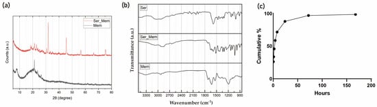
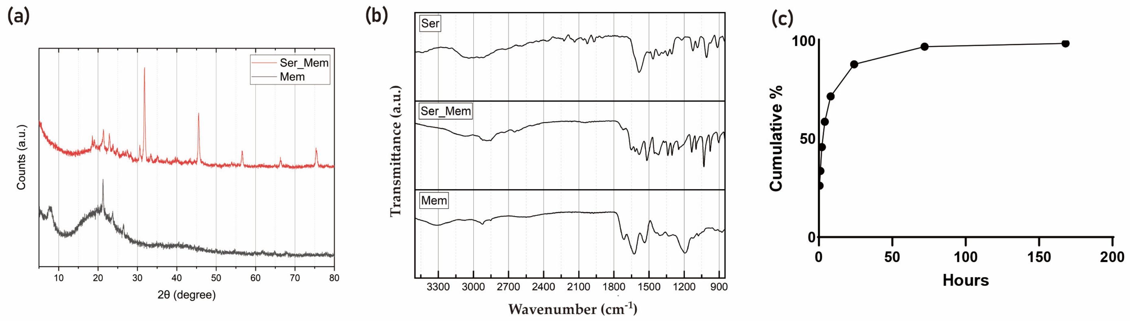
XRD analysis showed that the process-matched collagen membrane control (Mem) exhibited a broad diffraction background characteristic of the predominantly amorphous collagen matrix, whereas the L-serine-incorporated membrane (Ser_Mem) showed multiple sharp reflections superimposed on the collagen background (Figure 2a). Several prominent reflections in Ser_Mem coincided in 2θ position with reflections in the reference diffraction pattern for crystalline L-serine (ICDD PDF No. 00-027-1989) and were absent or markedly less evident in Mem. These findings are consistent with the presence of crystalline L-serine-containing domains in the loaded membrane. Because an experimental XRD pattern of the pure L-serine starting material was not available, the phase assignment should be regarded as supportive rather than definitive.
Figure 2.
Physicochemical characterization and in vitro release profile of the L-serine-incorporated collagen membrane. (a) X-ray diffraction patterns of the process-matched collagen membrane control (Mem) and L-serine-incorporated collagen membrane (Ser_Mem), together with the reference stick pattern for crystalline L-serine (ICDD PDF No. 00-027-1989). Mem exhibited a broad diffraction background characteristic of the predominantly amorphous collagen matrix, whereas Ser_Mem showed sharp reflections superimposed on the collagen background. Reflections in Ser_Mem at 2θ ≈ 18.5, 22.8, 25.6, 28, 30.6, 33.5 and 35° corresponded to reflections of the L-serine reference pattern (ICDD PDF No. 00-027-1989), with the strongest at ~22.8° and ~30.6°, and were absent from Mem. The experimental patterns were vertically offset for clarity. (b) Fourier-transform infrared (FT-IR) spectra of pure L-serine (Ser), the L-serine-incorporated membrane (Ser_Mem), and the process-matched collagen membrane control (Mem). Mem showed the characteristic collagen amide I (~1650 cm−1) and amide II (~1540 cm−1) bands. Ser_Mem retained this collagen envelope but showed additional absorption in the fingerprint region (1400–1200 cm−1) attributable to carboxyl and C–O-related vibrations of L-serine; these bands corresponded to bands in the pure L-serine spectrum and were absent from Mem. Ser_Mem also showed broadened absorption in the 3200–3400 cm−1 region (O–H/N–H stretching) and changes in the amide I and amide II regions relative to Mem. Together, these features are consistent with physical association between collagen and L-serine and possible non-covalent interactions, including hydrogen bonding; however, the spectra do not establish a specific bonding mechanism. (c) In vitro cumulative release profile of L-serine from the collagen membrane in phosphate-buffered saline at 37 °C over 168 h, determined by an L-serine ELISA assay. L-serine was released in a biphasic manner, with an initial burst within the first 8 h followed by a sustained release phase up to 168 h. Data are presented as mean ± standard deviation (n = 5).
FT-IR spectra were compared among the pure L-serine starting material (Ser), collagen membrane control (Mem), and L-serine-incorporated collagen membrane (Ser_Mem) (Figure 2b). The pure L-serine spectrum exhibited a broad N–H/O–H stretching envelope and multiple distinct absorption features in the fingerprint region. Ser_Mem retained the characteristic collagen amide I (~1650 cm−1) and amide II (~1540 cm−1) bands while also exhibiting spectral features corresponding to those observed in pure L-serine, including broadened absorption in the N–H/O–H stretching region and additional or enhanced bands in the fingerprint region. These L-serine-associated spectral features were not observed with the same pattern in Mem, supporting the presence of L-serine in the loaded membrane. Changes in the profiles of the amide I and amide II bands further suggest that L-serine loading altered the local molecular environment of the collagen matrix. Overall, direct comparison with the pure starting material supports successful incorporation of L-serine into the membrane, although the spectra alone do not establish a specific bonding mechanism or the strength or durability of the interaction.
The in vitro release profile showed that L-serine was released from the collagen membrane in a biphasic manner over 168 h (Figure 2c). An initial burst phase occurred within the first 8 h, during which approximately 72% of the loaded L-serine was released, with roughly 26% released within the first 30 min. This was followed by a sustained release phase from 8 to 168 h, over which the remaining fraction was gradually liberated as the interval release rate declined (from ~16% at 24 h to <2% by 168 h). The cumulative release reached 98.5% of the loaded amount (116.3 of 118 µg) by 168 h, indicating near-complete recovery of the incorporated L-serine. The pronounced initial burst is consistent with the dissolution of loosely bound or surface-associated L-serine—in agreement with the excess/unbound crystalline domains inferred from the XRD data—whereas the subsequent sustained release reflects the slower liberation of L-serine that interacted with the collagen matrix, as suggested by the amide I/II shifts in the FT-IR spectra. Because the release medium was exchanged in full at each sampling point, the L-serine concentration in the medium during a given interval could be derived from the released mass (molecular weight 105.09). The concentration was approximately 0.58 mM during the first 30 min, approximately 0.36 mM during the interval from 8 to 24 h, and below 0.05 mM after 72 h, so that the concentrations applied in the cell experiments lay within the range generated by the membrane. Because the culture medium already contained 0.40 mM L-serine, the added concentrations of 10, 100, and 1000 µM correspond to approximate total exposures of 0.41, 0.50, and 1.4 mM; the lowest added concentration therefore represents only a ~2.5% increase over baseline, consistent with the limited response observed at that dose, whereas the osteogenic response and its attenuation were most evident at the highest total concentration (~1.4 mM). The near-complete cumulative release also indicates that essentially all of the incorporated L-serine was releasable and that the membrane functions as a short-term delivery vehicle rather than a sustained-release depot.
3.2. L-Serine Transiently Enhances Osteogenic Marker Expression and Promotes Matrix Mineralization in MC3T3-E1 Cells
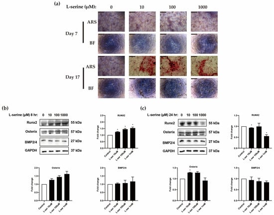
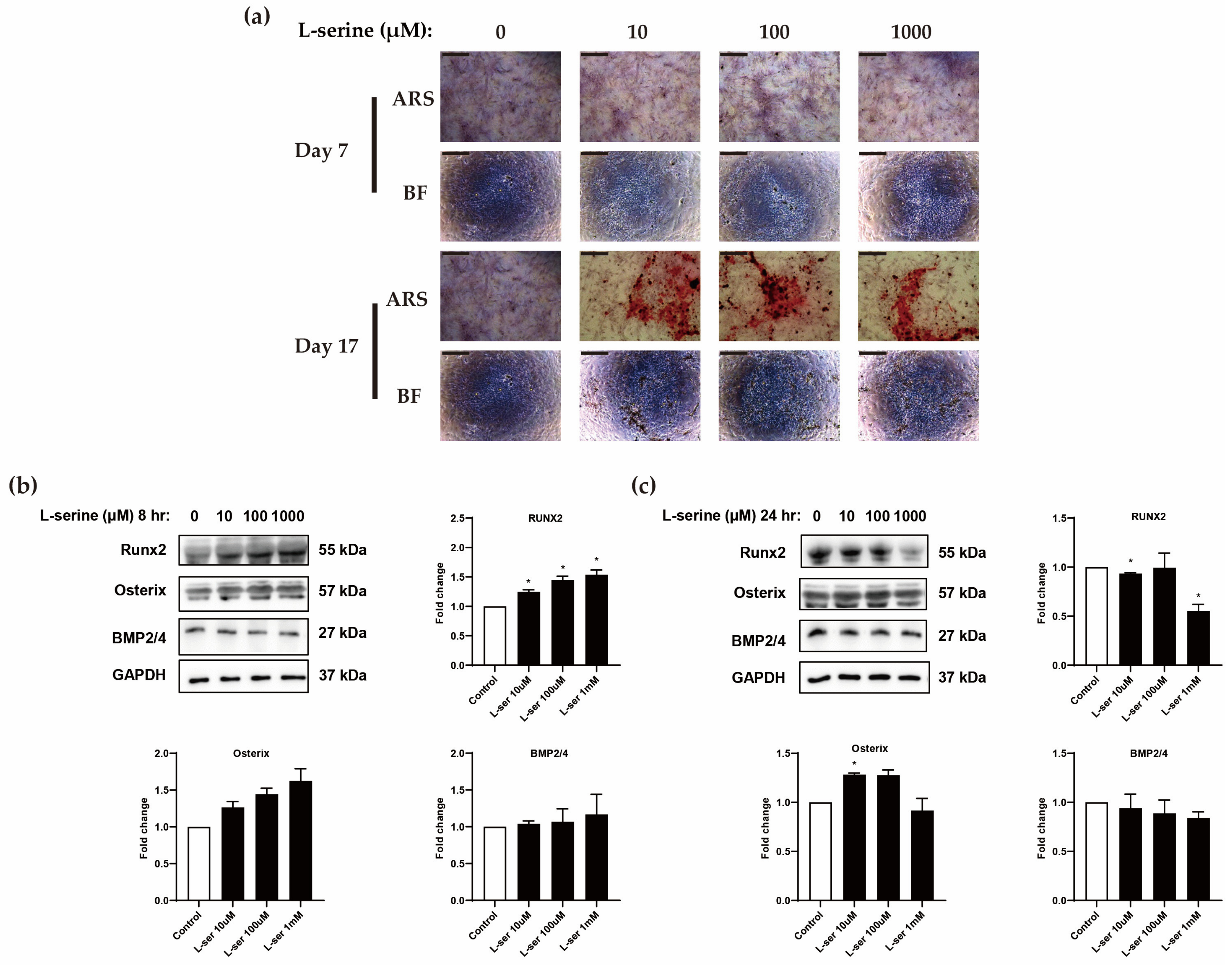
To evaluate the functional osteogenic outcome of L-serine treatment, matrix mineralization was assessed by Alizarin Red S staining (Figure 3a). At day 7, mineralization was minimal in all groups. By day 17, pronounced Alizarin Red–positive mineralized deposits were observed in all L-serine-treated groups (10, 100, and 1000 μM), whereas the untreated control remained largely unstained. These qualitative observations indicate that L-serine treatment promoted mineralized matrix formation by MC3T3-E1 cells. To examine the early transcriptional events underlying this response, the expression of osteogenic markers was then analyzed by Western blotting at 8 and 24 h (Figure 3b,c).
Figure 3.
L-serine modulates osteogenic marker expression and matrix mineralization in MC3T3-E1 cells in a time-dependent manner. (a) Representative Alizarin Red S (ARS) staining and corresponding brightfield (BF) images of MC3T3-E1 cells treated with L-serine (−, 10, 100, or 1000 μM) under osteogenic conditions at day 7 and day 17. Mineralization was minimal in all groups at day 7, whereas at day 17 pronounced Alizarin Red–positive mineralized deposits were evident in all L-serine-treated groups but remained minimal in the untreated control. Scale bars: 50 μm (b) Representative Western blots and densitometric analysis of Runx2, osterix, and BMP-2/4 in MC3T3-E1 cells treated with L-serine added at 10, 100, or 1000 μM for 8 h (corresponding to approximate total concentrations of 0.41, 0.50, and 1.4 mM in medium containing 0.40 mM L-serine). (c) Representative Western blots and densitometric analysis of Runx2, osterix, and BMP-2/4 in MC3T3-E1 cells treated with L-serine (10, 100, or 1000 μM) for 24 h. GAPDH served as the internal loading control. Relative protein expression was normalized to GAPDH and expressed as fold change versus the untreated control. Data are presented as mean ± standard deviation from four independent biological experiments (n = 4). * p < 0.05 compared to untreated control. Original (uncropped) Western blot images for Figure 3b,c are provided in Supplementary Figure S2.
This marker analysis revealed that L-serine modulated osteogenic marker expression in a time-dependent manner, with a more evident stimulatory pattern at 8 h after treatment than at 24 h (Figure 3b,c). At 8 h after L-serine treatment, Runx2 protein expression was increased in all L-serine-treated groups compared with the untreated control (Figure 3b). Densitometric analysis showed a dose-associated increase in Runx2 expression, with the highest expression observed in the 1 mM added L-serine group. Osterix expression also showed a gradual increasing tendency after L-serine treatment, particularly at 100 µM and 1 mM, although this change did not reach statistical significance. BMP2/4 expression showed only a mild increasing tendency, with relatively large variation, suggesting that the early response to L-serine was more clearly reflected by Runx2 and Osterix than by BMP2/4. Overall, the 8 h treatment condition showed a clear enhancement of osteogenic transcription factor expression, especially Runx2.
At 24 h after treatment, the response pattern was different. Runx2 expression was not increased compared with the control; rather, the 1 mM added L-serine group showed a marked reduction in Runx2 expression (Figure 3c). Osterix expression was increased in the 10 µM and 100 µM groups, with the 10 µM group showing a statistically significant increase, but this effect was not maintained at 1 mM. BMP2/4 expression did not increase at 24 h and showed a slight decreasing tendency in the L-serine-treated groups.
3.3. L-Serine-Incorporated Membrane Enhances Early Bone Regeneration in Calvarial Defects
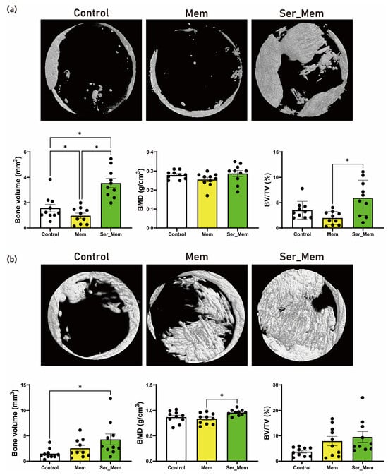
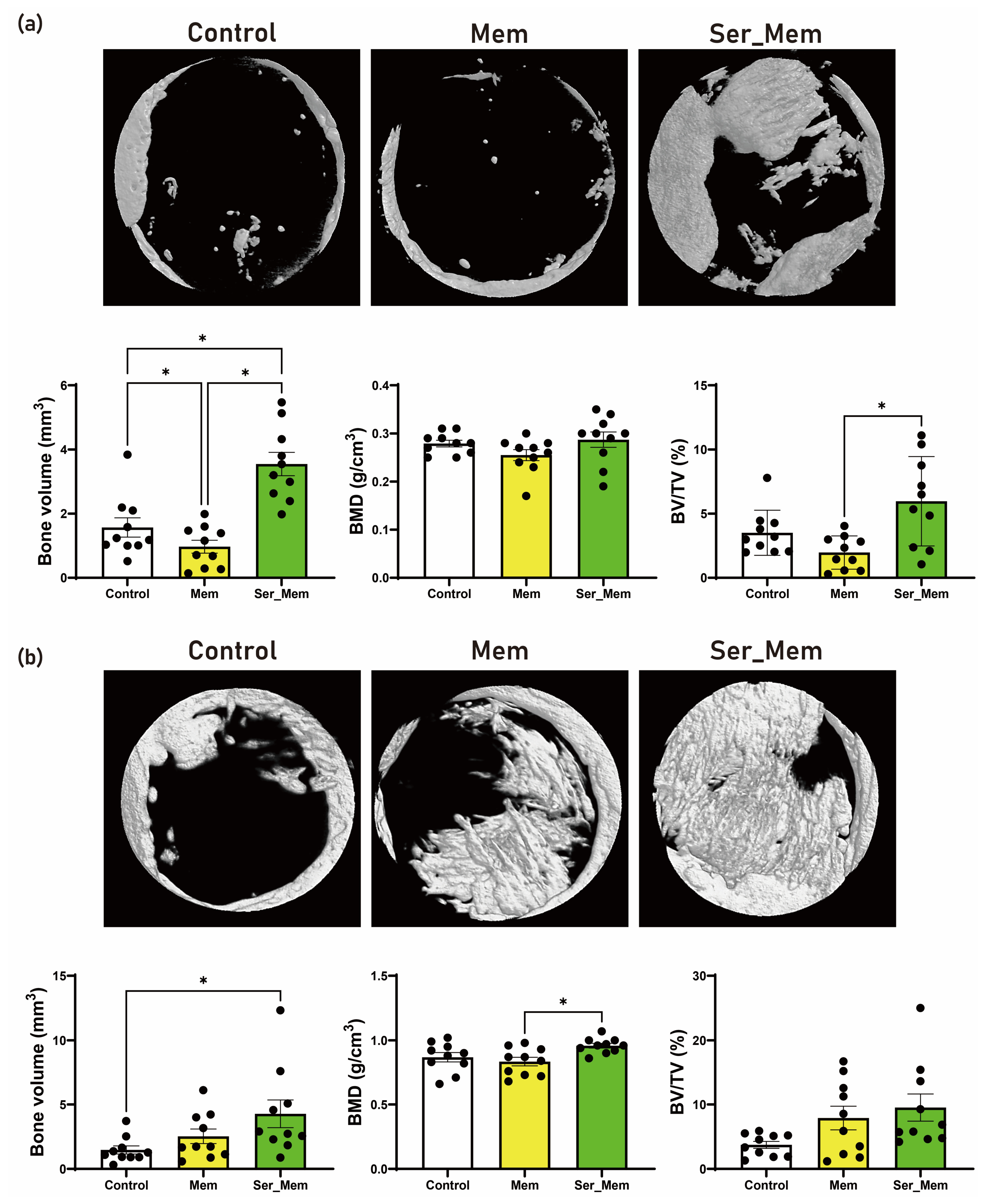
Micro-CT analysis demonstrated that bone regeneration was enhanced by the L-serine-incorporated membrane (Figure 4). In the 2D/3D reconstructed images, the untreated control defects showed limited new bone formation mainly along the defect margin, while the membrane-only group showed partial bone formation within the defect area. In contrast, the Ser_Mem group exhibited more extensive mineralized tissue formation, with newly formed bone bridging or occupying a larger portion of the defect region (Figure 4).
Figure 4.
Micro-CT evaluation of bone regeneration in rat calvarial defects. (a) Representative reconstructed micro-CT images and quantitative measurements at 4 weeks after treatment. Bone volume (BV) was significantly greater in the L-serine-incorporated collagen membrane group (Ser_Mem) than in both the untreated control and collagen membrane-only (Mem) groups. BV was also significantly lower in the Mem group than in the control group. Bone mineral density (BMD) did not differ significantly among the groups. Bone volume fraction (BV/TV) was significantly greater in the Ser_Mem group than in the Mem group, whereas the differences between Ser_Mem and control and between Mem and control were not statistically significant. (b) Representative reconstructed micro-CT images and quantitative measurements at 8 weeks after treatment. BV was significantly greater in the Ser_Mem group than in the control group. BMD was significantly greater in the Ser_Mem group than in the Mem group. Although the overall treatment effect for BV/TV was significant, none of the pairwise comparisons remained statistically significant after correction for multiple comparisons. Data are presented as mean ± standard deviation. n = 10 defects per group at each time point. Statistical comparisons were performed using an animal-block linear model, with animal included as a blocking factor, followed by pairwise treatment contrasts with Holm adjustment for multiple comparisons. * p < 0.05.
Quantitative analysis supported these image-based findings. There was a significant overall treatment effect for BV (F(2,13) = 20.12, p < 0.001) (Figure 4a). After accounting for animal as a blocking factor, BV was significantly greater in the Ser_Mem group than in the Control group, with an adjusted mean difference of 1.69 mm3 (95% CI 0.73 to 2.65; Holm-adjusted p = 0.0045), and greater than in the Mem group by 2.81 mm3 (95% CI 1.85 to 3.77; p < 0.001). BV was also lower in Mem than Control (adjusted difference −1.12 mm3; 95% CI −2.08 to −0.16; p = 0.026).
For BV/TV, the overall treatment effect was significant (F(2,13) = 6.48, p = 0.011). Ser_Mem showed significantly greater BV/TV than Mem, with an adjusted difference of 4.55 percentage points (95% CI 1.80 to 7.30; Holm-adjusted p = 0.010). However, the Ser_Mem–Control comparison was not significant after multiplicity correction (adjusted difference 1.83 percentage points; p = 0.173), nor was the Mem–Control comparison (p = 0.105). There was no significant overall treatment effect for BMD at 4 weeks (F(2,13) = 2.14, p = 0.157), and none of the pairwise comparisons reached statistical significance.
At 8 weeks, there was a significant overall treatment effect for BV (F(2,13) = 5.53, p = 0.018). BV was significantly greater in Ser_Mem than Control, with an adjusted mean difference of 3.79 mm3 (95% CI 1.31 to 6.27; Holm-adjusted p = 0.017). Neither Ser_Mem versus Mem (p = 0.227) nor Mem versus Control (p = 0.127) was statistically significant. For BV/TV, the overall treatment effect reached statistical significance (F(2,13) = 3.98, p = 0.045). However, none of the individual pairwise comparisons remained significant after Holm correction. Ser_Mem versus Control showed an adjusted difference of 6.69 percentage points (95% CI 1.03 to 12.36), but the Holm-adjusted p value was 0.072. Mem versus Control was similarly nonsignificant (adjusted difference 6.08 percentage points; p = 0.075), whereas Ser_Mem and Mem did not differ (p = 0.818).
For BMD, the overall treatment effect was significant (F(2,13) = 5.46, p = 0.019). Ser_Mem had significantly greater BMD than Mem, with an adjusted mean difference of 0.157 g/cm3 (95% CI 0.052 to 0.263; Holm-adjusted p = 0.020). Although Ser_Mem also had a higher BMD than Control by 0.109 g/cm3, this comparison was not statistically significant after multiplicity correction (p = 0.088). Mem and Control did not differ (p = 0.336) (Figure 4b).
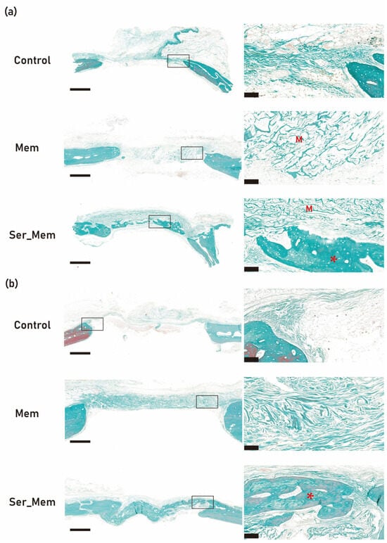
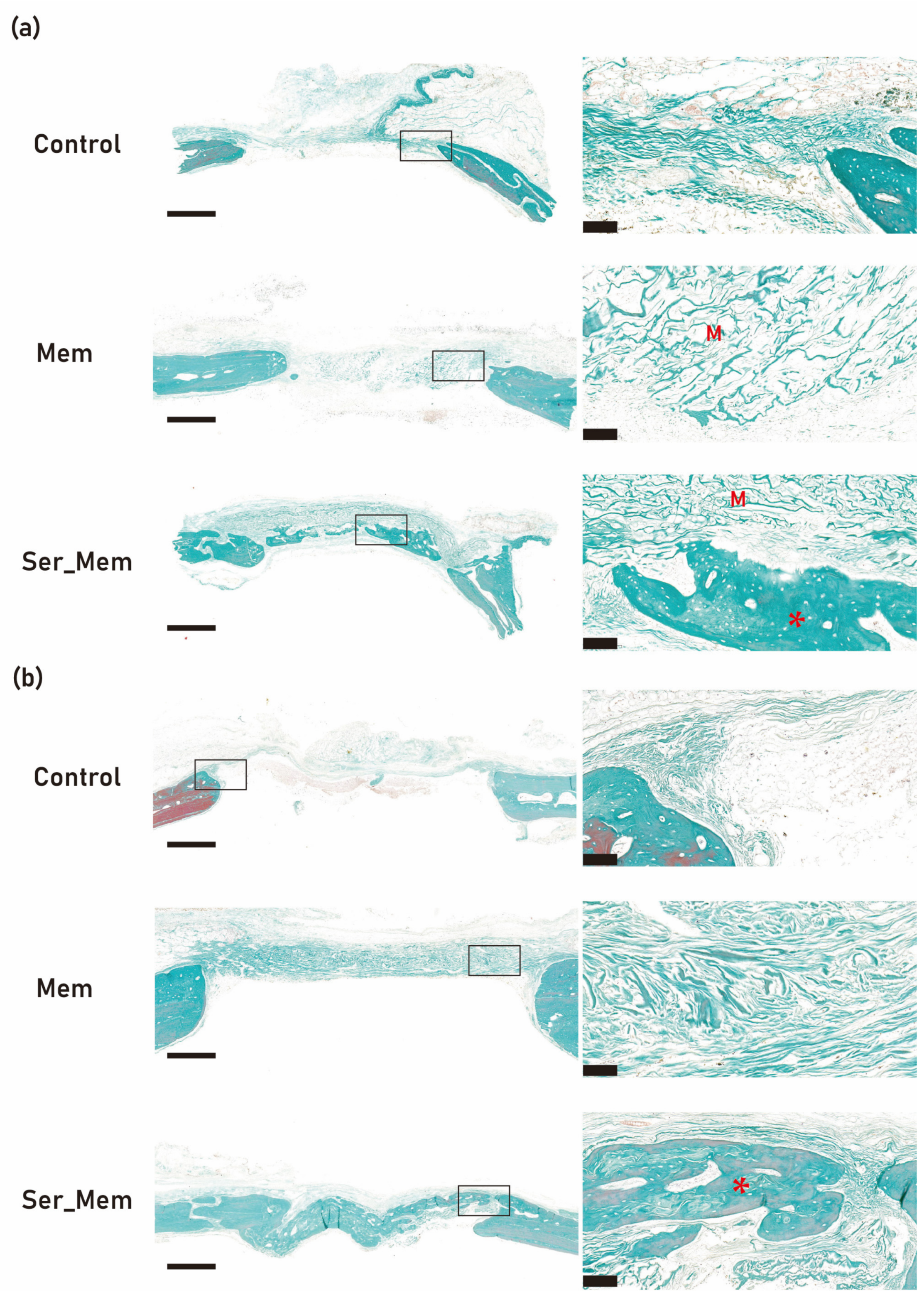
Histological evaluation by Goldner’s trichrome staining was consistent with the micro-CT findings (Figure 5). At 4 weeks, the control defects were bridged mainly by loose, disorganized connective tissue with only limited new bone at the defect margins (Figure 5a). The membrane-only group showed a more defined tissue layer spanning the defect, in which residual membrane material (M) was still evident, together with modest new bone formation adjacent to the native bone edges. In the Ser_Mem group, the defect was occupied by denser, more organized collagenous tissue, and newly formed bony islands (*) were observed within the defect in proximity to the degrading membrane (M), indicating active osteogenesis away from the native bone margins.
Figure 5.
Goldner’s trichrome staining of bone regeneration in rat calvarial defects. (a) Representative histologic images at 4 weeks after treatment in the control, collagen membrane-only (Mem), and L-serine-incorporated collagen membrane (Ser_Mem) groups. (b) Representative histologic images at 8 weeks after treatment in the control, Mem, and Ser_Mem groups. In each panel, low-magnification views show the overall defect area and high-magnification views correspond to the boxed regions. M indicates residual/degrading membrane material, and asterisks (*) indicate newly formed bony islands. Representative sections from the Ser_Mem group showed more extensive new bone formation and more advanced defect healing than those from the control and Mem groups. Scale bars: 1 mm (low magnification) and 100 μm (high magnification).
At 8 weeks, the overall tissue maturation increased in all groups, but intergroup differences were maintained (Figure 5b). The control group remained dominated by fibrous connective tissue with restricted bone formation, whereas the membrane-only group showed a moderate increase in newly formed bone within the defect. The Ser_Mem group exhibited the most extensive and mature bone formation, with newly formed bone extending toward the central region of the defect and greater continuity with the native bone. These qualitative histological observations were consistent with the micro-CT findings and showed more extensive and organized regenerated tissue in representative Ser_Mem defects.
3.4. L-Serine-Incorporated Membrane Increases BMP-2/4 and Runx2 Expression at 4 Weeks
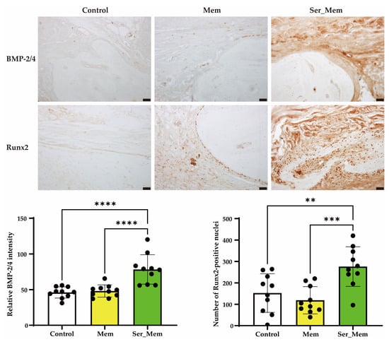
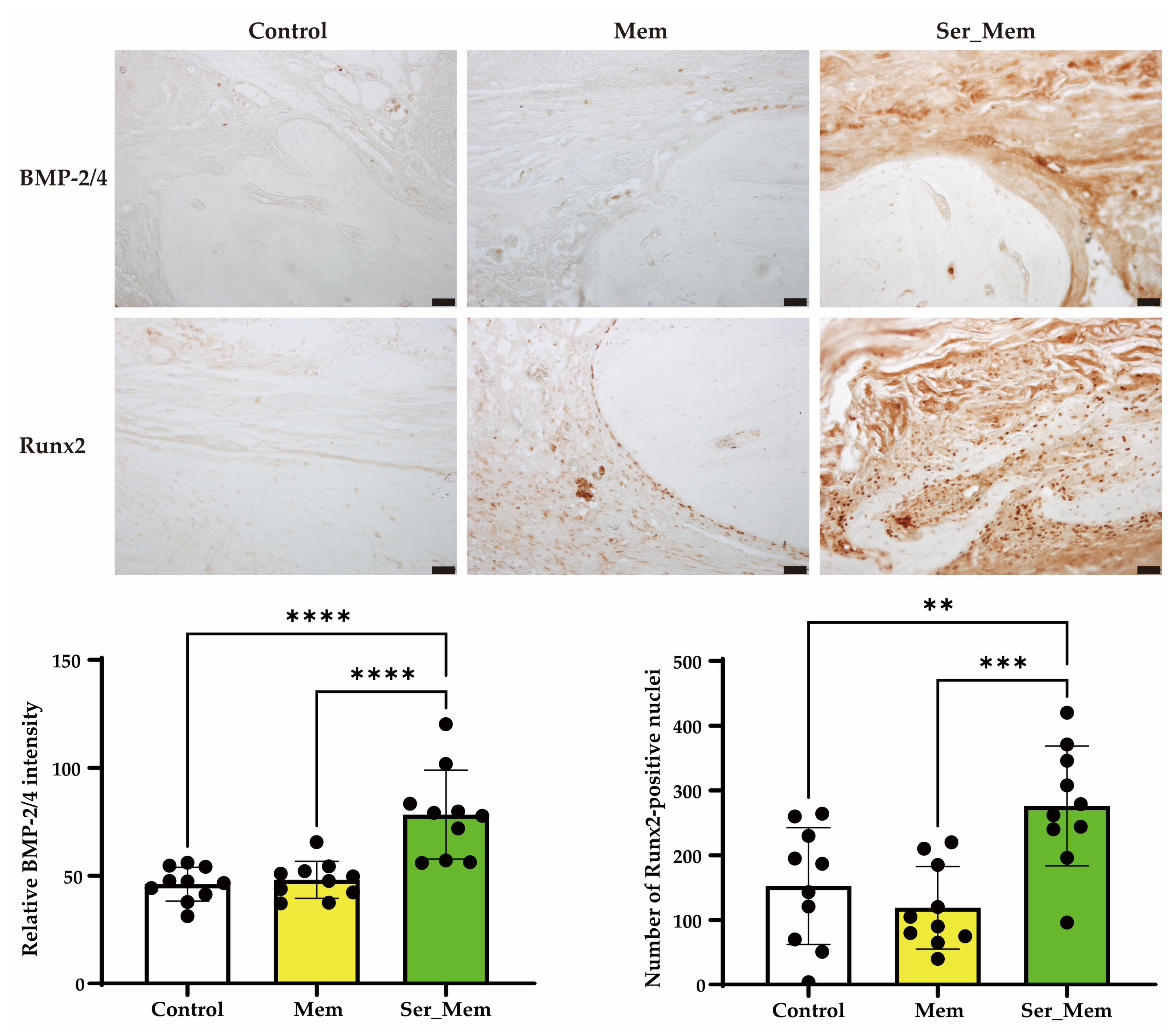
At 4 weeks, the Ser_Mem group showed stronger immunohistochemical expression of BMP-2/4 and Runx2 than the control and membrane-only groups (Figure 6). Representative images demonstrated increased BMP-2/4 immunoreactivity in the regenerated tissue of the Ser_Mem group, whereas only weak staining was observed in the control and Mem groups. Runx2 staining was also more prominent in the Ser_Mem group, with a greater number of positively stained nuclei. Quantitative analysis confirmed that the number of Runx2-positive nuclei and the relative intensity of BMP-2/4 staining were significantly higher in the Ser_Mem group than in both the control and Mem groups. These results indicate that L-serine incorporation into the collagen membrane promoted early osteogenic activity during bone regeneration.
Figure 6.
Immunohistochemical evaluation of BMP-2/4 and Runx2 expression at 4 weeks after treatment. Representative immunohistochemical images showing BMP-2/4 and Runx2 staining in the control, collagen membrane-only (Mem), and L-serine-incorporated collagen membrane (Ser_Mem) groups. The Ser_Mem group exhibited stronger BMP-2/4 immunoreactivity and more prominent Runx2-positive nuclear staining than the control and Mem groups. Quantitative analysis showed that the number of Runx2-positive nuclei and the relative intensity of BMP-2/4 staining were significantly higher in the Ser_Mem group than in both the control and Mem groups. Data are presented as mean ± standard deviation. n = 10 defects per group (** p < 0.01, *** p < 0.001, **** p < 0.0001). Scale bar: 50 μm.
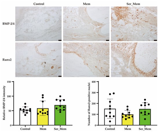
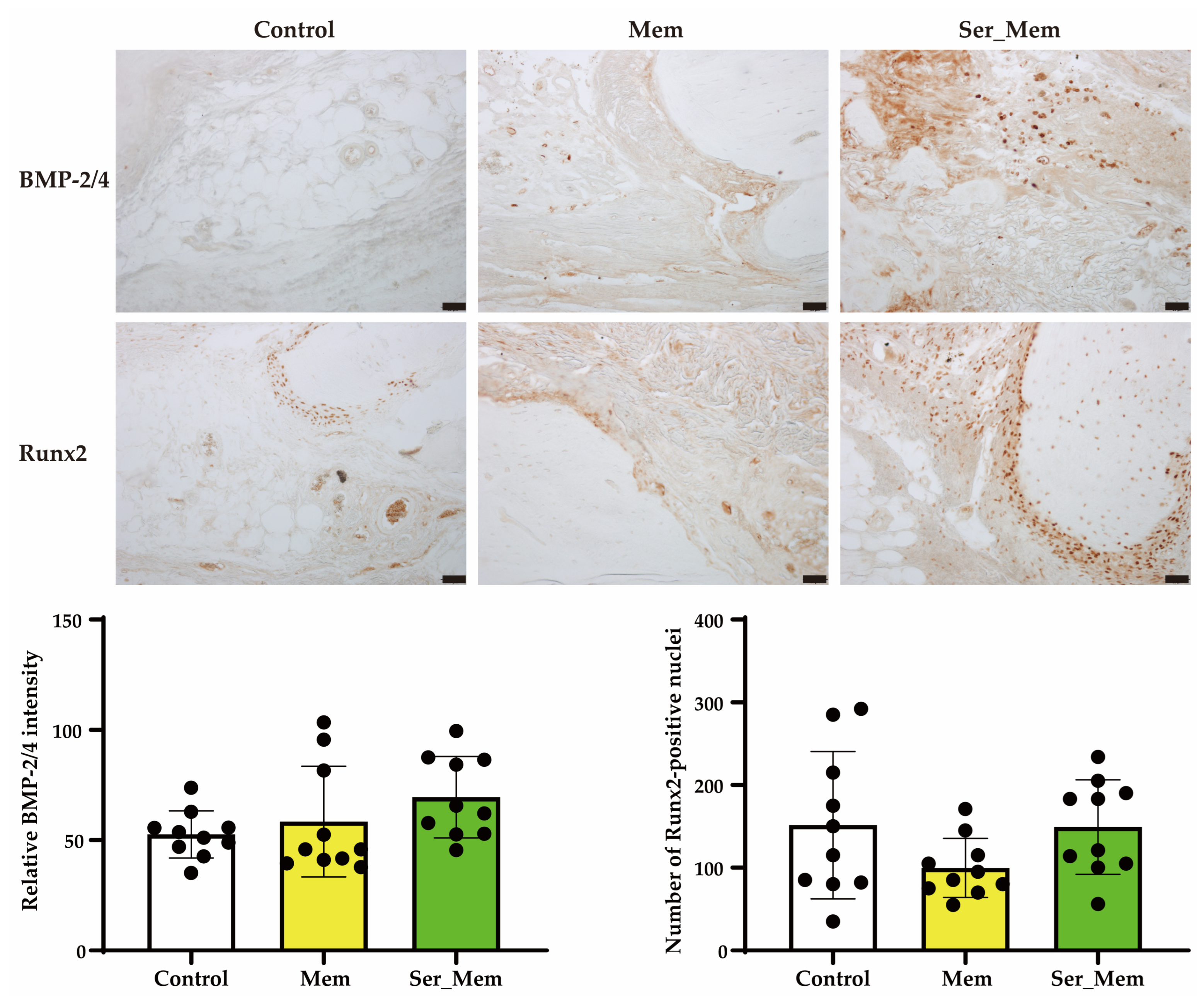
At 8 weeks, immunohistochemical staining for BMP-2/4 and Runx2 showed a trend toward higher expression in the Ser_Mem group than in the control and membrane-only groups (Figure 7). Representative images demonstrated more extensive BMP-2/4 immunoreactivity and denser Runx2-positive nuclear staining in the Ser_Mem group. However, quantitative analysis revealed no statistically significant differences among the groups in either the number of Runx2-positive nuclei or the relative intensity of BMP-2/4 staining. These findings suggest that although the L-serine-incorporated membrane continued to show a favorable trend in osteogenic marker expression at 8 weeks, the early enhancement observed at 4 weeks was attenuated over time.
Figure 7.
Immunohistochemical evaluation of BMP-2/4 and Runx2 expression at 8 weeks after treatment. Representative immunohistochemical images showing BMP-2/4 and Runx2 staining in the control, collagen membrane-only (Mem), and L-serine-incorporated collagen membrane (Ser_Mem) groups. The Ser_Mem group showed broader BMP-2/4 immunoreactivity and denser Runx2-positive nuclear staining than the control and Mem groups. However, quantitative analysis revealed no statistically significant differences among the groups in either the number of Runx2-positive nuclei or the relative intensity of BMP-2/4 staining. Data are presented as mean ± standard deviation. n = 10 defects per group at each time point. Scale bar: 50 μm.
4. Discussion
In the present study, incorporation of L-serine into a collagen membrane altered the physicochemical characteristics of the membrane and enhanced its bone regenerative performance in an ovariectomized rat calvarial defect model. The L-serine-incorporated membrane showed additional diffraction peaks on XRD and distinct spectral changes on FT-IR analysis (Figure 2), supporting the incorporation of L-serine into the collagen membrane. In the cellular experiment, L-serine transiently increased the expression of osteogenic markers, particularly Runx2 and osterix, at the early time point (Figure 3). In the animal experiment, the L-serine-incorporated membrane promoted greater bone formation than the unfilled control and collagen membrane alone, and this effect was accompanied by enhanced immunohistochemical expression of BMP-2/4 and Runx2 at 4 weeks (Figure 6). Collectively, these findings suggest that incorporation of L-serine can convert a conventional collagen membrane from a primarily passive barrier into a more biologically active scaffold that favors early osteogenic events during bone regeneration.
The physicochemical analyses support the incorporation of L-serine into the collagen membrane. Ser_Mem exhibited multiple sharp reflections superimposed on the broad diffraction background of the collagen membrane, and several reflections coincided in 2θ position with the reference powder-diffraction pattern for crystalline L-serine (ICDD PDF No. 00-027-1989). These findings are consistent with the presence of crystalline L-serine-containing domains in the loaded membrane. However, because the pure L-serine starting material was not independently analyzed by XRD in the present study, this phase assignment should be regarded as supportive rather than definitive. The FT-IR spectrum of Ser_Mem contained spectral features also observed in the experimentally measured pure L-serine starting material, together with changes in the O–H/N–H, amide I, amide II, and fingerprint regions relative to Mem. These observations further support the presence of L-serine and a change in the local molecular environment of the collagen matrix. The spectral changes are consistent with possible non-covalent interactions but do not establish a specific bonding mechanism. Previous characterization of blended collagen–L-serine composites showed that higher L-serine contents were associated with the appearance of crystalline reflections [15]. However, because L-serine was surface-loaded onto a pre-formed commercial membrane in the present study, comparison with the previously blended composites should be considered indicative rather than directly equivalent.
The relationship between the physicochemical findings and the release behavior should be interpreted cautiously. The near-complete cumulative release by 168 h indicates that most of the incorporated L-serine remained releasable under the present in vitro conditions. The initial burst release may reflect rapid dissolution of surface-associated or readily accessible L-serine, whereas the subsequent slower phase may arise from delayed release of L-serine located in less accessible regions of the membrane or weakly associated with the collagen matrix. However, XRD and FT-IR do not directly distinguish bound from unbound fractions and therefore do not establish the presence of two discrete L-serine populations. In addition, the release experiment was conducted under near-infinite sink conditions with complete replacement of the medium at each sampling point, thereby maintaining a high concentration gradient and potentially accelerating release relative to that at an implanted site. Overall, the XRD, FT-IR, and release data are consistent with predominantly physical and reversible incorporation of L-serine and a short-term delivery profile. This interpretation is also consistent with the transient in vitro response, which was most evident at 8 h, and with the stronger in vivo osteogenic response at 4 weeks than at 8 weeks.
The in vitro data further suggest that L-serine may act as an early regulator of osteogenic signaling (Figure 3). At 8 h, L-serine treatment increased Runx2 expression across all tested concentrations and showed a gradual increase in osterix expression [24], whereas BMP-2/4 showed only a mild upward tendency (Figure 3b). This pattern implies that the initial response to L-serine may be more closely related to early osteogenic transcriptional activation than to broad stimulation of all osteogenic markers. However, the response was not maintained uniformly at 24 h (Figure 3c). Runx2 was no longer increased, and the highest concentration of L-serine was associated with reduced Runx2 expression, while osterix was increased mainly at the lower concentrations. These findings indicate that the effects of L-serine on osteogenic marker expression are time- and concentration-dependent [17,25]. In this context, L-serine may act as an early metabolic cue that transiently supports osteoblastic differentiation rather than as a sustained osteoinductive factor [26,27]. The concentrations released from the membrane in vitro, approximately 0.05 to 0.6 mM during individual sampling intervals, fall within the range tested in the cell experiments, which supports the relevance of these observations to the membrane system. Although the early marker response was transient, Alizarin Red S staining showed enhanced matrix mineralization in the L-serine-treated cells at day 17, with minimal mineralization in all groups at day 7 (Figure 3a). This indicates that the brief early induction of Runx2 and osterix was nonetheless followed by a functional osteogenic outcome, namely increased mineralized matrix formation, over a longer culture period.
The in vivo results demonstrated that L-serine incorporation improved the regenerative efficacy of the collagen membrane beyond the effect of membrane placement alone. At 4 weeks, the Ser_Mem group showed significantly greater bone volume than both the control and membrane-only groups, while bone mineral density did not differ significantly among groups (Figure 4a). This suggests that, at the early stage, the principal effect of L-serine incorporation was to increase the amount of newly formed bone rather than to alter its mineral density. A similar pattern was reported previously in an ovariectomized rat calvarial defect model treated with an L-serine-incorporated gelatin sponge, in which L-serine incorporation enhanced bone regeneration under estrogen-deficient conditions [21]. In the present study, the regenerative advantage of L-serine incorporation appeared to persist over time, because at 8 weeks the Ser_Mem group continued to show enhanced bone regeneration, with significantly greater bone volume than the control group and significantly greater bone mineral density than the membrane-only group (Figure 4b). These findings indicate that the biologic advantage of the L-serine-incorporated membrane might not be limited to early tissue deposition, but might also contribute to later maturation of the regenerated tissue. Of note, the membrane-only group showed only limited improvement over the control under the present conditions, supporting the interpretation that the observed benefit was attributable to L-serine-mediated biofunctionalization rather than to the barrier effect of collagen membrane alone (Figure 5).
The relationship between the concentrations tested in vitro and the exposure achieved in vivo is less well defined. A 5-mm full-thickness calvarial defect corresponds to a volume of only approximately 12 µL, so that the local L-serine concentration immediately after implantation would be considerably higher than any concentration applied to the cell cultures, before falling rapidly through diffusion into the surrounding tissue and clearance by local perfusion. The local L-serine concentration immediately after implantation would be expected to be substantially higher than the concentrations tested in vitro and to decrease subsequently through diffusion, tissue uptake, and local perfusion. However, the magnitude and temporal profile of L-serine exposure in vivo were not directly measured in the present study. This interpretation is consistent with the transient nature of the response observed in vitro and, together with the reduction in Runx2 expression at 1 mM after 24 h, indicates that the loading dose used in the present study has not been optimized; determination of the optimal dose is a priority for further work. The loading amount of approximately 5 mg per membrane had been selected to ensure that L-serine remained present in excess throughout the early healing period, and was intended to establish proof of concept rather than to represent an optimized dose. At the loading used, the local L-serine concentration at the moment of implantation would be high and would produce a transient local hyperosmolarity at the defect site; because L-serine is a physiological, membrane-permeant amino acid that is actively transported and metabolized, this transient is expected to resolve rapidly as L-serine diffuses and is taken up by the surrounding tissue, consistent with the rapid release measured in vitro. Histologically, newly formed bony islands were observed within the defect in direct proximity to the residual, degrading L-serine-incorporated membrane at 4 weeks, indicating active osteogenesis at the membrane interface during the period of release, and the sections showed progressive bone formation and tissue maturation with no gross evidence of necrosis or destructive tissue change at either time point, consistent with the material being well tolerated over the observation period. Dedicated inflammatory scoring and the acute post-implantation window were not evaluated, and these, together with determination of the optimal loading, are included in our planned future work.
The immunohistochemical findings help explain the temporal pattern of bone regeneration observed in this study. At 4 weeks, the Ser_Mem group exhibited stronger BMP-2/4 expression and a greater number of Runx2-positive nuclei than the control and membrane-only groups, indicating enhanced early osteogenic activity within the defect (Figure 6). This interpretation is broadly consistent with previous reports that L-serine-related biomaterials enhance osteogenic responses under estrogen-deficient conditions [17]. Serine-rich sericin has been reported to increase BMP-2, osterix, and Runx2 expression [26]. Moreover, a previous study using an L-serine-incorporated gelatin sponge in ovariectomized rat calvarial defects demonstrated enhanced bone regeneration, together with suppression of cathepsin K and increased serine racemase activity, suggesting that the beneficial effect of L-serine may involve both promotion of bone formation and modulation of bone remodeling [21]. In the present study, these early marker changes likely reflect enhanced osteoblast recruitment or activation during the initial healing phase. At 8 weeks, BMP-2/4 and Runx2 expression still tended to be higher in the Ser_Mem group, but the differences were no longer statistically significant (Figure 7), reflecting progressive convergence of healing as the defects matured. Thus, the biologic contribution of L-serine appears to be most pronounced during the early phase of regeneration.
The ovariectomized model was selected because it represents a clinically relevant, estrogen-deficient condition in which any additional osteogenic stimulation provided by the membrane should be readily detectable. It should be noted that a recent study reported that short-term ovariectomized rats healed calvarial defects better than healthy animals [28]. In the present study, however, this pattern was not observed. A 5-mm rat calvarial defect is generally regarded as sub-critical and would be expected to undergo substantial spontaneous healing in a healthy animal by 8 weeks [29,30]; in our ovariectomized animals, the unfilled control defects nevertheless showed only limited new bone formation along the defect margin and did not heal completely at 8 weeks. This incomplete healing of a sub-critical defect is consistent with impaired, rather than enhanced, regeneration under estrogen deficiency and does not reproduce the accelerated healing reported for short-term ovariectomized rats [28]. The discrepancy may reflect differences in experimental conditions, including the post-ovariectomy interval, defect dimensions, and assessment time points. Importantly, the present study was designed entirely within the ovariectomized condition: all animals were ovariectomized, and the three treatments were compared under the same estrogen-deficient state, with two defects paired within each animal. Our conclusions therefore do not depend on a comparison with healthy or sham bone, and are based on treatment contrasts within the estrogen-deficient state.
From a translational perspective, these results suggest that incorporation of L-serine may represent a simple strategy for improving the biologic performance of collagen GBR membranes, particularly in compromised healing conditions such as estrogen deficiency. Conventional collagen membranes are widely used because of their favorable biocompatibility and handling characteristics, but their function has traditionally been regarded as primarily that of a passive barrier [28]. In contrast, the present results suggest that local delivery of L-serine can provide additional biologic stimulation that enhances early osteogenic events and ultimately improves bone regeneration (Figure 5).
Nevertheless, several limitations of this study should be acknowledged. First, only one animal model and one grafting condition were tested. Second, only a single loading amount of L-serine was evaluated in vivo. Third, the mechanistic pathway underlying the effect of L-serine was not fully investigated. Fourth, the cellular experiment was limited to selected osteogenic markers and short observation periods. Fifth, L-serine was introduced by aqueous loading without a covalent conjugation step, and its release was dominated by an initial burst phase; moreover, the loading dose used in vivo was not optimized. Sixth, treatments were assigned according to a fixed balanced cyclic sequence rather than by random allocation, and allocation was not concealed from the operator. Although this design balanced treatment representation, pairwise treatment combinations, and left/right positioning across animals, the absence of randomization and allocation concealment represents a potential source of bias. Seventh, only the osteogenic compartment was evaluated, and potential effects of L-serine on the soft-tissue side, such as fibroblast attachment or proliferation on the epithelial-facing surface, were not assessed; although no gross soft-tissue ingrowth into the defect was observed in the L-serine group, this was not quantified, and a dedicated evaluation of the soft-tissue response would be valuable. Eighth, the physical state and layer-wise distribution of L-serine within the bilayer membrane were not characterized; because L-serine was applied by aqueous drop-casting, it would be expected to distribute into both the dense and porous layers rather than selectively into one, and spatially resolved characterization is needed to confirm this. Ninth, the findings were obtained in a calvarial defect model, and bone of different embryonic origin and formation mode, such as endochondral long bone, may respond differently; the present conclusions should therefore be regarded as proof of concept for craniofacial guided bone regeneration rather than generalized to all bone defects. Future studies should compare different loading levels, directly characterize the physical state and distribution of L-serine within the membrane, and determine whether lower loading amounts can retain the osteogenic effect while reducing the initial burst release. Experimental XRD characterization of the pure starting material and additional analyses of L-serine–collagen interactions would also strengthen phase identification and clarify the molecular state of the incorporated L-serine. Further work should also assess membrane degradation behavior, examine covalent conjugation or crosslinking of L-serine to the collagen matrix, for which different crosslinking strategies have been characterized in collagen barrier membranes [29,30], as a means of extending release beyond the initial burst phase, include broader osteogenic and angiogenic markers, and confirm the regenerative efficacy of this approach in other defect models and larger preclinical systems [31,32,33]. Within these limitations, the present study supports the potential of an L-serine-incorporated collagen membrane as a bioactive GBR material for enhancing bone regeneration.
5. Conclusions
Incorporation of L-serine into a commercially available collagen membrane was achieved by simple aqueous loading and drying, resulting in physical association with the collagen matrix without covalent conjugation. In ovariectomized rats, the L-serine-incorporated membrane increased bone volume compared with the unfilled control at both 4 and 8 weeks and compared with the membrane-alone group at 4 weeks. BV/TV was greater than that of the membrane-alone group at 4 weeks, while BMD was greater than that of the membrane-alone group at 8 weeks. BMP-2/4 and Runx2 expression was also enhanced at 4 weeks. Because the L-serine release was essentially complete within one week and the in vitro and in vivo effects were most evident during the early healing phase, the membrane is best regarded as providing a short-term osteogenic stimulus rather than sustained delivery. Further studies are required to optimize the loading amount, prolong release, and confirm regenerative efficacy in additional defect models before clinical translation.
Supplementary Materials
The following supporting information can be downloaded at: https://www.mdpi.com/article/10.3390/biom16111689/s1, Figure S1: Masson’s trichrome staining of bone regeneration in rat calvarial defects. Figure S2. Uncropped Blot for Figure 3 with additional blots.
Author Contributions
Conceptualization, S.-G.K.; methodology, S.-G.K. and J.-Y.K.; formal analysis, M.-H.H. and Y.-Y.J.; investigation, Y.-E.J., J.-E.J., J.-H.O., S.K., J.C., M.-H.H. and Y.-Y.J.; data curation, J.-Y.K.; writing—original draft preparation, J.-Y.K.; writing—review and editing, S.-G.K. and U.G.; visualization, S.-G.K.; supervision, S.-G.K. and U.G.; funding acquisition, S.-G.K. and J.-H.O. All authors have read and agreed to the published version of the manuscript.
Funding
This research was supported by the National Research Foundation of Korea (NRF) grant funded by the Korea government (MSIT) (RS-2026-25477070); by the Cooperative Research Program for Agriculture Science and Technology Development (project no. RS-2026-25508046), Rural Development Administration, Republic of Korea; and by the ANCHOR program through the Gangwon ANCHOR Center, funded by the Ministry of Education (MOE) and Gangwon State, Republic of Korea (2026-ANCHOR-10-004).
Institutional Review Board Statement
The animal study protocol was approved by the Institutional Animal Care and Use Committee of Gangneung-Wonju National University, renamed Kangwon National University in March 2026 (protocol code GWNU-2025-20, approved on 3 November 2025). All procedures were performed in accordance with the institutional guidelines for the care and use of laboratory animals.
Informed Consent Statement
Not applicable.
Data Availability Statement
The datasets generated and/or analyzed during the current study are available from the corresponding author on reasonable request.
Conflicts of Interest
The authors declare no conflicts of interest. The funders had no role in the design of the study; in the collection, analyses, or interpretation of data; in the writing of the manuscript; or in the decision to publish the results.
Abbreviations
The following abbreviations are used in this manuscript:
| BMD | Bone mineral density |
| BMP | Bone morphogenetic protein |
| BV | Bone volume |
| BV/TV | Bone volume fraction |
| FT-IR | Fourier-transform infrared spectroscopy |
| GBR | Guided bone regeneration |
| micro-CT | Micro-computed tomography |
| PBS | Phosphate-buffered saline |
| ROI | Region of interest |
| Runx2 | Runt-related transcription factor 2 |
| XRD | X-ray diffraction |
References
- Duarte, N.D.; Frigério, P.B.; Chica, G.E.A.; Okamoto, R.; Buchaim, R.L.; Buchaim, D.V.; Messora, M.R.; Issa, J.P.M. Biomaterials for guided tissue regeneration and guided bone regeneration: A review. Dent. J. 2025, 13, 179. [Google Scholar] [CrossRef] [Scilit] [PubMed]
- Marian, D.; Toro, G.; D’Amico, G.; Trotta, M.C.; D’Amico, M.; Petre, A.; Lile, I.; Hermenean, A.; Fratila, A. Challenges and innovations in alveolar bone regeneration: A narrative review on materials, techniques, clinical outcomes, and future directions. Medicina 2025, 61, 20. [Google Scholar] [CrossRef] [Scilit] [PubMed]
- Liu, Y.; Zhang, H.; Zhang, L.; Han, J.; Yang, J. Design and applications of barrier membranes for guided bone regeneration. Bioact. Mater. 2026, 64, 655–685. [Google Scholar] [CrossRef] [Scilit] [PubMed]
- Liu, J.; Kerns, D.G. Mechanisms of guided bone regeneration: A review. Open Dent. J. 2014, 8, 56–65. [Google Scholar] [CrossRef] [Scilit] [PubMed]
- Elgali, I.; Omar, O.; Dahlin, C.; Thomsen, P. Guided bone regeneration: Materials and biological mechanisms revisited. Eur. J. Oral Sci. 2017, 125, 315–337. [Google Scholar] [CrossRef] [Scilit] [PubMed]
- Nyman, S.; Lindhe, J.; Karring, T.; Rylander, H. New attachment following surgical treatment of human periodontal disease. J. Clin. Periodontol. 1982, 9, 290–296. [Google Scholar] [CrossRef] [Scilit] [PubMed]
- Alauddin, M.S.; Abdul Hayei, N.A.; Sabarudin, M.A.; Mat Baharin, N.H. Barrier membrane in regenerative therapy: A narrative review. Membranes 2022, 12, 444. [Google Scholar] [CrossRef] [Scilit] [PubMed]
- Chen, S.; Wu, Z.; Huang, Z.; Liang, C.; Lee, S.J. Implantable dental barrier membranes as regenerative medicine in dentistry: A comprehensive review. Tissue Eng. Regen. Med. 2025, 22, 527–549. [Google Scholar] [CrossRef] [Scilit] [PubMed]
- Dimitriou, R.; Mataliotakis, G.I.; Calori, G.M.; Giannoudis, P.V. The role of barrier membranes for guided bone regeneration and restoration of large bone defects: Current experimental and clinical evidence. BMC Med. 2012, 10, 81. [Google Scholar] [CrossRef] [Scilit] [PubMed]
- Bujda, M.; Klíma, K. Enhancing guided bone regeneration with a novel carp collagen scaffold: Principles and applications. J. Funct. Biomater. 2024, 15, 150. [Google Scholar] [CrossRef] [Scilit] [PubMed]
- Alqahtani, A.M.; Moorehead, R.; Asencio, I.O. Guided tissue and bone regeneration membranes: A review of biomaterials and techniques for periodontal treatments. Polymers 2023, 15, 3355. [Google Scholar] [CrossRef] [Scilit] [PubMed]
- Han, Q.; Zhao, D.; Wang, X.; Shang, M.; Zhou, W.; Li, Q.; Song, H. Composite barrier membrane for bone regeneration: Advancing biomaterial strategies in defect repair. RSC Adv. 2025, 15, 1290–1299. [Google Scholar] [CrossRef] [Scilit] [PubMed]
- Omar, O.; Elgali, I.; Dahlin, C.; Thomsen, P. Barrier membranes: More than the barrier effect? J. Clin. Periodontol. 2019, 46, 103–123. [Google Scholar] [CrossRef] [Scilit] [PubMed]
- Ren, Y.; Fan, L.; Alkildani, S.; Liu, L.; Emmert, S.; Najman, S.; Rimashevskiy, D.; Schnettler, R.; Jung, O.; Xiong, X.; et al. Barrier membranes for guided bone regeneration (GBR): A focus on recent advances in collagen membranes. Int. J. Mol. Sci. 2022, 23, 14987. [Google Scholar] [CrossRef] [Scilit] [PubMed]
- Kim, S.Y.; Oh, J.H.; Hong, M.H.; Lee, J.H.; Jo, Y.Y.; Kim, S.G. L-serine-incorporated collagen scaffolds for modulating in vivo degradation behavior. J. Funct. Biomater. 2025, 16, 466. [Google Scholar] [CrossRef] [Scilit] [PubMed]
- Zhou, C.; Zhang, J.; Liu, Q.; Guo, Y.; Li, M.; Tao, J.; Peng, S.; Li, R.; Deng, X.; Zhang, G.; et al. Role of amino acid metabolism in osteoporosis: Effects on the bone microenvironment and treatment strategies. Mol. Med. Rep. 2025, 32, 212. [Google Scholar] [CrossRef] [Scilit] [PubMed]
- Kim, J.Y.; Che, X.; Kim, S.; Choi, J.; Lee, J.H.; Lee, J.H.; Kweon, H.; Lee, S.K.; Choi, J.Y.; Kim, S.G. Sericin’s potential in osteoporosis management: The roles of L-serine and D-serine in bone metabolism regulation. Nutrients 2025, 17, 574. [Google Scholar] [CrossRef] [Scilit] [PubMed]
- Kubota, T.; Hasuike, A.; Tsukune, N.; Ozawa, Y.; Yamamoto, T.; Min, S.; Naito, M.; Sato, S. Influence of estrogen deficiency on guided bone augmentation: Investigation of rat calvarial model and osteoblast-like MC3T3-E1 cells. Eur. J. Oral Sci. 2018, 126, 206–213. [Google Scholar] [CrossRef] [Scilit] [PubMed]
- Hsu, S.H.; Chen, L.R.; Chen, K.H. Primary osteoporosis induced by androgen and estrogen deficiency: The molecular and cellular perspective on pathophysiological mechanisms and treatments. Int. J. Mol. Sci. 2024, 25, 12139. [Google Scholar] [CrossRef] [Scilit] [PubMed]
- Kalu, D.N. The ovariectomized rat model of postmenopausal bone loss. Bone Miner. 1991, 15, 175–191. [Google Scholar] [CrossRef] [Scilit] [PubMed]
- Lee, Y.J.; Oh, J.H.; Park, S.; Choi, J.; Hong, M.H.; Kweon, H.; Chae, W.S.; Che, X.; Choi, J.Y.; Kim, S.G. The application of L-serine-incorporated gelatin sponge into the calvarial defect of the ovariectomized rats. Tissue Eng. Regen. Med. 2025, 22, 91–104. [Google Scholar] [CrossRef] [Scilit] [PubMed]
- Stegen, S.; Moermans, K.; Stockmans, I.; Thienpont, B.; Carmeliet, G. The serine synthesis pathway drives osteoclast differentiation through epigenetic regulation of NFATc1 expression. Nat. Metab. 2024, 6, 141–152. [Google Scholar] [CrossRef] [Scilit] [PubMed]
- Čandrlić, M.; Jerbić Radetić, A.T.; Omrčen, H.; Franović, B.; Batičić, L.; Gulić, T.; Čaljkušić-Mance, T.; Zoričić Cvek, S.; Malešić, L.; Cvijanović Peloza, O. Regeneration of critical calvarial bone defects using bovine xenograft, magnesium-enriched bovine xenograft and autologous dentin in rats: Micro-CT, gene expression and immunohistochemical analysis. J. Funct. Biomater. 2024, 15, 270. [Google Scholar] [CrossRef] [Scilit] [PubMed]
- Azarkina, K.; Gromova, E.; Malashicheva, A. “A friend among strangers” or the ambiguous roles of Runx2. Biomolecules 2024, 14, 1392. [Google Scholar] [CrossRef] [Scilit] [PubMed]
- Wan, W.Y.; Milner-White, E.J. A recurring two-hydrogen-bond motif incorporating a serine or threonine residue is found both at alpha-helical N termini and in other situations. J. Mol. Biol. 1999, 286, 1651–1662. [Google Scholar] [CrossRef] [Scilit] [PubMed]
- Jeong, H.E.; Choi, J.I.; Jeong, A.J.; Kim, M.S.; Jang, J.; Jung, H.J.; Lee, S.; Park, H.S.; Lee, J.H.; Lee, J.H.; et al. Comparative analysis of serine isomers and sericin sources in promoting osteoblastic differentiation. Int. J. Ind. Entomol. Biomater. 2025, 49, 55–65. [Google Scholar]
- Bertels, J.C.; He, G.; Long, F. Metabolic reprogramming in skeletal cell differentiation. Bone Res. 2024, 12, 57. [Google Scholar] [CrossRef] [Scilit] [PubMed]
- Balouch, A.P.; Marcet, E.; Bogle, J.; Wojda, S.J.; Donahue, S.W. Calvarial bone defects heal better in short-term ovariectomized rats compared to healthy rats. J. Musculoskelet. Neuronal Interact. 2025, 25, 392–406. [Google Scholar] [CrossRef] [Scilit] [PubMed]
- Spicer, P.P.; Kretlow, J.D.; Young, S.; Jansen, J.A.; Kasper, F.K.; Mikos, A.G. Evaluation of bone regeneration using the rat critical size calvarial defect. Nat. Protoc. 2012, 7, 1918–1929. [Google Scholar] [CrossRef] [Scilit] [PubMed]
- Parco, T.M.; Wagner, J.D.; Parbhoo, P.; Soviak, C.; Hu, Y.; Araiza, J.T.; O’Beirne, C.; Javaid, S.; Parachuru, P.; Lewis, M.; et al. Comprehensive evaluation of critical-size calvarial defect in athymic rat model. Front. Physiol. 2025, 16, 1662424. [Google Scholar] [CrossRef] [Scilit] [PubMed]
- Yang, Z.; Wu, C.; Shi, H.; Luo, X.; Sun, H.; Wang, Q.; Zhang, D. Advances in barrier membranes for guided bone regeneration techniques. Front. Bioeng. Biotechnol. 2022, 10, 921576. [Google Scholar] [CrossRef] [Scilit] [PubMed]
- Ren, Y.; Alkildani, S.; Burckhardt, K.; Köwitsch, A.; Radenkovic, M.; Stojanovic, S.; Najman, S.; Jung, O.; Liu, L.; Barbeck, M. The influence of different crosslinking agents on the physical properties, integration behavior and immune response of collagen-based barrier membranes. Front. Bioeng. Biotechnol. 2025, 12, 1506433. [Google Scholar] [CrossRef] [Scilit] [PubMed]
- Vladova, D.; Sbirkov, Y.; Stoyanova, E.; Chaprazov, T.; Dimitrov, K.K.; Hristov, H.; Kostov, D.; Veleva, P.; Stoeva, D.; Sarafian, V. Osteogenic potential of 3D bioprinted collagen scaffolds enriched with bone marrow stromal cells, BMP-2, and hydroxyapatite in a rabbit calvarial defect model. J. Funct. Biomater. 2026, 17, 68. [Google Scholar] [CrossRef] [Scilit] [PubMed]
Disclaimer/Publisher’s Note: The statements, opinions and data contained in all publications are solely those of the individual author(s) and contributor(s) and not of MDPI and/or the editor(s). MDPI and/or the editor(s) disclaim responsibility for any injury to people or property resulting from any ideas, methods, instructions or products referred to in the content. |
© 2026 by the authors. Licensee MDPI, Basel, Switzerland. This article is an open access article distributed under the terms and conditions of the Creative Commons Attribution (CC BY) license.