


Abstract
Methicillin-resistant Staphylococcus aureus (MRSA) causes intractable wound infections in which biofilms impede tissue repair. Curcumin (Cur) possesses antibacterial, anti-inflammatory and regenerative activities yet lacks specific targeting capacity. This work aimed to develop a biofilm-targeted curcumin conjugate for MRSA-infected wound therapy. Cur was conjugated to RNAIII-inhibiting peptide (RIP) through click chemistry. Cytotoxicity was evaluated in L929 fibroblasts; anti-biofilm performance was assayed against an MRSA strain, and a mouse full-thickness MRSA-infected wound model was used for in vivo assessment. RIP-Cur exhibited low cytotoxicity toward L929 cells. It inhibited MRSA biofilm formation by 71% and reduced the biomass of 48 h mature biofilms by 66.8%. In mouse wounds, RIP-Cur down-regulated pro-inflammatory and virulence factors and up-regulated healing-related markers, achieving improved wound-healing outcomes relative to the vancomycin group. This multifunctional RIP-Cur conjugate suppresses MRSA biofilm formation, modulates local inflammation and accelerates infected-wound tissue repair, supporting its potential as a candidate therapeutic agent for drug-resistant MRSA wound infections. Further work is required to fully dissect its complete mechanism of action and evaluate resistance-risk profiles.
1. Introduction
Targeting the bacterial quorum sensing (QS) system has gained extensive interest as an anti-biofilm strategy [1]. The QS system serves as a central regulatory network for bacterial communication [2]. RNAIII, transcribed from the agr locus, is a key regulator of virulence factor expression and biofilm formation in methicillin-resistant Staphylococcus aureus (MRSA) [3,4]. RNAIII-inhibiting peptide (RIP), a synthetic peptide interfering with the agr system, competitively binds to the target of RNAIII-activating protein(TRAP) [5,6], blocking RNAIII transcriptional activation and thereby inhibiting biofilm formation and attenuating virulence without directly killing MRSA [7]. However, clinical translation of RIP is limited by poor cell membrane permeability and insufficient in vivo stability [8]. Curcumin (Cur), a natural polyphenol derived from the rhizomes of Curcuma longa, has been investigated for its potential anti-inflammatory, antioxidant, and wound-repair properties [9]. Wound healing is a coordinated process involving inflammatory regulation, re-epithelialization, extracellular matrix remodeling, and angiogenesis. Experimental studies of plant-derived extracellular vesicles have reported improvements in wound repair-associated outcomes, including collagen deposition, epithelial regeneration, and vascularization in skin and oral mucosal wound models [10,11]. These findings provide broader context for investigating interventions that may influence host tissue repair, although the effects of such vesicle-based interventions cannot be directly extrapolated to RIP-Cur. In the present study, RIP was conjugated to Cur to combine RIP-mediated interference with MRSA quorum sensing and the potential host-directed activities of Cur. Whether RIP-Cur improves wound healing through bacterial virulence suppression, modulation of the host inflammatory response, or additional effects on tissue repair requires evaluation in the infected-wound model.
For the present study, we selected the clinical MRSA isolate Sa2, which was previously isolated from clinical wound specimens and deposited in the China General Microbiological Culture Collection Center. This strain exhibits strong biofilm-forming capacity, which recapitulates a key pathological feature of clinical MRSA wound infections and makes it suitable for evaluating anti-biofilm and anti-virulence therapeutic strategies.
The primary objective of this work was to construct the RIP-Cur peptide–curcumin conjugate via click-chemistry conjugation, characterize its in vitro cytocompatibility and anti-biofilm activity, and assess its therapeutic effects as a topical agent for MRSA-infected full-thickness skin wounds in mice. We aimed to distinguish the anti-virulence effects against MRSA and host-directed wound-repair-related responses induced by RIP-Cur.
Here, the conjugate was prepared via a click reaction. The cytocompatibility and in vitro anti-biofilm activity of RIP-Cur were systematically evaluated. A mouse model of MRSA-infected full-thickness skin wounds was established to investigate the therapeutic effects of RIP-Cur on wound healing [12]. The results are expected to provide an experimental basis for developing new therapeutic agents against MRSA-infected wounds.
2. Materials and Methods
2.1. MRSA Strain
The MRSA isolate Sa2 was isolated, identified and preserved by laboratory members. For long-term preservation, the strain was stored in brain–heart infusion (BHI) broth supplemented with 25% glycerol at −80 °C. For propagation, frozen stocks were streaked onto high-salt mannitol agar and cultured overnight at 37 °C. A single colony was picked and cultured in TSB at 37 °C, with shaking at 180 rpm. Bacterial suspensions were adjusted to target concentrations by measuring OD600. For in vitro assays, log-phase bacteria were diluted to 1 × 10⁶ CFU/mL in TSB. For in vivo wound infection, bacteria were washed and resuspended in BHI to 1 × 10⁸ CFU/mL. Actual bacterial concentrations were verified retrospectively by serial dilution and plate counting.
2.2. Reagents and Antibodies
RNAIII-inhibiting peptide (RIP) was synthesized by solid-phase peptide synthesis (purity > 95%) and kindly provided by the Lanzhou Institute of Chemical Physics, Chinese Academy of Sciences. Its amino acid sequence is Tyr-Ser-Pro-Trp-Thr-Asn-Phe, with a molecular weight of 913.97 Da [13].
Brain–heart infusion broth (BHI, B8130), MEM (12100-046), penicillin–streptomycin solution (P1400), tryptic soy broth (TSB, T8880-250), high-salt mannitol agar (HB4128), 0.1% crystal violet solution (G1064), and conventional streptavidin-peroxidase (SP) immunohistochemistry kit (SP0021) were from Solarbio (Beijing, China). Curcumin (C1386) was from Sigma-Aldrich (St. Louis, MO, USA). The RNA Easy Fast animal tissue total RNA extraction kit (DP451) was from Tiangen Biotech (Beijing, China). Rabbit anti-IL-10 monoclonal antibody (ab34843) was from Abcam (Cambridge, UK). Rat anti-GAPDH polyclonal antibody (10494-1-AP), rabbit anti-TGF-β monoclonal antibody (81746-4-Ig), rabbit anti-CD31 monoclonal antibody (66065-1-Ig), rabbit anti-α-SMA monoclonal antibody (14395-1-AP), and HRP-conjugated goat anti-rabbit and goat anti-mouse secondary antibodies were from Proteintech (Wuhan, China). 2× SYBR Green qPCR Master Mix (D7265) and the cDNA synthesis kit (D7270) were from Beyotime (Shanghai, China).
2.3. Synthesis, Purification, and Characterization of RIP-Cur Conjugate
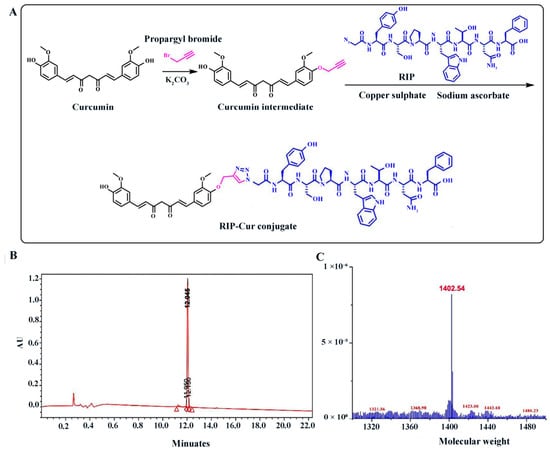
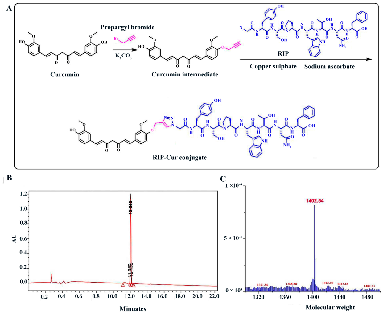
Curcumin (283 mg, ca. 65% purity, 0.5 mmol, 1.0 eq) in anhydrous DMF (4.0 mL) was treated with K2CO3 (69 mg, 0.5 mmol, 1.0 eq) and bromopropyne (43 μL, 80 wt% in toluene, 0.5 mmol, 1.0 eq), stirred at 0 °C for 0.5 h and then at room temperature for 42 h. The mixture was poured into water, extracted with dichloromethane (Sinopharm, Beijing, China), washed with brine, dried over Na2SO4, and concentrated in vacuo using a Concentrator Plus (Eppendorf, Hamburg, Germany). Purification by silica gel chromatography, eluting with 0.5–1% acetone in dichloromethane, gave an orange solid (92 mg, 45%). HRMS (ESI) gave m/z calcd. for [M+H]+ C24H23O6 407.1489 and found 407.1490.
The RIP peptide (azidoacetic acid-Tyr-Ser-Pro-Trp-Thr-Asn-Phe, 5 mg) and curcumin intermediate (2 mg) in water were treated with CuSO4·5H2O (5 mol%) and sodium ascorbate (10 mol%), stirred in the dark at room temperature for 28 h. The product was lyophilized (FreeZone 2.5 L freeze dryer, Labconco, Kansas City, MO, USA) and purified by HPLC (1260 Infinity II, Agilent, Santa Clara, CA, USA) on a Zorbax SB-C18 column (4.6 × 250 mm, 5 μm) using an acetonitrile/water gradient at 1.0 mL/min, with UV detection at 280 nm. The conjugate was confirmed by MALDI-TOF MS (Autoflex III, Bruker, Billerica, MA, USA) using α-cyano-4-hydroxycinnamic acid as matrix.
2.4. Cytotoxicity of RIP-Cur
L929 fibroblasts (HyCyte, Suzhou, China) were cultured in MEM supplemented with 10% fetal bovine serum (FBS) and 1% penicillin–streptomycin at 37 °C with 5% CO2 (Forma 3111, Thermo Fisher, Waltham, MA, USA). Cells in the log phase were subcultured every 2–3 days. For the cytotoxicity assay, cells were seeded in 96-well plates (5 × 10³/well) and incubated for 24 h to allow adherence, then treated with serial dilutions of RIP-Cur (1, 0.5, 0.25, 0.125, and 0.0625 mg/mL) or RIP (5, 2.5, 1.25, 0.625, and 0.3125 mg/mL) in 3% CMC-Na (100 μL/well, five replicates), together with blank and negative controls. After 24 or 48 h of treatment, 10 μL of CCK-8 (C6005, New Saimei, Suzhou, China) was added to each well and incubated for 2 h, and the absorbance at 450 nm (OD450) was recorded using a microplate reader (Synergy H1, BioTek, Winooski, VT, USA). The relative cell viability was calculated using the following formula, where OD_sample is the optical density of cells treated with RIP-Cur, OD_blank is the absorbance of blank medium, and OD_control is the absorbance of untreated cells. Cell viability (%) = [(OD_test − OD_blank)/(OD_negative − OD_blank)] × 100.
2.5. Cellular Uptake of RIP
L929 fibroblasts were seeded in 6-well plates at a density of 5 × 10⁵ cells/well. After 24 h of culture, cells were treated with 10 μg/mL FITC-labeled RIP for 3 h, washed with PBS, stained with DAPI for 10 min, and imaged using a CLSM (TCS SP8, Leica Microsystems, Wetzlar, Germany).
2.6. In Vitro Antibacterial Activity with RIP-Cur
The minimum inhibitory concentrations (MICs) of the test compounds against MRSA Sa2 were determined using a resazurin-based broth microdilution assay adapted from Sarker et al. [14]. Briefly, twofold serial dilutions of vancomycin, RIP (starting concentration, 5 mg/mL), curcumin (Cur; starting concentration, 5 mg/mL), and RIP-Cur (starting concentration, 1 mg/mL) were prepared in tryptic soy broth (TSB) in sterile 96-well plates. Each well received 100 μL of the bacterial suspension in the logarithmic growth phase, adjusted to a final concentration of 1 × 10⁶ CFU/mL. A growth control containing bacteria without the test compound and a sterility control containing TSB alone were included. Three replicate wells were used for each condition.
The plates were sealed and incubated at 37 °C for 16 h. Subsequently, 20 μL of 0.1% (w/v) resazurin sodium salt solution was added to each well, followed by a further 2 h of incubation. Resazurin served as a redox indicator: metabolically active bacteria reduced the blue indicator to pink, whereas wells without detectable bacterial growth remained blue. The MIC was defined as the lowest concentration of a test compound that completely inhibited bacterial growth, indicated by the absence of a color change from blue to pink, with all wells at higher concentrations also remaining blue. Each assay was performed in three independent experiments.
2.7. Antibiofilm Inhibition and Removal Test
Biofilm formation inhibition assay. MRSA Sa2 bacteria in the logarithmic growth phase were diluted 1:100 and inoculated into a 96-well plate containing 100 μL of tryptic soy broth (TSB) per well. RIP, curcumin (Cur), or RIP-Cur solution was then added to each well to achieve final concentrations of 5 mg/mL, 156.25 μg/mL, and 1 mg/mL, respectively. The concentrations of RIP and RIP-Cur were selected based on their maximum solubility under the experimental conditions, whereas the Cur concentration corresponded to its minimum inhibitory concentration (MIC) against MRSA Sa2. The plates were incubated statically at 37 °C for 48 h to evaluate the inhibitory effects of the compounds on biofilm formation.
Pre-formed biofilm biomass reduction assay. MRSA Sa2 biofilms were first allowed to mature for 48 h. The culture supernatant was then discarded, and the wells were gently washed twice with phosphate-buffered saline (PBS). Subsequently, 200 μL of the respective drug solution was added to each well, and the plates were incubated at 37 °C for a further 24 h to evaluate the effects of the compounds on pre-formed biofilm biomass. The negative control received 3% sodium carboxymethyl cellulose (CMC-Na), and the positive control received vancomycin at a final concentration of 3.125 μg/mL, corresponding to 1 × MIC against MRSA Sa2.
Biofilm quantification. After incubation, the culture medium was discarded, and the wells were washed three times with PBS. The attached biofilms were fixed with methanol for 15 min and air-dried. The biofilms were then stained with 0.1% crystal violet for 30 min, followed by rinsing with distilled water to remove excess stain. The bound crystal violet was dissolved in absolute ethanol for 5 min, and the absorbance of each well was measured at 570 nm using a microplate reader [15]. Six replicate wells were used per group, and the mean absorbance was used to calculate biofilm formation inhibition and pre-formed biofilm biomass reduction as follows:
Biofilm formation inhibition (%) = [1 − (ODtreated/ODcontrol)] × 100%
Pre-formed biofilm biomass reduction (%) = [1 − (ODtreated/ODcontrol)] × 100%
2.8. Animal Procedures and Surgeries
All animal procedures were approved by the Animal Experimental Ethics Committee of Gansu Agricultural University (Approval No. GSAU-Eth-VMC-2023-021) and conducted in accordance with ARRIVE 2.0 and NIH guidelines. Male BALB/c mice (6-8 weeks) were housed under specific pathogen-free conditions (22 ± 2 °C, 50% ± 10% humidity, 12 h light/dark cycle) with free access to food and water. After 7 days of acclimation, mice were anesthetized with 1% pentobarbital sodium (50 mg/kg, i.p., Sigma-Aldrich, St. Louis, MO, USA), shaved, and disinfected. A 6 mm full-thickness skin wound was created on the dorsum using a sterile punch. MRSA Sa2 (1 × 10⁸ CFU/mL in BHI) was applied to all wounds except uninfected controls (two doses within 10 min), delivering a total inoculum of 4 × 10⁶ CFU MRSA per wound.
Mice were randomly assigned to eight experimental groups using a random-number table method: uninfected, CMC-Na, vancomycin, Cur, RIP, RIP+Cur (RIP first, Cur 2 h later), Cur+RIP (Cur first, RIP 2 h later), and RIP-Cur. All therapeutic agents were prepared in a 3% CMC-Na vehicle. The working concentrations and delivered doses per wound were as follows: RIP (5 mg/mL, 200 μg/wound), curcumin (156.25 μg/mL, 6.25 μg/wound), RIP-Cur (1 mg/mL, 40 μg/wound), and vancomycin (3.125 μg/mL, 0.125 μg/wound). For the sequential-administration groups, each wound received 100 μg of RIP plus 3.125 μg of curcumin. Drug intervention started 12 h post-infection. For the sequential groups, 20 μL of each drug was applied at a 2 h interval; other groups received a single 40 μL dose. All groups maintained the same total topical application volume of 40 μL per wound to ensure comparable treatment conditions. Wounds were dressed daily for 14 days.
Tissue and blood samples were collected on days 3, 7, and 14. Serum was stored at −20 °C. Skin tissues were frozen at −80 °C (Antai freezer, Suzhou, China) or fixed in 4% paraformaldehyde (Thermo Fisher Scientific, Waltham, MA, USA) [16]. All animal experiments adopted a terminal-sacrifice sampling design; each mouse was sampled only once. Eight mice were initially assigned to each group, which were allocated to three sampling time points: day 3, day 7 and day 14 post-infection (n = 2 mice per group for day 3; n = 3 mice per group for day 7; n = 3 mice per group for day 14). The actual sample size for each assay was as follows:
- -
- Histopathological staining (H&E, Masson, IHC): n = 2 (day 3), n = 3 (day 7), n = 3 (day 14) per group
- -
- qPCR assay for bacterial virulence genes: n = 2 (day 3), n = 3 (day 7), n = 3 (day 14) per group
- -
- Western blot analysis: n = 3 per group at day 14
- -
- Serum ELISA assay: n = 3 per group at day 7
Wound-area quantification from wound photographs and histological assessments, including H&E, Masson’s trichrome and immunohistochemistry, were performed by an investigator blinded to group allocation information to reduce assessment bias. The sample size setting referred to previously published similar murine MRSA-infected full-thickness wound studies, taking into account potential animal loss, scheduled terminal-sacrifice sampling and biological variation in wound-healing phenotypes to achieve sufficient statistical power. Only pentobarbital sodium was used for surgical anesthesia. Post-operative systemic analgesia was not applied in this study to prevent analgesic agents from confounding local inflammatory and wound-healing-related experimental read-outs. Pre-defined humane endpoints included >20% loss of initial body weight, persistent lethargy, spontaneous bleeding or severe wound necrosis. Any animal satisfying these humane-endpoint criteria would be humanely euthanized immediately by CO2 asphyxiation.
2.9. Wound Healing Rate Measurement
Wound photographs were taken on days 1, 3, 5, 7, 10, and 14 post-infection with a ruler as a scale reference. Wound areas were measured by ImageJ (1.54 version), and the healing rate was calculated as Healing rate (%) = (1 − Area_t/Area_0) × 100.
2.10. Enzyme-Linked Immunosorbent Assay
Serum was collected on day 7. TNF-α, IL-1β, and MCP-1 were measured using ELISA kits (MM-0132M1, MM-0040M1, JL18442). Standards and samples (100 μL/well) were added to pre-coated plates and incubated at 37 °C for 1 h. After washing, a biotinylated antibody was added and incubated for 1 h, followed by HRP-conjugate (30 min) and TMB substrate (15 min, dark), with washing between each step. Absorbance was read at 450 nm, and concentrations (pg/mL) were calculated from standard curves. All samples were assayed in duplicate.
2.11. Real-Time Quantitative PCR Analysis
Skin tissues were homogenized and centrifuged twice (1000 rpm for 5 min, 12,000 rpm for 5 min) to collect bacterial pellets. Total RNA was extracted using the RNA Easy Fast Kit (DP451, Tiangen Biotech, Beijing, China), dissolved in 30 μL of RNase-free water, and stored at −80 °C. Reverse transcription used the BeyoRT™ II cDNA Synthesis Kit (D7270, Beyotime, Shanghai, China). RT-qPCR was performed on a LightCycler® 96 system (Shanghai Shen’an Medical Instrument, Shanghai, China) with 2× SYBR Green Master Mix (D7265, Beyotime) in 20 μL reactions containing primers for hla and psmα (Table 1, Sangon Biotech, Shanghai, China). The cycling protocol was 95 °C for 30 s, followed by 40 cycles of 95 °C for 5 s, 55 °C for 30 s, and 72 °C for 30 s. Melting curve analysis was performed after amplification to confirm specific amplification. Relative expression used the 2−ΔΔCt method with 16S rRNA as an internal control.
Table 1.
Nucleotide sequences of primers for 16S rRNA, psmα and hla genes.
2.12. Western Blotting Analysis
Total protein was extracted from skin tissues using RIPA lysis buffer containing phenylmethylsulfonyl fluoride (PMSF), quantified (ultra-micro spectrophotometer, Tianjin Test Instrument, Tianjin, China), mixed with loading buffer, and denatured at 98 °C for 15 min. Equal amounts (30 μg/lane) were separated by 12% SDS-PAGE and transferred to PVDF membranes. Membranes were blocked with 5% skim milk in TBST (1 h, room temperature), then incubated overnight at 4 °C with anti-TGF-β (1:5000), anti-IL-10 (1:2000), and anti-GAPDH (1:5000). After TBST washes, membranes were incubated with HRP-conjugated secondary antibodies (1:3000, 1 h, 37 °C). Bands were visualized by ECL and quantified with ImageJ, using GAPDH as an internal control.
2.13. Histological and Immunohistochemical Analysis
Skin tissues fixed in 4% paraformaldehyde were rinsed, dehydrated, embedded in paraffin, and sectioned at 4 μm. Sections were deparaffinized, rehydrated, and stained with H&E or Masson’s trichrome (G1346, Solarbio, Beijing, China). For immunohistochemistry, sections were incubated with anti-CD31 and anti-α-SMA antibodies, followed by HRP-conjugated secondary antibodies and DAB (3,3′-diaminobenzidine) development. All sections were dehydrated and mounted.
2.14. Statistical Analysis
The wound healing rate at day 14, as well as single-time-point data, including histopathological scores, qPCR, Western blot and ELISA results, were analyzed by one-way ANOVA followed by Tukey’s multiple comparisons test. Data are presented as mean ± SD, and statistical significance was set at * p < 0.05.
3. Results and Discussion
3.1. The Synthesis and Verification of RIP-Cur
The structure of RIP-Cur is shown in Figure 1A. HPLC gave a main peak at 12.045 min with 95.30% purity (Figure 1B). MS analysis gave [M+H]+ at m/z 1402.54, consistent with the theoretical value of 1402.55 (Figure 1C). These data confirm successful synthesis of RIP-Cur with high purity.
Figure 1.
Synthesis and characterization of the RIP-Cur conjugate. (A) Synthetic route of the RIP-Cur conjugate. (B) HPLC chromatogram of RIP-Cur. (C) Mass spectrometry spectrum of RIP-Cur. RIP-Cur, RNAIII inhibitory peptide–curcumin conjugate.
3.2. Biocompatibility and Cytotoxicity of RIP-Cur
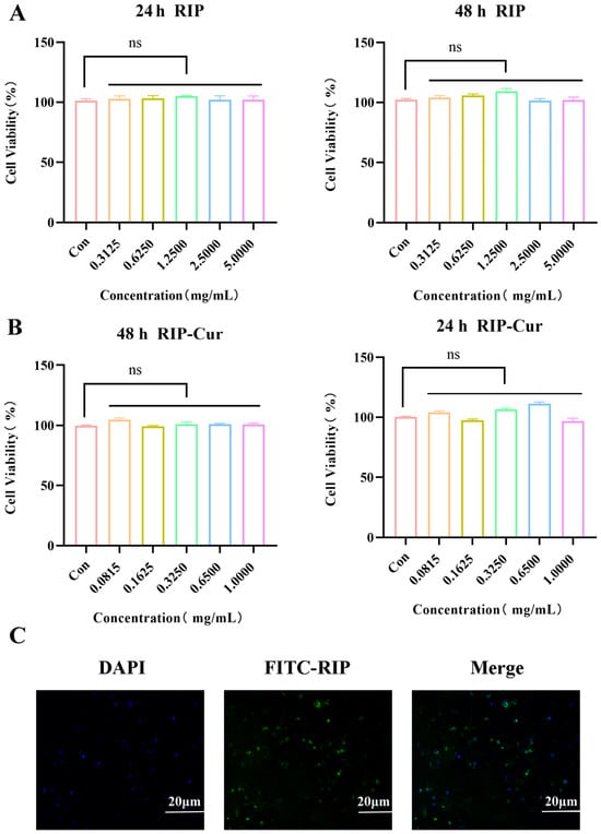
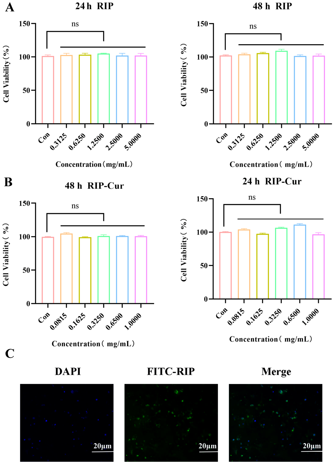
The CCK-8 assay showed that RIP-Cur was non-cytotoxic to L929 fibroblasts at 0.08125–1 mg/mL, with viability >90% (Figure 2B), indicating favorable in vitro biocompatibility of RIP-Cur. FITC-labeled free RIP peptide was internalized by L929 cells after 3 h, as shown by green cytoplasmic fluorescence under CLSM (Figure 2C). DAPI staining revealed intact nuclei under this incubation condition, demonstrating that free FITC-RIP peptide did not induce obvious cellular damage.
Figure 2.
Biocompatibility and cellular uptake of RIP-Cur. (A) Effect of RIP on the viability of L929 cells as determined by CCK-8 assay. (B) Effect of RIP-Cur on the viability of L929 cells as determined by CCK-8 assay. (C) CLSM images of L929 cells incubated with FITC-labeled RIP for 3 h, showing cytoplasmic distribution (green) and nuclear counterstaining with DAPI (blue). Scale bar, 20 μm. ns, not significant.
3.3. Antibacterial Activity of RIP-Cur In Vitro
The activity of RIP-Cur against MRSA Sa2 was evaluated by broth microdilution (Table 2). Individual replicate MIC measurements are provided in Table S2. The MIC values of both RIP and RIP-Cur against MRSA Sa2 were greater than 512 μg/mL under the conditions tested, indicating that neither compound exhibited measurable growth-inhibitory activity within the tested concentration range. This is consistent with RIP’s mechanism of interfering with quorum sensing rather than killing bacteria directly.
Table 2.
Minimum inhibitory concentrations of vancomycin, RIP, Cur, and RIP-Cur against MRSA Sa2.
3.4. Antibiofilm Properties of RIP-Cur
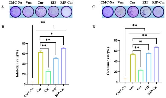
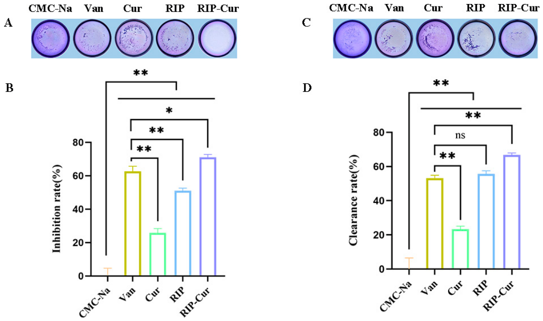
Anti-biofilm activity was evaluated by crystal violet staining. The absorbance values of biofilm inhibition and eradication assays are shown in (Table S1). RIP-Cur showed the strongest inhibition of nascent biofilm formation (71%), versus 50.8% for RIP and 43.5% for Cur (Figure 3). For pre-formed mature biofilms, RIP-Cur reduced biofilm biomass by 66.8%, which was lower than its inhibitory effect on nascent biofilms. Thus, RIP-Cur inhibited biofilm formation and reduced the biomass of mature biofilms, demonstrating greater potency against nascent biofilms.
Figure 3.
Detection of the biofilm biomass reduction of the Sa2 strain. (A,C) Crystal violet staining images of biofilms in 96-well plates. (B) Detection of the inhibition rate of biofilm formation of the Sa2 strain by different drugs. (D) Detection of the biofilm biomass reduction of the Sa2 strain by different drugs. CMC-Na, vehicle control; Van, vancomycin; Cur, curcumin; RIP, RNAIII-inhibiting peptide; RIP + Cur and Cur + RIP, physical mixtures; RIP-Cur, RNAIII inhibitory peptide–curcumin conjugate. * p < 0.05, ** p < 0.01, ns, not significant.
3.5. In Vivo Wound Healing Analysis
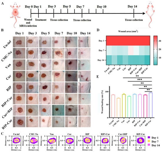
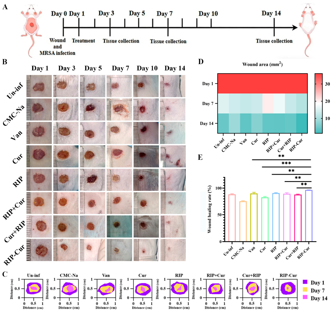
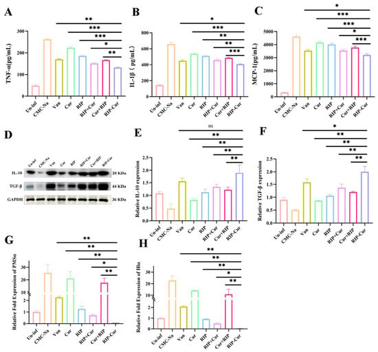
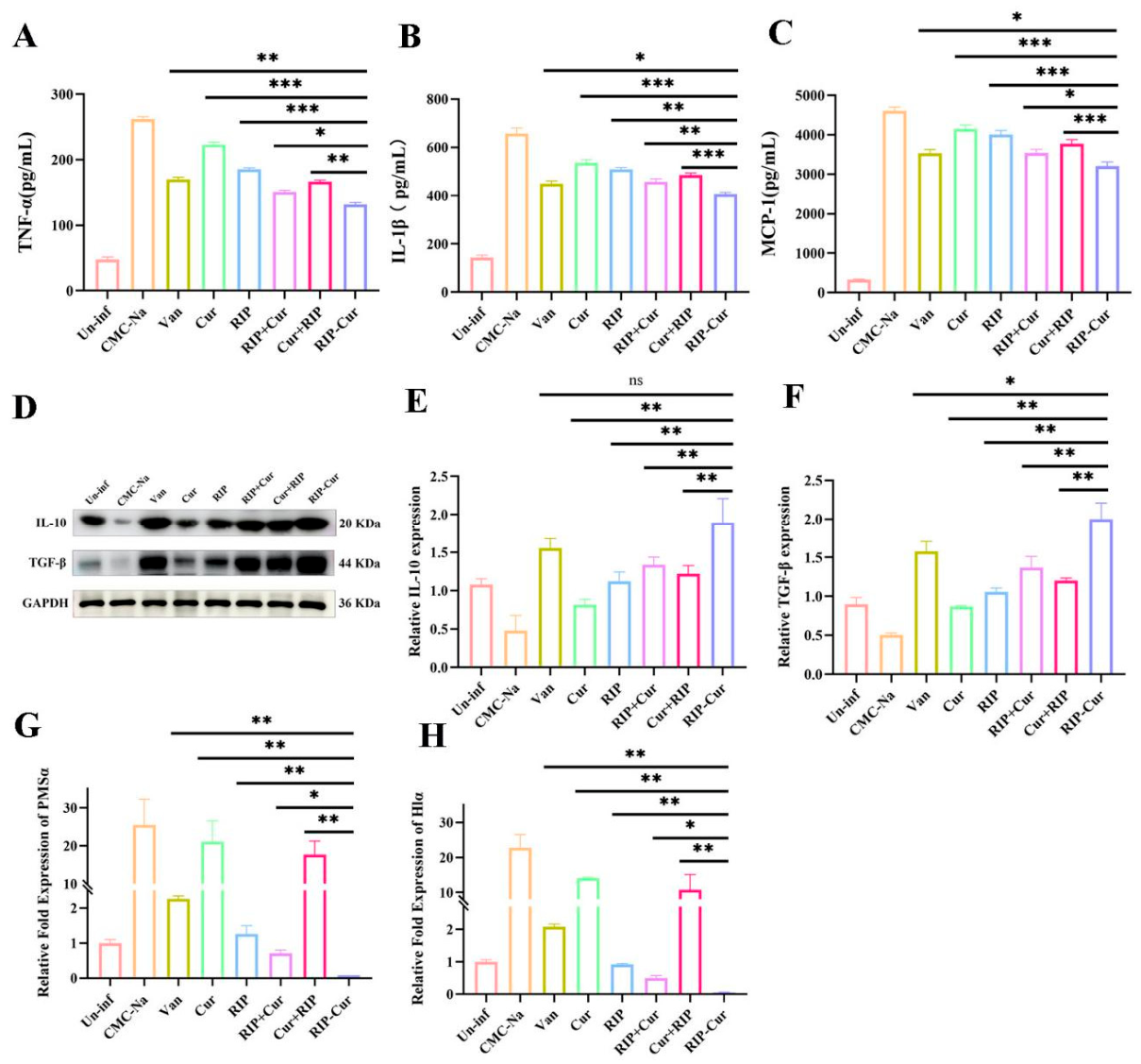
RIP-Cur was evaluated in a mouse model of MRSA-infected skin wounds. On day 14, the RIP-Cur group showed the highest healing rate (96.5%) among all treatments (Figure 4B–E). ELISA on day 7 showed that RIP-Cur reduced TNF-α, IL-1β, and MCP-1 versus CMC-Na and vancomycin. Western blotting on day 14 showed that RIP-Cur upregulated TGF-β (2.0-fold) and IL-10 (1.89-fold) (Figure 5D–F). qPCR on day 7 showed that RIP-Cur nearly abolished hla and psmα transcription, which remained high in the controls (Figure 5G,H).
Figure 4.
Evaluation of wound healing in mice with MRSA-infected wounds following different treatments. (A) Representative wound photographs; (B) Wound contour maps; (C) Heatmap visualization; (D) Quantitative analysis of wound healing rates on day 14. Wound healing rates were calculated as \((1 − \text{Area}_t/\text{Area}_0) × 100\%\). Data are presented as mean ± SD. The numbers of mice contributing wound measurements per group were n = 8 on days 1 and 3, n = 6 on days 5 and 7, and n = 3 on days 10 and 14. Wound healing rates on day 14 were analyzed using one-way ANOVA followed by Tukey’s multiple comparisons test. * p < 0.05, ** p < 0.01, *** p < 0.001.
Figure 5.
In vivo anti-inflammatory, anti-virulence, and pro-repair effects of RIP-Cur in MRSA-infected wound tissue. (A–C) ELISA quantification of TNF-α, IL-1β, and MCP-1 at day 7. (D–F) Western blotting and densitometric analysis of IL-10 and TGF-β at day 14. (G,H) qPCR analysis of psmα and hlα expression at day 7. Un-inf, uninfected control; CMC-Na, vehicle control; Van, vancomycin; Cur, curcumin; RIP, RNAIII-inhibiting peptide; RIP + Cur and Cur + RIP, physical mixtures; RIP-Cur, RNAIII inhibitory peptide–curcumin conjugate. Data are presented as mean ± SD. * p < 0.05, ** p < 0.01, *** p < 0.001, ns, not significant. The original western blot images can be found in Supplementary Materials.
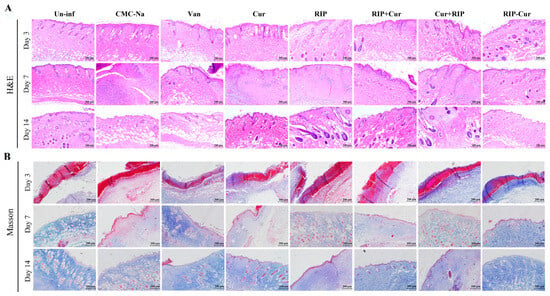
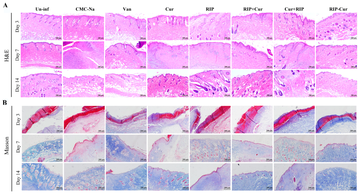
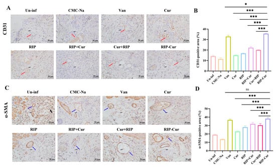
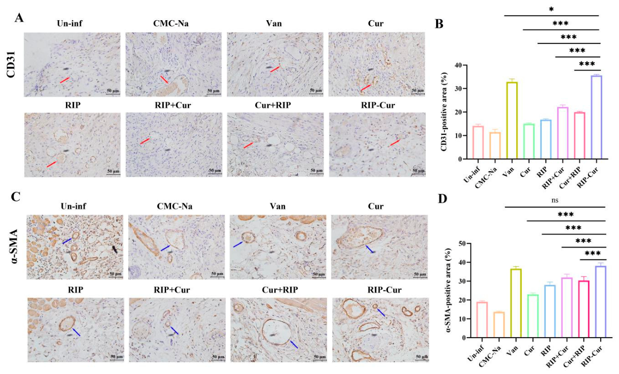
Histologically, RIP-Cur-treated wounds on day 14 formed a continuous, well-stratified epithelium with less inflammation and more neovascularization (H&E, Figure 6A). Masson’s trichrome showed abundant, organized collagen in the dermis (Figure 6B). Immunohistochemical analysis showed an increased CD31-positive area in RIP-Cur-treated wounds (*** p < 0.001, Figure 7). Changes in α-SMA staining were also observed. Together with the histological findings, these results suggest that RIP-Cur treatment was associated with improved wound-healing outcomes and changes in tissue remodeling.
Figure 6.
Histological assessment of wound repair and tissue remodeling in MRSA-infected wounds. (A) Observation of the healing effect of RIP-Cur on skin tissues around the wound at days 3, 7, and 14 using H&E staining. (B) Observation of the healing effect of RIP-Cur on skin tissues around the wound at days 3, 7, and 14 using Masson staining; magnification 40×, scale bar 200 μm. Un-inf, uninfected control; CMC-Na, vehicle control; Van, vancomycin; Cur, curcumin; RIP, RNAIII-inhibiting peptide; RIP + Cur and Cur + RIP, physical mixtures; RIP-Cur, RNAIII inhibitory peptide–curcumin conjugate.
Figure 7.
Immunohistochemical staining of angiogenesis-related markers in wound tissues. (A) Representative images of CD31 immunohistochemical staining. (B) Quantification of CD31-positive area (%). (C) Representative images of α-SMA immunohistochemical staining. (D) Quantification of α-SMA-positive area (%). Wound tissues were collected on day 14. Scale bars = 50 μm. Data are presented as mean ± SD. * p < 0.05, *** p < 0.001. ns, not significant.
The clinical treatment of MRSA-infected wounds faces substantial challenges due to the strong biofilm-forming capacity and multidrug resistance of MRSA [17]. Conventional antibiotics poorly penetrate biofilms and readily induce resistance [18]. The covalently conjugated RIP-Cur developed in this study achieves both anti-biofilm and tissue repair-promoting functions, offering a promising alternative strategy for clinical management.
As a quorum sensing inhibitor, RIP disrupts mature biofilms [16], but its anti-inflammatory and tissue repair properties remain unclear. Curcumin possesses antibacterial, anti-inflammatory, and antioxidant activities, yet its poor water solubility restricts clinical translation [19]. In this study, RIP was covalently conjugated with Cur to construct a bifunctional conjugate that preserves RIP’s agr-interfering activity and incorporates Cur’s anti-inflammatory potency. CLSM confirmed that free FITC-labeled RIP peptide could be internalized by fibroblasts; however, direct cellular uptake evidence for the complete RIP-Cur conjugate is absent in the present study [20]. CCK-8 confirmed that both RIP and RIP-Cur showed no cytotoxicity at effective concentrations, indicating favorable biocompatibility [21].
Notably, RIP-Cur showed no detectable direct bactericidal activity against MRSA Sa2 at the concentrations tested (MIC > 512 μg/mL), supporting an anti-virulence rather than a conventional bactericidal mode of action under the conditions of this study [22,23]. To further characterize its potential effects on bacterial virulence, we examined the expression of hla (α-hemolysin) and psmα (phenol-soluble modulin α), which are associated with host tissue damage and inflammatory responses [24]. RIP-Cur treatment was associated with reduced expression of hla and psmα in wound tissue. These findings are consistent with an effect on virulence-associated gene expression; however, because bacterial burden was not quantified in vivo and inhibition of the agr/RNAIII pathway in wound tissue was not independently confirmed, they do not establish bacterial eradication or directly demonstrate pathway inhibition. Importantly, anti-virulence activity and host tissue repair are related but distinct aspects of infected-wound treatment. The observed improvement in wound healing may reflect reduced bacterial virulence, changes in the host inflammatory environment, effects on tissue repair, or a combination of these processes.
On the host side, pro-inflammatory cytokines, including TNF-α, IL-1β, and MCP-1, are associated with local inflammatory responses, while IL-10 and TGF-β are involved in inflammation resolution and tissue remodeling. CD31 and α-SMA are commonly used as markers associated with vascularization and myofibroblast-related tissue remodeling, respectively [25]. In the present study, RIP-Cur treatment was associated with reduced levels of TNF-α, IL-1β, and MCP-1, together with increased levels of IL-10 and TGF-β, as well as an increased CD31-positive area and changes in α-SMA staining. Histological observations also suggested improvements in epithelial continuity and collagen organization. Collectively, these findings suggest that RIP-Cur treatment was associated with changes in inflammatory and tissue-repair-related indicators in MRSA-infected wounds. However, these observations do not establish a direct causal relationship between cytokine modulation, vascularization, collagen deposition, and wound closure. Moreover, because the present study did not independently distinguish the effects of RIP from those of Cur, the respective contributions of bacterial virulence modulation and potential host-directed effects remain to be clarified.
Broader experimental studies have also investigated plant-derived extracellular vesicles in wound repair. In rabbit skin and tongue wound models, these interventions were associated with changes in histological repair and vascularization-related outcomes. Although these studies used different interventions and wound models, they provide broader context for evaluating multiple aspects of tissue repair. Their findings should not, however, be interpreted as direct evidence for the efficacy or mechanism of RIP-Cur in MRSA-infected wounds.
Several previous studies have reported anti-biofilm and wound-repair effects of curcumin-based derivatives against MRSA infections. Many curcumin formulations exert antibacterial activity by directly disrupting bacterial membranes, yet high effective concentrations and poor biological availability limit their practical application. Peptide-modified curcumin conjugates have been developed to improve targeting; however, most reported constructs focus on bactericidal effects and rarely combine quorum-sensing interference with host-directed wound-repair functions. RIP, as an agr-quorum-sensing inhibitor, can attenuate MRSA virulence without killing bacteria, but its standalone application suffers from poor tissue retention in wound microenvironments. In our work, covalent conjugation of RIP and curcumin integrates quorum-sensing suppression, biofilm inhibition, and curcumin-mediated anti-inflammatory and tissue-repair capacities into one molecule. Compared with separate administration or physical mixtures of RIP and Cur, our covalent conjugate achieved stronger inhibition of nascent and mature MRSA biofilms and a more robust improvement in in vivo wound healing efficiency. This dual-function design represents a distinct strategy from conventional bactericidal antimicrobials and simple curcumin formulations, providing an alternative therapeutic candidate for MRSA-infected wound management. Compared with vancomycin, RIP-Cur showed more pronounced modulation of host repair-related indicators. Vancomycin exerts its therapeutic effect primarily via bactericidal activity; however, it may not fully suppress bacterial virulence factors and exhibits limited capacity to resolve excessive inflammation or promote tissue regeneration. As an anti-virulence-targeting conjugate, RIP-Cur does not possess direct bactericidal activity. It represses MRSA-derived virulence-gene expression and remodels wound cytokine balance without relying on bacterial lysis. The higher healing rate (96.5%) and stronger up-regulation of CD31 and α-SMA observed in our model highlight the merit of RIP-Cur, which targets both bacterial pathogenicity and host tissue-repair responses. Future studies should clarify intracellular metabolism and synergistic mechanisms [26]. In addition, we only assessed the cellular internalization of free RIP peptide in this work; direct labeling experiments targeting the intact RIP-Cur conjugate should be performed in future investigations. Additionally, this study focused on acute healing; long-term evaluation of appendage regeneration, scar formation, biosafety, and pharmacokinetics is needed [27]. The photoinstability of curcumin may compromise storage stability; thus, formulation optimization is warranted [28]. Furthermore, in vivo viable bacterial burden was not quantified in this study; future investigations should include wound-tissue CFU plating to directly assess in vivo antimicrobial activity.
It is important to acknowledge several limitations of the present study. First, statistical comparison of wound healing was restricted to the endpoint (day 14). Group-by-time interaction across all observation time points was not evaluated, which represents a limitation of the present analysis. Second, all experiments were performed using a single clinical MRSA isolate, Sa2. Different MRSA lineages exhibit divergent virulence profiles and biofilm-forming capacities, so findings obtained from one strain cannot be fully generalized to other MRSA isolates. Third, the mouse full-thickness wound model has inherent limitations for predicting human wound responses; differences in skin structure, immune background, and wound-healing kinetics between mice and humans restrict direct clinical extrapolation. Fourth, our evaluation focused on short-term acute wound healing within 14 days. Long-term observations, including biosafety profiling, scar-tissue formation, and skin appendage regeneration, were not assessed in this work, which should be addressed in follow-up investigations. Finally, in vivo bacterial burden was not quantified by CFU enumeration in wound tissue, which limits direct interpretation of bacterial load changes under RIP-Cur treatment. Collectively, these points highlight key gaps that warrant further exploration in future pre-clinical work.
4. Conclusions
In summary, our data show that RIP-Cur suppresses MRSA biofilm formation and reduces virulence-gene transcript levels in infected wounds, alongside modulation of local inflammatory responses and enhancement of angiogenesis and collagen deposition, which collectively support accelerated closure of MRSA-infected wounds [29,30]. Distinct from bactericidal antibiotics such as vancomycin, RIP-Cur functions via an anti-virulence-associated profile without direct bactericidal activity. These observations support the therapeutic potential of this conjugate; however, further investigations are needed to fully confirm its in vivo mode of action and assess whether this strategy limits resistance emergence.
Supplementary Materials
The following supporting information can be downloaded at https://www.mdpi.com/article/10.3390/biom16111690/s1, Table S1: OD values of MRSA biofilm inhibition and eradication assays under different treatments. Table S2: MIC values of vancomycin, RIP, curcumin, and RIP–Cur against MRSA. The original western blot images can be found in Supplementary Materials.
Author Contributions
L.X. and Y.W.: Conceptualization. X.S.: Methodology. J.Y.: Visualization. D.Z.: Software. X.P.: Data curation. J.Z.: Validation. R.L. (Rui Luo): Formal analysis. R.L. (Rong Li): Investigation. Y.H.: Conceptualization, supervision, funding acquisition, project administration, resources, writing—review & editing. All authors have read and agreed to the published version of the manuscript.
Funding
This research was supported by the Youth Talent Program of “Fuxi” (Gaufx-03Y02), the Scientific Research Start-up Funds for Openly-Recruited Doctors of Gansu Agricultural University (2017RCZX-13), the National Natural Science Foundation of China (31902339), the Foundation for Youth Doctor of Gansu Province (2021QB-026), the Special Funds for Discipline Construction of Gansu Agricultural University (GAU-XKJS-2018-067), and the Program of the Local Science and Technology Development (Gansu province) Guided by the Central Government (03322026).
Institutional Review Board Statement
All animal experimental procedures were approved by the Animal Experimental Ethics Committee of Gansu Agricultural University (Approval No. GSAU-Eth-VMC-2023-021, Approval date: 9 March 2023) and conducted in accordance with the ARRIVE guidelines 2.0 and the National Institutes of Health Guide for the Care and Use of Laboratory Animals. All efforts were made to minimize animal suffering and reduce the number of animals used.
Informed Consent Statement
Not applicable.
Data Availability Statement
The data presented in this study are available in the Supplementary Materials.
Acknowledgments
I am grateful to He Yuxuan for her guidance and to Li Hongyan’s group at the Institute of Modern Physics for their assistance in drug synthesis. During the preparation of this work, the authors used DeepSeek (DeepSeek-V3; available at https://deepseek.com) to assist with language polishing and grammatical refinement of the manuscript.
Conflicts of Interest
The authors declare no conflicts of interest.
References
- Khan, M.A.; Wang, S.; Zhu, H. Targeting quorum sensing: Natural product-based inhibition and quenching for antimicrobial strategies. FUTURE Microbiol. 2025, 20, 1049–1068. [Google Scholar] [CrossRef] [Scilit]
- Liu, S.; Feng, X.; Zhang, H.; Li, P.; Yang, B.; Gu, Q. Decoding bacterial communication: Intracellular signal transduction, quorum sensing, and cross-kingdom interactions. Microbiol. Res. 2025, 292, 127995. [Google Scholar] [CrossRef] [Scilit]
- Vuong, C.; Saenz, H.L.; Götz, F.; Otto, M. Impact of the agr quorum-sensing system on adherence to polystyrene in Staphylococcus aureus. J. Infect. Dis. 2000, 182, 1688–1693. [Google Scholar] [CrossRef] [Scilit]
- Novick, R.P.; Ross, H.F.; Projan, S.J.; Kornblum, J.; Kreiswirth, B.; Moghazeh, S. Synthesis of staphylococcal virulence factors is controlled by a regulatory RNA molecule. EMBO J. 1993, 12, 3967–3975. [Google Scholar] [CrossRef] [Scilit]
- Balaban, N.; Goldkorn, T.; Nhan, R.T.; Dang, L.B.; Scott, S.; Ridgley, R.M.; Rasooly, A.; Wright, S.C.; Larrick, J.W.; Rasooly, R.; et al. Autoinducer of virulence as a target for vaccine and therapy against Staphylococcus aureus. Science 1998, 280, 438–440. [Google Scholar] [CrossRef] [Scilit]
- Balaban, N.; Stoodley, P.; Fux, C.A.; Wilson, S.; Costerton, J.W.; Dell’Acqua, G. Prevention of staphylococcal biofilm-associated infections by the quorum sensing inhibitor RIP. Clin. Orthop. Relat. Res. 2005, 437, 48–54. [Google Scholar] [CrossRef] [Scilit]
- Balaban, N.; Cirioni, O.; Giacometti, A.; Ghiselli, R.; Braunstein, J.B.; Silvestri, C.; Mocchegiani, F.; Saba, V.; Scalise, G. Treatment of Staphylococcus aureus biofilm infection by the quorum-sensing inhibitor RIP. Antimicrob. Agents Chemother. 2007, 51, 2226–2229. [Google Scholar] [CrossRef] [Scilit]
- Rasmussen, T.B.; Givskov, M. Quorum sensing inhibitors: A bargain of effects. Microbiology 2006, 152, 895–904. [Google Scholar] [CrossRef] [Scilit]
- Mohanty, C.; Sahoo, S.K. Curcumin and its topical formulations for wound healing applications. Drug Discov. Today 2017, 22, 1582–1592. [Google Scholar] [CrossRef] [Scilit]
- Adel, N.; Kolenda, J.; Thulesen, J.; Stankovic, N.; Thulesen, I.V.; Saucillo, J.R.; Jo, Y.K. Plant Exosome Injection with or without Low Level Laser Therapy Promotes Skin Wound Healing: An Experimental Study. Plast. Reconstr. Surgery. Glob. Open 2026, 14, e7891. [Google Scholar] [CrossRef] [Scilit]
- Adel, N.; Thulesen, J.; Kolenda, J.; Stankovic, N.; Llano, F.; Jo, Y.K.; Thulesen, I.V.; Ragaei, A.; Sabry, M.A. Plant Exosome Injections in Tongue Wounds: A Comparative Histological and Immunohistochemical Experimental Study. Plast. Reconstr. Surgery. Glob. Open 2026, 14, e7616. [Google Scholar] [CrossRef] [Scilit]
- Simonetti, O.; Cirioni, O.; Ghiselli, R.; Goteri, G.; Scalise, A.; Orlando, F.; Silvestri, C.; Riva, A.; Saba, V.; Madanahally, K.D.; et al. RNAIII-inhibiting peptide enhances healing of wounds infected with methicillin-resistant Staphylococcus aureus. Antimicrob. Agents Chemother. 2008, 52, 2205–2211. [Google Scholar] [CrossRef] [Scilit]
- Palit, S.; Sanapalli, B.K.R.; Manda, S.B.; Munnangi, V.; Deshpande, A.; Sanapalli, V. Quorum-Sensing Inhibitors as Potential Antibacterial Agents Against Staphylococcus aureus: A Comprehensive Review. Chem. Biodivers. 2026, 23, e02013. [Google Scholar] [CrossRef] [Scilit]
- Sarker, S.D.; Nahar, L.; Kumarasamy, Y. Microtitre plate-based antibacterial assay incorporating resazurin as an indicator of cell growth, and its application in the in vitro antibacterial screening of phytochemicals. Methods 2007, 42, 321–324. [Google Scholar] [CrossRef] [Scilit]
- Stepanovic, S.; Vukovic, D.; Dakic, I.; Savic, B.; Svabic-Vlahovic, M. A modified microtiter-plate test for quantification of staphylococcal biofilm formation. J. Microbiol. Methods 2000, 40, 175–179. [Google Scholar] [CrossRef] [Scilit]
- Garong, C.J.C.; Burigsay, N.P.F.; Gapultos, R.S.; Mandawe, J.L.B.; Pedrosa, R.M.V.; Dayrit, G.B. Optimization of Resazurin Microplate Assay (REMA) in Evaluating Anti-MRSA and Anti-MSSA Activities. Acta Medica Philipp. 2025, 59, 55–61. [Google Scholar] [CrossRef] [Scilit]
- Otto, M. Staphylococcal Biofilms. Microbiol. Spectr. 2018, 6, 4. [Google Scholar] [CrossRef] [Scilit]
- Bin, L.; McGiffin, D.; Nguyen, T.; Wang, L.; Sun, Y.; Ye, L.; Han, M.; Sheng, C.; Lee, T.H.; Aguilar, M.R.E.I.; et al. Accurate quantitation of antibiotic penetration through staphylococcal biofilms. Biofilm 2025, 10, 100316. [Google Scholar] [CrossRef] [Scilit]
- Hegde, M.; Girisa, S.; BharathwajChetty, B.; Vishwa, R.; Kunnumakkara, A.B. Curcumin Formulations for Better Bioavailability: What We Learned from Clinical Trials Thus Far? ACS Omega 2023, 8, 10713–10746. [Google Scholar] [CrossRef] [Scilit]
- Zhao, K.; Vasquez, J.K.; Blackwell, H.E. Potent pan-group quorum sensing inhibitors in Staphylococcus aureus revealed by N-terminal tailoring of peptidomimetics. Chem. Commun. 2023, 59, 587–590. [Google Scholar] [CrossRef] [Scilit]
- Alenazi, N.A.; Aleanizy, F.S.; Alqahtani, F.Y.; Aldossari, A.A.; Alanazi, M.M.; Alfaraj, R. Anti-quorum sensing activity of poly-amidoamine dendrimer generation 5 dendrimer loaded kinase inhibitor peptide against methicillin-resistant Staphylococcus aureus. Saudi Pharm. J. SPJ Off. Publ. Saudi Pharm. Soc. 2024, 32, 101932. [Google Scholar] [CrossRef] [Scilit]
- Touati, A.; Ibrahim, N.A.; Idres, T. Disarming Staphylococcus aureus: Review of Strategies Combating This Resilient Pathogen by Targeting Its Virulence. Pathogens 2025, 14, 386. [Google Scholar] [CrossRef] [Scilit]
- Alum, E.U.; Gulumbe, B.H.; Izah, S.C.; Uti, D.E.; Aja, P.M.; Igwenyi, I.O.; Offor, C.E. Natural product-based inhibitors of quorum sensing: A novel approach to combat antibiotic resistance. Biochem. Biophys. Rep. 2025, 43, 102111. [Google Scholar] [CrossRef] [Scilit]
- Gless, B.H.; Sereika-Bejder, B.S.; Jensen, I.; Bojer, M.S.; Tsiko, K.; Schmied, S.H.; Vitolo, L.; Toledo-Silva, B.; De Vliegher, S.; Ingmer, H.; et al. Mapping of quorum sensing interaction network of commensal and pathogenic staphylococci. mBio 2025, 16, e0096725. [Google Scholar] [CrossRef] [Scilit]
- Lu, J.; Wang, Z.; Cai, D.; Lin, X.; Huang, X.; Yuan, Z.; Zhang, Y.; Lei, H.; Wang, P. Carrier-Free Binary Self-Assembled Nanomedicines Originated from Traditional Herb Medicine with Multifunction to Accelerate MRSA-Infected Wound Healing by Antibacterial, Anti-Inflammation and Promoting Angiogenesis. Int. J. Nanomed. 2023, 18, 4885–4906. [Google Scholar] [CrossRef] [Scilit]
- Wang, D.; Yin, F.; Li, Z.; Zhang, Y.; Shi, C. Current progress and remaining challenges of peptide-drug conjugates (PDCs): Next generation of antibody-drug conjugates (ADCs)? J. Nanobiotechnology 2025, 23, 305. [Google Scholar] [CrossRef] [Scilit]
- Eltaher, S.S.; Khattab, Z.; Walid, G.; Loay, O.; Emad, R.; Hakim, C.; Elhadidy, M. Hijacking the signal: A critical evaluation of quorum sensing inhibitors as a next-generation approach against Staphylococcus aureus. CELL Commun. Signal. 2026, 24, 135. [Google Scholar] [CrossRef] [Scilit]
- Zhu, Z.; Liu, J.; Hu, Y.; Dong, Y.; Zhang, L.; Chen, X.; Zou, L. Tailoring curcumin ternary complex nanocrystals via microfluidic mediated assembly: Stability, solubility, bioaccessibility and formation mechanism. Food Chem. 2025, 480, 143920. [Google Scholar] [CrossRef] [Scilit]
- Khan, M.A.; Zhu, L.; Zhu, H. Peptide-based approaches to quorum-sensing disruption: Emerging trends and applications in antimicrobial therapy. BIOORGANIC Med. Chem. 2026, 133, 118496. [Google Scholar] [CrossRef] [Scilit]
- Deng, X.; Ratnayake, J.; Ali, A. Curcumin-Loaded Drug Delivery Systems for Acute and Chronic Wound Management: A Review. Bioengineering 2025, 12, 860. [Google Scholar] [CrossRef] [Scilit]

Disclaimer/Publisher’s Note: The statements, opinions and data contained in all publications are solely those of the individual author(s) and contributor(s) and not of MDPI and/or the editor(s). MDPI and/or the editor(s) disclaim responsibility for any injury to people or property resulting from any ideas, methods, instructions or products referred to in the content. |
© 2026 by the authors. Licensee MDPI, Basel, Switzerland. This article is an open access article distributed under the terms and conditions of the Creative Commons Attribution (CC BY) license.