


Abstract
Type 2 diabetes mellitus (T2DM) and its vascular complications remain a leading cause of morbidity and mortality worldwide. Endothelial dysfunction is a common pathological denominator underlying diabetic microvascular and macrovascular disease. Major facilitator superfamily domain-containing protein 2A (MFSD2A) is a lysophosphatidylcholine transporter essential for blood–brain barrier integrity that also functions in the retina, liver, placenta, and kidney, participating in lipid homeostasis, inflammation resolution, and barrier maintenance. Emerging evidence links MFSD2A to diabetic complications. Its expression is reduced in the retina and kidney yet upregulated in the fatty liver, while its role in the vascular endothelium under diabetic conditions remains unexplored. In parallel, ferroptosis, an iron-dependent cell death driven by lipid peroxidation, is implicated in diabetic vascular disease. This hypothesis-oriented narrative review examines MFSD2A structure, transport mechanisms, and tissue-specific functions, synthesises current evidence linking MFSD2A to diabetic complications, critically evaluates whether MFSD2A-mediated lipid transport modulates endothelial ferroptosis sensitivity, and identifies key knowledge gaps and therapeutic opportunities. This review identifies that MFSD2A is dysregulated in a tissue-specific pattern in diabetes and finds that the interplay between MFSD2A-mediated DHA transport and endothelial ferroptosis sensitivity represents a critical knowledge gap that is tractable through the tissue-specific, conditionally testable framework developed in this review.
1. Introduction
Type 2 diabetes mellitus (T2DM) has reached pandemic proportions, with an estimated global prevalence exceeding 500 million adults and projections rising steadily [1]. The clinical burden of T2DM is driven not merely by hyperglycaemia, but by its chronic vascular complications. These are traditionally dichotomised into macrovascular diseases (coronary artery disease, cerebrovascular disease, and peripheral arterial disease, all of which share atherosclerosis as their pathological foundation) and microvascular diseases including diabetic retinopathy (DR), diabetic kidney disease (DKD), and diabetic neuropathy [2,3]. Among these, cardiovascular disease remains the leading cause of death in patients with T2DM [4].
A unifying early event across the spectrum of diabetic vascular complications is endothelial dysfunction [5]. Under chronic hyperglycaemia and dyslipidaemia, the endothelium loses its capacity to maintain vascular homeostasis, whereby nitric oxide (NO) bioavailability declines, reactive oxygen species (ROS) production escalates, pro-inflammatory cytokines and adhesion molecules are upregulated, and the endothelial barrier becomes increasingly permeable [5,6]. These changes set the stage for monocyte recruitment, lipid deposition, and foam cell formation in the arterial intima (the hallmarks of atherogenesis), as well as for capillary basement membrane thickening and pericyte loss in the microvasculature.
The role of lipid metabolism in diabetic endothelial dysfunction has received considerable attention, yet one dimension remains particularly underexplored, namely the transport and compartmentalisation of specific lipid species across the endothelial membrane. Major facilitator superfamily domain-containing protein 2A (MFSD2A) occupies a key position at this interface. First identified as a transcript highly induced in brown adipose tissue during fasting and cold exposure [7], MFSD2A was subsequently characterised as the primary transporter responsible for brain uptake of the conditionally essential omega-3 fatty acid docosahexaenoic acid (DHA), in the form of lysophosphatidylcholine (LPC) [8]. In the decade since this discovery, MFSD2A has been established as a critical determinant of BBB integrity, through its ability to suppress caveolae-mediated transcytosis via lipid-dependent membrane remodelling [9,10].
Despite this rich body of work in neurobiology, the role of MFSD2A in systemic metabolic disease, and specifically in T2DM and its complications, has only recently begun to attract attention. Landmark studies have demonstrated that MFSD2A is functionally important beyond the BBB, including in hepatic lipid homeostasis [11,12], retinal vascular integrity, renal tubular repair, placental DHA transfer [13], and intestinal inflammation resolution. Critically, MFSD2A expression is altered in several tissues under diabetic conditions. It is downregulated in the retina in DR, reduced in the kidneys of patients with DKD, and suppressed in the placenta in gestational diabetes mellitus (GDM) [13]. In striking contrast, MFSD2A expression is upregulated in the liver of patients with non-alcoholic fatty liver disease (NAFLD, also referred to as metabolic dysfunction-associated steatotic liver disease, MASLD), indicating that MFSD2A regulation in diabetes is tissue-specific and context-dependent.
In parallel, a growing body of literature has established ferroptosis, which is an iron-dependent and lipid peroxidation-driven form of regulated cell death, as a key pathogenic mechanism in diabetic vascular disease [14,15]. Endothelial cells exposed to high glucose and pro-inflammatory cytokines undergo ferroptosis through the p53-xCT-glutathione (GSH) axis [16], and heme oxygenase-1 (HMOX1)-mediated iron overload has been directly implicated in ferroptotic endothelial injury in diabetic atherosclerosis [17]. Ferroptosis is fundamentally a lipid-driven process in which the peroxidation of polyunsaturated fatty acid-containing phospholipids (PUFA-PLs), synthesised via acyl-CoA synthetase long-chain family member 4 (ACSL4) and lysophosphatidylcholine acyltransferase 3 (LPCAT3), generates the lethal lipid signals that, when unchecked by glutathione peroxidase 4 (GPX4), propagate membrane destruction [18,19]. This lipid-centric mechanism places MFSD2A, which directly governs the flux of highly peroxidisable DHA-containing phospholipids into cellular membranes, at a potential regulatory nexus for ferroptosis sensitivity.
A recent review by Li et al. [20] comprehensively surveyed MFSD2A biology with a focus on the BBB and ischaemia–reperfusion injury. The present review differed in several respects. It explicitly emphasised T2DM and diabetic complications rather than the BBB and ischaemia–reperfusion injury. It included a tissue-by-tissue analysis of MFSD2A dysregulation across seven organ systems. It proposed and critically evaluated the MFSD2A-ferroptosis hypothesis. Li et al. [20] noted, in the context of ischaemia–reperfusion injury, that MFSD2A downregulation may exacerbate neuronal ferroptosis, but did not develop this into a testable framework for the diabetic endothelium. It provided a comprehensive survey of MFSD2A transcriptional and post-translational regulatory networks. It also discussed tissue-specific therapeutic strategies for MFSD2A modulation in diabetic complications.
It is important to state upfront that this is a hypothesis-oriented narrative review, not an evidence-synthesising review. The direct evidence linking MFSD2A to diabetic complications is limited. Only one study has functionally interrogated MFSD2A in DR; the DKD evidence is confined to human snRNA-seq correlation data and a non-diabetic IRI mouse model; and the role of MFSD2A in diabetic atherosclerosis is entirely unexplored experimentally. The MFSD2A-ferroptosis connection discussed herein is a hypothesis that has not yet been tested. The value of this review lies not in summarising a mature body of work, but in systematically mapping what is known, identifying critical knowledge gaps, and providing a conceptual framework for hypothesis-driven investigation of this nascent field. This review aims to establish where MFSD2A sits in the biology of T2DM and its complications and to specify what would be required to determine whether its lipid transport function influences endothelial ferroptosis. Throughout, the strength of the evidence is indicated by the label that opens each relevant paragraph: Established (demonstrated directly and independently replicated), Supported indirectly (correlative, single-dataset, or derived from a related but non-equivalent setting), Hypothesised (not tested experimentally) and No direct evidence (no experimental data available).
2. Mfsd2a: Gene, Protein Structure, and Physiological Functions
2.1. Gene, Tissue Distribution, and Subcellular Localisation
The human MFSD2A gene (also known as NLS1, MCPH15, SLC59A1) is located on chromosome 1p34.2 and encodes a 530-amino-acid integral membrane protein belonging to the major facilitator superfamily (MFS), one of the largest families of secondary transporters across all domains of life [7,8,21]. Unlike most MFS transporters, which handle water-soluble substrates, MFSD2A is atypical in that it transports lysophospholipids, making it one of only a handful of proposed lipid-translocating MFS members, alongside MFSD2B, Spns2, and Spns1 [22,23].
The tissue distribution of MFSD2A is broader than initially appreciated. It is most highly expressed in the endothelium of brain microvessels, where it is localised specifically to the BBB and is essentially absent from pericytes, astrocytes, and neurons [8,9]. Outside the brain, MFSD2A is abundantly expressed in the retinal pigment epithelium (RPE) and the endothelium of the blood–retinal barrier; in the syncytiotrophoblast layer of the placenta [13,24]; in hepatocytes [11,12]; in brown adipose tissue, particularly under fasting or cold-stimulated conditions [7]; in the S3 segment of the renal proximal tubule; and in intestinal microvascular endothelial cells. According to the Human Protein Atlas and GTEx data, MFSD2A transcripts are also detectable in testis and adipose tissue [25]. Subcellularly, MFSD2A localises predominantly to the plasma membrane, with enrichment at the basolateral (abluminal) side of brain endothelial cells [9].
2.2. Protein Architecture and Transport Mechanism
MFSD2A comprises 12 transmembrane (TM) α-helices organised into two pseudosymmetric six-helix bundles forming the N-terminal domain (TM1–6) and the C-terminal domain (TM7–12). This topology is a hallmark of the MFS fold [21,26]. Structures of mouse, chicken, zebrafish, and human MFSD2A determined by single-particle cryo-electron microscopy (cryo-EM) have collectively elucidated the molecular architecture underlying its unique lipid transport function [22,23,26,27,28] (Figure 1A).
Figure 1.
MFSD2A structure and transport mechanism. (A) Transmembrane topology with key structural domains. (B) The “trap-and-flip” transport cycle.
Key structural features include (i) a distinctive extracellular domain formed by an elongated loop between TM5 and TM6 (EL3) and a second loop between TM11 and TM12 (EL6), which are linked by a conserved C216–C464 disulfide bridge; this arrangement of two extracellular loops joined by a disulfide is not found in other MFS transporters; (ii) a conserved sodium-binding site involving residues Glu312 and Asp92 that coordinates the Na+ ion essential for coupled transport; (iii) a large central cavity that accommodates the zwitterionic LPC headgroup via salt-bridge interactions with Lys436, while a hydrophobic cleft lines the path for the fatty acyl tail; (iv) lateral openings between TM5/TM8 and TM2/TM11 that allow substrate entry from the outer leaflet of the plasma membrane; and (v) three sets of ionic locks that stabilise the outward-open conformation [22,23,26,27,28].
Mechanistically, MFSD2A operates via a “trap-and-flip” adaptation of the classical alternating-access model [22,23,27]. The transport cycle proceeds through three conformational states: outward-facing (LPC enters from the outer leaflet via the TM5/TM8 lateral opening, accompanied by Na+ binding to Glu312), occluded (the substrate is engulfed within the central cavity and stabilised by an aromatic network comprising Phe329, Phe60, and Phe61), and inward-facing (the intracellular gate Phe399/Trp403/Met182 opens, releasing LPC into the inner leaflet). A Na+-relay mechanism, involving Na+ transfer from Glu312 to Asp92, drives the occluded-to-inward transition [27]. Direct biochemical evidence from reconstituted liposome assays has confirmed that MFSD2A functions as a lysolipid flippase, translocating lysophosphatidylserine (and by extension LPC) from the outer to the inner leaflet in a sodium-dependent manner [22] (Figure 1B).
Established. Substrate specificity studies have demonstrated that MFSD2A preferentially transported LPC species carrying long-chain (≥C14) fatty acyl chains, including LPC-oleate (18:1), LPC-palmitate (16:0), LPC-linoleate (18:2), and especially LPC-DHA (22:6), with substantially reduced activity toward medium-chain species, likely owing to geometric constraints requiring sufficient hydrophobic character for membrane integration [8,26,27]. The zwitterionic headgroup is essential: MFSD2A does not transport unesterified fatty acids or lysolipids lacking a negative charge. However, the phosphate moiety can be partially substituted, as evidenced by the transport of structurally related acylcarnitines [26].
Because DHA is the principal substrate of MFSD2A and the lipid species at the centre of the hypothesis developed below, its provenance and handling warrant a brief separate treatment. DHA (22:6 n-3) is not a dietary essential fatty acid in the strict sense. It can be synthesised endogenously from the parent n-3 fatty acid α-linolenic acid (ALA, 18:3 n-3) by sequential Δ6-desaturation (FADS2), elongation, Δ5-desaturation (FADS1), two further elongations to 24:5n-3, a second Δ6-desaturation, and a final peroxisomal β-oxidation that shortens the 24-carbon intermediate to DHA [29]. This pathway is inefficient in humans, and the fraction of ingested ALA converted to DHA is generally estimated to be below a few per cent, which is why DHA is best regarded as conditionally essential and why preformed dietary DHA is the principal determinant of tissue DHA status [29]. The main dietary sources are oily fish and other seafood, with algal oils providing a vegetarian source [30]. DHA circulates in plasma esterified within phospholipids and in lysophosphatidylcholine. MFSD2A transports the lysophosphatidylcholine form, and not unesterified fatty acid [8]. In the brain and retina, PUFAs are mainly supplied by the blood rather than synthesised locally [29]. Uptake of LPC-DHA by MFSD2A across the blood–brain and blood–retinal barriers is therefore the dominant route of acquisition [8,31].
DHA handling is altered in type 2 diabetes. Erythrocyte membrane fatty acid composition and the activity of the desaturases that act in this pathway have been related to the incidence of type 2 diabetes in a nested case–cohort analysis within the EPIC–Potsdam Study. Higher Δ6-desaturase and stearoyl-CoA desaturase activity were associated with increased risk, whereas Δ5-desaturase activity was inversely associated [32]. In the largest synthesis to date, an individual-participant pooling project of 20 prospective cohorts, higher circulating biomarkers of the seafood-derived n-3 fatty acids EPA, DPA and DHA were each associated with a lower incidence of T2DM (hazard ratios per interquintile range of 0.92, 0.79 and 0.82, respectively), whereas the plant-derived precursor α-linolenic acid showed no association [33]. The demonstration that DHA status itself tracks with diabetes risk is relevant here because measured n-3 status integrates both dietary intake and endogenous metabolism, and therefore reflects the same balance between supply and handling that MFSD2A participates in. Two implications follow for the present review. First, circulating LPC-DHA availability, and therefore the substrate presented to MFSD2A, is itself altered in T2DM. The transporter operates on a background of a changed lipid environment rather than a constant one. Second, because tissue DHA status reflects the balance between transporter-mediated uptake and endogenous synthesis, a reduction in MFSD2A expression, as reported in the diabetic retina and kidney, would be expected to be most consequential in tissues that cannot compensate by synthesising DHA locally.
2.3. Physiological Functions Across Organ Systems
2.3.1. Blood–Brain Barrier Integrity and Brain Development
The canonical function of MFSD2A is the maintenance of BBB impermeability. MFSD2A is highly and selectively expressed in cerebral capillary endothelial cells, where it transports LPC-DHA into the brain [8]. Because the brain cannot synthesise DHA de novo, it must import this fatty acid from the circulation. MFSD2A-mediated LPC transport accounts for the vast majority of this uptake [8]. In Mfsd2a global knockout (KO) mice, brain LPC-DHA uptake is reduced by 80–90%, total brain DHA content is markedly diminished, and the animals develop microcephaly, hippocampal and cerebellar neuronal loss, cognitive deficits, and severe anxiety [8].
Established. The mechanism by which MFSD2A maintains BBB integrity extends beyond DHA delivery. Andreone [10] demonstrated that the lipid transport activity of MFSD2A is directly coupled to the suppression of caveolae-mediated transcytosis in brain endothelial cells. By establishing a DHA-enriched lipid microenvironment in the plasma membrane, MFSD2A creates membrane microdomains that disfavour caveolin-1 oligomerisation and caveolae formation, thereby physically blocking the transcellular route of BBB leakage. In Mfsd2a-deficient mice, the BBB exhibits a dramatic increase in transcytotic vesicle density without disruption of tight junctions, indicating that transcytosis rather than paracellular permeability represents the primary pathway regulated by MFSD2A. This mechanism has been corroborated in the context of intracerebral haemorrhage, where MFSD2A overexpression attenuates BBB disruption by inhibiting vesicular transcytosis [34].
The critical importance of MFSD2A for human brain development is demonstrated by the finding that biallelic loss-of-function mutations in MFSD2A cause autosomal recessive primary microcephaly type 15 (MCPH15), a severe neurodevelopmental disorder characterised by profound microcephaly, hypomyelination, intellectual disability, and often early lethality [35]. Patients with MFSD2A mutations exhibit elevated plasma LPC levels, reflecting impaired tissue uptake, and brain imaging shows simplified gyral patterns and markedly reduced white matter volume [35].
2.3.2. Retinal Homeostasis and Visual Function
The eye, like the brain, is enriched in DHA-containing phospholipids. In photoreceptor outer segment disc membranes, DHA constitutes up to 50% of total fatty acyl chains in phosphatidylethanolamine and is critical for rhodopsin packaging and phototransduction [31,36]. Wong et al. [31] demonstrated that MFSD2A is highly expressed in the RPE during embryonic development, preceding photoreceptor maturation, and serves as the primary site of LPC-DHA uptake in the eye. Mfsd2a KO mice display DHA-deficient retinas, shortened and disorganised outer rod segments, mislocalised rhodopsin, and compensatory upregulation of lipogenic and cholesterogenic gene programmes driven by SREBP-1 and SREBP-2.
Importantly, eye DHA uptake depends on RPE-expressed MFSD2A rather than endothelial MFSD2A, as demonstrated by the normal retinal DHA levels in endothelium-specific Mfsd2a KO mice. This tissue-specific partitioning of MFSD2A function has implications for drug targeting strategies.
2.3.3. Hepatic Lipid Homeostasis and Protection Against Steatosis
Established. The liver is increasingly recognised as a major site of MFSD2A function. Early work using global Mfsd2a KO mice showed reduced hepatic very-low-density lipoprotein (VLDL) secretion, decreased plasma triglyceride levels after prolonged fasting, and smaller adipocytes in white adipose tissue, pointing to a systemic role in lipid trafficking [12]. More recently, Chin et al. [11] provided a detailed mechanistic dissection using liver-specific Mfsd2a KO (L2aKO) mice. They found that hepatic MFSD2A was induced in both NAFLD patients and high-fat-diet-fed mice via glucocorticoid receptor signalling, and that MFSD2A imports blood-derived LPC into hepatocytes to sustain the hepatic phosphatidylcholine (PC) pool.
Strikingly, L2aKO mice challenged with a high-fat diet developed a strong nonalcoholic steatohepatitis (NASH)-like phenotype within only two weeks, characterised by steatosis, hepatocyte ballooning, inflammation, and fibrosis, associated with a specific reduction in linoleic acid-containing phospholipids. Mechanistically, LPC species containing oleate and linoleate promoted lipid droplet biogenesis and contributed to the phospholipid pool, while LPC-DHA promoted lipid droplet formation and simultaneously suppressed lipogenesis. These findings establish hepatic MFSD2A as a hepatoprotective factor that prevents the progression from simple steatosis to steatohepatitis by ensuring adequate phospholipid availability for lipid droplet packaging and VLDL assembly.
2.3.4. Brown Adipose Tissue and Energy Metabolism
MFSD2A was originally identified through a screen for genes induced in brown adipose tissue (BAT) during fasting and adaptive thermogenesis [7]. In Mfsd2a global KO mice, BAT displays a histologically abnormal appearance with fewer multilocular lipid droplets, reduced total triglyceride content with significantly increased oxidative capacity as measured by ex vivo fatty acid oxidation assays, and normal maintenance of body temperature during cold challenge [12]. Hypothesised. These observations, together with the reduced fat mass and smaller adipocytes in KO mice, suggest that MFSD2A participates in the mobilisation and utilisation of lipid fuel in thermogenic adipose tissue. Whether the molecular role of MFSD2A in BAT is analogous to its hepatic function in LPC uptake for phospholipid synthesis, or involves a distinct mechanism, remains to be determined. Whether MFSD2A-dependent BAT lipid handling is altered in T2DM, a condition characterised by impaired thermogenesis and BAT whitening, has not been explored.
2.3.5. Inflammation Resolution in the Gut Vasculature
Beyond its roles in barrier maintenance and lipid homeostasis, MFSD2A also participates in inflammation resolution. Ungaro et al. [37] demonstrated that MFSD2A was expressed in intestinal microvascular endothelial cells and that its overexpression promotes DHA epoxygenation into pro-resolving lipid mediators, including resolvins and protectins, via the cytochrome P450 pathway. MFSD2A-overexpressing endothelial progenitor cells reduced colitis severity in mice, and MFSD2A expression was lower in colonic tissues from ulcerative colitis patients than from healthy controls. These findings position MFSD2A at the interface of lipid transport and inflammation biology. Whether intestinal MFSD2A dysfunction contributes to the gut barrier impairment and systemic inflammation observed in T2DM has not been investigated.
2.3.6. Placental Transfer and Gestational Diabetes
The placenta constitutes another barrier tissue where MFSD2A plays a critical transport role. MFSD2A is expressed in the syncytiotrophoblast layer and mediates the transfer of DHA from the maternal to the foetal circulation [13,24]. In pregnancies complicated by GDM, placental MFSD2A expression is significantly reduced, and this reduction correlates with decreased DHA levels in cord blood, even when maternal DHA status is adequate [13]. A cryo-EM structure of the human MFSD2A–syncytin-2 complex demonstrated that MFSD2A independently mediated lipid transport while also interacting with syncytin-2 to facilitate syncytiotrophoblast formation [24]. Whether reduced placental MFSD2A in GDM affects foetal neurodevelopment warrants investigation.
2.3.7. Renal Function
Supported indirectly. The most recent addition to the MFSD2A functional repertoire concerns the kidney. Loke et al. [38] demonstrated that MFSD2A was expressed in the S3 segment of the renal proximal tubule, the region most vulnerable to ischaemia–reperfusion injury (IRI), and that Mfsd2a haploinsufficient mice exhibit impaired recovery of kidney function following IRI, associated with a deficiency in DHA-containing phospholipids. LPC-DHA treatment post-injury improved renal function and restored phospholipid DHA levels. Importantly, single-nucleus RNA-sequencing data from a DKD cohort showed that MFSD2A expression was significantly reduced in proximal tubular cells of DKD patients relative to controls. It should be noted that the DKD evidence for MFSD2A downregulation derived from a single snRNA-seq dataset, whilst the functional data came from a nondiabetic ischaemia–reperfusion injury model. The extent to which these findings reflect MFSD2A biology in progressive human DKD has not been independently replicated. In parallel, ferroptosis has been increasingly implicated in DKD pathogenesis, with proximal tubular epithelial cells being particularly vulnerable to ferroptotic injury under diabetic conditions [14,15]. It has not been investigated whether MFSD2A downregulation in DKD proximal tubules modulates ferroptosis sensitivity, either by reducing the peroxidisable DHA-PL substrate pool or by impairing DHA-derived pro-resolving mediator production. As discussed for DR (Section 5.1), the simultaneous occurrence of MFSD2A downregulation and ferroptosis activation in DKD is likely explained by Pathway A (transcytosis and inflammation resolution) dominating the pathogenic consequences, rather than by a direct causal link through the substrate pool for ferroptotic lipid peroxidation (see Section 5.3.3). These observations suggest that the MFSD2A/LPC-DHA axis may be important for tubular epithelial repair and that its deficiency could contribute to the progression of DKD.
2.4. Summary of Physiological Roles
The picture that emerges from the current literature is that MFSD2A serves as a multifunctional lipid transporter whose physiological role is exquisitely tailored to the specific demands of each tissue (Table 1). In barrier endothelia (BBB, BRB), it suppresses transcytosis to maintain impermeability. In the liver, it provides phospholipid precursors essential for lipid droplet packaging and VLDL assembly. In the gut, it fuels the production of pro-resolving lipid mediators. In the placenta, it supports both DHA transfer and syncytialisation. In the kidney, it enables tubular repair. Across all these contexts, the common thread is the sodium-dependent import of LPC, yet the downstream consequences diverge dramatically depending on the cellular machinery available in each tissue. Understanding this tissue-specific functional logic is essential for evaluating how MFSD2A dysregulation may contribute to the diverse manifestations of diabetic complications (Figure 2).
Table 1.
MFSD2A expression changes across tissues, with the type and disease context of the supporting evidence.
Figure 2.
Tissue-specific physiological functions of MFSD2A. Schematic overview of MFSD2A functions across organ systems: BBB, retina, liver, placenta, intestine, kidney, and BAT.
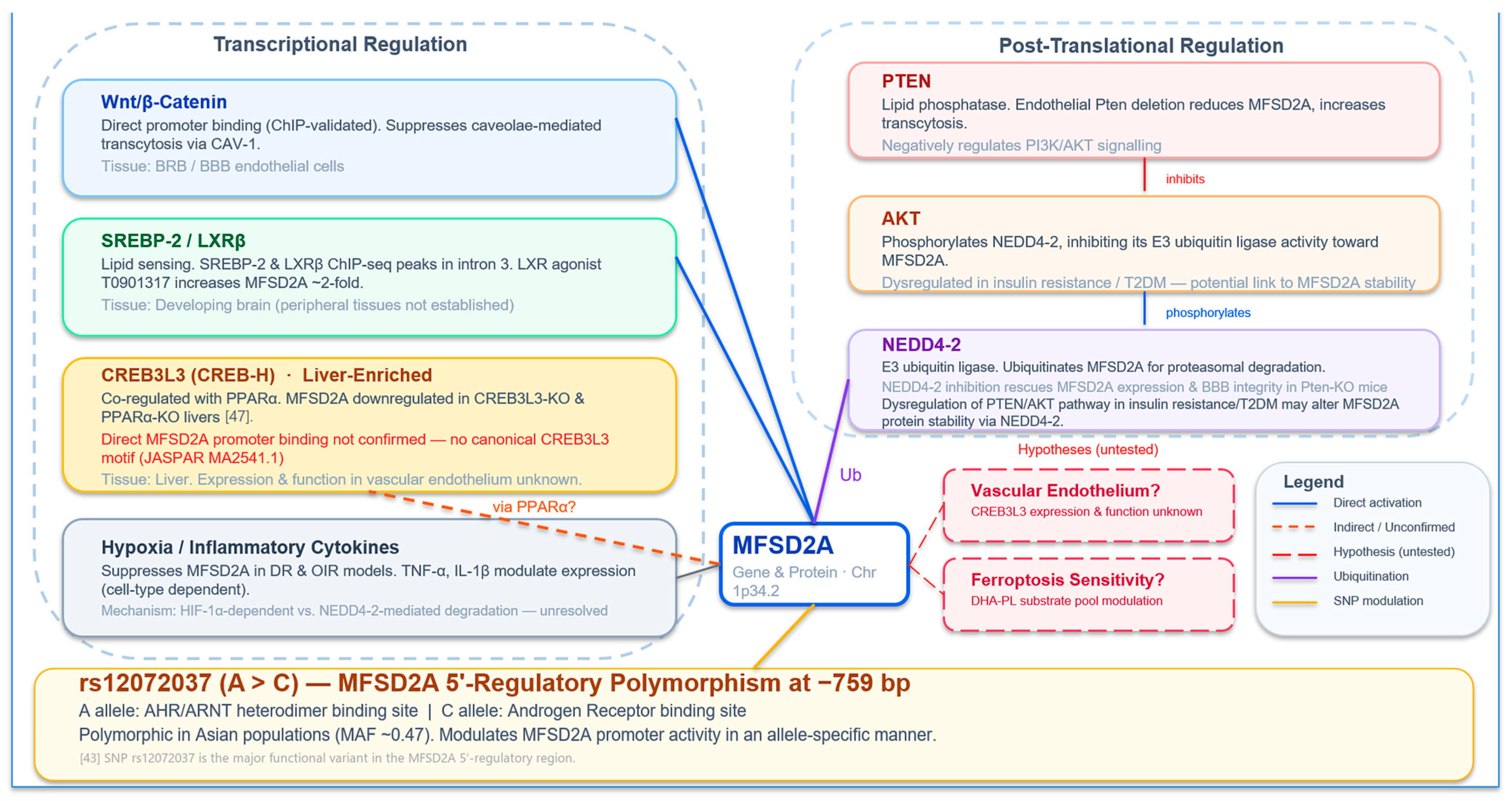
3. Transcriptional and Post-Translational Regulation of MFSD2A
The tissue-specific and context-dependent expression patterns of MFSD2A imply a complex regulatory architecture. Over the past decade, several distinct transcriptional and post-translational control mechanisms have been identified (Figure 3).
3.1. Wnt/β-Catenin Signalling
The most direct transcriptional regulator of MFSD2A identified to date is the canonical Wnt/β-catenin pathway. Wang et al. [40] demonstrated that MFSD2A was a direct transcriptional target of β-catenin in retinal endothelial cells. In mice lacking either the Wnt co-receptor LRP5 (Lrp5−/−) or the Wnt ligand Norrin (Ndpy/−), retinal MFSD2A expression was significantly reduced, leading to increased endothelial transcytosis and blood–retinal barrier (BRB) breakdown. β-catenin was shown to bind directly to the MFSD2A promoter via ChIP, and MFSD2A overexpression was sufficient to rescue the transcytosis defect in Wnt-deficient endothelial cells, confirming the functional significance of this interaction. Mechanistically, the Wnt/β-catenin/MFSD2A axis suppresses transcytosis through a caveolin-1 (CAV-1)-positive caveolae pathway. Wnt signalling-deficient retinas also exhibited reduced omega-3 fatty acid levels, reflecting the dual transport and barrier functions of MFSD2A.
This finding has provocative implications for diabetic vascular disease. Wnt/β-catenin signalling is dysregulated in diabetes (activated in vascular calcification, suppressed in wound healing), and the net effect on endothelial MFSD2A expression may depend on the local balance of Wnt ligands, receptor availability, and downstream transcriptional co-factors. Whether Wnt/β-catenin signalling regulates MFSD2A expression in the systemic vasculature under diabetic or atherogenic conditions remains to be investigated.
3.2. SREBP/LXR Regulation and Lipogenic Feedback
MFSD2A expression in the brain is tightly coupled to cellular lipid status through the sterol regulatory element-binding protein (SREBP) and liver X receptor (LXR) pathways. Chan et al. [41] demonstrated that MFSD2A in the developing brain is regulated by SREBP-1, SREBP-2, and LXRβ. ChIP-seq data mining identified SREBP-2 and LXRβ binding sites in intron 3 of the mouse Mfsd2a gene, and treatment with the LXR agonist T0901317 increased brain MFSD2A protein levels by approximately twofold. This regulatory logic is physiologically coherent: when cellular lipid levels are low (signalled by SREBP activation), MFSD2A is upregulated to enhance import of exogenous lipid. When lipid levels are sufficient, SREBP is suppressed and MFSD2A expression declines.
It is important to note that SREBP/LXR regulation of MFSD2A has been demonstrated in the developing brain; the extent to which this mechanism operates in peripheral tissues, including the liver and vasculature, has not been established. In the liver, hepatic MFSD2A expression is primarily driven by glucocorticoid receptor signalling rather than SREBP, underscoring the tissue-specificity of MFSD2A regulatory mechanisms.
3.3. PTEN/AKT/NEDD4-2 Axis: Post-Translational Control
In addition to transcriptional regulation, MFSD2A protein levels are subject to post-translational control through ubiquitin-mediated degradation. Cui et al. [42] identified a PTEN/AKT/NEDD4-2 signalling axis in brain endothelial cells that governs MFSD2A stability. In this pathway, the E3 ubiquitin ligase NEDD4-2 interacts with and ubiquitinates MFSD2A, targeting it for proteasomal degradation. AKT-mediated phosphorylation of NEDD4-2 regulates its ligase activity toward MFSD2A. The physiological significance of this axis was demonstrated in mice with cerebrovascular endothelial-specific Pten deletion. These animals exhibited reduced MFSD2A protein levels, increased transcytosis, and elevated BBB permeability, while pharmacological inhibition of NEDD4-2 restored MFSD2A expression and BBB integrity.
The PTEN/AKT pathway is centrally involved in insulin signalling, and its dysregulation is a hallmark of insulin resistance and T2DM. It is therefore conceivable that aberrant AKT signalling in the diabetic endothelium could alter MFSD2A protein stability via the NEDD4-2 pathway, contributing to the tissue-specific expression changes observed in diabetic complications.
3.4. Polymorphic Variation and Epigenetic Regulation
A functional single-nucleotide polymorphism (SNP) in the MFSD2A 5′-regulatory region, rs12072037 (A > C), modulates MFSD2A promoter activity in a cell-type-specific manner [43]. The A allele creates binding sites for the AHR/ARNT heterodimer, whereas the C allele creates a binding site for the androgen receptor [43]. This polymorphism is in linkage disequilibrium with the MYCL1-EcoRI polymorphism and is polymorphic in Asian but not Caucasian populations, raising the possibility of population-specific effects on MFSD2A expression.
3.5. Hypoxia and Inflammatory Regulation
In the context of DR, MFSD2A expression is suppressed by hypoxia. Exposure of brain endothelial cells (bEnd.3) to hypoxic conditions significantly decreased MFSD2A protein levels, and similar hypoxia-mediated MFSD2A downregulation was observed in the oxygen-induced retinopathy (OIR) model. The signalling pathways mediating hypoxia-induced MFSD2A suppression (whether HIF-1α-dependent transcriptional repression, enhanced NEDD4-2-mediated degradation, or a combination of mechanisms) remain to be defined. Inflammatory cytokines, including tumour necrosis factor-α (TNF-α) and interleukin-1β (IL-1β), have also been reported to modulate MFSD2A expression in endothelial cells, although the published data are limited and sometimes contradictory across cell types [37,44].
3.6. CREB3L3: A Liver-Enriched Transcriptional Regulator of MFSD2A with Implications for Diabetic Vascular Disease
Established. An additional transcriptional regulator of MFSD2A, identified outside the context of the BBB and retina, is cyclic AMP-responsive element-binding protein 3 like 3 (CREB3L3, also known as CREB-H). CREB3L3 is a basic leucine zipper transcription factor of the CREB/ATF family that is expressed primarily in the liver and small intestine and is proteolytically activated upon ER stress or metabolic demand to regulate genes governing triglyceride metabolism, fatty acid oxidation, ketogenesis, and apolipoprotein biosynthesis [45,46]. Transcriptional profiling of livers from CREB3L3 knockout and PPARα knockout mice identified MFSD2A as one of a small number of genes under dual control of PPARα and CREB3L3. MFSD2A was significantly downregulated in the livers of both CREB3L3−/− and PPARα−/− mice, and adenoviral overexpression of CREB3L3 markedly upregulated hepatic MFSD2A expression [47]. This places MFSD2A within the CREB3L3 transcriptional programme, alongside established targets such as Fgf21, Cpt1a, and Bdh1. Notably, the CREB3L3 and MFSD2A regulatory networks share several upstream inputs. Both are induced by glucocorticoid receptor signalling [11,46], both are responsive to fasting and metabolic stress, and both converge on hepatic phospholipid homeostasis and VLDL assembly (Section 2.3.3). Whether CREB3L3 directly binds the MFSD2A promoter or acts through intermediary factors such as PPARα remains to be clarified. A bioinformatic scan of the 3-kb proximal MFSD2A promoter does not identify a canonical high-affinity CREB3L3 consensus motif (JASPAR MA2541.1), suggesting that CREB3L3 either binds a non-canonical site, operates through a distal enhancer element, or regulates MFSD2A indirectly via PPARα co-activation.
The relevance of the CREB3L3–MFSD2A axis extends beyond hepatic lipid metabolism. CREB3L3 deficiency in mice, through either global or liver-specific knockout, results in severe hypertriglyceridaemia, accelerated atherosclerosis on the Ldlr−/− background, and exacerbation of diet-induced obesity and insulin resistance, while hepatic CREB3L3 overexpression normalises dyslipidaemia and halts atherosclerosis in diabetic mouse models [46,48]. These metabolic phenotypes overlap substantially with the tissue-specific MFSD2A dysregulation observed in diabetes (Section 4.1), raising the hypothesis that CREB3L3 may coordinate a broader transcriptional programme involving MFSD2A that protects against diabetic complications. Whether CREB3L3 regulates MFSD2A expression in tissues beyond the liver, particularly in the vascular endothelium, where CREB3L3 expression has not been characterised and where MFSD2A’s role remains unresolved (Section 5.2), represents a testable hypothesis. Furthermore, given the emerging interplay between MFSD2A-mediated DHA transport and ferroptosis sensitivity (Section 5.3), the possibility that CREB3L3-dependent MFSD2A regulation modulates endothelial ferroptotic susceptibility in diabetic atherosclerosis warrants investigation. Direct experimental evidence for the CREB3L3–MFSD2A–ferroptosis axis in the vasculature is currently lacking, and its elucidation represents a priority for future research.
Figure 3.
Transcriptional and post-translational regulation of MFSD2A. Key regulatory inputs include Wnt/β-catenin, SREBP/LXR, PTEN/AKT/NEDD4-2, CREB3L3 (hepatic), hypoxia and inflammatory cytokines (TNF-α, IL-1β), and the rs12072037 SNP. The grey connector denotes a reported regulation whose mechanism remains unresolved (Section 3.5) [43,47].
4. MFSD2A in Type 2 Diabetes Mellitus
4.1. Altered MFSD2A Expression Across Tissues in Diabetes
One of the most striking observations from the current literature is that MFSD2A expression is altered in diabetes, but the direction of change is profoundly tissue-dependent (Table 1). Understanding the logic behind this tissue specificity is essential for evaluating MFSD2A as a therapeutic target.
In the liver, MFSD2A is upregulated at the mRNA and protein levels in both NAFLD patients and high-fat-diet-fed mice. This upregulation is mediated, at least in part, by glucocorticoid receptor signalling and likely represents a compensatory response to increased demand for phospholipid precursors under conditions of hepatic lipid overload. The functional importance of this upregulation is demonstrated by the rapid progression to NASH in liver-specific Mfsd2a KO mice, indicating that MFSD2A induction is a protective adaptation that, when absent or insufficient, unmasks severe hepatic pathology. Nevertheless, in the context of the severe oxidative stress that accompanies steatohepatitis, increased hepatic DHA import via MFSD2A could in principle expand the substrate pool for lipid peroxidation. This consideration does not negate the net hepatoprotective role of MFSD2A demonstrated by Chin et al. [11], but warrants investigation. As discussed in Section 3.6, the liver-enriched transcription factor CREB3L3 also regulates hepatic MFSD2A expression [47], and its contribution to the observed MFSD2A upregulation in NAFLD, relative to that of glucocorticoid receptor signalling, has not been dissected.
In the retina, MFSD2A expression at both the mRNA and protein levels was significantly decreased at 2 and 4 months after diabetes induction in the streptozotocin (STZ)-induced diabetic mouse model, compared with age-matched controls. This downregulation was also observed in the oxygen-induced retinopathy (OIR) model, which recapitulates the ischaemic phase of proliferative DR. Immunofluorescence staining confirmed that retinal MFSD2A was expressed in vascular endothelial cells, and its reduction was associated with increased vascular leakage and pathological neovascularisation.
In the kidney, single-nucleus RNA-sequencing data from human DKD samples showed that MFSD2A expression in proximal tubular cells, specifically in the S3 segment where MFSD2A is normally enriched, is significantly reduced compared to healthy controls. The magnitude and cell-type specificity of this reduction suggest that MFSD2A loss may contribute to the impaired tubular repair capacity characteristic of progressive DKD.
In the placenta, MFSD2A protein expression is significantly lower in women with GDM than in normoglycaemic pregnancies, and this reduction correlates with decreased DHA levels in cord blood [13]. This finding suggests that maternal hyperglycaemia directly or indirectly suppresses placental MFSD2A, potentially compromising foetal DHA supply during a critical window of neurodevelopment.
The picture in the systemic vasculature is less well-characterised. Transcriptomic profiling of endothelial cells treated with Ox-PAPC has demonstrated widespread transcriptional remodelling involving genes linked to lipid metabolism, inflammation, and redox homeostasis [49]. Whether MFSD2A is among the Ox-PAPC-responsive genes in the vascular endothelium, and whether its expression is altered under diabetic conditions in vivo, has not been reported (see Section 5.2).
4.2. MFSD2A, Insulin Resistance, and Systemic Lipid Dysregulation
The systemic metabolic phenotype of global Mfsd2a KO mice provides insight into the role of MFSD2A in whole-body lipid homeostasis. Beyond the well-characterised neurological phenotype, Mfsd2a KO mice exhibit reduced hepatic VLDL-triglyceride secretion (particularly after prolonged fasting), decreased plasma triglyceride levels, reduced body fat mass with smaller adipocytes, decreased BAT triglyceride content with increased oxidative capacity, and elevated circulating LPC concentrations [8,12]. Intriguingly, humans with biallelic MFSD2A loss-of-function mutations also display approximately 80% higher plasma LPC levels compared to carriers and non-carriers [35], confirming that MFSD2A-dependent tissue LPC uptake is a quantitatively important determinant of circulating LPC concentrations.
This elevation of circulating LPC in MFSD2A deficiency is noteworthy because plasma LPC species, particularly LPC (16:0), LPC (18:1), and LPC (18:2), have been identified as independent biomarkers and potential mediators of insulin resistance and T2DM risk in large-scale metabolomics studies [50,51]. Whether altered MFSD2A expression or activity in diabetes contributes to the dysregulated plasma LPC profile observed in T2DM patients remains an open question.
Beyond its systemic metabolic effects, the tissue-specific alterations in MFSD2A expression have direct implications for endothelial function, the common pathological substrate of diabetic vascular complications.
4.3. MFSD2A and Endothelial Dysfunction in the Diabetic Milieu
Endothelial dysfunction in diabetes is characterised by a cluster of interrelated abnormalities, including reduced NO bioavailability, elevated ROS production, increased expression of adhesion molecules (VCAM-1, ICAM-1) and pro-inflammatory cytokines (IL-6, IL-1β, TNF-α), enhanced endothelial permeability, and a shift towards a pro-thrombotic phenotype [5,6]. How might altered MFSD2A expression intersect with these processes?
First, as the primary transporter of LPC-DHA into endothelial cells, MFSD2A directly governs the availability of DHA for conversion into anti-inflammatory and pro-resolving lipid mediators, including resolvins, protectins, and maresins, via cyclooxygenase (COX), lipoxygenase (LOX), and cytochrome P450 pathways. When MFSD2A is downregulated, as occurs in the retina and kidney in diabetes, this anti-inflammatory lipid mediator production would be expected to decline, perpetuating endothelial inflammation.
Second, MFSD2A-mediated LPC-DHA uptake suppresses de novo lipogenesis through SREBP pathway inhibition, as demonstrated in the developing brain and eye [31,41]. In the diabetic endothelium, where aberrant lipogenesis and lipotoxicity contribute to dysfunction, loss of this suppressive mechanism could exacerbate metabolic stress.
Third, the transcytosis-suppressing function of MFSD2A, which operates through caveolin-1-dependent pathways, may be relevant to diabetic macrovascular permeability. Increased endothelial permeability to macromolecules, particularly LDL, is an early event in atherogenesis that is exacerbated by diabetes. Although the role of transcytosis (as opposed to paracellular leakage) in diabetic atherosclerosis has not been specifically investigated, the demonstration that MFSD2A suppresses transcytosis in brain and retinal endothelia raises the possibility that altered MFSD2A expression in the systemic vasculature could modulate LDL transcytosis. The direction of this modulation would depend on whether MFSD2A is up- or downregulated in the diabetic arterial endothelium in vivo, a question that remains unresolved. If MFSD2A is upregulated, transcytosis would be expected to decrease, which could be protective, whereas if MFSD2A is downregulated (as occurs in the retina and kidney), loss of transcytosis control could contribute to enhanced LDL deposition.
Fourth, MFSD2A may influence endothelial cell fate through modulation of ferroptosis sensitivity. Ferroptosis is fundamentally a lipid-driven process in which the peroxidation of PUFA-containing phospholipids propagates membrane destruction. As the primary transporter of the highly peroxidisable DHA into cells, MFSD2A is positioned at a potential regulatory nexus for ferroptosis sensitivity. This hypothesis is explored in detail in Section 5.3.
5. MFSD2A in Diabetic Complications
The tissue-specific expression changes in MFSD2A in diabetic complications are summarised in Figure 4.
Figure 4.
Dysregulation of MFSD2A in type 2 diabetes and complications. Summary of MFSD2A expression changes (↑/↓ or ?) and pathological outcomes across diabetes-affected tissues [11,13,38,39].
5.1. Diabetic Retinopathy
Diabetic retinopathy is the most common microvascular complication of diabetes and a leading cause of blindness in working-age adults. Its pathogenesis involves pericyte loss, capillary basement membrane thickening, endothelial proliferation, BRB breakdown, and, in the proliferative stage, pathological neovascularisation driven largely by VEGF [36]. However, approximately 27% of DR patients have a poor visual outcome despite anti-VEGF therapy, and about 50% still exhibit macular oedema after two years of ranibizumab treatment, highlighting the need for VEGF-independent therapeutic strategies.
Supported indirectly. Zhang et al. [39] provided the first, and, to date, only, direct experimental evidence linking MFSD2A to DR (disease context: d, e). They found that the ratio of DHA-derived metabolites to total DHA was lower in vitreous humour from DR patients than from macular hole controls, suggesting impaired DHA metabolism despite adequate substrate availability. In both the STZ-induced diabetic mouse model and the OIR model, retinal MFSD2A expression was significantly reduced. Mechanistically, MFSD2A overexpression in brain endothelial cells (bEnd.3) inhibited cell proliferation, migration, and vesicular transcytosis, which are three processes central to DR pathogenesis. In vivo, the combination of MFSD2A overexpression via AAV-mediated gene delivery and dietary DHA supplementation was more effective at reducing pathological neovascularisation and vascular leakage than either intervention alone.
It should be noted that Zhang et al. [39] employed the streptozotocin-induced type 1 diabetes model and the oxygen-induced retinopathy model, neither of which fully recapitulated the metabolic features of T2DM. Validation in high-fat-diet/streptozotocin or db/db models was not performed. These results carry two important clinical implications. First, DHA supplementation alone may be insufficient to improve DR in patients with low retinal MFSD2A expression, because of the transporter required for DHA uptake into retinal vascular endothelial cells, where MFSD2A exerts its barrier-protective and anti-angiogenic functions, is deficient. It is worth noting that total retinal DHA accretion depends primarily on RPE-expressed MFSD2A (Section 2.3.2) [31], whereas the therapeutic effects of MFSD2A overexpression in DR models appear to be mediated through endothelial MFSD2A, highlighting distinct roles for MFSD2A in the RPE and the retinal vasculature. This tissue-compartment specificity could explain, in part, why omega-3 fatty acid supplementation trials for DR have produced inconsistent results. Second, MFSD2A is positioned as a potential adjunctive therapeutic target, particularly for patients with poor response to anti-VEGF monotherapy.
Hypothesised. Ferroptosis has been implicated in RPE degeneration and retinal capillary injury under diabetic conditions [15]. Given that MFSD2A is highly expressed in the RPE and that RPE-expressed MFSD2A accounts for the majority of ocular DHA uptake (Section 2.3.2), altered MFSD2A activity in the diabetic RPE may modulate RPE ferroptosis sensitivity. Whether MFSD2A-derived DHA incorporation sensitises or protects RPE cells against ferroptotic stimuli has not been investigated. The interplay between the established transcytosis-suppressing function of MFSD2A and its potential ferroptosis-modulating role in the retinal vasculature warrants direct examination.
5.2. Diabetic Atherosclerosis
Atherosclerosis in the setting of diabetes is accelerated, more diffusely distributed, and associated with higher plaque vulnerability than in nondiabetic individuals [4]. The metabolic abnormalities of T2DM, hyperglycaemia, insulin resistance, dyslipidaemia, and chronic low-grade inflammation, converge on the arterial endothelium to promote endothelial dysfunction, the initiating step in atherogenesis [5]. Oxidised low-density lipoprotein (Ox-LDL) and oxidised phospholipids such as Ox-PAPC, which accumulate in the subendothelial space, activate endothelial cells to express adhesion molecules and chemoattractants, recruiting monocytes that subsequently differentiate into lipid-laden foam cells [49].
No direct evidence: Critically, the role of MFSD2A in diabetic atherosclerosis is entirely unexplored in the published literature (disease context: none). Ox-PAPC induced extensive transcriptional reprogramming in human aortic endothelial cells (HAECs) involving over 1000 genes [49]. Whether MFSD2A ranked among these responsive genes has not been reported. A recent analysis of the GTEx cohort identified 619 and 356 differentially expressed genes associated with diabetes in the thoracic aorta and tibial artery, respectively, yet MFSD2A was not among them [52]. It should be noted that the GTEx data were derived from bulk arterial tissue, in which endothelial-specific transcripts may be diluted below detection thresholds. Whether MFSD2A protein is expressed in the systemic arterial endothelium at baseline, and whether its expression is altered under diabetic conditions in vivo, therefore remains unresolved. Li et al. [20] noted that MFSD2A was absent from cerebral arteriolar endothelium, consistent with the broader observation that its expression is concentrated in capillary-level endothelia (BBB, BRB, intestinal microvasculature, placenta) rather than in large-vessel endothelium. However, direct evidence for or against MFSD2A expression in the arterial endothelium outside the CNS has not been published.
Supported indirectly. This knowledge gap is significant for several reasons. The established contribution of ferroptosis to diabetic endothelial injury through p53-mediated xCT suppression and HMOX1-driven iron overload places endothelial lipid peroxidation at the centre of the disease process [16,17]. Lipid peroxidation is the biochemical executioner of ferroptotic cell death, dependent on the abundance of peroxidisable PUFA-containing phospholipids in cellular membranes. MFSD2A, as the primary gatekeeper of DHA import, governs the supply of one of the most peroxidation-susceptible fatty acids to particular phospholipid pools, although whether DHA-containing species rather than arachidonic acid- and adrenic acid-containing phosphatidylethanolamines are the peroxidised substrates in ferroptosis remains unresolved (Section 5.3.1). Resolving whether MFSD2A is expressed in the systemic arterial wall therefore represents a critical prerequisite for the hypothesis examined in Section 5.3. If MFSD2A is absent from the arterial endothelium, the MFSD2A-ferroptosis nexus may be more relevant to microvascular complications (DR, DKD) or to atherosclerosis-associated microvasculature, such as the vasa vasorum, where capillary-level endothelial cells reside.
5.3. MFSD2A at the Interface of Lipid Transport and Ferroptosis
5.3.1. Ferroptosis in Diabetic Endothelial Injury: Established Evidence
Established. Ferroptosis is an iron-dependent form of regulated cell death executed through the ACSL4/LPCAT3/GPX4 axis [18,19]. ACSL4 ligates free polyunsaturated fatty acids (PUFAs) to coenzyme A, LPCAT3 incorporates the resulting PUFA-CoAs into membrane phospholipids, and GPX4 reduces phospholipid hydroperoxides to non-toxic lipid alcohols using glutathione as a cofactor [18,19,53]. When GPX4 activity is compromised, or when the supply of oxidation-prone PUFA-phospholipids exceeds the antioxidant capacity, iron-catalysed lipid peroxidation propagates autocatalytically and culminates in membrane rupture [53]. Among PUFA substrates, DHA (22:6) possesses five bis-allylic methylene positions, compared with three in arachidonic acid (20:4), which renders it intrinsically more susceptible to peroxidation once esterified into membrane phospholipids. It should be noted, however, that ACSL4 exhibited higher affinity for arachidonic acid (20:4) than for DHA (22:6) in biochemical assays [54], and the extent to which MFSD2A-imported DHA is channelled through ACSL4 versus alternative metabolic routes, such as ACSL3-mediated esterification or pro-resolving mediator synthesis, has not been determined.
Supported indirectly. A growing body of evidence has established ferroptosis as a key pathogenic mechanism in diabetic vascular disease [14,15]. Endothelial cells exposed to high glucose and pro-inflammatory cytokines undergo ferroptosis through the p53-xCT-glutathione (GSH) axis [16], and HMOX1-mediated iron overload has been directly implicated in ferroptotic endothelial injury in diabetic atherosclerosis [17]. These findings establish the ACSL4/LPCAT3/GPX4 machinery as a functional pathway in the diabetic endothelium. Which lipid species actually carry the peroxidation in this setting remains an open question. DHA-containing phospholipids are candidate substrates given their high peroxidation susceptibility. That they are the responsible species in the diabetic endothelium is Hypothesised rather than established. The experiments that would test it are set out in Section 5.3.4.
5.3.2. Hypothesis: MFSD2A as a Modulator of Ferroptosis Sensitivity
MFSD2A, as the primary transporter of LPC-DHA in tissues where it is expressed [8], directly governs the supply of DHA for phospholipid incorporation. This positions MFSD2A at a regulatory nexus that could influence ferroptosis sensitivity in two opposing directions (Table 2).
Table 2.
Potential bidirectional roles of MFSD2A in ferroptosis regulation.
Several factors complicate this model and preclude a simple prediction. First, MFSD2A transports multiple LPC species, including LPC-oleate (18:1) and LPC-palmitate (16:0) [8,26]. The proportion of DHA-containing LPC transported by MFSD2A in the diabetic endothelium has not been determined, and LPC species with lower unsaturation may dilute the peroxidisable substrate pool. Second, the metabolic fate of imported DHA depends on the enzymatic repertoire of the recipient cell: channelling into phospholipids (via LPCAT3) favours ferroptosis sensitivity, whereas channelling into pro-resolving mediator synthesis (via COX, LOX, and cytochrome P450 pathways) would be expected to exert anti-ferroptotic effects. Third, cells possess multiple ferroptosis defence systems beyond GPX4, including FSP1/CoQ10, DHODH, and the GCH1/BH4 pathway [53]. Whether any of these are co-regulated with MFSD2A under diabetic conditions is unknown.
Hypothesised. The net effect of altered MFSD2A expression in the diabetic arterial wall, whether it promotes ferroptotic injury or serves a compensatory protective function, is the central unresolved question in this field. Distinguishing between the pro- and anti-ferroptotic scenarios will require direct experimental manipulation of endothelial MFSD2A expression under diabetic conditions, combined with comprehensive lipidomic profiling, redox status assessment, and ferroptosis phenotyping. The possibility that MFSD2A functions as a context-dependent modulator whose net effect on ferroptosis sensitivity is determined by the local balance of LPC species, downstream enzymatic machinery, and co-regulated defence systems deserves particular consideration.
5.3.3. Reconciling the Tissue Paradox
An apparent paradox arises when the MFSD2A-ferroptosis hypothesis is juxtaposed with the tissue-specific expression data summarised in Table 1. In the retina and kidney, MFSD2A expression is markedly downregulated under diabetic conditions [38,39], yet ferroptosis is reportedly increased in these same tissues [15]. If MFSD2A downregulation were to reduce DHA import and thereby deplete the substrate pool for ferroptotic lipid peroxidation, a decrease in ferroptosis sensitivity would be predicted. The opposite is observed.
Hypothesised. This paradox can be resolved by distinguishing two functionally independent pathways through which MFSD2A dysregulation contributes to diabetic tissue injury. Pathway A operates through transcytosis control and inflammation resolution. MFSD2A downregulation disinhibits caveolae-mediated transcytosis and reduces the production of DHA-derived pro-resolving lipid mediators, thereby driving barrier breakdown and sustained inflammation independently of ferroptosis. Pathway B operates through modulation of the substrate pool for ferroptotic lipid peroxidation. Altered MFSD2A-mediated LPC-DHA import changes the abundance of peroxidation-susceptible phospholipids in cellular membranes, thereby influencing ferroptosis sensitivity.
In tissues where MFSD2A is downregulated (retina, kidney), the pathogenic consequences are likely dominated by Pathway A. The ferroptosis observed in these tissues is driven by diabetes-induced GPX4 suppression and ACSL4 upregulation [14,15], which operate independently of MFSD2A-mediated substrate supply. The MFSD2A downregulation and the ferroptosis activation in these tissues are therefore parallel events that share a common upstream trigger (hyperglycaemia and oxidative stress) rather than occupying sequential positions on the same causal chain.
In tissues where MFSD2A expression is preserved or upregulated (liver, and potentially other sites), Pathway B may become functionally relevant. In the NAFLD liver, MFSD2A upregulation is clearly hepatoprotective through maintenance of phospholipid pools, suggesting that the net effect of MFSD2A in this context is anti-ferroptotic. Whether the same holds true in other tissues where MFSD2A expression is retained under diabetic conditions remains to be determined.
An important implication of this framework is that the MFSD2A-ferroptosis hypothesis should not be applied indiscriminately to all tissues. It is likely most relevant to tissues in which MFSD2A expression is maintained or induced, where the balance between substrate supply for lipid peroxidation and anti-ferroptotic mediator production becomes a meaningful determinant of cell fate.
5.3.4. Falsifiability Conditions
The bidirectional model presented in Table 2 and the tissue-specific framework described above each carry specific conditions under which they would be substantially weakened or refuted.
For the bidirectional model, three conditions are identified. First, if endothelial-specific Mfsd2a overexpression or silencing in diabetic mouse models were to produce no alteration in lipid peroxidation markers (malondialdehyde, 4-hydroxynonenal) or ferroptosis markers (Ptgs2 mRNA, GPX4 activity) in the tissue of interest, the model would lack experimental support. Second, if lipidomic profiling were to reveal no change in DHA-containing phospholipid species upon MFSD2A modulation, the premise that MFSD2A activity meaningfully alters the substrate pool for ferroptotic lipid peroxidation would be challenged. Third, if MFSD2A protein levels in human diabetic arterial endothelium were found not to differ from those in nondiabetic controls, the clinical relevance of the hypothesis to macrovascular disease would be diminished.
For the CREB3L3–MFSD2A–ferroptosis axis proposed in Section 3.6, two independent conditions are identified. One is anatomical. If CREB3L3 protein were found not to be expressed in the vascular endothelium, the axis would lack its cellular substrate and the hypothesis would be confined to hepatic lipid biology. The other is regulatory. Even if CREB3L3 were present in endothelial cells, its gain- or loss-of-function would need to demonstrably alter MFSD2A expression and lipid peroxidation markers in these cells, otherwise the causal chain from CREB3L3 through MFSD2A to ferroptosis would be broken. Convergent positive evidence at both levels would provide independent support for the hypothesis.
For the tissue-specific framework, two predictions are relevant. The prediction that Pathway A (transcytosis and inflammation resolution) drives the harmful effects of MFSD2A downregulation would be wrong if restoring transcytosis control (e.g., via NEDD4-2 inhibition) or giving resolvins did not reduce tissue damage in DR or DKD models, even when ferroptosis stays active. The prediction that Pathway B (ferroptosis substrate modulation) was tissue-dependent would be challenged if MFSD2A overexpression in the retina, where endogenous MFSD2A is downregulated, were to sensitise retinal endothelial cells to ferroptosis rather than protect them.
It is acknowledged that the absence of currently available direct experimental evidence for either pathway in the diabetic endothelium means that these falsifiability conditions remain, at present, theoretical guardrails for future investigation rather than criteria that can be applied to existing data. Table 3 decomposes the causal chain from MFSD2A activity to endothelial ferroptosis sensitivity into four links. For each link it sets out the claim, its current evidence status, the experiments required to test it, and the decision rule that would falsify or qualify it.
Table 3.
Causal chain from MFSD2A activity to endothelial ferroptosis sensitivity, with current evidence status and required experiments.
6. Therapeutic Implications and Open Questions
The prospect of therapeutically modulating MFSD2A in diabetic complications is attractive but complicated by its tissue-specific roles (Table 4). AAV-mediated MFSD2A overexpression has already reduced retinal vascular pathology in preclinical DR models, and the use of endothelial cell-specific promoters or organ-specific AAV serotypes could enable targeted delivery while avoiding off-target effects in tissues where MFSD2A upregulation might be detrimental.
Table 4.
Therapeutic strategies relevant to MFSD2A in diabetic complications, graded by evidence maturity.
Rather than targeting MFSD2A directly, modulation of its transcriptional regulators may offer greater tissue selectivity. Wnt/β-catenin pathway agonists could upregulate MFSD2A in the BRB for DR, hepatic overexpression of CREB3L3 normalises dyslipidaemia and halts atherosclerosis in diabetic mice [48], but the reported atheroprotection was attributed to reduced circulating remnant lipoproteins, no requirement for MFSD2A was demonstrated in that setting, and no pharmacological CREB3L3 activator has yet been described, so the possibility that CREB3L3 activation exerts atheroprotection through MFSD2A induction (Section 3.6) remains a hypothesis; the NEDD4-2 inhibitor approach has been validated for BBB restoration and could be evaluated in diabetic complications. In tissues where MFSD2A is downregulated, such as the retina and kidney, combining MFSD2A overexpression with LPC-DHA supplementation may be synergistic, as demonstrated in DR models; the development of stable, bioavailable LPC-DHA formulations remains an active area of investigation. Finally, if MFSD2A upregulation promotes endothelial ferroptosis, inhibitors including ferrostatin-1, liproxstatin-1, deferoxamine, and vitamin E analogs could mitigate downstream consequences by targeting the pathological endpoint rather than the transporter itself.
Notwithstanding these therapeutic possibilities, the tissue-specificity of MFSD2A regulation presents a central challenge for therapeutic translation. MFSD2A is downregulated in the retina and kidney yet upregulated in the liver in diabetes, meaning that systemic MFSD2A modulation could produce divergent, and potentially deleterious, effects across tissues. The development of tissue-specific delivery strategies, whether through organ-specific AAV serotypes, endothelial cell-specific promoters, or targeted transcriptional modulators, will be essential for realising the therapeutic potential of MFSD2A modulation. Additionally, the potential of MFSD2A or the plasma LPC species profile as a biomarker for stratifying patients with diabetic vascular complications warrants investigation.
7. Conclusions
MFSD2A has travelled a notable path, from an orphan transporter identified in a brown adipose tissue screen, to a central gatekeeper of the blood–brain barrier, to an emerging player in systemic metabolic disease. The current evidence establishes that MFSD2A is not merely a brain-specific lipid transporter, but a multifunctional protein with tissue-adapted roles spanning transcytosis suppression, phospholipid homeostasis, inflammation resolution, and potentially ferroptosis modulation.
In T2DM, MFSD2A expression is dysregulated in a tissue-specific pattern. It is suppressed in the retina (DR), kidney (DKD), and placenta (GDM), while it is upregulated in the liver (NAFLD/NASH). In the vasculature, whether MFSD2A expression is altered in response to atherogenic stimuli and under diabetic conditions remains largely unexplored. This divergence reflects the complex interplay among local metabolic stress, tissue-specific transcriptional programmes, and the adaptive versus maladaptive consequences of altered MFSD2A activity.
8. Future Perspectives
The most significant gap in the literature, and the area of greatest opportunity, concerns MFSD2A in diabetic atherosclerosis. The convergence of Ox-PAPC-driven endothelial transcriptional reprogramming with the established role of ferroptosis in diabetic atherosclerosis raises the question of whether MFSD2A-mediated DHA import contributes to ferroptotic endothelial injury. Testing this connection in vivo represents a priority for future investigation.
Several outstanding questions emerge from this review. First, whether additional transcriptional regulators of MFSD2A operate in the vascular endothelium beyond those characterised to date remains to be determined. Second, the net effect of MFSD2A dysregulation in the arterial wall, whether it promotes ferroptotic injury or serves a compensatory protective function, remains unresolved and awaits in vivo validation.
Key priorities for future investigation include identifying novel transcriptional regulators of MFSD2A in the vascular endothelium, including CREB3L3, whose role outside the liver remains unknown (Section 3.6), using ChIP-seq or CUT&RUN under diabetic conditions; assessing the functional consequences of MFSD2A modulation on endothelial ferroptosis sensitivity through lipidomic, redox, and cell-death profiling; generating and characterising endothelium-specific Mfsd2a conditional knockout mice on the ApoE-deficient diabetic atherosclerosis background; investigating the Wnt/β-catenin/MFSD2A axis in diabetic macrovascular disease; profiling MFSD2A expression and plasma LPC species in well-phenotyped T2DM patient cohorts; and exploring the therapeutic combination of MFSD2A gene therapy with LPC-DHA supplementation in DR and DKD.
The study of MFSD2A in T2DM and its complications is at an early stage, but the available evidence, spanning structural biology, in vivo genetics, and clinical correlative studies, provides a solid foundation for the hypothesis-driven investigations that will define this emerging field. Given the immense clinical burden of diabetic vascular complications and the persistent unmet need for therapies beyond glucose control, MFSD2A represents a promising and largely untapped frontier in diabetes research.
9. Methodology
A hypothesis-oriented narrative literature review was conducted following the Scale for the Assessment of Narrative Review Articles (SANRA) guidelines [58]. The review aimed to synthesise evidence linking MFSD2A to type 2 diabetes and its complications and to critically evaluate the hypothesis that MFSD2A-mediated lipid transport modulates ferroptosis sensitivity in the diabetic endothelium. Consistent with this aim, articles providing mechanistic data on ferroptotic lipid peroxidation and its substrate specificity were actively sought, even when originating from non-diabetic disease models, to inform the hypothesis framework developed herein. The criteria used to identify and select the cited manuscripts are set out below, together with the search strategy, so that the basis on which evidence was included or excluded can be assessed independently of the narrative. A literature search was performed in PubMed and Scopus for articles published from database inception to July 2026. The primary search used the query: Mfsd2a OR “major facilitator superfamily domain-containing protein 2A” OR NLS1 (All Fields in PubMed; Title-Abstract-Keywords in Scopus), yielding 364 and 774 records, respectively. Secondary searches combined the primary MFSD2A query with terms related to type 2 diabetes, diabetic complications, ferroptosis, endothelial dysfunction, lysophosphatidylcholine, and docosahexaenoic acid. The literature was identified through a section-by-section approach: for each subsection, targeted searches were conducted within the retrieved records and reference lists of key articles. Articles were selected for inclusion if they met one or more of the following criteria: (i) reported original experimental data or provided a substantive review directly addressing MFSD2A structure, function, regulation, or tissue-specific roles; (ii) investigated MFSD2A in the context of metabolic disease, including T2DM, NAFLD/MASLD, GDM, or diabetic complications; (iii) examined ferroptosis mechanisms relevant to the MFSD2A-ferroptosis hypothesis, including studies in diabetic vascular or tissue injury, vascular disease models, or foundational investigations of ferroptotic lipid peroxidation and its substrate specificity; or (iv) were key conceptual or methodological references necessary for contextualising MFSD2A biology or diabetic complications. Articles were excluded when they (i) mentioned MFSD2A only in passing (e.g., as one entry among hundreds in a transcriptomic or proteomic dataset without dedicated functional investigation or discussion); (ii) focused exclusively on MFSD2B or other MFS family members without providing data or discussion specific to MFSD2A; (iii) were conference abstracts, editorials, or commentaries lacking original data or substantive synthesis; or (iv) were not available in English. The selection rationale for each included article was documented according to which of the above criteria it satisfied and which subsection it supported. Articles retrieved by the primary search that were ultimately not included were primarily those in which MFSD2A appeared as a peripheral finding in high-throughput screens without dedicated follow-up, a pattern that accounted for the majority of exclusions. Articles identified through secondary and reference-based searches were selected because the primary MFSD2A search did not capture them—typically studies whose main topic was ferroptosis in diabetes, endothelial dysfunction in atherosclerosis, or LPC-DHA metabolism, for which MFSD2A was not included among their indexed keywords. The reference list comprises the sources cited in this review. Given the early stage of this field, no formal exclusion criteria based on study design were applied, and both original research and review articles were considered. Titles and abstracts were evaluated by Z.Z. for relevance, and full texts were retrieved for detailed assessment.
Author Contributions
Conceptualization, J.L. and Q.Z.; formal analysis, S.J. and Z.Z.; investigation, S.J., Z.Z., J.H., Y.F., Q.C., W.W.C., Y.X., M.F. and A.Y.S.; resources, J.L. and Q.Z.; writing—original draft preparation, S.J. and Z.Z.; writing—review and editing, all authors; supervision, J.L. and Q.Z.; funding acquisition, J.L. and Q.Z. All authors have read and agreed to the published version of the manuscript.
Funding
This research was funded by the Royal Society of New Zealand Catalyst Fund (grant numbers CSG-UOA2108 and CSG-UOA2310) and the Medical and Health Science Program of Zhejiang Province (grant number 2025KY1583). The funding bodies had no role in the design of the study; in the collection, analyses, or interpretation of data; in the writing of the manuscript; or in the decision to publish the results.
Institutional Review Board Statement
Not applicable.
Informed Consent Statement
Not applicable.
Data Availability Statement
The data supporting the findings of this study are available within the article.
Acknowledgments
During the preparation of this manuscript, the authors used ChatGPT (OpenAI, GPT-5.5) for the purpose of improving the grammar and language of the manuscript. The authors have reviewed and edited the output and take full responsibility for the content of this publication.
Conflicts of Interest
The authors declare no conflicts of interest.
References
- International Diabetes Federation. IDF Diabetes Atlas, 10th ed.; International Diabetes Federation: Brussels, Belgium, 2021. [Google Scholar]
- Forbes, J.M.; Cooper, M.E. Mechanisms of diabetic complications. Physiol. Rev. 2013, 93, 137–188. [Google Scholar] [CrossRef] [Scilit]
- Brownlee, M. Biochemistry and molecular cell biology of diabetic complications. Nature 2001, 414, 813–820. [Google Scholar] [CrossRef] [Scilit]
- Einarson, T.R.; Acs, A.; Ludwig, C.; Panton, U.H. Prevalence of cardiovascular disease in type 2 diabetes: A systematic literature review of scientific evidence from across the world in 2007–2017. Cardiovasc. Diabetol. 2018, 17, 83. [Google Scholar] [CrossRef] [Scilit]
- Shi, Y.; Vanhoutte, P.M. Macro- and microvascular endothelial dysfunction in diabetes. J. Diabetes 2017, 9, 434–449. [Google Scholar] [CrossRef] [Scilit]
- Meza, C.A.; La Favor, J.D.; Kim, D.-H.; Hickner, R.C. Endothelial dysfunction: Is there a hyperglycaemia-induced imbalance of NOX and NOS? Int. J. Mol. Sci. 2019, 20, 3775. [Google Scholar] [CrossRef] [Scilit]
- Angers, M.; Uldry, M.; Kong, D.; Gimble, J.M.; Jetten, A.M. Mfsd2a encodes a novel major facilitator superfamily domain-containing protein highly induced in brown adipose tissue during fasting and adaptive thermogenesis. Biochem. J. 2008, 416, 347–355. [Google Scholar] [CrossRef] [Scilit]
- Nguyen, L.N.; Ma, D.; Shui, G.; Wong, P.; Cazenave-Gassiot, A.; Zhang, X.; Wenk, M.R.; Goh, E.L.K.; Silver, D.L. Mfsd2a is a transporter for the essential omega-3 fatty acid docosahexaenoic acid. Nature 2014, 509, 503–506. [Google Scholar] [CrossRef] [Scilit]
- Ben-Zvi, A.; Lacoste, B.; Kur, E.; Andreone, B.J.; Mayshar, Y.; Yan, H.; Gu, C. Mfsd2a is critical for the formation and function of the blood–brain barrier. Nature 2014, 509, 507–511. [Google Scholar] [CrossRef] [Scilit]
- Andreone, B.J.; Chow, B.W.; Tata, A.; Lacoste, B.; Ben-Zvi, A.; Bullock, K.; Deik, A.A.; Ginty, D.D.; Clish, C.B.; Gu, C. Blood-Brain Barrier Permeability Is Regulated by Lipid Transport-Dependent Suppression of Caveolae-Mediated Transcytosis. Neuron 2017, 94, 581–594.e585. [Google Scholar] [CrossRef] [Scilit]
- Chin, C.F.; Galam, D.L.A.; Gao, L.; Tan, B.C.; Wong, B.H.; Chua, G.-L.; Loke, R.Y.J.; Lim, Y.C.; Wenk, M.R.; Lim, M.-S.; et al. Blood-derived lysophospholipid sustains hepatic phospholipids and fat storage necessary for hepatoprotection in overnutrition. J. Clin. Investig. 2023, 133, e171267. [Google Scholar] [CrossRef] [Scilit]
- Berger, J.H.; Charron, M.J.; Silver, D.L. Major facilitator superfamily domain-containing protein 2a (MFSD2A) has roles in body growth, motor function, and lipid metabolism. PLoS ONE 2012, 7, e50629. [Google Scholar] [CrossRef] [Scilit]
- Prieto-Sánchez, M.T.; Ruiz-Palacios, M.; Blanco-Carnero, J.E.; Pagan, A.; Hellmuth, C.; Uhl, O.; Peissner, W.; Ruiz-Alcaraz, A.J.; Parrilla, J.J.; Koletzko, B.; et al. Placental MFSD2a transporter is related to decreased DHA in cord blood of women with treated gestational diabetes. Clin. Nutr. 2017, 36, 513–521. [Google Scholar] [CrossRef] [Scilit]
- Li, X.; Fang, M.; Liu, X.; Jiang, J.; Wang, S.; Mao, X.; Zou, Z.; Jin, W. Ferroptosis in diabetes mellitus and its complications: Overview of clinical and preclinical research. Cell Death Discov. 2025, 11, 504. [Google Scholar] [CrossRef] [Scilit]
- Liu, P.; Zhang, Z.; Cai, Y.; Li, Z.; Zhou, Q.; Chen, Q. Ferroptosis: Mechanisms and role in diabetes mellitus and its complications. Ageing Res. Rev. 2024, 94, 102201. [Google Scholar] [CrossRef] [Scilit]
- Luo, E.-F.; Li, H.-X.; Qin, Y.-H.; Qiao, Y.; Yan, G.-L.; Yao, Y.-Y.; Li, L.-Q.; Hou, J.-T.; Tang, C.-C.; Wang, D. Role of ferroptosis in the process of diabetes-induced endothelial dysfunction. World J. Diabetes 2021, 12, 124–137. [Google Scholar] [CrossRef] [Scilit]
- Meng, Z.; Liang, H.; Zhao, J.; Gao, J.; Liu, C.; Ma, X.; Liu, J.; Liang, B.; Jiao, X.; Cao, J.; et al. HMOX1 upregulation promotes ferroptosis in diabetic atherosclerosis. Life Sci. 2021, 284, 119935. [Google Scholar] [CrossRef] [Scilit]
- Doll, S.; Proneth, B.; Tyurina, Y.Y.; Panzilius, E.; Kobayashi, S.; Ingold, I.; Irmler, M.; Beckers, J.; Aichler, M.; Walch, A.; et al. ACSL4 dictates ferroptosis sensitivity by shaping cellular lipid composition. Nat. Chem. Biol. 2017, 13, 91–98. [Google Scholar] [CrossRef] [Scilit]
- Dixon, S.J.; Lemberg, K.M.; Lamprecht, M.R.; Skouta, R.; Zaitsev, E.M.; Gleason, C.E.; Patel, D.N.; Bauer, A.J.; Cantley, A.M.; Yang, W.S.; et al. Ferroptosis: An iron-dependent form of nonapoptotic cell death. Cell 2012, 149, 1060–1072. [Google Scholar] [CrossRef] [Scilit]
- Li, C.; Ibrahim, M.M.; Fang, C. MFSD2A: A molecular nexus linking blood–brain barrier, lipid metabolism, and ischemia–reperfusion injury. Cell. Mol. Biol. Lett. 2025, 30, 148. [Google Scholar] [CrossRef] [Scilit]
- Yan, N. Structural biology of the major facilitator superfamily transporters. Annu. Rev. Biophys. 2015, 44, 257–283. [Google Scholar] [CrossRef] [Scilit]
- Chua, G.-L.; Tan, B.C.; Loke, R.Y.J.; He, M.; Chin, C.-F.; Wong, B.H.; Kuk, A.C.Y.; Ding, M.; Wenk, M.R.; Guan, L.; et al. Mfsd2a utilizes a flippase mechanism to mediate omega-3 fatty acid lysolipid transport. Proc. Natl. Acad. Sci. USA 2023, 120, e2215290120. [Google Scholar] [CrossRef] [Scilit]
- Nguyen, C.; Lei, H.-T.; Lai, L.T.F.; Gallenito, M.J.; Mu, X.; Matthies, D.; Gonen, T. Lipid flipping in the omega-3 fatty-acid transporter. Nat. Commun. 2023, 14, 2571. [Google Scholar] [CrossRef] [Scilit]
- Martinez-Molledo, M.; Nji, E.; Reyes, N. Structural insights into the lysophospholipid brain uptake mechanism and its inhibition by syncytin-2. Nat. Struct. Mol. Biol. 2022, 29, 604–612. [Google Scholar] [CrossRef] [Scilit]
- Uhlén, M.; Fagerberg, L.; Hallström, B.M.; Lindskog, C.; Oksvold, P.; Mardinoglu, A.; Sivertsson, Å.; Kampf, C.; Sjöstedt, E.; Asplund, A.; et al. Tissue-based map of the human proteome. Science 2015, 347, 1260419. [Google Scholar] [CrossRef] [Scilit]
- Quek, D.Q.Y.; Nguyen, L.N.; Fan, H.; Silver, D.L. Structural insights into the transport mechanism of the human sodium-dependent lysophosphatidylcholine transporter MFSD2A. J. Biol. Chem. 2016, 291, 9383–9394. [Google Scholar] [CrossRef] [Scilit]
- Bergman, S.; Cater, R.J.; Plante, A.; Mancia, F.; Khelashvili, G. Substrate binding-induced conformational transitions in the omega-3 fatty acid transporter MFSD2A. Nat. Commun. 2023, 14, 3391. [Google Scholar] [CrossRef] [Scilit]
- Cater, R.J.; Chua, G.L.; Erramilli, S.K.; Keener, J.E.; Choy, B.C.; Tokarz, P.; Chin, C.F.; Quek, D.Q.Y.; Kloss, B.; Pepe, J.G.; et al. Structural basis of omega-3 fatty acid transport across the blood–brain barrier. Nature 2021, 595, 315–319. [Google Scholar] [CrossRef] [Scilit]
- Bazinet, R.P.; Layé, S. Polyunsaturated fatty acids and their metabolites in brain function and disease. Nat. Rev. Neurosci. 2014, 15, 771–785. [Google Scholar] [CrossRef] [Scilit]
- Saini, R.K.; Prasad, P.; Sreedhar, R.V.; Akhilender Naidu, K.; Shang, X.; Keum, Y.-S. Omega−3 Polyunsaturated Fatty Acids (PUFAs): Emerging Plant and Microbial Sources, Oxidative Stability, Bioavailability, and Health Benefits—A Review. Antioxidants 2021, 10, 1627. [Google Scholar] [CrossRef] [Scilit]
- Wong, B.H.; Chan, J.P.; Cazenave-Gassiot, A.; Poh, R.W.; Foo, J.C.; Galam, D.L.A.; Ghosh, S.; Nguyen, L.N.; Barathi, V.A.; Yeo, S.W.; et al. Mfsd2a is a transporter for the essential ω-3 fatty acid docosahexaenoic acid (DHA) in eye and is important for photoreceptor cell development. J. Biol. Chem. 2016, 291, 10501–10514. [Google Scholar] [CrossRef] [Scilit]
- Kröger, J.; Zietemann, V.; Enzenbach, C.; Weikert, C.; Jansen, E.H.J.M.; Döring, F.; Joost, H.-G.; Boeing, H.; Schulze, M.B. Erythrocyte membrane phospholipid fatty acids, desaturase activity, and dietary fatty acids in relation to risk of type 2 diabetes in the European Prospective Investigation into Cancer and Nutrition (EPIC)–Potsdam Study123. Am. J. Clin. Nutr. 2011, 93, 127–142. [Google Scholar] [CrossRef] [Scilit]
- Qian, F.; Ardisson Korat, A.V.; Imamura, F.; Marklund, M.; Tintle, N.; Virtanen, J.K.; Zhou, X.; Bassett, J.K.; Lai, H.; Hirakawa, Y.; et al. n-3 Fatty Acid Biomarkers and Incident Type 2 Diabetes: An Individual Participant-Level Pooling Project of 20 Prospective Cohort Studies. Diabetes Care 2021, 44, 1133–1142. [Google Scholar] [CrossRef] [Scilit]
- Yang, Y.R.; Xiong, X.Y.; Liu, J.; Wu, L.R.; Zhong, Q.; Zhou, K.; Meng, Z.Y.; Liu, L.; Wang, F.X.; Gong, Q.W.; et al. Mfsd2a (Major Facilitator Superfamily Domain Containing 2a) Attenuates Intracerebral Hemorrhage–Induced Blood–Brain Barrier Disruption by Inhibiting Vesicular Transcytosis. J. Am. Heart Assoc. 2017, 6, e005811. [Google Scholar] [CrossRef] [Scilit]
- Guemez-Gamboa, A.; Nguyen, L.N.; Yang, H.; Zaki, M.S.; Kara, M.; Ben-Omran, T.; Akizu, N.; Rosti, R.O.; Rosti, B.; Scott, E.; et al. Inactivating mutations in MFSD2A, required for omega-3 fatty acid transport in brain, cause a lethal microcephaly syndrome. Nat. Genet. 2015, 47, 809–813. [Google Scholar] [CrossRef] [Scilit]
- Shindou, H.; Hishikawa, D.; Harayama, T.; Yuki, K.; Shimizu, T. Recent progress on acyl CoA: Lysophospholipid acyltransferase research. J. Lipid Res. 2009, 50, S46–S51. [Google Scholar] [CrossRef] [Scilit]
- Ungaro, F.; Tacconi, C.; Massimino, L.; Corsetto, P.A.; Correale, C.; Fonteyne, P.; Piontini, A.; Garzarelli, V.; Calcaterra, F.; Della Bella, S.; et al. MFSD2A promotes endothelial generation of inflammation-resolving lipid mediators and reduces colitis in mice. Gastroenterology 2017, 153, 1363–1377.e1366. [Google Scholar] [CrossRef] [Scilit]
- Loke, R.Y.J.; Chin, C.F.; Liang, G.; Wong, B.H.; Galam, D.L.A.; Tan, B.C.; Chua, G.-L.; Minegishi, S.; Morisawa, N.; Sidorov, I.; et al. Mfsd2a-mediated lysolipid transport is important for renal recovery after acute kidney injury. J. Lipid Res. 2023, 64, 100416. [Google Scholar] [CrossRef] [Scilit]
- Zhang, C.-L.; Wang, H.-L.; Li, P.-C.; Hong, C.-D.; Chen, A.-Q.; Qiu, Y.-M.; Zeng, A.-P.; Zhou, Y.-F.; Hu, B.; Li, Y.-N. Mfsd2a overexpression alleviates vascular dysfunction in diabetic retinopathy. Pharmacol. Res. 2021, 171, 105755. [Google Scholar] [CrossRef] [Scilit]
- Wang, Z.; Liu, C.-H.; Huang, S.; Fu, Z.; Tomita, Y.; Britton, W.R.; Cho, S.S.; Chen, C.T.; Sun, Y.; Ma, J.-x.; et al. Wnt signaling activates MFSD2A to suppress vascular endothelial transcytosis and maintain blood-retinal barrier. Sci. Adv. 2020, 6, eaba7457. [Google Scholar] [CrossRef] [Scilit]
- Chan, J.P.; Wong, B.H.; Chin, C.F.; Galam, D.L.A.; Foo, J.C.; Wong, L.C.; Ghosh, S.; Wenk, M.R.; Cazenave-Gassiot, A.; Silver, D.L. The lysolipid transporter Mfsd2a regulates lipogenesis in the developing brain. PLoS Biol. 2018, 16, e2006443. [Google Scholar] [CrossRef] [Scilit]
- Cui, Y.; Wang, Y.; Song, X.; Ning, H.; Zhang, Y.; Teng, Y.; Wang, J.; Yang, X. Brain endothelial PTEN/AKT/NEDD4-2/MFSD2A axis regulates blood-brain barrier permeability. Cell Rep. 2021, 36, 109327. [Google Scholar] [CrossRef] [Scilit]
- Colombo, F.; Falvella, F.S.; Galvan, A.; Frullanti, E.; Kunitoh, H.; Ushijima, T.; Dragani, T.A. A 5′-region polymorphism modulates promoter activity of the tumor suppressor gene MFSD2A. Mol. Cancer 2011, 10, 81. [Google Scholar] [CrossRef] [Scilit]
- Eser Ocak, P.; Ocak, U.; Sherchan, P.; Zhang, J.H.; Tang, J. Insights into major facilitator superfamily domain-containing protein-2a (Mfsd2a) in physiology and pathophysiology. What do we know so far? J. Neurosci. Res. 2020, 98, 29–41. [Google Scholar] [CrossRef] [Scilit]
- Omori, Y.; Imai, J.-I.; Watanabe, M.; Komatsu, T.; Suzuki, Y.; Kataoka, K.; Watanabe, S.; Tanigami, A.; Sugano, S. CREB-H: A novel mammalian transcription factor belonging to the CREB/ATF family and functioning via the box-B element with a liver-specific expression. Nucleic Acids Res. 2001, 29, 2154–2162. [Google Scholar] [CrossRef] [Scilit]
- Nakagawa, Y.; Araki, M.; Han, S.-i.; Mizunoe, Y.; Shimano, H. CREBH systemically regulates lipid metabolism by modulating and integrating cellular functions. Nutrients 2021, 13, 3204. [Google Scholar] [CrossRef] [Scilit]
- Ruppert, P.M.M.; Park, J.-G.; Xu, X.; Hur, K.Y.; Lee, A.-H.; Kersten, S. Transcriptional profiling of PPARα−/− and CREB3L3−/− livers reveals disparate regulation of hepatoproliferative and metabolic functions of PPARα. BMC Genom. 2019, 20, 199. [Google Scholar] [CrossRef] [Scilit]
- Shimizu-Albergine, M.; Basu, D.; Kanter, J.E.; Kramer, F.; Kothari, V.; Barnhart, S.; Thornock, C.; Mullick, A.E.; Clouet-Foraison, N.; Vaisar, T.; et al. CREBH normalizes dyslipidemia and halts atherosclerosis in diabetes by decreasing circulating remnant lipoproteins. J. Clin. Investig. 2021, 131, e153285. [Google Scholar] [CrossRef] [Scilit]
- Romanoski, C.E.; Che, N.; Yin, F.; Mai, N.; Pouldar, D.; Civelek, M.; Pan, C.; Lee, S.; Vakili, L.; Yang, W.-P.; et al. Network for activation of human endothelial cells by oxidised phospholipids: A critical role of heme oxygenase 1. Circ. Res. 2011, 109, e27–e41. [Google Scholar] [CrossRef] [Scilit]
- Wang-Sattler, R.; Yu, Z.; Herder, C.; Messias, A.C.; Floegel, A.; He, Y.; Heim, K.; Campillos, M.; Holzapfel, C.; Thorand, B.; et al. Novel biomarkers for pre-diabetes identified by metabolomics. Mol. Syst. Biol. 2012, 8, MSB201243. [Google Scholar] [CrossRef] [Scilit]
- Floegel, A.; Stefan, N.; Yu, Z.; Mühlenbruch, K.; Drogan, D.; Joost, H.-G.; Fritsche, A.; Häring, H.-U.; Hrabě de Angelis, M.; Peters, A.; et al. Identification of serum metabolites associated with risk of type 2 diabetes using a targeted metabolomic approach. Diabetes 2013, 62, 639–648. [Google Scholar] [CrossRef] [Scilit]
- Shouma, A.; Giannoudi, M.; Conning-Rowland, M.; Drozd, M.; Brown, O.I.; Cheng, C.; Sukumar, P.; Bridge, K.I.; Levelt, E.; Bailey, M.A.; et al. Analysis of the diabetic arterial transcriptome to define novel biomarkers of macrovascular disease. medRxiv 2026. [Google Scholar] [CrossRef] [Scilit]
- Stockwell, B.R.; Friedmann Angeli, J.P.; Bayir, H.; Bush, A.I.; Conrad, M.; Dixon, S.J.; Fulda, S.; Gascón, S.; Hatzios, S.K.; Kagan, V.E.; et al. Ferroptosis: A regulated cell death nexus linking metabolism, redox biology, and disease. Cell 2017, 171, 273–285. [Google Scholar] [CrossRef] [Scilit]
- Kuwata, H.; Hara, S. Role of acyl-CoA synthetase ACSL4 in arachidonic acid metabolism. Prostaglandins Other Lipid Mediat. 2019, 144, 106363. [Google Scholar] [CrossRef] [Scilit]
- Kagan, V.E.; Mao, G.; Qu, F.; Angeli, J.P.F.; Doll, S.; Croix, C.S.; Dar, H.H.; Liu, B.; Tyurin, V.A.; Ritov, V.B.; et al. Oxidized arachidonic and adrenic PEs navigate cells to ferroptosis. Nat. Chem. Biol. 2017, 13, 81–90. [Google Scholar] [CrossRef] [Scilit]
- di Blasio, L.; Vara-Messler, M.; Peracino, B.; Masti, E.; Cepas-López, V.; Trusolino, L.; Puliafito, A.; Bertotti, A.; Monica, V.; Primo, L. Omega-3 fatty acid DHA induces ferroptosis in colorectal cancer patient-derived organoids and drug-tolerant cells. Cell Death Dis. 2026, 17, 464. [Google Scholar] [CrossRef] [Scilit]
- Filiberto, A.C.; Ladd, Z.; Leroy, V.; Su, G.; Elder, C.T.; Pruitt, E.Y.; Hensley, S.E.; Lu, G.; Hartman, J.B.; Zarrinpar, A.; et al. Resolution of inflammation via RvD1/FPR2 signaling mitigates Nox2 activation and ferroptosis of macrophages in experimental abdominal aortic aneurysms. FASEB J. 2022, 36, e22579. [Google Scholar] [CrossRef] [Scilit]
- Baethge, C.; Goldbeck-Wood, S.; Mertens, S. SANRA—A scale for the quality assessment of narrative review articles. Res. Integr. Peer Rev. 2019, 4, 5. [Google Scholar] [CrossRef] [Scilit]
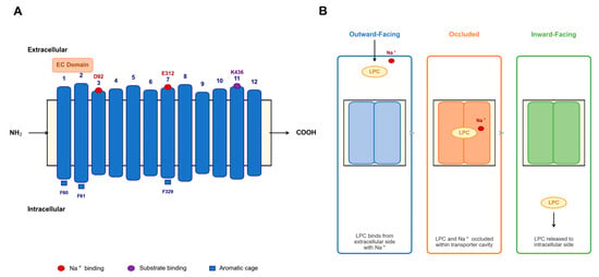
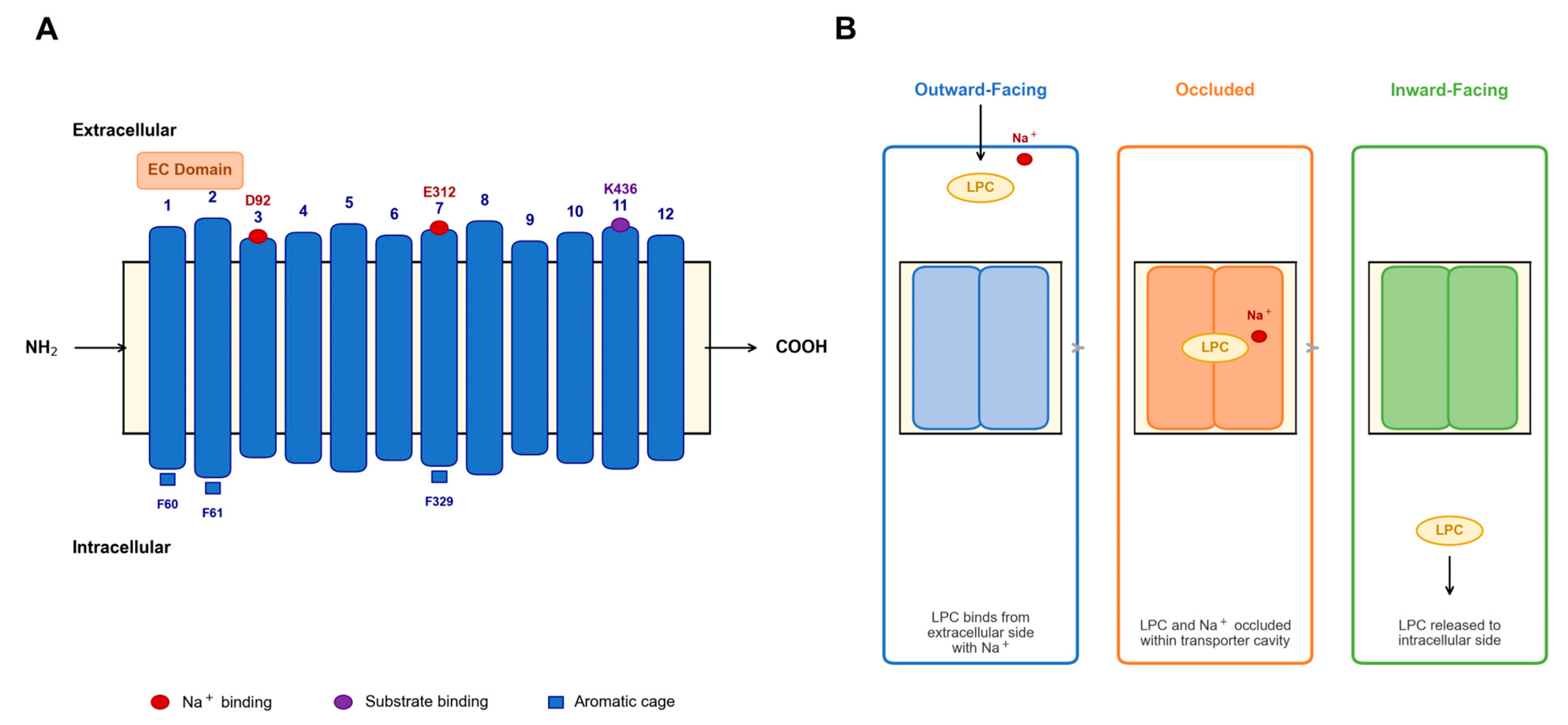
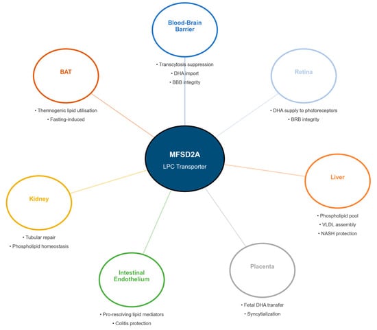
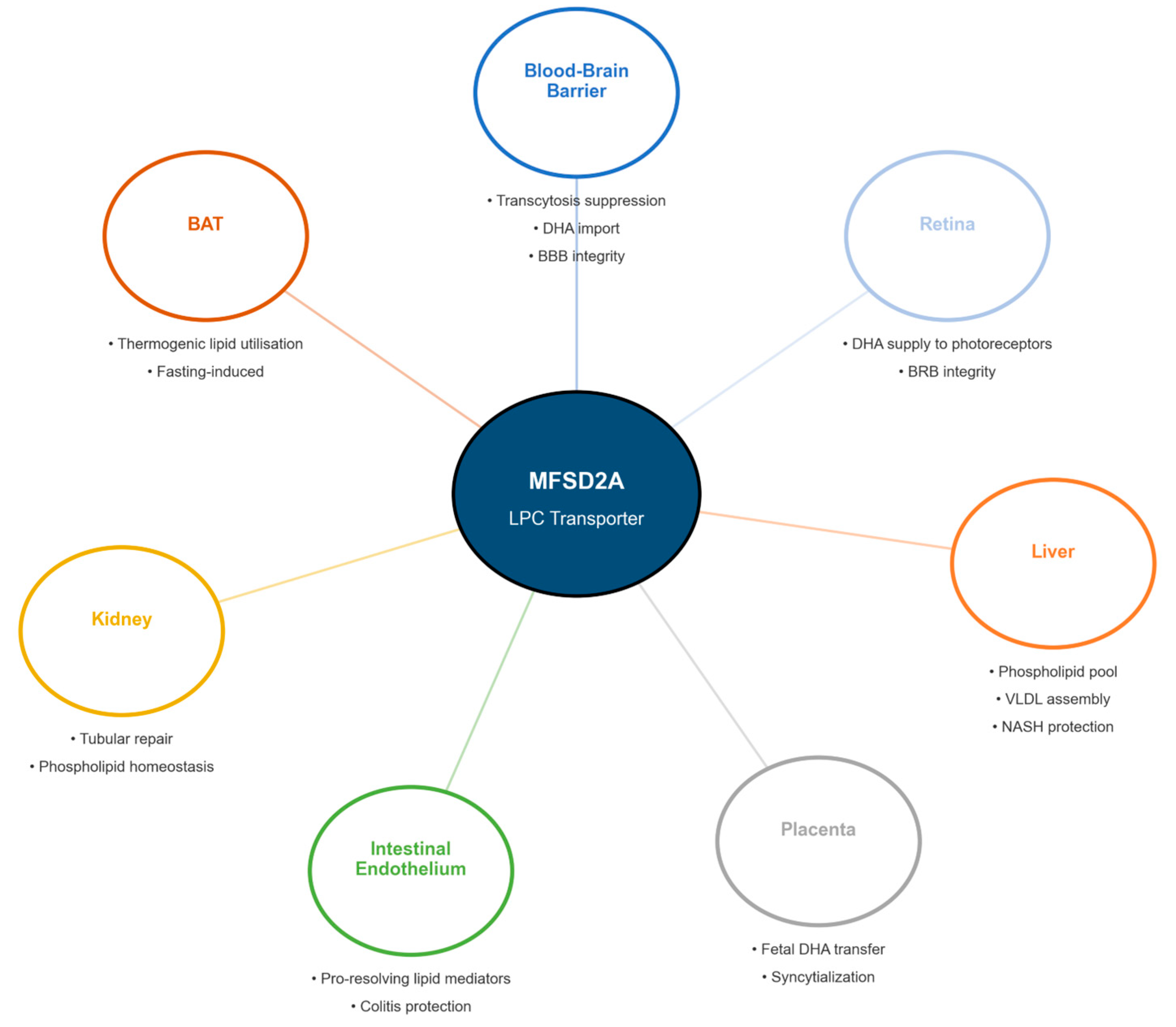
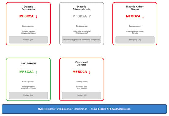
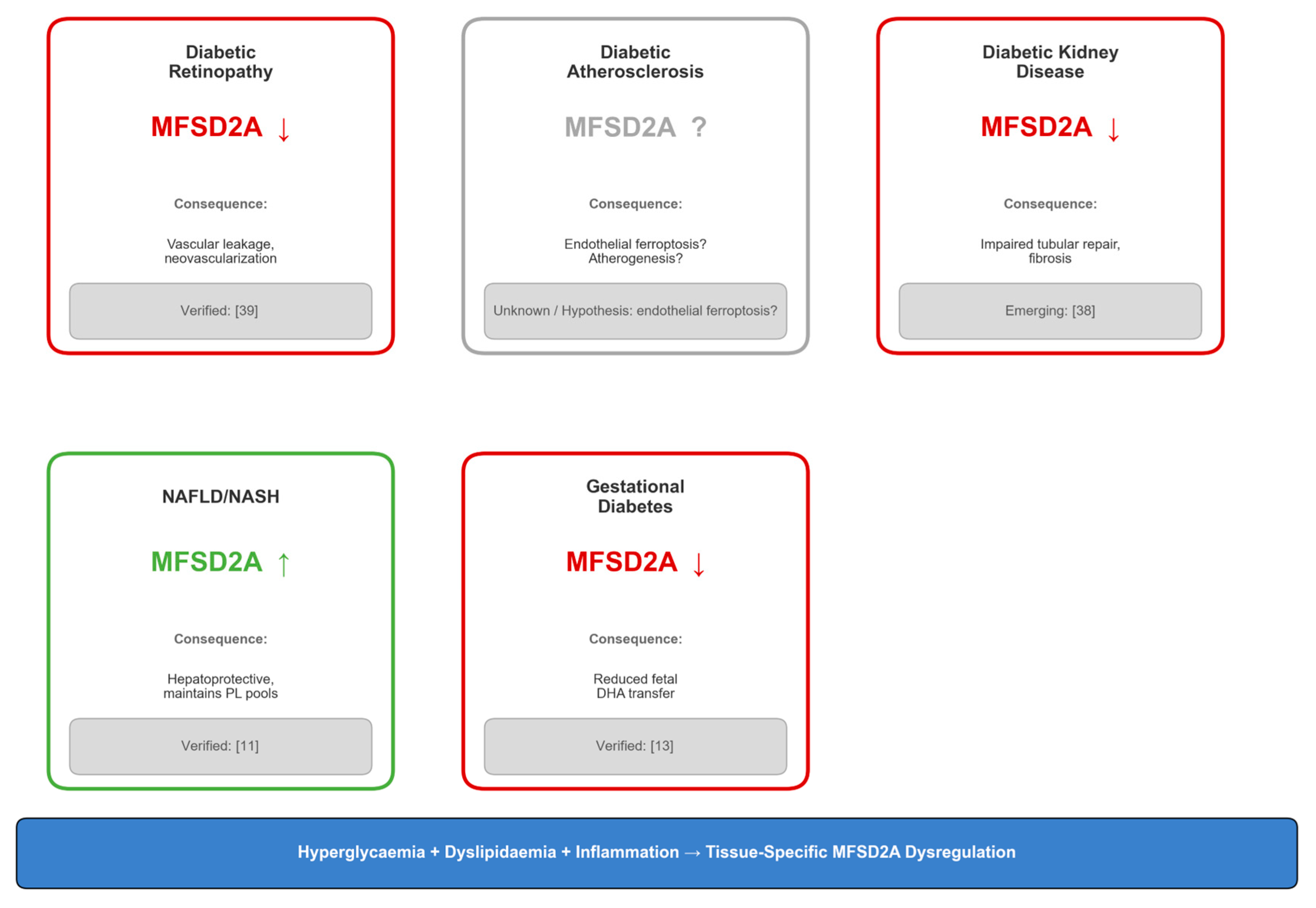

Disclaimer/Publisher’s Note: The statements, opinions and data contained in all publications are solely those of the individual author(s) and contributor(s) and not of MDPI and/or the editor(s). MDPI and/or the editor(s) disclaim responsibility for any injury to people or property resulting from any ideas, methods, instructions or products referred to in the content. |
© 2026 by the authors. Licensee MDPI, Basel, Switzerland. This article is an open access article distributed under the terms and conditions of the Creative Commons Attribution (CC BY) license.