


Abstract
DNA and RNA G-quadruplexes (G4s and rG4s, respectively) are well-known modulators of transcription and splicing. G4s are enriched in transcriptionally active chromatin domains that produce transcripts susceptible to alternative splicing (AS), at least in higher vertebrates. These domains are arranged close to nuclear speckles (NSs), where key AS regulators are stored. Recent findings have revealed new aspects of cotranscriptional AS, emphasizing the advantages of transient rG4s and revealing the connection between excessive rG4s and disease. Here, we discuss how (r)G4s’ presumed ability to promote liquid–liquid phase separation at active chromatin and their affinity for dual regulators of transcription and splicing has brought them to the forefront of communication between NSs and their associated chromatin domains. We also discuss how NS components may contribute to maintaining balanced (r)G4 folding, provide examples of imbalance, and analyze the prospects of G4-based AS reprogramming.
1. Introduction
G-quadruplexes are noncanonical structures found in guanine-rich DNA (G4s) and RNA (rG4s) [1,2,3]. The key sequence requirement for G4 folding is the presence of G3+ tracts (four in unimolecular structures), which are separated by reasonably short loops (typically up to 7 or 12 nucleotides). Guanine residues from different tracts form Hoogsteen-bonded G-tetrads that can coordinate metal cations and stack on top of each other. The resulting quadruple helices have grooves and facets that differ from those of canonical structures. Depending on guanosine configurations, tract orientations, and loop arrangement, they adopt various topologies and therefore show a distinctive yet broad interactome [4].
Intramolecular (r)G4 motifs are abundant in the human genome and transcriptome, and their folding is related to the regulation of key biological processes [4,5]. The relevance of G4s to transcription is supported by their enrichment in promoters [6,7], (super)enhancers [8,9,10], and insulators [11], their ability to block RNA polymerase II (Pol II) or stabilize R-loops [7,12], and their affinity for multiple transcription factors (TFs) [13] and chromatin remodelers [14]. The relevance of rG4s to alternative splicing (AS) is supported by their enrichment at exon/intron boundaries [15] and their affinity for AS regulators, including heterogeneous nuclear ribonucleoproteins (hnRNPs) and serine-arginine-rich splicing factors (SRSFs) [16].
Most (r)G4s are transient due to the activity of specific helicases [17,18,19] and competition with canonical secondary structures among other factors. The local impact of G4 folding/unwinding on transcription is position- and context-dependent and can be negative [20] or positive [21]. In general, however, G4s are associated with transcription activation [22,23] and have been detected in active, superenhancer-governed, GC-rich topologically associating domains (TADs) [10]. Recent studies suggest that GC-rich transcripts produced from such TADs are particularly prone to co-transcriptional AS [24,25]. The precise regulation of AS in higher vertebrates [26] requires the timely recruitment of regulatory factors (hnRNPs and SRSFs) from their “depots,” which are membraneless organelles known as nuclear speckles (NSs) [27,28]. Consistently, GC-rich active TADs are physically close to NSs and can therefore be referred to as speckle-associated domains (SPADs) [29,30].
In this review, we summarize the recent evidence of G4/rG4 folding in SPADs and SPAD-derived transcripts, as well as the contributions of (r)G4s to NS-SPAD juxtaposition through interactions with NS-associated proteins. We also discuss the importance of these interactions for transcription and AS, as well as the consequences of excessive (r)G4 formation upon the expansion of G-rich genomic repeats. Most aspects of (r)G4 contributions to transcription and AS have been reviewed previously [31,32,33]. Here, we contextualize these contributions spatially, focusing on the narrow spatial region (the “interface”) between NSs and SPADs in higher vertebrates. Additionally, we mention key technologies developed in the last decade that gave rise to the concept of NS-SPAD communication and illuminated the role of (r)G4s in this communication.
2. Spatiotemporal Overlaps Between Nuclear Speckles, Active Chromatin and G-Quadruplexes
2.1. Active Chromatin Domains with GC-Rich Isochores Are Associated with Nuclear Speckles
The SPAD concept emerged due to progress in measuring cytological distances [29,34]. This technology is based on labeling DNA proximal to organelles of interest via catalytic tyramide activation (tyramide signal amplification, TSA), which has recently been implemented in proximity ligation assays and in situ hybridization to invigorate spatial molecular biology [35]. In TSA assays, tyramide is typically conjugated to biotin for eventual pulldown and is activated in situ by a peroxidase fused to an antibody to the organelle protein marker. Labeling efficiency reflects proximity to the protein marker because it is determined by the diffusion-dependent local concentration of active tyramide radicals, which decreases exponentially with distance [29]. Initially, TSA was mainly applied in proteomics. A major breakthrough that made it applicable to SPAD mapping was optimizing the conditions for DNA labeling and subsequent sequencing (TSA-seq) [29]. Current gold-standard protocols ensure efficient DNA labeling due to saturating protein labeling (TSA-seq 2.0) [36]. Consequently, the normalized density of sequencing reads can be converted to cytological distances on a micron scale.
Another approach to SPAD mapping relies on chromatin immunoprecipitation and sequencing (ChIP-seq) or cleavage under targets and tagmentation (CUT&Tag) [37] with antibodies to NS-associated proteins. The CUT&Tag technique also uses a protein A/G-fused transposase. When activated by magnesium ions, the transposase cleaves DNA under target proteins (e.g., NS-associated proteins) and adds tags (sequencing adapters). The ChIP-seq/CUT&Tag approach enables SPAD identification based on interactome data rather than actual distances, which might cause some bias. Thus, despite its widespread use in mapping various chromatin compartments, this approach is suboptimal for spatial biology and could not generate the respective concepts. The development of the SPAD concept was only possible with the implementation of TSA-seq. TSA-seq-based SPAD mapping (the red leaf in the shamrock scheme, Figure 1) has been reported for several species and cell lines, including human embryonic stem cells and immortalized cells (H1 and HEK-293), human cancer cell lines (K562 and HCT116), human neonatal (HFF) and adult (HDFa) fibroblasts, and mouse embryonic fibroblasts (3T3) among others [24,25,29,36]. Pairwise comparisons between human cell lines revealed moderate (∼10%) differences in SPAD profiles that followed gene expression differences [36]. CUT&Tag-based SPAD mapping has only been performed on mouse embryonic cells, focusing on different developmental stages [38]. Thus, the profiles obtained by these two approaches cannot be compared directly. However, both have been compared to the profiles of active TADs obtained using conventional chromosome conformation capture methods [29,38,39].
Figure 1.
Evidence for the contributions of DNA/RNA G-quadruplexes (G4s/rG4s) to co-transcriptional speckle-dependent alternative splicing (AS). (Top): a shamrock-like scheme summarizing three lines of research that converged to support the role of transient rG4s and G4s from speckle-associated domains (SPADs) in AS regulation. The key types of assays are underlined, and the respective methods are italicized. (Middle): a timeline of related recent advances in studies of G4s (green), rG4s (blue), and SPADs (red). The two selected advances in enhancer studies (grey) are supposedly relevant to the role of SPAD-associated (r)G4s in AS regulation. Superscripts link the selected advances to references. Green: a, [22]; b, [13]; c, [14]; d, [40,41,42,43,44,45,46,47]. Red: a, [29]; b, [36]; c, [24]; d, [48]; e, [25]. Blue: a, [49,50]; b, [51]; c, [15]; d, [52]; e, [53]. Grey: a, [54]; b, [55]. (Bottom): schematic summary of the hypothetical contributions (dashed arrows) of G4s and rG4s to the speckle-SPAD communication during co-transcriptional AS. The presumed (r)G4 functions are italicized, and the key outcomes are underlined.
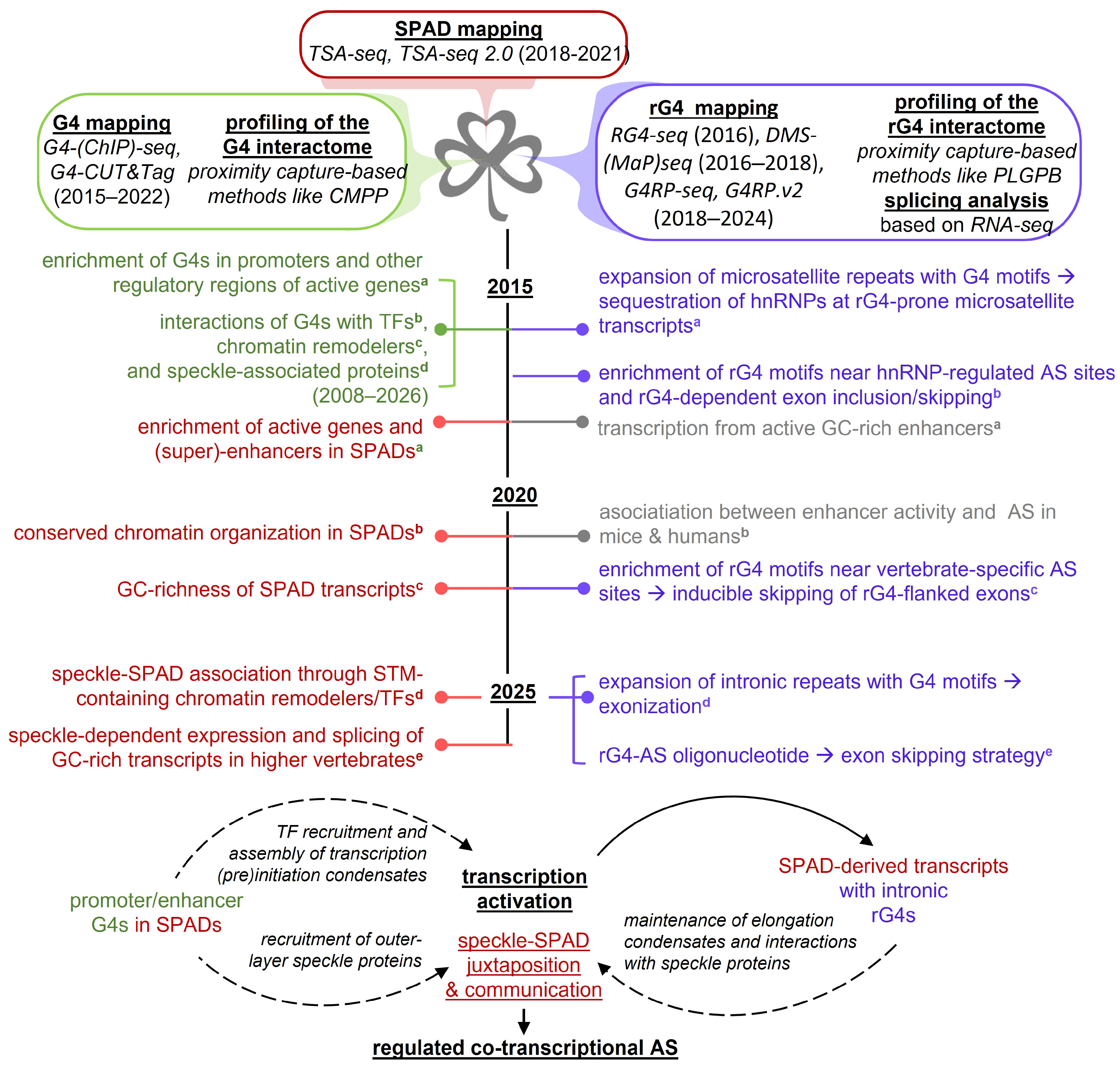
SPADs are very similar to active TADs; however, their boundaries do not fully coincide. As evidenced by CUT&Tag assays, SPADs emerge earlier than TADs during embryogenesis; at least some SPADs have well-defined boundaries while TAD boundaries are still obscure [38]. This implies that NSs do not merely “adjust” to the 3D chromatin structure but rather assist in shaping it. Analysis of SPADs mapped by TSA-seq techniques revealed the prevalence of transcriptionally active regions (>95% euchromatin) with similarly high GC content (GC isochores) [24,25], suggesting the presence of GC-isochore-specific secondary structures, such as G4s in SPADs and rG4s in SPAD-derived transcripts.
2.2. G-Quadruplexes Are Enriched in Active Chromatin Domains and Related Transcripts
The enrichment of G4s in active TADs and rG4s in TAD-derived transcripts has been evidenced using diverse mapping techniques over the past decade (Figure 1: the green and blue leaves in the shamrock scheme). Initial G4 mapping in genomic DNA relied on polymerase stop assays under G4-favoring versus G4-disfavoring conditions, followed by sequencing of primer extension products (G4-seq) [56]. Although the resulting maps were arguably more biologically relevant than sequence-based predictions, they only indicated G4 folding potential in a double-stranded DNA context and did not account for the impact of nucleosome occupancy or other cell-type-specific epigenetic marks. The development of G4-specific antibodies [57,58] has enabled G4 verification in a given epigenetic context using ChIP-seq [59] and CUT&Tag [60] assays. The former assays have been performed on human embryonic stem cells and downstream lineages [61], multiple cancer cell lines, normal fibroblasts [59], etc. The latter assays have been performed on mouse embryonic stem cells and downstream lineages [60]. Both types of assays have evidenced G4 colocalization with active chromatin marks, as well as prevalence in promoters and enhancers, especially in stem cells. Although a subset of G4s was lost during differentiation, colocalization with active chromatin marks remained genome-wide. Thus, the results were mostly consistent with each other and qualitatively agreed with G4-seq data. In contrast, transcriptomic studies raised some controversy.
First-generation high-throughput rG4 mapping methods relied on reverse transcriptase stop assays and sequencing of primer extension products (rG4-seq) [62]. Similar to G4-seq, rG4-seq data can be regarded as maps of potential rather than established structures. Although cell-line-specific RNA processing is intrinsically taken into account in rG4-seq experiments, other specific factors, such as the interplay between helicases and rG4-stabilizing proteins, are not recapitulated. The rG4-seq results reported for the transcriptome of human cancer cells (HeLa) [62] did not align well with classical in-cell dimethyl sulfate (DMS) footprinting [63], and this was attributed to the transient nature of rG4 folding. Despite their generally high thermodynamic stability [64], rG4s appear to be in a more complex equilibrium with other secondary structures than G4s due to RNA’s enhanced conformational polymorphism [65]. A conclusive interpretation of rG4-seq awaits verification using modern DMS-based techniques [66,67], such as mutational profiling (MaP-seq) [68], which uses a specific reverse transcriptase to encode DMS modifications as mismatches [69]. These methods have already confirmed several SPAD-derived therapeutically relevant rG4s [70] but they have yet to be applied in transcriptome-wide studies.
In parallel to DMS-MaP-seq, another rG4 mapping technique called G4-RNA-specific precipitation and sequencing (G4RP-seq) [71] was developed to address the aforementioned RNA polymorphism challenge. G4RP-seq inherits from G4-ChIP-seq, but it also requires initial fixation of the transient rG4 state through cross-linking using a functionalized G4-specific small-molecule probe. This probe is eventually recognized by a corresponding antibody [72]. The application of G4RP-seq and its recent update, G4RP.v2 [73], to the transcriptome of MCF-7 cancer cells confirmed that rG4 folding correlates with transcript G-richness and G4 folding in transcript-encoding DNA. Taken together with G4 mapping results, these findings support the presence of rG4-rich transcripts at active SPADs. However, the transient nature of these rG4s should be noted.
2.3. Transient G-Quadruplexes Are Likely Present near Speckles in G and S Phases
The relatively short lifespan of most (r)G4s hinders their quantification in dynamic processes. Additional limitations stem from the inability of commercially available antibodies, such as BG4 and its derivatives [58], to distinguish between RNA and DNA structures when used in combination with fluorescently labeled secondary antibodies for (r)G4 imaging. RNase treatment and the use of selective G4 and rG4-stabilizing small molecules (such as pyridostatin and its carboxy derivative, respectively) enable partial decoupling of the G4 and rG4 contributions [74]. Nevertheless, the variation in the rG4 fraction with the cell cycle has yet to be fully characterized.
The G4 fraction approaches zero in M phase, increases from G0 to G1, peaks in S phase, and declines in G2 phase [75]. S-phase-specific G4s must be transient because their persistence would impede replication forks [76]. These S-phase-specific G4s differ from G4s in replication origins, which may play a role in replication pre-initiation and origin licensing during the G1 phase [77]. Other G4s are enriched in promoters, enhancers, and untranslated regions (UTRs) [78]. They may be formed in active TADs throughout G-phases to fine-tune transcription [13]. Promoter G4 folding is predetermined by chromatin context and precedes transcription initiation [23], while UTR G4s can precede or follow transcription initiation and are prone to regulation by nascent transcripts through the formation of hybrid DNA/RNA structures, such as R-loops and G-loops [79].
Correlating the overall G4 dynamics with the dynamics of active TADs is challenging. Transcription-related G4s are overshadowed by replication-induced ones [58,75], while TADs simply transform into replication timing domains during the G1-to-S transition [80]. Active TADs form in early G1, achieve maximum insulation from inactive TADs by late G1, and demonstrate enhanced segregation in G2. SPAD dynamics is supposed to be a convolution of TAD and NS dynamics, essentially repeating TAD dynamics because NSs are more or less stable throughout interphase [81]. During M phase, NSs disassemble due to the hyperphosphorylation of their SR-rich core components by SR-specific kinases, Cdc2-like kinases, and casein kinase 2 [82,83,84]. Internal NS dynamics, i.e., reshaping of subdomains scaffolded by different SR-rich proteins [85,86] cannot be ruled out, though partial verification revealed no such reshaping [87].
In summary, promoter/enhancer G4s, as well as some UTR G4s, appear to be present within SPADs from the beginning of interphase. The spatiotemporal proximity of G4s and NSs is an important prerequisite for the hypothesis that G4s contribute to NS positioning or chromatin organization around NSs (the bottom scheme in Figure 1). Along with rG4s, these G4s are presumed to participate in NS-SPAD communication by recruiting speckle-associated proteins, as discussed in the following sections. Hereafter, we focus on the proposed causative relations illustrated by the dashed arrows in Figure 1, noting that they can be bidirectional. In particular, we emphasize the roles of prefolded G4s in activating transcription but also acknowledge that this activation facilitates G4 folding under certain conditions. Similarly, we emphasize the contributions of G4s and rG4s to the recruiting NS proteins, but we also note that these proteins can modulate G4 formation, at least in vitro. Thus, the proposed causative relations shape the core hypothesis, and its future refinements may contain dynamic feedback networks.
3. G-Quadruplex-Dependent Biocondensates at Speckle-Associated Active Chromatin
3.1. Transcriptional Biocondensates Form at Speckle-Associated Chromatin
The presence of long, multivalent enhancers (superenhancers) enables SPADs to accumulate TFs and chromatin remodelers to the critical concentration required for liquid–liquid phase separation [88]. This process can be visualized as the assembly of dynamic protein hubs (Figure 2a) on hyperacetylated promoters and enhancers that are decorated by the bromodomain-containing chromatin remodeler, BRD4. Prior to transcription initiation, the mediator complex connects BRD4 and accessory activators to the initially non-phosphorylated (inactive) Pol II and major TFs [89]. The mediator, BRD4, and several TFs, including those with zinc-finger domains, engage in transient multivalent interactions through their intrinsically disordered regions (IDRs). A combination of non-specific IDR contacts and specific interactions involving bromodomains, zinc finger domains, or other domains holds the biocondensate together [90].
Figure 2.
G-quadruplexes at the forefront of communication between speckles (NSs) and their associated chromatin domains (SPADs). (a) (Left): Schematic representation of the relative positions of NSs and active versus inactive topologically associating chromatin domains (TADs). (Right): Schematic representation of the interface between NSs and transcriptional condensates at SPADs. (b) Putative G4s (green) and rG4s (blue) at the NS-SPAD interface: the positions of the key motif clusters and examples of confirmed structures.
The nucleation step of the biocondensate assembly requires the simultaneous recruitment of multiple TFs. This requirement is only met at superenhancers, which can contact several promoters simultaneously [90,91]. Their juxtaposition is enabled by DNA loop extrusion by cohesin, with loop borders marked by the insulator protein CTCF [92]. Subsequent maturation of the condensates during transcription initiation may involve the incorporation of enhancer RNA that transiently interacts with the IDRs of the condensate proteins.
This view of superenhancer-governed transcription activation stems from a comparison of 3D chromatin maps revealed by chromosome conformation capture-based methods and Pol II/TF occupancy revealed by classical ChIP-seq and RNA sequencing. Recent updates to these methods, which supported the specific roles of superenhancers and GC-rich enhancer RNA, include high-sensitivity nascent transcript sequencing [93] and cap analysis of gene expression [54]. Super-resolution imaging of cells expressing biocondensate components with photoconvertible fluorescent tags confirmed the co-separation of these components [89]. Tracking clusters of tagged components after photoconversion or monitoring fluorescence recovery after photobleaching revealed the dynamics of the biocondensate interior, consistent with its presumed viscoelastic state [94]. Combined immunofluorescence and DNA fluorescence in situ hybridization demonstrated the positioning of the biocondensates at superenhancers [89,95]. Thus, several lines of evidence suggest that at least some SPADs serve as scaffolds for biocondensate formation. It should be noted that the key evidence was obtained in mouse embryonic stem cells [89,95]. However, the majority of transient protein clusters—presumed transcriptional biocondensates—persisted during differentiation and resembled those previously observed in differentiated human cells [96,97]. Therefore, the thesis about biocondensate formation likely applies to multiple cell lines and species.
3.2. G-Quadruplexes Assist in Assembling the Transcriptional Condensates
Although G4 enrichment in promoters and (super)enhancers is well-documented [6,7,8,9,10], the contribution of these G4s and rG4s to biocondensate formation by increasing the multivalency of promoter DNA and enhancer RNA has only recently been recognized [98,99]. This contribution is partly due to their semi-specific binding affinity toward chromatin remodelers/TFs with bromodomains or zinc-finger domains, including the transcriptional condensate scaffolding proteins BRD4 [100] and CTCF [101,102], as well as the ubiquitous GC-box-targeting TF SP1 [103].
CTCF recruitment to G4s has been confirmed to promote chromatin looping, as evidenced by the formation of new 3D chromatin contacts after a G4 insertion [104]. While not a general prerequisite for biocondensate formation, chromatin looping is crucial at (super)enhancers because it enables enhancer-promoter communication [105]. Whether BRD4 is crucial for G4-mediated enhancer-promoter communication remains to be verified in cells. Recent immunoprecipitation of enhancer quadruplexes revealed the potential for protein-independent, G4-mediated communication [9] through intermolecular folding of G-tracts from promoters and enhancers.
SP1 is not a well-established contributor to enhancer-promoter interactions. Although it facilitates the assembly of the preinitiation complex by interacting with the TATA-binding protein, its ability to recruit transcriptional repressors makes the outcome condition-dependent [106]. Furthermore, SP1 recruitment to canonical double-stranded GC-rich motifs versus G4 motifs is context-dependent. Nevertheless, SP1 is strongly associated with G4 motif-containing oncogene promoters and with superenhancers in several human cancer cell lines, where it activates transcription by promoting biocondensate formation [107]. Whether SP1 is recruited to promoter/superenhancer G4s or double stranded GC-rich motifs awaits clarification. Recently, SP1 was confirmed to colocalize with Pol II and the mediator and to facilitate biocondensate formation in a G4-dependent manner at least in the case of Bcl3 oncogene promoter [108].
In addition to semi-specific interactions with zinc-finger domains, bromodomains and probably some other domains of TFs and chromatin remodelers, G4s are prone to non-specific contacts with protein IDRs through π-π and cation-π interactions, which are among the most frequent in biomolecular condensates [109].
In summary, G4s could assist in assembling transcriptional biocondensates in multiple ways, but further investigation is needed. The most direct evidence of G4-mediated intracellular phase separation reported thus far is probably the formation of speckle-like puncta following the addition of exogenous G4s. However, this evidence was obtained in artificial cells [98]. Additionally, although G4 enrichment in promoters and superenhancers is well-established for multiple cell lines [10,22], the fact that G4s precede transcription initiation has only been definitively confirmed for the K562 cell line [23], which had been used in most early G4 mapping experiments [78,110].
3.3. G-Quadruplexes Are at Play in Both Initiation- and Elongation-Related Condensates
Liquid–liquid phase separation at superenhancers suggests that the NS-SPAD interface is a two- condensate interface, or a three-condensate interface if transcription (pre)initiation condensates and elongation condensates are distinguished [111]. The assembly of the latter can be considered a maturation of the former. The key component present in both (pre)initiation and elongation condensates is Pol II [112]. Initiation requires phosphorylation of its C-terminal domain by the cyclin-dependent kinase subunit of TFIIH, while elongation requires further phosphorylation by the cyclin-dependent kinase subunit of P-TEFb [113]. Eventual hyperphosphorylation alters co-separation preferences of Pol II [114], which is consistent with changes in the biocondensate content.
According to the “scaffold-client” model [115], the mediator complex, BRD4, and inactive Pol II are scaffolds of (pre)initiation condensates [89], while active (hyperphosphorylated) Pol II and some accessory proteins (e.g., the histone chaperone FACT) are scaffolds of elongation condensates [116]. Most TFs appear to be “clients,” entering condensates due to their affinity for DNA and scaffolds [117]. The defining feature of a scaffold is the presence of long IDRs. These IDRs are typically low-complexity sequences showing specific amino acid biases and repeated patterns, such as the YSPTSPS heptad repeats in the C-terminal tail of Pol II.
DNA is not a canonical scaffold; rather, it has been referred to as a “surface” for condensate assembly [118]. However, it can be described using the sticker-spacer framework proposed for scaffolds. Within this framework, scaffolds are composed of “inert” flexible elements (spacers) interspersed with interaction-prone elements (stickers) [119]. Key SPAD stickers are canonical TF binding sites and G4s within (super)enhancers and promoters [23]. Whether G4s in UTRs and first introns are folded prior to transcription and can act as stickers in (pre)initiation condensates is an open question. There is strong evidence that clustered intronic and intergenic G4s contribute to drug resistance-related gene activation in ovarian cancer cells, where these clusters act similarly to superenhancers [120].
In general, however, clusters of quadruplex motifs downstream of the transcription start site (Figure 2b) [121] may correspond predominantly to rG4s and co-transcriptionally folded G4s in R-loops. Interestingly, the ability of coding strand G4s to stabilize R-loops [12] results in a positive feedback-like relationship. However, R-loop-related DNA damage remains low under normal conditions [79,122], because this positive feedback is balanced by the activity of helicases and NS-associated proteins, as reviewed below. Regarding transient rG4s in intronic motif clusters, one can expect them to support Pol II-scaffolded biocondensates during transcription elongation, given the tendency of rG4s to promote liquid–liquid phase separation of IDR-containing proteins in artificial models [123].
3.4. Nuclear Speckles and Transcriptional Condensates Are Interdependent
The major scaffolds of NSs are SR-rich proteins. These include the nearly entirely disordered SRRM2 and SON [86,124] as well as the SRSFs [85]. The latter contain RNA recognition motifs (RRMs) in addition to SR motifs. Together with noncoding RNAs (primarily MALAT1), they constitute the inner (core) parts of NSs [125]. Other RNAs and small nuclear ribonucleoproteins (snRNPs) act as clients and are distributed between the inner and outer layers of NSs. The outer layer is enriched with heterogeneous ribonucleoproteins (hnRNPs) [126], which can be regarded as secondary scaffolds. Similar to SRSFs, they contain RRMs and IDRs, but these IDRs are more diverse in terms of amino acid bias (e.g., acidic or aromatic IDRs, prion-like domains, and RGG motifs).
The multilayer organization of NSs was initially identified using canonical in situ hybridization, immunostaining, and immunoblotting methods [87], and was recently confirmed using TSA-based methods combined with mass spectrometry [127]. Outer layer proteins likely shuttle between NSs and SPADs. Many of these proteins affect transcription elongation (e.g., hnRNP A1 and hnRNP R [128]) and/or termination (e.g., hnRNP K [129]), as well as pre-mRNA polyadenylation [129], splicing [130] and trafficking [131]. Thus, hnRNPs act both as transcription regulators and as RNA processing factors, accounting for the communication between elongation condensates and NSs.
The interdependence of NSs and (pre)initiation condensates is supported by the association between AS and the activity of multiple transcriptional enhancers, at least in mice and humans, as summarized in the Visualizing Enhancer-AS Associations (VEnAS) database [55]. This could be attributed to enhancer RNA, which either recruits NS components or enters NSs itself (Figure 2a). Another aspect of the NS-SPAD communication involves the ability of major transcription initiation factors, such as TAF2, and architectural proteins, such as CTCF, to enter NSs under certain conditions, which affects AS [132,133]. For instance, AS reprogramming has been reported in response to SRRM2 interactions with CTCF aggregates in senescent cells [133].
The propensity of TFs and chromatin remodelers to enter NSs has recently been linked to the presence of specific signaling motifs termed “speckle-targeting motifs” (STMs) [48]. Common STMs, identified by aligning the sequences of proteins with confirmed NS localization, are ~30-mer motifs with proline residues at defined positions. Future studies are needed to further elucidate the sequence requirements, and the set of NS-associated proteins may be refined or expanded. Nevertheless, the interdependence of NSs and SPAD biocondensates due to the exchange of the outer-layer proteins is firmly established. Below, we review evidence that many of these proteins are G4- and/or rG4-interactors, suggesting that (r)G4s contribute to NS-SPAD communication.
4. Interactions Between G-Quadruplexes and Speckle-Associated Proteins
4.1. G-Quadruplexes Recruit Speckle Proteins to Transcriptional Condensates
SPAD-associated components of transcriptional condensates, such as TAF2 and CTCF, are recruited to speckles through transient interactions of IDRs [132,133], which may include STMs. In contrast, the recruitment of NS components to transcriptional condensates involves specific interactions between NS components’ RRMs and RNA/DNA. Importantly, RRMs hardly recognize B-form DNA and most unstructured single-stranded DNA, but they can recognize G4s and rG4s. This explains why some NS components, particularly hnRNPs, modulate transcription initiation at G4-harboring promoters [40]. Initially, such G4-hnRNP interactions were revealed by classical, non-high-throughput methods like mass spectrometry-based analysis of mixtures obtained by pull-down assays using magnetic beads or peptide-based scaffolds decorated with G4s [41,42]. Subsequently, these interactions were confirmed by in situ proximity capture-based methods including high-throughput assays [43] like co-binding-mediated protein profiling (CMPP), a method that employs a functionalized G4-specific ligand to crosslink G4s with protein interactors [44]. Examples of hnRNP-binding G4s shown in Figure 2b are the KRAS oncogene promoter structures recognized by hnRNP A1 [45].
Most hnRNPs are best known as rG4 binders [46,47]. Interactions of hnRNPs with UTR rG4s, especially those that overlap with internal ribosome entry sites (IRES), affect translation [134], whereas interactions with rG4s at intron/exon boundaries affect spliceosome assembly and eventually AS [32]. Interestingly, SRSFs rarely interact with G4s in transcription (pre)initiation condensates but likely interact with rG4s in nascent transcripts [135]. This is probably because only the SRSF-poor outer layer of the speckles partially intermixes with transcription (pre)initiation condensates. In contrast, nascent transcripts can enter both layers, according to the “interfacial” splicing model (Figure 3a), hence the accessibility of rG4s to inner NS proteins.
Figure 3.
G-quadruplexes in co-transcriptional splicing: regulatory roles, consequences of expansion, and application in splicing reprogramming. (a) Normal contributions of (r)G4s from speckle-associated chromatin (SPAD) to transcription and co-transcriptional alternative splicing (AS) through interactions with the transcriptional machinery and the components of nuclear speckles (NS). Solid arrows indicate relations supported by multiple lines of evidence, including assays on cell cultures. Dotted and dashed arrows indicate hypothetical relations and those supported by in vitro assays in cell-free systems, respectively. (b) The impact of rG4 accumulation due to pathogenic expansion of G4-prone genomic repeats (e.g., G4C2 repeats in C9orf72) on NS morphology and AS. (c) Reprogramming AS by inducing an rG4 within the exonic splicing enhancer using a G-rich antisense oligonucleotide (G-ASO).
Examples of SRSF1-binding rG4s shown in Figure 2b are the IRES-regulating rG4 from 5′-UTR of the actin-related gene ARPC2 [136] and the rG4 from the first intron of the C9orf72 gene [135]. The latter also has affinity to hnRNP A1 [137]. Like G4-interacting proteins from NSs, most rG4-interacting proteins were first identified by pull-down assays [41] and then confirmed by high-throughput, proximity-capture-based methods such as eCLIP (enhanced cross-linking and immunoprecipitation) [138] or PLGPB (G-quadruplex-interacting proteins specific biotin-ligation procedure) [139].
4.2. Speckle Proteins Unwind G-Quadruplexes in Transcriptional Condensates
One underinvestigated issue regarding NS proteins is their (r)G4 helicase-like activity [40,135,140,141]. Unwinding of promoter/(super)enhancer G4s associated with active transcription seems counterintuitive, yet it may facilitate the transition to productive elongation. The details await clarification. The domain structures of the NS proteins, specifically the presence of RRMs and IDRs, allow for several possible explanations of the helicase-like activity [126,142]. First, RRMs may recognize G-tracts as unstructured single strands (linear motifs), shifting the equilibrium between G4 folding and unfolding towards the latter. Second, RRMs may initially recognize loop sequences; the subsequent engagement of G-tracts could then distort the G4 geometry. Third, together with adjacent IDRs, RRMs could recognize the secondary structure rather than the sequence and destabilize the G4 by disrupting its core with IDR side chains.
The first two hypotheses can be partially verified by comparing G4 motifs to the canonical linear binding motifs of NS proteins. Considering the common tandem RRM arrangement in SRSFs and hnRNPs, these canonical binding motifs can be bipartite. However, tandem RRMs tend to act in concert while adopting different orientations and showing different specificities, depending on the linker between their binding sites [143]. One example is TDP-43. It recognizes sequences that are different from the tandem arrangement of its isolated RRM1 and RRM2 binding motifs [144] and exhibits pronounced affinity for GT-rich single-stranded DNA (ssDNA), as well as parallel-stranded G4s. Another representative example is hnRNP A1. Its RRMs have high affinity for purine-rich sequences. In complex with the intronic splicing silencer of the SMN2 transcript, they recognize the silencer elements GGAA and GAC [145]. These elements resemble, but do not fully match, the hnRNP A1 consensus binding motifs selected in vitro: 5′-UAGGGA/U-3′ in RNA and 5′-TAGGGA/T-3′ in DNA [146]. Some (r)G4s recognized by hnRNP A1 comply with the consensus motif; others do not, suggesting both sequence- and structure-specific interactions. Examples of the former include telomeric (r)G4s folded from TTAGG repeats [147]. Examples of the latter include G4s from the nuclease-hypersensitive element of the KRAS promoter, 32R (AG3CG2TGTG3AAGAG3AAGAG5AG2) [45], which contains multiple G-tracts and exists as a mixture of G4 conformers, modelled by the G-to-T substitution variants G9T and G25T (Figure 2b). Thus, the recognition of G-tracts or loop sequences as linear motifs can only explain unwinding of a subset of G4s, which indirectly argues for structure-specific binding and unwinding.
Conclusive evidence of structure-specific versus sequence-specific binding can be obtained by comparing the interactomes of G4s and their analogs with guanosine residues substituted with 7-deazaguanosine [148], 8-oxo-guanosine [149], or other derivatives that impair Hoogsteen bonding while largely preserving Watson–Crick bonding. Reported high-throughput assays of this kind lacked focus on NS proteins, and the borderline thermodynamic stability of the analyzed 2-tetrad G4s complicated the interpretation of the results [150]. Additional challenges arose from the conformational polymorphism of (r)G4s. Although most of them are parallel-stranded, i.e., adopt topologies of the same type [151], non-uniform G-tracts (excessive, extended, truncated, or interrupted) enhance the topological diversity due to register shifts and various core defects, such as bulges or imperfect tetrads. This can be seen in the KRAS G4 variants (Figure 2b). With all limitations taken into account, comparative analyses of interactomic data obtained for (r)G4s and their non-G4 analogs with modified nucleobases [150] argue that at least some interactions are structure-specific.
Other recent studies suggest that arginine-rich RRM-neighboring IDRs play a role in both G4 recognition and unwinding [152,153,154]. These arginine-rich IDRs are SR motifs present in all SRSFs [135,152] and RG/RGG motifs present in multiple hnRNPs, including hnRNP A1 [154], hnRNP A2/B1 [155], FUS [156], TDP-43 [157], hnRNP K [158], hnRNP U [159], etc. The mechanisms have been partially disclosed based on binding assays with SR/RGG mutants [160] and characterization of the wild-type complexes using classical biophysical methods (e.g., NMR spectroscopy) and modern single-molecule techniques [155]. Arginine residues appear to be the principal residues involved in recognition. Cation-π interactions between IDR arginine guanidino groups and the outer (r)G-tetrad appear to promote (r)G4 unwinding by facilitating further electrostatic contacts with the internucleotide phosphate linkages of the (r)G4 backbone, altering the backbone packing.
In summary, multiple NS-associated proteins exhibit an (r)G4 helicase-like activity due to the combined effects of their RRMs and IDRs. G4 unwinding is likely followed by protein dissociation, consistent with the dynamic nature of the NS-SPAD communication. The consequences of the repositioning of NS-associated proteins to nascent transcripts, where rG4s are mostly clustered at intronic boundaries [121], are discussed in the following section.
5. Splicing-Related Consequences of G-Quadruplex Interactions with Speckle Proteins
5.1. G-Quadruplexes Act Alongside Canonical Splicing Enhancers and Silencers
The fact that both SRSFs [152,155] and hnRNPs [46,137,161] bind to rG4s suggests a potential competition between these factors at intronic boundaries (Figure 2b). Although cis-regulatory elements are commonly classified as intronic splicing silencers versus exonic splicing enhancers, the bound AS regulator rather than position alone defines the regulatory outcome. Thus, the clustering of rG4 motifs in introns does not automatically mean they repress splicing. Some cis-regulatory sites have comparable affinities for SRSFs and hnRNPs, and rG4 motifs appear to be among these sites [16]. Moreover, rG4s can exist in equilibrium with alternative secondary structures and unfolded RNA, which results in complex competition between hnRNPs and SRSFs for linear motifs and rG4s (Figure 3a).
The role of intronic rG4s in AS has been demonstrated and partially decoupled from that of linear regulatory sites in human neurons [15]. Enhanced skipping of G4-flanked exons, as evidenced by RNA-seq, was observed after treating neurons with potassium chloride. This treatment reduces membrane potential and triggers intracellular Ca2+ influx, supposedly affecting hnRNPs that are phosphorylated by Ca2+-sensitive calmodulin-dependent kinases [162] and their partners or competitors. eCLIP assays revealed the accumulation of RGG-containing hnRNPs, including hnRNP K and hnRNP U at the boundaries of the potassium-sensitive differentially skipped exons [15]. Thus, the skipping could be attributed to the recruitment of splicing inhibitors by exonic rG4s. Incorporating representative differentially skipped exons and their G4-prone or mutant (G-rich non-G4) flanks into artificial minigenes enabled a direct comparison of AS outcomes and confirmed the importance of rG4 folding. This example illustrates the membrane potential-dependent rG4 contribution to AS regulation, which may be of particular importance in excitable cells.
In several immortalized non-cancer cell lines, analysis of RNA binding protein footprints suggests that major clusters of rG4motifs recruit RGG-free hnRNP H and hnRNP F [51]. Unlike most family members, these hnRNPs promote exon inclusion. Like most family members, they bind folded rG4s more efficiently than linear G-rich motifs. Consistently, mutations that disrupt intronic rG4 folding while maintaining linear G-rich motifs promoted skipping of G4-flanked exons, as evidenced by minigene reporter assays [51]. Similar exon skipping has been observed upon hnRNP H/F knockdown or cell treatment with hnRNP-displacing ligands [163].
It can be summarized that rG4s act alongside canonical intronic splicing enhancers and silencers, mostly in an hnRNP-mediated manner. The benefits of their unwinding by a subset of hnRNPs are likely linked to translation rather than AS [164]. The role of SRSFs is either underinvestigated or less pronounced, probably because key G4 clusters contact hnRNPs first, as suggested by intron positioning at the periphery of NSs in the “interfacial” splicing model (Figure 3a) [165]. Finally, it should be noted that although intron boundaries are the most common positions of rG4 motifs and motif clusters (Figure 2b), individual motifs and folded rG4s are also present in exons. The effects of such rG4s on AS are similarly context-dependent and defined by the proteins recruited [32].
5.2. Excessive G-Quadruplexes Alter Speckles and Disrupt Alternative Splicing
Transient rG4s contribute to the balanced AS by recruiting NS proteins without disrupting NS morphology. In contrast, excessive rG4s resulting from the expansion of G-rich microsatellites are associated with pronounced NS distortions [27]. Two relevant examples are GGC and G4C2 expansions.
GGC expansions occur in diverse genomic loci (UTRs/introns of at least six genes) and cause various pathologies, the most common of which are myopathy and neurological disorders [166,167]. Expansions within open reading frames lead to the accumulation of toxic polyglycine- or polyalanine-containing proteins, which often affect NSs and other biocondensates [167]. For instance, the polyalanine-containing polyadenylate-binding protein nuclear-1 forms toxic aggregates and disrupts NSs by extracting poly-A mRNA [168]. Alternatively, its aggregation may occur within NSs. GGC repeats can theoretically fold into G4s or rG4s [169], but these structures are rarely observed in genomic DNA, probably due to the insufficient GC skew and the destabilizing effects of CpG methylation. The most common structures in GGC repeat-containing RNA are noncanonical loops, which recruit and sequester non-NS proteins, such as the microRNA biogenesis regulator DGCR8 and its partners [170].
The expansion of G4C2 repeats in the C9orf72 intron 1 is the most common genetic cause of several neurodegenerative diseases, including amyotrophic lateral sclerosis and frontotemporal dementia [171]. Similar to GGC expansion, the G4C2 expansion triggers pathology through a multi-pathway mechanism, involving both RNA repeats and the products of their translation [172]—poly-GA, poly-GR, poly-GP, poly-PR, and poly-PA—among other factors. The transcripts fold into rG4s, aggregate through multimerization [173], and promote aggregation of recruited proteins [49]. The relevance of the RNA repeats to pathology is supported by the detection of RNA foci [174], which accumulate and sequester RNA processing-related proteins, including NS proteins [49,50,175] (Figure 3b). This explains AS aberrations in patients with amyotrophic lateral sclerosis or frontotemporal dementia caused by repeat expansions [49].
The role of the poly-dipeptides [176] is supported by the fact that preventing translation of respective RNAs by editing the repeat-preceding CUG codon [177] alleviates most symptoms [178]. Among the dipeptide repeats, arginine-rich ones appear particularly toxic because they disrupt NSs and other intracellular biocondensates due to their high propensity for cation-π interactions [179]. The exact mechanism of their accumulation has long been puzzling. Recently, it was demonstrated that multiple rG4s can reshuffle NS layers and recruit SRSF1 to the C9orf72 intron 1 (Figure 3b). This activates a cryptic splice site (Figure 3b), resulting in the exonization of the expanded repeats [52]. This finding does not contradict the proposed role of the CUG codon as a target for genome editing [178]. However, it suggests that at least some dipeptide repeats are produced through the canonical translation of the extended exon 1. This is a rare but significant example of rG4-driven AS reprogramming in an SRSF-mediated manner.
5.3. Inducing G4 Formation Within Splicing Enhancers Enables Controlled Exon Skipping
Recently, substantial effort has been devoted to AS reprogramming for preventing the inclusion of exons that introduce premature stop codons [180]. Such stop codons may result from nonsense or frameshift mutations. Well-known examples include DMD mutations causing Duchenne muscular dystrophy through a loss of dystrophin function [181]. Skipping the damaged exon or the adjacent one can restore protein function if the remaining exons align in-frame to restore translation. For instance, skipping mutant exon 23 in the mouse dystrophin transcript and skipping single or multiple exons from the DMD-related deletion hotspot of the human dystrophin transcript (ex. 45–55) [182,183] produce partially functional proteins. This strategy has been implemented using heteroduplex-forming antisense oligonucleotides that target the exonic splicing enhancer, as well as some peptide-oligonucleotide conjugates [183,184].
To date, four heteroduplex-forming antisense oligonucleotides, all with a morpholino phosphoramidate backbone, have been approved for muscular dystrophy treatment in patients with respective exon-skipping-amendable mutations [185]. Limitations of these drugs include relatively narrow patient coverage (below 15% for each drug) and modest therapeutic efficiency. Due to the accelerated FDA approval and the lack of long-term comparisons, it is difficult to make conclusive remarks about the overall clinical benefits. However, dystrophin recovery induced by these drugs is low in most cases, suggesting inefficient exon skipping, probably because the antisense oligonucleotides cannot fully shield the splicing enhancer and/or displace the splicing-activating protein. In search of more reliable ways to distort splicing enhancers, an rG4-based splice switching strategy was developed [53] (Figure 3c).
In this strategy, the antisense oligonucleotide module is combined with a G-rich, rG4-inducing module. The latter is the key functional unit, and the former accounts for specificity. A similar double-module design was recently used to inhibit the translation of monoamine oxidase-encoding RNA. The thermodynamically unstable 2-tetrad unimolecular rG4 in its UTR served as a weak translation inhibitor, but it was rearranged into a more stable and potent 3-tetrad bimolecular rG4 by a G-rich oligonucleotide conjugated to an antisense module [164]. In the case of rG4-based dystrophin splice switching (Figure 3c), the complete four-tract G4 motif does not need to be present in the target sequence. A conjugate with a G-rich module comprising several G-tracts can be directed to a single G-tract located up to 70 nt from the antisense-targeted sequence [53]. A key limitation of rG4-inducing antisense oligonucleotides comprising multiple G-tracts is their propensity for multimerization. This problem can be mitigated by using interrupted G-tracts, which produce G4s with bulges.
The tested bulged G4-inducing antisense oligonucleotides were highly efficient (induced approximately 80% exon skipping) in cultured cells and moderately efficient (induced nearly 15% exon skipping) in a mouse model [53]. Direct comparisons between these oligonucleotides and conventional heteroduplex-forming antisense oligonucleotides [184] are needed to determine if G4-based splice switching therapy significantly improves protein function recovery. The prospects of its translation to clinics also depend on the drug delivery strategy, which in turn depends on the potential optimization of the oligonucleotide backbone structure. The 2′-OMe modification selected for rG4-inducing oligonucleotides has disadvantages (e.g., reduced penetration through cellular membranes) as well as advantages (e.g., the wider commercial availability) over the morpholino modification. Variations in the backbone structure can profoundly affect the rG4-inducing potential.
In summary, inducing rG4 folding within splicing enhancers is an exciting, albeit under-explored, splice-switching strategy that will hopefully be further developed in upcoming studies. In view of the complex and context-dependent role of rG4s in NS-dependent splicing, elucidating the interactions of induced rG4s with NS-associated proteins—especially splicing-enhancing versus splicing-repressing hnRNPs—could advance progress in the field.
6. Conclusions
In this review, we summarized the potential contributions of (r)G4s to the regulation of transcription and splicing in higher vertebrates. A distinctive aspect of this regulation is the accumulation of regulatory machinery in interdependent biomolecular condensates, namely NSs and transcriptional condensates at SPADs. The latter appear to be rich in G4s and produce G-rich transcripts, which form rG4s. Studies in model systems suggest that (r)G4s facilitate the formation of transcriptional condensates and their communication with NSs. This communication involves incorporating the rG4-rich enhancer RNA and SPAD-derived transcripts into NSs during transcription initiation and elongation, respectively. It also involves the shuttling of some transcription factors or (r)G4-binding outer-layer NS proteins between transcriptional condensates and NSs during transcription initiation or elongation, respectively. Some of these shuttling proteins unwind G4s and rG4s, which may facilitate productive transcription elongation and the eventual translation of processed transcripts. Like many other cis-regulatory elements, G4s and rG4s recruit trans-regulators in a context-dependent manner. However, their key hotspots in promoters and introns, respectively, are generally associated with transcription activation and exon inclusion.
Thus, although (r)G4s are not crucial for transcription and splicing in general, they recruit regulators of these processes, especially dual regulators. This facilitates co-transcriptional alternative splicing (AS) and enables timely AS reprogramming in response to stimuli. While this offers evolutionary benefits, it also predisposes higher vertebrates to pathology if the intricate balance between (r)G4 folding and unwinding is disrupted. Excessive (r)G4s—for example, those formed by expanded G-rich repeats—can sequester NS proteins, alter NS architecture, and cause widespread splicing defects. This has been demonstrated in models of neurodegenerative diseases driven by G4C2 repeat expansion. In contrast, controlled (r)G4 folding within canonical exonic splicing enhancers enables splice switching, as demonstrated by the programming of exon skipping in the mouse dystrophin transcript in the Duchenne muscular dystrophy model. Further research on (r)G4s in the context of NS-SPAD communication and AS control could advance splice-switching strategies and related therapeutic approaches. Specifically, elucidating the sequence and structural determinants of NS protein-rG4 interactions is necessary to clarify their targetability. A deeper understanding of how transient versus persistent/excessive rG4s impact NSs and transcriptional condensates, as well as the development of new rG4-destabilizing molecules, may inspire new approaches to mitigating the effects of G-rich repeat expansion.
Author Contributions
Conceptualization, A.V.; writing—original draft preparation, P.R., J.K. and A.V.; writing—review and editing, P.R., J.K. and A.V. All authors have read and agreed to the published version of the manuscript.
Funding
This work was funded by Russian Science Foundation (22-15-00129-P).
Institutional Review Board Statement
Not applicable.
Informed Consent Statement
Not applicable.
Data Availability Statement
No new data were created or analyzed in this study. Data sharing is not applicable to this article.
Conflicts of Interest
The authors declare no conflicts of interest.
Abbreviations
The following abbreviations are used in this manuscript:
| AS | Alternative splicing |
| ChIP-seq | Chromatin immunoprecipitation and sequencing |
| cMPP | co-binding-mediated protein profiling |
| CUT&Tag | Cleavage under targets and tagmentation |
| DMS | Dimethyl sulfate |
| MAP-seq | Mutational profiling and sequencing |
| eCLIP | Enhanced cross-linking and immunoprecipitation |
| G4 | DNA G-quadruplex |
| G4RP | G4-RNA-specific precipitation |
| G4-seq | G4 mapping based on polymerase stop assays and sequencing |
| G-ASO | Antisense oligonucleotide with a G-rich module |
| hnRNP | Heterogeneous nuclear ribonucleoprotein |
| IDR | Intrinsically disordered region |
| NS | Nuclear speckle |
| PLGPB | G-quadruplex-interacting proteins specific biotin-ligation procedure |
| Pol II | RNA polymerase II |
| rG4 | RNA G-quadruplex |
| RG4-seq | rG4 mapping based on reverse transcriptase stop assays and sequencing |
| RRM | RNA recognition motif |
| snRNP | Small nuclear ribonucleoprotein |
| SPAD | Speckle-associated domain |
| SRSF | Serine-arginine-rich splicing factor |
| STM | Speckle targeting motif |
| TAD | Topologically associated domain |
| TF | Transcription factor |
| TSA | Tyramide signal amplification |
| UTR | Untranslated region |
References
- Burge, S.; Parkinson, G.N.; Hazel, P.; Todd, A.K.; Neidle, S. Quadruplex DNA: Sequence, Topology and Structure. Nucleic Acids Res. 2006, 34, 5402–5415. [Google Scholar] [CrossRef] [Scilit] [PubMed]
- Huppert, J.L.; Balasubramanian, S. Prevalence of Quadruplexes in the Human Genome. Nucleic Acids Res. 2005, 33, 2908–2916. [Google Scholar] [CrossRef] [Scilit] [PubMed]
- Varshney, D.; Spiegel, J.; Zyner, K.; Tannahill, D.; Balasubramanian, S. The Regulation and Functions of DNA and RNA G-Quadruplexes. Nat. Rev. Mol. Cell Biol. 2020, 21, 459–474. [Google Scholar] [CrossRef] [Scilit] [PubMed]
- Kwok, C.K.; Merrick, C.J. G-Quadruplexes: Prediction, Characterization, and Biological Application. Trends Biotechnol. 2017, 35, 997–1013. [Google Scholar] [CrossRef] [Scilit] [PubMed]
- Zhang, R.; Wang, Y.; Wang, C.; Sun, X.; Mergny, J.-L. G-Quadruplexes as Pivotal Components of Cis-Regulatory Elements in the Human Genome. BMC Biol. 2024, 22, 177. [Google Scholar] [CrossRef] [Scilit] [PubMed]
- Huppert, J.L.; Balasubramanian, S. G-Quadruplexes in Promoters throughout the Human Genome. Nucleic Acids Res. 2007, 35, 406–413. [Google Scholar] [CrossRef] [Scilit] [PubMed]
- Esnault, C.; Zine El Aabidine, A.; Robert, M.-C.; Cucchiarini, A.; Magat, T.; Pigeot, A.; Bouchouika, S.; Garcia-Oliver, E.; Gawron, K.; Basyuk, E.; et al. G-Quadruplexes Are Promoter Elements Controlling Nucleosome Exclusion and RNA Polymerase II Pausing. Nat. Genet. 2025, 57, 1981–1993. [Google Scholar] [CrossRef] [Scilit] [PubMed]
- Hegyi, H. Enhancer-Promoter Interaction Facilitated by Transiently Forming G-Quadruplexes. Sci. Rep. 2015, 5, 9165. [Google Scholar] [CrossRef] [Scilit] [PubMed]
- DeMeis, J.D.; Roberts, J.T.; Delcher, H.A.; Godang, N.L.; Coley, A.B.; Brown, C.L.; Shaw, M.H.; Naaz, S.; Dahal, A.; Alqudah, S.Y.; et al. Long G4-Rich Enhancers Target Promoters via a G4 DNA-Based Mechanism. Nucleic Acids Res. 2025, 53, gkae1180. [Google Scholar] [CrossRef] [Scilit] [PubMed]
- Huang, H.; Xiao, K.; Wang, Z.; Zhu, W.; Gu, Y.; Sun, X. G-Quadruplex Structures Are Transcriptional Regulatory Hubs of Super-Enhancers. Int. J. Biol. Macromol. 2026, 339, 150074. [Google Scholar] [CrossRef] [Scilit] [PubMed]
- Hou, Y.; Li, F.; Zhang, R.; Li, S.; Liu, H.; Qin, Z.S.; Sun, X. Integrative Characterization of G-Quadruplexes in the Three-Dimensional Chromatin Structure. Epigenetics 2019, 14, 894–911. [Google Scholar] [CrossRef] [Scilit] [PubMed]
- Lee, C.-Y.; McNerney, C.; Ma, K.; Zhao, W.; Wang, A.; Myong, S. R-Loop Induced G-Quadruplex in Non-Template Promotes Transcription by Successive R-Loop Formation. Nat. Commun. 2020, 11, 3392. [Google Scholar] [CrossRef] [Scilit] [PubMed]
- Lago, S.; Nadai, M.; Cernilogar, F.M.; Kazerani, M.; Domíniguez Moreno, H.; Schotta, G.; Richter, S.N. Promoter G-Quadruplexes and Transcription Factors Cooperate to Shape the Cell Type-Specific Transcriptome. Nat. Commun. 2021, 12, 3885. [Google Scholar] [CrossRef] [Scilit] [PubMed]
- Lawler, N.B.; Ou, A.; King, J.J.; Evans, C.W.; Iyer, K.S.; Smith, N.M. G4-DNA Formation and Chromatin Remodelling Are Interdependent in Human Cells. Chem. Sci. 2023, 14, 7681–7687. [Google Scholar] [CrossRef] [Scilit] [PubMed]
- Georgakopoulos-Soares, I.; Parada, G.E.; Wong, H.Y.; Medhi, R.; Furlan, G.; Munita, R.; Miska, E.A.; Kwok, C.K.; Hemberg, M. Alternative Splicing Modulation by G-Quadruplexes. Nat. Commun. 2022, 13, 2404. [Google Scholar] [CrossRef] [Scilit] [PubMed]
- Huang, H.; Zhou, X. Advantages and Challenges of G-Quadruplexes in Regulating Alternative Splicing. JACS AU 2026, 6, 2161–2175. [Google Scholar] [CrossRef] [Scilit] [PubMed]
- Mendoza, O.; Bourdoncle, A.; Boulé, J.-B.; Brosh, R.M.; Mergny, J.-L. G-Quadruplexes and Helicases. Nucleic Acids Res. 2016, 44, 1989–2006. [Google Scholar] [CrossRef] [Scilit] [PubMed]
- Sauer, M.; Paeschke, K. G-Quadruplex Unwinding Helicases and Their Function in Vivo. Biochem. Soc. Trans. 2017, 45, 1173–1182. [Google Scholar] [CrossRef] [Scilit] [PubMed]
- Caterino, M.; Paeschke, K. Action and Function of Helicases on RNA G-Quadruplexes. Methods 2022, 204, 110–125. [Google Scholar] [CrossRef] [Scilit] [PubMed]
- González, V.; Hurley, L.H. The C-Terminus of Nucleolin Promotes the Formation of the c- MYC G-Quadruplex and Inhibits c- MYC Promoter Activity. Biochemistry 2010, 49, 9706–9714. [Google Scholar] [CrossRef] [Scilit] [PubMed]
- Shklover, J.; Weisman-Shomer, P.; Yafe, A.; Fry, M. Quadruplex Structures of Muscle Gene Promoter Sequences Enhance in Vivo MyoD-Dependent Gene Expression. Nucleic Acids Res. 2010, 38, 2369–2377. [Google Scholar] [CrossRef] [Scilit] [PubMed]
- Hänsel-Hertsch, R.; Beraldi, D.; Lensing, S.V.; Marsico, G.; Zyner, K.; Parry, A.; Di Antonio, M.; Pike, J.; Kimura, H.; Narita, M.; et al. G-quadruplex structures mark human regulatory chromatin. Nat. Genet. 2016, 48, 1267–1272. [Google Scholar] [CrossRef] [Scilit] [PubMed]
- Shen, J.; Varshney, D.; Simeone, A.; Zhang, X.; Adhikari, S.; Tannahill, D.; Balasubramanian, S. Promoter G-Quadruplex Folding Precedes Transcription and Is Controlled by Chromatin. Genome Biol. 2021, 22, 143. [Google Scholar] [CrossRef] [Scilit] [PubMed]
- Barutcu, A.R.; Wu, M.; Braunschweig, U.; Dyakov, B.J.A.; Luo, Z.; Turner, K.M.; Durbic, T.; Lin, Z.-Y.; Weatheritt, R.J.; Maass, P.G.; et al. Systematic mapping of nuclear domain-associated transcripts reveals speckles and lamina as hubs of functionally distinct retained introns. Mol. Cell 2022, 82, 1035–1052.e9. [Google Scholar] [CrossRef] [Scilit] [PubMed]
- Małszycki, M.; Martina, L.; Ilık, İ.A.; Salgado Figueroa, D.; Dasgupta, N.; Çoşar, M.F.; Kim, K.-T.; Carraco, G.; Fauler, B.; Meierhofer, D.; et al. Nuclear Speckles Enable Processing of RNA from GC-Rich Isochores. Cell 2026, 189, 2024–2039.e25. [Google Scholar] [CrossRef] [Scilit] [PubMed]
- Amit, M.; Donyo, M.; Hollander, D.; Goren, A.; Kim, E.; Gelfman, S.; Lev-Maor, G.; Burstein, D.; Schwartz, S.; Postolsky, B.; et al. Differential GC Content between Exons and Introns Establishes Distinct Strategies of Splice-Site Recognition. Cell Rep. 2012, 1, 543–556. [Google Scholar] [CrossRef] [Scilit] [PubMed]
- Galganski, L.; Urbanek, M.O.; Krzyzosiak, W.J. Nuclear Speckles: Molecular Organization, Biological Function and Role in Disease. Nucleic Acids Res. 2017, 45, 10350–10368. [Google Scholar] [CrossRef] [Scilit] [PubMed]
- Chaturvedi, P.; Belmont, A.S. Nuclear Speckle Biology: At the Cross-Roads of Discovery and Functional Analysis. Curr. Opin. Cell Biol. 2024, 91, 102438. [Google Scholar] [CrossRef] [Scilit] [PubMed]
- Chen, Y.; Zhang, Y.; Wang, Y.; Zhang, L.; Brinkman, E.K.; Adam, S.A.; Goldman, R.; van Steensel, B.; Ma, J.; Belmont, A.S. Mapping 3D Genome Organization Relative to Nuclear Compartments Using TSA-Seq as a Cytological Ruler. J. Cell Biol. 2018, 217, 4025–4048. [Google Scholar] [CrossRef] [Scilit] [PubMed]
- Bhat, P.; Chow, A.; Emert, B.; Ettlin, O.; Quinodoz, S.A.; Strehle, M.; Takei, Y.; Burr, A.; Goronzy, I.N.; Chen, A.W.; et al. Genome Organization around Nuclear Speckles Drives MRNA Splicing Efficiency. Nature 2024, 629, 1165–1173. [Google Scholar] [CrossRef] [Scilit] [PubMed]
- Robinson, J.; Raguseo, F.; Nuccio, S.P.; Liano, D.; Di Antonio, M. DNA G-Quadruplex Structures: More than Simple Roadblocks to Transcription? Nucleic Acids Res. 2021, 49, 8419–8431. [Google Scholar] [CrossRef] [Scilit] [PubMed]
- Peynet, A.; Le Sénéchal, R.; Keruzoré, M.; Lejault, P.; Quillévéré, A.; Loaëc, N.; Martin, N.; Lepan, T.; Teulade-Fichou, M.-P.; Corcos, L.; et al. Emerging Role of G-Quadruplexes in Alternative Splicing Regulation. Trends Genet. 2026, 42, 701–721. [Google Scholar] [CrossRef] [Scilit] [PubMed]
- Nuccio, S.P.; Stasi, M.; Cadoni, E.; Di Antonio, M. Function through Shape: An Overview of DNA G-Quadruplexes in Transcriptional Regulation. Curr. Opin. Chem. Biol. 2026, 93, 102718. [Google Scholar] [CrossRef] [Scilit] [PubMed]
- Kumar, P.; Gholamalamdari, O.; Zhang, Y.; Zhang, L.; Vertii, A.; van Schaik, T.; Peric-Hupkes, D.; Sasaki, T.; Gilbert, D.M.; van Steensel, B.; et al. Nucleolus and Centromere Tyramide Signal Amplification-Seq Reveals Variable Localization of Heterochromatin in Different Cell Types. Commun. Biol. 2024, 7, 1135. [Google Scholar] [CrossRef] [Scilit] [PubMed]
- Michel, H.A.; McCallum, P.; Wu, W.; Qiu, H.; Lee, J.L.; Luo, S.; Chan, C.N.; Schaffenrath, J.; Yeo, Y.Y.; Yiu, S.P.T.; et al. PASTA: Versatile Tyramide-Oligonucleotide Amplification for Multimodal Spatial Biology. Nat. Methods 2026, 23, 1769–1774. [Google Scholar] [CrossRef] [Scilit] [PubMed]
- Zhang, L.; Zhang, Y.; Chen, Y.; Gholamalamdari, O.; Wang, Y.; Ma, J.; Belmont, A.S. TSA-Seq Reveals a Largely Conserved Genome Organization Relative to Nuclear Speckles with Small Position Changes Tightly Correlated with Gene Expression Changes. Genome Res. 2021, 31, 251–264. [Google Scholar] [CrossRef] [Scilit] [PubMed]
- Kaya-Okur, H.S.; Wu, S.J.; Codomo, C.A.; Pledger, E.S.; Bryson, T.D.; Henikoff, J.G.; Ahmad, K.; Henikoff, S. CUT&Tag for Efficient Epigenomic Profiling of Small Samples and Single Cells. Nat. Commun. 2019, 10, 1930. [Google Scholar] [CrossRef] [Scilit] [PubMed]
- Kang, M.; Zhang, T.; Ning, C.; Bao, Y.; Liu, Z.; Gao, L.; Luan, L.; Wang, C.; Liu, J.; Ke, Y. Step-Wise Organization of Genomic Nuclear Speckle-Associated Domains during Mammalian Embryonic Development. Protein Cell 2025, 16, 815–821. [Google Scholar] [CrossRef] [Scilit] [PubMed]
- Ke, Y.; Xu, Y.; Chen, X.; Feng, S.; Liu, Z.; Sun, Y.; Yao, X.; Li, F.; Zhu, W.; Gao, L.; et al. 3D Chromatin Structures of Mature Gametes and Structural Reprogramming during Mammalian Embryogenesis. Cell 2017, 170, 367–381.e20. [Google Scholar] [CrossRef] [Scilit] [PubMed]
- Paramasivam, M.; Membrino, A.; Cogoi, S.; Fukuda, H.; Nakagama, H.; Xodo, L.E. Protein HnRNP A1 and Its Derivative Up1 Unfold Quadruplex DNA in the Human KRAS Promoter: Implications for Transcription. Nucleic Acids Res. 2009, 37, 2841–2853. [Google Scholar] [CrossRef] [Scilit] [PubMed]
- Pipier, A.; Devaux, A.; Lavergne, T.; Adrait, A.; Couté, Y.; Britton, S.; Calsou, P.; Riou, J.F.; Defrancq, E.; Gomez, D. Constrained G4 Structures Unveil Topology Specificity of Known and New G4 Binding Proteins. Sci. Rep. 2021, 11, 13469. [Google Scholar] [CrossRef] [Scilit] [PubMed]
- Cogoi, S.; Paramasivam, M.; Spolaore, B.; Xodo, L.E. Structural Polymorphism within a Regulatory Element of the Human KRAS Promoter: Formation of G4-DNA Recognized by Nuclear Proteins. Nucleic Acids Res. 2008, 36, 3765–3780. [Google Scholar] [CrossRef] [Scilit] [PubMed]
- Abdelhady, A.M.; Sato, S.; Masuzawa, T.; Deguchi, K.; Oba, M.; Sato, T.; Mase, N.; Yamanaka, T.; Osman, J.A.; Kato, M.; et al. Photocatalytic Proximity Labeling for the Identification of G-Quadruplex DNA-Interacting Proteins. Commun. Chem. 2026, 9, 286. [Google Scholar] [CrossRef] [Scilit] [PubMed]
- Zhang, X.; Spiegel, J.; Martínez Cuesta, S.; Adhikari, S.; Balasubramanian, S. Chemical Profiling of DNA G-Quadruplex-Interacting Proteins in Live Cells. Nat. Chem. 2021, 13, 626–633. [Google Scholar] [CrossRef] [Scilit] [PubMed]
- Ferino, A.; Marquevielle, J.; Choudhary, H.; Cinque, G.; Robert, C.; Bourdoncle, A.; Picco, R.; Mergny, J.-L.; Salgado, G.F.; Xodo, L.E. HnRNPA1/UP1 Unfolds KRAS G-Quadruplexes and Feeds a Regulatory Axis Controlling Gene Expression. ACS Omega 2021, 6, 34092–34106. [Google Scholar] [CrossRef] [Scilit] [PubMed]
- Carey, J.L.; Hayashi, M.; Welebob, E.; Ganser, L.R.; Wang, H.; Buckhaults, K.; DePierro, J.A.; Shi, Z.; Shorter, J.; Myong, S.; et al. RNA G-Quadruplexes Function as a Tunable Switch of FUS Phase Separation. Nucleic Acids Res. 2026, 54, gkag594. [Google Scholar] [CrossRef] [Scilit] [PubMed]
- Othman, Z.; Ranz, M.; Cortolezzis, Y.; Lourenço, P.; Moreira, D.; Daher, A.; Cruz, C.; Di Giorgio, E.; Xodo, L.E.; Salgado, G.F. Recognition and Resolution of KRAS 5’-UTR RNA G-Quadruplexes by HnRNPA1. bioRxiv 2026. [Google Scholar] [CrossRef] [Scilit]
- Alexander, K.A.; Yu, R.; Skuli, N.; Coffey, N.J.; Nguyen, S.; Faunce, C.L.; Huang, H.; Dardani, I.P.; Good, A.L.; Lim, J.; et al. Nuclear Speckles Regulate Functional Programs in Cancer. Nat. Cell Biol. 2025, 27, 322–335. [Google Scholar] [CrossRef] [Scilit] [PubMed]
- Conlon, E.G.; Lu, L.; Sharma, A.; Yamazaki, T.; Tang, T.; Shneider, N.A.; Manley, J.L. The C9ORF72 GGGGCC Expansion Forms RNA G-Quadruplex Inclusions and Sequesters HnRNP H to Disrupt Splicing in ALS Brains. eLife 2016, 5, e17820. [Google Scholar] [CrossRef] [Scilit] [PubMed]
- Kumar, V.; Hasan, G.M.; Hassan, M.I. Unraveling the Role of RNA Mediated Toxicity of C9orf72 Repeats in C9-FTD/ALS. Front. Neurosci. 2017, 11, 711. [Google Scholar] [CrossRef] [Scilit] [PubMed]
- Huang, H.; Zhang, J.; Harvey, S.E.; Hu, X.; Cheng, C. RNA G-Quadruplex Secondary Structure Promotes Alternative Splicing via the RNA-Binding Protein HnRNPF. Genes Dev. 2017, 31, 2296–2309. [Google Scholar] [CrossRef] [Scilit] [PubMed]
- Yang, S.; Wijegunawardana, D.; Sheth, U.; Veire, A.M.; Salgado, J.M.S.; Arab, T.; Agrawal, M.; Zhou, J.; Pereira, J.D.; Gendron, T.F.; et al. Aberrant Splicing Exonizes C9orf72 Repeat Expansion in ALS/FTD. Nat. Neurosci. 2025, 28, 2034–2043. [Google Scholar] [CrossRef] [Scilit] [PubMed]
- Iwase, R.; Ishiguro, T.; Hara, R.I.; Nagata, T.; Yokota, T. G-Quadruplex-Based Splice Switching as a Therapeutic Approach in Duchenne Muscular Dystrophy. ACS Chem. Biol. 2025, 20, 670–679. [Google Scholar] [CrossRef] [Scilit] [PubMed]
- Xiong, L.; Kang, R.; Ding, R.; Kang, W.; Zhang, Y.; Liu, W.; Huang, Q.; Meng, J.; Guo, Z. Genome-wide Identification and Characterization of Enhancers Across 10 Human Tissues. Int. J. Biol. Sci. 2018, 14, 1321–1332. [Google Scholar] [CrossRef] [Scilit] [PubMed]
- Shiau, C.-K.; Huang, J.-H.; Liu, Y.-T.; Tsai, H.-K. Genome-Wide Identification of Associations between Enhancer and Alternative Splicing in Human and Mouse. BMC Genom. 2022, 22, 919. [Google Scholar] [CrossRef] [Scilit] [PubMed]
- Chambers, V.S.; Marsico, G.; Boutell, J.M.; Di Antonio, M.; Smith, G.P.; Balasubramanian, S. High-Throughput Sequencing of DNA G-Quadruplex Structures in the Human Genome. Nat. Biotechnol. 2015, 33, 877–881. [Google Scholar] [CrossRef] [Scilit] [PubMed]
- Henderson, A.; Wu, Y.; Huang, Y.C.; Chavez, E.A.; Platt, J.; Johnson, F.B.; Brosh, R.M.; Sen, D.; Lansdorp, P.M. Detection of G-Quadruplex DNA in Mammalian Cells. Nucleic Acids Res. 2014, 42, 860–869. [Google Scholar] [CrossRef] [Scilit] [PubMed]
- Biffi, G.; Tannahill, D.; McCafferty, J.; Balasubramanian, S. Quantitative Visualization of DNA G-Quadruplex Structures in Human Cells. Nat. Chem. 2013, 5, 182–186. [Google Scholar] [CrossRef] [Scilit] [PubMed]
- Hänsel-Hertsch, R.; Spiegel, J.; Marsico, G.; Tannahill, D.; Balasubramanian, S. Genome-Wide Mapping of Endogenous G-Quadruplex DNA Structures by Chromatin Immunoprecipitation and High-Throughput Sequencing. Nat. Protoc. 2018, 13, 551–564. [Google Scholar] [CrossRef] [Scilit] [PubMed]
- Lyu, J.; Shao, R.; Kwong Yung, P.Y.; Elsässer, S.J. Genome-Wide Mapping of G-Quadruplex Structures with CUT&Tag. Nucleic Acids Res. 2022, 50, e13. [Google Scholar] [CrossRef] [Scilit] [PubMed]
- Zyner, K.G.; Simeone, A.; Flynn, S.M.; Doyle, C.; Marsico, G.; Adhikari, S.; Portella, G.; Tannahill, D.; Balasubramanian, S. G-Quadruplex DNA Structures in Human Stem Cells and Differentiation. Nat. Commun. 2022, 13, 142. [Google Scholar] [CrossRef] [Scilit] [PubMed]
- Kwok, C.K.; Marsico, G.; Sahakyan, A.B.; Chambers, V.S.; Balasubramanian, S. RG4-Seq Reveals Widespread Formation of G-Quadruplex Structures in the Human Transcriptome. Nat. Methods 2016, 13, 841–844. [Google Scholar] [CrossRef] [Scilit] [PubMed]
- Guo, J.U.; Bartel, D.P. RNA G-Quadruplexes Are Globally Unfolded in Eukaryotic Cells and Depleted in Bacteria. Science 2016, 353, aaf5371. [Google Scholar] [CrossRef] [Scilit] [PubMed]
- Zaccaria, F.; Fonseca Guerra, C. RNA versus DNA G-Quadruplex: The Origin of Increased Stability. Chem.—Eur. J. 2018, 24, 16315–16322. [Google Scholar] [CrossRef] [Scilit] [PubMed]
- Chakraborty, A.; Roy, S. Conformational and Environmental Determinants of RNA Solvation Dynamics: Roles of Intrinsic Flexibility, Allostery, and Protein Binding. Phys. Chem. Chem. Phys. 2026, 28, 6259–6271. [Google Scholar] [CrossRef] [Scilit] [PubMed]
- Oleynikov, M.; Jaffrey, S.R. RNA Tertiary Structure and Conformational Dynamics Revealed by BASH MaP. eLife 2024, 13, RP98540. [Google Scholar] [CrossRef] [PubMed]
- Bohn, P.; Gribling-Burrer, A.-S.; Ambi, U.B.; Smyth, R.P. Nano-DMS-MaP Allows Isoform-Specific RNA Structure Determination. Nat. Methods 2023, 20, 849–859. [Google Scholar] [CrossRef] [Scilit] [PubMed]
- Strobel, E.J.; Yu, A.M.; Lucks, J.B. High-Throughput Determination of RNA Structures. Nat. Rev. Genet. 2018, 19, 615–634. [Google Scholar] [CrossRef] [Scilit] [PubMed]
- Zubradt, M.; Gupta, P.; Persad, S.; Lambowitz, A.M.; Weissman, J.S.; Rouskin, S. DMS-MaPseq for Genome-Wide or Targeted RNA Structure Probing in Vivo. Nat. Methods 2017, 14, 75–82. [Google Scholar] [CrossRef] [Scilit] [PubMed]
- Miyashita, E.; Onizuka, K.; Chen, Y.; Yoshida, H.; Hatayama, H.; Ishikawa, S.; Yan, P.; Hasegawa, T.; Ozawa, M.; Maeta, K.; et al. Systematic Identification of Variant-Specific RNA Structure-Small Molecule Interactions Exemplified by RNA G-Quadruplexes. Nat. Commun. 2026, 17, 2243. [Google Scholar] [CrossRef] [Scilit] [PubMed]
- Yang, S.Y.; Lejault, P.; Chevrier, S.; Boidot, R.; Robertson, A.G.; Wong, J.M.Y.; Monchaud, D. Transcriptome-Wide Identification of Transient RNA G-Quadruplexes in Human Cells. Nat. Commun. 2018, 9, 4730. [Google Scholar] [CrossRef] [Scilit] [PubMed]
- Yang, S.Y.; Monchaud, D.; Wong, J.M.Y. Global Mapping of RNA G-Quadruplexes (G4-RNAs) Using G4RP-Seq. Nat. Protoc. 2022, 17, 870–889. [Google Scholar] [CrossRef] [Scilit] [PubMed]
- Mitteaux, J.; Monchaud, D. Protocol for Cellular RNA G-Quadruplex Profiling Using G4RP.V2. STAR Protoc. 2024, 5, 103480. [Google Scholar] [CrossRef] [Scilit] [PubMed]
- Biffi, G.; Di Antonio, M.; Tannahill, D.; Balasubramanian, S. Visualization and Selective Chemical Targeting of RNA G-Quadruplex Structures in the Cytoplasm of Human Cells. Nat. Chem. 2014, 6, 75–80. [Google Scholar] [CrossRef] [Scilit] [PubMed]
- King, J.J.; Irving, K.L.; Evans, C.W.; Chikhale, R.V.; Becker, R.; Morris, C.J.; Peña Martinez, C.D.; Schofield, P.; Christ, D.; Hurley, L.H.; et al. DNA G-Quadruplex and i-Motif Structure Formation Is Interdependent in Human Cells. J. Am. Chem. Soc. 2020, 142, 20600–20604. [Google Scholar] [CrossRef] [Scilit] [PubMed]
- Lerner, L.K.; Sale, J.E. Replication of G Quadruplex DNA. Genes 2019, 10, 95. [Google Scholar] [CrossRef] [Scilit] [PubMed]
- Valton, A.-L.; Prioleau, M.-N. G-Quadruplexes in DNA Replication: A Problem or a Necessity? Trends Genet. 2016, 32, 697–706. [Google Scholar] [CrossRef] [Scilit] [PubMed]
- Spiegel, J.; Cuesta, S.M.; Adhikari, S.; Hänsel-Hertsch, R.; Tannahill, D.; Balasubramanian, S. G-Quadruplexes Are Transcription Factor Binding Hubs in Human Chromatin. Genome Biol. 2021, 22, 117. [Google Scholar] [CrossRef] [Scilit] [PubMed]
- Wulfridge, P.; Sarma, K. Intertwining Roles of R-Loops and G-Quadruplexes in DNA Repair, Transcription and Genome Organization. Nat. Cell Biol. 2024, 26, 1025–1036. [Google Scholar] [CrossRef] [Scilit] [PubMed]
- Pope, B.D.; Ryba, T.; Dileep, V.; Yue, F.; Wu, W.; Denas, O.; Vera, D.L.; Wang, Y.; Hansen, R.S.; Canfield, T.K.; et al. Topologically Associating Domains Are Stable Units of Replication-Timing Regulation. Nature 2014, 515, 402–405. [Google Scholar] [CrossRef] [Scilit] [PubMed]
- Ilık, İ.A.; Aktaş, T. Nuclear Speckles: Dynamic Hubs of Gene Expression Regulation. FEBS J. 2022, 289, 7234–7245. [Google Scholar] [CrossRef] [Scilit] [PubMed]
- Zhou, Z.; Fu, X.-D. Regulation of Splicing by SR Proteins and SR Protein-Specific Kinases. Chromosoma 2013, 122, 191–207. [Google Scholar] [CrossRef] [Scilit] [PubMed]
- Song, M.; Pang, L.; Zhang, M.; Qu, Y.; Laster, K.V.; Dong, Z. Cdc2-like Kinases: Structure, Biological Function and Therapeutic Targets for Diseases. Signal Transduct. Target. Ther. 2023, 8, 148. [Google Scholar] [CrossRef] [Scilit] [PubMed]
- Rai, A.K.; Chen, J.-X.; Selbach, M.; Pelkmans, L. Kinase-Controlled Phase Transition of Membraneless Organelles in Mitosis. Nature 2018, 559, 211–216. [Google Scholar] [CrossRef] [Scilit] [PubMed]
- Tripathi, V.; Song, D.Y.; Zong, X.; Shevtsov, S.P.; Hearn, S.; Fu, X.-D.; Dundr, M.; Prasanth, K.V. SRSF1 Regulates the Assembly of Pre-MRNA Processing Factors in Nuclear Speckles. Mol. Biol. Cell 2012, 23, 3694–3706. [Google Scholar] [CrossRef] [Scilit] [PubMed]
- Ilik, İ.A.; Malszycki, M.; Lübke, A.K.; Schade, C.; Meierhofer, D.; Aktaş, T. SON and SRRM2 Are Essential for Nuclear Speckle Formation. eLife 2020, 9, e60579. [Google Scholar] [CrossRef] [Scilit] [PubMed]
- Fei, J.; Jadaliha, M.; Harmon, T.S.; Li, I.T.S.; Hua, B.; Hao, Q.; Holehouse, A.S.; Reyer, M.; Sun, Q.; Freier, S.M.; et al. Quantitative Analysis of Multilayer Organization of Proteins and RNA in Nuclear Speckles at Super Resolution. J. Cell Sci. 2017, 130, 4180–4192. [Google Scholar] [CrossRef] [Scilit] [PubMed]
- Lim, B.; Levine, M.S. Enhancer-Promoter Communication: Hubs or Loops? Curr. Opin. Genet. Dev. 2021, 67, 5–9. [Google Scholar] [CrossRef] [Scilit] [PubMed]
- Cho, W.-K.; Spille, J.-H.; Hecht, M.; Lee, C.; Li, C.; Grube, V.; Cisse, I.I. Mediator and RNA Polymerase II Clusters Associate in Transcription-Dependent Condensates. Science 2018, 361, 412–415. [Google Scholar] [CrossRef] [Scilit] [PubMed]
- Wang, R.; Li, A.; Pang, Z. Current Progress and Future Perspective of Super-Enhancers: A Viable and Effective Bridge between the Transcriptional Apparatus and Disease. Front. Genet. 2025, 16, 1611905. [Google Scholar] [CrossRef] [Scilit] [PubMed]
- Blayney, J.W.; Francis, H.; Rampasekova, A.; Camellato, B.; Mitchell, L.; Stolper, R.; Cornell, L.; Babbs, C.; Boeke, J.D.; Higgs, D.R.; et al. Super-Enhancers Include Classical Enhancers and Facilitators to Fully Activate Gene Expression. Cell 2023, 186, 5826–5839.e18. [Google Scholar] [CrossRef] [Scilit] [PubMed]
- Kubo, N.; Ishii, H.; Xiong, X.; Bianco, S.; Meitinger, F.; Hu, R.; Hocker, J.D.; Conte, M.; Gorkin, D.; Yu, M.; et al. Promoter-Proximal CTCF Binding Promotes Distal Enhancer-Dependent Gene Activation. Nat. Struct. Mol. Biol. 2021, 28, 152–161. [Google Scholar] [CrossRef] [Scilit] [PubMed]
- Bressin, A.; Jasnovidova, O.; Arnold, M.; Altendorfer, E.; Trajkovski, F.; Kratz, T.A.; Handzlik, J.E.; Hnisz, D.; Mayer, A. High-Sensitive Nascent Transcript Sequencing Reveals BRD4-Specific Control of Widespread Enhancer and Target Gene Transcription. Nat. Commun. 2023, 14, 4971. [Google Scholar] [CrossRef] [Scilit] [PubMed]
- Alberti, S.; Arosio, P.; Best, R.B.; Boeynaems, S.; Cai, D.; Collepardo-Guevara, R.; Dignon, G.L.; Dimova, R.; Elbaum-Garfinkle, S.; Fawzi, N.L.; et al. Current Practices in the Study of Biomolecular Condensates: A Community Comment. Nat. Commun. 2025, 16, 7730. [Google Scholar] [CrossRef] [Scilit] [PubMed]
- Sabari, B.R.; Dall’Agnese, A.; Boija, A.; Klein, I.A.; Coffey, E.L.; Shrinivas, K.; Abraham, B.J.; Hannett, N.M.; Zamudio, A.V.; Manteiga, J.C.; et al. Coactivator Condensation at Super-Enhancers Links Phase Separation and Gene Control. Science 2018, 361, eaar3958. [Google Scholar] [CrossRef] [Scilit] [PubMed]
- Cisse, I.I.; Izeddin, I.; Causse, S.Z.; Boudarene, L.; Senecal, A.; Muresan, L.; Dugast-Darzacq, C.; Hajj, B.; Dahan, M.; Darzacq, X. Real-Time Dynamics of RNA Polymerase II Clustering in Live Human Cells. Science 2013, 341, 664–667. [Google Scholar] [CrossRef] [Scilit] [PubMed]
- Cho, W.-K.; Jayanth, N.; English, B.P.; Inoue, T.; Andrews, J.O.; Conway, W.; Grimm, J.B.; Spille, J.-H.; Lavis, L.D.; Lionnet, T.; et al. RNA Polymerase II Cluster Dynamics Predict MRNA Output in Living Cells. eLife 2016, 5, e13617. [Google Scholar] [CrossRef] [Scilit] [PubMed]
- Liu, X.; Xiong, Y.; Zhang, C.; Lai, R.; Liu, H.; Peng, R.; Fu, T.; Liu, Q.; Fang, X.; Mann, S.; et al. G-Quadruplex-Induced Liquid–Liquid Phase Separation in Biomimetic Protocells. J. Am. Chem. Soc. 2021, 143, 11036–11043. [Google Scholar] [CrossRef] [Scilit] [PubMed]
- Zhang, Y.; Yang, M.; Duncan, S.; Yang, X.; Abdelhamid, M.A.S.; Huang, L.; Zhang, H.; Benfey, P.N.; Waller, Z.A.E.; Ding, Y. G-Quadruplex Structures Trigger RNA Phase Separation. Nucleic Acids Res. 2019, 47, 11746–11754. [Google Scholar] [CrossRef] [Scilit] [PubMed]
- Pavlova, I.I.; Ivanova, O.M.; Iudin, M.S.; Surdina, A.V.; Barinov, N.A.; Bogomiakova, M.E.; Oreshkov, S.D.; Shenkarev, Z.O.; Severov, V.V.; Klinov, D.V.; et al. G-Quadruplexes as Potential Traps for Superenhancer Marker BRD4: Ligand-Sensitive Binding and Co-Separation in Vitro. Nucleic Acids Res. 2025, 53, gkaf726. [Google Scholar] [CrossRef] [Scilit] [PubMed]
- Tikhonova, P.; Pavlova, I.; Isaakova, E.; Tsvetkov, V.; Bogomazova, A.; Vedekhina, T.; Luzhin, A.V.; Sultanov, R.; Severov, V.; Klimina, K.; et al. DNA G-Quadruplexes Contribute to CTCF Recruitment. Int. J. Mol. Sci. 2021, 22, 7090. [Google Scholar] [CrossRef] [Scilit] [PubMed]
- Samaniego-Castruita, D.; Han, I.; Morgan, R.C.; Carpenter, S.; Williams, B.; Chakraborty, A.; Radhakrishnan, I.; Ay, F.; Myers, S.A.; Rao, A.; et al. CTCF Directly Binds G-Quadruplex Structures to Regulate Genome Topology and Gene Expression. Proc. Natl. Acad. Sci. USA 2026, 123, e2509164123. [Google Scholar] [CrossRef] [Scilit] [PubMed]
- Raiber, E.-A.; Kranaster, R.; Lam, E.; Nikan, M.; Balasubramanian, S. A non-canonical DNA structure is a binding motif for the transcription factor SP1 in vitro. Nucleic Acids Res. 2012, 40, 1499. [Google Scholar] [CrossRef] [Scilit] [PubMed]
- Roy, S.S.; Bagri, S.; Vinayagamurthy, S.; Sengupta, A.; Then, C.R.; Kumar, R.; Sridharan, S.; Chowdhury, S. Artificially Inserted G-Quadruplex DNA Secondary Structures Induce Long-Distance Chromatin Activation. eLife 2024, 13, RP96216. [Google Scholar] [CrossRef] [Scilit]
- Popay, T.M.; Dixon, J.R. Coming Full Circle: On the Origin and Evolution of the Looping Model for Enhancer–Promoter Communication. J. Biol. Chem. 2022, 298, 102117. [Google Scholar] [CrossRef] [Scilit] [PubMed]
- O’Connor, L.; Gilmour, J.; Bonifer, C. The Role of the Ubiquitously Expressed Transcription Factor Sp1 in Tissue-specific Transcriptional Regulation and in Disease. Yale J. Biol. Med. 2016, 89, 513. [Google Scholar] [PubMed] [PubMed Central]
- Shan, L.; Wang, W.; Du, L.; Li, D.; Wang, Y.; Xie, Y.; Li, H.; Wang, J.; Shi, Z.; Zhou, Y.; et al. SP1 Undergoes Phase Separation and Activates RGS20 Expression through Super-Enhancers to Promote Lung Adenocarcinoma Progression. Proc. Natl. Acad. Sci. USA 2024, 121, e2401834121. [Google Scholar] [CrossRef] [Scilit] [PubMed]
- Yoo, W.; Song, Y.W.; Bansal, V.; Kim, K.K. G-Quadruplex-Dependent Transcriptional Regulation by Molecular Condensation in the Bcl3 Promoter. Nucleic Acids Res. 2025, 53, gkaf827. [Google Scholar] [CrossRef] [Scilit] [PubMed]
- Pavlova, I.; Iudin, M.; Surdina, A.; Severov, V.; Varizhuk, A. G-Quadruplexes in Nuclear Biomolecular Condensates. Genes 2023, 14, 1076. [Google Scholar] [CrossRef] [Scilit] [PubMed]
- Mao, S.-Q.; Ghanbarian, A.T.; Spiegel, J.; Martínez Cuesta, S.; Beraldi, D.; Di Antonio, M.; Marsico, G.; Hänsel-Hertsch, R.; Tannahill, D.; Balasubramanian, S. DNA G-Quadruplex Structures Mold the DNA Methylome. Nat. Struct. Mol. Biol. 2018, 25, 951–957. [Google Scholar] [CrossRef] [Scilit] [PubMed]
- Guo, C.; Luo, Z.; Lin, C. Phase Separation, Transcriptional Elongation Control, and Human Diseases. J. Mol. Cell Biol. 2021, 13, 314–318. [Google Scholar] [CrossRef] [Scilit] [PubMed]
- Boehning, M.; Dugast-Darzacq, C.; Rankovic, M.; Hansen, A.S.; Yu, T.; Marie-Nelly, H.; McSwiggen, D.T.; Kokic, G.; Dailey, G.M.; Cramer, P.; et al. RNA Polymerase II Clustering through Carboxy-Terminal Domain Phase Separation. Nat. Struct. Mol. Biol. 2018, 25, 833–840. [Google Scholar] [CrossRef] [Scilit] [PubMed]
- Kwak, H.; Lis, J.T. Control of Transcriptional Elongation. Annu. Rev. Genet. 2013, 47, 483–508. [Google Scholar] [CrossRef] [Scilit] [PubMed]
- Lu, H.; Yu, D.; Hansen, A.S.; Ganguly, S.; Liu, R.; Heckert, A.; Darzacq, X.; Zhou, Q. Phase-Separation Mechanism for C-Terminal Hyperphosphorylation of RNA Polymerase II. Nature 2018, 558, 318–323. [Google Scholar] [CrossRef] [Scilit] [PubMed]
- Banani, S.F.; Rice, A.M.; Peeples, W.B.; Lin, Y.; Jain, S.; Parker, R.; Rosen, M.K. Compositional Control of Phase-Separated Cellular Bodies. Cell 2016, 166, 651–663. [Google Scholar] [CrossRef] [Scilit] [PubMed]
- Li, Z.; Burgos-Bravo, F.; Xu, K.; Li, C.; Kwan, K.Y.; Tong, A.B.; Shan, Z.; Wang, H.; Takaku, M.; Li, J.; et al. Phase-Separated NDF−FACT Condensates Facilitate Transcription Elongation on Chromatin. Nat. Cell Biol. 2025, 27, 1938–1951. [Google Scholar] [CrossRef] [Scilit] [PubMed]
- Boija, A.; Klein, I.A.; Sabari, B.R.; Dall’Agnese, A.; Coffey, E.L.; Zamudio, A.V.; Li, C.H.; Shrinivas, K.; Manteiga, J.C.; Hannett, N.M.; et al. Transcription Factors Activate Genes through the Phase-Separation Capacity of Their Activation Domains. Cell 2018, 175, 1842–1855.e16. [Google Scholar] [CrossRef] [Scilit] [PubMed]
- Pancholi, A.; Klingberg, T.; Zhang, W.; Prizak, R.; Mamontova, I.; Noa, A.; Sobucki, M.; Kobitski, A.Y.; Nienhaus, G.U.; Zaburdaev, V.; et al. RNA Polymerase II Clusters Form in Line with Surface Condensation on Regulatory Chromatin. Mol. Syst. Biol. 2021, 17, MSB202110272. [Google Scholar] [CrossRef] [Scilit] [PubMed]
- Ginell, G.M.; Holehouse, A.S. An Introduction to the Stickers-and-Spacers Framework as Applied to Biomolecular Condensates. In Phase-Separated Biomolecular Condensates; Springer: Berlin/Heidelberg, Germany, 2023; pp. 95–116. [Google Scholar]
- Robinson, J.; Flint, G.; Garner, I.; Galli, S.; Maher, T.E.; Kuimova, M.K.; Vilar, R.; McNeish, I.A.; Brown, R.; Keun, H.; et al. G-Quadruplex Structures Regulate Long-Range Transcriptional Reprogramming to Promote Drug Resistance in Ovarian Cancer Cells. Genome Biol. 2025, 26, 183. [Google Scholar] [CrossRef] [Scilit] [PubMed]
- Maizels, N.; Gray, L.T. The G4 Genome. PLoS Genet. 2013, 9, e1003468. [Google Scholar] [CrossRef] [Scilit] [PubMed]
- De Magis, A.; Manzo, S.G.; Russo, M.; Marinello, J.; Morigi, R.; Sordet, O.; Capranico, G. DNA Damage and Genome Instability by G-Quadruplex Ligands Are Mediated by R Loops in Human Cancer Cells. Proc. Natl. Acad. Sci. USA 2019, 116, 816–825. [Google Scholar] [CrossRef] [Scilit] [PubMed]
- Wang, W.; Xu, Q.; Zhang, Y.; Liu, F.; Sui, G.; Li, D. G-Quadruplexes: Structural Diversity and Emerging Roles in Biomolecular Condensation. Adv. Sci. 2026, 13, e76663. [Google Scholar] [CrossRef] [Scilit] [PubMed]
- Zhang, M.; Gu, Z.; Guo, S.; Sun, Y.; Ma, S.; Yang, S.; Guo, J.; Fang, C.; Shu, L.; Ge, Y.; et al. SRRM2 Phase Separation Drives Assembly of Nuclear Speckle Subcompartments. Cell Rep. 2024, 43, 113827. [Google Scholar] [CrossRef] [Scilit] [PubMed]
- Shinn, M.K.; Tomares, D.T.; Liu, V.; Pant, A.; Qiu, Y.; Vitalis, A.; Song, Y.J.; Ayala, Y.; Ruff, K.M.; Strout, G.W.; et al. Nuclear Speckle Proteins Form Intrinsic and MALAT1-Dependent Microphases. Cell 2026, 189, 832–852.e24. [Google Scholar] [CrossRef] [Scilit] [PubMed]
- Geuens, T.; Bouhy, D.; Timmerman, V. The HnRNP Family: Insights into Their Role in Health and Disease. Hum. Genet. 2016, 135, 851–867. [Google Scholar] [CrossRef] [Scilit] [PubMed]
- Dopie, J.; Sweredoski, M.J.; Moradian, A.; Belmont, A.S. Tyramide Signal Amplification Mass Spectrometry (TSA-MS) Ratio Identifies Nuclear Speckle Proteins. J. Cell Biol. 2020, 219, e201910207. [Google Scholar] [CrossRef] [Scilit] [PubMed]
- Ji, C.; Deng, C.; Antor, K.; Bischler, T.; Schneider, C.; Fischer, U.; Sendtner, M.; Briese, M. HnRNP R Negatively Regulates Transcription by Modulating the Association of P-TEFb with 7SK and BRD4. EMBO Rep. 2022, 23, EMBR202255432. [Google Scholar] [CrossRef] [Scilit] [PubMed]
- Mikula, M.; Bomsztyk, K.; Goryca, K.; Chojnowski, K.; Ostrowski, J. Heterogeneous Nuclear Ribonucleoprotein (HnRNP) K Genome-Wide Binding Survey Reveals Its Role in Regulating 3′-End RNA Processing and Transcription Termination at the Early Growth Response 1 (EGR1) Gene through XRN2 Exonuclease*. J. Biol. Chem. 2013, 288, 24788–24798. [Google Scholar] [CrossRef] [Scilit] [PubMed]
- Choi, Y.D.; Grabowski, P.J.; Sharp, P.A.; Dreyfuss, G. Heterogeneous Nuclear Ribonucleoproteins: Role in RNA Splicing. Science 1986, 231, 1534–1539. [Google Scholar] [CrossRef] [Scilit] [PubMed]
- Ma, A.S.W.; Moran-Jones, K.; Shan, J.; Munro, T.P.; Snee, M.J.; Hoek, K.S.; Smith, R. Heterogeneous Nuclear Ribonucleoprotein A3, a Novel RNA Trafficking Response Element-Binding Protein. J. Biol. Chem. 2002, 277, 18010–18020. [Google Scholar] [CrossRef] [Scilit] [PubMed]
- Bhuiyan, T.; Arecco, N.; Mendoza Sanchez, P.K.; Kim, J.; Schwan, C.; Weyrauch, S.; Nizamuddin, S.; Prunotto, A.; Tekman, M.; Biniossek, M.L.; et al. TAF2 Condensation in Nuclear Speckles Links Basal Transcription Factor TFIID to RNA Splicing Factors. Cell Rep. 2025, 44, 115616. [Google Scholar] [CrossRef] [Scilit] [PubMed]
- Palikyras, S.; Varamogiani-Mamatsi, V.; Zhu, Y.; Mizi, A.; Ramasamy, S.; Grini, J.V.; Liebermann, I.; Stavropoulou, A.; Puig, A.P.; Ogurlu-Tank, T.S.; et al. Senescent Cells Cluster CTCF on Nuclear Speckles to Instruct an Alternative Splicing Program. Nat. Aging 2026, 6, 1592–1610. [Google Scholar] [CrossRef] [Scilit] [PubMed]
- Kamzeeva, P.N.; Alferova, V.A.; Korshun, V.A.; Varizhuk, A.M.; Aralov, A.V. 5′-UTR G-Quadruplex-Mediated Translation Regulation in Eukaryotes: Current Understanding and Methodological Challenges. Int. J. Mol. Sci. 2025, 26, 1187. [Google Scholar] [CrossRef] [Scilit] [PubMed]
- De Silva, N.I.U.; Lehman, N.; Fargason, T.; Paul, T.; Zhang, Z.; Zhang, J. Unearthing a Novel Function of SRSF1 in Binding and Unfolding of RNA G-Quadruplexes. Nucleic Acids Res. 2024, 52, 4676–4690. [Google Scholar] [CrossRef] [Scilit] [PubMed]
- Al-Zeer, M.A.; Dutkiewicz, M.; von Hacht, A.; Kreuzmann, D.; Röhrs, V.; Kurreck, J. Alternatively Spliced Variants of the 5’-UTR of the ARPC2 MRNA Regulate Translation by an Internal Ribosome Entry Site (IRES) Harboring a Guanine-Quadruplex Motif. RNA Biol. 2019, 16, 1622–1632. [Google Scholar] [CrossRef] [Scilit] [PubMed]
- Vedekhina, T.; Svetlova, J.; Pavlova, I.; Barinov, N.; Alieva, S.; Malakhova, E.; Rubtsov, P.; Shtork, A.; Klinov, D.; Varizhuk, A. Cross-Effects in Folding and Phase Transitions of HnRNP A1 and C9Orf72 RNA G4 In Vitro. Molecules 2024, 29, 4369. [Google Scholar] [CrossRef] [Scilit] [PubMed]
- Adlhart, M.; Hoffmann, D.; Polyansky, A.A.; Žagrović, B. Coding Relationship Links RNA G-Quadruplexes and Protein RGG Motifs in RNA-Binding Protein Autoregulation. Proc. Natl. Acad. Sci. USA 2025, 122, e2413721122. [Google Scholar] [CrossRef] [Scilit] [PubMed]
- Lu, Z.; Xie, S.; Su, H.; Han, S.; Huang, H.; Zhou, X. Identification of G-Quadruplex-Interacting Proteins in Living Cells Using an Artificial G4-Targeting Biotin Ligase. Nucleic Acids Res. 2024, 52, e37. [Google Scholar] [CrossRef] [Scilit] [PubMed]
- Fukuda, H.; Katahira, M.; Tsuchiya, N.; Enokizono, Y.; Sugimura, T.; Nagao, M.; Nakagama, H. Unfolding of Quadruplex Structure in the G-Rich Strand of the Minisatellite Repeat by the Binding Protein UP1. Proc. Natl. Acad. Sci. USA 2002, 99, 12685–12690. [Google Scholar] [CrossRef] [Scilit] [PubMed]
- Zhang, Q.-S.; Manche, L.; Xu, R.-M.; Krainer, A.R. HnRNP A1 Associates with Telomere Ends and Stimulates Telomerase Activity. RNA 2006, 12, 1116–1128. [Google Scholar] [CrossRef] [Scilit] [PubMed]
- Wagner, R.E.; Frye, M. Noncanonical Functions of the Serine-arginine-rich Splicing Factor (SR) Family of Proteins in Development and Disease. BioEssays 2021, 43, e2000242. [Google Scholar] [CrossRef] [Scilit] [PubMed]
- Jain, N.; Lin, H.-C.; Morgan, C.E.; Harris, M.E.; Tolbert, B.S. Rules of RNA Specificity of HnRNP A1 Revealed by Global and Quantitative Analysis of Its Affinity Distribution. Proc. Natl. Acad. Sci. USA 2017, 114, 2206–2211. [Google Scholar] [CrossRef] [Scilit] [PubMed]
- Furukawa, Y.; Suzuki, Y.; Fukuoka, M.; Nagasawa, K.; Nakagome, K.; Shimizu, H.; Mukaiyama, A.; Akiyama, S. A Molecular Mechanism Realizing Sequence-Specific Recognition of Nucleic Acids by TDP-43. Sci. Rep. 2016, 6, 20576. [Google Scholar] [CrossRef] [Scilit] [PubMed]
- Beusch, I.; Barraud, P.; Moursy, A.; Cléry, A.; Allain, F.H.-T. Tandem HnRNP A1 RNA Recognition Motifs Act in Concert to Repress the Splicing of Survival Motor Neuron Exon 7. eLife 2017, 6, e25736. [Google Scholar] [CrossRef] [Scilit] [PubMed]
- Burd, C.G.; Dreyfuss, G. RNA Binding Specificity of HnRNP A1: Significance of HnRNP A1 High-affinity Binding Sites in Pre-mRNA Splicing. EMBO J. 1994, 13, 1197–1204. [Google Scholar] [CrossRef] [Scilit] [PubMed]
- Ling, X.; Yao, Y.; Ding, L.; Ma, J. The Mechanism of UP1 Binding and Unfolding of Human Telomeric DNA G-Quadruplex. Biochim. Biophys. Acta (BBA)-Gene Regul. Mech. 2023, 1866, 194985. [Google Scholar] [CrossRef] [Scilit] [PubMed]
- Weldon, C.; Behm-Ansmant, I.; Hurley, L.H.; Burley, G.A.; Branlant, C.; Eperon, I.C.; Dominguez, C. Identification of G-Quadruplexes in Long Functional RNAs Using 7-Deazaguanine RNA. Nat. Chem. Biol. 2017, 13, 18–20. [Google Scholar] [CrossRef] [Scilit] [PubMed]
- Campbell, L.; Lowran, K.; Cismas, E.; Wu, C.G. 8-Oxoguanine Disrupts G-Quadruplex DNA Stability and Modulates FANCJ AKKQ Peptide Binding. Molecules 2025, 30, 3424. [Google Scholar] [CrossRef] [Scilit] [PubMed]
- Martyr, J.G.; Guzmán, B.B.; Jimenez, A.; Rothacher, R.L.; Cavazos, F.F.; Hu, Y.; Thompson, R.D.; Mustoe, A.M.; Aleman, M.M.; Dominguez, D. Massively Parallel Characterization of RNA G-Quadruplex Stability and Molecular Recognition. Nucleic Acids Res. 2026, 54, gkag821. [Google Scholar] [CrossRef] [Scilit] [PubMed]
- Chen, J.; Cheng, M.; Salgado, G.F.; Stadlbauer, P.; Zhang, X.; Amrane, S.; Guédin, A.; He, F.; Šponer, J.; Ju, H.; et al. The Beginning and the End: Flanking Nucleotides Induce a Parallel G-Quadruplex Topology. Nucleic Acids Res. 2021, 49, 9548–9559. [Google Scholar] [CrossRef] [Scilit] [PubMed]
- De Silva, N.I.U.; Kunwar, P.; Rifat Rahman, M.I.; Kwao, J.K.; Lehman, N.; Zhang, Z.; Paul, T.; Cheng, C.; Truex, N.; Lee, H.; et al. Sequence Determinants of RNA G-quadruplex Unfolding by Arg-rich Regions. FEBS Lett. 2026, 600, 1373–1385. [Google Scholar] [CrossRef] [Scilit] [PubMed]
- Patra, S.; Claude, J.-B.; Naubron, J.-V.; Wenger, J. Fast Interaction Dynamics of G-Quadruplex and RGG-Rich Peptides Unveiled in Zero-Mode Waveguides. Nucleic Acids Res. 2021, 49, 12348–12357. [Google Scholar] [CrossRef] [Scilit] [PubMed]
- Cogoi, S.; Rapozzi, V.; Cauci, S.; Xodo, L.E. Critical Role of HnRNP A1 in Activating KRAS Transcription in Pancreatic Cancer Cells: A Molecular Mechanism Involving G4 DNA. Biochim. Biophys. Acta (BBA)-Gen. Subj. 2017, 1861, 1389–1398. [Google Scholar] [CrossRef] [Scilit] [PubMed]
- Shahatibieke, D.; Tang, X.; Zheng, X.; Liu, Y.; Abula, A. Molecular Recognition and Induced Dimerization of HnRNP A2/B1 Truncations by G-Quadruplex Single Strand DNA. Sci. Rep. 2026, 16, 10970. [Google Scholar] [CrossRef] [Scilit] [PubMed]
- Ulum, L.L.; Karikome, Y.; Yagi, R.; Kawashima, T.; Ishihara, A.; Oyoshi, T. DNA G-Quadruplex-Binding Protein Developed Using the RGG Domain of Translocated in Liposarcoma/Fused in Sarcoma Inhibits Transcription of Bcl-2. ACS Omega 2023, 8, 10459–10465. [Google Scholar] [CrossRef] [Scilit] [PubMed]
- Ishiguro, A.; Ishihama, A. Essential Roles and Risks of G-Quadruplex Regulation: Recognition Targets of ALS-Linked TDP-43 and FUS. Front. Mol. Biosci. 2022, 9, 957502. [Google Scholar] [CrossRef] [Scilit] [PubMed]
- Barboro, P.; Ferrari, N.; Balbi, C. Emerging Roles of Heterogeneous Nuclear Ribonucleoprotein K (HnRNP K) in Cancer Progression. Cancer Lett. 2014, 352, 152–159. [Google Scholar] [CrossRef] [Scilit] [PubMed]
- Kletzien, O.A.; Wuttke, D.S.; Batey, R.T. The RNA-Binding Domain of HnRNP U Extends beyond the RGG/RG Motifs. Biochemistry 2024, 63, 460–474. [Google Scholar] [CrossRef] [Scilit] [PubMed]
- Grasso, N.; Graziano, R.; Marzano, S.; D’Aria, F.; Merlino, F.; Grieco, P.; Randazzo, A.; Pagano, B.; Amato, J. Unveiling the Interaction between DNA G-Quadruplexes and RG-Rich Peptides. Int. J. Biol. Macromol. 2023, 253, 126749. [Google Scholar] [CrossRef] [Scilit] [PubMed]
- Vo, T.; Brownmiller, T.; Hall, K.; Jones, T.L.; Choudhari, S.; Grammatikakis, I.; Ludwig, K.R.; Caplen, N.J. HNRNPH1 Destabilizes the G-Quadruplex Structures Formed by G-Rich RNA Sequences That Regulate the Alternative Splicing of an Oncogenic Fusion Transcript. Nucleic Acids Res. 2022, 50, 6474–6496. [Google Scholar] [CrossRef] [Scilit] [PubMed]
- Li, B.; Wen, M.; Gao, F.; Wang, Y.; Wei, G.; Duan, Y. Regulation of HNRNP family by post-translational modifications in cancer. Cell Death Discov. 2024, 10, 427. [Google Scholar] [CrossRef] [Scilit] [PubMed]
- Dardenne, E.; Polay Espinoza, M.; Fattet, L.; Germann, S.; Lambert, M.-P.; Neil, H.; Zonta, E.; Mortada, H.; Gratadou, L.; Deygas, M.; et al. RNA Helicases DDX5 and DDX17 Dynamically Orchestrate Transcription, MiRNA, and Splicing Programs in Cell Differentiation. Cell Rep. 2014, 7, 1900–1913. [Google Scholar] [CrossRef] [Scilit] [PubMed]
- Turcotte, M.-A.; Perreault, J.-P. RNA G-Quadruplex Reprogramming with Guanine-Rich Antisense Oligonucleotides Inhibits Monoamine Oxidase B’s Translation. ACS Bio Med. Chem. AU 2025, 5, 403–414. [Google Scholar] [CrossRef] [Scilit] [PubMed]
- Liao, S.E.; Regev, O. Splicing at the Phase-Separated Nuclear Speckle Interface: A Model. Nucleic Acids Res. 2021, 49, 636–645. [Google Scholar] [CrossRef] [Scilit] [PubMed]
- Ishiura, H.; Shibata, S.; Yoshimura, J.; Suzuki, Y.; Qu, W.; Doi, K.; Almansour, M.A.; Kikuchi, J.K.; Taira, M.; Mitsui, J.; et al. Noncoding CGG Repeat Expansions in Neuronal Intranuclear Inclusion Disease, Oculopharyngodistal Myopathy and an Overlapping Disease. Nat. Genet. 2019, 51, 1222–1232. [Google Scholar] [CrossRef] [Scilit] [PubMed]
- Boivin, M.; Yu, J.; Eura, N.; Schmitt, L.; Pietri, D.; Grandgirard, E.; Goetz-Reiner, P.; Plassard, D.; Nahy, C.; Maglott, A.; et al. GGC Repeat Expansions within New Open Reading Frames Are Translated into Toxic Polyglycine Proteins in Oculopharyngodistal Myopathy. Nat. Genet. 2026, 58, 517–529. [Google Scholar] [CrossRef] [Scilit] [PubMed]
- Klein, P.; Oloko, M.; Roth, F.; Montel, V.; Malerba, A.; Jarmin, S.; Gidaro, T.; Popplewell, L.; Perie, S.; Lacau St Guily, J.; et al. Nuclear Poly(A)-Binding Protein Aggregates Misplace a Pre-MRNA Outside of SC35 Speckle Causing Its Abnormal Splicing. Nucleic Acids Res. 2016, 44, 10929–10945. [Google Scholar] [CrossRef] [Scilit] [PubMed]
- Teng, Y.; Zhu, M.; Qiu, Z. G-Quadruplexes in Repeat Expansion Disorders. Int. J. Mol. Sci. 2023, 24, 2375. [Google Scholar] [CrossRef] [Scilit] [PubMed]
- Sellier, C.; Freyermuth, F.; Tabet, R.; Tran, T.; He, F.; Ruffenach, F.; Alunni, V.; Moine, H.; Thibault, C.; Page, A.; et al. Sequestration of DROSHA and DGCR8 by Expanded CGG RNA Repeats Alters MicroRNA Processing in Fragile X-Associated Tremor/Ataxia Syndrome. Cell Rep. 2013, 3, 869–880. [Google Scholar] [CrossRef] [Scilit] [PubMed]
- Balendra, R.; Isaacs, A.M. C9orf72-Mediated ALS and FTD: Multiple Pathways to Disease. Nat. Rev. Neurol. 2018, 14, 544–558. [Google Scholar] [CrossRef] [Scilit] [PubMed]
- Baud, A.; Derbis, M.; Tutak, K.; Sobczak, K. Partners in Crime: Proteins Implicated in RNA Repeat Expansion Diseases. WIREs RNA 2022, 13, e1709. [Google Scholar] [CrossRef] [Scilit] [PubMed]
- Raguseo, F.; Wang, Y.; Li, J.; Petrić Howe, M.; Balendra, R.; Huyghebaert, A.; Vadukul, D.M.; Tanase, D.A.; Maher, T.E.; Malouf, L.; et al. The ALS/FTD-Related C9orf72 Hexanucleotide Repeat Expansion Forms RNA Condensates through Multimolecular G-Quadruplexes. Nat. Commun. 2023, 14, 8272. [Google Scholar] [CrossRef] [Scilit] [PubMed]
- Peters, O.M.; Cabrera, G.T.; Tran, H.; Gendron, T.F.; McKeon, J.E.; Metterville, J.; Weiss, A.; Wightman, N.; Salameh, J.; Kim, J.; et al. Human C9ORF72 Hexanucleotide Expansion Reproduces RNA Foci and Dipeptide Repeat Proteins but Not Neurodegeneration in BAC Transgenic Mice. Neuron 2015, 88, 902–909. [Google Scholar] [CrossRef] [Scilit] [PubMed]
- McEachin, Z.T.; Parameswaran, J.; Raj, N.; Bassell, G.J.; Jiang, J. RNA-Mediated Toxicity in C9orf72 ALS and FTD. Neurobiol. Dis. 2020, 145, 105055. [Google Scholar] [CrossRef] [Scilit] [PubMed]
- Frottin, F.; Pérez-Berlanga, M.; Hartl, F.U.; Hipp, M.S. Multiple Pathways of Toxicity Induced by C9orf72 Dipeptide Repeat Aggregates and G4C2 RNA in a Cellular Model. eLife 2021, 10, e62718. [Google Scholar] [CrossRef] [Scilit] [PubMed]
- Tabet, R.; Schaeffer, L.; Freyermuth, F.; Jambeau, M.; Workman, M.; Lee, C.-Z.; Lin, C.-C.; Jiang, J.; Jansen-West, K.; Abou-Hamdan, H.; et al. CUG Initiation and Frameshifting Enable Production of Dipeptide Repeat Proteins from ALS/FTD C9ORF72 Transcripts. Nat. Commun. 2018, 9, 152. [Google Scholar] [CrossRef] [Scilit] [PubMed]
- Jiang, X.; Schaeffer, L.; Patni, D.; Russo, T.; Lee, C.-Z.; Aguilar, C.; Marques, C.; Jansen-West, K.; Hruska-Plochan, M.; Ray-Soni, A.; et al. Blocking RAN Translation without Altering Repeat RNAs Rescues C9ORF72 -Related ALS and FTD Phenotypes. Science 2026, 391, eadv2600. [Google Scholar] [CrossRef] [Scilit] [PubMed]
- Lee, K.-H.; Zhang, P.; Kim, H.J.; Mitrea, D.M.; Sarkar, M.; Freibaum, B.D.; Cika, J.; Coughlin, M.; Messing, J.; Molliex, A.; et al. C9orf72 Dipeptide Repeats Impair the Assembly, Dynamics, and Function of Membrane-Less Organelles. Cell 2016, 167, 774–788.e17. [Google Scholar] [CrossRef] [Scilit] [PubMed]
- Stamm, S. Alternative Splicing and Human Disease. In Encyclopedia of Life Sciences; Wiley: Hoboken, NJ, USA, 2017; pp. 1–9. [Google Scholar]
- García-Rodríguez, R.; Hiller, M.; Jiménez-Gracia, L.; van der Pal, Z.; Balog, J.; Adamzek, K.; Aartsma-Rus, A.; Spitali, P. Premature Termination Codons in the DMD Gene Cause Reduced Local MRNA Synthesis. Proc. Natl. Acad. Sci. USA 2020, 117, 16456–16464. [Google Scholar] [CrossRef] [Scilit] [PubMed]
- Hoffman, E.P.; McNally, E.M. Exon-Skipping Therapy: A Roadblock, Detour, or Bump in the Road? Sci. Transl. Med. 2014, 6, 230fs14. [Google Scholar] [CrossRef] [Scilit] [PubMed]
- Hanson, B.; Wood, M.J.A.; Roberts, T.C. Molecular Correction of Duchenne Muscular Dystrophy by Splice Modulation and Gene Editing. RNA Biol. 2021, 18, 1048–1062. [Google Scholar] [CrossRef] [Scilit] [PubMed]
- Hasegawa, J.; Nagata, T.; Ihara, K.; Tanihata, J.; Ebihara, S.; Yoshida-Tanaka, K.; Yanagidaira, M.; Ohara, M.; Sasaki, A.; Nakayama, M.; et al. Heteroduplex Oligonucleotide Technology Boosts Oligonucleotide Splice Switching Activity of Morpholino Oligomers in a Duchenne Muscular Dystrophy Mouse Model. Nat. Commun. 2024, 15, 7530. [Google Scholar] [CrossRef] [Scilit] [PubMed]
- Moriyama, H.; Moriyama, S.; Yokota, T. FDA-Approved Antisense Oligonucleotide Therapies for Duchenne Muscular Dystrophy: Current Status and Future Outlook. RNA Biol. 2026, 23, 1–17. [Google Scholar] [CrossRef] [Scilit] [PubMed]
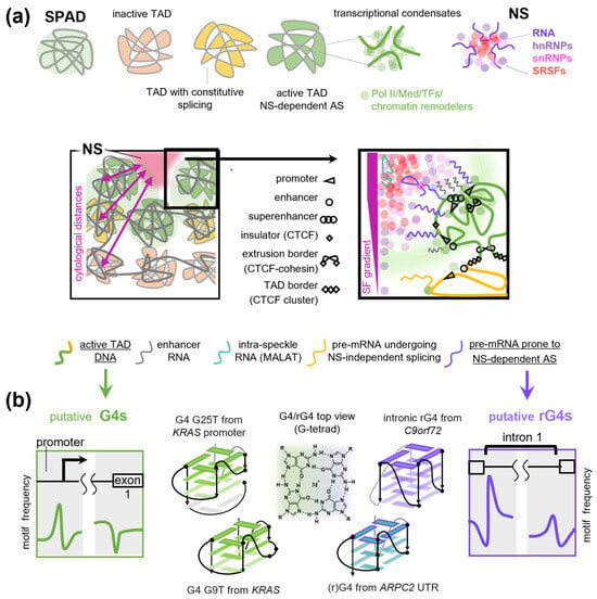
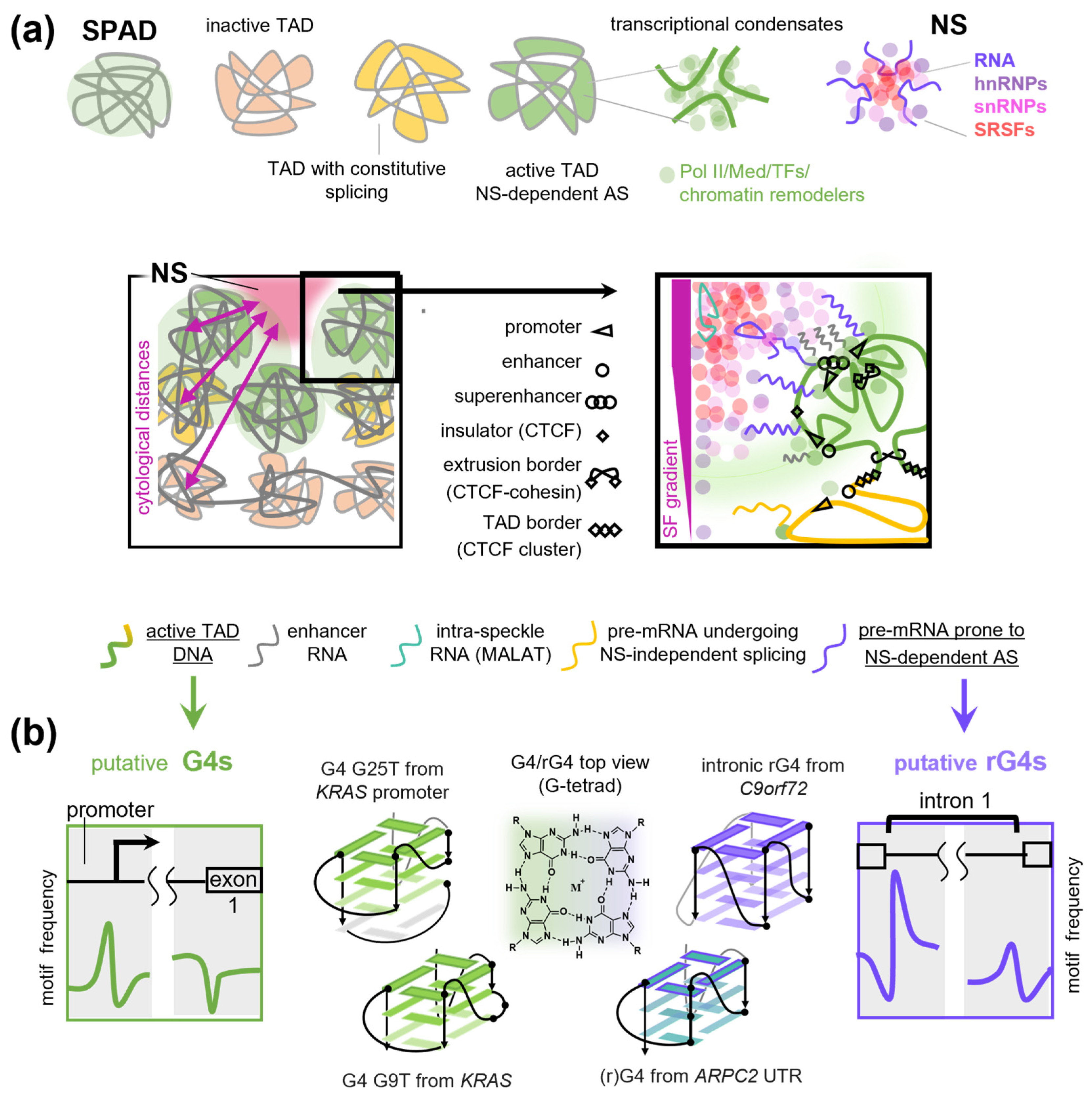
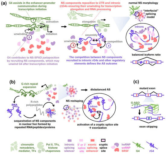
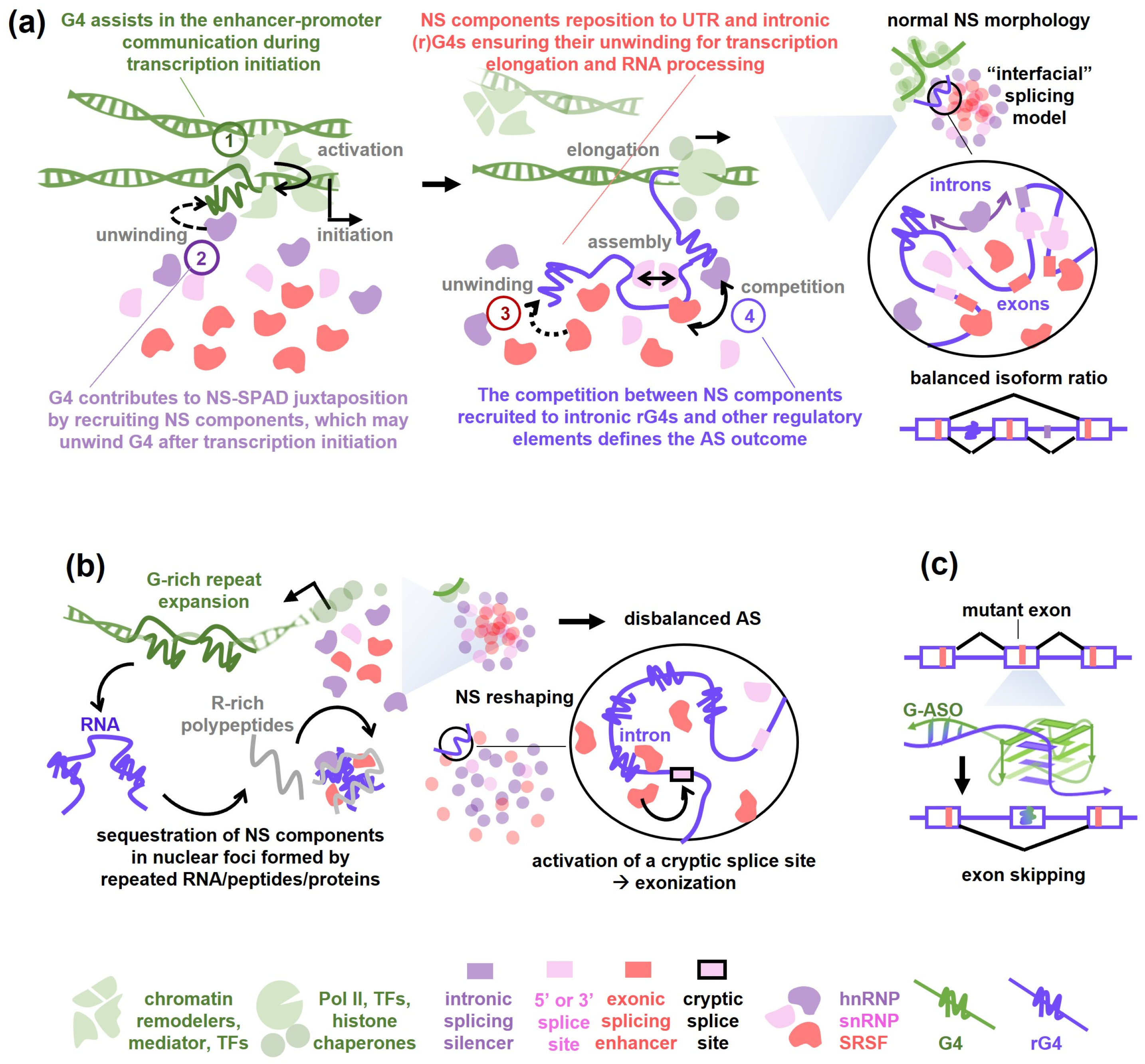
Disclaimer/Publisher’s Note: The statements, opinions and data contained in all publications are solely those of the individual author(s) and contributor(s) and not of MDPI and/or the editor(s). MDPI and/or the editor(s) disclaim responsibility for any injury to people or property resulting from any ideas, methods, instructions or products referred to in the content. |
© 2026 by the authors. Licensee MDPI, Basel, Switzerland. This article is an open access article distributed under the terms and conditions of the Creative Commons Attribution (CC BY) license.