


Abstract
Background: Myoclonus, a sudden brief shock-like involuntary movement, represents a common yet under-recognized manifestation across many inherited metabolic disorders. Although its occurrence has been reported in case series and small cohorts, the overall spectrum, pathophysiological mechanisms, and therapeutic relevance of metabolic myoclonus have not been systematically summarized. Methods: A systematic search of PubMed was conducted for English-language publications from 2014 to 2025 using predefined MeSH terms related to myoclonus, movement disorders, and inborn errors of metabolism. Titles and abstracts were screened independently by three reviewers. After removal of duplicates, 27 articles were included, complemented by 65 additional references addressing individual disorders. Data were organized according to the International Classification of Inherited Metabolic Disorders (ICIMD). Results: Myoclonus was documented across six ICIMD categories, including intermediary metabolism, mitochondrial energy metabolism, lipid metabolism, disorders of complex molecules and organelles, cofactor and mineral metabolism, and metabolic cell signaling disorders. Clinical presentation ranged from isolated jerks to progressive myoclonic epilepsies. Several conditions—such as GLUT1 deficiency, cerebrotendinous xanthomatosis, and folate receptor α deficiency—are treatable through dietary or pharmacological interventions. Conclusions: Recognition of myoclonus as a presenting feature of inherited errors of metabolism (IEMs) is critical for timely diagnosis and treatment. Metabolic screening should be considered in all unexplained cases of myoclonus, particularly when accompanied by developmental delay or systemic abnormalities.
1. Introduction
Myoclonus is defined as a sudden, brief, shock-like involuntary movement caused by either bursts of muscle activity (positive myoclonus) or transient muscle inhibition (negative myoclonus). It is not a disease entity but a symptom that may occur at rest, during voluntary action, or in response to external stimuli such as auditory, tactile, or emotional triggers [1,2]. Myoclonus is typically irregular, lasting 10–50 ms, and may vary in distribution and amplitude. Depending on the anatomical generator, it can originate from cortical, subcortical or segmental (peripheral) structures. Although myoclonus is classically associated with epileptic and neurodegenerative conditions, it is also a prominent neurological sign in several inherited metabolic disorders [1,3]. The International Classification of Inherited Metabolic Disorders (ICIMD) lists over 1400 disorders grouped into 24 categories and more than 120 subgroups [4]. Many of these diseases can disrupt neuronal excitability and neurotransmitter balance, leading to myoclonic jerks, seizures, or progressive myoclonic epilepsies. Despite its clinical importance, the metabolic etiology of myoclonus remains underrecognized [2]. Reports are often limited to isolated case studies, and comprehensive analyses are lacking. Moreover, several metabolic diseases presenting with myoclonus, such as propionic acidemia, GLUT1 deficiency, or cerebrotendinous xanthomatosis, are treatable if diagnosed early, using dietary modification, vitamin or cofactor supplementation, or targeted pharmacotherapy [5]. This review aims to provide a systematic synthesis of the literature describing inborn errors of metabolism (IEMs) associated with myoclonus. We summarize the clinical, neurophysiological, and imaging characteristics of these disorders, discuss shared mechanisms of neuronal dysfunction, and highlight diagnostic and therapeutic strategies. Ultimately, our goal is to promote earlier recognition of metabolic causes in patients presenting with unexplained myoclonus and to emphasize treatable conditions within this broad group.
2. Materials and Methods
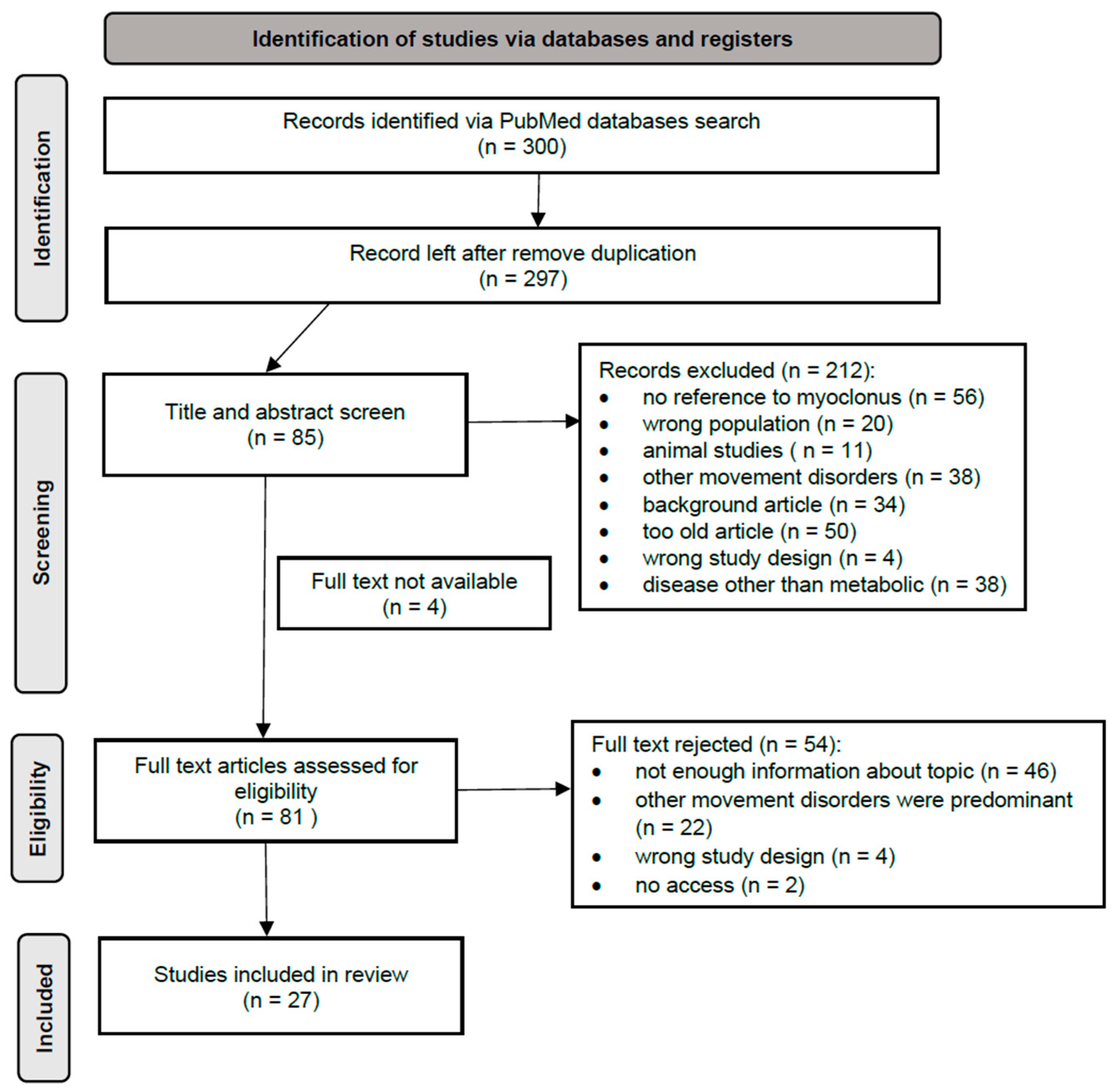
A comprehensive literature search was conducted using PubMed in March 2025. At the beginning of the project, we decided to use works from the last 10 years. However, several works from 2014 proved to be very helpful, so they were included. This search strategy included the following keywords along with their medical subject headings (MeSH terms): (“myoclonus” OR “myoclonic jerks” OR “involuntary movements” OR “movement disorders” OR “myoclonic seizures”) AND (“children” OR “pediatric” OR “infants”) AND (“metabolic disorders” OR “inborn errors of metabolism” OR “mitochondrial disorders” OR “lysosomal storage diseases” OR “urea cycle disorders” OR “organic acidemias” OR “aminoacidopathies” OR “peroxisomal disorders” OR “glycogen storage diseases” OR “fatty acid oxidation disorders” OR “purine metabolism disorders” OR “pyrimidine metabolism disorders” OR “congenital disorders of glycosylation” OR “sterol metabolism disorders” OR “neurotransmitter metabolism disorders” OR “Wilson disease” OR “Menkes disease” OR “homocystinuria” OR “galactosemia”). Only articles written in English and describing human subjects were included. Studies performed exclusively in animal models were excluded. Three independent reviewers screened all titles and abstracts. Following removal of duplicates, 297 records were screened, 85 underwent full-text evaluation, and 27 met inclusion criteria. This review was conducted in accordance with the PRISMA 2020 guidelines (Figure 1). An additional 65 references were used to complement disease-specific details, including biochemical and genetic information. Disorders were classified according to the International Classification of Inherited Metabolic Disorders (ICIMD). Data extracted from each publication included type and localization of myoclonus, EEG characteristics, MRI findings, biochemical diagnostics and treatment response. Given the heterogeneity of the study designs, a narrative synthesis rather than quantitative meta-analysis was performed.
Figure 1.
PRISMA flow diagram for screening and selection.
3. Results
3.1. Intermediary Metabolism: Nutrient Pathways
This category encompasses disorders of amino acid, organic acid, and carbohydrate metabolism [4], where several disorders can include myoclonus as a symptom. A detailed list of diseases in this category and those described below can be found in Table 1.
Organic acidurias are characterized by abnormal excretion of organic acids in urine and include propionic acidemia and glutaric aciduria type I, both of which can manifest with myoclonic seizures [6,7,8].
Propionic acidemia results from mutations in PCCA or PCCB, leading to deficiency of propionyl-CoA carboxylase. Neurological manifestations result from accumulation of propionic acid with its metabolites and include developmental delay, hypotonia, encephalopathy, and multiple seizure types—particularly myoclonic, focal, and atypical absence seizures. EEG may demonstrate frontotemporal slow-wave activity and 7–9 Hz central comb-like rhythms. MRI often shows basal ganglia lesions, delayed myelination, or cerebellar atrophy [7,9]. Early dietary protein restriction and L-carnitine supplementation mitigate metabolic crises and improve neurological outcomes [10].
Glutaric aciduria type I arises from GCDH mutations leading to glutaryl-CoA dehydrogenase deficiency, causing accumulation of neurotoxic glutaric and 3-hydroxyglutaric acids [10]. Patients exhibit developmental delay, dystonia, and seizures, including myoclonic episodes. EEG often reveals temporoparietal discharges, while MRI demonstrates frontotemporal hypoplasia, subdural collections, and caudate-putamen signal abnormalities [7,8]. Early identification through newborn screening enables treatment with a low-lysine diet and carnitine supplementation, reducing the risk of encephalopathic crises and severe neurological damage [10].
GLUT1 deficiency syndrome, caused by SLC2A1 mutations, represents another treatable metabolic cause of myoclonus [11,12]. Impaired glucose transport across the blood–brain barrier leads to an energy-deficient brain state [6]. Clinically, patients present with infantile-onset seizures, developmental delay, microcephaly, and movement disorders including myoclonus and ataxia [6,11,12]. EEG may show generalized spike-and-wave discharges, and MRI can reveal white matter abnormalities or cerebellar atrophy [12]. The ketogenic diet remains the mainstay therapy, effectively controlling seizures and movement episodes [10].
In the non-classical GLUT1 phenotype, also known as paroxysmal exercise-induced dyskinesia (PED), patients experience transient myoclonic or dystonic movements provoked by exertion or fasting [5,10]. The modified Atkins diet and triheptanoin supplementation have shown efficacy in reducing episode frequency [10,11,13].
Table 1.
Inherited metabolic disorders associated with myoclonus, summarized by ICIMD category.
3.2. Energy Metabolism: Mitochondrial Disorders
Mitochondrial diseases affect oxidative phosphorylation and energy production [4]. Within this group, myoclonic epilepsy with ragged-red fibres (MERRF) and POLG-related disorders are the principal syndromes associated with myoclonus.
MERRF is caused by pathogenic variants in mitochondrial tRNA genes, most commonly m.8344A > G in MT-TK [22]. The classical phenotype includes myoclonus, generalized epilepsy, ataxia, and ragged-red fibres on muscle biopsy. Myoclonic seizures are the hallmark, often accompanied by generalized spike-and-wave EEG patterns with background slowing. MRI typically demonstrates brain atrophy and basal ganglia calcifications [23]. Laboratory findings include elevated lactate and CSF protein levels. Treatment is supportive, focusing on seizure control, and valproic acid should be avoided, especially in coexistent POLG mutations. Some benefit has been observed with coenzyme Q10, idebenone, and L-carnitine supplementation, although evidence remains limited [22]. While MERRF is the paradigmatic “myoclonic” mitochondrial disease, recent evidence underscores the need to differentiate it from MELAS. According to GeneReviews, myoclonus occurs in 25–49% of patients with MELAS, making it a frequent but not defining feature, unlike in MERRF where it is a core symptoms [70]. In MELAS, focal seizures and stroke-like episodes predominate, whereas MERRF is characterized by generalized myoclonus and ataxia. Understanding these phenotypic overlaps and distinctions is crucial, as both conditions require avoidance of mitochondrial toxins (e.g., valproic acid) and may benefit from metabolic stabilization.
POLG-related disorders arise from mutations in the POLG gene encoding mitochondrial DNA polymerase gamma. These include a broad clinical spectrum: AHS (Alpers-Huttenlocher syndrome), presenting in early childhood with intractable myoclonic epilepsy, liver failure, and encephalopathy; MEMSA (myoclonic epilepsy, myopathy, sensory ataxia), with generalized myoclonus and progressive ataxia; and ANS (ataxia neuropathy spectrum), characterized by cerebellar ataxia, neuropathy, and occasional myoclonus [26]. EEG findings and MRI changes vary according to the phenotype and are described in detail in Table 1. Prognosis depends on age of onset and disease severity. There is no curative treatment; symptomatic management and avoidance of valproate are essential to prevent hepatic failure [26].
3.3. Lipid Metabolism and Transport
Disorders of lipid metabolism and transport comprise abnormalities in fatty acid oxidation, bile acid synthesis, and sterol metabolism [4]. Among these, cerebrotendinous xanthomatosis (CTX) stands out as a prominent cause of myoclonus within this group.
CTX results from CYP27A1 mutations leading to impaired bile acid synthesis and accumulation of cholestanol and cholesterol [71]. The disease presents with a wide clinical spectrum that includes childhood-onset cataracts, chronic diarrhea, tendon xanthomas, and progressive neurological decline in adolescence or adulthood. Neurological involvement is marked by ataxia, parkinsonism, cognitive impairment, and myoclonus, which may mimic progressive myoclonic epilepsy [71,72].
Electrophysiological recordings confirm subcortical myoclonus originating from dentate-basal ganglia dysfunction. MRI findings often reveal cerebellar and cortical atrophy and symmetric dentate nucleus hyperintensities [73]. Importantly, CTX is treatable: administration of chenodeoxycholic acid (CDCA) normalizes bile acid metabolism, reduces cholestanol accumulation, and can stabilize or even reverse neurological symptoms, including myoclonus. Early treatment is associated with the most favorable outcomes [10].
3.4. Complex Molecule and Organelle Metabolism
This category consists of congenital disorders of glycosylation (CDG), peroxisomal and lysosomal diseases, and defects in organelle biogenesis. Myoclonus or myoclonic seizures have been described in several entities: PMM2-CDG, ALG11-CDG, ALG-13-CDG, PIGA-CDG, PIGN-CDG, NGLY1 deficiency, Zellweger spectrum disorders, gangliosidosis type 1 and 2, Gaucher disease type III, sialidosis type I, ceroid lipofuscinosis 2, 6A, 11, 14 and Niemann-Pick disease type C (NPC).
NGLY1 deficiency together with sialidosis, neuronal ceroid lipofuscinosis and MERRF syndrome forms a group of heterogeneous disorders called Progressive Myoclonus Epilepsies (PME). They are characterized by myoclonus, generalized epilepsy and neurological deterioration, including dementia and ataxia [74]. NGLY1 deficiency in particular is caused by mutations in NGLY1, which encodes N-glycanase 1—an enzyme responsible for deglycosylating misfolded glycoproteins prior to proteasomal degradation [45]. Disruption of this pathway leads to accumulation of aberrant glycoproteins and impaired cellular stress responses. Clinically, affected patients exhibit hypo- or alacrimia, hypotonia, peripheral neuropathy, microcephaly, liver dysfunction (indicated by elevated serum transaminases and liver fibrosis) and myoclonic seizures. EEG findings show multifocal epileptiform discharges, particularly in frontocentral regions, often correlated with jerks on video–EEG [46]. MRI frequently reveals progressive cerebral and cerebellar atrophy and delayed myelination. Treatment is symptomatic, though emerging studies suggest potential benefit from AMPA receptor antagonists such as perampanel, which may improve action myoclonus [75].
Zellweger syndrome represents the severe end of the spectrum of peroxisomal biogenesis disorders (PBDs), standing in contrast to single-enzyme peroxisomal defects [47,48]. It is caused by mutations in PEX genes essential for organelle assembly, leading to defective formation and function of peroxisomes. While myoclonus has been linked to PEX13, PEX19, and PEX26, it is also observed in phenotypes associated with other genes, including PEX1. The clinical presentation includes global developmental delay, hypotonia, ataxia, epilepsy, myoclonic seizures, sensorineural hearing loss, and ocular abnormalities such as retinopathy, cataracts, glaucoma, progressive blindness and tunnel vision. EEG often reveals burst-suppression or hypsarrhythmic patterns, consistent with severe epileptic encephalopathy. MRI shows white matter hypomyelination, neocortical dysplasia, delayed myelination, germinolytic cysts, leukodystrophy and ventriculomegaly. Biochemically, patients exhibit elevated very-long-chain fatty acids and abnormal bile acid intermediates. Deficiency of plasmalogens is detectable in erythrocytes, and diagnostic confirmation is obtained through biochemical testing and skin fibroblast analysis [71]. There is currently no curative therapy; management is supportive, focusing on seizure control and nutritional support.
Niemann-Pick disease type C (NPC) is a lysosomal storage disorder caused by mutations in NPC1 (95%) or NPC2 leading to defective intracellular lipid trafficking and accumulation of cholesterol and glycosphingolipids [48]. It may manifest itself as progressive cognitive decline (78%), gait and limb ataxia (70%), dysarthria, dystonia, dysphagia, and cataplexy. Vertical supranuclear gaze palsy is a robust clinical indicator of NP-C [64]. EEG often reveals generalized slowing and interictal discharges, while MRI demonstrates diffuse cerebral atrophy [64]. Biochemical markers such as oxysterol, bile acid derivatives and lyso-sphingomyelin-509 aid diagnosis. The only currently approved treatment with miglustat, an inhibitor of glycosphingolipid synthesis, slows neurological progression. Gene therapy and combination strategies are under investigation, but early initiation of treatment appears critical to improving outcomes [65]. While miglustat, an inhibitor of glycosphingolipid synthesis, is approved in many jurisdictions (including Europe and Japan) to slow neurological progression, it is not FDA-approved for NPC in the United States. Recently, regulatory agencies have authorized new agents; the United States FDA has approved both levacetylleucine and arimoclomol for the treatment of NPC. In Europe, the EMA recommended granting marketing authorization for levacetylleucine (Aqneursa) in July 2025. It is indicated for the treatment of neurological manifestations and, in the EU, is used in combination with miglustat, or as a monotherapy in patients where miglustat is not tolerated. Preclinical studies suggest that levacetylleucine enhances ATP production and mitochondrial energy metabolism, contributing to improved motor function and reduced myoclonus.
3.5. Cofactor and Mineral Metabolism
Cofactor and mineral metabolism disorders are inherited neurometabolic conditions broadly divided into two groups: disorders of vitamin and cofactor metabolism, and disorders of trace element and metal metabolism. In our review, myoclonus was reported in tetrahydrobiopterin deficiency, cerebral folate transport deficiency (FOLR1 deficiency), X-linked cobalamin disorder, pyridoxine-dependent and pyridoxal phosphate-responsive epilepsies, methylenetetrahydrofolate reductase deficiency, biotinidase deficiency and Menkes disease. In most of these, myoclonic seizures dominate the presentation.
FOLR1 deficiency arises from biallelic mutations in FOLR1, encoding folate receptor α (FR α), leading to reduced CNS folate levels despite normal systemic concentrations [76]. Affected children present after infancy with developmental delay, speech and language impairment, ataxia, nystagmus, hypotonia, gait abnormalities and myoclonic or tonic seizures. Seizures can be severe, precipitated by fever, or progressing to status epilepticus [76,77]. Behavioral disturbances, including autistic features, may also be present [76]. EEG frequently shows generalized epileptiform discharges or slow spike-wave complexes, consistent with a myoclonic–astatic epilepsy phenotype [78]. MRI reveals white matter hyperintensities and cerebellar atrophy [76,79,80,81]. Diagnosis is confirmed by low CSF 5-methyltetrahydrofolate levels. Treatment with oral or intramuscular 5-formyltetrahydrofolate often improves neurological signs, especially when started early; folic acid is contraindicated, mostly in the case of low but important residual levels of FOLR1 transport activity as it may competitively block FRα [76,77].
Vitamin B6-related epilepsies include pyridoxine-dependent epilepsy (PDE) due to ALDH7A1 deficiency and pyridoxal phosphate-responsive epilepsy (PNPO deficiency) [82,83]. Both typically present neonatally with drug-resistant myoclonic or tonic seizures and burst-suppression EEG patterns. MRI may show non-specific delayed myelination or ventriculomegaly [14,83]. Diagnosis in both conditions rests on demonstrating seizure responsiveness to supraphysiologic pyridoxine or pyridoxal-5′-phosphate (PLP) (PNPO), coupled with biomarkers (e.g., α-AASA in PDE) and confirmatory genetic testing [83]. Lifelong treatment is essential: PDE responds to pyridoxine (typically 30 mg/kg/day up to 500 mg/day) often with excellent seizure control, and adjuncts such as a lysine-restricted diet and arginine supplementation can enhance developmental outcome [84]. PNPO deficiency, by contrast, usually requires lifelong PLP (or pyridoxine) therapy for seizure control in around 60% of cases, though developmental delay remains common if diagnosis is delayed [83]. It is important to remember that certain anti-seizure medications (such as carbamazepine, valproate, phenytoin and phenobarbital) can cause low plasma concentration of PLP and should be avoided [83].
Myoclonus is also well documented in Menkes disease that is caused by mutations in ATP7A and leads to defective copper transport and deficiency of copper-dependent enzymes. Clinically, it may appear as multifocal or generalized myoclonic seizures in early infancy, sometimes alongside focal seizures and later epileptic spasms [85]. Additionally, infants develop neurodegeneration, hypotonia and connective-tissue abnormalities. For diagnosis, beyond copper and ceruloplasmin assays, early testing should include plasma and, where feasible, CSF catecholamine profiling, with elevated dopamine/noradrenaline and DOPAC:DHPG ratios providing sensitive neonatal biomarkers that can be abnormal even before neurological decline [86,87]. MRI may show cerebral and cerebellar atrophy and vascular tortuosity [88]. Treatment with subcutaneous copper-histidine can partially ameliorate symptoms if started early, though efficacy is limited in advanced disease [89,90].
3.6. Metabolic Cell Signaling Disorders
This final category includes disorders affecting neurotransmitter synthesis and degradation and endocrine metabolic disorders. Myoclonus occurs most notably in tyrosine hydroxylase deficiency (THD) and succinic semialdehyde dehydrogenase deficiency (SSADH).
THD results from TH mutations disrupting dopamine synthesis [91]. The phenotype includes two major forms, both presenting with hypokinesia, bradykinesia, delayed motor development, oculogyric crises, ptosis and autonomic disturbances [10,91]. Type A usually occurs before the age of 1 and it has a more classical course for DRD: dystonia beginning in lower limbs, progressing to arms, face and the oropharyngeal area. Patients also present with rigidity and tremor. Type B is considered to be more severe, with an onset within the first few weeks of birth, and it develops rapidly and can be manifested by dystonia, tremor and myoclonus. EEG findings are generally non-specific; MRI may show frontotemporal atrophy or ventriculomegaly [92]. Diagnosis relies on CSF neurotransmitter profiling showing low homovanillic acid with normal pterins. Treatment with levodopa combined with other medications such as carbidopa, benzhexol, benserazide, selegiline and biperiden often yields marked improvement, particularly in type A patients [92].
SSADH deficiency, caused by ALDH5A1 mutations, impairs GABA metabolism, leading to accumulation of γ-hydroxybutyric acid [93]. Clinical features include developmental delay, non-progressive encephalopathy, dystonia, dyskinesia hypotonia, ataxia, and seizures—often myoclonic or generalized tonic–clonic. Sleep disturbances, neurobehavioral/psychiatric manifestations such as autism spectrum disorder, attention deficits, obsessive-compulsive behavior and anxiety can also be present [93,94]. MRI demonstrates dentate nucleus, subthalamic nucleus and globus pallidus hyperintensities, and EEG shows diffuse slowing with multifocal spikes [7,94]. Treatment is symptomatic; seizures can be controlled with antiepileptic drugs; however, vigabatrin is not recommended and valproate should be used only in seizures with a drug-resistant generalized spike-wave EEG pattern [94].
4. Discussion
This systematic review demonstrates that myoclonus is a recurrent, cross-cutting feature among multiple categories of inherited metabolic diseases. Although the underlying biochemical pathways differ, the pathophysiological mechanisms converge on common neuronal dysfunction—particularly cortical hyperexcitability, impaired inhibitory neurotransmission, and disrupted energy metabolism. The presence of myoclonus, whether isolated or as part of PME, should alert clinicians to possible metabolic etiologies, many of which are amenable to specific treatments. Across the ICIMD classification, myoclonus is especially prevalent in disorders of intermediary and mitochondrial energy metabolism, as well as in lysosomal and vitamin-related diseases. Several disorders, such as phenylketonuria, organic acidemias and CDGs, can be detected by newborn screening, allowing for earlier initiation of treatment and reducing the burden of the disease. The versatile biochemical biomarker for myoclonus is missing due to heterogeneity of its cause. However, choosing a proper diagnostic pathway, according to clinical presentation and family history, may accelerate the diagnosis. Electrophysiologically, cortical myoclonus predominates in mitochondrial and lysosomal disorders, while subcortical and spinal generators are more frequent in lipid and neurotransmitter-related diseases. MRI correlates, including dentate nucleus hyperintensities in CTX and basal ganglia lesions in organic acidurias, further support the concept of region-specific vulnerability linked to metabolic stress. Despite these insights, several limitations persist. Published data remain fragmented, often derived from small series or anecdotal reports. Heterogeneity in diagnostic criteria and lack of unified reporting impede cross-comparison. Furthermore, treatment efficacy is rarely assessed systematically, and long-term outcomes are poorly documented. The establishment of international multicentre registries and standardized neurophysiological protocols would enhance our understanding of prevalence, natural history, and treatment responses. Importantly, the recognition of treatable causes should guide diagnostic algorithms. In every case of unexplained myoclonus, particularly when accompanied by developmental delay, movement abnormalities, or multisystem involvement, clinicians should explore the following metabolic diagnostics: amino acid, lactate and pyruvate analyses from plasma and CSF; analyses of urine organic acids, CSF neurotransmitters and very long-chain fatty acids; lumbar puncture for glucose concentration simultaneously with plasma glucose concentration; acylcarnitine profiling from plasma; analysis of transferrin isoforms; and evaluation of storage disorders. This should be followed by a confirmatory genetic test such as next-generation sequencing.
5. Conclusions
Myoclonus constitutes an important and often overlooked manifestation of inherited metabolic diseases. It occurs across virtually all ICIMD categories, reflecting diverse yet converging metabolic mechanisms affecting neuronal excitability. Crucially, several of these conditions are treatable, and early recognition can substantially modify disease trajectory. Clinicians should maintain a high index of suspicion for metabolic causes in any patient presenting with unexplained myoclonus (especially in children and young adults) and promptly initiate biochemical and genetic investigations. Early diagnosis not only enables targeted therapy but also provides families with accurate prognostic and genetic counseling information.
Author Contributions
Conceptualization; methodology; writing—original draft preparation; writing—review and editing, A.O., J.P., E.M. and Z.Z. All authors contributed to the conception and design of the study, data acquisition, analysis, and interpretation. All authors critically revised the manuscript for intellectual content and approved the final version. All authors have read and agreed to the published version of the manuscript.
Funding
This research received no external funding. Statutory work of Medical University of Silesia in Katowice, Poland No BNW-1-180/K/4/K. The authors confirm independence from any sponsors; the content of the article was not influenced by external funding sources.
Institutional Review Board Statement
Not applicable.
Informed Consent Statement
Not applicable.
Data Availability Statement
No new data were generated or analyzed in this study. All data supporting the findings of this review are available within the cited literature.
Conflicts of Interest
The authors declare no conflicts of interest.
Abbreviations
The following abbreviations are used in this manuscript:
| 5-HIAA | 5-Hydroxyindoleacetic Acid |
| 5-HTP | 5-Hydroxytryptophan |
| 5-MTHF | 5-methyltetrahydrofolate |
| α-AASA | α-amino adipic semialdehyde |
| AAV | adenovirus |
| AHS | Alpers-Huttenlocher Syndrome |
| Ala | Alanine |
| ALT | Alanine aminotransferase |
| ANS | Ataxia neuropathy spectrum |
| AST | Aspartate aminotransferase |
| AR-GTPCHD | Autosomal Recessive GTP Cyclohydrolase I Deficiency |
| BCKA | Branched-Chain Alpha Ketoacids |
| BCAA | Branched-Chain Amino Acid |
| CDCA | Chenodeoxycholic acid |
| CDG | Congenital Disorders of Glycosylation |
| CLN | Neuronal Ceroid Lipofuscinosis |
| CNS | Central nervous system |
| COMT inhibitors | Catechol-O-Methyltransferase Inhibitors |
| CSF | Cerebrospinal Fluid |
| CTX | Cerebrotendinous Xanthomatosis |
| DA | Dopamine |
| DBS | Dried Blood Spot |
| DC inhibitor | Dopa Decarboxylase Inhibitor |
| DD | Developmental Disability |
| DHPG | Dihydroxyphenylglycol |
| DHPRD | Dihydropteridine Reductase Deficiency |
| DOPAC | Dihydroxyphenylacetic acid |
| DRD | Dopa-responsive Dystonia |
| ESE | Electrographic Status Epilepticus |
| FOLR1 deficiency | Cerebral folate transport deficiency |
| FRα | Folate receptor α |
| GCH1 | Gene encoding GTP Cyclohydrolase I |
| GGTP | Gamma-glutamyl transferase |
| GHB | Gamma-Hydroxybutyric Acidura |
| GI reflux | Gastrointestinal reflux |
| GLUT1 | Glucose transporter type 1 |
| Gly | Glycine |
| GM1 | Gangliosidosis type 1 |
| GM2 | Gangliosidosis type 2 |
| GTC | Generalized Tonic–Clonic |
| HEX | Hexoaminidase |
| HPA | Hyperphenylalaninemia |
| HVA | Homovanillic Acid |
| ICIMD | International Classification of Inherited Metabolic Disorders |
| ID | Intellectual Disability |
| IEMs | Inherited errors of metabolism |
| JME | Juvenile myoclonic epilepsy |
| MAO | Monoamine Oxidase |
| MEMSA | Myoclonic epilepsy myopathy sensory ataxia |
| MPI | Mannose Phosphate Isomerase |
| NGLY1 | N-glycanase type 1 |
| NPC | Niemman-Pick disease type C |
| P1 | Patients with GTP Cyclohydrolase I Deficiency |
| PCBD1 | gene encoding Pterin-4a-Carbinolamine Dehydratase |
| PCDD | Pterin-4a-Carbinolamine Dehydratase Deficiency |
| PDE | Pyridoxine-dependent epilepsy |
| PED | paroxysmal exercise-induced dyskinesia |
| Phe | Phenylalanine |
| PLGA | NPs Poly(lactic-co-glycolic acid) nanoparticles |
| PLP | Pyridoxal 5′-phosphate |
| PME | Progressive myoclonic epilepsy |
| POLG | Polymerase gamma |
| PNPO | Pyridox(am)ine 5′-phosphate oxidase |
| PTPSD | 6-Pyruvoyl-Tetrahydropterin Synthase Deficiency |
| PTS | gene encoding 6-Pyruvoyl-Tetrahydropterin Synthase |
| QDPR g | gene encoding Quinoid Dihydropteridine Reductase |
| SCAE | Spino-cerebellar ataxia with epilepsy |
| SEP | Somatosensory Evoked Potential |
| Ser | Serine |
| SLEs | Stroke-Like Episodes |
| SPR | gene encoding Sepiapterin Reductase |
| SR | Sepiapterin Reductase Deficiency |
| SSADH | Succinic Semialdehyde Dehydrogenase Deficiency |
| SSRIs | Selective Serotonin Reuptake Inhibitors |
| THD | Tyrosine Hydroxylase Deficiency |
| IUGR | Intrauterine Growth Retardation |
| VA | Valproic Acid |
References
- Riva, A.; D’Onofrio, G.; Ferlazzo, E.; Pascarella, A.; Pasini, E.; Franceschetti, S.; Panzica, F.; Canafoglia, L.; Vignoli, A.; Coppola, A.; et al. Myoclonus: Differential diagnosis and current management. Epilepsia Open 2024, 9, 486–500. [Google Scholar] [CrossRef] [Scilit] [PubMed]
- Sanders, A.E.; Zafar, N.; Sharma, S. Myoclonus. In StatPearls [Internet]; StatPearls Publishing: Treasure Island, FL, USA, 2025. Available online: http://www.ncbi.nlm.nih.gov/books/NBK537015/ (accessed on 1 September 2025).
- Eberhardt, O.; Topka, H. Myoclonic Disorders. Brain Sci. 2017, 7, 103. [Google Scholar] [CrossRef] [Scilit]
- Ferreira, C.R.; Rahman, S.; Keller, M.; Zschocke, J. An International Classification of Inherited Metabolic Disorders (ICIMD). J. Inherit. Metab. Dis. 2021, 44, 164–177. [Google Scholar] [CrossRef] [Scilit] [PubMed]
- Ebrahimi-Fakhari, D.; Van Karnebeek, C.; Münchau, A. Movement Disorders in Treatable Inborn Errors of Metabolism. Mov. Disord. 2019, 34, 598–613. [Google Scholar] [CrossRef] [Scilit]
- Almannai, M.; Al Mahmoud, R.A.; Mekki, M.; El-Hattab, A.W. Metabolic Seizures. Front. Neurol. 2021, 12, 640371. [Google Scholar] [CrossRef] [Scilit] [PubMed]
- Latzer, I.T.; Blau, N.; Ferreira, C.R.; Pearl, P.L. Clinical and biochemical footprints of inherited metabolic diseases. XV. Epilepsies. Mol. Genet. Metab. 2023, 140, 107690. [Google Scholar] [CrossRef] [Scilit]
- Neurometabolic Disorders-Related Early Childhood Epilepsy: A Single-Center Experience in Saudi Arabia—ClinicalKey [Internet]. 2025. Available online: https://www.clinicalkey.com/#!/content/playContent/1-s2.0-S1875957215000510?returnurl=https:%2F%2Flinkinghub.elsevier.com%2Fretrieve%2Fpii%2FS1875957215000510%3Fshowall%3Dtrue&referrer=https:%2F%2Fpubmed.ncbi.nlm.nih.gov%2F (accessed on 20 August 2025).
- Galarreta Aima, C.I.; Shchelochkov, O.A.; Jerves Serrano, T.; Venditti, C.P. Propionic Acidemia. In GeneReviews® [Internet]; Adam, M.P., Feldman, J., Mirzaa, G.M., Pagon, R.A., Wallace, S.E., Amemiya, A., Eds.; University of Washington: Seattle, WA, USA, 1993. Available online: http://www.ncbi.nlm.nih.gov/books/NBK92946/ (accessed on 27 August 2025).
- Jinnah, H.A.; Albanese, A.; Bhatia, K.P.; Cardoso, F.; Prat, G.D.; de Koning, T.J.; Espay, A.J.; Fung, V.; Garcia-Ruiz, P.J.; Gershanik, O.; et al. Treatable Inherited Rare Movement Disorders. Mov. Disord. Off. J. Mov. Disord. Soc. 2018, 33, 21–35. [Google Scholar] [CrossRef] [Scilit]
- Christensen, C.K.; Walsh, L. Movement Disorders and Neurometabolic Diseases. Semin. Pediatr. Neurol. 2018, 25, 82–91. [Google Scholar] [CrossRef] [Scilit]
- Hully, M.; Vuillaumier-Barrot, S.; Le Bizec, C.; Boddaert, N.; Kaminska, A.; Lascelles, K.; de Lonlay, P.; Cances, C.; des Portes, V.; Roubertie, A.; et al. From splitting GLUT1 deficiency syndromes to overlapping phenotypes. Eur. J. Med. Genet. 2015, 58, 443–454. [Google Scholar] [CrossRef] [Scilit]
- Xu, J.J.; Chen, Y.L.; Yu, H.; Chen, D.F.; Li, H.F.; Wu, Z.Y. Genetic and Clinical Features of SLC2A1-Related Paroxysmal Exercise-Induced Dyskinesia. Pediatr. Neurol. 2025, 170, 31–37. [Google Scholar] [CrossRef] [Scilit]
- Nunes, M.L.; Yozawitz, E.G.; Zuberi, S.; Mizrahi, E.M.; Cilio, M.R.; Moshé, S.L.; Plouin, P.; Vanhatalo, S.; Pressler, R.M.; Task Force on Neonatal Seizures; et al. Neonatal seizures: Is there a relationship between ictal electroclinical features and etiology? A critical appraisal based on a systematic literature review. Epilepsia Open 2019, 4, 10–29. [Google Scholar] [CrossRef] [Scilit]
- Kantamneni, T.; Mondok, L.; Parikh, S. Inborn Errors of Metabolism with Movement Disorders: Defects in Metal Transport and Neurotransmitter Metabolism. Pediatr. Clin. N. Am. 2018, 65, 301–315. [Google Scholar] [CrossRef] [Scilit]
- Bhandary, S.; Aguan, K. Pyruvate dehydrogenase complex deficiency and its relationship with epilepsy frequency—An overview. Epilepsy Res. 2015, 116, 40–52. [Google Scholar] [CrossRef] [Scilit]
- Divahia, G.M.; Córdova, N.M.; García, E.E. Pyruvate Dehydrogenase Deficiency: With More Phenotypes Than We Believe. J. Pediatr. Neonatol. 2022, 23, 71–79. [Google Scholar] [CrossRef] [Scilit]
- Abraham, M.S.; Gladson, N.; Koshy, J.T. Girl with intermittent ataxia, myoclonus and poor scholastic performance—A case report. IP Indian J. Neurosci. 2023, 9, 227–230. [Google Scholar] [CrossRef] [Scilit]
- Jafari, N.; Mosallanejad, A.; Ghobadifar, A.; Karimzadeh, P.; Ghassemabadi, R.G.; Nasehi, M.; Shakiba, M.; Tabatabaee, S. Utility of Seizure Pattern and Related Clinical Features in the Diagnosis of Neurometabolic Disorders. Iran. J. Child. Neurol. 2020, 14, 123–132. [Google Scholar] [PubMed]
- Narayan, V.; Mahay, S.B.; Verma, I.C.; Puri, R.D. Case Series of Creatine Deficiency Syndrome due to Guanidinoacetate Methyltransferase Deficiency. Ann. Indian Acad. Neurol. 2020, 23, 347. [Google Scholar] [CrossRef] [Scilit]
- Alhomsi, D.; Abdalsalam, D.; Sulaiman, R.; Bakleh, S.; Alasmar, D. Novel guanidinoacetate methyltransferase (GAMT) mutation associated with cerebral creatine deficiency syndrome in a Syrian child: A case report. Ann. Med. Surg. 2023, 85, 1906–1910. [Google Scholar] [CrossRef] [Scilit]
- Velez-Bartolomei, F.; Lee, C.; Enns, G. MERRF. In GeneReviews® [Internet]; Adam, M.P., Feldman, J., Mirzaa, G.M., Pagon, R.A., Wallace, S.E., Amemiya, A., Eds.; University of Washington: Seattle, WA, USA, 1993. Available online: http://www.ncbi.nlm.nih.gov/books/NBK1520/ (accessed on 29 August 2025).
- Lamperti, C.; Zeviani, M. Myoclonus epilepsy in mitochondrial disorders. Epileptic Disord. 2016, 18, S94–S102. [Google Scholar] [CrossRef] [Scilit]
- Finsterer, J.; Zarrouk-Mahjoub, S. Management of epilepsy in MERRF syndrome. Seizure 2017, 50, 166–170. [Google Scholar] [CrossRef] [Scilit]
- Finsterer, J.; Zarrouk-Mahjoub, S.; Shoffner, J.M. MERRF Classification: Implications for Diagnosis and Clinical Trials. Pediatr. Neurol. 2018, 80, 8–23. [Google Scholar] [CrossRef] [Scilit]
- Rahman, S.; Copeland, W.C. POLG-related disorders and their neurological manifestations. Nat. Rev. Neurol. 2019, 15, 40–52. [Google Scholar] [CrossRef] [Scilit]
- Papandreou, A.; Rahman, S.; Fratter, C.; Ng, J.; Meyer, E.; Carr, L.J.; Champion, M.; Clarke, A.; Gissen, P.; Hemingway, C.; et al. Spectrum of movement disorders and neurotransmitter abnormalities in paediatric POLG disease. J. Inherit. Metab. Dis. 2018, 41, 1275–1283. [Google Scholar] [CrossRef] [Scilit]
- Rajakulendran, S.; Pitceathly, R.D.S.; Taanman, J.W.; Costello, H.; Sweeney, M.G.; Woodward, C.E.; Jaunmuktane, Z.; Holton, J.L.; Jacques, T.S.; Harding, B.N.; et al. A Clinical, Neuropathological and Genetic Study of Homozygous A467T POLG-Related Mitochondrial Disease. PLoS ONE 2016, 11, e0145500. [Google Scholar] [CrossRef] [Scilit]
- van Westrhenen, A.; Cats, E.A.; van den Munckhof, B.; van der Salm, S.M.A.; Teunissen, N.W.; Ferrier, C.H.; Leijten, F.S.S.; Geleijns, K.P.W. Specific EEG markers in POLG1 Alpers’ syndrome. Clin. Neurophysiol. Off. J. Int. Fed. Clin. Neurophysiol. 2018, 129, 2127–2131. [Google Scholar] [CrossRef] [Scilit]
- Cohen, B.H.; Chinnery, P.F.; Copeland, W.C. POLG-Related Disorders. In GeneReviews® [Internet]; Adam, M.P., Feldman, J., Mirzaa, G.M., Pagon, R.A., Wallace, S.E., Amemiya, A., Eds.; University of Washington: Seattle, WA, USA, 1993. Available online: http://www.ncbi.nlm.nih.gov/books/NBK26471/ (accessed on 19 August 2025).
- Fiumara, A.; Barone, R.; Del Campo, G.; Striano, P.; Jaeken, J. Electroclinical Features of Early-Onset Epileptic Encephalopathies in Congenital Disorders of Glycosylation (CDGs). JIMD Rep. 2016, 27, 93–99. [Google Scholar] [CrossRef] [Scilit]
- Pereira, A.G.; Bahi-Buisson, N.; Barnerias, C.; Boddaert, N.; Nabbout, R.; de Lonlay, P.; Kaminska, A.; Eisermann, M. Epileptic spasms in congenital disorders of glycosylation. Epileptic Disord. 2017, 19, 15–23. [Google Scholar] [CrossRef] [Scilit] [PubMed]
- Haanpää, M.K.; Ng, B.G.; Gallant, N.M.; Singh, K.E.; Brown, C.; Kimonis, V.; Freeze, H.H.; Muller, E.A., 2nd. ALG11-CDG syndrome: Expanding the phenotype. Am. J. Med. Genet. Part A 2019, 179, 498–502. [Google Scholar] [CrossRef] [Scilit] [PubMed]
- Ng, B.G.; Eklund, E.A.; Shiryaev, S.A.; Dong, Y.Y.; Abbott, M.A.; Asteggiano, C.; Bamshad, M.J.; Barr, E.; Bernstein, J.A.; Chelakkadan, S.; et al. Predominant and novel de novo variants in 29 individuals with ALG13 deficiency: Clinical description, biomarker status, biochemical analysis, and treatment suggestions. J. Inherit. Metab. Dis. 2020, 43, 1333–1348. [Google Scholar] [CrossRef] [Scilit]
- Gámez, A.; Serrano, M.; Gallego, D.; Vilas, A.; Pérez, B. New and potential strategies for the treatment of PMM2-CDG. Biochim. Biophys. Acta BBA Gen. Subj. 2020, 1864, 129686. [Google Scholar] [CrossRef] [Scilit] [PubMed]
- Bayat, A.; Knaus, A.; Pendziwiat, M.; Afenjar, A.; Barakat, T.S.; Bosch, F.; Callewaert, B.; Calvas, P.; Ceulemans, B.; Chassaing, N.; et al. Lessons learned from 40 novel PIGA patients and a review of the literature. Epilepsia 2020, 61, 1142–1155. [Google Scholar] [CrossRef] [Scilit] [PubMed]
- Lipiński, P.; Tylki-Szymańska, A. Congenital Disorders of Glycosylation: What Clinicians Need to Know? Front. Pediatr. 2021, 9, 715151. [Google Scholar] [CrossRef] [Scilit]
- Paprocka, J.; Jezela-Stanek, A.; Tylki-Szymańska, A.; Grunewald, S. Congenital Disorders of Glycosylation from a Neurological Perspective. Brain Sci. 2021, 11, 88. [Google Scholar] [CrossRef] [Scilit] [PubMed]
- Serrano, M. Stroke-like Episodes in PMM2-CDG: When the Lack of Other Evidence Is the Only Evidence. Front. Pediatr. 2021, 9, 717864. [Google Scholar] [CrossRef] [Scilit] [PubMed]
- Muthusamy, K.; Perez-Ortiz, J.M.; Ligezka, A.N.; Altassan, R.; Johnsen, C.; Schultz, M.J.; Patterson, M.C.; Morava, E. Neurological manifestations in PMM2-congenital disorders of glycosylation (PMM2-CDG): Insights into clinico-radiological characteristics, recommendations for follow-up, and future directions. Genet. Med. 2024, 26, 101027. [Google Scholar] [CrossRef] [Scilit]
- Erdal, A.E.; Ceylan, A.C.; Gücüyener, K.; Öktem, R.M.; Kıreker Köylü, O.; Kasapkara, Ç.S. ALG11-CDG: Novel variant and review of the literature. J. Pediatr. Endocrinol. Metab. JPEM. 2023, 36, 409–413. [Google Scholar] [CrossRef] [Scilit]
- Quelhas, D.; Jaeken, J. Treatment of congenital disorders of glycosylation: An overview. Mol. Genet. Metab. 2024, 143, 108567. [Google Scholar] [CrossRef] [Scilit]
- Lam, C.; Krasnewich, D.M. PMM2-CDG. In GeneReviews® [Internet]; Adam, M.P., Feldman, J., Mirzaa, G.M., Pagon, R.A., Wallace, S.E., Amemiya, A., Eds.; University of Washington: Seattle, WA, USA, 1993. Available online: http://www.ncbi.nlm.nih.gov/books/NBK1110/ (accessed on 20 August 2025).
- Panneman, D.M.; Wortmann, S.B.; Haaxma, C.A.; van Hasselt, P.M.; Wolf, N.I.; Hendriks, Y.; Küsters, B.; van Emst-de Vries, S.; van de Westerlo, E.; Koopman, W.J.H.; et al. Variants in NGLY1 lead to intellectual disability, myoclonus epilepsy, sensorimotor axonal polyneuropathy and mitochondrial dysfunction. Clin. Genet. 2020, 97, 556–566. [Google Scholar] [CrossRef] [Scilit]
- Kariminejad, A.; Shakiba, M.; Shams, M.; Namiranian, P.; Eghbali, M.; Talebi, S.; Makvand, M.; Jaeken, J.; Najmabadi, H.; Hennekam, R.C. NGLY1 deficiency: Novel variants and literature review. Eur. J. Med. Genet. 2021, 64, 104146. [Google Scholar] [CrossRef] [Scilit]
- Sonoda, Y.; Fujita, A.; Torio, M.; Mukaino, T.; Sakata, A.; Matsukura, M.; Yonemoto, K.; Hatae, K.; Ichimiya, Y.; Chong, P.F.; et al. Progressive myoclonic epilepsy as an expanding phenotype of NGLY1-associated congenital deglycosylation disorder: A case report and review of the literature. Eur. J. Med. Genet. 2024, 67, 104895. [Google Scholar] [CrossRef] [Scilit]
- Klouwer, F.C.C.; Berendse, K.; Ferdinandusse, S.; Wanders, R.J.A.; Engelen, M.; Poll-The, B.T. Zellweger spectrum disorders: Clinical overview and management approach. Orphanet J. Rare Dis. 2015, 10, 151. [Google Scholar] [CrossRef] [Scilit] [PubMed]
- Alayoubi, A.M.; Ijaz, A.; Wali, A.; Hashmi, J.A.; Alharbi, A.; Basit, S. Zellweger syndrome; identification of mutations in PEX19 and PEX26 gene in Saudi families. Ann. Med. 2025, 57, 2447400. [Google Scholar] [CrossRef] [Scilit]
- Karimzadeh, P.; Habibi, P. An Approach to Neurometabolic Epilepsy in Children with an Underlying Neurometabolic Disorder. Iran. J. Child. Neurol. 2020, 14, 79–86. [Google Scholar] [PubMed]
- Er, E.; Canda, E.; Yazıcı, H.; Eraslan, C.; Sözmen, E.; Uçar, S.K.; Çoker, M. An Evalution of the Demographic and Clinical Characterictics of Patients with GM2 Gangliosidosis. J. Pediatr. Res. 2018, 5, 12–16. [Google Scholar] [CrossRef] [Scilit]
- Tim-Aroon, T.; Wichajarn, K.; Katanyuwong, K.; Tanpaiboon, P.; Vatanavicharn, N.; Sakpichaisakul, K.; Kongkrapan, A.; Eu-Ahsunthornwattana, J.; Thongpradit, S.; Moolsuwan, K.; et al. Infantile onset Sandhoff disease: Clinical manifestation and a novel common mutation in Thai patients. BMC Pediatr. 2021, 21, 22. [Google Scholar] [CrossRef] [Scilit]
- Regier, D.S.; Tifft, C.J.; Rothermel, C.E. GLB1-Related Disorders. In GeneReviews® [Internet]; Adam, M.P., Feldman, J., Mirzaa, G.M., Pagon, R.A., Wallace, S.E., Amemiya, A., Eds.; University of Washington: Seattle, WA, USA, 1993. Available online: http://www.ncbi.nlm.nih.gov/books/NBK164500/ (accessed on 25 August 2025).
- Hughes, D.A.; Pastores, G.M. Gaucher Disease. In GeneReviews® [Internet]; Adam, M.P., Feldman, J., Mirzaa, G.M., Pagon, R.A., Wallace, S.E., Amemiya, A., Eds.; University of Washington: Seattle, WA, USA, 1993. Available online: http://www.ncbi.nlm.nih.gov/books/NBK1269/ (accessed on 26 August 2025).
- Caciotti, A.; Melani, F.; Tonin, R.; Cellai, L.; Catarzi, S.; Procopio, E.; Chilleri, C.; Mavridou, I.; Michelakakis, H.; Fioravanti, A.; et al. Type I sialidosis, a normosomatic lysosomal disease, in the differential diagnosis of late-onset ataxia and myoclonus: An overview. Mol. Genet. Metab. 2020, 129, 47–58. [Google Scholar] [CrossRef] [Scilit]
- Ebrahimi-Fakhari, D.; Hildebrandt, C.; Davis, P.E.; Rodan, L.H.; Anselm, I.; Bodamer, O. The Spectrum of Movement Disorders in Childhood-onset Lysosomal Storage Diseases. Mov. Disord. Clin. Pract. 2018, 5, 149–155. [Google Scholar] [CrossRef] [Scilit] [PubMed]
- Mastrangelo, M.; Sartori, S.; Simonati, A.; Brinciotti, M.; Moro, F.; Nosadini, M.; Pezzini, F.; Doccini, S.; Santorelli, F.M.; Leuzzi, V. Progressive myoclonus epilepsy and ceroidolipofuscinosis 14, The multifaceted phenotypic spectrum of KCTD7-related disorders. Eur. J. Med. Genet. 2019, 62, 103591. [Google Scholar] [CrossRef] [Scilit]
- Williams, R.E.; Adams, H.R.; Blohm, M.; Cohen-Pfeffer, J.L.; de los Reyes, E.; Denecke, J.; Drago, K.; Fairhurst, C.; Frazier, M.; Guelbert, N.; et al. Management Strategies for CLN2 Disease. Pediatr. Neurol. 2017, 69, 102–112. [Google Scholar] [CrossRef] [Scilit]
- Rus, C.M.; Weissensteiner, T.; Pereira, C.; Susnea, I.; Danquah, B.D.; Morales Torres, G.; Giese, A.K.; Thiels, C.; Radunovic, M.; Schlotawa, L.; et al. Clinical and genetic characterization of a cohort of 97 CLN6 patients tested at a single center. Orphanet J. Rare Dis. 2022, 17, 179. [Google Scholar] [CrossRef] [Scilit]
- Saleh, M.M.; Hamhom, A.M.; Al-Otaibi, A.; AlGhamdi, M.; Housawi, Y.; Aljadhai, Y.I.; Al-Hassnan, Z.N.; Al-Nababteh, A.M.; Alshammari, M.S.; AlBakheet, A.; et al. Clinical and Molecular Characteristics of Neuronal Ceroid Lipofuscinosis in Saudi Arabia. Pediatr. Neurol. 2024, 155, 149–155. [Google Scholar] [CrossRef] [Scilit] [PubMed]
- Schulz, A.; Specchio, N.; de Los Reyes, E.; Gissen, P.; Nickel, M.; Trivisano, M.; de Saint-Martin, A.; Williams, M.E.; Cohen-Pfeffer, J.L.; Kohlschütter, A.; et al. Safety and efficacy of cerliponase alfa in children with neuronal ceroid lipofuscinosis type 2 (CLN2 disease): An open-label extension study. Lancet Neurol. 2024, 23, 60–70. [Google Scholar] [CrossRef] [Scilit]
- Thuppanattumadam Ananthasubramanian, S.; Padmanabha, H.; Ravindranadh, C.M.; Kenchiah, R.; Bhatia, S.; Santhoshkumar, R.; Govindaraj, P.; Narayanan, D.L.; Polavarapu, K.; Vengalil, S.; et al. Genetic spectrum of neuronal ceroid lipofuscinosis & its genotype-phenotype correlation—A single centre experience of 56 cases. J. Neurol. Sci. 2025, 468, 123338. [Google Scholar] [CrossRef] [Scilit]
- Zeineddin, S.; Matar, G.; Abosaif, Y.; Abunada, M.; Aldabbour, B. A novel pathogenic variant in the KCTD7 gene in a patient with neuronal ceroid lipofuscinosis (CLN14): A case report and review of the literature. BMC Neurol. 2024, 24, 367. [Google Scholar] [CrossRef] [Scilit]
- Nóbrega, P.R.; Paiva, A.R.B.; Amorim Junior, A.D.; Lima, P.L.G.S.B.; Cabral, K.S.S.; Barcelos, I.P.; Lin, J.; Maciel, R.; Coimbra Neto, A.R.; Gonçalves, M.W.; et al. Further description of the phenotypic spectrum of neuronal ceroid lipofuscinosis type 11. Genet. Med. 2025, 27, 101291. [Google Scholar] [CrossRef] [Scilit]
- Kumawat, B.L.; Saini, P.K.; Sharma, C.M.; Sharma, M.; Manu, L.S. Juvenile Onset Niemann-Pick Type C Disease with Refractory Seizures. Ann. Indian. Acad. Neurol. 2019, 22, 539–540. [Google Scholar] [CrossRef] [Scilit] [PubMed]
- Sitarska, D.; Tylki-Szymańska, A.; Ługowska, A. Treatment trials in Niemann-Pick type C disease. Metab. Brain Dis. 2021, 36, 2215–2221. [Google Scholar] [CrossRef] [Scilit]
- U.S. Food and Drug Administration. FDA Approves New Drug to Treat Niemann-Pick Disease, Type, C. 2024. Available online: https://www.fda.gov/news-events/press-announcements/fda-approves-new-drug-treat-niemann-pick-disease-type-c (accessed on 24 August 2025).
- Bozaci, A.E.; Er, E.; Yazici, H.; Canda, E.; Kalkan Uçar, S.; Güvenc Saka, M.; Eraslan, C.; Demir, K.; Çoker, M. Tetrahydrobiopterin deficiencies: Lesson from clinical experience. JIMD Rep. 2021, 59, 42–51. [Google Scholar] [CrossRef] [Scilit]
- Scalais, E.; Osterheld, E.; Weitzel, C.; De Meirleir, L.; Mataigne, F.; Martens, G.; Desviat, L.R.; Gnanapavan, S.; Heales, S.; Thöny, B.; et al. X-Linked Cobalamin Disorder (HCFC1) Mimicking Nonketotic Hyperglycinemia with Increased Both Cerebrospinal Fluid Glycine and Methylmalonic Acid. Pediatr. Neurol. 2017, 71, 65–69. [Google Scholar] [CrossRef] [Scilit]
- Biswas, A.; McNamara, C.; Gowda, V.K.; Gala, F.; Sudhakar, S.; Sidpra, J.; Vari, M.S.; Striano, P.; Blaser, S.; Severino, M.; et al. Neuroimaging Features of Biotinidase Deficiency. AJNR Am. J. Neuroradiol. 2023, 44, 328–333. [Google Scholar] [CrossRef] [Scilit] [PubMed]
- El-Hattab, A.W.; Almannai, M.; Scaglia, F. MELAS. In GeneReviews® [Internet]; Adam, M.P., Feldman, J., Mirzaa, G.M., Pagon, R.A., Wallace, S.E., Amemiya, A., Eds.; University of Washington: Seattle, WA, USA; pp. 1993–2025. Available online: https://www.ncbi.nlm.nih.gov/books/NBK1233/ (accessed on 23 November 2025).
- Nie, S.; Chen, G.; Cao, X.; Zhang, Y. Cerebrotendinous xanthomatosis: A comprehensive review of pathogenesis, clinical manifestations, diagnosis, and management. Orphanet J. Rare Dis. 2014, 9, 179. [Google Scholar] [CrossRef] [Scilit]
- Stelten, B.M.L.; van de Warrenburg, B.P.C.; Wevers, R.A.; Verrips, A. Movement disorders in cerebrotendinous xanthomatosis. Park. Relat. Disord. 2019, 58, 12–16. [Google Scholar] [CrossRef] [Scilit]
- Lagarde, J.; Roze, E.; Apartis, E.; Pothalil, D.; Sedel, F.; Couvert, P.; Arzel-Hézode, M.; Le Guillou, K.; Billette de Villemeur, T.; Vidailhet, M.; et al. Myoclonus and dystonia in cerebrotendinous xanthomatosis. Mov. Disord. 2012, 27, 1805–1810. [Google Scholar] [CrossRef] [Scilit]
- Orsini, A.; Valetto, A.; Bertini, V.; Esposito, M.; Carli, N.; Minassian, B.A.; Bonuccelli, U.; Peroni, D.; Striano, P. The best evidence for progressive myoclonic epilepsy: A pathway to precision therapy. Seizure 2019, 71, 247–257. [Google Scholar] [CrossRef] [Scilit]
- Assenza, G.; Nocerino, C.; Tombini, M.; Di Gennaro, G.; D’Aniello, A.; Verrotti, A.; Addante, S.; Ricci, L.; Lanzone, J.; Di Lazzaro, V. Perampanel Improves Cortical Myoclonus and Disability in Progressive Myoclonic Epilepsies: A Case Series and a Systematic Review of the Literature. Front. Neurol. 2021, 12, 630366. [Google Scholar] [CrossRef] [Scilit]
- Goldman, I.D. FOLR1-Related Cerebral Folate Transport Deficiency. In GeneReviews® [Internet]; Adam, M.P., Feldman, J., Mirzaa, G.M., Pagon, R.A., Wallace, S.E., Amemiya, A., Eds.; University of Washington: Seattle, WA, USA, 1993. Available online: http://www.ncbi.nlm.nih.gov/books/NBK599286/ (accessed on 24 August 2025).
- Pope, S.; Artuch, R.; Heales, S.; Rahman, S. Cerebral folate deficiency: Analytical tests and differential diagnosis. J. Inherit. Metab. Dis. 2019, 42, 655–672. [Google Scholar] [CrossRef] [Scilit]
- Cerebral Folate Transport Deficiency in 2 Cases with Intractable Myoclonic Epilepsy [Internet]. 2025. Available online: https://www.j-epilepsy.org/journal/view.php?year=2024&vol=14&page=29 (accessed on 1 September 2025).
- Gowda, V.K.; Natarajan, B.; Srinivasan, V.M.; Shivappa, S.K. Treatable Neurodegenerative Disorder: Cerebral Folate Transport Deficiency––Two Children from Southern India. J. Pediatr. Neurosci. 2021, 16, 273. [Google Scholar] [CrossRef] [Scilit] [PubMed]
- Nicolai, J.; van Kempen, M.J.A.; Postma, A.A. Teaching NeuroImages: White matter hypomyelination and progressive calcifications in cerebral folate deficiency. Neurology 2016, 87, e4–e5. [Google Scholar] [CrossRef] [Scilit] [PubMed]
- Mafi, S.; Laroche-Raynaud, C.; Chazelas, P.; Lia, A.S.; Derouault, P.; Sturtz, F.; Favard, S. Pharmacoresistant Epilepsy in Childhood: Think of the Cerebral Folate Deficiency, a Treatable Disease. Brain Sci. 2020, 10, 762. [Google Scholar] [CrossRef] [Scilit] [PubMed]
- van Karnebeek, C.D.M.; Tiebout, S.A.; Niermeijer, J.; Poll-The, B.T.; Ghani, A.; Coughlin, C.R.; van Gerwe, P.; Hartmann, H.; Stockler-Ipsiroglu, S.; Wolf, N.I. Pyridoxine-Dependent Epilepsy: An Expanding Clinical Spectrum. Pediatr. Neurol. 2016, 59, 6–12. [Google Scholar] [CrossRef] [Scilit]
- Plecko, B.; Mills, P. PNPO Deficiency. In GeneReviews® [Internet]; Adam, M.P., Feldman, J., Mirzaa, G.M., Pagon, R.A., Wallace, S.E., Amemiya, A., Eds.; University of Washington: Seattle, WA, USA, 1993. Available online: http://www.ncbi.nlm.nih.gov/books/NBK581452/ (accessed on 1 September 2025).
- Friedman, S.D.; Ishak, G.E.; Poliachik, S.L.; Poliakov, A.V.; Otto, R.K.; Shaw, D.W.W.; Saneto, R.P. Callosal alterations in pyridoxine-dependent epilepsy. Dev. Med. Child. Neurol. 2014, 56, 1106–1110. [Google Scholar] [CrossRef] [Scilit] [PubMed]
- Verrotti, A.; Carelli, A.; Coppola, G. Epilepsy in Children with Menkes Disease: A Systematic Review of Literature. J. Child. Neurol. 2014, 29, 1757–1764. [Google Scholar] [CrossRef] [Scilit]
- Kaler, S.G.; DiStasio, A.T. ATP7A-Related Copper Transport Disorders. In GeneReviews® [Internet]; Adam, M.P., Feldman, J., Mirzaa, G.M., Pagon, R.A., Wallace, S.E., Amemiya, A., Eds.; University of Washington: Seattle, WA, USA, 1993. Available online: http://www.ncbi.nlm.nih.gov/books/NBK1413/ (accessed on 17 October 2025).
- Goldstein, D.S.; Holmes, C.S.; Kaler, S.G. Relative Efficiencies of Plasma Catechol Levels and Ratios for Neonatal Diagnosis of Menkes Disease. Neurochem. Res. 2009, 34, 1464–1468. [Google Scholar] [CrossRef] [Scilit]
- Zhu, J.; Liao, Y.; Li, X.; Jia, F.; Ma, X.; Qu, H. Brain and the whole-body bone imaging appearances in Menkes disease: A case report and literature review. BMC Pediatr. 2024, 24, 411. [Google Scholar] [CrossRef] [Scilit]
- Sentynl Therapeutics, Inc. Copper Histidinate Treatment for Menkes Disease [Internet]. clinicaltrials.gov; 2025 June. Report No.: NCT04074512. Available online: https://clinicaltrials.gov/study/NCT04074512 (accessed on 17 October 2025).
- Sentynl Therapeutics Announces, U.S. FDA Acceptance and Priority Review of New Drug Application for CUTX-101 (Copper Histidinate) Product Candidate for Treatment of Menkes Disease [Internet]. Sentynl Therapeutics, Inc. Available online: https://sentynl.com/news/sentynl-therapeutics-announces-u-s-fda-acceptance-and-priority-review-of-new-drug-application-for-cutx-101-copper-histidinate-product-candidate-for-treatment-of-menkes-disease/ (accessed on 17 October 2025).
- Wijemanne, S.; Jankovic, J. Dopa-responsive dystonia—Clinical and genetic heterogeneity. Nat. Rev. Neurol. 2015, 11, 414–424. [Google Scholar] [CrossRef] [Scilit] [PubMed]
- Dong, H.Y.; Feng, J.Y.; Yue, X.J.; Shan, L.; Jia, F.Y. Dopa-responsive dystonia caused by tyrosine hydroxylase deficiency: Three cases report and literature review. Medicine 2020, 99, e21753. [Google Scholar] [CrossRef] [Scilit] [PubMed]
- Dong, H.; Ma, X.; Chen, Z.; Zhang, H.; Song, J.; Jin, Y.; Peng, X.; Liu, Y.; Yang, Y.; Wu, Y.; et al. Clinical features and ALDH5A1 gene findings in 13 Chinese cases with succinic semialdehyde dehydrogenase deficiency. BMC Med. Genom. 2024, 17, 158. [Google Scholar] [CrossRef] [Scilit]
- Tokatly Latzer, I.; Pearl, P.L.; Roullet, J.B. Succinic Semialdehyde Dehydrogenase Deficiency. In GeneReviews® [Internet]; Adam, M.P., Feldman, J., Mirzaa, G.M., Pagon, R.A., Wallace, S.E., Amemiya, A., Eds.; University of Washington: Seattle, WA, USA, 1993. Available online: http://www.ncbi.nlm.nih.gov/books/NBK1195/ (accessed on 30 August 2025).
Disclaimer/Publisher’s Note: The statements, opinions and data contained in all publications are solely those of the individual author(s) and contributor(s) and not of MDPI and/or the editor(s). MDPI and/or the editor(s) disclaim responsibility for any injury to people or property resulting from any ideas, methods, instructions or products referred to in the content. |
© 2026 by the authors. Licensee MDPI, Basel, Switzerland. This article is an open access article distributed under the terms and conditions of the Creative Commons Attribution (CC BY) license.