


Abstract
Background/Objectives: Metabolic dysfunction-associated steatotic liver disease (MASLD) and atherosclerosis often occur together and share the same metabolic risk factors. The aim of our study was to assess the prevalence of MASLD and factors associated with it in primary care patients with dyslipidemia (n = 600) and, in the subgroup with ultrasound-confirmed carotid atherosclerosis (n = 407), to check whether MASLD is associated with the extent of atherosclerotic changes and to explore the role of non-invasive liver indices (Fibrosis-4 index [FIB-4], aspartate aminotransferase-to-platelet ratio index [APRI], De Ritis ratio and Fatty Liver Index [FLI]). Methods: The study included 600 people with diagnosed dyslipidemia enrolled in the cardiology coordinated-care pathway in primary care. Atherosclerotic burden was analyzed in the subgroup with carotid atherosclerosis (n = 407). MASLD was diagnosed based on abdominal ultrasound. Multivariable logistic regression, adjusted for age, sex and cardiometabolic factors, was used to identify factors associated with MASLD. Associations of MASLD with atherosclerotic burden were assessed by linear and ordinal regression adjusted for age and sex and, in additional models, adjusted for diabetes, hypertension, body mass index (BMI), smoking, low-density lipoprotein cholesterol (LDL-C), statin use and estimated glomerular filtration rate (eGFR). FIB-4, APRI, the De Ritis ratio and the Fatty Liver Index (FLI) were calculated for all patients. Results: MASLD was present in 45% of patients. Patients with MASLD had higher BMI, waist circumference, triglycerides, uric acid and liver enzymes and lower high-density lipoprotein cholesterol (HDL-C) (p < 0.001 for all). Carotid atherosclerosis itself was not associated with MASLD after adjustment for age, sex, BMI, diabetes, hypertension, triglycerides, HDL-C and uric acid (odds ratio (OR) 0.90, 95% confidence interval (CI) 0.52–1.55). Age, BMI, type 2 diabetes (OR 1.85), hypertension (OR 2.01) and triglycerides were factors associated with MASLD. In patients with confirmed carotid atherosclerosis (n = 407), MASLD was associated with greater atherosclerotic burden after adjustment for age and sex (bilateral involvement OR 1.80, stenosis ≥ 50% (21 events) OR 3.64, 95% CI 1.14–11.66 (Firth), larger plaque diameter; all p < 0.05), and the association with the burden score and plaque diameter persisted after additional adjustment for diabetes, hypertension, BMI, smoking, LDL-C, statin use and eGFR (burden score β = 0.35, 95% CI 0.19–0.52), whereas the association with bilateral involvement was attenuated after adjustment for diabetes and hypertension (OR 1.55, 95% CI 0.93–2.59) and recovered in the full model (OR 1.90, 95% CI 1.08–3.32). There was no evidence of an association with plaque hypoechogenicity (p = 0.87). No such association was found for FIB-4, APRI or the De Ritis ratio after adjustment for age. FLI showed modest accuracy in identifying patients with MASLD (area under the curve (AUC) 0.72, 95% CI 0.68–0.76); the Youden cut-off (FLI ≥ 47) was unstable and exploratory (positive predictive value (PPV) 61%, negative predictive value (NPV) 73%), and at the conventional cut-offs, NPV was 77% for FLI < 30 and PPV 65% for FLI ≥ 60. Conclusions: In primary care patients with dyslipidemia, MASLD is associated mainly with classic metabolic risk factors and not with the presence of atherosclerosis itself. In patients who already have atherosclerosis, MASLD is associated with more extensive atherosclerotic changes, with no evidence of an association with plaque hypoechogenicity. Larger, prospective studies are needed to confirm these findings.
1. Introduction
The growing burden of chronic cardiometabolic diseases has substantially increased the demand for specialist healthcare services, making effective risk stratification and optimal utilization of healthcare resources one of the major challenges facing modern healthcare systems. Atherosclerotic cardiovascular disease remains the leading cause of morbidity and mortality worldwide, despite substantial advances in prevention and treatment [1]. Atherosclerosis is a chronic, progressive inflammatory disease of the arterial wall characterized by lipid accumulation, immune cell activation, and remodeling of vascular structures, ultimately leading to plaque formation and clinical events such as myocardial infarction and stroke [2]. The overall atherosclerotic burden can be assessed using localized measures of atherosclerotic plaque in large arteries. The carotid arteries represent the most studied and well-established site for such assessment, with the presence and extent of carotid atherosclerosis serving as markers of systemic atherosclerotic burden and being associated with an increased risk of cardiovascular events [3]. Increasing evidence suggests that cardiometabolic disorders traditionally considered as affecting separate organ systems may share common pathophysiological pathways contributing to vascular injury and atherosclerosis [4,5].
Metabolic dysfunction-associated steatotic liver disease (MASLD) is currently defined as the presence of hepatic steatosis accompanied by at least one cardiometabolic risk factor in the absence of alternative causes of steatosis. Introduced by the multisociety Delphi consensus [6], the term MASLD replaced non-alcoholic fatty liver disease (NAFLD) to emphasize the central role of metabolic dysfunction in disease development while shifting the focus from an alcohol exclusion-based definition toward the recognition of metabolic dysfunction as a key driver of disease. MASLD is not only a liver condition, but it is also associated with significantly increased cardiovascular disease (CVD) risk [7,8] and risk of both all-cause and CVD-related mortality [9,10]. This association may be explained by common underlying pathophysiological mechanisms, including insulin resistance, pro-atherogenic dyslipidemia, endothelial dysfunction, oxidative stress and systemic inflammation [11]. Importantly, MASLD may therefore be considered not merely a marker of cardiometabolic risk but as a systemic metabolic disorder that may contribute to the development and progression of atherosclerosis through multiple interconnected pathways.
A major methodological challenge in evaluating the relationship between MASLD and atherosclerotic disease is the extensive overlap between MASLD and established cardiovascular risk factors. MASLD frequently coexists with obesity, type 2 diabetes, hypertension, dyslipidemia, insulin resistance, and metabolic syndrome, all of which independently contribute to the development and progression of atherosclerosis [8,11]. Therefore, it remains important to determine whether the association between MASLD-related parameters and atherosclerotic plaque characteristics reflects an independent contribution of hepatic metabolic dysfunction or is primarily driven by accompanying cardiometabolic risk factors. The cardiovascular risk associated with MASLD may also vary according to disease severity, with the stage of liver fibrosis emerging as a potentially important, yet underrecognized, determinant of cardiovascular risk [11].
In particular, the severity of liver fibrosis appears to be associated with the presence and burden of subclinical atherosclerosis. In a large cross-sectional and longitudinal study of patients with MASLD, liver fibrosis was independently associated with carotid atherosclerosis and carotid plaque, while progression to a higher probability of advanced fibrosis was associated with an increased risk of incident carotid atherosclerosis during follow-up [12]. Similarly, in the ILERVAS cohort, higher non-invasive fibrosis scores, including the Fibrosis-4 (FIB-4) and the NAFLD Fibrosis Score, were associated with a greater carotid atherosclerotic burden, including plaque presence, number, and total plaque area, even after adjustment for traditional cardiovascular risk factors [13]. Consistently, in patients with MASLD, both FIB-4 and liver stiffness measured by transient elastography were associated with carotid atherosclerosis, while higher fibrosis-related parameters were also associated with a greater prevalence of significant coronary artery stenosis [14]. These findings suggest that MASLD severity, particularly the presence of advanced fibrosis, may be relevant to the development and extent of atherosclerotic disease. However, the association between the severity of MASLD and atherosclerotic burden among patients with established atherosclerosis remains insufficiently characterized, particularly in the primary care setting.
The stage of liver fibrosis is considered the principal determinant of prognosis in patients with MASLD, as it is the strongest predictor of liver-related outcomes and mortality and is also associated with an increased risk of extrahepatic complications, including cardiovascular disease [15]. Among available non-invasive tests, the FIB-4 index and the Fatty Liver Index (FLI) have gained widespread clinical use due to their simplicity, availability, and reliance on routinely obtained clinical and laboratory parameters.
The FIB-4 index is calculated using age, aspartate aminotransferase (AST), alanine aminotransferase (ALT), and platelet count to estimate the likelihood of advanced liver fibrosis. Its diagnostic performance for identifying advanced fibrosis in patients with NAFLD/MASLD has been extensively validated, with consistently high negative predictive value across multiple studies [16]. Accordingly, current international guidelines recommend FIB-4 as the first-line test for fibrosis risk stratification in individuals with MASLD, particularly in primary care and other low-prevalence settings [15]. Patients with indeterminate or high FIB-4 values should subsequently undergo second-line assessment using liver stiffness measurement, most commonly by vibration-controlled transient elastography, to improve the identification of advanced fibrosis and optimize referral to specialist care [15].
The Fatty Liver Index (FLI) is a simple, non-invasive algorithm based on body mass index, waist circumference, serum triglyceride levels, and gamma-glutamyl transferase that was developed to predict the presence of hepatic steatosis in the general population [17]. Since its introduction, FLI has been extensively validated and is currently recognized as a practical tool for identifying individuals with a high probability of hepatic steatosis, while also reflecting an adverse cardiometabolic risk profile [17,18].
Primary care represents a key setting for the identification and risk stratification of patients with MASLD, as individuals with metabolic risk factors are frequently first evaluated and longitudinally managed in this context. Given the high prevalence of MASLD among patients with obesity, type 2 diabetes, dyslipidemia, hypertension, and other cardiometabolic conditions, primary care physicians play an essential role in early recognition of the disease and prevention of its complications. Moreover, because MASLD is closely associated with cardiovascular disease, primary care provides an opportunity for integrated cardiometabolic risk assessment, combining evaluation of liver-related risk with established cardiovascular prevention strategies. The coordinated care and preventive health programs in Polish primary care create an opportunity to move cardiovascular prevention beyond conventional risk-factor assessment towards earlier detection of subclinical vascular disease. The “Moje Zdrowie” (“My Health”) program promotes systematic preventive evaluation and identification of individuals at increased cardiometabolic risk. At coordinated care, carotid and peripheral arterial Doppler ultrasonography are available and may complement conventional risk assessment by enabling the detection of subclinical atherosclerotic lesions, including atherosclerotic plaque, which may remain clinically silent despite an elevated underlying cardiovascular risk. Such an integrated approach may improve risk stratification and facilitate timely, individualized preventive management [19,20].
The relationship between non-invasive liver indices and the burden of established atherosclerotic disease in real-world primary care populations remains insufficiently explored.
The primary aim of our study was to assess the prevalence of MASLD and its metabolic determinants in primary care patients with dyslipidemia and to check whether MASLD is associated with the extent of carotid atherosclerotic changes in patients with confirmed carotid atherosclerosis. Secondary aims were to explore whether non-invasive indices (FIB-4, APRI, the De Ritis ratio and FLI) are associated with atherosclerotic burden and to evaluate the accuracy of FLI for ultrasound-diagnosed MASLD.
2. Materials and Methods
2.1. Study Design and Population
We conducted a cross-sectional, retrospective analysis of medical records collected between January and September 2026 from patients enrolled in the cardiology coordinated-care pathway at a primary care practice in Mysłowice, Poland. Within this pathway, patients with cardiological diagnoses, most commonly essential hypertension, but also other conditions such as chronic ischemic heart disease or heart failure, are referred for carotid Doppler ultrasound as part of cardiovascular risk stratification. Patients underwent the examination consecutively, in an order unrelated to disease severity. Of the 927 patients who underwent carotid Doppler ultrasound during the study period, 600 met the inclusion criteria for the present analysis and were included. The reasons for exclusion are detailed in the study flow chart (Figure 1).
Figure 1.
Flow chart of patient selection.
All included patients had undergone carotid Doppler ultrasound, and all except one had also undergone abdominal ultrasound, as part of routine care within the coordinated-care pathway. This was a retrospective analysis of routinely collected clinical data. In response to an official inquiry, the Bioethics Committee of the Medical University of Silesia in Katowice confirmed that the analysis does not constitute a medical experiment and therefore does not require formal approval (BNW/NWN/0052/KB/310/26). The use of data for research purposes was authorized by the practice. Data were extracted by the treating investigator and de-identified at the point of extraction: the analysis dataset contained no direct identifiers (names, personal identification numbers, addresses or dates), only sequential record numbers. All procedures complied with the General Data Protection Regulation (GDPR, Regulation (EU) 2016/679) and the principles of the Declaration of Helsinki.
2.2. Inclusion and Exclusion Criteria
Eligibility criteria included dyslipidemia, defined in accordance with the 2019 ESC/EAS Guidelines for the management of dyslipidemias [21] as plasma LDL cholesterol (LDL-C) > 115 mg/dL and/or triglycerides (TG) > 150 mg/dL, ongoing lipid-lowering treatment or a documented prior diagnosis of dyslipidemia. Lipid-lowering treatment was defined as the use of a statin (atorvastatin, rosuvastatin or pitavastatin) and/or ezetimibe at the study visit. At the baseline visit, LDL-C > 115 mg/dL and/or triglycerides > 150 mg/dL were present in 368 patients (61.3%; both in 75); 181 further patients (30.2%) had values below these thresholds on lipid-lowering treatment (statin and/or ezetimibe; 286 treated patients in total, 105 of whom still exceeded the thresholds); and 51 patients (8.5%) met neither criterion: 29 had a documented prior diagnosis (dyslipidemia previously managed non-pharmacologically, with LDL-C having fallen below the thresholds) and 22 had LDL-C exceeding the goal for their ESC/EAS cardiovascular risk.
Patients were excluded in the presence of uncompensated thyroid disease; chronic pancreatitis; acute exacerbation of autoimmune disorders; acute or chronic inflammatory liver disease, suspected on the basis of aminotransferase levels more than three times the upper limit of normal (these patients were referred for further diagnostic work-up, as this finding alone does not establish an alternative liver disease, and were not included in the analysis); positive anti-HCV antibodies or positive HBsAg; significant alcohol consumption (>30 g/day in men or >20 g/day in women), consistent with alcohol-related liver disease; use of medications known to cause secondary hepatic steatosis (e.g., corticosteroids, methotrexate, or valproic acid); or use of medications known to cause secondary hyperlipidemia (e.g., hormonal therapy, antipsychotics, cyclosporine, or antiretroviral therapy). HBsAg and anti-HCV were tested in all patients. Alcohol consumption was assessed by a medical interview conducted by the physician and recorded in the medical documentation.
2.3. Laboratory and Anthropometric Measurements
All measurements were obtained at the baseline visit within the coordinated-care pathway. Body weight and height were measured according to standard procedures, and BMI was calculated as body weight in kilograms divided by height in meters squared (kg/m2). Waist and hip circumferences were measured at standard anatomical landmarks, and the waist-to-hip ratio (WHR) was calculated. All anthropometric measurements were performed by qualified nursing staff.
Arterial blood pressure (BP) was measured twice after a 5-min rest period, with the patient in a seated position, in the arm without vascular access, using an automated device (Microlife BP A2 Basic). The mean of the two measurements was used for analysis. Estimated glomerular filtration rate (eGFR) was calculated using the CKD-EPI equation and expressed as mL/min/1.73 m2.
Liver fibrosis and steatosis surrogate indices were calculated from routinely obtained laboratory parameters. The Fibrosis-4 (FIB-4) index was calculated using age, aminotransferase levels, and platelet count [16]. The AST-to-Platelet Ratio Index (APRI) was calculated as (AST/upper limit of normal)/platelet count (×109/L) × 100 [22]. The De Ritis ratio was calculated as AST/ALT [23]. The Fatty Liver Index (FLI) was calculated from triglycerides, BMI, gamma-glutamyl transferase (GGT), and waist circumference using the formula proposed by Bedogni et al. [17]. FIB-4, APRI and the De Ritis ratio are used as indices of liver injury/fibrosis risk, whereas FLI is a steatosis index.
Routine laboratory analyses were performed in a certified laboratory. Venous blood samples were collected after a 12-h overnight fast, between 7:00 and 8:00 a.m.
2.4. Carotid Atherosclerotic Plaque Examination
Carotid ultrasound examination and assessment of carotid intima-media thickness (CIMT) in the extracranial carotid arteries were performed using B-mode ultrasound with a linear probe (5–13 MHz) on a GE Vivid T9 ultrasound system, following the imaging protocol of the Atherosclerosis Risk in Communities (ARIC) Study. All carotid ultrasound examinations were performed by a single certified physician (M.H.) experienced in vascular ultrasound. Both carotid arteries were scanned in longitudinal and transverse planes, including the common carotid artery, bulb, and the internal and external carotid arteries. Carotid atherosclerotic plaque was defined as CIMT > 1.5 mm or the presence of a focal plaque, in accordance with the Mannheim Carotid Intima-Media Thickness and Plaque Consensus [24]. The largest plaque diameter was defined as the maximum plaque thickness (in mm), measured perpendicular to the vessel wall in a longitudinal view, of the largest plaque. Carotid stenosis was quantified using peak systolic velocity (PSV) criteria, with stenosis ≥ 50% defined as a PSV > 125 cm/s in the internal carotid artery, in accordance with the consensus criteria of the Society of Radiologists in Ultrasound [25]. Plaque echogenicity was assessed visually in B-mode by a single examiner. A patient was classified as having hypoechogenic plaque if at least one plaque in either carotid artery was visibly less echogenic than the adjacent sternocleidomastoid muscle; the reference structure commonly used for the visual assessment of plaque echogenicity with no grading scale (e.g., Gray-Weale) was applied. Hypoechogenicity was used as a surrogate marker of plaque vulnerability; quantitative grey-scale median analysis, assessment of surface irregularity or ulceration, and contrast-enhanced ultrasound were not performed. CIMT was assessed during the examination only to identify plaque (CIMT > 1.5 mm); continuous CIMT values were not recorded in the medical documentation and were therefore not available for analysis.
2.5. Hepatic Steatosis Examination
Abdominal ultrasound was used to assess hepatic steatosis, in accordance with the imaging approach recommended by the EASL-EASD-EASO Clinical Practice Guidelines. All examinations were performed using a GE Vivid T9 ultrasound system with a curved-array probe (2–5 MHz). Hepatic steatosis was assessed by a qualitative visual comparison of hepatic and right renal cortical echogenicity; no quantitative hepatorenal ratio was measured. Images were acquired via the right intercostal approach in the midaxillary line, with both the liver and right kidney visualized simultaneously.
MASLD was diagnosed according to the 2023 multi-society Delphi consensus nomenclature as the presence of hepatic steatosis on ultrasound together with at least one of five cardiometabolic risk factors (overweight/obesity, impaired fasting glucose or type 2 diabetes, hypertension, hypertriglyceridemia, or low HDL cholesterol), in the absence of significant alcohol consumption or other competing etiologies of hepatic steatosis, as defined in the exclusion criteria [6]. The following operational thresholds were used: (1) BMI ≥ 25 kg/m2 or waist circumference > 94 cm in men and > 80 cm in women; (2) fasting glucose ≥ 100 mg/dL, HbA1c ≥ 5.7%, type 2 diabetes or glucose-lowering treatment; (3) blood pressure ≥ 130/85 mmHg or antihypertensive treatment; (4) triglycerides ≥ 150 mg/dL or lipid-lowering treatment; (5) HDL-C ≤ 40 mg/dL in men/≤ 50 mg/dL in women or lipid-lowering treatment. Elevated LDL-C alone is not a criterion. All patients with ultrasound-detected steatosis met at least one criterion (one criterion in 3, two in 15, three in 51, four in 49 and five in 152 patients), so MASLD status was equivalent to ultrasound-detected steatosis in the absence of competing etiologies. All abdominal ultrasound examinations were performed by a single physician (M.H.). Carotid and abdominal ultrasound were performed during the same visit by the same examiner, who was not blinded to the findings of the other examination or to clinical and laboratory data; the results analyzed were those recorded in the medical documentation at the visit, and stored images were not re-evaluated as part of the present study.
2.6. Composite Atherosclerotic Burden Score
In patients with confirmed carotid atherosclerosis, a composite atherosclerotic burden score (range 1–4) was constructed to capture the overall extent of carotid disease. The score was developed for this study and has not been previously validated. It combines four binary components, each representing a recognized marker of carotid plaque extent or severity: presence of carotid plaque (1 point), bilateral carotid involvement, stenosis ≥ 50%, and largest plaque diameter at or above the sample median (2.2 mm). The score therefore ranges from 0 (no plaque; used only in the whole-cohort analysis) to 4; because plaque presence is constant in patients with confirmed atherosclerosis, the score ranges from 1 to 4 in this subgroup and is equivalent to a 0–3 score of the three remaining components. The components were weighted equally because no validated weighting exists; each component was also analyzed separately, and alternative scoring (continuous plaque diameter and the first principal component of the three components) was used in sensitivity analyses. Bilateral involvement was defined as the presence of an atherosclerotic plaque (including CIMT > 1.5 mm, which was classified as plaque) in the common, internal or external carotid artery on both sides. Largest plaque diameter could not be measured in 8 patients: 3 had carotid occlusion and were assigned to the upper diameter category, and 5 had incomplete measurements (diameter component coded as absent). Bilateral involvement and stenosis were missing in 4 patients each. Missing components were coded as absent, and a complete-case sensitivity analysis gave consistent results.
2.7. Identification of Comorbidities
Diagnoses of type 2 diabetes and hypertension were established based on documented ongoing pharmacological treatment for these conditions or, in newly identified cases, according to standard diagnostic criteria. Chronic kidney disease (CKD) was defined by a documented diagnosis in the medical records and/or (eGFR < 60 mL/min/1.73 m2 or albuminuria (urine albumin-to-creatinine ratio ≥ 30 mg/g)) and classified into G-stages according to eGFR. Smoking status was based on self-reported history recorded in the medical documentation at the study visit.
2.8. Statistical Analysis
Data were processed using Statistica TIBCO Software Inc. (2017) version 13.3 software, licensed by the Medical University of Silesia in Katowice. To assess the normality of distributions, we used the Shapiro–Wilk test. Values are presented as median and interquartile range (IQR), given their non-normal distribution, and were compared between groups using the Mann–Whitney U test. Categorical variables are presented as counts and percentages and compared using the chi-square test with Yates’ continuity correction. Multivariable logistic regression was used to identify factors associated with MASLD, with results expressed as odds ratios (OR) and 95% confidence intervals (CI). The association between MASLD and the burden score was assessed using multivariable linear regression, adjusted for age and sex, with results expressed as beta coefficients with 95% CI. To assess the influence of cardiometabolic confounders, additional models were fitted: model 2 added type 2 diabetes and hypertension; model 3 added BMI and current smoking; and model 4 added LDL-C, statin use and eGFR. In a sensitivity analysis, glucose-lowering therapy was added to model 4. The adjustment strategy proceeded from demographic variables (model 1) to shared metabolic diseases (type 2 diabetes and hypertension; model 2), adiposity and smoking (BMI and current smoking; model 3), and lipid and renal status and treatment (LDL-C, statin use and eGFR; model 4). Factors such as BMI, diabetes and hypertension are shared causes of MASLD and atherosclerosis (confounders) but may also lie on the causal pathway from MASLD to atherosclerosis (mediators); the fully adjusted model therefore estimates the association independent of these factors and may over-adjust. Waist circumference replaced BMI and glucose-lowering therapy was added in sensitivity analyses. Statin use was defined as current treatment with atorvastatin, rosuvastatin or pitavastatin at baseline. Stenosis ≥ 50% was analyzed with Firth penalized logistic regression because of the small number of events (n = 21). As the burden score is an ordinal variable, ordinal logistic regression (proportional odds model), adjusted for age and sex, was used as a complementary analysis. Model assumptions were assessed using residual diagnostics (skewness, Breusch–Pagan test) and robust (HC3) standard errors for the linear model and cut-point-specific odds ratios and a likelihood-ratio test against a multinomial model for the proportional-odds assumption. The analysis was performed in the full sample and repeated in a complete-case sensitivity analysis (Supplementary Table S2). Individual components of burden-score were additionally analyzed separately using age/sex-adjusted logistic (binary components) or linear (plaque diameter) regression. Correlations between liver-derived indices and the burden score were assessed with Spearman’s rank correlation, both unadjusted and age/sex-adjusted; 95% confidence intervals for correlation coefficients were obtained by bootstrap (2000 resamples). Adjusted correlations were computed on residuals obtained by regressing each variable on age and sex. The discriminative accuracy of FLI for confirmed MASLD was evaluated by using receiver operating characteristic (ROC) curve analysis, with the area under the curve (AUC) reported and the optimal cutoff determined using the Youden index. Sensitivity, specificity, positive (PPV) and negative predictive values (NPV) were calculated for the Youden-optimal cut-off and for the conventional FLI cut-offs (FLI < 30 to rule out and FLI ≥ 60 to rule in steatosis; for the rule-out threshold, sensitivity and specificity refer to FLI ≥ 30 as a positive test). The 95% confidence interval of the AUC and the stability of the Youden cut-off were assessed by bootstrap resampling (2000 samples). Performance was also examined separately in women and men. In patients with MASLD, FIB-4 was also analyzed in age-specific categories (<1.3 if <65 years, <2.0 if ≥65 years, otherwise higher) using ordinal regression adjusted for age and sex. We assumed a p-value of less than 0.05 was statistically significant. Figure 1, Figure 2 and Figure 3 were performed and generated in Python (ver. 3.13.18) (statsmodels, scikit-learn, matplotlib); the code was written with the assistance of a generative AI tool (see Acknowledgments), and all outputs were checked by the authors against the analysis results.
3. Results
3.1. Study Group Characteristics
The study population consisted of 600 patients. The median age was 63.0 years (IQR 49.0–71.0), and 64.2% of patients were female. All patients had an established diagnosis of dyslipidemia, previously known in 472 patients (78.7%) and newly identified at the visit in 128 patients (21.3%). Coexisting comorbidities included hypertension in 80.2%, type 2 diabetes in 23.5%, and prediabetes in 40.7% of patients. Heart failure was present in 17.5% of the cohort overall, with heart failure with preserved ejection (HFpEF) predominating (61.0% of heart failure cases). Chronic kidney disease was present in 58 patients (9.7%); stage G3a in 33 (5.5%), G3b in 15 (2.5%) and G4 in 3 (0.5%). The remaining 7 patients (1.2%) had an eGFR ≥ 60 mL/min/1.73 m2 and were classified as having CKD based on albuminuria. Current smokers comprised 15.2% of the cohort.
MASLD was diagnosed in 270 patients (45%). Detailed baseline characteristics stratified by MASLD status are presented in Table 1. Patients with MASLD had a significantly higher prevalence of type 2 diabetes (36.3% vs. 13%, p < 0.001), hypertension (91.9% vs. 70.6%, p < 0.001), and confirmed carotid atherosclerosis (78.9% vs. 58.8%, p < 0.001) compared to patients without MASLD. Patients with MASLD were also more frequently treated with glucose-lowering drugs (46.6% vs. 19.6%, p < 0.001), including metformin (32.3% vs. 10.7%), SGLT2 inhibitors (28.8% vs. 7.7%) and GLP-1 receptor agonists (including dual GIP/GLP-1 agonists; 4.5% vs. 2.1%, p = 0.10); 11 of the 19 patients treated with GLP-1 receptor agonists did not have type 2 diabetes). No significant differences were observed for sex distribution or current smoking status.
Table 1.
Baseline characteristics of the study population stratified by MASLD status.
3.2. Factors Associated with MASLD
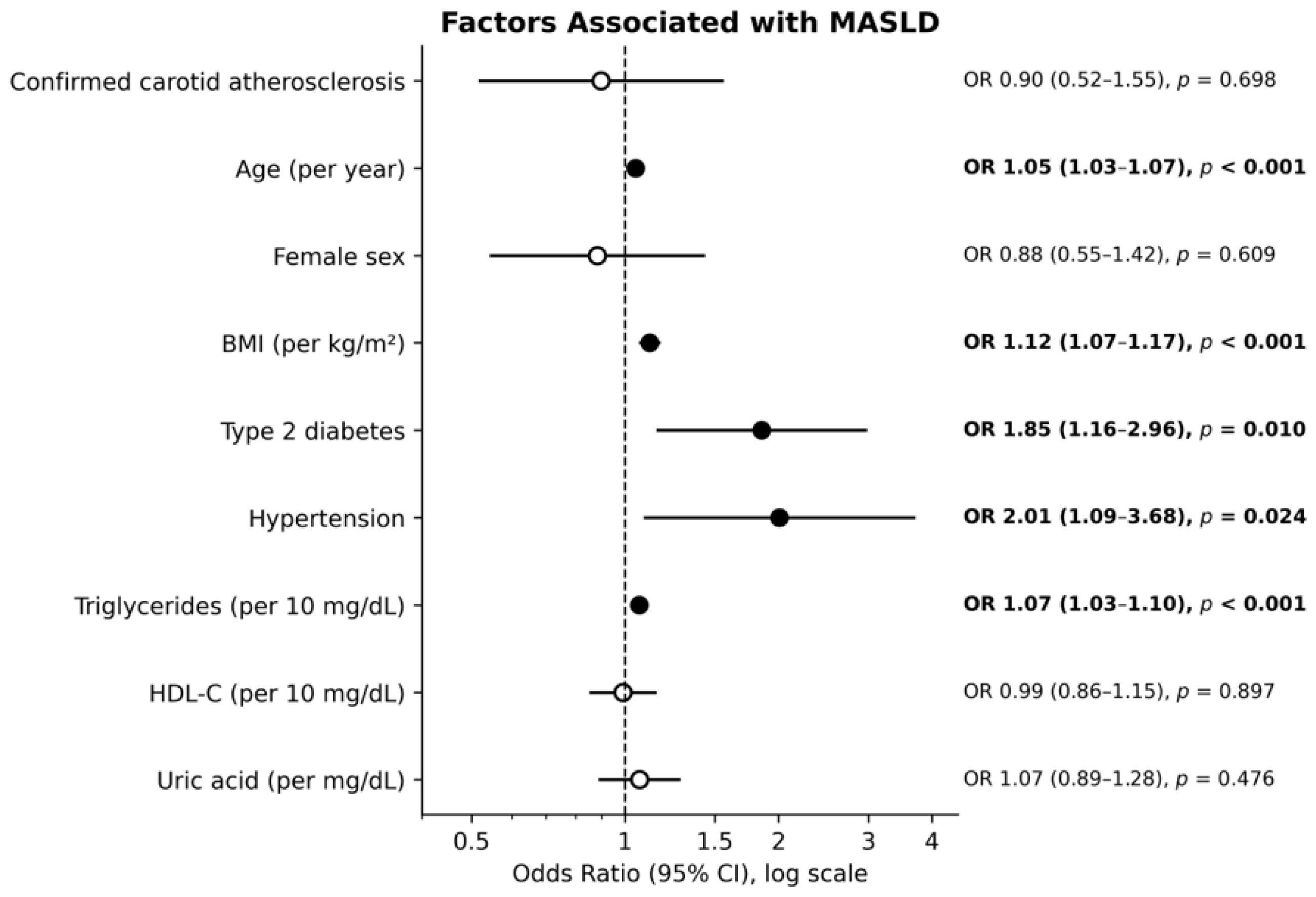
In a multivariable logistic regression model adjusted for confirmed carotid atherosclerosis, age, sex, BMI, type 2 diabetes, hypertension, triglycerides, HDL-C, and uric acid, confirmed carotid atherosclerosis was not associated with MASLD (OR 0.90, 95% CI 0.52–1.55, p = 0.698). Factors associated with MASLD were age (OR 1.05 per year, 95% CI 1.03–1.07, p < 0.001), BMI (OR 1.12 per kg/m2, 95% CI 1.07–1.17, p < 0.001), type 2 diabetes (OR 1.85, 95% CI 1.16–2.96, p = 0.010), hypertension (OR 2.01, 95% CI 1.09–3.68, p = 0.024) and triglycerides (OR 1.07 per 10 mg/dL, 95% CI 1.03–1.10, p < 0.001). Sex, HDL-C and uric acid were not associated with MASLD after adjustment. Detailed results are presented in Figure 2.
Figure 2.
Factors associated with MASLD in multivariable logistic regression. Odds ratios (ORs) with 95% confidence intervals (CIs) are shown on a logarithmic scale; filled circles indicate statistically significant associations (p < 0.05) and open circles non–significant associations. ORs are expressed per year (age), per kg/m2 (BMI), per 10 mg/dL (triglycerides and HDL cholesterol) and per 1 mg/dL (uric acid). BMI, body mass index; HDL, high–density lipoprotein.
3.3. Carotid Plaque Characteristics and Burden According to MASLD Status
In patients with confirmed carotid atherosclerosis (n = 407), plaque characteristics differed significantly according to MASLD status. Patients with MASLD had larger plaque diameter (p < 0.001), a higher prevalence of bilateral carotid involvement (p < 0.001), and a higher prevalence of stenosis ≥ 50% (p = 0.004). There was no evidence of a difference in plaque hypoechogenicity (p = 0.402). Detailed results are presented in Table 2. In the whole cohort (n = 600; score 0 assigned to patients without plaque), MASLD was also associated with a higher burden score (β = 0.27, 95% CI 0.11–0.43, p < 0.001), whereas the presence of plaque itself was not (adjusted OR 1.21, 95% CI 0.76–1.94).
Table 2.
Carotid plaque phenotype and atherosclerotic burden by MASLD status.
The composite atherosclerotic burden score was significantly higher in patients with MASLD than in those without (p < 0.001). After adjustment for age and sex, MASLD remained associated with a higher burden score (β = 0.313, 95% CI 0.157–0.469, p < 0.001); ordinal logistic regression gave a consistent result (OR 2.05, 95% CI 1.40–3.00), as did a complete-case analysis excluding the 8 patients with a missing score component (n = 399: β = 0.31, 95% CI 0.15–0.46; fully adjusted model 4, n = 395: β = 0.34, 95% CI 0.17–0.51). Residuals of the linear model were mildly left-skewed (skewness −0.22) without marked heteroscedasticity (Breusch-Pagan p = 0.066), and the estimate was unchanged with robust (HC3) standard errors (95% CI 0.156–0.470). For the ordinal model, cut-point-specific odds ratios were 1.77 (score ≥ 2), 2.17 (≥3) and 5.93 (≥4; 95% CI 1.32–26.58), and the proportional-odds assumption was marginally violated (likelihood-ratio test p = 0.033), driven by the sparse highest category (n = 19); after merging scores 3 and 4, the departure was no longer significant (p = 0.058) and the odds ratio was similar (1.92, 95% CI 1.30–2.83). The association remained significant after additional adjustment for type 2 diabetes and hypertension (β = 0.29, 95% CI 0.13–0.45) and after further adjustment for BMI, smoking, LDL-C, statin use and eGFR (β = 0.35, 95% CI 0.19–0.52). Results were similar when waist circumference replaced BMI (β = 0.30, 95% CI 0.14–0.47) and after additional adjustment for glucose-lowering therapy (β = 0.33, 95% CI 0.16–0.50). Coding the three patients with carotid occlusion as absent for the diameter component, or excluding them, did not change the association (β = 0.30, 95% CI 0.15–0.46 and β = 0.30, 95% CI 0.14–0.45, respectively). The estimate for bilateral involvement was attenuated after adjustment for diabetes and hypertension (OR 1.55, 95% CI 0.93–2.59) but remained similar in the fully adjusted model (OR 1.90, 95% CI 1.08–3.32). The pattern for bilateral involvement across models 1–4 was non-monotonic (OR 1.80, 1.55, 1.86 and 1.90; Supplementary Table S1), with broadly overlapping confidence intervals, and should not be over-interpreted; the estimates for the burden score and plaque diameter were stable across models. The estimate for stenosis ≥ 50% increased with further adjustment (Firth OR 3.64, 5.07, 6.50 and 7.15 in models 1–4; Supplementary Table S1) but was very imprecise (21 events for up to nine covariates in model 4, i.e., about two events per variable) and should be considered exploratory. Each individual component of the burden score was also associated with MASLD after adjustment: bilateral involvement (OR 1.80, 95% CI 1.10–2.95, p = 0.020), stenosis ≥ 50% (OR 4.14, 95% CI 1.17–14.62, p = 0.027; this estimate is based on only 21 events and is imprecise, Firth penalized logistic regression: OR 3.64, 95% CI 1.14–11.66), and plaque diameter (β = 0.41 mm, 95% CI 0.25–0.58, p < 0.001). There was no evidence of an association between MASLD and plaque hypoechogenicity after adjustment (OR 0.97, 95% CI 0.63–1.47, p = 0.874).
3.4. Correlation Between Non-Invasive Liver Indices and Atherosclerotic Burden
Within the subgroup with confirmed carotid atherosclerosis, FIB-4 showed a significant unadjusted correlation with the composite atherosclerotic burden score (Spearman’s ρ = 0.256, 95% CI 0.164–0.346, p < 0.001), which was not retained after adjustment for age and sex (ρ = 0.004, 95% CI −0.085 to 0.101, p = 0.934). APRI (unadjusted ρ = 0.071, 95% CI −0.025 to 0.166; adjusted ρ = 0.006, 95% CI −0.091 to 0.104) and the De Ritis ratio (unadjusted ρ = 0.078, 95% CI −0.017 to 0.170; adjusted ρ = −0.032, 95% CI −0.133 to 0.070) showed no significant correlation with the burden score (p > 0.05 for all). FLI was likewise not correlated with the burden score (unadjusted ρ = −0.030, 95% CI −0.124 to 0.064; adjusted ρ = −0.012, 95% CI −0.114 to 0.090; p > 0.05). Among patients with MASLD, age-specific FIB-4 category (<1.3 if <65 years; <2.0 if ≥65 years) was not associated with burden score (OR per category 0.96, 95% CI 0.67–1.36, p = 0.82).
3.5. FLI as a Non-Invasive Screening Tool for MASLD
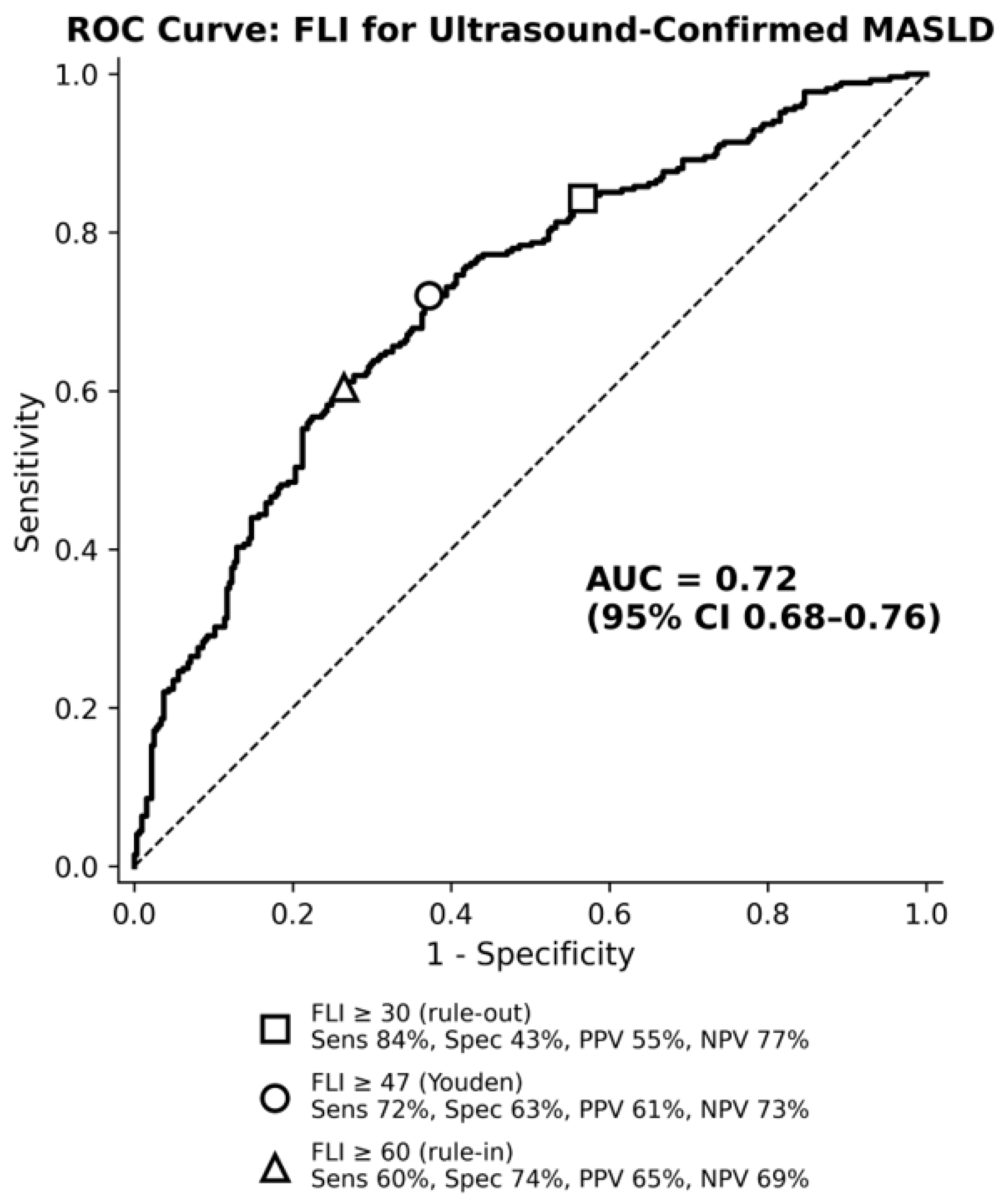
FLI showed modest discriminative accuracy for ultrasound-confirmed MASLD (AUC 0.72, 95% CI 0.68–0.76; n = 593; 7 patients were excluded because of missing GGT, waist circumference or triglycerides). The Youden cut-off of FLI ≥ 47 (sensitivity 72.0%, 95% CI 66.4–77.0; specificity 62.8%, 95% CI 57.4–67.8; PPV 61.5%, NPV 73.1%) was derived and evaluated in the same dataset and should be considered exploratory: it was unstable on bootstrap resampling (median 56, 95% range 40–67), with an optimism of the Youden index of 0.024 (optimism-corrected 0.323 vs. 0.348). At the conventional cut-offs, FLI ≥ 30 had a sensitivity of 84.3% and a specificity of 43.4% (NPV of FLI < 30, 77.0%), whereas FLI ≥ 60 had a sensitivity of 60.4%, specificity of 73.5% and PPV of 65.3% (Figure 3). The prevalence of MASLD was 23.0% in the FLI < 30 group (30.9% of patients), 39.5% in the intermediate zone (FLI 30–59; 27.3% of patients) and 65.3% in the FLI ≥ 60 group (41.8% of patients). Performance was somewhat higher in women (AUC 0.74, 95% CI 0.69–0.79) than in men (AUC 0.69, 95% CI 0.61–0.75); the Youden cut-offs were 56.6 and 66.8, respectively.
Figure 3.
Receiver operating characteristic (ROC) curve of the Fatty Liver Index (FLI) for ultrasound confirmed MASLD (AUC = 0.72, 95% CI 0.68–0.76; n = 593). Markers indicate the conventional rule-out (FLI ≥ 30; square) and rule-in (FLI ≥ 60; triangle) cut–offs and the Youden–optimal cut–off (FLI ≥ 47; circle), with corresponding sensitivity (Sens), specificity (Spec), positive predictive value (PPV) and negative predictive value (NPV). AUC, area under the curve; CI, confidence interval (bootstrap, 2000 samples).
4. Discussion
In industrialized societies, atherosclerotic cardiovascular disease (ASCVD), including coronary artery disease (CAD), peripheral artery disease (PAD), and cerebrovascular disease, remains among the leading causes of morbidity and mortality. Worldwide, according to the Global Burden of Disease-based projections, approximately 20.5 million cardiovascular deaths were expected in 2025, rising to 35.6 million by 2050 [26]. Atherosclerosis is the principal underlying pathology of these conditions, accounting for approximately three-quarters of all cardiovascular deaths [27]. Cardiovascular Disease (CVD) development and progression are closely intertwined with metabolic dysfunction, obesity, metabolic dysfunction-associated steatotic liver disease (MASLD) and diabetes mellitus acting as interconnected drivers of increased cardiovascular incidence and mortality [7,28]. Beyond their frequent coexistence, these metabolic disorders appear to share common underlying pathophysiological mechanisms (insulin resistance, pro-atherogenic dyslipidemia, endothelial dysfunction, oxidative stress, and systemic inflammation), which may account for the parallel increase in all-cause and cardiovascular mortality observed in patients with MASLD [10,11].
In this cross-sectional study of 600 primary care patients with dyslipidemia enrolled in a cardiology coordinated-care pathway, MASLD was present in 45% of patients and was associated with age, BMI, type 2 diabetes, hypertension, and triglycerides. Confirmed carotid atherosclerosis itself was not associated with MASLD after adjustment for these factors. However, in patients who already had confirmed carotid atherosclerosis, MASLD was associated with a greater atherosclerotic burden (bilateral involvement, significant stenosis, and larger plaque diameter), with no evidence of an association with plaque hypoechogenicity, a single marker of plaque vulnerability. This association was not reproduced by FIB-4, APRI, or the De Ritis ratio after adjusting for age. FLI showed modest discriminative accuracy for identifying patients with MASLD using data routinely available in primary care.
Globally, the prevalence of steatotic liver disease has been rising steadily and is now estimated at approximately 30% of the adult population, making it the most common chronic liver disease worldwide [29]. The prevalence observed in our cohort (45%) was considerably higher than this general-population estimate, which likely reflects the more heavily burdened baseline risk profile of our study population: unlike unselected population samples, all patients included in this study had an established diagnosis of dyslipidemia, and a substantial proportion also had hypertension or type 2 diabetes.
The independent metabolic factors identified in our cohort (age, BMI, type 2 diabetes, hypertension, and triglyceride levels) closely reflect the core components of metabolic syndrome and are consistent with the current understanding of MASLD as its hepatic manifestation [30]. Hypertension, (pre)diabetes, and hypertriglyceridemia have also emerged as some of the strongest predictors of metabolic burden and adverse outcomes in recent MASLD cohorts [31]. This concordance is consistent with the established metabolic phenotype of MASLD and supports the plausibility of our findings.
In our cohort, confirmed carotid atherosclerosis was not associated with MASLD after adjustment for age, sex, BMI, type 2 diabetes, hypertension, triglycerides, HDL-C, and uric acid (p = 0.698). This finding should not be read as contradicting the established association between MASLD and cardiovascular risk reported elsewhere in the literature, since most prior studies address the reverse question, namely whether MASLD predicts future cardiovascular events [32], whereas our analysis asked whether the presence of atherosclerosis itself is associated with MASLD once classic metabolic risk factors are accounted for. Our result instead supports a common soil hypothesis: MASLD and carotid atherosclerosis may represent parallel consequences of the same underlying metabolic dysfunction (insulin resistance, visceral adiposity, and atherogenic dyslipidemia) rather than one condition directly driving the other. This interpretation is consistent with recent evidence showing that coronary atherosclerosis progression in MASLD is driven by the cumulative burden of cardiometabolic risk factors rather than by any single dominant factor [33] and is further supported by Mendelian randomization data indicating that genetically proxied blood pressure and fasting glucose, rather than liver-specific markers, are causally associated with carotid atherosclerosis [34]. The lower LDL-C and total cholesterol observed in patients with MASLD most probably reflect more frequent and more intensive lipid-lowering treatment (58.5% vs. 38.8%), as among untreated patients LDL-C was similar in the two groups (148 vs. 150.5 mg/dL). Patients with MASLD were also more frequently treated with glucose-lowering drugs (including SGLT2 inhibitors and metformin), which may influence body weight, liver enzymes and, potentially, vascular findings; this may have attenuated or confounded some associations, although adjustment for lipid-lowering and glucose-lowering therapy did not alter the association between MASLD and atherosclerotic burden. In summary, these findings suggest that in a primary care population already engaged in cardiometabolic risk management, the liver and the vasculature should not be approached as independent diagnostic targets. These findings support the hypothesis that MASLD and carotid atherosclerosis reflect a shared cardiometabolic substrate. Consequently, carotid ultrasound performed routinely within the cardiology coordinated-care pathway should not be relied upon as an incidental screening cue for MASLD; rather, MASLD case-finding should be pursued proactively through direct assessment of metabolic risk factors, regardless of whether atherosclerosis has already been identified.
In our study population, among patients with confirmed carotid atherosclerosis (n = 407), MASLD was associated with a greater atherosclerotic burden, reflected by higher rates of bilateral involvement, stenosis ≥ 50%, as well as larger plaque diameter after adjustment for age and sex (p < 0.05). The association persisted after additional adjustment for diabetes, hypertension, BMI, smoking, LDL-C, statin use and eGFR, which suggests that shared cardiometabolic risk factors do not fully explain it; however, residual confounding cannot be excluded, and adjustment for BMI, which forms part of the MASLD definition, may be considered over-adjustment. Notably, there was no evidence of an association between MASLD and plaque hypoechogenicity (OR 0.97, 95% CI 0.63–1.47) detected; however, the confidence interval does not exclude a moderate increase or decrease in odds, and hypoechogenicity is only one imperfect marker of plaque vulnerability. Evidence linking MASLD to plaque vulnerability is limited and derives mainly from studies using CT angiography or high-resolution MRI, which reported an independent association with high-risk coronary and carotid plaque features [35,36].
In contrast, data on ultrasound-assessed plaque echogenicity in MASLD are scarce, and most ultrasound-based studies have assessed only plaque presence, thickness or overall burden [37]. Our finding of no evidence of an association with hypoechogenicity, a coarser marker, is therefore not necessarily at odds with these reports.
This pattern suggests that MASLD is more closely associated with the extent of established atherosclerosis than with sonographic features associated with plaque vulnerability. A plausible biological explanation is that the chronic, systemic metabolic dysregulation characteristic of MASLD (lipotoxicity and low-grade inflammation) promotes gradual, diffuse atherosclerotic progression over time, manifesting as more numerous, bilateral, and larger plaques, whereas plaque destabilization is governed by more localized processes (focal inflammatory infiltration, matrix metalloproteinase activity, and fibrous cap thinning) that may not be equally sensitive to systemic metabolic burden [38,39]; this explanation is hypothetical and requires longitudinal evaluation. In this sense, MASLD may increase the quantity rather than necessarily the biological aggressiveness, although this cannot be concluded from a single sonographic marker of atherosclerotic disease, consistent with recent machine learning-based work identifying classic metabolic parameters (age, systolic blood pressure, total cholesterol, sex, and fasting glucose), rather than liver-specific markers, as the dominant predictors of carotid plaque in MASLD populations [40]. A similar metabolically-driven pattern of carotid plaque progression in patients with MASLD has been reported in early middle-aged adults, particularly among those without other overt metabolic risk factors [41], further suggesting that MASLD may reflect cumulative metabolic exposure. These findings are hypothesis-generating; whether patients with MASLD and carotid plaque benefit from different follow-up requires prospective studies.
Non-invasive liver indices demonstrated a differential association with MASLD and carotid atherosclerotic burden in our cohort. FIB-4 and APRI were higher in patients with MASLD, whereas the De Ritis ratio did not differ significantly between groups. Notably, FIB-4 was initially associated with carotid atherosclerotic burden, but this association was no longer significant after adjustment for age. This finding is particularly relevant because age is a component of the FIB-4 formula and is itself strongly associated with the development and progression of atherosclerosis. The influence of age on the interpretation of FIB-4 is well established, with biopsy-validated data demonstrating a substantial age-related decline in its specificity for advanced fibrosis, particularly among individuals aged ≥65 years [42]. More recently, in a large cohort of 12,095 individuals, Zhang et al. reported that age showed superior predictive performance for carotid atherosclerosis compared with non-invasive liver fibrosis markers. Within the NAFLD subgroup, higher FIB-4 was positively associated with carotid atherosclerosis, whereas APRI was not independently associated after multivariable adjustment [43]. In a large cohort of patients with MASLD aged <50 years, higher FIB-4 was also associated with subclinical carotid atherosclerosis, further suggesting that the relationship between hepatic fibrosis markers and carotid disease may vary across age groups [44]. Taken together, these findings suggest that the attenuation of the FIB-4 atherosclerosis association after adjustment for age in our study may, at least in part, reflect the shared association of FIB-4 and atherosclerotic burden with increasing age rather than an independent association between the fibrosis score and atherosclerosis. Our findings should nevertheless be considered in the context of studies reporting associations between hepatic fibrosis and carotid atherosclerosis. Su et al. reported independent associations between several non-invasive fibrosis markers, including FIB-4, and carotid atherosclerosis in 3302 patients with NAFLD [45]. Similarly, a recent cross-sectional and longitudinal study in patients with MASLD found that liver fibrosis was associated with prevalent carotid atherosclerosis, while progression to a high-intermediate probability of advanced fibrosis based on FIB-4 was associated with the development of carotid atherosclerosis during follow-up [12]. However, these studies primarily addressed the presence of carotid atherosclerosis or its development over time, whereas our analysis focused on the extent of atherosclerotic burden, including plaque burden, bilateral involvement, plaque diameter and clinically relevant stenosis. This distinction may partly account for differences between our findings and previous reports and underscores that an association between a fibrosis index and the presence of atherosclerosis does not necessarily imply an independent relationship with the extent of atherosclerotic disease. The lack of a significant association between APRI or the De Ritis ratio and atherosclerotic burden further argues against a consistent relationship between routinely calculated biochemical liver scores and the extent of carotid atherosclerosis in our population. Importantly, this should not be interpreted as evidence against a biological link between hepatic fibrosis and cardiovascular disease. Rather, our findings suggest that commonly used fibrosis scores may largely reflect established risk factors for atherosclerotic burden, particularly age and cardiometabolic risk factors. The specific age dependence of FIB-4 may be especially relevant in older populations such as ours. Accordingly, while non-invasive fibrosis scores may contribute to cardiovascular risk characterization in patients with MASLD, their relationship with carotid atherosclerotic burden should be interpreted in the context of age and other established cardiovascular risk factors, rather than as an independent measure of atherosclerotic disease severity.
In our study, the fatty liver index (FLI) demonstrated moderate discrimination for identifying ultrasound-confirmed MASLD with AUC 0.72. Although this value was lower than those reported in several recent studies [46,47,48,49], it is comparable to the performance observed in some other clinical populations. For example, Thomson et al. [50] reported an AUC of 0.653 (95% CI 0.540–0.766) in patients with metabolic syndrome and ultrasound-confirmed steatosis. Similarly, Fujii et al. [51], using the controlled attenuation parameter as the reference standard in a health-check population, reported an AUROC of 0.786 for MASLD, with marked sex-related differences (0.729 in men and 0.886 in women). These differences may be related, at least in part, to the characteristics and metabolic enrichment of the study populations, as well as differences in the reference methods used to identify hepatic steatosis. In particular, our cohort consisted of patients with established dyslipidemia, with a high prevalence of hypertension, impaired glucose metabolism, and other cardiovascular risk factors, which differs substantially from relatively lower-risk health-check populations in which higher FLI discrimination has been reported [49]. Furthermore, FLI incorporates BMI, waist circumference, triglycerides, and GGT, all of which are closely related to the metabolic phenotype underlying MASLD; therefore, its discriminative capacity may be reduced in populations in which metabolic risk factors are highly prevalent across both MASLD and non-MASLD groups. Differences in sex distribution may also contribute to between-study variability, as recent studies have demonstrated substantially different optimal FLI thresholds between women and men [46,51]. Finally, the use of ultrasonography as the reference method in our study may contribute to differences in observed diagnostic performance compared with studies using Controlled Attenuation Parameter (CAP) or other quantitative techniques; notably, Fujii et al. reported only 78.3% agreement between ultrasonography and CAP for the detection of steatotic liver disease. The AUC of 0.72 observed in our study appears consistent with the variability reported for FLI in contemporary MASLD populations and suggests that FLI provides only modest information: even at the conventional cut-offs, 23% of patients with FLI < 30 had MASLD and 35% of those with FLI ≥ 60 did not, and 27% fell into the intermediate zone.
Our findings have several potential clinical implications. First, the association between MASLD and a greater atherosclerotic burden, without evidence of a corresponding association with plaque hypoechogenicity, is hypothesis-generating. These cross-sectional findings do not allow conclusions about management; they suggest that MASLD is more closely related to plaque burden than to hypoechogenicity, which should be tested in prospective studies. We found no evidence of an association with hypoechogenicity, a single and imperfect marker of plaque vulnerability.
Second, FLI—calculated from routinely available anthropometric and biochemical data (BMI, waist circumference, triglycerides, GGT)—showed modest discriminatory performance for MASLD (AUC 0.72) in our cohort. Although it has negligible marginal cost and is immediately available at the point of care, FLI cannot replace ultrasonography in this population: at best, a high FLI (≥60) may help prioritize patients for ultrasound, whereas a low FLI does not reliably exclude MASLD in dyslipidemic patients.
Third, our findings regarding FIB-4 argue against using this index, in isolation, as a marker of atherosclerotic burden in this population: its apparent association with vascular disease was substantially explained by age rather than indicating an age-independent association between FIB-4 and atherosclerotic burden. Clinically, an elevated FIB-4 in an older dyslipidemic patient should prompt consideration of hepatic fibrosis risk on its own merits but should not be interpreted as independent evidence of greater carotid disease severity.
Overall, these findings are consistent with considering hepatic and vascular risk together within cardiometabolic care. FLI alone is insufficient for MASLD case finding, whereas carotid plaque characterization appears more informative for assessing atherosclerotic burden than any single liver fibrosis score.
This study has several strengths: consecutive, real-world enrollment from a routine cardiology-linked care pathway; concurrent hepatic and carotid ultrasound assessment in the same patients by a single examiner; a panel of validated noninvasive liver indices (FIB-4, APRI, De Ritis, FLI) calculated directly rather than taken from diagnoses; a composite measure of atherosclerotic burden based on bilaterality, stenosis, and plaque size; and multivariable adjustment with sensitivity analyses that gave consistent results for the burden score.
These strengths should nonetheless be weighed against several limitations inherent to the study design. It was a single-center, cross-sectional study, which precludes causal inference, and the cohort was limited to dyslipidemic patients recruited from a single care pathway, which limits generalizability. Because patients were enrolled through a cardiology-linked care pathway, mainly on the basis of hypertension and other cardiological diagnoses, and underwent carotid Doppler as part of risk stratification, the cohort is likely to have a higher cardiometabolic risk than the general dyslipidemic or primary care population; the prevalences of MASLD (45%) and carotid atherosclerosis (68%) should therefore not be extrapolated to unselected populations, and selection bias cannot be excluded. No formal sample-size calculation was performed; the sample comprised all eligible consecutive patients, and the study was therefore not powered for the secondary and exploratory analyses. Statistical power was modest for less frequent outcomes (e.g., stenosis ≥ 50%, n = 21), reflected in wide confidence intervals; the odds ratio for stenosis should therefore be interpreted as exploratory. MASLD was diagnosed by ultrasound rather than biopsy or MRI-PDFF, and ultrasound has limited sensitivity for mild steatosis (<20–30% hepatic fat), which may have led to misclassification of some patients as non-MASLD; one patient without abdominal ultrasound (FLI 12.5) was classified as not having MASLD; excluding this patient did not change the results (MASLD prevalence 45.1%, 270/599; OR for carotid atherosclerosis 0.90, 95% CI 0.52–1.55; burden score β = 0.32, 95% CI 0.16–0.47). Fibrosis was estimated by serum indices rather than histology or elastography, with FIB-4 mathematically embedding age, which complicates age-adjusted interpretation. All abdominal and carotid ultrasound examinations were performed by a single unblinded examiner, and inter-observer variability was not assessed. Because both examinations were performed by the same unblinded examiner, correlated measurement error between the two readings cannot be excluded and may have inflated the association between MASLD and carotid findings. Plaque vulnerability was approximated by a single visually assessed B-mode feature (the presence of at least one hypoechogenic plaque) without a standardized grading scale; the null finding should therefore not be interpreted as evidence of no association. Continuous CIMT values were not available for analysis, and missing components of the burden score were coded as absent (a complete-case analysis gave consistent results). The composite burden score was developed for this study, has not been validated, and assumes equal weighting of its components; however, the results were consistent when the components were analyzed separately and with alternative scoring. Burden analyses were restricted to patients with established carotid atherosclerosis, which may introduce selection bias and limit inference to this subgroup; an analysis in the whole cohort, with patients without plaque scored as 0, gave a consistent association. Because BMI, glycemia, blood pressure and triglycerides are components of the cardiometabolic criteria of MASLD, their association with MASLD is partly definitional; however, in our cohort, MASLD status was virtually equivalent to ultrasound steatosis. Residual confounding (e.g., diet, physical activity, duration of metabolic disease) cannot be excluded, and statin use and glucose-lowering therapy were recorded at baseline only; treatment intensity, duration and adherence were not assessed. Finally, the confidence interval for the association between carotid atherosclerosis and MASLD does not exclude a modest effect in either direction.
To address these limitations, prospective, multi-center, longitudinal studies are needed to determine whether MASLD predicts incident carotid plaque and plaque progression, building on early signals reported in younger cohorts. More precise quantification of hepatic steatosis and fibrosis, e.g., by elastography or MRI-PDFF, would refine the exposure assessment, which in the present study relied on surrogate indices, and more objective assessment of plaque vulnerability (e.g., grey-scale median analysis or contrast-enhanced ultrasound) would strengthen conclusions about plaque stability. Finally, mechanistic studies incorporating markers of insulin resistance, systemic inflammation, and endothelial dysfunction could directly test the common-soil hypothesis, rather than inferring it from cross-sectional associations.
5. Conclusions
In conclusion, in this dyslipidemic primary care cohort, MASLD was associated with greater carotid atherosclerotic burden among patients with established carotid atherosclerosis, with no evidence of an association with plaque hypoechogenicity. The attenuation of the association between FIB-4 and atherosclerotic burden after adjustment for age highlights the importance of considering age when interpreting non-invasive fibrosis indices in this setting. FLI showed modest discriminatory performance for ultrasound-detected MASLD and cannot replace ultrasonography. Prospective studies are needed to determine whether MASLD is associated with subsequent plaque progression and to clarify the clinical value of combining hepatic and vascular assessment in cardiometabolic care.
Supplementary Materials
The following supporting information can be downloaded at: https://www.mdpi.com/article/10.3390/metabo16110892/s1, Table S1: Association of MASLD with the atherosclerotic burden score and its components across adjustment models 1–4. Table S2: Summary of the statistical models: sample sizes, covariates and sensitivity analyses.
Author Contributions
Conceptualization, M.H.; methodology, M.H.; formal analysis, M.H.; investigation, M.H.; resources, M.H.; data curation, M.H.; writing—original draft preparation, M.H. and M.W.; writing—review and editing, M.H. and M.W.; visualization, M.H. and M.W.; supervision, B.O.; project administration, M.H. All authors have read and agreed to the published version of the manuscript.
Funding
This research received no external funding.
Institutional Review Board Statement
The study was a retrospective analysis of de-identified routine clinical data. The Bioethics Committee of the Medical University of Silesia in Katowice confirmed, in response to an official inquiry, that it does not constitute a medical experiment and does not require approval (BNW/NWN/0052/KB/310/26). Data use was authorized by the practice, and the study complied with the GDPR and the Declaration of Helsinki.
Informed Consent Statement
Patient consent was waived because of the retrospective analysis of de-identified routine clinical data.
Data Availability Statement
The individual-level dataset is not publicly available because it contains sensitive health data of patients of a single primary care practice, and public deposition was not authorized by the practice; re-identification risk cannot be fully excluded despite de-identification. De-identified data may be made available to qualified researchers on reasonable request to the corresponding author, subject to approval by the practice and a data use agreement.
Acknowledgments
Statistical analyses were performed as described in Section 2.8. Figure 2 and Figure 3 were generated programmatically in Python (matplotlib) directly from the analyzed dataset and model outputs, with code written with the assistance of Claude (Anthropic; Sonnet 5.5/2026). Figure 1 was drawn with the same tool from the patient-flow counts. The authors verified that the figures correspond to the reported statistical results and take full responsibility for their content. AI tools were not used to generate or alter study data or results.
Conflicts of Interest
The authors declare no conflicts of interest.
Abbreviations
The following abbreviations are used in this manuscript:
| MASLD | Metabolic dysfunction-associated steatotic liver disease |
| NAFLD | Non-alcoholic fatty liver disease |
| CVD | Cardiovascular disease |
| FIB-4 | Fibrosis-4 |
| FLI | Fatty Liver Index |
| AST | Aspartate aminotransferase |
| ALT | Alanine aminotransferase |
| BMI | Body mass index |
| WHR | Waist-to-hip ratio |
| BP | Blood pressure |
| eGFR | Estimated glomerular filtration rate |
| APRI | AST-to-Platelet Ratio Index |
| GGT | Gamma-glutamyl transferase |
| CIMT | Carotid intima-media thickness |
| ARIC | Atherosclerosis Risk in Communities |
| PSV | Peak Systolic Velocity |
| IQR | Interquartile Range |
| OR | Odds Ratios |
| CI | Confidence Intervals |
| ROC | Receiver Operating Characteristic |
| AUC | Area under the curve |
| AI | Artificial Intelligence |
| SGLT2 | Sodium-glucose cotransporter 2 |
| GLP-1 | Glucagon-like peptide-1 |
| GIP | Glucose-dependent insulinotropic polypeptide |
| HFpEF | Heart failure with preserved ejection fraction |
| LDL-C | Low-density lipoprotein cholesterol |
References
- Roth, G.A.; Mensah, G.A.; Johnson, C.O.; Addou, A.A.; Ammirati, E.; Baddour, L.M.; Barengo, N.C.; Beaton, A.Z.; Benjamin, E.J.; Benziger, C.P.; et al. Global Burden of Cardiovascular Diseases and Risk Factors, 1990–2019: Update from the GBD 2019 Study. J. Am. Coll. Cardiol. 2020, 76, 2982–3021. [Google Scholar] [CrossRef] [Scilit] [PubMed]
- Libby, P. The changing landscape of atherosclerosis. Nature 2021, 592, 524–533. [Google Scholar] [CrossRef] [Scilit] [PubMed]
- Nambi, V.; Chambless, L.; Folsom, A.R.; He, M.; Hu, Y.; Mosley, T.; Volcik, K.; Boerwinkle, E.; Ballantyne, C.M. Carotid intima-media thickness and presence or absence of plaque improves prediction of coronary heart disease risk: The ARIC (Atherosclerosis Risk In Communities) study. J. Am. Coll. Cardiol. 2010, 55, 1600–1607. [Google Scholar] [CrossRef] [Scilit] [PubMed]
- Boccatonda, A.; Bray, A.; Marozzi, M.S.; Cipollone, A.; Lauletta, G.; D’Ardes, D.; Cicco, S.; Cipollone, F. From liver to cardiovascular system: The MASLD-ASCVD continuum. Intern. Emerg. Med. 2026. Epub ahead of print. [Google Scholar] [CrossRef] [Scilit] [PubMed]
- Chew, N.W.S.; Mehta, A.; Goh, R.S.J.; Zhang, A.; Chen, Y.; Chong, B.; Chew, H.S.J.; Shabbir, A.; Brown, A.; Dimitriadis, G.K.; et al. Cardiovascular-Liver-Metabolic Health: Recommendations in Screening, Diagnosis, and Management of Metabolic Dysfunction-Associated Steatotic Liver Disease in Cardiovascular Disease via Modified Delphi Approach. Circulation 2025, 151, 98–119. [Google Scholar] [CrossRef] [Scilit] [PubMed]
- Rinella, M.E.; Lazarus, J.V.; Ratziu, V.; Francque, S.M.; Sanyal, A.J.; Kanwal, F.; Romero, D.; Abdelmalek, M.F.; Anstee, Q.M.; Arab, J.P.; et al. A multisociety Delphi consensus statement on new fatty liver disease nomenclature. J. Hepatol. 2023, 79, 1542–1556. [Google Scholar] [CrossRef] [PubMed]
- Lee, H.H.; Lee, H.A.; Kim, E.J.; Kim, H.Y.; Kim, H.C.; Ahn, S.H.; Lee, H.; Kim, S.U. Metabolic dysfunction-associated steatotic liver disease and risk of cardiovascular disease. Gut 2024, 73, 533–540. [Google Scholar] [CrossRef] [Scilit] [PubMed]
- Zheng, H.; Sechi, L.A.; Navarese, E.P.; Casu, G.; Vidili, G. Metabolic dysfunction-associated steatotic liver disease and cardiovascular risk: A comprehensive review. Cardiovasc. Diabetol. 2024, 23, 346. [Google Scholar] [CrossRef] [Scilit] [PubMed]
- Oh, R.; Kim, S.; Cho, S.H.; Kim, J.; Lee, Y.-B.; Jin, S.-M.; Hur, K.Y.; Kim, G.; Kim, J.H. Metabolic Dysfunction-Associated Steatotic Liver Disease and All-Cause and Cause-Specific Mortality. Diabetes Metab. J. 2025, 49, 80–91. [Google Scholar] [CrossRef] [Scilit] [PubMed]
- Yang, J.; Kim, Y.R.; Na, S.K.; Kim, S.; An, J.; Shim, J.H. Mortality and Cardiovascular Outcomes in Patients with MAFLD Compared with Patients with MASLD: A Systematic Review and Meta-Analysis. Gut Liver 2026, 20, 137–152. [Google Scholar] [CrossRef] [Scilit] [PubMed]
- Ratti, C.; Malaguti, M.; Emanuele, D.; Bellasi, A.; Sanna, G. Understanding MASLD—From molecular pathogenesis to cardiovascular risk: A concise review for the clinical cardiologist. Atherosclerosis 2025, 409, 120495. [Google Scholar] [CrossRef] [Scilit] [PubMed]
- Zhang, H.; Lin, F.; Miao, M.; Yu, C.; Guo, W. Liver fibrosis increases the risk of subclinical atherosclerosis in patients with MASLD: A cross-sectional and longitudinal study. Nutr. Metab. 2025, 22, 68. [Google Scholar] [CrossRef] [Scilit] [PubMed]
- León-Mengíbar, J.; Malagón, M.M.; Bermúdez-López, M.; Valdivielso, J.M.; Pamplona, R.; Torres, G.; Mauricio, D.; Castro, E.; Fern, E.; Caix, A.; et al. Association of estimated liver fibrosis with carotid but not femoral atherosclerotic burden: The ILERVAS cohort. Front. Endocrinol. 2026, 16, 1651689. [Google Scholar] [CrossRef] [Scilit] [PubMed]
- Kubotsu, Y.; Sakamoto, Y.; Tago, M.; Chihara, A.; Norita, M.; Inadomi, C.; Inoue, K.; Takayanagi, H.; Tanaka, K.; Isoda, H.; et al. FIB-4 Index and Liver Stiffness Measurement are Potential Predictors of Atherosclerosis in Metabolic Dysfunction-Associated Steatotic Liver Disease. J. Atheroscler. Thromb. 2025, 32, 239–252. [Google Scholar] [CrossRef] [Scilit] [PubMed]
- European Association for the Study of the Liver (EASL); European Association for the Study of Diabetes (EASD); European Association for the Study of Obesity (EASO). EASL-EASD-EASO Clinical Practice Guidelines on the management of metabolic dysfunction-associated steatotic liver disease (MASLD). J. Hepatol. 2024, 81, 492–542. [Google Scholar] [CrossRef] [Scilit] [PubMed]
- Han, S.; Choi, M.; Lee, B.; Lee, H.-W.; Kang, S.H.; Cho, Y.; Ahn, S.B.; Song, D.S.; Jun, D.W.; Lee, J.; et al. Accuracy of Noninvasive Scoring Systems in Assessing Liver Fibrosis in Patients with Nonalcoholic Fatty Liver Disease: A Systematic Review and Meta-Analysis. Gut Liver 2022, 16, 952–963. [Google Scholar] [CrossRef] [Scilit] [PubMed]
- Bedogni, G.; Bellentani, S.; Miglioli, L.; Masutti, F.; Passalacqua, M.; Castiglione, A.; Tiribelli, C. The Fatty Liver Index: A simple and accurate predictor of hepatic steatosis in the general population. BMC Gastroenterol. 2006, 6, 33. [Google Scholar] [CrossRef] [Scilit] [PubMed]
- Carli, F.; Sabatini, S.; Gaggini, M.; Sironi, A.M.; Bedogni, G.; Gastaldelli, A. Fatty Liver Index (FLI) Identifies Not Only Individuals with Liver Steatosis but Also at High Cardiometabolic Risk. Int. J. Mol. Sci. 2023, 24, 14651. [Google Scholar] [CrossRef] [Scilit] [PubMed]
- Narodowy Fundusz Zdrowia. Opieka Koordynowana w POZ. Available online: https://www.nfz.gov.pl/dla-pacjenta/informacje-o-swiadczeniach/podstawowa-opieka-zdrowotna/opieka-koordynowana-w-poz/ (accessed on 15 September 2026).
- Ministerstwo Zdrowia. Moje Zdrowie—Bilans Zdrowia Osoby Dorosłej. Available online: https://www.gov.pl/web/zdrowie/moje-zdrowie-bilans-zdrowia-osoby-doroslej (accessed on 15 September 2026).
- Mach, F.; Baigent, C.; Catapano, A.L.; Koskinas, K.C.; Casula, M.; Badimon, L.; Chapman, M.J.; De Backer, G.G.; Delgado, V.; Ference, B.A.; et al. 2019 ESC/EAS Guidelines for the Management of Dyslipidaemias: Lipid Modification to Reduce Cardiovascular Risk. Eur. Heart J. 2020, 41, 111–188. [Google Scholar] [CrossRef] [Scilit] [PubMed]
- Wai, C.T.; Greenson, J.K.; Fontana, R.J.; Kalbfleisch, J.D.; Marrero, J.A.; Conjeevaram, H.S.; Lok, A.S. A simple noninvasive index can predict both significant fibrosis and cirrhosis in patients with chronic hepatitis C. Hepatology 2003, 38, 518–526. [Google Scholar] [CrossRef] [Scilit] [PubMed]
- De Ritis, F.; Coltorti, M.; Giusti, G. An enzymic test for the diagnosis of viral hepatitis: The transaminase serum activities. Clin. Chim. Acta 1957, 2, 70–74. [Google Scholar] [CrossRef] [Scilit] [PubMed]
- Touboul, P.-J.; Hennerici, M.G.; Meairs, S.; Adams, H.; Amarenco, P.; Bornstein, N.; Csiba, L.; Desvarieux, M.; Ebrahim, S.; Hernandez Hernandez, R.; et al. Mannheim Carotid Intima-Media Thickness and Plaque Consensus (2004–2006–2011). Cerebrovasc. Dis. 2012, 34, 290–296. [Google Scholar] [CrossRef] [Scilit] [PubMed]
- Grant, E.G.; Benson, C.B.; Moneta, G.L.; Alexandrov, A.V.; Baker, J.D.; Bluth, E.I.; Carroll, B.A.; Eliasziw, M.; Gocke, J.; Hertzberg, B.S.; et al. Carotid Artery Stenosis: Gray-Scale and Doppler US Diagnosis—Society of Radiologists in Ultrasound Consensus Conference. Radiology 2003, 229, 340–346. [Google Scholar] [CrossRef] [Scilit] [PubMed]
- Chong, B.; Jayabaskaran, J.; Jauhari, S.M.; Chan, S.P.; Goh, R.; Kueh, M.T.W.; Li, H.; Chin, Y.H.; Kong, G.; Anand, V.V.; et al. Global Burden of Cardiovascular Diseases: Projections from 2025 to 2050. Eur. J. Prev. Cardiol. 2025, 32, 1001–1015. [Google Scholar] [CrossRef] [Scilit] [PubMed]
- Nedkoff, L.; Briffa, T.; Zemedikun, D.; Herrington, S.; Wright, F.L. Global Trends in Atherosclerotic Cardiovascular Disease. Clin. Ther. 2023, 45, 1087–1091. [Google Scholar] [CrossRef] [Scilit] [PubMed]
- Kivimäki, M.; Kuosma, E.; Ferrie, J.E.; Luukkonen, R.; Nyberg, S.T.; Alfredsson, L.; Batty, G.D.; Brunner, E.J.; Fransson, E.; Goldberg, M.; et al. Overweight, Obesity, and Risk of Cardiometabolic Multimorbidity: Pooled Analysis of Individual-Level Data for 120 813 Adults from 16 Cohort Studies from the USA and Europe. Lancet Public Health 2017, 2, e277–e285. [Google Scholar] [CrossRef] [Scilit] [PubMed]
- Younossi, Z.M.; Golabi, P.; Paik, J.M.; Henry, A.; Van Dongen, C.; Henry, L. The Global Epidemiology of Nonalcoholic Fatty Liver Disease (NAFLD) and Nonalcoholic Steatohepatitis (NASH): A Systematic Review. Hepatology 2023, 77, 1335–1347. [Google Scholar] [CrossRef] [Scilit] [PubMed]
- Pustjens, J.; van Kleef, L.A.; Janssen, H.L.A.; de Knegt, R.J.; Brouwer, W.P. Differential Prevalence and Prognostic Value of Metabolic Syndrome Components among Patients with MASLD. JHEP Rep. 2024, 6, 101193. [Google Scholar] [CrossRef] [Scilit] [PubMed]
- Alnees, M.; Awwad, M.; Najajra, D.; Darwish, A.; Heih, O.; Al-Ali, T.; Abu Hamdeh, N.; Abdullah, A.; Ahmad, J.; Jarrar, K.; et al. Predictive Factors for Metabolic Syndrome in Patients with Metabolic Dysfunction-Associated Steatotic Liver Disease (MASLD). BMC Gastroenterol. 2025, 25, 562. [Google Scholar] [CrossRef] [Scilit] [PubMed]
- Bhatia, M.K.; Garg, A.; Ali, J.; Bhatia, M.; Singh, B.; Bassi, M.; Broder, A.; Adler, D.G. Association of NAFLD and Cardiovascular Events: A Systematic Review and Meta-Analysis. J. Clin. Gastroenterol. 2026, 60, 101–114. [Google Scholar] [CrossRef] [Scilit] [PubMed]
- Kim, A.; Kang, D.; Choi, S.C.; Sinn, D.H.; Gwak, G.-Y. Cardiometabolic Risk Factors and Coronary Atherosclerosis Progression in Patients with Metabolic Dysfunction-Associated Steatotic Liver Disease: The Influential Role of Quantity over Type. J. Gastroenterol. Hepatol. 2025, 40, 258–264. [Google Scholar] [CrossRef] [Scilit] [PubMed]
- Peng, W.-H.; Zhang, J.; Ruan, Y.-H.; Li, Y.; Liu, Y.; Dai, M.-J.; Jin, J.-K.; Yan, X.-B.; Ji, F. Risk Factors for Atherosclerotic Plaque Formation in MASLD Patients: Insights from Cross-Sectional and Mendelian Randomization Analyses. Nutr. Metab. Cardiovasc. Dis. 2026, 36, 104815. [Google Scholar] [CrossRef] [Scilit] [PubMed]
- Xu, T.; Guo, B.; Li, S.; Zhang, S.; Wang, X. Non-Alcoholic Fatty Liver Disease Is a Strong Predictor of Carotid High-Risk Plaques as Assessed by High-Resolution Magnetic Resonance Imaging. Quant. Imaging Med. Surg. 2025, 15, 898–910. [Google Scholar] [CrossRef] [Scilit] [PubMed]
- Puchner, S.B.; Lu, M.T.; Mayrhofer, T.; Liu, T.; Pursnani, A.; Ghoshhajra, B.B.; Truong, Q.A.; Wiviott, S.D.; Fleg, J.L.; Hoffmann, U.; et al. High-Risk Coronary Plaque at Coronary CT Angiography Is Associated with Nonalcoholic Fatty Liver Disease, Independent of Coronary Plaque and Stenosis Burden: Results from the ROMICAT II Trial. Radiology 2015, 274, 693–701. [Google Scholar] [CrossRef] [Scilit] [PubMed]
- Huang, Y.; Wang, Y.; Xiao, Z.; Yao, S.; Tang, Y.; Zhou, L.; Wang, Q.; Xie, Y.; Zhang, L.; Zhou, Y.; et al. The Association between Metabolic Dysfunction-Associated Steatotic Liver Disease, Cardiovascular and Cerebrovascular Diseases and the Thickness of Carotid Plaque. BMC Cardiovasc. Disord. 2023, 23, 554. [Google Scholar] [CrossRef] [Scilit] [PubMed]
- Iturbe-Rey, S.; Maccali, C.; Arrese, M.; Aspichueta, P.; Oliveira, C.P.; Castro, R.E.; Lapitz, A.; Izquierdo-Sanchez, L.; Bujanda, L.; Perugorria, M.J.; et al. Lipotoxicity-Driven Metabolic Dysfunction-Associated Steatotic Liver Disease (MASLD). Atherosclerosis 2025, 400, 119053. [Google Scholar] [CrossRef] [Scilit] [PubMed]
- Shah, P.K. Mechanisms of Plaque Vulnerability and Rupture. J. Am. Coll. Cardiol. 2003, 41, 15S–22S. [Google Scholar] [CrossRef] [Scilit] [PubMed]
- Zhai, S.-M.; Wang, X.-L.; Zhang, H.; Zuo, Y.-Q. Predicting Carotid Plaques in Metabolic Dysfunction-Associated Steatotic Liver Disease Using Machine Learning and SHAP Interpretation. Sci. Rep. 2025, 15, 36074. [Google Scholar] [CrossRef] [Scilit] [PubMed]
- Xiao, W.; Sun, X.; Lv, H.; Liu, X.; Zhu, J. Association between Metabolic Dysfunction-Associated Steatotic Liver Disease (MASLD) and Short-Term Progression of Carotid Atherosclerosis among Early Middle Age Adults. Diabetes Metab. Syndr. Obes. 2025, 18, 4535–4546. [Google Scholar] [CrossRef] [Scilit] [PubMed]
- McPherson, S.; Hardy, T.; Dufour, J.-F.; Petta, S.; Romero-Gomez, M.; Allison, M.; Oliveira, C.P.; Francque, S.; Van Gaal, L.; Schattenberg, J.M.; et al. Age as a Confounding Factor for the Accurate Non-Invasive Diagnosis of Advanced NAFLD Fibrosis. Am. J. Gastroenterol. 2017, 112, 740–751. [Google Scholar] [CrossRef] [Scilit] [PubMed]
- Zhang, S.; Liang, H.; Liu, J.; Zhu, Y. The Association between Liver Fibrosis Markers and Carotid Atherosclerosis in Non-Alcoholic Fatty Liver Disease Is Strongly Influenced by Age. BMC Cardiovasc. Disord. 2026, 26, 424. [Google Scholar] [CrossRef] [Scilit] [PubMed]
- Zhang, S.; Liang, H.; Liu, J.; Zhu, Y. The Association between Hepatic Fibrosis Indicators and Subclinical Carotid Atherosclerosis in the Population with Metabolic Dysfunction-Associated Steatotic Liver Disease under 50 Years Old. Nutr. Metab. 2025, 22, 136. [Google Scholar] [CrossRef] [Scilit] [PubMed]
- Su, J.; Zhou, L.; Liu, J.; Wang, Y.; Wang, G. Noninvasive Liver Fibrosis Markers Are Independently Associated with Carotid Atherosclerosis Risk in Patients with Nonalcoholic Fatty Liver Disease. Scand. J. Gastroenterol. 2024, 59, 961–971. [Google Scholar] [CrossRef] [Scilit] [PubMed]
- Crudele, L.; De Matteis, C.; Novielli, F.; Di Buduo, E.; Petruzzelli, S.; De Giorgi, A.; Antonica, G.; Berardi, E.; Moschetta, A. Fatty Liver Index (FLI) Is the Best Score to Predict MASLD with 50% Lower Cut-Off Value in Women than in Men. Biol. Sex. Differ. 2024, 15, 43. [Google Scholar] [CrossRef] [Scilit] [PubMed]
- Demirci, S.; Sezer, S. Fatty Liver Index vs. Biochemical-Anthropometric Indices: Diagnosing Metabolic Dysfunction-Associated Steatotic Liver Disease with Non-Invasive Tools. Diagnostics 2025, 15, 565. [Google Scholar] [CrossRef] [Scilit] [PubMed]
- Vamja, R.; M, Y.; Vala, V.; Ramachandran, A.; Nagda, J. Diagnostic Accuracy of Fatty Liver Index (FLI) for Detecting Metabolic Associated Fatty Liver Disease (MAFLD) in Adults Attending a Tertiary Care Hospital, a Cross-Sectional Study. Clin. Diabetes Endocrinol. 2024, 10, 46. [Google Scholar] [CrossRef] [Scilit] [PubMed]
- Miwa, T.; Tajirika, S.; Imamura, N.; Adachi, M.; Horita, R.; Hanai, T.; Ng, C.H.; Siddiqui, M.S.; Fukao, T.; Shimizu, M.; et al. Usefulness of Health Checkup-Based Indices in Identifying Metabolic Dysfunction-Associated Steatotic Liver Disease. JGH Open 2024, 8, e13110. [Google Scholar] [CrossRef] [Scilit] [PubMed]
- Thomson, E.S.; Oommen, A.T.; S, S.V.; Pillai, G. Comparison of Non-Invasive Liver Fat Scoring Systems as Markers of Metabolic Dysfunction-Associated Liver Disease. Cureus 2024, 16, e72222. [Google Scholar] [CrossRef] [Scilit] [PubMed]
- Fujii, H.; Uchida-Kobayashi, S.; Kanamori, A.; Nadatani, Y.; Kawamura, E.; Kimura, T.; Fukumoto, S.; Watanabe, T. Defining Optimal Fatty Liver Index Thresholds for MASLD and MetALD Using Controlled Attenuation Parameter as Reference. J. Gastroenterol. 2025, 60, 1296–1309. [Google Scholar] [CrossRef] [Scilit] [PubMed]
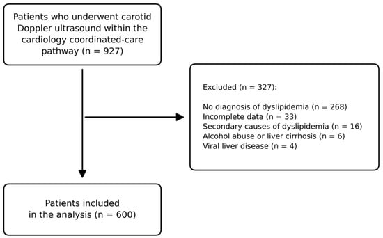
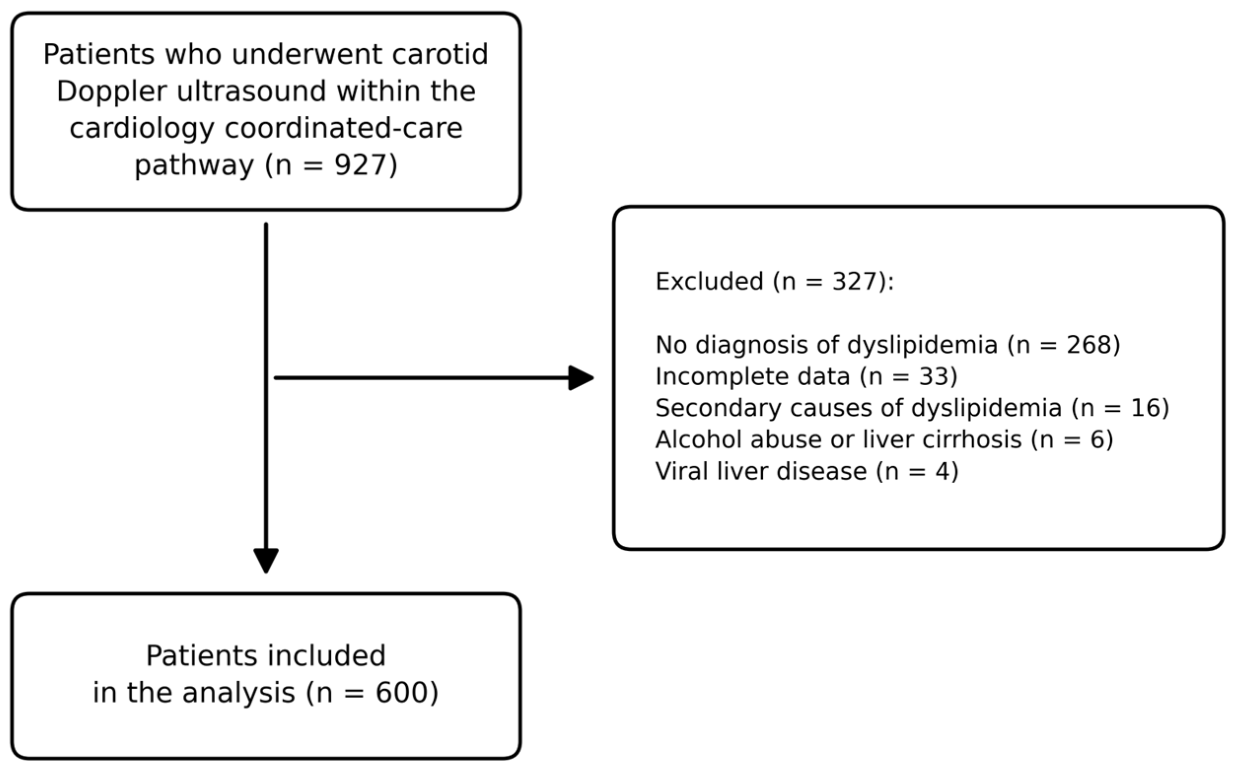

Disclaimer/Publisher’s Note: The statements, opinions and data contained in all publications are solely those of the individual author(s) and contributor(s) and not of MDPI and/or the editor(s). MDPI and/or the editor(s) disclaim responsibility for any injury to people or property resulting from any ideas, methods, instructions or products referred to in the content. |
© 2026 by the authors. Licensee MDPI, Basel, Switzerland. This article is an open access article distributed under the terms and conditions of the Creative Commons Attribution (CC BY) license.