


Abstract
(1) Background: The nasolabial fold is a key determinant of midfacial aging, influenced by both static volume loss and dynamic muscular activity. Botulinum toxin type A (BoNT-A) and hyaluronic acid (HA) fillers are widely used in aesthetic medicine, but limited evidence exists regarding the sequential use of these treatments to address both static and dynamic components of the nasolabial fold. (2) Methods: This prospective interventional case series included 23 patients with clinically evident nasolabial folds and depressor anguli oris (DAO) hyperactivity. All patients received BoNT-A injections into the DAO muscle, followed by HA filler augmentation in selected cases according to baseline severity. Patients were classified into three treatment groups in accordance with the treatment strategy selected at baseline: BoNT-A alone, BoNT-A plus 0.5 mL HA per side, and BoNT-A plus 1 mL HA per side. Outcomes were assessed using the Wrinkle Severity Rating Scale (WSRS) in both static and dynamic conditions and the Global Aesthetic Improvement Scale (GAIS), evaluated by two independent blinded physicians. (3) Results: All treatment groups demonstrated a reduction in dynamic WSRS scores following BoNT-A injection, reflecting decreased DAO activity and reduced dynamic accentuation of the nasolabial fold. Patients receiving combined BoNT-A and HA treatment showed additional improvement in static fold severity. Overall, GAIS scores indicated much to very much aesthetic improvement, with the highest outcomes observed in patients receiving 1 mL HA augmentation. Interobserver agreement was good across evaluations. (4) Conclusions: Botulinum toxin treatment of the DAO was linked to reduced dynamic accentuation of the nasolabial fold, while hyaluronic acid augmentation led to additional improvement in static fold severity. These preliminary findings indicate that sequential treatment might address both dynamic and static components of nasolabial fold appearance. However, larger randomized studies with longer follow-up are necessary to confirm these findings.
1. Introduction
Nasolabial folds are a pair of skin creases that extend bilaterally from the nasal wings to the corners of the mouth and may continue inferiorly. Their name derives from the anatomical regions they originate from. These folds are a natural anatomical boundary between the cheek and perioral soft tissues. The nasolabial folds extend from the lateral aspect of the nose, near the nasal ala, inferolaterally toward the oral commissures. With aging, they become more pronounced, increasing in both length and depth [1].
An established anatomical framework for describing the organization of facial soft tissues is the layered SCALP model, which identifies five distinct strata. The outermost layer is the skin (layer 1), followed by the subcutaneous connective tissue rich in adipose content (layer 2). The third layer is the aponeurotic or musculoaponeurotic system, which contributes to facial expression and structural support. Beneath this lies a layer of loose areolar connective tissue (layer 4), allowing for mobility between superficial and deep structures. The deepest layer is the periosteum or deep fascia (layer 5), which closely adheres to the underlying bone. This model provides a comprehensive basis for anatomical understanding relevant to both diagnostic and surgical contexts [2,3,4].
The superficial musculoaponeurotic system (SMAS), corresponding to the third layer in the SCALP model, functions as a structural interface between the mimic muscles and the overlying skin. Ghassemi et al. classified the SMAS into two main morphological subtypes. SMAS Type I is characterized by fibrous septa that anchor the mimic muscles to the skin and extend laterally toward the nasolabial fold. In contrast, SMAS Type II displays a denser fibrous network interspersed with adipose tissue and is located medially to the nasolabial fold [5].
Histological investigations by Sandulescu et al. further characterize the nasolabial fold as a prominent cutaneous depression demarcating the boundary between SMAS Type I and Type II. Lateral to the nasolabial fold, the subcutaneous tissue comprises vertically oriented connective tissue septa that link the facial musculature to the dermis, consistent with the architecture of SMAS Type I. Medial to the fold, particularly in the upper lip area, these fibrous structures become shorter and more compact, forming robust attachments between the zygomaticus major muscle and the skin. The intervening spaces are occupied by fat lobules, reinforcing the anatomical and functional distinction between the two SMAS configurations [2,3].
The development of nasolabial folds is multifactorial, involving anatomical, structural, and dynamic elements. One contributing factor is volume deficiency in adjacent facial compartments (including the deep midfacial region). This condition results from a reduced amount of superficial fat beneath the fold, often associated with the retraction of the jawbone into the canine fossa, which might be responsible for increased fold depth and altered midfacial contour. While previous correction strategies relied on implants or autologous fat grafts, modern approaches favor dermal fillers due to their superior elasticity and ability to achieve more natural and precise volume restoration [1,3].
The second mechanism involves differential skin attachment and tissue sagging. Beneath the nasolabial fold, the skin is tightly anchored to the underlying orbicularis oris muscle, limiting downward migration. In contrast, above the fold, the dermal attachments are looser, allowing the anterior buccal fat pad to descend and create a convex appearance. Age-related changes, including fat atrophy below the fold and superficial fat accumulation above it, further exacerbate the difference in thickness and contour of these tissues [1,4].
In addition, the activity of the levator muscles of the upper lip (such as the levator labia superioris nasi, levator labii superioris, and zygomaticus major and minor muscles) can accentuate the depth of the nasolabial fold through repetitive muscle contractions and facial animation. These muscles elevate the upper lip, accentuating the medial portion of the fold [6] and influence both the dynamic and static appearance of the fold, thereby affecting facial aesthetics and symmetry [1]. This theory is supported by observations in patients with facial nerve paralysis, who lose the nasolabial fold on the affected side, providing strong evidence that facial animation plays a key role in its formation.
Given the diverse etiologies, treatment of the nasolabial fold must be individualized, taking into consideration the patient’s specific anatomical features and functional dynamics. In addition, to reduce the risk of side effects, careful attention must be paid during the procedure to respect the regional vascular anatomy.
The facial artery is the primary vascular supply to the mid and lower face, originating from the external carotid artery and traversing an important anatomical path. It extends upward from the lower border of the mandible, moving medially toward the nasolabial fold, where it forms the superior labial, inferior labial, and angular branches. This path positions the artery in proximity to common target areas in aesthetic medicine, particularly during dermal filler injections [7].
Considering the variable and potentially superficial course of the facial artery and its branches in the nasolabial region, this is a high-risk area for vascular complications during injection procedures. Accidental intravascular injection of fillers can cause arterial occlusion, potentially resulting in local ischemia, tissue necrosis, or, in severe cases, retrograde embolization involving the ophthalmic artery and subsequent vision loss. Risk-reduction strategies include thorough knowledge of regional vascular anatomy, slow and controlled injection, use of appropriate injection planes and volumes, and the use of blunt cannulas rather than sharp needles to reduce the likelihood of vascular penetration [8,9].
The nasolabial fold represents an important indicator of midfacial aging, often appearing as one of the earliest signs of volume loss in the facial soft tissues. Despite decades of surgical innovations aimed at improving its appearance [1,6], this anatomical region remains difficult to fully correct, often undertreated by conventional facelift techniques [6]. Age-related loss of subcutaneous and dermal volume significantly contributes to the deepening of the fold and the formation of wrinkles.
With the rise in minimally invasive procedures in aesthetic medicine, the use of neuromodulators and soft-tissue fillers has increased over the past two decades [10]. Botulinum toxin injections have become the most performed nonsurgical aesthetic procedure, driven by the growing demand for less invasive treatments that offer rapid recovery and predictable, safe results. Over the past decade, botulinum toxin has emerged as the preferred treatment for dynamic rhytids, with extensive research and clinical application in neuromodulating the frontalis, corrugator supercilii, procerus, lateral orbicularis oculi, nasalis, and depressor anguli oris muscles [6].
Among nonsurgical options, Botulinum toxin type A (BoNT-A) has become one of the most commonly performed procedures in aesthetic medicine. By treating upper facial areas, botulinum toxin has demonstrated exceptional efficacy, with high reported rates of patient satisfaction. Benefits of this treatment include consistent efficacy, minimal downtime and rejuvenating effects, leading to the exploration of other applications beyond its indications. This evolution has led to the development of different dilution protocols, personalized dosing strategies, and the customization of injection patterns adapted to individual anatomical variations. Current strategies target multiple regions, including the lower face, such as platysma bands and the depressor anguli oris (DAO) muscle. Relaxation of the depressor muscles can achieve a lifting effect, jawline definition and improved resting facial expression. Noninvasive lower face rejuvenation necessitates a detailed understanding of facial anatomy and individualized injection techniques to achieve natural outcomes [11].
The current study aimed to assess the short-term effect of botulinum toxin injections targeting the DAO muscles on the dynamic component of the nasolabial fold.
Changes in the appearance of the nasolabial fold during active DAO contraction were evaluated, together with changes in static fold severity and overall aesthetic improvement following botulinum toxin treatment alone or followed by hyaluronic acid augmentation. The study was designed to investigate the clinical contribution of DAO activity to the dynamic appearance of the nasolabial fold rather than to establish a causal mechanism of tissue migration or long-term facial aging.
2. Materials and Methods
This prospective interventional case series included 23 consecutive patients seeking consultation for nasolabial fold correction. All participants signed informed consent prior to inclusion. They were fully informed that botulinum toxin treatment for the DAO muscle is an off-label use, and they voluntarily consented to this treatment.
The study was conducted in accordance with the Declaration of Helsinki and approved by the Re-search Ethics Committee of MedLife Group for studies conducted at Humanitas MedLife Hospital (Approval No. 9/21.07.2025, approved on 21 July 2025). The study was not prospectively registered in a publicly accessible clinical trial registry.
The study was initiated in August 2025 and conducted over a period of three months (August–October 2025), enrolling consecutive eligible patients during this timeframe.
All injection procedures were performed under standardized clinical and aseptic conditions.
Standardized photographs and clinical assessments were obtained at baseline, after BoNT-A treatment, and at the final visit 4–8 weeks after the last treatment session. The intermediate assessment was performed 2 weeks after BoNT-A injection, once the neuromodulatory effect was considered clinically established; patients in Groups 2 and 3 subsequently received HA augmentation following this intermediate evaluation. At each time point, standardized photographic documentation was obtained under identical lighting conditions, facial expression control, and patient positioning to ensure reproducibility; the follow-up visit included acquisition of the second post-treatment image set (Figures S1–S5 in Supplementary Materials).
All patients received botulinum toxin injections into the DAO muscle using BoNT-A (Alluzience®, abobotulinumtoxin A). According to the treatment strategy selected at baseline, patients were classified into three treatment groups. Each patient underwent baseline static and dynamic assessment followed by DAO BoNT-A injection and an intermediate assessment performed 2 weeks later to confirm the effect of neuromodulation. Seven patients received botulinum toxin alone (Group 1), five patients received botulinum toxin followed by 0.5 mL of HA (Restylane® Defyne) injected into each nasolabial fold (Group 2), and eleven patients received botulinum toxin followed by 1 mL of HA (Restylane® Defyne) injected into each nasolabial fold (Group 3). Patients in Group 2 and Group 3 received HA augmentation, followed by the final post-treatment assessment.
Treatment allocation was not randomized: treatment strategy and HA volume were chosen in accordance with baseline NLF severity, facial anatomy and individual aesthetic goals. Therefore, treatment groups were not intended to be directly comparable with respect to baseline disease severity.
Inclusion criteria comprised adults presenting with visible nasolabial folds and clinical evidence of DAO hyperactivity who requested minimally invasive facial rejuvenation. Exclusion criteria included pregnancy or breastfeeding, neuromuscular disorders, active local infection, previous botulinum toxin treatment within the preceding 4 months, hypersensitivity to botulinum toxin or hyaluronic acid products, and inability to provide informed consent.
Each patient underwent a detailed clinical evaluation, assessing:
- Malar volume and projection;
- Distribution and condition of the superficial fat compartments;
- Smile dynamics and smile type classification;
- Muscular activity of the DAO and platysma.
All clinical assessments and treatment planning were performed by the same experienced injector to ensure consistency in patient evaluation and injection technique.
In patients exhibiting hyperactivity of the DAO muscle, identified by voluntary depression of the oral commissures (i.e., pulling the mouth corners downward), botulinum toxin injection was administered first. It has been hypothesized that chronic hyperactivity of the DAO and platysma muscles, together with the tethering effect of the SMAS, may contribute to descent of the superficial malar fat compartments toward the nasolabial fold; however, this biomechanical hypothesis was not objectively assessed in the present study (no imaging or tissue-tracking evaluation was performed). Clinically, the nasolabial fold appeared more pronounced when patients were asked to perform the DAO action by voluntarily pulling the corners of the mouth downward, consistent with dynamic accentuation during muscle contraction.
The initial phase of treatment, with botulinum toxin, was designed to evaluate its effect on the nasolabial fold. This was followed by photographic documentation taken before and after the botulinum toxin treatment. The botulinum toxin was injected using a 4 mm, 32 G needle approximately 1 cm lateral and 1 cm inferior to the modiolus, at a depth of approximately 4 mm, with 2–3 units of botulinum toxin injected into each side.
The second phase of treatment consisted of hyaluronic acid injection into the nasolabial fold. In patients who received 0.5 mL HA per side, 0.2 mL was administered as a deep bolus at the piriform fossa in contact with the underlying bone, followed by 0.3 mL injected into the nasolabial fold using a 25 G, 38 mm cannula. In patients who received 1 mL HA per side, 0.4 mL was administered as a deep bolus at the piriform fossa, followed by 0.6 mL injected into the nasolabial fold using the same cannula technique. Following cannula passage to establish the injection plane, the filler was deposited in the subcutaneous plane using a fanning technique. The cannula insertion point was directed along the fold, approximately 5 mm from its caudal margin.
Clinical outcomes were assessed using the Wrinkle Severity Rating Scale (WSRS) [12] and the Global Aesthetic Improvement Scale (GAIS) [13]. Standardized photographs were obtained before treatment and at follow-up under identical lighting conditions, camera settings, patient positioning, and predefined facial expressions instructions.
The severity of the nasolabial folds at rest was assessed using the validated 5-point Wrinkle Severity Rating Scale (WSRS), ranging from 1 (absent) to 5 (extremely severe). To explore the dynamic component of the NLF, we applied a study-specific modification of the WSRS during maximal voluntary contraction of the DAO muscle. This dynamic assessment was exploratory and has not been independently validated. Therefore, dynamic WSRS results were interpreted as an exploratory measure of NLF accentuation during DAO contraction rather than as a validated outcome measure.
Overall aesthetic improvement following treatment was assessed using the GAIS, a validated 5-point subjective scale ranging from 1 (worse) to 5 (very much improved). GAIS scores reflected the overall aesthetic outcome by comparing post-treatment appearance with baseline photographs.
All WSRS and GAIS evaluations were performed independently by two experienced physicians who were not involved in the treatment procedures. Static WSRS scores were assigned with the face in a neutral resting position, whereas dynamic WSRS scores were recorded during maximal voluntary contraction of the depressor anguli oris muscle. The left and right nasolabial folds were evaluated separately, and the mean score was used for statistical analysis. The dynamic evaluation was a study-specific exploratory application of the WSRS, despite not being formally validated. Each patient was instructed to perform maximal voluntary contraction of the DAO muscle by actively depressing the oral commissures. Lighting, camera settings, facial positioning and facial expression instructions were the same between assessments. The intensity of voluntary DAO contraction was not objectively quantified (with the use of electromyography or another instrumental method). After independent assessment, the two evaluators reached a consensus GAIS score.
The primary outcome was the reduction in dynamic accentuation of the nasolabial folds during active DAO contraction. Secondary outcomes included improvement in static nasolabial fold appearance and physician assessment based on standardized photographs.
Statistical analysis was performed using Microsoft Excel. Pre- and post-treatment comparisons of static and dynamic WSRS scores were conducted using the Wilcoxon signed-rank test for paired non-parametric data, applied separately within each treatment group and for the overall cohort. The association between hyaluronic acid (HA) volume and changes in WSRS scores (Δ = post-treatment minus pre-treatment) was assessed using Spearman’s rank correlation coefficient. Interobserver reliability for static and dynamic WSRS assessments was evaluated using the intraclass correlation coefficient (ICC), based on a two-way random-effects, absolute-agreement model: ICC(2,1). All statistical tests were two-tailed, and a p-value < 0.05 was considered statistically significant.
During the preparation of this manuscript, the authors used ChatGPT (GPT-5.6 Luna, OpenAI) for minor language editing, including spelling and grammar checking and improving the linguistic clarity of selected sentences. All content was critically reviewed and approved by the authors, who take full responsibility for the accuracy, integrity, and final content of the manuscript.
3. Results
A total of 23 patients with prominent nasolabial folds associated with depressor anguli oris (DAO) hyperactivity were included in this case series. All patients received botulinum toxin injections into the DAO muscle using abobotulinumtoxinA (Alluzience®), at doses of 2–3 units per side, according to baseline clinical assessment of DAO activity.
Seven patients (30.4%) received botulinum toxin alone (Group 1), five patients (21.7%) received botulinum toxin combined with 0.5 mL hyaluronic acid (HA) per nasolabial fold (Group 2), and eleven patients (47.8%) received botulinum toxin combined with 1 mL HA per nasolabial fold (Group 3).
The cohort comprised 19 women (82.6%) and 4 men (17.4%). The mean age was 31.2 years in Group 1, 33.4 years in Group 2, and 35.8 years in Group 3. Group 1 included six women (85.7%) and one man (14.3%), Group 2 included four women (80.0%) and one man (20.0%), and Group 3 included nine women (81.8%) and two men (18.2%). Baseline demographic characteristics are summarized in Table 1.
Table 1.
Baseline demographic characteristics of the study populations.
Baseline assessment demonstrated the presence of a dynamic component in all patients, with dynamic WSRS scores consistently higher than static WSRS scores, confirming clinically relevant DAO hyperactivity across the cohort. Patients in Group 1 presented with low baseline static severity (median static WSRS = 1) and a moderate dynamic component (median dynamic WSRS = 2). In Group 2, baseline severity was higher, with median static and dynamic WSRS scores of 2 and 3, respectively, while Group 3 demonstrated the most severe baseline presentation, with median static and dynamic WSRS scores of 3 and 4, respectively.
Interobserver reliability was excellent for both WSRS components, with intraclass correlation coefficients ICC(2,1) of 0.939 for static and 0.964 for dynamic assessments, confirming high reproducibility between the two independent evaluators.
Following treatment, all groups demonstrated improvement in nasolabial fold appearance, with a consistent and marked reduction in the dynamic component across the entire cohort. In Group 1 (BoNT-A alone), static WSRS remained unchanged (mean 1.0 to 1.0), whereas dynamic WSRS decreased significantly from 2.21 to 1.0 (Wilcoxon signed-rank test, p = 0.016), corresponding to an approximate 55% reduction. This finding indicates a selective effect of botulinum toxin on DAO-mediated dynamic fold formation, without measurable influence on static severity.
In Group 2 (BoNT-A + 0.5 mL HA), both static and dynamic WSRS improved following treatment, although the changes did not reach statistical significance (p = 0.063 for both components), likely reflecting the limited sample size.
In contrast, Group 3 (BoNT-A + 1 mL HA) demonstrated statistically significant improvement in both static and dynamic components (both p = 0.001), with a mean reduction in static WSRS of 2.09 points, indicating a marked reduction in static WSRS severity.
Across the entire cohort, both static and dynamic WSRS scores improved significantly after treatment (both p < 0.001).
A strong association was found between HA volume and change in WSRS scores (Δ = post-treatment minus pre-treatment; Spearman ρ = −0.973, p < 0.001). However, because HA volume was selected in accordance with the baseline NLF severity, this association should not be interpreted as a causal dose–response relationship. The observed association may reflect confounding by indication, as patients with more severe folds received higher HA volumes. A significant inverse association was also observed between HA volume and change in dynamic WSRS (ρ = −0.777, p < 0.001). Likewise, considering that HA volume was selected depending on baseline severity, neither association should be regarded as evidence of a causal dose–response relationship.
Global aesthetic improvement, assessed using the Global Aesthetic Improvement Scale (GAIS), showed high levels of satisfaction across all groups. Median GAIS scores were 4 in Groups 1 and 2 and 5 in Group 3, indicating much to very much improvement, with the greatest aesthetic benefit observed in the group receiving higher-volume filler augmentation.
Considering that treatment allocation relied on baseline severity and individual clinical assessment, the groups had different baseline NLF severity and should therefore not be regarded as randomized comparative treatment groups.
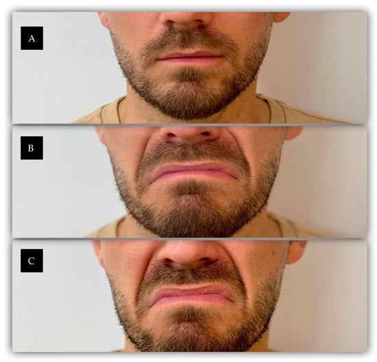
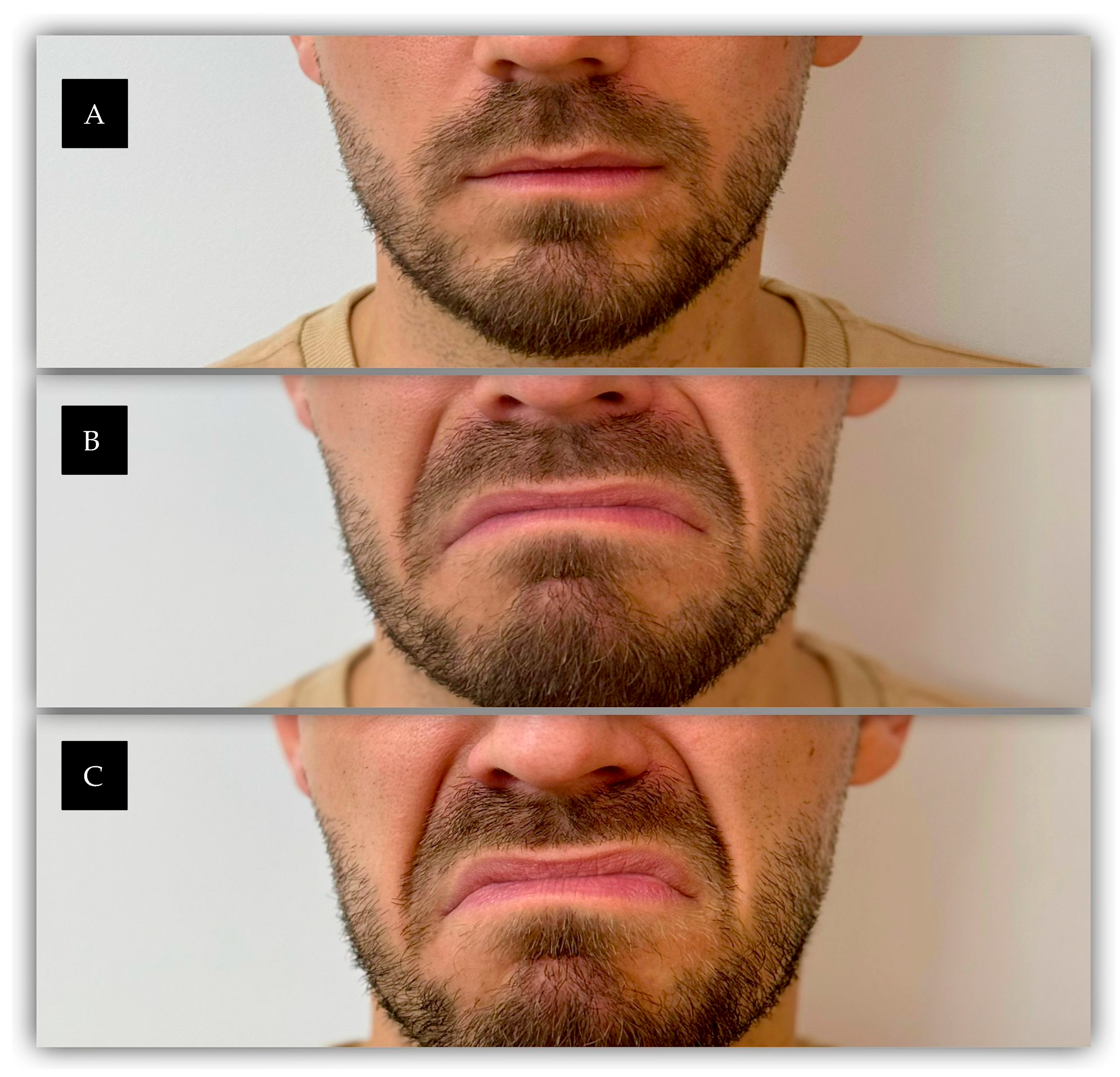
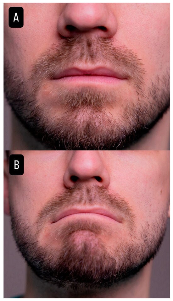
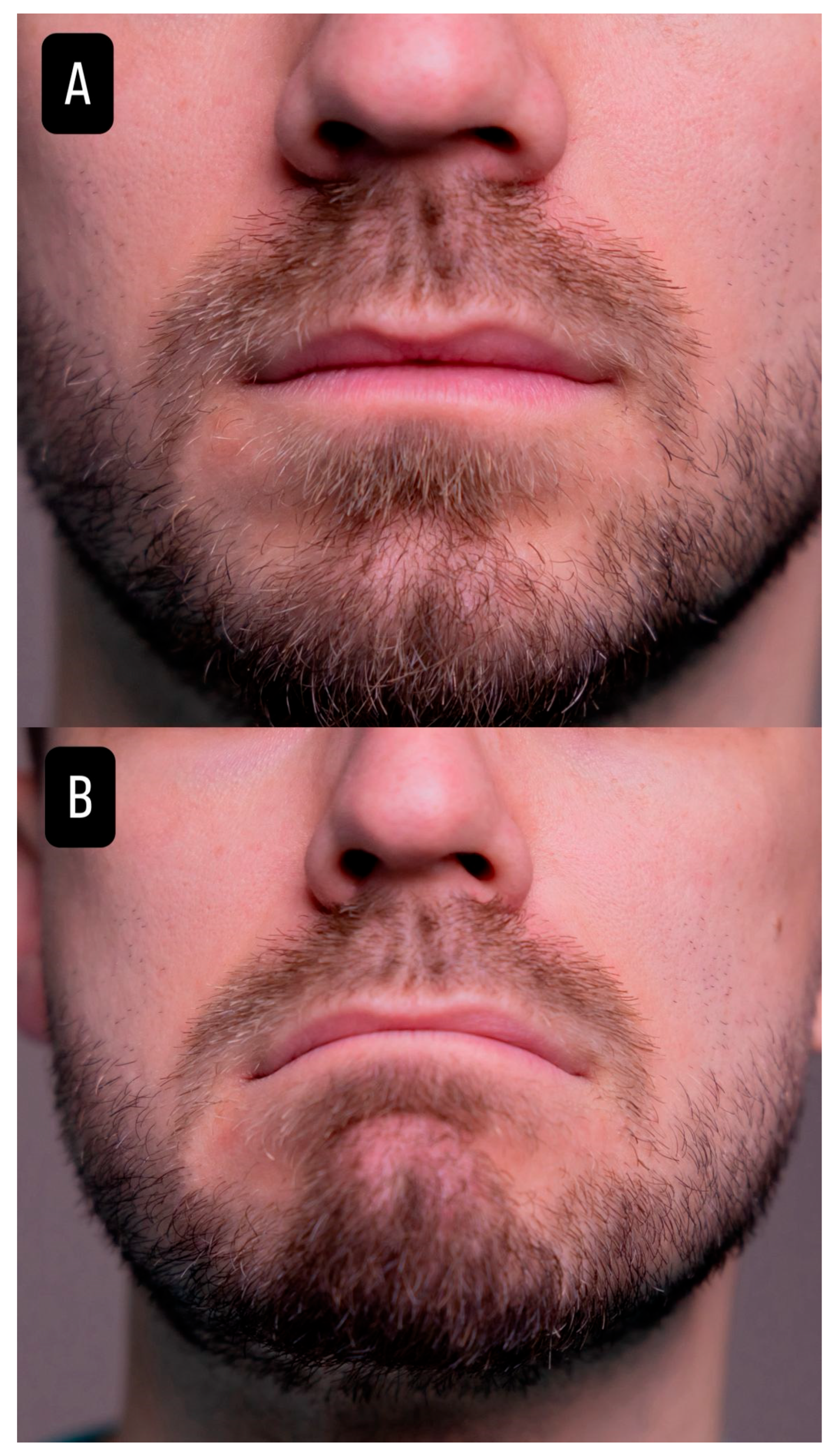
Standardized photographic assessment confirmed consistent clinical improvement, with reduction in nasolabial fold prominence during facial animation and a global improvement in lower facial appearance across all treatment groups (Figure 1 and Figure 2, Figures S1–S5 in Supplementary Materials).
Figure 1.
Baseline assessment. (A) Static position. Early formation of the nasolabial folds can be observed, particularly in the perinasal region. (B) Active contraction of the DAO muscles. Increased prominence of the lower two-thirds of the nasolabial folds is evident. (C) Active contraction of both the DAO muscles and the upper lip elevator muscles. A marked accentuation of the entire length of the nasolabial folds can be observed. In this context, the nasolabial folds demonstrate distinct patterns of dynamic enhancement depending on the muscles involved. Contraction of the depressor muscles predominantly accentuates the lower portion of the folds, whereas contraction of the upper lip elevator muscle mainly increases the prominence of the upper portion.
Figure 2.
After botulinum toxin injection into the DAO muscles. (A) Static position, shown for comparison with the image before the intervention (Figure 1A). No significant change in nasolabial fold appearance at rest was observed in this treatment group, consistent with the static WSRS results (Group 1). (B) Active contraction of the DAO muscles no longer produces dynamic accentuation of the nasolabial folds. The lower portion of the folds appears less pronounced during facial animation, reflecting reduced depressor muscle activity following botulinum toxin injection.
Patients were systematically assessed at each follow-up visit for BoNT-A-related complications, including oral commissure asymmetry, lower lip dysfunction, and speech or smile alteration, as well as for HA-related complications, including bruising, edema, nodules, infection, or vascular complications. No major adverse events or treatment-related complications were recorded during the follow-up period. All procedures were well tolerated, and no patients required medical intervention related to the injections.
4. Discussion
4.1. Overview of the Main Findings
This study suggests that botulinum toxin injection into the DAO muscle significantly reduces dynamic accentuation of nasolabial folds, with additional improvement achieved through adjunctive hyaluronic acid volumization in patients with higher baseline static severity. The combined approach resulted in consistent aesthetic improvement across all treatment groups.
Overall aesthetic improvement was graded favorably by the independent evaluators. The highest GAIS scores occurred in patients who received the higher-volume HA treatment. Still, these findings should be interpreted in the context of baseline differences between treatment groups.
While the botulinum toxin treatment did not produce an immediate, visible effect on the static appearance of the nasolabial folds, its benefits became apparent in dynamic expressions, particularly during the contraction of the DAO muscle.
In our study, the most evident effect of botulinum toxin was a decrease in dynamic accentuation of the lower portion of the nasolabial fold during active DAO contraction. Whether repeated reduction in DAO activity can influence long-term soft-tissue displacement or the progression of marionette or nasolabial lines remains hypothetical and requires further investigation.
4.2. Anatomical and Functional Considerations
The human face relies on a delicate balance between the muscles that lift and lower facial features. Increased activity of depressor muscles, such as the platysma muscle and the DAO muscle, can contribute to dynamic wrinkles and a tired facial expression. While the muscles may naturally weaken with age, their activity may be more pronounced on less resistant tissues and dermis, potentially leading to alterations in the smile. Recent vectorial analyses of platysma contraction mechanisms in healthy adults have revealed a bidirectional contraction pattern, with the superior fibers pulling the lower face caudally and the inferior fibers elevating the skin of the neck and upper chest. All these forces converge at a predictable horizontal plane along the mid-cervical region. This anatomical behavior suggests that the platysma contributes not only to dynamic descent of the jawline and marionette lines, but may also apply (indirectly) tension that leads to an accentuation of the nasolabial fold. Traditionally, the platysma has been considered primarily in relation to platysmal bands and descent of the jaw [14]. However, these findings support a broader role, functionally contributing to lower facial aging. In our study, we focused specifically on the effect of DAO neuromodulation on dynamic nasolabial fold appearance.
DAO muscle plays an important role in depressing the oral commissures, leading to a negative facial expression and the development of several lines, including the marionette lines and the labiomental fold. Botulinum toxin injections targeting the DAO may allow the lip elevators to predominate, improving the contour of the lower face. Still, the procedure remains off-label and should be performed with caution due to the close anatomical relations between the DAO and the depressor labii inferioris, which, if affected, can result in lower lip asymmetry. According to Moradi et al., a three-point injection technique targeted at the upper half of the DAO is associated with favorable outcomes and low incidence of adverse effects. While this therapeutic approach was previously discussed in the context of marionette lines, their study explored its impact on the nasolabial fold as well, stating that some dynamic interactions between the DAO, platysma, and perioral musculature also influence the depth and presentation of the nasolabial fold [15].
The combined use of BoNT-A and dermal filler might enable separate targeting of dynamic muscle activity and static soft-tissue deficiency. In the present study, BoNT-A was related to improvement in dynamic nasolabial fold accentuation, while subsequent HA augmentation was associated with greater improvement in static fold severity. The study design limits drawing conclusions with respect to comparative efficacy, durability, or safety advantages of the sequential approach.
Temporary relaxation of these depressor muscles induced by BoNT-A attenuates the underlying muscle activity, creating a smoother surface on which dermal fillers can be precisely applied. Thus, it leads to a more balanced, rejuvenated appearance [16].
4.3. The Aging Process of the Face
Facial aging represents a complex, multifactorial process characterized by progressive degeneration and structural alteration of facial components. This includes intrinsic aging of the skin, as well as atrophy and remodeling of the underlying soft tissues, including subcutaneous fat, fascia, and muscles. Skin quality is determined by several factors, including thickness, elasticity, degree of actinic damage, and hydration status. Facial soft tissue volume is primarily maintained by the subcutaneous fat that is distributed in the anatomical compartments of the face, the SMAS, and the connective tissue supporting the different anatomical planes. With aging, these structures undergo qualitative and quantitative changes, including loss of elasticity, volume depletion, and reduced structural support. As a result, soft tissues progressively descend under the influence of gravity, leading to characteristic morphological changes associated with facial aging. These structural changes contribute to the clinical manifestations of facial aging [3,4,11].
Skeletal support of the face represents another determinant of facial aging, as it also undergoes changes during the aging process. Bone structures undergo resorption and remodeling with age. At the orbital level, the enlargement of the orbital cavity occurs as a consequence of bone resorption affecting the margins of the orbital aperture, particularly the frontal, zygomatic, and maxillary bones [11].
Facial aging is not only caused by gravitational descent of facial fat compartments, progressive weakening of the supporting ligaments of the face and skeletal remodeling; it is also significantly influenced by the activity of the depressor muscles. Various muscles such as the DAO, the inferior labialis, and the platysma contribute over time to downward movement of the lower facial tissues (and a tired, sad, or aged appearance) [4,11].
The superficial fat compartments of the face are highly dynamic and more mobile than the deeper fat layers. These fat deposits are influenced by the resting and dynamic tension of the facial muscles, which are responsible for facial expression. The facial muscles create both subtle and pronounced movements such as smiling, frowning, and speaking, which continuously influence the arrangement of the superficial fat deposits. Over time, these repetitive mechanical forces contribute to alterations in facial contour and promote the formation of lines, folds, and wrinkles. In addition, as the face ages, the volume and elasticity of these fat deposits decrease. When combined with the repeated tension caused by facial muscle activity, these changes contribute to soft tissue descent and further modification of facial morphology, ultimately accentuating the clinical features of facial aging [6,17,18].
Moreover, the effects of gravity on skin laxity and soft tissue atrophy over time are significant contributors to facial creases such as nasolabial folds and lip lines. These folds, along with other age-related changes, are the result of a combination of gravitational forces, muscle movements, and the natural thinning of the dermis. Repetitive muscle movements, such as smiling or frowning, create dynamic creases such as the crow’s feet because they repeatedly stretch the dermis and alter its elasticity. This understanding of facial aging has led to the adoption of advanced techniques such as soft tissue redistribution, malar fat pad lift, and SMAS lift, all of which are now common strategies for rejuvenating the aging face. In addition, soft tissue augmentation has gained increasing popularity as a means of restoring volume loss and improving facial contours, thereby contributing to a more youthful appearance. This combination of lifting and volumizing techniques aims to restore both structural support and soft tissue volume, resulting in more natural and balanced aesthetic outcomes [2,3,19].
4.4. Limitations
This study has several limitations.
First, the relatively small sample size (23 patients) limits the generalizability of the findings and limits statistical precision and generalizability.
Second, the short follow-up period does not allow for evaluation of long-term outcomes, particularly regarding the durability of treatment effects and any potential long-term influence on facial soft-tissue changes. Repeated neuromodulation of the depressor facial muscles may theoretically contribute to delayed descent of midfacial tissues and attenuation of nasolabial fold progression over time; however, such long-term preventive effects require extended follow-up periods that were not included in this study.
In addition, statistical analyses were performed using Microsoft Excel, which provides limited advanced statistical capabilities compared with dedicated software packages, which limited the feasibility of more advanced statistical modelling and sensitivity analyses.
The relatively small sample size, particularly in Group 2, reduces statistical power and might reduce the ability to detect statistically significant differences despite clinically relevant directional changes. The use of non-parametric tests, while appropriate for small and ordinal datasets such as WSRS, limits the ability to adjust for potential confounding variables.
Another methodological limitation is represented by the non-randomized allocation of patients to treatment groups. Treatment intensity was chosen according to baseline NLF severity, with patients presenting more severe static folds receiving greater HA volumes. Thus, the observed association between HA volume and WSRS improvement cannot be interpreted as a true dose–response relationship. Baseline severity and treatment allocation were linked, introducing potential confounding by indication. The results should consequently be regarded as exploratory associations instead of evidence that higher HA volumes produce greater clinical improvement.
Moreover, a limitation of this study is the exploratory modification of the WSRS used to evaluate NLF prominence during DAO contraction. Although the conventional WSRS is validated, this dynamic application has not been formally validated. The dynamic WSRS findings should be regarded as hypothesis-generating and require confirmation using validated dynamic facial evaluation methods in future studies.
Finally, subgroup analyses should be interpreted with caution due to uneven group distribution and limited sample size, which may affect the generalizability of the findings.
Further longitudinal studies with larger cohorts and prolonged observation are necessary to validate these hypotheses and assess the durability of both functional and aesthetic outcomes.
5. Conclusions
Overall, this prospective study highlights that botulinum toxin injection into the DAO muscle could reduce the dynamic accentuation of the nasolabial fold during active DAO contraction. In cases presenting greater baseline static fold severity, subsequent hyaluronic acid augmentation was linked to additional improvement in static nasolabial fold appearance. The combined treatment approach addresses two clinically distinct components of nasolabial fold appearance: dynamic muscular activity and static soft-tissue deficiency.
However, because treatment allocation was non-randomized, HA volume was associated with baseline severity. Follow-up was limited, so the observed associations should be interpreted as preliminary. Larger randomized studies using validated dynamic assessment methods and longer follow-up are required to explore the independent contribution of DAO neuromodulation, the optimal HA volume and the durability of treatment effects.
Supplementary Materials
The following supporting information can be downloaded at: https://www.mdpi.com/article/10.3390/cosmetics13050247/s1, Figures S1–S5: Case-specific evaluation of facial muscle activity.
Author Contributions
Conceptualization, I.C.P., R.A.I., V.P. and M.V.M.; methodology, I.C.P., R.A.I., V.P. and M.V.M.; validation, P.A.P. and S.S.; formal analysis, I.C.P. and R.A.I.; investigation, I.C.P. and M.V.M.; data curation, I.C.P. and R.A.I.; visualization, I.C.P. and R.A.I.; writing—original draft preparation, all authors; writing—review and editing, all authors; supervision, M.V.M.; project administration, I.C.P. and M.V.M.; funding acquisition, I.C.P. All authors have read and agreed to the published version of the manuscript.
Funding
The publication of this paper was supported by the “Iuliu Hațieganu” University of Medicine and Pharmacy Cluj-Napoca, subject to institutional reimbursement procedures.
Institutional Review Board Statement
The study was conducted in accordance with the Declaration of Helsinki and approved by the Research Ethics Committee of MedLife Group for studies conducted at Humanitas MedLife Hospital (Approval No. 9/21.07.2025, approved on 21 July 2025).
Informed Consent Statement
Informed consent was obtained from all subjects involved in the study. Written informed consent has been obtained from the patients to publish this paper.
Data Availability Statement
The raw data supporting the conclusions of this article will be made available by the authors on request.
Acknowledgments
(1) The authors thank Neola Pharma for providing the injectable products through an unrestricted educational sponsorship. Injectable products used in this study were provided through an unrestricted educational sponsorship by Neola Pharma, the official distributor of Galderma products in Romania. The support was provided exclusively to promote medical education and facilitate the clinical application presented in this article. The sponsor had no role in the study design, patient selection, data collection, data analysis, interpretation of the results, manuscript preparation, or the decision to submit the manuscript for publication. The authors conducted the study independently, and the scientific content and conclusions were not influenced by the sponsor. (2) The authors used ChatGPT (GPT-5.6 Luna, OpenAI) for minor editorial assistance, including rephrasing and language polishing. Scientific content, analyses, interpretations and conclusions were developed by the authors.
Conflicts of Interest
The authors declare no conflicts of interest.
References
- Hong, G.-W.; Song, S.; Park, S.Y.; Lee, S.-B.; Wan, J.; Hu, K.-S.; Yi, K.-H. Why Do Nasolabial Folds Appear? Exploring the Anatomical Perspectives and the Role of Thread-Based Interventions. Diagnostics 2024, 14, 716. [Google Scholar] [CrossRef] [Scilit] [PubMed]
- Sandulescu, T.; Buechner, H.; Rauscher, D.; Naumova, E.; Arnold, W.-H. Histological, SEM and three-dimensional analysis of the midfacial SMAS—New morphological insights. Ann. Anat.—Anat. Anz. 2019, 222, 70–78. [Google Scholar] [CrossRef] [Scilit] [PubMed]
- Sandulescu, T.; Franzmann, M.; Jast, J.; Blaurock-Sandulescu, T.; Spilker, L.; Klein, C.; Naumova, E.A.; Arnold, W.H. Facial fold and crease development: A new morphological approach and classification. Clin. Anat. 2019, 32, 573–584. [Google Scholar] [CrossRef] [Scilit] [PubMed]
- Fratila, A.A.M.; Schenck, T.L.; Redka-Swoboda, W.; Zilinsky, I.; Pavicic, T.; Cotofana, S. The anatomy of the aging face: A review. Facial Plast. Surg. 2016, 32, 253–260. [Google Scholar] [CrossRef] [Scilit] [PubMed]
- Ghassemi, A.; Prescher, A.; Riediger, D.; Axer, H. Anatomy of the SMAS revisited. Aesthetic Plast. Surg. 2003, 27, 258–264. [Google Scholar] [CrossRef] [Scilit] [PubMed]
- Snider, C.C.; Amalfi, A.N.; Hutchinson, L.E.; Sommer, N.Z. New Insights into the Anatomy of the Midface Musculature and its Implications on the Nasolabial Fold. Aesthetic Plast. Surg. 2017, 86, 53–1090. [Google Scholar] [CrossRef] [Scilit] [PubMed]
- de Maio, M.; DeBoulle, K.; Braz, A.; Rohrich, R.J. Facial Assessment and Injection Guide for Botulinum Toxin and Injectable Hyaluronic Acid Fillers: Focus on the Midface. Plast. Reconstr. Surg. 2017, 140, 540e–550e. [Google Scholar] [CrossRef] [Scilit] [PubMed]
- Ozturk, C.N.; Li, Y.; Tung, R.; Parker, L.; Piliang, M.P.; Zins, J.E. Complications following injection of soft-tissue fillers. Aesthetic Surg. J. 2013, 33, 862–877. [Google Scholar] [CrossRef] [Scilit] [PubMed]
- Colon, J.; Mirkin, S.; Hardigan, P.; Elias, M.J.; Jacobs, R.J.; Elias, M. Adverse Events Reported from Hyaluronic Acid Dermal Filler Injections to the Facial Region: A Systematic Review and Meta-Analysis. Cureus 2023, 15, e38286. [Google Scholar] [CrossRef] [Scilit] [PubMed]
- Beer, G.M.; Manestar, M.; Mihic-Probst, D. The causes of the nasolabial crease: A histomorphological study. Clin. Anat. 2013, 26, 196–203. [Google Scholar] [CrossRef] [Scilit] [PubMed]
- Lorenc, Z.P.; Corduff, N.; van Loghem, J.; Yoelin, S. Creating Lift in the Lower Face with Botulinum Toxin A Treatment: An Anatomical Overview with Videos and Case Studies Illustrating Patient Evaluation and Treatment. Aesthetic Surg. J. Open Forum 2022, 4, ojac034. [Google Scholar] [CrossRef] [Scilit] [PubMed]
- Day, D.J.; Littler, C.M.; Swift, R.W.; Gottlieb, S. The wrinkle severity rating scale: A validation study. Am. J. Clin. Dermatol. 2004, 5, 49–52. [Google Scholar] [CrossRef] [Scilit] [PubMed]
- DiBernardo, G.A.; DiBernardo, B.E. Prediction of Treatment Outcomes for Neck Rejuvenation Utilizing a Unique Classification System of Treatment Approach Using a 1440-nm Side-Firing Laser. Aesthetic Surg. J. 2018, 38, S43–S51. [Google Scholar] [CrossRef] [Scilit] [PubMed]
- Ohana, O.; Fezza, J.P.; Sykes, J.; Yoelin, S.G.; Lee, J.H.; Fezza, R.M.; Lee, W.W. The Platysma Contraction Pattern in Healthy Adults: A Vector Analysis Study. Plast. Reconstr. Surg. 2024, 153, 601–607. [Google Scholar] [CrossRef] [Scilit] [PubMed]
- Moradi, A.M.; Shirazi, A. A Retrospective and Anatomical Study Describing the Injection of Botulinum Neurotoxins in the Depressor Anguli Oris. Plast. Reconstr. Surg. 2022, 149, 850–857. [Google Scholar] [CrossRef] [Scilit] [PubMed]
- Hall, C. Optimizing Facial Aesthetics: Sequential Application of Botulinum Toxin A and Dermal Fillers for Enhanced Results. Plast. Aesthetic Nurs. 2024, 44, 70–71. [Google Scholar] [CrossRef] [Scilit] [PubMed]
- Swift, A.; Liew, S.; Weinkle, S.; Garcia, J.K.; Silberberg, M.B. The Facial Aging Process from the “Inside Out”. Aesthetic Surg. J. 2021, 41, 1107–1119. [Google Scholar] [CrossRef] [Scilit] [PubMed]
- Choi, Y.-J.B.; Kim, J.-S.D.; Gil, Y.-C.; Phetudom, T.; Kim, H.-J.D.; Tansatit, T.M.; Hu, K.-S.D. Anatomical considerations regarding the location and boundary of the depressor anguli oris muscle with reference to botulinum toxin injection. Plast. Reconstr. Surg. 2014, 134, 917–921. [Google Scholar] [CrossRef] [Scilit] [PubMed]
- Narins, R.S.; Brandt, F.S.; Dayan, S.H.; Hornfeldt, C.S. Persistence of nasolabial fold correction with a hyaluronic acid dermal filler with retreatment: Results of an 18-month extension study. Dermatol. Surg. 2011, 37, 644–650. [Google Scholar] [CrossRef] [Scilit] [PubMed]

Disclaimer/Publisher’s Note: The statements, opinions and data contained in all publications are solely those of the individual author(s) and contributor(s) and not of MDPI and/or the editor(s). MDPI and/or the editor(s) disclaim responsibility for any injury to people or property resulting from any ideas, methods, instructions or products referred to in the content. |
© 2026 by the authors. Licensee MDPI, Basel, Switzerland. This article is an open access article distributed under the terms and conditions of the Creative Commons Attribution (CC BY) license.