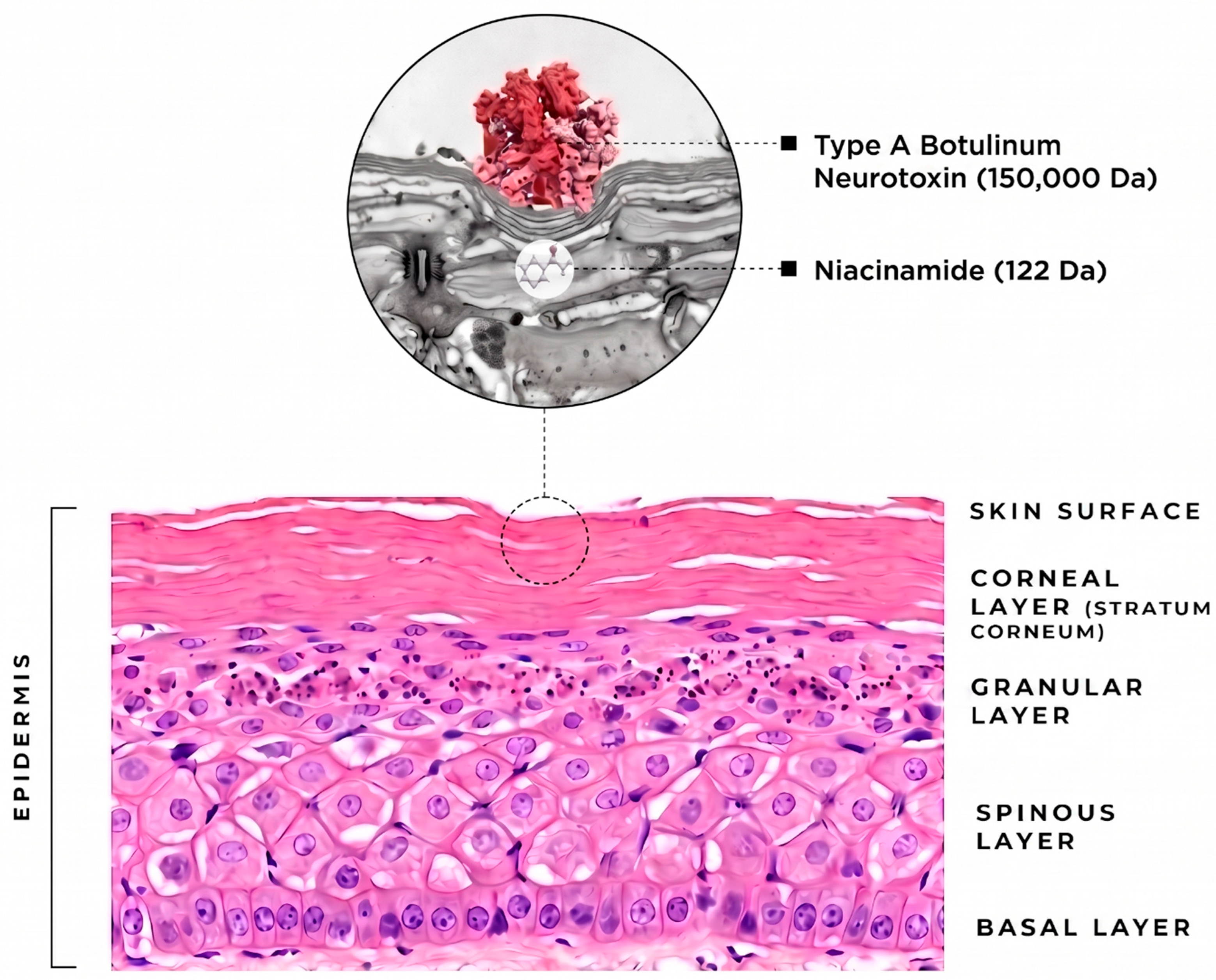
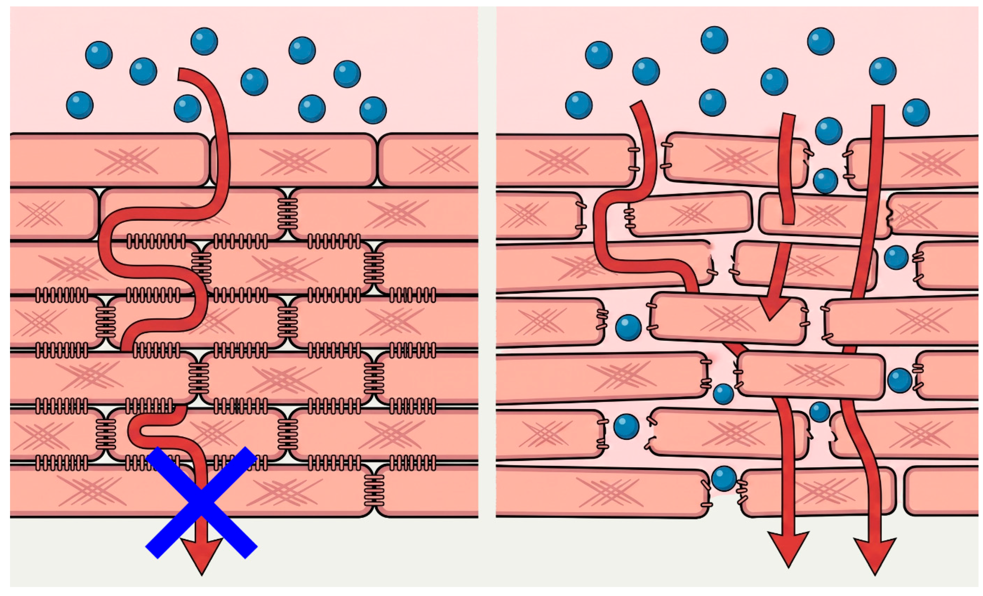
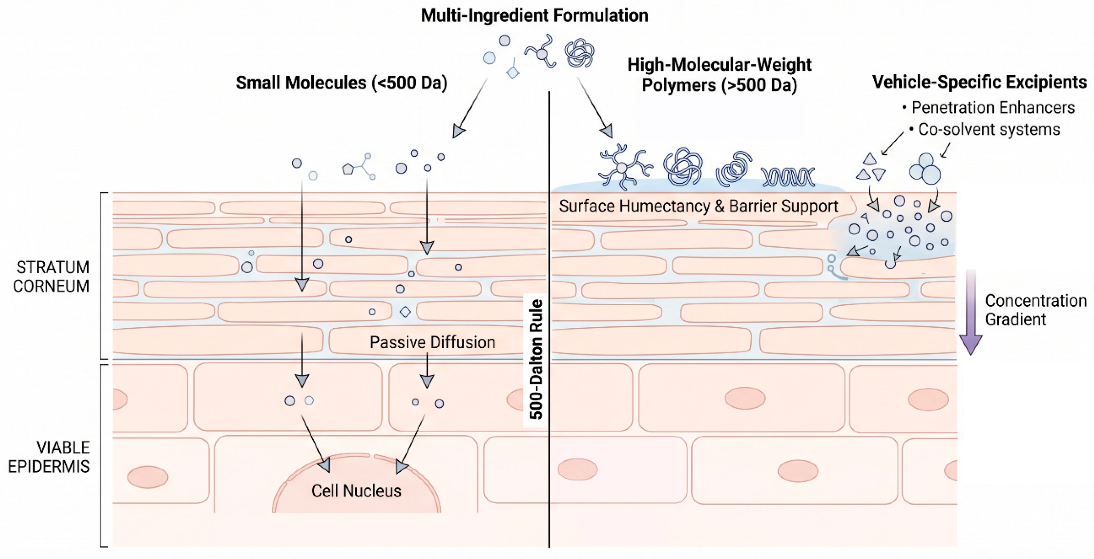
While the 500 Da rule should be regarded as a useful heuristic rather than an immutable biological cutoff, it nevertheless highlights one important constraint underlying many topical skin booster claims. Passive cutaneous transport is governed by the combined effects of molecular size, lipophilicity, ionization, aqueous solubility, vehicle composition, thermodynamic activity, and barrier condition; molecular size, therefore, should not be interpreted in isolation. Nevertheless, many proposed skin booster bioactives are both large and highly hydrophilic, a physicochemical combination that makes efficient passive permeation across intact SC particularly unfavorable. Accordingly, such macromolecular actives generally exhibit negligible or highly limited passive penetration unless the barrier is circumvented or permeability is enhanced by an appropriate delivery strategy [
16].
The most widely studied and clinically relevant approach involves deliberate disruption or bypass of the SC (
Table 3), thereby creating transient pathways that permit the delivery of macromolecules otherwise excluded from passive diffusion across intact skin [
17]. Even simple microneedling transiently compromises the SC barrier, while more advanced modalities—including AFXL and RF microneedling—can further augment permeability and clinical effects. By creating temporary microchannels, these procedures substantially reduce the barrier imposed by the SC and permit enhanced delivery of selected macromolecular agents. Importantly, evidence of enhanced macromolecular transport should be interpreted as specific to the formulation, molecular form and labeling strategy, delivery device, treatment parameters, and experimental model actually studied; such findings should not be generalized to untested topical formulations or related macromolecules.
5.1. Microneedling-Enhanced Transepidermal Transport of Macromolecules
Mechanical disruption of the SC using microneedling and related microneedle platforms is a well-established technique for creating transient microchannels that bypass the SC and facilitate transcutaneous transport of topically applied agents [
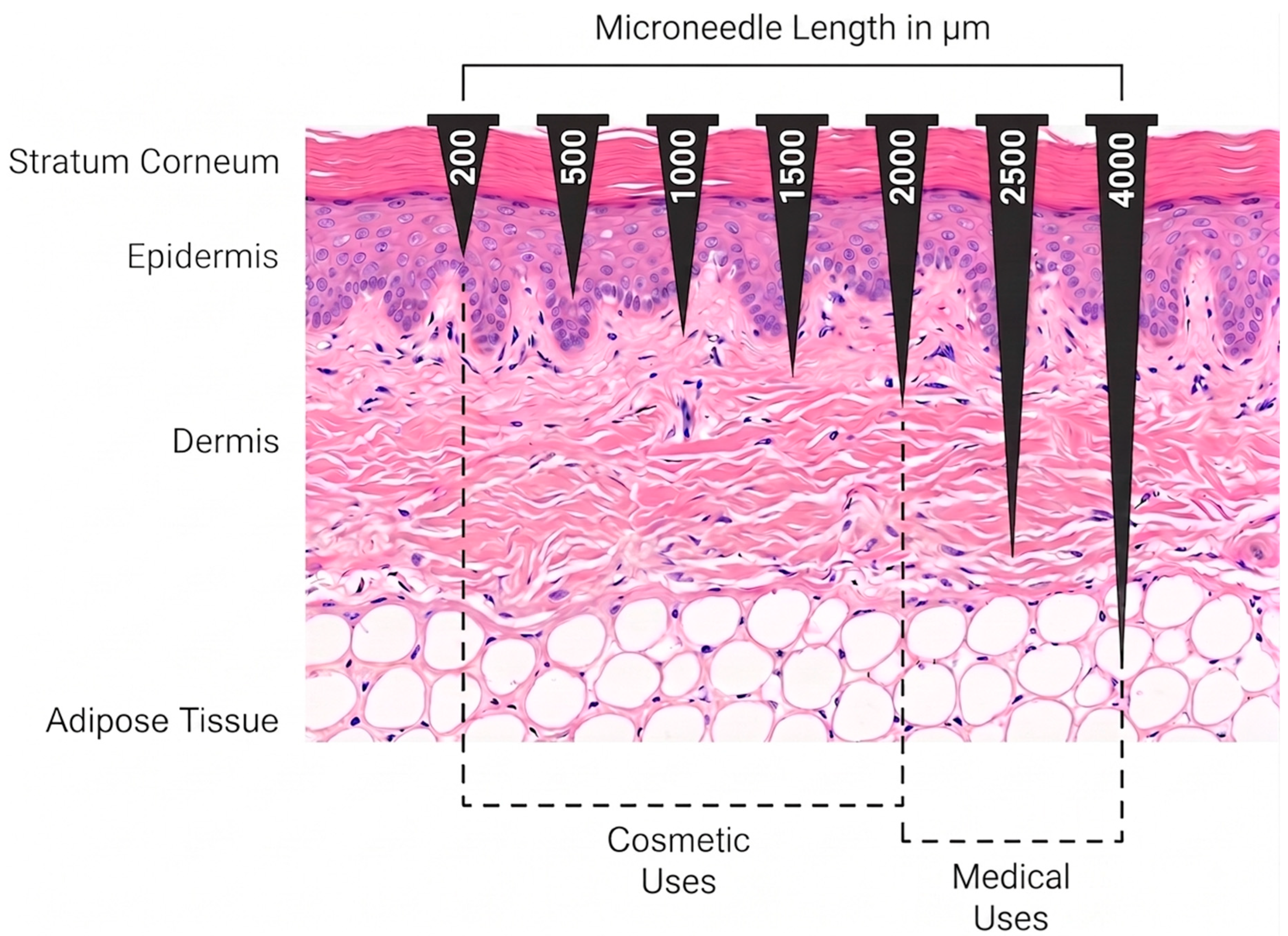
18]. Microchannel formation, however, establishes a potential transport pathway rather than analyte-specific delivery to a defined tissue compartment; increased permeability or tracer transport should therefore be distinguished from tissue deposition, target engagement, and clinical efficacy. Nominal microneedle length influences the skin compartment accessed (
Figure 2), but actual insertion depth and channel behavior vary with device mechanics, anatomical site, skin thickness, needle geometry, and insertion technique [
19,
20].
Early studies characterized the formation and closure of microneedle-created channels [
21] and established the clinical and histologic basis of percutaneous collagen induction [
22,
23]. Subsequent drug-delivery studies showed that these channels can enhance transcutaneous transport of topically applied compounds [
24,
25]. Together, these data support microneedling as a barrier-bypass strategy, while the magnitude of transport remains molecule-specific.
Figure 2.
Conceptual relationship between microneedle length and penetration into human skin. Longer microneedles generally permit access to deeper cutaneous compartments, but actual insertion depth may be substantially less than nominal needle length because of skin deformation and device-specific insertion mechanics. Penetration depth also varies with anatomical site, skin thickness, needle geometry, and insertion technique; therefore, the depicted ranges should be interpreted schematically rather than as fixed anatomical thresholds. Conceptual ranges informed by [
26].
Figure 2.
Conceptual relationship between microneedle length and penetration into human skin. Longer microneedles generally permit access to deeper cutaneous compartments, but actual insertion depth may be substantially less than nominal needle length because of skin deformation and device-specific insertion mechanics. Penetration depth also varies with anatomical site, skin thickness, needle geometry, and insertion technique; therefore, the depicted ranges should be interpreted schematically rather than as fixed anatomical thresholds. Conceptual ranges informed by [
26].
The resulting permeability window is brief: microneedle-induced channels begin to reseal within minutes, with substantial barrier recovery over the following hours and complete closure within hours to days depending on occlusion and experimental conditions [
21].
HA has been extensively investigated in microneedle-based delivery systems. Fluorescence-tracer, confocal imaging, and ex vivo permeation studies provide proof-of-principle for enhanced transport or tissue localization under specific experimental conditions [
27], but much of this evidence derives from HA-containing dissolving microneedles, fluorescently labeled HA, or model compounds rather than separately applied native high-molecular-weight HA after conventional microneedling [
28]. Device geometry and effective insertion depth further influence transport [
20,
29]. Accordingly, current evidence supports pathway-level transport or localization rather than quantitative dermal exposure or target engagement of separately applied native high-molecular-weight HA, and findings from specialized or labeled systems should not be generalized to conventional topical HA.
PDRN has been studied as a microneedling adjunct in an animal model and an early clinical trial [
30,
31], supporting biological plausibility but not establishing the amount, depth, molecular integrity, or biologically active concentration reaching the target tissue. Favorable clinical outcomes have likewise been reported when exosome-containing formulations are applied after microneedling [
32,
33], but these studies do not demonstrate intact-vesicle deposition or functional cargo delivery to a defined viable skin compartment. For both PDRN- and EV-containing formulations, mechanistic plausibility and adjunctive clinical benefit therefore remain distinct from direct evidence of target-compartment exposure or target engagement.
Overall, microneedling creates transient barrier-bypass pathways and can enhance transport of selected compounds and macromolecular model systems, but the magnitude and depth of transport remain molecule-, formulation-, and device-specific. Current evidence does not establish that microneedle-assisted topical delivery of HA-, PDRN-, or EV-containing formulations achieves target-compartment exposure or pharmacokinetic performance comparable to direct intradermal injection, which remains the more predictable route for dermal drug exposure (
Table 4).
5.2. Ablative Fractional Laser Resurfacing
Laser-assisted drug delivery (LADD) has emerged as a well-established therapeutic platform in dermatology, leveraging controlled fractional laser-induced disruption of the skin barrier to enhance the cutaneous and transdermal delivery of topically applied agents [
35]. The foundational framework of LADD is based predominantly on AFXL technology, particularly the 10,600 nm carbon dioxide (CO
2) and 2940 nm erbium: yttrium–aluminum–garnet (Er:YAG) laser systems, which generate controlled microscopic treatment zones that transiently bypass the SC and facilitate topical drug delivery [
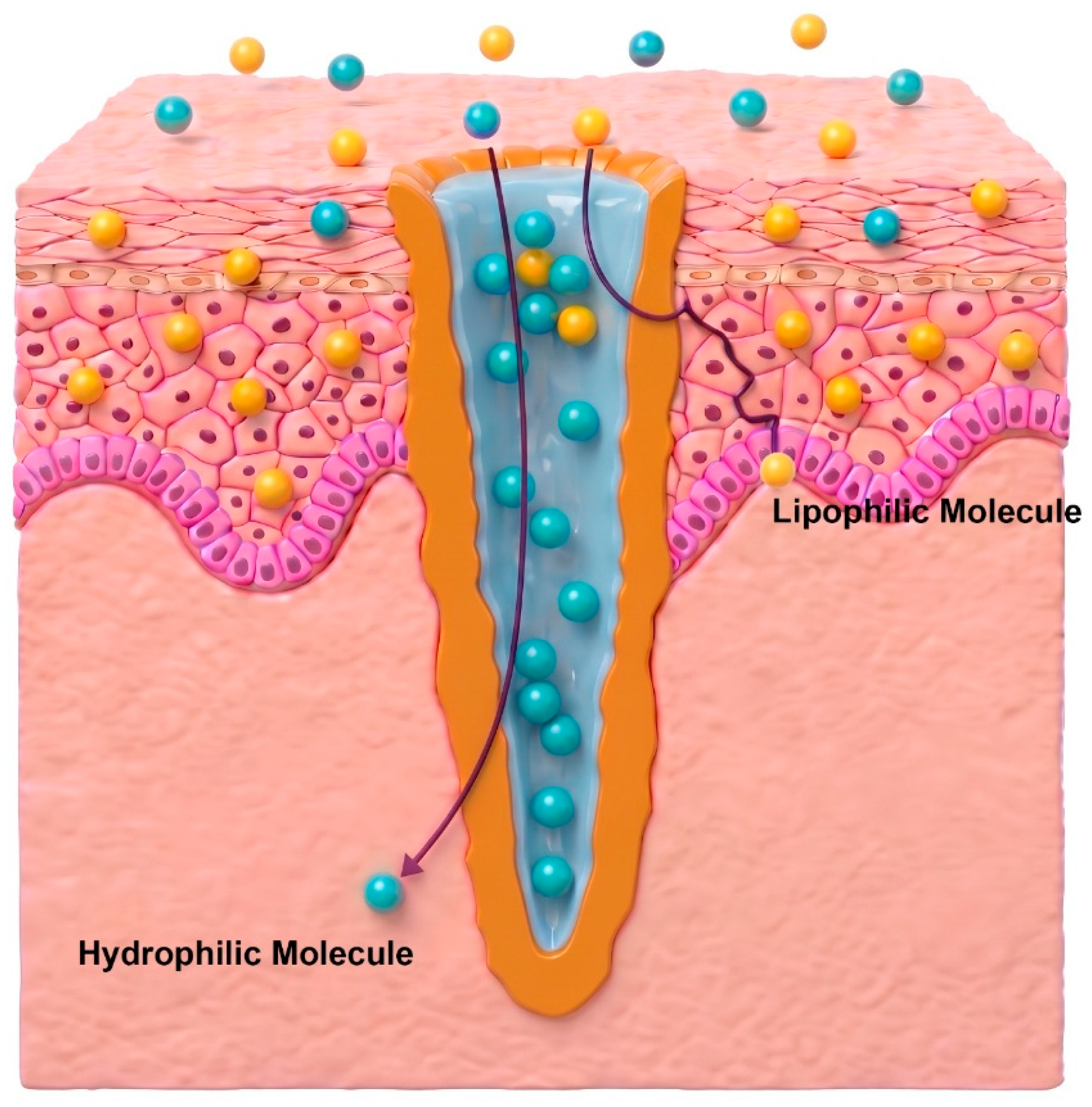
35]. These vertically oriented, water-filled microchannels provide a transient pathway for topical agents to bypass the SC, particularly facilitating the delivery of hydrophilic molecules that otherwise penetrate intact skin poorly [
36] (
Figure 3). Furthermore, the peripheral zone of thermal coagulation surrounding each microscopic ablation column may serve as a transient drug reservoir, modulating the retention and subsequent diffusion of topically applied therapeutics [
35,
36].
Unfortunately, the clinical optimization of LADD is limited by the temporal dynamics of epidermal resealing. A 2017 channel-kinetics investigation using optical coherence tomography and transepidermal water loss (TEWL) mapped microchannel morphology over time [
37]. AFX-induced channels were fully patent only for ~30 min, the peak channel-mediated transport window. Patency dropped to ~75% by 6 h and to <3% by 24–48 h. Fluorescence similarly diminished within 30 min and was no longer detectable by 48 h, indicating that the immediate post-treatment window—particularly the first ~30 min—represents the period of maximal transport potential [
37].
The clinical evidence base for LADD is strongest for relatively low-molecular-weight therapeutics, including corticosteroids, 5-fluorouracil, aminolevulinic acid, and methotrexate [
35]. These compounds possess physicochemical properties that are inherently favorable for cutaneous transport, and AFXL can further enhance their cutaneous transport by transiently bypassing the SC [
35]. Melasma has become a major focus of LADD-related clinical research in dermatology. Split-face randomized trials have evaluated fractional CO
2 laser combined with tranexamic acid delivered topically or by microinjection, depending on the study protocol [
38,
39]. Other comparative trials have specifically evaluated laser-assisted topical tranexamic acid against topical ascorbic acid [
40,
41]. One study found that combining low-power fractional CO
2 with Q-switched neodymium-doped yttrium aluminium garnet (QS-Nd:YAG) laser “toning” reduced punctate leukoderma risk while maintaining comparable melasma-improving efficacy to QS-Nd:YAG alone [
42]. Waibel et al. [
43] further demonstrated in a randomized split-face trial that immediate application of a vitamin C/E/ferulic acid serum following fractional ablative CO
2 laser resurfacing accelerated clinical wound healing, reduced postoperative erythema and edema, and significantly increased local basic fibroblast growth factor expression, supporting the concept that LADD can augment early regenerative responses. Pretreatment with fractional Er:YAG laser has been shown to improve topical anesthetic efficacy, yielding substantially lower pain scores than topical anesthesia alone [
44]. For hypertrophic scars and keloids, clinical series and reports support the feasibility of AFXL-assisted corticosteroid delivery as an alternative adjunct to direct intralesional injection [
45,
46], though one randomized trial reported no clear long-term advantage over laser monotherapy [
47].
Peptides weighing several hundred daltons have been shown to penetrate effectively when topically applied to AFXL-treated skin. A low-fluence fractional Er:YAG laser enhanced peptide skin permeation by increasing follicular uptake, and the magnitude of this effect depended on peptide size and sequence, with smaller, more hydrophilic peptides showing the greatest enhancement [
48]. Similarly, a randomized, blinded split-face study found that applying a tripeptide/hexapeptide anhydrous gel before and after AFXL accelerated barrier recovery and improved acne-scar outcomes compared with moisturizer control [
49]. This demonstrates an adjunctive clinical effect but does not directly quantify peptide deposition within the skin.
Clinical studies suggest that AFXL can facilitate biological effects of topically applied agents even in the kilodalton range, most notably botulinum neurotoxin type A, although direct target-compartment quantification and the overall evidence for active-specific dermal delivery of such high–molecular-weight compounds remain limited. A randomized split-face trial demonstrated that combining fractional CO
2 laser with topical application of reconstituted botulinum neurotoxin yields superior improvements in skin roughness, hydration, elasticity, and overall rejuvenation compared with laser treatment alone [
50]. AFXL-assisted delivery of topical botulinum neurotoxin has likewise shown promise in a small case series, achieving safe reductions of approximately 50–80% in palmar sweating over three months in patients with primary palmar hyperhidrosis [
51]. This finding was confirmed in a couple of randomized, intrapatient trials in patients with primary localized hyperhidrosis [
52,
53,
54].
AFXL creates aqueous microchannels that can facilitate transport of a broader range of agents, including selected hydrophilic macromolecules; however, the extent and depth of transport and tissue localization remain analyte-, formulation-, and parameter-specific [
35,
36,
55]. Nguyen et al. [
55] provided one of the strongest in vivo demonstrations of AFXL-enhanced HA tissue localization using multiphoton tomography with fluorescence lifetime imaging. Topically applied HA was visualized throughout the viable epidermis and into the upper dermis 30 min after fractional CO
2 laser treatment, with residual HA detectable 30 days later [
55]. These findings provide direct in vivo evidence that AFXL can facilitate localization of selected HA formulations in the viable epidermis and upper dermis under studied conditions [
55], although the concentration–time profile and functional target engagement require further investigation. Their follow-up analysis showed that both CO
2 and 1927 nm thulium fractional lasers enhance topical HA penetration into the upper dermis, with CO
2 producing larger HA aggregates but more prolonged inflammation, whereas thulium produced comparable upper-dermal localization with substantially less downtime [
56]. Clinically, a randomized split-face study demonstrated that applying HA immediately after ablative fractional CO
2 laser treatment yielded greater improvements in skin texture and firmness than saline, supporting the clinical feasibility of combining AFXL with topical HA for facial rejuvenation but not independently establishing HA deposition [
57]. Combined fractional Er:YAG treatment with topical HA has also been described in limited clinical reporting [
58]. In evidentiary terms, the imaging data support spatial localization of HA under the studied conditions, whereas quantitative target-compartment pharmacokinetics and biological target engagement remain separate, incompletely characterized endpoints. These findings should therefore be interpreted within the specific HA preparation, labeling approach, laser parameters, and imaging model studied and should not be extrapolated to all topical HA formulations or treatment settings.
For PDRN and related polynucleotides, strong evidence for LADD is still lacking. A randomized comparative trial found that combining fractional CO
2 laser treatment with a topical formulation containing polynucleotide together with exosome-mimetic nanovesicles produced greater acne-scar improvement, fewer adverse effects, and higher patient satisfaction than CO
2 laser monotherapy [
59]. Because the adjunct contained multiple biologically active components, the observed benefit cannot be attributed to polynucleotide alone, and direct dermal exposure was not quantified. Nonetheless, animal models have demonstrated that applying PDRN following AFXL can accelerate wound healing, increase granulation-tissue thickness, and increase neovascularization compared with fractional treatment alone [
60].
5.3. Radiofrequency Microneedling
RF microneedling has emerged as a distinct and mechanistically unique modality within the paradigm of device-assisted drug delivery [
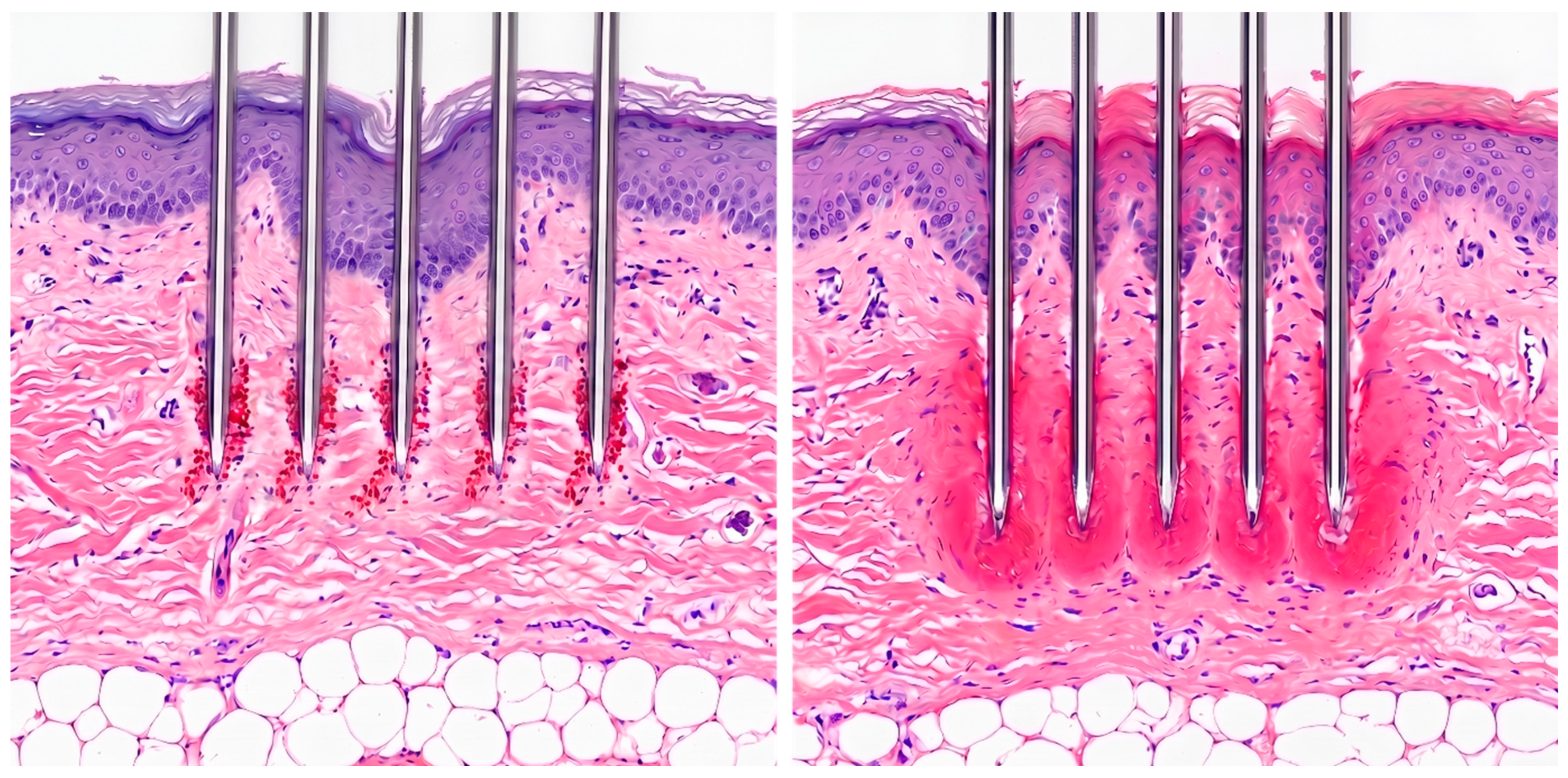
61]. Unlike standard mechanical microneedling or AFXL, RF microneedling induces two simultaneous yet distinct tissue events that govern drug permeation. The physical penetration of the needles generates mechanical microchannels that bypass the SC, functioning identically to traditional microneedling to facilitate the initial entry of topically applied agents. Concurrently, the deposition of RF energy creates thermal coagulation zones around each needle tip [
62] (
Figure 4). This perilesional, thermally altered tissue resembles the coagulative collar seen in AFXL [
63].
However, the spatial distribution of these tissue events critically influences transdermal bioavailability. Modern insulated-needle RF microneedling platforms largely spare the epidermis by confining RF-induced thermal injury to the dermis. Consequently, unlike AFXL that generates continuous thermally ablated columns, topical drug delivery after insulated RF microneedling is thought to occur primarily through the transient mechanical needle-entry microchannels [
63,
64], while the contribution of the surrounding RF-induced coagulation zone to molecular transport remains incompletely defined. The available device-physics literature supports epidermal sparing with insulated needles but does not establish a quantified open-channel geometry or lifetime [
64]. Accordingly, the window for enhanced passive topical delivery may differ from that achieved with AFXL-assisted drug delivery, but its magnitude and duration remain uncertain. Broader reviews of transdermal biologic delivery likewise emphasize persistent evidence gaps for macromolecular transport across noninjectable platforms [
65].
In melasma, randomized studies have tested RF microneedling with adjunctive topical small-molecule therapy [
66], and combination protocols with autologous platelet-rich plasma (PRP) have also reported clinical benefit [
67]. A split-face, double-blind, randomized controlled trial evaluated the application of topical insulin, a peptide of approximately 5.8 kDa, immediately following fractional RF microneedling for atrophic acne scars [
68]. At the three-month follow-up, the treatment side demonstrated statistically significant improvements in clinical scar grading alongside histologically confirmed increases in dermal collagen density. This study provides indirect pharmacodynamic evidence consistent with enhanced delivery of a peptide substantially larger than 500 Da, although intradermal insulin exposure was not directly quantified. Topical exosome treatments applied immediately after RF microneedling have recently been described in case series as producing marked improvements in acne, including notable reductions in both overall severity and lesion counts [
69], while preliminary split-face studies using plant-derived EV preparations suggest additional benefits for skin remodeling and texture [
70]. Nevertheless, these clinical outcome studies do not directly demonstrate vesicle transport or target-compartment exposure. The evidence base for RF microneedling-assisted delivery of topical adjuncts remains largely preliminary and suggestive. A randomized split-face study by Cheng et al. [
71] reported that combining bipolar RF with microneedle-assisted application of an extracellular matrix (ECM) produced significantly greater improvements in periorbital wrinkle severity, skin elasticity, and skin thickness than microneedle-assisted application alone. The authors attributed these synergistic effects to the complementary actions of potential adjunct transport through needle-created pathways and RF-induced dermal remodeling, although direct tissue penetration of the ECM components was not measured.
Clinically, one of the major methodological limitations of the current RF microneedling literature is the intrinsic regenerative activity of the procedure itself. Independent of any adjunctive topical therapy, needle penetration and RF-induced thermal coagulation activate wound-healing pathways that promote neocollagenesis, neoelastogenesis, ECM remodeling, and regenerative immune responses [
72,
73]. Accordingly, clinical improvements observed in uncontrolled or open-label combination studies constitute indirect pharmacodynamic indications rather than proof of adjunct delivery, reinforcing the need for split-face randomized controlled trials. Furthermore, the channel-kinetics framework established for AFXL, which quantified open-channel dwell times [
37], has not been replicated for RF microneedling. In the literature identified by this review, we found no peer-reviewed study that directly quantified the exact dimensions, temporal patency, or absolute molecular-weight limits of RF-induced microchannels.
5.4. Cold Atmospheric Plasma
Unlike microneedling or AFXL, CAP does not primarily enhance permeability by creating persistent physical channels. Experimental studies instead indicate that CAP can transiently modify SC lipid organization, membrane and junctional permeability, and other aspects of barrier physiology through reactive oxygen and nitrogen species, charged species, and transient electric fields (
Figure 5) [
74,
75,
76,
77]. CAP-associated changes in surface pH and cutaneous microcirculation have also been reported [
78,
79,
80], although their direct contribution to topical drug transport remains uncertain. These observations establish biological plausibility for transient barrier modulation but should not, by themselves, be interpreted as evidence that a particular active reaches a defined viable-skin compartment at a clinically relevant concentration.
CAP evidence can be considered at four levels: (1) barrier modification or increased nonspecific permeability; (2) enhanced transport of a defined analyte across or into skin; (3) localization or quantification within a specified target compartment; and (4) target engagement or clinical benefit. These levels are not interchangeable: changes in TEWL, lipid organization, junctional proteins, or electrical permeability establish barrier modulation, not dermal delivery of a specific active. Because barrier recovery and transport kinetics vary by endpoint, analyte, device, and experimental system [
77,
81], no universal CAP “delivery window” can presently be defined.
The most direct analyte-specific evidence for CAP-assisted topical transport is currently strongest for selected relatively small molecules whose overall physicochemical and formulation characteristics are compatible with cutaneous transport. For lidocaine, quantitative permeation experiments demonstrated an approximately 1.97-fold increase in transdermal flux after CAP pretreatment [
82], and a randomized split-face clinical study subsequently showed greater topical anesthetic effect [
83]. Experimental studies have also reported enhanced percutaneous absorption of model compounds in porcine skin [
84], enhanced niacinamide transport [
81], and clinical use of a combined cold-plasma/electroporation/microjet platform to augment nano-molecule delivery [
85]. In a hairless-mouse model, low-intensity CAP combined with topical ascorbic acid produced greater pigment reduction than either intervention alone, supporting preclinical synergy rather than direct ascorbate pharmacokinetic proof [
86]. For selected supra-500 Da analytes, experimental studies have demonstrated enhanced transport or tissue localization of epidermal growth factor, cyclosporine A, and 3–5 kDa dextran under specific CAP conditions [
77,
87,
88]. These findings provide proof-of-feasibility for particular analyte-device combinations rather than evidence of a general molecular-size range that CAP can reliably deliver.
For skin-booster macromolecules such as HA and PDRN, the evidence is substantially less mature. Human skin-explant data support CAP-enhanced transport of topically applied HA [
89], while nanocarrier and plasma-engineered formulations have shown increased transport or cellular uptake in experimental systems [
8,
76,
90]. However, these findings remain formulation-specific and do not establish quantitative in vivo dermal exposure of native topical HA or PDRN. Clinical reports using a PCL microsphere formulation likewise demonstrate feasibility rather than direct particle deposition [
91]. No randomized human study identified in this review directly quantified dermal HA or PDRN after CAP-assisted topical application.
The CAP literature reviewed here includes both argon- and helium-fed systems, underscoring substantial device heterogeneity. Reproducible CAP-assisted topical-delivery protocols will therefore require more precise dosimetry-controlled output, including standardized reactive-species flux, exposure duration, and treatment area.
Table 5 summarizes the highest level of evidence identified for representative analyte-platform pairings and explicitly separates barrier modification, analyte-specific transport or localization, target-compartment exposure, and adjunctive clinical outcomes.