


Abstract
The demand for naturally derived ingredients in cosmetics is constantly growing, with marine algae emerging as promising candidates due to their rich antioxidant and protective metabolites. Skin aging, driven by oxidative stress, impaired hydration, and weakening of the dermal–epidermal junction (DEJ), manifests as dryness, loss of elasticity, and reduced density. Conventional synthetic antioxidants raise safety concerns, highlighting the need for effective natural alternatives. Ecklonia cava, an edible brown seaweed abundant in phlorotannins, has been reported to possess strong antioxidant and anti-inflammatory properties, yet its effects on key molecular markers of hydration and DEJ integrity remain underexplored. In this study, we standardized an E. cava extract (ECE) and evaluated its antioxidant activity, gene regulatory effects, and clinical efficacy. ECE was standardized by measuring total polyphenols and flavonoids, evaluated by DPPH scavenging assay, and shown to upregulate DEJ-related genes, reinforcing dermal–epidermal cohesion. A randomized clinical trial further confirmed that topical application of ECE improved hydration, elasticity, and density compared with placebo, with benefits evident within weeks. Collectively, these findings establish ECE as a multifunctional cosmetic ingredient capable of protecting the skin while enhancing structural and functional aspects of skin health, supporting its potential application in anti-aging skincare.
1. Introduction
The global cosmetic industry has continuously grown, accompanied by increasing demand for natural ingredients with multifunctional properties for skin health. In particular, marine-derived compounds are considered promising candidates due to their diverse structural characteristics and broad biological activities [1,2,3,4]. In recent years, concerns regarding the safety of synthetic antioxidants have further increased interest in natural alternatives with proven efficacy and safety [5]. Marine algae are particularly rich in bioactive compounds, including polyphenols, phlorotannins, and sulfated polysaccharides, which exhibit strong radical scavenging activity as well as anti-inflammatory and photo-protective effects [6]. These multifunctional properties highlight their potential as cosmetic ingredients for maintaining skin homeostasis and protecting against environmental stressors. Brown algae, which thrive under extreme marine conditions such as strong ultraviolet radiation, high salinity, and constant water currents, produce potent antioxidant and protective metabolites to adapt to these environments. Owing to these properties, marine algae have attracted considerable attention as consumers are looking for active ingredients for attenuating skin aging, preferably by improving hydration and strengthening the skin barrier [7].
Skin aging is characterized by gradual changes in skin appearance and function, typically manifesting as dryness, reduced elasticity, and decreased density [8,9,10]. These changes are closely associated with reduced epidermal moisture content, degradation of the extracellular matrix (ECM), and weakening of the dermal–epidermal junction (DEJ) [11,12]. Oxidative stress has been identified as a central mechanism driving these processes, as excessive reactive oxygen species (ROS) promote collagen and elastin degradation, impair DEJ integrity, and accelerate wrinkle formation and barrier dysfunction [13,14,15]. In addition to oxidative stress, impaired epidermal water transport and reduced dermal–epidermal cohesion are critical contributors to skin aging. Aquaporin 3 (AQP3), the most extensively studied aquaporin in the epidermis, plays a key role in transporting both water and glycerol, and its upregulation is directly associated with enhanced hydration and elasticity [16,17]. Deficiency of AQP3 leads to reduced glycerol levels, resulting in dry skin, decreased elasticity, and delayed wound healing. Likewise, the DEJ is composed of protein complexes that anchor and stabilize the epidermis and dermis, with collagen type IV alpha 1 chain (COL4A1), collagen type XVII alpha 1 chain (COL17A1), and laminin subunit alpha 5 (LAMA5) being essential structural components [18,19]. Reduced expression of these genes is closely linked to wrinkle formation, sagging, and barrier dysfunction. Therefore, strategies that not only mitigate oxidative stress but also enhance AQP3 expression and reinforce DEJ integrity are crucial for delaying and managing skin aging [20,21].
Ecklonia cava, an edible brown seaweed native to the coastal regions of Korea and Japan, is especially notable for its abundance of phlorotannins, a unique class of polyphenols [22]. Previous studies have reported that E. cava extracts (ECE) and isolated phlorotannins, such as dieckol, eckol, 6,6′-bieckol, and phlorofucofuroeckol-A, possess strong antioxidant activity, anti-inflammatory effects, and protective functions against ultraviolet-induced damage [23,24,25,26,27,28,29,30]. They have also been shown to modulate metabolic processes and promote skin regeneration. However, despite these well-documented properties, systematic investigations into the effects of ECE on key molecular markers of skin hydration and structural components of the DEJ remain limited. Given the growing demand for natural cosmetic ingredients with multifunctional efficacy, elucidating the broader biological potential of E. cava is of considerable importance. Therefore, the present study aimed to evaluate the antioxidant activity of ECE using the DPPH radical scavenging assay, and to investigate its effects on the expression of hydration-related (AQP3) and DEJ-associated genes (COL4A1, COL17A1, and LAMA5). Furthermore, clinical evaluations were conducted to assess improvements in skin hydration, elasticity, and density. Through this integrated approach, we sought to establish scientific evidence supporting the applicability of ECE as a functional cosmetic ingredient for anti-aging skin care.
2. Materials and Methods
2.1. Preparation of ECE
Dried thalli of Ecklonia cava were subjected to a pretreatment process to remove chlorophylls. 10 g of raw material was immersed in 500 g of solvent, a mixture of 70% ethanol and 30% glycerin at room temperature for 24 h, followed by thorough washing with 500 g of purified water, twice. The pretreated material was dried at 60 °C for 24 h. Pretreatment removing chlorophylls generally gave slight loss of biomass but increased the content of polyphenols and flavonoids in the final extract in our experiences 9.82 g of dried pretreated E. cava was then extracted in 98.2 g of solvent, a mixture of 60% purified water and 40% 1,3-butylene glycol, to make an extraction ratio of 1:10. The extraction was carried out at 50 °C for 72 h, with ultrasonic-assisted extraction (25 kHz, 1 h per day) to enhance efficiency. Finally, 80 g of extract was acquired by 0.45-μm filtration (Advantec, Saijyo City, Japan) to remove insoluble residues. We designed an optimal extraction process and the solvent composition to facilitate application in cosmetic formulations. Conventional botanical extracts prepared with organic solvents or hot water are often difficult to disperse and solubilize in such formulations. In contrast, our ECE could be directly applied in both in vitro experiments and clinical studies.
2.2. Determination of Polyphenol and Flavonoid Contents
Total polyphenol content was determined using the Folin–Ciocalteu method with minor modifications [31]. Briefly, 50 μL of sample was mixed with 50 μL of 0.2 N Folin–Ciocalteu reagent, prepared by diluting 2 N Folin–Ciocalteu’s phenol reagent (Sigma-Aldrich, St. Louis, MO, USA, F9252) with distilled water, followed by the addition of 100 μL of 10% sodium carbonate solution (Sigma-Aldrich, S2127). The reaction mixture was incubated at room temperature in the dark for 30 min, and absorbance was measured at 725 nm using a microplate reader. Gallic acid (Sigma-Aldrich, G7384) was used as the calibration standard prepared by serial dilution, and total polyphenol content was expressed as mg gallic acid equivalents (GAE)/g extract.
Total flavonoid content was determined using the diethylene glycol (DEG) colorimetric method [32]. Briefly, 20 μL of sample, 160 μL of diethylene glycol (Sigma-Aldrich, 32160), and 20 μL of 1 N NaOH were sequentially added to a 96-well plate and mixed thoroughly. After incubation at room temperature for 3 min, absorbance was measured at 420 nm using a microplate reader. Quercetin (Sigma-Aldrich, Q4951) was used as the calibration standard prepared by serial dilution. Total flavonoid content was expressed as mg quercetin equivalents (QE)/g extract.
2.3. DPPH Radical Scavenging Assay
The antioxidant activity of ECE was evaluated using the 2,2-diphenyl-1-picrylhydrazyl (DPPH) radical scavenging method. Samples were mixed with 0.2 mM DPPH solution and incubated in the dark at room temperature for 20 min. Absorbance was measured at 517 nm using a microplate reader [33]. Ascorbic acid served as a positive control. Radical scavenging activity (%) was calculated relative to the control, and the half-maximal scavenging concentration (SC50) values were obtained from dose–response curves. All experiments were performed in triplicate (n = 3).
2.4. Cell Culture and Treatment
Human neonatal dermal fibroblasts (HDFn) were purchased from the American Type Culture Collection (ATCC, Manassas, VA, USA; Cat. No. PCS-201-010) and HaCaT cells were from AddexBio (San Diego, CA, USA; Cat. No. T0020001). Cells were maintained in DMEM supplemented with 10% fetal bovine serum (FBS) and 1% penicillin-streptomycin at 37 °C in a humidified 5% CO2 incubator. Cells were seeded into 96-well plates at densities of 2 × 10⁴ cells/well (HDFn) and 3 × 10⁴ cells/well (HaCaT), allowed to adhere overnight, and treated with ECE at final concentrations of 0.5, 1, and 2% for 24 h. The solvent-only (40% 1,3-butylene glycol) group served as the control. Following treatment, cells were harvested for RNA extraction and subsequent reverse transcription (RT)-qPCR analysis to evaluate gene expression changes.
2.5. RNA Extraction and cDNA Synthesis
RNA extraction and cDNA synthesis were performed using the SuperPrep™ II Cell Lysis & RT Kit for qPCR (TOYOBO, Osaka, Japan) according to the manufacturer’s instructions. Following cell lysis, total RNA contained within the lysate was reverse-transcribed into cDNA in a single-tube reaction, enabling direct downstream qPCR analysis. Briefly, 4 μL of cell lysate was mixed with 16 μL of RT reaction mixture, and reverse transcription was carried out under the following conditions: 37 °C for 15 min, 50 °C for 5 min, and 98 °C for 5 min, followed by a 4 °C hold. The concentration and purity of cDNA was assessed using a NanoDrop spectrophotometer (Thermo Fisher Science, Waltham, MA, USA).
2.6. Quantitative Real-Time PCR
Quantitative real-time PCR (qPCR) was carried out using the FastStart Essential DNA Green Master (Roche Diagnostics, Mannheim, Germany) on a real-time PCR system according to the manufacturer’s instructions. Amplification of AQP3, COL4A1, COL17A1, and LAMA5 was performed using gene-specific primers, with β-actin serving as the reference gene for normalization, whereas vehicle-treated cells served as the experimental control group. The primer sequences are listed in Table 1.
Table 1.
Primer sequences used for qPCR.
Each PCR reaction was prepared in a total volume 20 μL, consisting of 10 μL SYBR Green Master Mix, 5 μL cDNA template, 1 μL forward primer (10 μM), 1 μL reverse primer (10 μM), 3 μL PCR-grade water. The amplification conditions were as follows: initial denaturation at 95 °C for 600 s, followed by 45 cycles of denaturation at 95 °C for 10 s, annealing at 60 °C for 10 s and extension at 72 °C for 10 s. Melting curve analysis was subsequently performed under the following conditions: 95 °C for 10 s, 65 °C for 60 s, and 98 °C for 1 s, followed by cooling at 37 °C for 30 s. Amplification specificity and melting curve analysis were evaluated using LightCycler 96 SW 1.1 (Roche Diagnostics).
Relative gene expression levels were calculated using the 2−ΔΔCt method as previously described by Livak and Schmittgen [34]. Three independent biological replicates were analyzed, and each qPCR reaction was performed in technical duplicate.
2.7. Statistical Analyses of In Vitro Experiments
Data are presented as the mean ± standard deviation (SD) from at least three independent biological replicates. Statistical significance among multiple groups was analyzed using ordinary one-way analysis of variance (ANOVA) followed by Tukey’s honestly significant difference (HSD) or Dunnett’s post hoc test where appropriate, by GraphPad Prism Version 11.0.2 (San Diego, CA, USA). *, p < 0.05; **, p < 0.01; ***, p < 0.001.
2.8. In Vivo Study Design
A randomized, split-face, controlled clinical trial was conducted with 22 healthy female volunteers aged 20–65 years. All subjects provided written informed consent, and the study was performed in accordance with Good Clinical Practice guidelines. Environmental conditions were maintained at 22 ± 2 °C and 50 ± 5% relative humidity. Participants were instructed to apply equal quantities of the ECE-containing (0.1%) formulation to one side of the face and the placebo formulation that was identical in all ingredients except ECE (Table S1), to the contralateral side twice daily for 4 weeks. Skin hydration, elasticity, and density were assessed at baseline, 2 weeks, and 4 weeks using standardized non-invasive instruments.
2.9. Skin Hydration Assessment
Skin hydration was assessed at baseline and after 2 and 4 weeks of product application using a non-invasive corneometer (Corneometer® CM 825, Courage + Khazaka electronic GmbH, Germany). At each assessment time point, measurements were performed three times at each test site, and the mean value was used for analysis. Skin hydration values were expressed in arbitrary units (A.U.), with higher values indicating increased stratum corneum hydration.
2.10. Skin Elasticity Assessment
Skin elasticity was assessed at baseline and after 2 and 4 weeks of product application using a cutometer (Cutometer® MPA580, Courage + Khazaka electronic GmbH, Köln, Germany). Measurements were performed three times at each assessment site, and the mean value of the R2 parameter was used for analysis. During each measurement, a constant negative pressure of 450 mbar was applied to the skin for 2 s over three consecutive cycles.
2.11. Skin Density Assessment
Skin density was evaluated at baseline and after 2 and 4 weeks of product application using an ultrasound-based imaging system (DermaLab® Series SkinLab Combo, Cortex Technology, Aalborg, Denmark). Measurements were performed separately on the test and control areas using an ultrasound probe, and skin density was expressed as a percentage (%). Higher values indicated increased skin density.
3. Results
3.1. Antioxidant Activity
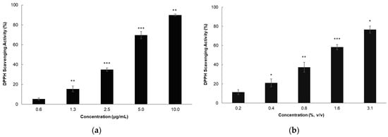
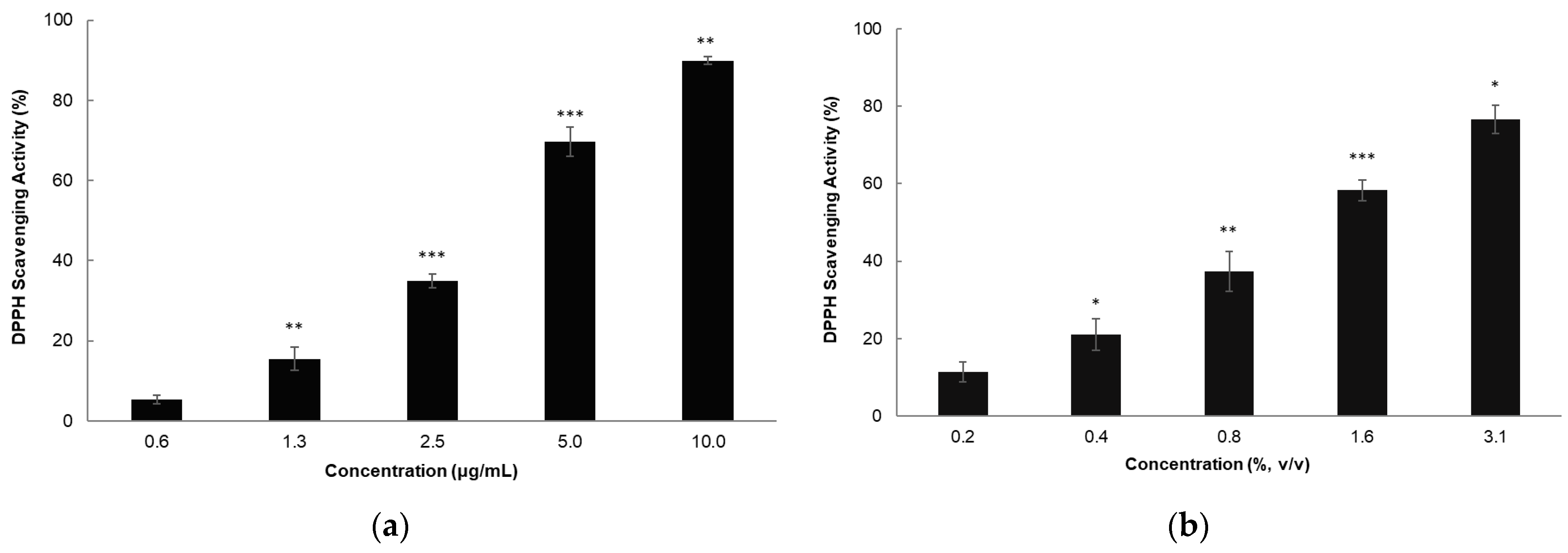
First, we measured the total polyphenol and flavonoid contents of ECE (Table 2) as a standardization of the plant extract for subsequent studies. Although the extraction solvent was optimized for easy application in the cosmetic formulation, inclusion of polyol made it impossible to calculate extraction yield by dry weight. Instead, the contents of these natural antioxidants were useful to standardize ECE. Then we assessed the antioxidant activity of ECE using the DPPH radical scavenging assay, with ascorbic acid serving as a positive control (Figure 1). Both samples showed concentration-dependent scavenging activity. One-way ANOVA revealed significant differences among treatment groups for both ascorbic acid and ECE, and Tukey’s HSD post hoc analysis confirmed significant concentration-dependent increases in radical scavenging activity. The SC50 (half maximal scavenging concentration) values were 3.1 μg/mL for ascorbic acid and 1.14% for ECE.
Table 2.
Total polyphenol and flavonoid contents of ECE.
Figure 1.
Antioxidant activity of ascorbic acid and ECE as measured by DPPH assay. (a) Ascorbic acid and (b) ECE exhibited concentration-dependent DPPH radical scavenging activity. The SC50 values were 3.1 μg/mL for ascorbic acid and 1.14% for ECE, respectively. Both samples showed a progressive increase in scavenging activity with increasing concentrations. The results were analyzed using one-way ANOVA followed by Tukey’s HSD post hoc test. *, p < 0.05; **, p < 0.01; ***, p < 0.001.
3.2. Effect on AQP3 Gene Expression
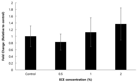
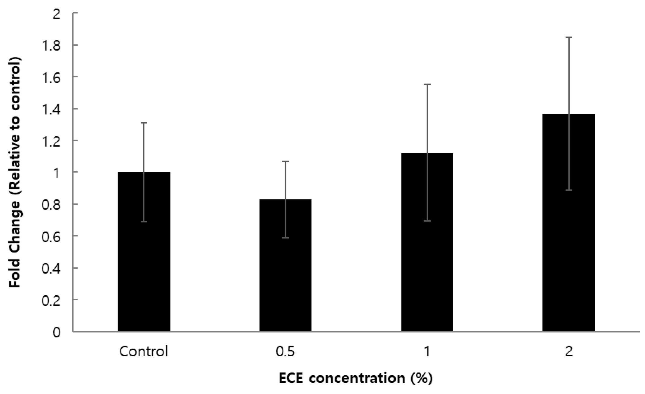
To evaluate the moisturizing effect of ECE in vitro, we analyzed the expression of the AQP3 gene in human dermal fibroblasts (HDFn). The results proposed the induction of AQP3 expression in a concentration-dependent manner (Figure 2). The slight reduction in AQP3 levels at the minimum concentration likely reflects an initial adaptive phase of the cells to the complex phytochemical environment of ECE. This minor fluctuation at low doses often preceded the robust, dose-dependent induction observed at higher concentrations of plant extracts. However, the induction of the AQP3 gene in HDFn was not statistically significant in our study.
Figure 2.
Effect of ECE on AQP3 mRNA expression in HDFn cells. HDFn cells were treated with solvent control or ECE at 0.5, 1, and 2% for 24 h, and AQP3 mRNA levels were determined by RT-qPCR and normalized to β-actin. Relative expression levels were calculated using the 2−ΔΔCt method and presented as fold changes relative to the control. The results were analyzed using one-way ANOVA followed by Dunnett’s post hoc test.
3.3. Effect on DEJ-Related Gene Expression
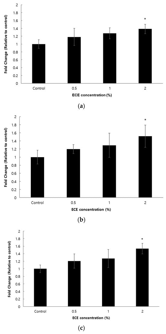
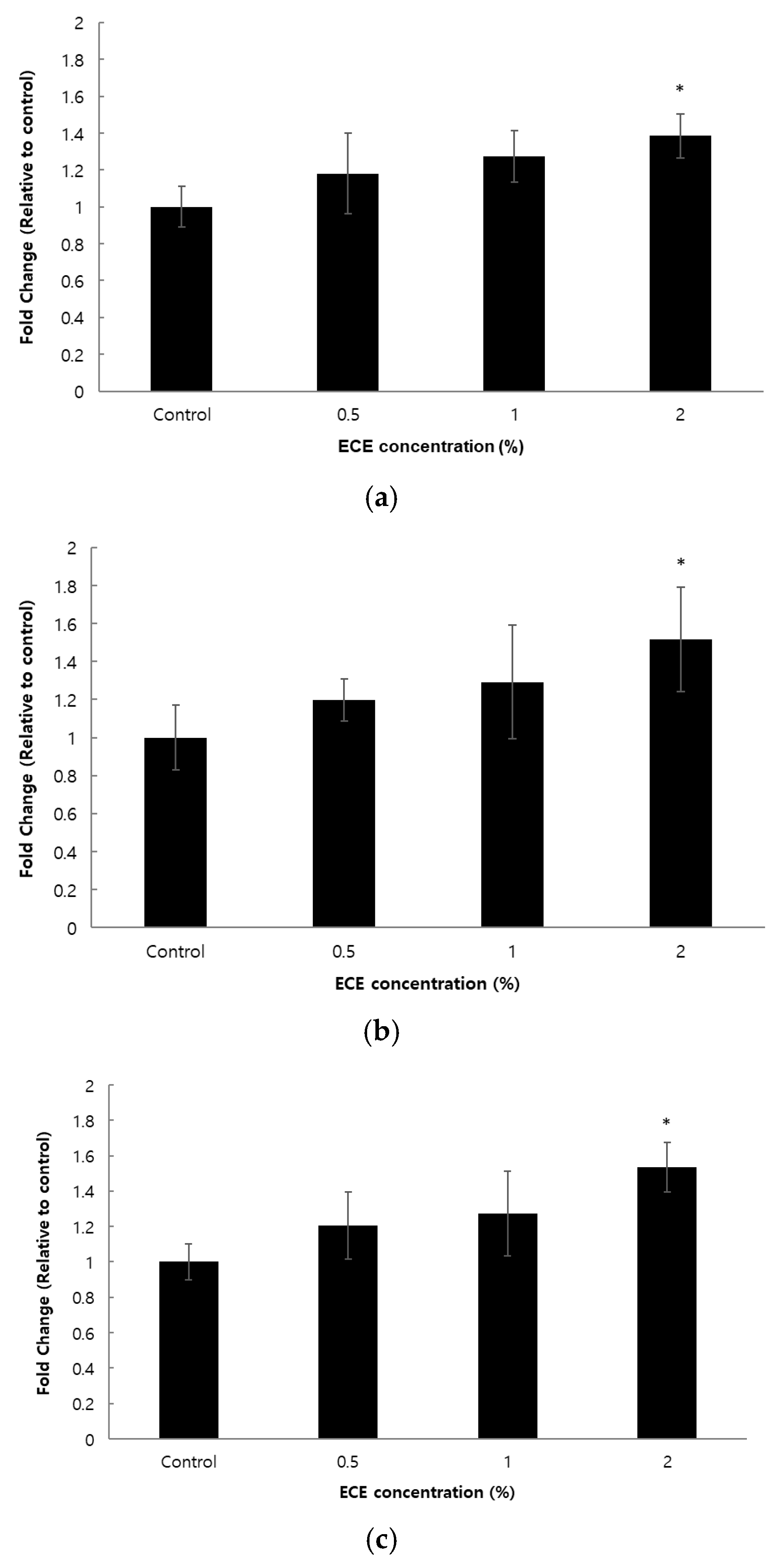
To further explore the molecular mechanisms underlying the benefits of ECE on skin health, we examined its effects on DEJ-related genes in human keratinocytes (HaCaT). After 24 h of treatment with 0.5–2% of the extract, the mRNA levels of COL4A1, COL17A1, and LAMA5 showed concentration-dependent increases (Figure 3). Treatment with 2% ECE increased COL4A1, COL17A1, and LAMA5 expression levels by 38.5%, 51.6%, and 53.7%, respectively, with statistical significance based on Dunnett’s post hoc test. These three genes are well-established structural components of the DEJ, and the observed upregulation suggested that ECE could contribute to improved dermal-epidermal cohesion and skin firmness.
Figure 3.
Effect of ECE on DEJ-related gene expression in HaCaT cells. HaCaT cells were treated with solvent control or ECE at 0.5, 1, and 2% for 24 h, mRNA levels of (a) COL4A1, (b) COL17A1, and (c) LAMA5 were determined by qPCR and normalized to β-actin. Relative expression levels were calculated using the 2−ΔΔCt method and presented as fold changes relative to the control. Treatment with ECE increased the expression of DEJ-related genes in a concentration-dependent manner. The results were analyzed using one-way ANOVA followed by Dunnett’s post hoc test. *, p < 0.05.
3.4. In Vivo Effects of the ECE-Containing Formulation
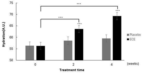
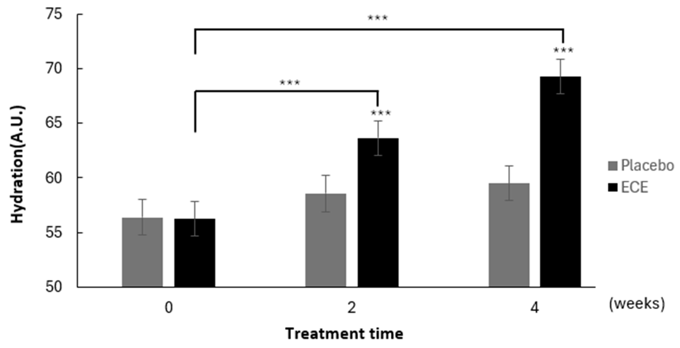
Skin hydration was first assessed by comparing the ECE-treated area with the placebo-treated area. Over the 4-week application period, hydration in the ECE-treated area increased progressively (Figure 4). Compared with baseline, hydration increased significantly at both 2 weeks and 4 weeks of treatment (p < 0.001), with a more pronounced improvement observed at week 4. In contrast, although the placebo group showed slight improvement, the changes were not statistically significant throughout the study period. It is possible that functional ingredients such as emulsifiers and polyols in the formulations might improve skin hydration at the marginal level (Table S1).
Figure 4.
Comparison of skin hydration between ECE-containing formulation and placebo. A split-face study design was conducted, where the ECE-containing formulation was applied to one side of the face and the placebo cream was applied to the contralateral side. Skin hydration was assessed using a non-invasive instrumental method. Data are represented as mean ± SD. Statistical significance was assessed using a paired Student’s t-test (*** p < 0.001).
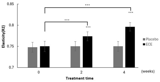
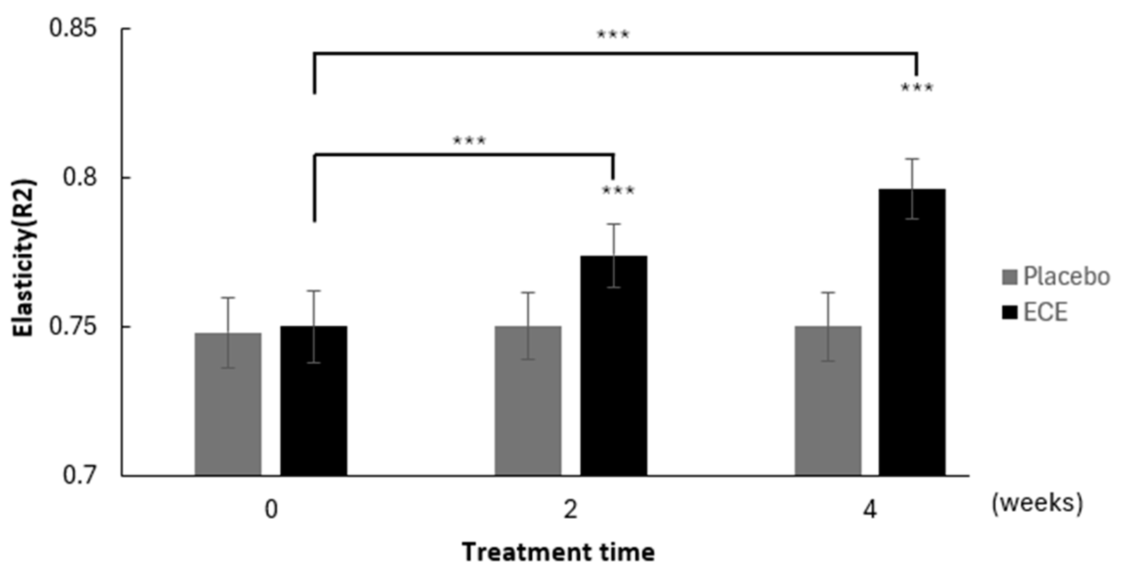
We also examined skin elasticity, and the ECE-treated area showed significant increases during the study period (Figure 5). Relative to baseline, elasticity increased significantly at both 2 weeks and 4 weeks (p < 0.001). In contrast, almost no changes in elasticity were observed in the placebo group. When changes from baseline were compared between the two areas, the increase in skin elasticity was significantly greater in the ECE-treated area compared with the control area at both time points (p < 0.001).
Figure 5.
Comparison of skin elasticity between ECE-containing formulation and placebo. A split-face study design was conducted, where the ECE-containing formulation was applied to one side of the face and the placebo cream was applied to the contralateral side. Skin elasticity was assessed using a non-invasive instrumental method. Data are represented as mean ± SD. Statistical significance was assessed using a paired Student’s t-test (*** p < 0.001).
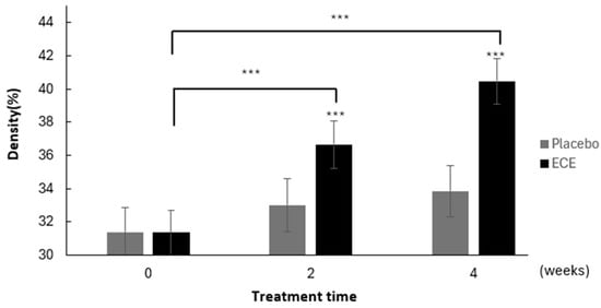
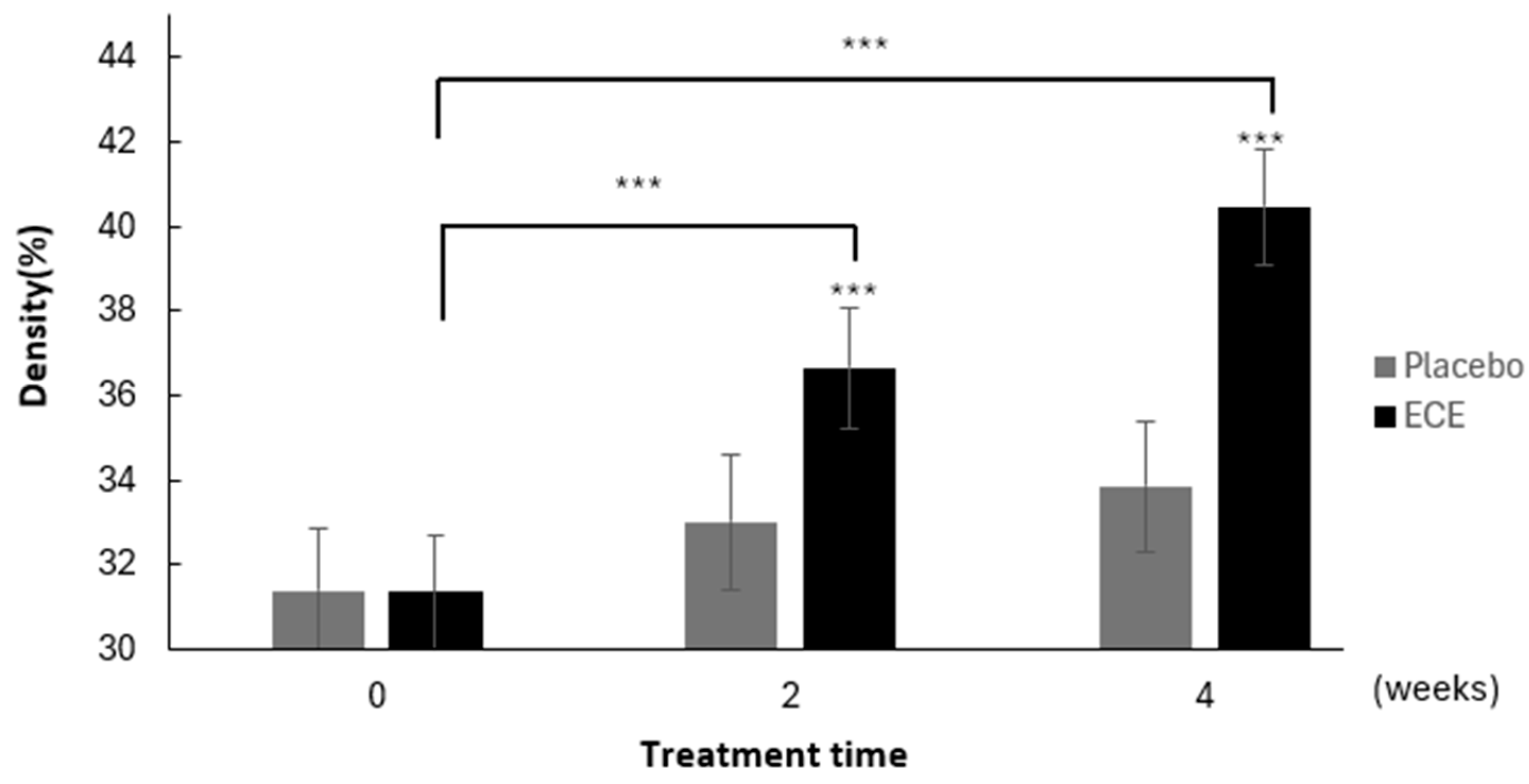
Finally, skin density measurements revealed a similar pattern, with progressive increases observed in the ECE-treated area relative to baseline (Figure 6). These changes reached statistical significance at both 2 weeks and 4 weeks (p < 0.001). The placebo group also exhibited minor improvement in density as well, but these changes were not statistically significant. Direct comparison confirmed that the increase in skin density was significantly greater in the ECE-treated area than in the placebo group at both time points (p < 0.001).
Figure 6.
Comparison of skin density between ECE-containing formulation and placebo. A split-face study design was conducted, where the ECE-containing formulation was applied to one side of the face and the placebo cream was applied to the contralateral side. Skin density was assessed using a non-invasive instrumental method. Data are represented as mean ± SD. Statistical significance was assessed using a paired Student’s t-test (*** p < 0.001).
4. Discussion
The present study demonstrated that ECE exerts beneficial effects on skin hydration, elasticity, and density. Previous studies on E. cava have consistently shown that its extracts possess strong antioxidant activity, including DPPH radical scavenging, and that this activity is associated with their rich phenolic composition; moreover, earlier reports have described high total polyphenol and flavonoid contents together with cellular protective effects, supporting the potential of E. cava as a functional cosmetic ingredient [23,27,35,36]. While the DPPH radical scavenging assay indicated that our ECE does not exhibit strong antioxidant activity, it was optimally prepared for direct application in cosmetic formulations. Considering that extracts with higher antioxidant activity often face solubility issues that limit their effective incorporation into cosmetic products, the direct applicability of our ECE represents a practical advantage despite its relatively modest antioxidant potential [37]. It is also plausible that the modest antioxidant activity observed may be attributable to other bioactive constituents within the extract, which could contribute to the biological effects noted in our additional experiments.
At the molecular level, no statistically significant induction of AQP3 expression was detected in HDFn. Although prior studies have suggested that plant-derived extracts may enhance AQP3 expression and thereby improve epidermal hydration capacity [38], our clinical study demonstrated a skin hydration effect of ECE, which warrants further molecular investigation to elucidate the underlying mechanisms. On the other hand, ECE could increase the expression of DEJ-related genes, including COL4A1, COL17A1, and LAMA5, in keratinocytes. The dermal–epidermal junction plays a crucial role in maintaining skin structural integrity and mechanical stability, and its deterioration is closely associated with skin aging and loss of firmness [39]. Previous studies have suggested that polyphenols can preserve dermal architecture by protecting ECM components from oxidative damage and by promoting collagen synthesis [36]. In this context, the upregulation of DEJ-associated genes by ECE suggests a plausible molecular basis for the observed improvements in skin elasticity and density.
The in vivo split-face study further corroborated the in vitro results. Similar improvements in skin hydration and barrier function have been reported in clinical studies evaluating topical formulations containing flavonoids, which demonstrated significant increases in skin hydration and reductions in transepidermal water loss compared with placebo formulations [40]. Notably, elasticity improvements were particularly pronounced, highlighting the potential of ECE to enhance skin resilience, possibly through its combined antioxidant and DEJ-stabilizing effects. These results collectively demonstrate that ECE can deliver measurable cosmetic benefits when incorporated into topical formulations.
While these findings are promising, several limitations should be acknowledged. The study was conducted over a relatively short duration (4 weeks), and longer-term studies are needed to confirm the sustained efficacy of ECE. In addition, the sample size was limited, and future research should include larger and more diverse populations to validate and generalize the results, with more appropriate statistics. Finally, although molecular assays provided mechanistic insights, further studies are warranted to elucidate the signaling pathways involved in ECE-mediated gene regulation.
In conclusion, ECE significantly upregulated the expression of DEJ-associated genes in vitro, and finally enhanced skin hydration, elasticity, and density in vivo. These findings support the potential application of ECE as a functional ingredient in cosmetic products aimed at improving skin health and appearance. Future studies should focus on long-term efficacy, safety, and mechanistic exploration to fully establish the value of ECE in dermatological and cosmetic applications.
Supplementary Materials
The following supporting information can be downloaded at: https://www.mdpi.com/article/10.3390/cosmetics13030136/s1, Table S1. Compositions of ECE-containing and placebo creams.
Author Contributions
Conceptualization, D.-H.K. and J.C.; investigation, H.J., D.-H.G. and S.-I.K.; resources, D.-H.K. and J.C.; writing—original draft preparation, H.J., D.-H.G. and S.-I.K.; writing—review and finalization, J.C.; supervision, D.-H.K. and J.C. All authors have read and agreed to the published version of the manuscript.
Funding
This research received no external funding.
Institutional Review Board Statement
The study was conducted in accordance with the Declaration of Helsinki and approved by the Institutional Review Board of Prederma (PDGE-035-CT-25915, 24 October 2025).
Informed Consent Statement
Informed consent was obtained from all subjects involved in the study.
Data Availability Statement
The original contributions presented in this study are included in the article/Supplementary Material. Further inquiries can be directed to the corresponding authors.
Acknowledgments
The authors would like to thank the study participants for their valuable contribution to this research.
Conflicts of Interest
Hyeonjung Jung, and Joonseok the authors are employees of The Garden of Naturalsolution. Da-Hye Gam, Su-In Kwon, and Dong-Hyun Kim the authors are employees of T&L Co. Ltd. The authors declare that the research was conducted in the absence of any commercial or financial relationships that could be construed as a potential conflict of interest. The funders had no role in the design of the study; in the collection, analyses or interpretation of data; in the writing of the manuscript; or in the decision to publish the results.
Abbreviations
The following abbreviations are used in this manuscript:
| ECE | Ecklonia cava extract |
| AQP3 | Aquaporin 3 |
| DEJ | Dermal-epidermal junction |
| ECM | Extracellular matrix |
| ROS | Reactive oxygen species |
| DPPH | 2,2-diphenyl-1-picrylhydrazyl |
| HDFn | Human dermal fibroblasts, neonatal |
| FBS | Fetal bovine serum |
| RT | Reverse Transcription |
| qPCR | Quantitative real-time PCR |
References
- Costa, J.P.; Custódio, L.; Reis, C.P. Exploring the potential of using marine-derived ingredients: From the extraction to cutting-edge cosmetics. Mar. Drugs 2023, 21, 620. [Google Scholar] [CrossRef] [Scilit]
- Alves, A.; Sousa, E.; Kijjoa, A.; Pinto, M. Marine-derived compounds with potential use as cosmeceuticals and nutricosmetics. Molecules 2020, 25, 2536. [Google Scholar] [CrossRef] [Scilit]
- Majura, J.J.; Cao, W.; Chen, Z.; Htwe, K.K.; Li, W.; Du, R.; Zhang, P.; Zheng, H.; Gao, J. The current research status and strategies employed to modify food-derived bioactive peptides. Front. Nutr. 2022, 9, 950823. [Google Scholar] [CrossRef] [Scilit]
- Vieira, H.; Lestre, G.M.; Solstad, R.G.; Cabral, A.E.; Botelho, A.; Helbig, C.; Coppola, D.; de Pascale, D.; Robbens, J.; Raes, K.; et al. Current and Expected Trends for the Marine Chitin/Chitosan and Collagen Value Chains. Mar. Drugs 2023, 21, 605. [Google Scholar] [CrossRef] [Scilit] [PubMed]
- Michalak, M. Plant-Derived Antioxidants: Significance in Skin Health and the Ageing Process. Int. J. Mol. Sci. 2022, 23, 585. [Google Scholar] [CrossRef] [Scilit] [PubMed]
- Li, Y.X.; Wijesekara, I.; Li, Y.; Kim, S.K. Phlorotannins as bioactive agents from brown algae. Process Biochem. 2011, 46, 2219–2224. [Google Scholar] [CrossRef] [Scilit]
- Jayawardhana, H.H.A.C.K.; Jayawardena, T.U.; Sanjeewa, K.K.A.; Liyanage, N.M.; Nagahawatta, D.P.; Lee, H.G.; Kim, J.I.; Jeon, Y.J. Marine algal polyphenols as skin protective agents: Current status and future prospectives. Mar. Drugs 2023, 21, 285. [Google Scholar] [CrossRef] [Scilit]
- Gu, Y.; Han, J.; Jiang, C.; Zhang, Y. Biomarkers, oxidative stress and autophagy in skin aging. Ageing Res. Rev. 2020, 59, 101036. [Google Scholar] [CrossRef] [Scilit]
- Shin, J.W.; Kwon, S.H.; Choi, J.Y.; Na, J.I.; Huh, C.H.; Choi, H.R.; Park, K.-C. Molecular Mechanisms of Dermal Aging and Antiaging Approaches. Int. J. Mol. Sci. 2019, 20, 2126. [Google Scholar] [CrossRef] [Scilit]
- Sharma, P.; Dhiman, T.; Negi, R.S.; Anshad, O.C.; Gupta, K.; Bhatti, J.S.; Thareja, S. A comprehensive review of the molecular mechanisms driving skin photoaging and the recent advances in therapeutic interventions involving natural polyphenols. S. Afr. J. Bot. 2024, 166, 466–482. [Google Scholar] [CrossRef] [Scilit]
- Quan, T.; Fisher, G.J. Role of age-associated alterations of the dermal extracellular matrix microenvironment in human skin aging: A mini-review. Gerontology 2015, 61, 427–434. [Google Scholar] [CrossRef] [Scilit] [PubMed]
- Draelos, Z.D. Cosmeceuticals: Efficacy and safety. Dermatol. Ther. 2018, 31, e12602. [Google Scholar]
- Papaccio, F.; Caputo, S.; Bellei, B. Focus on the contribution of oxidative stress in skin aging. Antioxidants 2022, 11, 1121. [Google Scholar] [CrossRef] [Scilit]
- Rinnerthaler, M.; Bischof, J.; Streubel, M.K.; Trost, A.; Richter, K. Oxidative stress in aging human skin. Biomolecules 2015, 5, 545–589. [Google Scholar] [CrossRef] [Scilit]
- Poljšak, B.; Dahmane, R.; Godić, A. Intrinsic skin aging: The role of oxidative stress. Acta Dermatovenerol. APA 2012, 21, 33–36. [Google Scholar]
- Patel, R.; Heard, L.K.; Chen, X.; Bollag, W.B. Aquaporins in the skin. Aquaporins 2017, 969, 173–191. [Google Scholar]
- Hara-Chikuma, M.; Verkman, A.S. Roles of aquaporin-3 in the epidermis. J. Investig. Dermatol. 2008, 128, 2145–2151. [Google Scholar] [CrossRef] [Scilit]
- Langton, A.K.; Halai, P.; Griffiths, C.E.; Sherratt, M.J.; Watson, R.E. The impact of intrinsic ageing on the protein composition of the dermal-epidermal junction. Mech. Ageing Dev. 2016, 156, 14–16. [Google Scholar] [CrossRef] [Scilit]
- Wiradjaja, F.; DiTommaso, T.; Smyth, I. Basement membranes in development and disease. Birth Defects Res. Part C 2010, 90, 8–31. [Google Scholar]
- Roig-Rosello, E.; Dayan, G.; Bovio, S.; Manissier, P.; Errazuriz, E.; Rousselle, P. Dermal stiffness governs the topography of the epidermis and the underlying basement membrane in young and old human skin. Aging Cell 2024, 23, e14096. [Google Scholar] [CrossRef] [Scilit] [PubMed]
- Jeong, S.; Yoon, S.; Kim, S.; Jung, J.; Kor, M.; Shin, K.; Lim, C.; Han, H.S.; Lee, H.; Park, K.-Y.; et al. Anti-Wrinkle Benefits of Peptides Complex Stimulating Skin Basement Membrane Proteins Expression. Int. J. Mol. Sci. 2020, 21, 73. [Google Scholar] [CrossRef] [Scilit] [PubMed]
- Wijesekara, I.; Yoon, N.Y.; Kim, S.K. Phlorotannins from Ecklonia cava (Phaeophyceae): Biological activities and potential health benefits. Biofactors 2010, 36, 408–414. [Google Scholar] [CrossRef] [Scilit] [PubMed]
- Li, Y.; Qian, Z.J.; Ryu, B.; Lee, S.H.; Kim, M.M.; Kim, S.K. Chemical components and its antioxidant properties in vitro: An edible marine brown alga, Ecklonia cava. Bioorg. Med. Chem. 2009, 17, 1963–1973. [Google Scholar] [CrossRef] [Scilit]
- Jung, W.K.; Heo, S.J.; Jeon, Y.J.; Lee, C.M.; Park, Y.M.; Byun, H.G.; Choi, Y.H.; Park, S.G.; Choi, I.L.W. Inhibitory effects and molecular mechanism of dieckol isolated from marine brown alga on COX-2 and iNOS in microglial cells. J. Agric. Food Chem. 2009, 57, 4439–4446. [Google Scholar] [CrossRef] [Scilit]
- Heo, S.-J.; Ko, S.-C.; Cha, S.-H.; Kang, D.-H.; Park, H.-S.; Choi, Y.-U.; Kim, D.; Jung, W.-K.; Jeon, Y.-J. Effect of phlorotannins isolated from Ecklonia cava on melanogenesis and their protective effect against photo-oxidative stress induced by UV-B radiation. Toxicol. Vitr. 2009, 23, 1123–1130. [Google Scholar] [CrossRef] [Scilit]
- Bu, H.J.; Ham, Y.M.; Kim, J.M.; Lee, S.J.; Hyun, J.W.; Lee, N.H. Elastase and hyaluronidase inhibition activities of phlorotannins isolated from Ecklonia cava. Korean J. Pharmacogn. 2006, 27, 92–96. [Google Scholar]
- Yoo, C.Y.; Kim, S.Y.; Park, J.W.; Sung, S.A.; Kim, D.A.; Park, J.H.; Xuan, S.H.; Park, S.N. Antioxidant and Cellular Protective Activities of Ecklonia cava Extracts against Reactive Oxyegen Species. J. Soc. Cosmet. Sci. Korea 2015, 41, 287–294. [Google Scholar] [CrossRef] [Scilit]
- He, Y.-L.; Xiao, Z.; Yang, S.; Zhou, C.; Sun, S.; Hong, P.; Qian, Z.-J. A Phlorotanin, 6,6′-bieckol from Ecklonia cava, Against Photoaging by Inhibiting MMP-1, -3 and -9 Expression on UVB-induced HaCaT Keratinocytes. Photochem. Photobiol. 2022, 98, 1131–1139. [Google Scholar] [CrossRef] [Scilit]
- Wang, L.; Je, J.G.; Yang, H.W.; Jeon, Y.J.; Lee, S. Dieckol, an Algae-Derived Phenolic Compound, Suppresses UVB-Induced Skin Damage in Human Dermal Fibroblasts and Its Underlying Mechanisms. Antioxidants 2021, 10, 352. [Google Scholar] [CrossRef] [Scilit] [PubMed]
- Batsukh, S.; Oh, S.; Lee, J.M.; Joo, J.H.J.; Son, K.H.; Byun, K. Extracellular vesicles from Ecklonia cava and phlorotannin promote rejuvenation in aged skin. Mar. Drugs 2024, 22, 223. [Google Scholar] [CrossRef] [Scilit]
- Torres, P.; Osaki, S.; Silveira, E.; dos Santos, D.Y.A.C.; Chow, F. Comprehensive evaluation of Folin-Ciocalteu assay for total phenolic quantification in algae (Chlorophyta, Phaeophyceae, and Rhodophyta). Algal Res. 2024, 80, 103503. [Google Scholar] [CrossRef] [Scilit]
- Nicolescu, A.; Bunea, C.I.; Mocan, A. Total flavonoid content revised: An overview of past, present, and future determinations in phytochemical analysis. Anal. Biochem. 2025, 700, 115794. [Google Scholar] [CrossRef] [Scilit]
- Gulcin, İ.; Alwasel, S.H. DPPH Radical Scavenging Assay. Processes 2023, 11, 2248. [Google Scholar] [CrossRef] [Scilit]
- Livak, K.J.; Schmittgen, T.D. Analysis of relative gene expression data using real-time quantitative PCR and the 2−ΔΔCT method. Methods 2001, 25, 402–408. [Google Scholar] [CrossRef] [Scilit] [PubMed]
- Athukorala, Y.; Kim, K.N.; Jeon, Y.J. Antiproliferative and antioxidant properties of an enzymatic hydrolysate from brown alga, Ecklonia cava. Food Chem. Toxicol. 2006, 44, 1065–1074. [Google Scholar] [CrossRef] [Scilit] [PubMed]
- Dwevedi, D.; Srivastava, A. Molecular Mechanisms of Polyphenols in Management of Skin Aging. Curr. Aging Sci. 2024, 17, 180–188. [Google Scholar] [CrossRef] [Scilit]
- Susanti, I.; Pratiwi, R.; Rosandi, Y.; Hasanah, A.N. Separation Methods of Phenolic Compounds from Plant Extract as Antioxidant Agents Candidate. Plants 2024, 13, 965. [Google Scholar] [CrossRef] [Scilit] [PubMed]
- Gunia-Krzyżak, A. Aquaporins in the Skin: Molecular Regulators of Hydration and Potential Targets for Cosmetic Applications. Cosmetics 2025, 12, 263. [Google Scholar] [CrossRef] [Scilit]
- Roig-Rosello, E.; Rousselle, P. The Human Epidermal Basement Membrane: A Shaped and Cell Instructive Platform That Aging Slowly Alters. Biomolecules 2020, 10, 1607. [Google Scholar] [CrossRef] [Scilit]
- Ariffin, N.H.M.; Hasham, R.; Sarmidi, M.R. Safety profiling and skin barrier characterization of a topical herbal cream containing Ficus deltoidea leaf extract. Int. J. Cosmet. Sci. 2025. [Google Scholar] [CrossRef] [Scilit] [PubMed]

Disclaimer/Publisher’s Note: The statements, opinions and data contained in all publications are solely those of the individual author(s) and contributor(s) and not of MDPI and/or the editor(s). MDPI and/or the editor(s) disclaim responsibility for any injury to people or property resulting from any ideas, methods, instructions or products referred to in the content. |
© 2026 by the authors. Licensee MDPI, Basel, Switzerland. This article is an open access article distributed under the terms and conditions of the Creative Commons Attribution (CC BY) license.