


Abstract
Background: Floating barbed threads are commonly used for minimally invasive midface lifting and rely on mobile subcutaneous tissue for support, which may limit stability. Fixation is primarily achieved by barb engagement within the subcutaneous fat and fibrous septa of the retinacula cutis. Objectives: To describe an anatomically guided modification of the APTOS Excellence Visage Soft (PLLA/PCL) thread technique, positioning the terminal segment posterior to the zygomatic retaining ligament line with the aim to enhancing mechanical stability. This technical report presents the anatomical rationale, procedural steps, and illustrative clinical cases demonstrating feasibility. Methods: The modified technique uses a single-entry point at the superior zygomatic margin, with five threads per hemiface. After linear insertion, the cannula is rotated laterally and inferiorly to position the terminal barbs posterior to the zygomatic retaining ligament line, thereby transferring tensile load toward a more fixed anatomical structure. Representative cases were documented and are presented. Results: Illustrative cases showed immediate midface elevation with improved malar projection and softening of the nasolabial and mentolabial folds. Standardized 3D imaging and vector analysis demonstrated a superolateral pattern of soft tissue displacement along the intended vectors, consistent with the proposed fixed-anchorage concept. The procedure was well tolerated, with only mild and transient local effects observed. One illustrative case included photographic follow-up at 12 months, in which preservation of midface contour and malar projection was visually appreciable. Conclusions: Redirecting the terminal thread segment posterior to the zygomatic retaining ligament line is a feasible modification that may contribute to improved vector stability by engaging a fixed fascial structure. Observations—including one case with 12-month follow-up—support the anatomical plausibility of the approach, although controlled studies with objective endpoints are necessary to confirm long-term efficacy and reproducibility.
1. Introduction
The aging process of the face affects all layers: skin, subcutaneous fat, muscles, and skeleton, and the signs of aging depend mainly on which anatomical layer is predominantly involved. Progressive bone resorption in the maxilla and pyriform aperture reduces structural support, promoting tissue descent and flattening of the contour [1,2,3]. These changes affect the position of superficial and deep fat compartments, accentuating the deep nasolabial and infraorbital folds. Concurrently, skin firmness and elasticity decline, and the activity of midfacial muscles becomes less efficient [2,4].
The midface is one of the earliest facial regions to exhibit visible signs of aging. Minimally invasive strategies have been developed to counteract midface aging, aiming to restore contour, support, and projection without the need for surgical intervention. Recent data suggest a growing preference for less invasive approaches [5]. According to industry projections published by Grand View Research (2024), the global market for aesthetic threads is projected to grow at a compound annual growth rate (CAGR) of 6.8% from 2024 to 2030, reflecting the sustained increase in demand for thread-based rejuvenation procedures [6].
Threads composed of biocompatible polymers such as polycaprolactone (PCL) and poly-L-lactic acid (PLLA) provide immediate mechanical fat repositioning with lifting effect and stimulate collagen production along the insertion path, contributing to gradual tissue tightening and improved dermal density over time [7,8]. Traditional barbed-thread techniques rely on floating fixation, in which the thread is positioned within mobile subcutaneous tissue and engages the retinacula cutis. While clinically effective, these methods depend on tissue elasticity and septal integrity, which may vary among individuals and may diminish with age. As a result, vector relaxation or early loss of lifting effect may occur in some patients.
To address these anatomical and biomechanical considerations, we describe a modified thread lifting technique that repositions the terminal segment of the APTOS Excellence Visage Soft (PLLA/PCL) thread posterior to the zygomatic retaining ligament line. This modification aims to engage a more stable fascial region rather than relying solely on mobile subcutaneous tissues, thereby potentially improving vector stability while preserving the minimally invasive nature of thread lifting.
In addition to vector stability, redirecting the trajectory toward a ligament-supported region may theoretically broaden the functional distribution of bioactive stimulation, as PLLA/PCL threads induce collagen deposition along their pathways.
2. Materials and Methods
This paper is a technical report describing an anatomically guided modification of the PLLA/PCL thread-lifting technique for midface repositioning. The objective is to present the anatomical rationale, procedural steps, and illustrative clinical application of the modified fixed-anchorage approach.
The thread used in this study belongs to the category of floating threads, composed of poly-L-lactic acid (PLLA) and polycaprolactone (PCL) (Excellence Visage Soft, Aptos LLC, Tbilisi, Georgia). These devices are usually implanted in the subcutaneous tissue without direct fixation to stable structures. Fixation is achieved through multidirectional barb engagement within the mobile subcutaneous tissues, supported by fibrous septa of the retinacula cutis. While clinically applicable, floating placements are susceptible to gradual loss of lift due to tissue stretching and barb disengagement.
The conceptual innovation of the proposed technique converts a floating placement into a fixed anchorage configuration by securing the thread within a nonmobile, ligament-supported segment of the midface—at or near the zygomatic retaining ligament line.
The report does not constitute a clinical trial and was not designed to evaluate efficacy or compare outcomes with conventional techniques.
Between March 2025 and November 2025, 40 patients underwent the described modified fixed-anchorage midface thread-lifting technique in the clinic. Most patients received multiple simultaneous treatments. Of the 10 cases performed using threads only with the presented technique, three cases are included as illustrative cases, selected based on the availability of standardized photographic documentation, 3D imaging, patient consent and follow-up data.
According to local regulations, descriptive technical reports that do not involve experimental intervention or prospective data collection do not require formal ethics committee approval. All patients provided written informed consent for treatment and for the use of anonymized images for scientific publication.
2.1. Anatomical Rationale
The retaining ligaments of the face are dense fibrous structures that anchor the skin and the superficial musculoaponeurotic system (SMAS) and the underlying soft tissues to fix to the periosteum (true retaining ligaments) or fascial points. According to contemporary classification, they are divided into osteocutaneous ligaments (e.g., zygomatic, mandibular) and fasciocutaneous ligaments (e.g., masseteric, parotid) [9]. These structures define transition zones of mobility, which directly correspond to the clinical vectors of facial aging.
The concept of fixed fibrous attachment within the midface was first introduced by McGregor (1959), who described a “fibrous patch” between the parotid fascia and the cheek dermis—later termed McGregor’s patch [10,11]. This region, corresponding to the zygomatic and upper masseteric ligament complex, delineates the transition from mobile to fixed midfacial tissue [12]. In the present technique, anchorage posterior to the zygomatic retaining ligament line operates on the same biomechanical principle, ensuring traction along a non-mobile fascial plane.
Cadaveric dissections consistently demonstrate the zygomatic and masseteric ligaments in predictable topographic positions, located on average 5 cm and 4.8 cm anterior to the tragus, respectively [13]. The mandibular retaining ligament represents another key osteocutaneous anchor that limits the anterior extent of jowl formation.
Recent three-dimensional reconstructions based on real-color sectioned images confirmed that even non-osseous structures, such as the platysma-auricular and the cervicomental suspensory ligaments, exhibit functional fixation to the SMAS and platysma, supporting their inclusion among clinically relevant retaining structures [14].
Lore’s (platysma-auricular) fascia has also been described as a distinct structure originating from the auricular cartilage of the tragus and located anterior to it. Although it does not insert directly into the periosteum and therefore is not considered a true retaining ligament, its relationship with the masseteric ligament (inserted into it) and parotid region is relevant for surgical and minimally invasive procedures [9,14]. For thread lifting, recognition of Lore’s fascia is important as it represents a transition zone of fascial attachments that may influence tissue mobility and vector stability.
From a clinical perspective, thread lifting differs from surgical facelifts in that these ligaments are preserved rather than released. By anchoring threads to these ligamentous structures, the technique leverages natural anatomical fixation points to achieve predictable lifting while minimizing risk to crucial structures such as facial nerve branches and vascular structures [9].
Understanding the anatomical course of the retaining ligaments allows optimized design of thread vectors. The modified technique exploits the natural relationship between mobile and fixed facial zones, ensuring that traction is transferred to a more stable fascial region rather than to mobile fat compartments.
Vectors are aligned parallel to the masseteric ligament, ensuring vertical stabilization of the lower cheek and jawline while maintaining a safe corridor lateral to the facial vein [9]. The mandibular ligament system marks the inferior boundary of action, preventing overcorrection and preserving the cervicomental angle [15].
In the preauricular transition, the parotid-masseteric fascia and Lore’s fascia serve as gliding interfaces that distribute tension and allow harmonious adaptation of the overlying skin [16].
Maintaining the cannula in the sub-SMAS, supraperiosteal plane over the zygoma and in the superficial subcutaneous plane over the midface allows efficient barb engagement within the retinacular septa while preserving vascular and neural integrity [4].
This applied understanding of facial ligament anatomy ensures that the modified technique achieves stable, anatomically directed lift with minimal morbidity, while preserving the natural dynamic of the midface.
2.2. Technical Procedure
Step 1 (Marking): In the upright position marking is performed to allow natural soft tissue descent and accurate vector planning. Five vectors per hemiface are marked, converging toward the superior zygomatic margin, located approximately 1 cm anterior to the hairline. Vector orientation follows the anatomic direction of midfacial descent and parallels the zygomatic and masseteric retaining ligament lines.
Step 2 (Anesthesia): After skin antisepsis, a local infiltration with 2% lidocaine with epinephrine (1:200,000) is performed at the entry point and along the planned cannula path to ensure comfort and minimize bleeding.
Step 3 (Insertion): The barbed threads, preloaded in 21 G blunt-tip cannulas, are introduced into the subcutaneous plane and advanced distally along the marked vectors. Threads are sequentially placed from inferior to superior vectors, allowing progressive lifting and repositioning of the overlying tissue. Gentle counter-traction toward the entry point is maintained during retrograde withdrawal to optimize barb engagement within the retinacular septa.
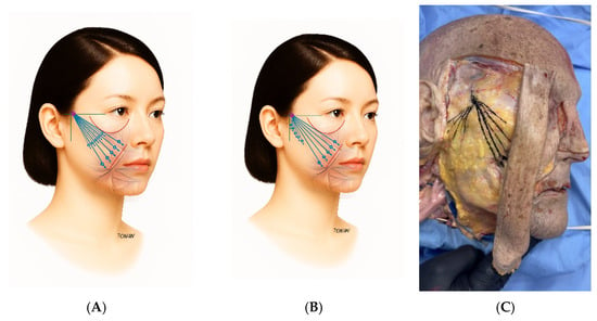
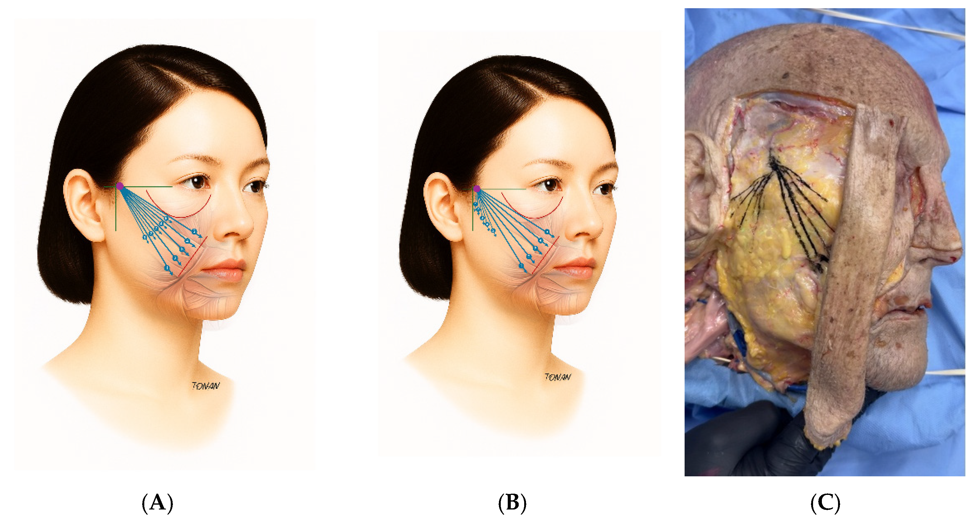
Modified anchorage approach: The average distance from the entry point to the nasolabial fold is 6–7 cm, leaving approximately 3 cm of thread within the cannula at the end of linear insertion. In the traditional “floating” technique, this remaining segment is re-inserted into the mobile subcutaneous tissue toward the nasolabial fold (NLF), forming a short “V” configuration (Figure 1A). In the modified technique the cannula is rotated laterally and inferiorly so that the terminal barbs are positioned posterior to the zygomatic retaining ligament line (Figure 1B). This maneuver aims to shift tensile load to a more fixed area, non-mobile structure.
Figure 1.
Comparative overview of the traditional floating-thread placement and the modified fixed-anchorage technique. The illustrations contrast the trajectory and terminal positioning of the thread in each approach, and the cadaveric image demonstrates the anatomical location of the retroligamentous anchorage zone. The entry point is marked as purple dot and the blue lines represent the trajectory of the threads, with terminal barbed segments positioned within the subcutaneous and superficial musculoaponeurotic system (SMAS) plane to achieve mechanical lifting and tissue repositioning. (A). Traditional floating technique with anterior reinsertion into mobile subcutaneous tissue. (B). Modified technique redirecting the terminal segment posterior to the zygomatic retaining ligament line. (C). Cadaveric image showing the anatomical region corresponding to the posterior anchorage site.
Step 4 (Post-procedural care): Standard instructions include cold compresses, avoidance of excessive facial movements for 48 h, refraining from massage or pressure on the treated area, and oral analgesics if necessary.
A cadaveric dissection was performed to visually confirm the anatomical position of the anchorage zone. Figure 1C illustrates the placement of the terminal barb segment posterior to the zygomatic retaining ligament line, demonstrating the relationship between the cannula path and the fixed retroligamentous fascia.
This anatomical correlation supports the feasibility of directing tension toward a stable fascial layer rather than relying on mobile subcutaneous fat.
Anchoring posterior to the ligament line also broadens the bio-stimulatory field of the PLLA/PCL thread, promoting neocollagenesis along a mechanically active pathway [17].
3. Results
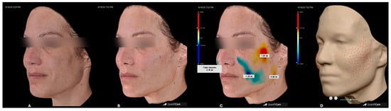
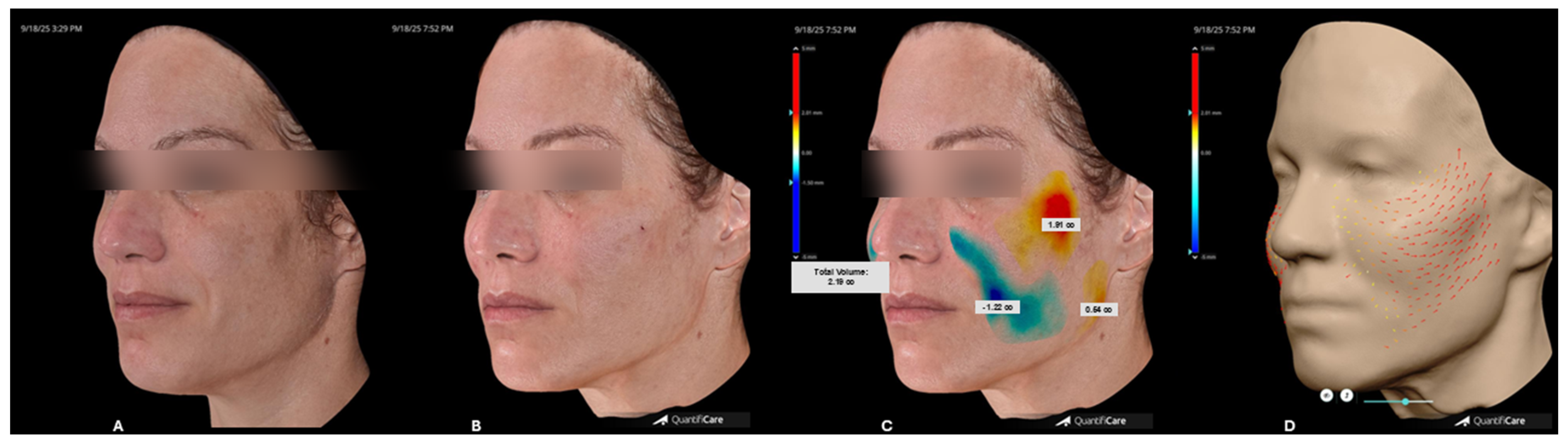
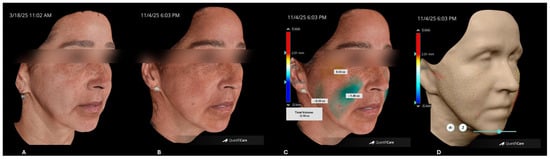
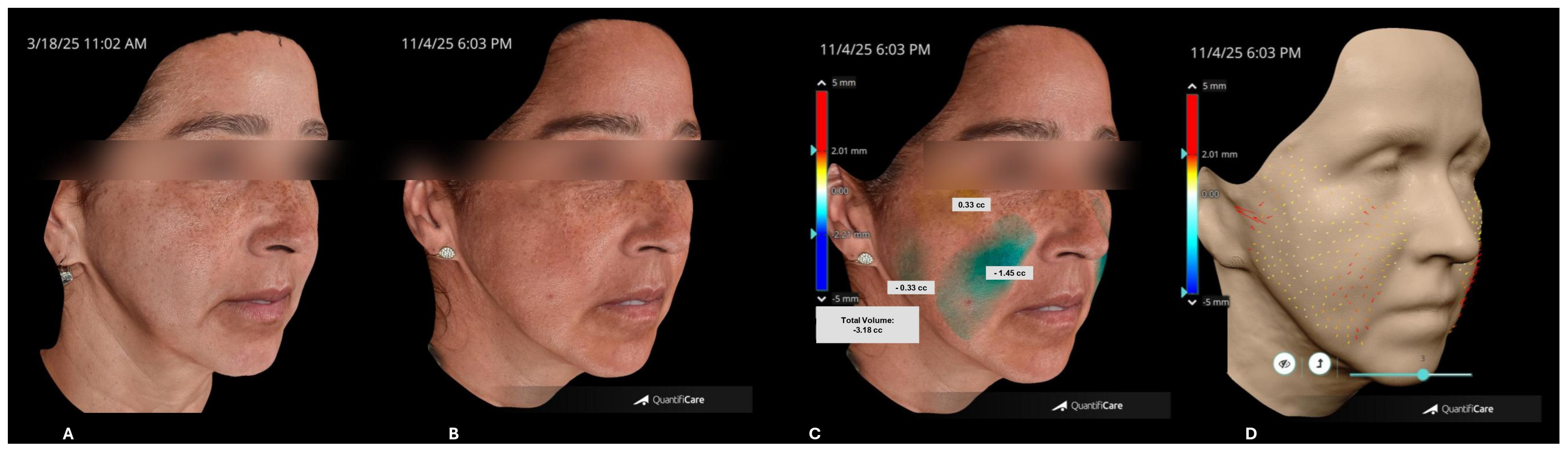
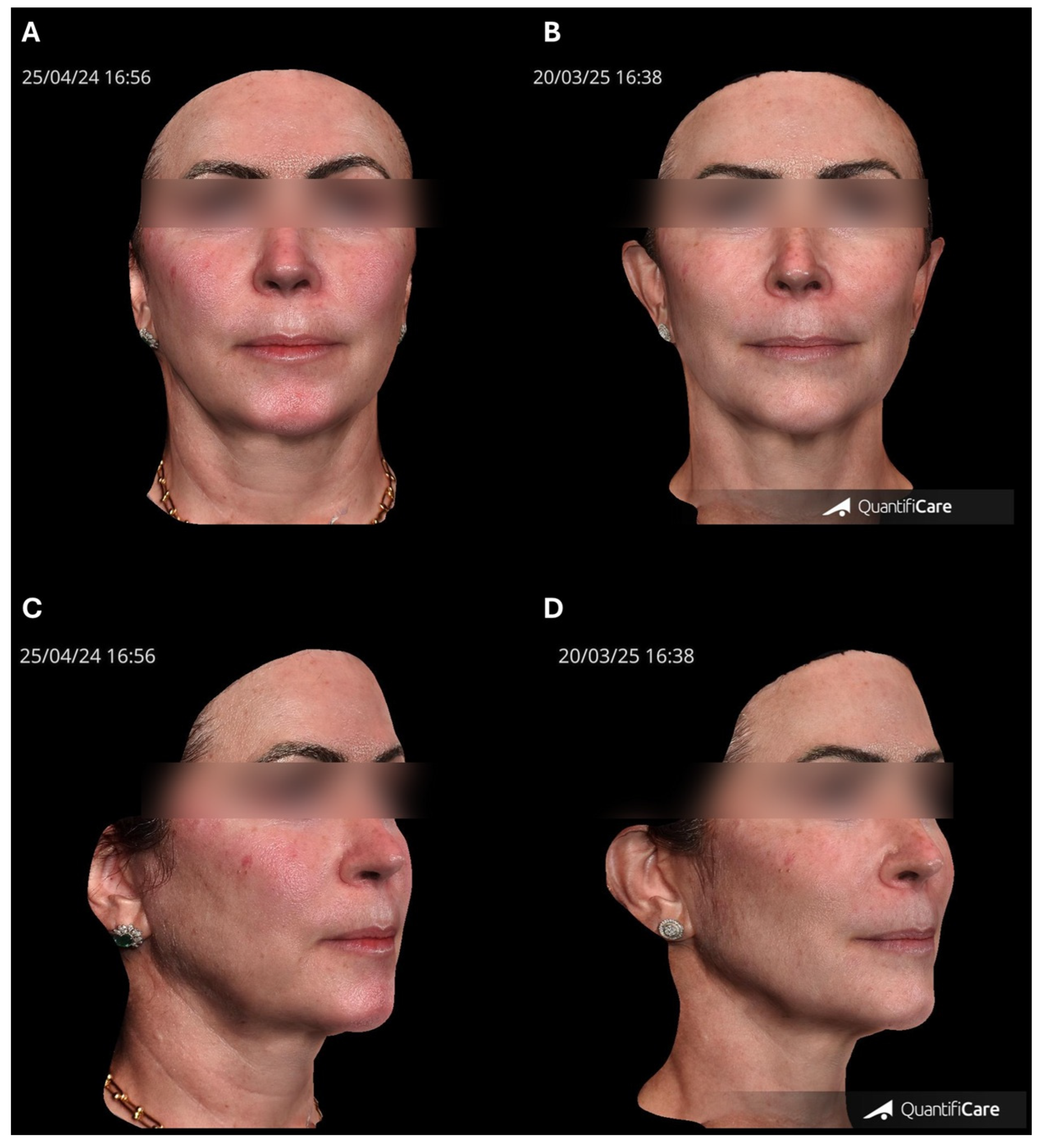
Representative clinical cases demonstrated the practical application and biomechanical effect of the modified fixed-anchorage technique. Anchorage posterior to the zygomatic retaining ligament line resulted in immediate midface elevation, with improved malar projection and softening of the nasolabial and mentolabial folds (Figure 2 and Figure 3).
Figure 2.
3D Quantificare assessment of an illustrative case. (A). Pre-procedure image. (B). Immediate post-procedure result showing midface elevation. (C). Volumetric map demonstrating areas of positive soft tissue displacement following vector repositioning. (D). Vector analysis displaying direction and magnitude of tissue mobilization induced by the thread trajectories.
Figure 3.
3D Quantificare assessment of an illustrative case with 240 days follow-up imaging. (A). Pre-procedure image. (B). 240 days post-procedure result showing midface fat repositioning with improvement in nasolabial and mento-labial folds. (C). Volumetric map demonstrates areas of positive soft tissue displacement following vector repositioning. (D). Vector analysis displaying direction and magnitude of tissue mobilization induced by thread trajectories.
Standardized 3D imaging demonstrated a reproducible pattern of positive volumetric displacement along the treated vectors, consistent with mechanical repositioning of the malar fat compartments. Quantificare vector analysis demonstrated upward and superolateral redirection of soft tissues, corresponding to the intended anchorage line posterior to the zygomatic retaining ligament.
Quantificare analyses were used qualitatively to illustrate vector direction and soft tissue displacement patterns. No numerical thresholds, measurement error analysis, or reproducibility testing were performed, as the purpose was to demonstrate biomechanical plausibility rather than quantitative clinical outcomes.
Patients reported high tolerance during and after the procedure. Mild edema and localized tenderness were reported, resolving spontaneously within 1 to 2 weeks. No hematomas, dimpling, asymmetry, or vascular complications were observed.
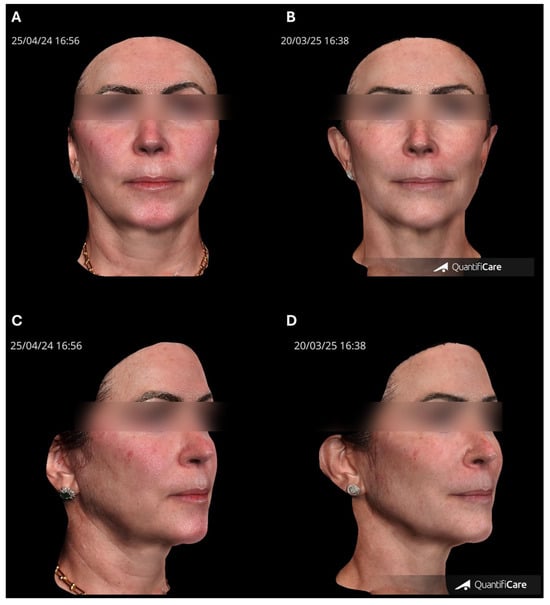
In one patient with 12-month photographic follow-up (Figure 4), contour preservation was visually appreciable, particularly in the malar and midface regions. While this finding suggests potential stability of the vectored lift, the absence of quantitative measurements limits interpretation regarding durability, and no conclusions can be drawn about long-term efficacy.
Figure 4.
Representative case of the modified fixed-anchorage thread-lifting technique with 1-year follow-up. (A,C) Baseline images. (B,D) One-year post-procedure views showing midface elevation, malar projection improvement, and softening of the nasolabial fold.
Overall, the aggregated observations support the feasibility and mechanical plausibility of the modified technique. However, the descriptive nature of the sample precludes generalization, and controlled studies are required to determine comparative performance against traditional floating-thread placement.
4. Discussion
Thread lifting has undergone substantial refinement since its introduction, supported by a growing understanding of facial retaining ligaments, soft tissue biomechanics, and material properties of absorbable threads. Traditionally, barbed sutures rely on floating fixation, in which lift is achieved through barb engagement within the mobile subcutaneous layer and the retinacula cutis [18,19,20]. The present modification builds upon this anatomical understanding, aiming to enhance the mechanical stability of midface repositioning by transferring tension from the mobile subcutaneous layer to the fixed ligamentous framework.
The rationale aligns with classic descriptions of McGregor’s patch, a dense fibrous zone between the parotid and malar regions [11,12], and with contemporary analyses that map the zygomatic and superior masseteric ligaments as boundaries between fixed and mobile midfacial tissues [9,21]. In principle, the present fixed-anchorage technique operates on the same biomechanical rationale—using an anatomically stable region to convert linear traction into vertical lift, reducing relapse and improving contour definition. Cadaveric dissections and modern 3D anatomical reconstructions further confirm that these ligaments occupy predictable positions and serve as natural anchorage points for facial soft tissue support [13,14].
The cadaveric demonstration included in this report reinforces the spatial feasibility of redirecting the terminal barb segment into the retroligamentous plane, supporting the theoretical shift from a purely floating system to a hybrid configuration that partially engages a fixed fascial structure. This differs from surgical facelift techniques, in which retaining ligaments are released; in thread lifting, they are preserved and can therefore function as stabilizing boundaries for vector orientation [1].
Across the three illustrative cases, the technique demonstrated consistent mechanical patterns on standardized 3D imaging. Quantificare volumetric and vector analyses showed superolateral tissue redistribution along the intended vectors, corresponding to midface elevation and improved malar definition. These findings are compatible with the biomechanical expectation of anchoring tension closer to a fixed ligament line. Patients experienced only mild, transient edema or tenderness, and no complications such as dimpling, hematoma, migration, or neurovascular events were observed, findings that align with the known safety profile of PLLA/PCL threads [7,8].
One patient demonstrated 12-month photographic maintenance of contour and malar projection. Although encouraging, this observation must be interpreted cautiously because no quantitative longitudinal measurements were obtained, and the sample size is descriptive rather than comparative. The biostimulatory effects of PLLA/PCL—namely neocollagenesis and neoelastogenesis—may contribute to contour maintenance [17,22], but the present data are insufficient to establish this relationship.
Ultimately, the aggregated observations support the anatomical plausibility and procedural feasibility of repositioning the terminal segment posterior to the zygomatic retaining ligament line. The technique aligns with the natural architecture of facial retaining structures and appears to produce predictable mechanical effects in the midface. However, larger controlled studies with standardized imaging, objective outcomes, and long-term follow-up are needed to determine whether this modification offers measurable advantages over traditional floating-thread placement.
This technical report presents several inherent limitations that must be acknowledged when interpreting the findings. The sample consisted of only illustrative, non-consecutive cases, selected based on the availability of photographic documentation rather than predetermined criteria, which restricts generalizability. The absence of a comparator group precludes any assessment of differences between this modified approach and traditional floating-thread placement. In addition, outcomes were evaluated qualitatively without objective measurements such as ultrasound imaging, elastography, or longitudinal 3D volumetric quantification, limiting the ability to draw conclusions regarding long-term efficacy or tissue behavior. Follow-up intervals also varied among cases, with only one patient documented at 12 months, further constraining the interpretation of durability.
An additional limitation is the absence of evaluation in anatomically distinct populations. Facial skeletal morphology varies across ethnic groups, particularly between Asian and Caucasian populations, with differences in zygomatic width, malar projection, skin thickness, as well as culturally influenced aesthetic preferences [23]. Given that the present approach relies on ligament-based anchorage rather than volumetric augmentation, vector orientation can be deliberately modulated to address distinct anatomical and aesthetic objectives, including the avoidance of excessive malar prominence. The illustrative cases included in this report were treated in Brazil, a population characterized by significant ethnic heterogeneity, and did not specifically include a dedicated Asian cohort. In individuals with prominent zygomatic architecture, such as many Asian patients, an entry point positioned inferior to the zygomatic arch may offer a more favorable vector orientation, potentially reducing the risk of excessive zygomatic fullness. Evaluation of this modification in populations with distinct skeletal and soft tissue characteristics represents an important direction for future investigation. Given these considerations, the observations presented here should be interpreted as evidence of anatomical feasibility rather than clinical effectiveness.
5. Conclusions
This technical report presents an anatomically guided modification of midface thread implantation, redirecting the terminal segment of a PLLA/PCL barbed thread posterior to the zygomatic retaining ligament line. The approach demonstrated procedural feasibility and predictable immediate lifting effects, supported by 3D imaging and cadaveric correlation. By extending the trajectory into the retroligamentous zone, the modification also broadens the area of collagen stimulation and neocollagenesis, potentially enhancing the tissue remodeling effect associated with PLLA/PCL threads. Further controlled studies with imaging and validated outcome measures are warranted to confirm the long-term efficacy and reproducibility of this fixed-anchorage concept.
Author Contributions
Conceptualization, L.T.; methodology, L.T., A.H. and L.E.A.; validation, L.T.; investigation, A.H. and L.E.A.; anatomical analysis and cadaveric procedures, A.H. and L.E.A.; writing—original draft preparation, R.V.; writing—review and editing, R.V., A.H. and L.T.; supervision, L.T. and L.E.A.; project administration, R.V. All authors have read and agreed to the published version of the manuscript.
Funding
The threads utilized in the cadaver study were provided by Ilikia Brasil.
Institutional Review Board Statement
This paper reports a technical experience based on cases treated during routine clinical practice. No prospective data collection, research protocol, or experimental intervention was implemented. The described technique has been routinely performed in the clinical setting. Between March 2025 and November 2025, 40 patients underwent the described modified fixed-anchorage midface thread-lifting technique in the clinic. Most patients received multiple simultaneous treatments. Of the 10 cases performed using threads only with the presented technique, three cases are selected as illustrative cases, selected based on the availability of standardized photographic documentation, 3D imaging, patient consent and follow-up data. All data presented are fully anonymized. Written informed consent for image publication was obtained from all illustrated patients. According to local regulations, descriptive technical reports that do not involve experimental intervention or prospective data collection do not require formal ethics committee approval. All patients provided written informed consent for treatment and for the use of anonymized images for scientific publication.
Informed Consent Statement
Informed consent was obtained from all subjects involved in the study. Written informed consent has been obtained from the patients to publish this paper.
Data Availability Statement
The data supporting the conclusions of this article are contained within the manuscript. Further information may be requested from the corresponding author, subject to ethical and privacy considerations.
Conflicts of Interest
Luiz Tonon and Alessandra Haddad are speakers for APTOS Global. Luiz Tonon is affiliated with Tonon Medical Group. Alessandra Haddad is affiliated with A Haddad Medical and Aesthetic Clinics. Renata Viana is a scientific consultant for Ilikia Brasil, the distributor of the thread in Brazil. Luiz Eduardo Avelar is affiliated with Domani Clinic. The authors declare that the research was conducted in the absence of any commercial or financial relationships that could be construed as a potential conflict of interest.
References
- Mendelson, B.; Wong, C.-H. Changes in the Facial Skeleton With Aging: Implications and Clinical Applications in Facial Rejuvenation. Aesthetic Plast. Surg. 2012, 36, 753–760. [Google Scholar] [CrossRef] [Scilit]
- Toledo Avelar, L.E.; Cardoso, M.A.; Santos Bordoni, L.; de Miranda Avelar, L.; de Miranda Avelar, J.V. Aging and Sexual Differences of the Human Skull. Plast. Reconstr. Surg. Glob. Open 2017, 5, e1297. [Google Scholar] [CrossRef] [Scilit]
- Sato, M.; Muniz, M.; Ferreira, L.R. Treatment of Mid-Face Aging with Calcium Hydroxylapatite: Focus on Retaining Ligament Support. Clin. Cosmet. Investig. Dermatol. 2024, 17, 2545–2553. [Google Scholar] [CrossRef] [Scilit] [PubMed]
- Frank, K.; Moellhoff, N.; Engerer, N.; Kaye, K.O.; Alfertshofer, M.; Gotkin, R.H.; Kassirer, S.; Weinmann, C.; Casabona, G.; Cotofana, S. Understanding the Interplay Between Skin, Fascia, and Muscles of the Midface in Facial Aging. Aesthetic Plast. Surg. 2024, 48, 3568–3576. [Google Scholar] [CrossRef] [Scilit] [PubMed]
- Shehan, J.N.; Ryll, L.S.; LeClair, J.; Ezzat, W.H. Current Practices and Trends in Midface Rejuvenation. Ann. Plast. Surg. 2023, 90, 118–122. [Google Scholar] [CrossRef] [Scilit]
- Grand View Research. Aesthetic Threads Market (2024–2030): Size, Share & Trends Analysis Report by Product Type, by Application, by End-Use, by Region, and Segment Forecasts; Grand View Research: San Francisco, CA, USA, 2024; Available online: https://www.grandviewresearch.com/industry-analysis/aesthetic-threads-market-report (accessed on 17 December 2025).
- Rezaee Khiabanloo, S.; Nabie, R.; Aalipour, E. Outcomes in thread lift for face, neck, and nose: A prospective chart review study with APTOS. J. Cosmet. Dermatol. 2020, 19, 2867–2876. [Google Scholar] [CrossRef] [Scilit] [PubMed]
- Turkevych, M.; Turkevych, A.; Kadjaya, A.; Gold, M.H.; Lotti, T.; Sulamanidze, G. Pathomorphological criteria of use efficiency of resorbable and permanent implants in aesthetic medicine and cosmetic dermatology. J. Cosmet. Dermatol. 2018, 17, 731–735. [Google Scholar] [CrossRef] [Scilit]
- Alghoul, M.; Codner, M.A. Retaining Ligaments of the Face. Aesthet. Surg. J. 2013, 33, 769–782. [Google Scholar] [CrossRef] [Scilit]
- Kaye, B.L. The Extended Facelift with Ancillary Procedures. Ann. Plast. Surg. 1981, 6, 335–346. [Google Scholar] [CrossRef] [Scilit]
- McGregor, M. Face lift techniques. In Proceedings of the 1st Annual Meeting of the California Society of Plastic Surgeons, Yosemite, CA, USA, 1959. [Google Scholar]
- Seo, Y.S.; Song, J.K.; Oh, T.S.; Kwon, S.I.; Tansatit, T.; Lee, J.H. Review of the Nomenclature of the Retaining Ligaments of the Cheek: Frequently Confused Terminology. Arch. Plast. Surg. 2017, 44, 266–275. [Google Scholar] [CrossRef] [Scilit]
- Rossell-Perry, P.; Paredes-Leandro, P. Anatomic Study of the Retaining Ligaments of the Face and Applications for Facial Rejuvenation. Aesthetic Plast. Surg. 2013, 37, 504–512. [Google Scholar] [CrossRef] [Scilit] [PubMed]
- Park, J.S.; Noel, G. Facial retaining ligaments based on real color sectioned images with 3D models: Toward a more precise classification. J. Cranio-Maxillofac. Surg. 2025, 53, 1638–1646. [Google Scholar] [CrossRef] [Scilit]
- Kang, M.S.; Kang, H.G.; Nam, Y.S.; Kim, I.-B. Detailed anatomy of the retaining ligaments of the mandible for facial rejuvenation. J. Cranio-Maxillofac. Surg. 2016, 44, 1126–1130. [Google Scholar] [CrossRef] [Scilit] [PubMed]
- O’Brien, J.X.; Rozen, W.M.; Whitaker, I.S.; Ashton, M.W. Lore’s fascia and the platysma-auricular ligament are distinct structures. J. Plastic Reconstr. Aesthet. Surg. 2012, 65, e241–e245. [Google Scholar] [CrossRef] [Scilit] [PubMed]
- Kwon, T.R.; Han, S.W.; Yeo, I.K.; Kim, J.H.; Kim, J.M.; Hong, J.Y.; Lee, B.C.; Lee, S.E.; Moon, H.S.; Kwon, H.J.; et al. Biostimulatory effects of polydioxanone, poly-d-lactic acid, and polycaprolactone fillers in mouse model. J. Cosmet. Dermatol. 2019, 18, 1002–1008. [Google Scholar] [CrossRef] [Scilit]
- Sakata, A.; Abe, K.; Mizukoshi, K.; Gomi, T.; Okuda, I. Relationship between the retinacula cutis and sagging facial skin. Ski. Skin. Res. Technol. 2018, 24, 93–98. [Google Scholar] [CrossRef] [Scilit]
- Sulamanidze, M.A.; Shiffman, M.A.; Paikidze, T.G.; Sulamanidze, G.M.; Gavasheli, L.G. Facial Lifting with APTOS Threads. Int. J. Cosmetic Surg. Aesthet. Dermatol. 2001, 3, 275–281. [Google Scholar] [CrossRef] [Scilit]
- Sulamanidze, M.; Sulamanidze, G. APTOS Suture Lifting Methods: 10 Years of Experience. Clin. Plast. Surg. 2009, 36, 281–306. [Google Scholar] [CrossRef] [Scilit]
- Braz, A.; Palermo, E.; Issa, M.C. Revisiting the Ligament Line of the Face. Dermatol. Clin. 2024, 42, 97–102. [Google Scholar] [CrossRef] [Scilit]
- Soen, M.; Hidayat, M.; Widowati, W. Enhancing dermal collagen density towards youthfulness: A comparative study of PCL, PLLA, and PDO thread implantation in aging rats model. Iran. J. Basic. Med. Sci. 2025, 28, 151–157. [Google Scholar] [CrossRef] [Scilit] [PubMed] [PubMed Central]
- Rhee, S.C. Differences between Caucasian and Asian attractive faces. Ski. Skin. Res. Technol. 2018, 24, 73–79. [Google Scholar] [CrossRef] [Scilit] [PubMed]
Disclaimer/Publisher’s Note: The statements, opinions and data contained in all publications are solely those of the individual author(s) and contributor(s) and not of MDPI and/or the editor(s). MDPI and/or the editor(s) disclaim responsibility for any injury to people or property resulting from any ideas, methods, instructions or products referred to in the content. |
© 2026 by the authors. Licensee MDPI, Basel, Switzerland. This article is an open access article distributed under the terms and conditions of the Creative Commons Attribution (CC BY) license.