


Simple Summary
Listeria monocytogenes is a foodborne bacterium that can cause severe illness, particularly in pregnant women, newborns, older adults, and people with weakened immune systems. This study examined 26 strains collected from hospital patients in Bydgoszcz, Poland, between 2017 and 2024 to determine their antibiotic susceptibility, ability to form biofilm, and genetic characteristics linked to disease and survival. Most strains were susceptible to all tested antibiotics, although four were resistant to trimethoprim–sulfamethoxazole, and three of these were also resistant to erythromycin. All strains formed biofilm, with half showing strong formation. The strains also varied in their genetic profiles, including genes associated with virulence. Combined phenotypic and genotypic analyses may support a better understanding of strain diversity, pathogenic potential, and traits associated with persistence and survival.
Abstract
Listeria monocytogenes is the etiological agent of listeriosis, a disease that disproportionately affects pregnant women, newborns, older adults, and immunocompromised individuals. The present study provides a phenotypic and genotypic characterization of 26 clinical L. monocytogenes isolates collected in Bydgoszcz, Poland, in 2017–2024. Antibiotic susceptibility was assessed using the disk diffusion method, biofilm formation was evaluated using the 24-well crystal violet assay, and PCR was performed to detect serogroups, selected virulence genes, and stress survival islets (SSIs). Most isolates (22; 84.6%) were susceptible to five tested antibiotics (penicillin, ampicillin, meropenem, erythromycin, trimethoprim–sulfamethoxazole). Resistance to trimethoprim–sulfamethoxazole was detected in four isolates (15.4%), three of which (11.5% of all isolates) were also resistant to erythromycin. All isolates were classified as biofilm-forming under the conditions of the crystal violet assay, including 13 (50.0%) strong, 9 (34.6%) moderate, and 4 (15.4%) weak biofilm producers. PCR-based serogroups, which classify genetically related serotypes according to characteristic marker-gene profiles, were distributed as follows: Serogroup 1/2b–3b–7 was predominant and included 13 isolates (50.0%), followed by serogroup 4b–4d–4e, detected in 11 isolates (42.3%), and serogroup 1/2a–3a, found in two isolates (7.7%). No isolates belonging to serogroup 1/2c–3c were identified. Four virulence gene profiles characterized all 26 isolates: In total, 12 strains (46.2%) were detected to have all 10 analyzed virulence genes, 2 (7.7%) did not have mpl, 1 (3.8%) did not have iap, and 11 (42.3%) lacked both fbpA and mpl, as determined by the applied PCR assays. At the stress survival islet locus, SSI-1 was detected in 8 isolates (30.8%), SSI-2 was not detected, and the 1.1 kbp (SSI-1-negative locus) variant was detected in 18 isolates (69.2%). The distribution of SSI-1 differed among PCR serogroups, whereas no association between PCR serogroup and biofilm category was detected. Overall, the findings demonstrate phenotypic and targeted genotypic diversity within this single-center collection of clinical L. monocytogenes isolates. Combined phenotypic and genotypic analyses may support a better understanding of strain diversity, pathogenic potential, and traits associated with persistence and survival.
1. Introduction
Listeria monocytogenes is a Gram-positive, non-spore-forming, rod-shaped bacterium that is widely distributed in the natural environment. It can be isolated from soil, water, and wastewater treatment plants [1]. This species has multiple adaptive mechanisms that enable it to survive under diverse environmental stress conditions, including variable pH (4.0–9.6), high salinity (up to 20% salt), temperatures ranging from −0.4 °C to 45 °C, and exposure to reactive oxygen species [2].
L. monocytogenes is an intracellular pathogen capable of breaching three major host barriers. Following ingestion of contaminated food, the bacterium can cross the intestinal barrier and disseminate through the lymphatic system into the bloodstream. It can also traverse the blood–brain and placental barriers [3]. Moreover, L. monocytogenes is adapted to invade and replicate within epithelial cells, macrophages, and dendritic cells [4].
L. monocytogenes is the etiological agent of listeriosis, a disease transmitted primarily through food. The main sources of this pathogen include unpasteurized dairy products, meat, fish, and ready-to-eat (RTE) foods [5]. In humans, infection may manifest as mild gastrointestinal illness or as invasive disease, including sepsis, central nervous system infections, and perinatal listeriosis. Cutaneous listeriosis is a rare form of infection that occurs mainly among farmers and veterinarians exposed to secretions from animals infected with L. monocytogenes [6]. Individuals at particularly high risk include older adults, immunocompromised patients, pregnant women, and newborns [6]. Non-invasive listeriosis may present with nausea, vomiting, diarrhea, headache, abdominal pain, joint pain, drowsiness, and loss of appetite and is usually self-limiting. Neurolisteriosis typically presents as meningitis or encephalitis and, in some cases, may lead to brain abscesses or brainstem inflammation; clinical symptoms include headache, fever, and neck stiffness [6,7]. In pregnant women, nonspecific symptoms such as uterine contractions, preterm labor, fetal arrhythmias, and miscarriage may occur in addition to fever and diarrhea. Early-onset neonatal listeriosis develops between the first and sixth day of life and is associated with bacteremia, pneumonia, and meningitis. By contrast, late-onset disease occurs between 5 and 20 days of life and may manifest as fever, diarrhea, colitis, and meningitis [5,6,7]. The case-fatality rate of listeriosis remains high, at approximately 20–30% [8].
In 2024, 3041 cases of listeriosis were reported in the European Union, corresponding to a notification rate of 0.69 cases per 100,000 population and making listeriosis the fourth most frequently reported zoonotic disease [9]. Most cases were confirmed during the summer months. Despite its relatively low incidence, listeriosis remained the most severe zoonosis in terms of the proportion of hospitalizations and the case-fatality rate: Notably, 97.3% of patients required hospitalization, and 301 deaths were recorded, 80% of which occurred among individuals aged over 65 years [9]. Moreover, the number of reported cases increased significantly over the preceding five years, a trend that may partly reflect population aging and changes in dietary habits [9].
Although L. monocytogenes comprises multiple serotypes distinguished by their somatic (O) and flagellar (H) antigens, four serotypes—1/2a, 1/2b, 1/2c, and 4b—account for the majority of human listeriosis cases. Serotypes 1/2a and 1/2c are frequently recovered from food and food-processing environments, whereas serotypes 1/2b and 4b are particularly important in clinical infections; notably, serotype 4b has been associated with numerous major outbreaks of invasive listeriosis [10].
The first-line antibiotics used for the treatment of listeriosis are β-lactams, such as penicillin, ampicillin, or amoxicillin, often administered in combination with gentamicin. Alternative therapeutic options include trimethoprim–sulfamethoxazole (co-trimoxazole) and meropenem, while erythromycin may be used in pregnant women [6]. L. monocytogenes exhibits intrinsic resistance to fosfomycin, fusidic acid, oxacillin, cephalosporins, and lincosamides. In addition, the emergence and dissemination of acquired resistance may be driven by both vertical transmission and horizontal transfer of resistance genes carried on mobile genetic elements, including plasmids, transposons, and conjugative elements, potentially between L. monocytogenes and other bacteria. Together with the extensive use of antimicrobials in animal husbandry, these processes have contributed to the emergence of strains resistant to quinolones, β-lactams, aminoglycosides, tetracyclines, phenicols, macrolides, trimethoprim, and biocides [11,12].
Given the clinical significance of L. monocytogenes, the ongoing concern regarding antimicrobial resistance, and the need for better understanding of its virulence-associated characteristics, continued surveillance of clinical isolates is essential. Therefore, this study aimed to characterize the phenotypic and selected genotypic features of L. monocytogenes strains isolated from clinical specimens in Bydgoszcz, Poland, focusing on antimicrobial susceptibility, biofilm formation, serogroup distribution, selected virulence genes, and stress survival islets. The findings may contribute to a better understanding of the characteristics of locally circulating clinical strains and provide a basis for further epidemiological investigations.
2. Materials and Methods
2.1. Bacterial Isolates
The study included 26 archived clinical L. monocytogenes isolates from the collection of the Department of Microbiology, Ludwik Rydygier Collegium Medicum in Bydgoszcz, Nicolaus Copernicus University in Toruń (Poland). The isolates had originally been recovered during routine clinical diagnostic procedures from patients hospitalized at Antoni Jurasz University Hospital No. 1 in Bydgoszcz, Poland, during 2017–2024 and subjected to microbiological identification in accordance with the hospital procedures applicable at the time of sampling. Each isolate originated from a different patient.
2.2. Molecular Identification
2.2.1. Preparation of Strains for Analysis
The tested strains were stored at −70 °C in brain heart infusion broth (BHI; Becton Dickinson, Franklin Lakes, NJ, USA) supplemented with 15.0% glycerol (Avantor, Gliwice, Poland). Before analysis, the frozen strains were streaked onto Columbia agar supplemented with 5% sheep blood (CAB; Graso, Krąg, Poland) and incubated at 37 °C for 24 h. To ensure culture purity and obtain growth derived from a single colony, one well-isolated colony from the primary culture was subcultured onto fresh CAB (Graso, Krąg, Poland) and incubated under the same conditions. Bacterial growth from this second passage was used for all subsequent analyses.
2.2.2. Genomic DNA Isolation
Genomic DNA was isolated using a column-based method according to the manufacturer’s instructions (EurX, Gdańsk, Poland).
Briefly, a single bacterial colony from CAB (Graso, Krąg, Poland) was suspended in 200 µL of Tris–EDTA buffer (Promega, Madison, WI, USA), supplemented with 10 µL of lysozyme (EurX, Gdańsk, Poland), and incubated at 37 °C for 40 min. Subsequently, 20 µL of proteinase K (Promega, Madison, WI, USA) and 220 µL of Sol V buffer (EurX, Gdańsk, Poland) were added, and the mixture was vortexed and incubated at 50 °C for 10 min. After brief centrifugation, 250 µL of 96% ethanol (Chempur, Piekary Śląskie, Poland) was added, and the sample was mixed and incubated at room temperature for 1 min. The lysate was transferred to a binding column and centrifuged at 8000× g for 1 min. The column was washed sequentially with 500 µL of Wash V1 buffer (EurX, Gdańsk, Poland) and 500 µL of RBW buffer (EurX, Gdańsk, Poland), with centrifugation at 8000× g for 1 min after each wash, followed by centrifugation at maximum speed for 2 min to remove residual wash buffer. DNA was eluted by applying 50 µL of RNase-free water (EurX, Gdańsk, Poland) directly to the membrane, incubating at room temperature for 1 min, and centrifuging at maximum speed for 2 min. The purified DNA was stored at 4 °C until further analysis.
2.2.3. Species Identification
Species identification was performed using matrix-assisted laser desorption/ionization time-of-flight mass spectrometry (MALDI-TOF MS) during routine laboratory testing at Antoni Jurasz University Hospital No. 1 in Bydgoszcz (Poland). Mass spectra were acquired using a Microflex LT/SH mass spectrometer (Bruker, Billerica, MA, USA) and analyzed with MALDI Biotyper software version 4.1 incorporating the Bruker Taxonomy database. Briefly, a single colony from a fresh culture was transferred onto a MALDI target plate, overlaid with 1 µL of the matrix solution (α-cyano-4-hydroxycinnamic acid) (Bruker, Billerica, MA, USA) and allowed to air-dry. Mass spectra were then acquired and compared with the reference spectral library supplied with the MALDI-TOF MS system. Identification was accepted in accordance with the manufacturer’s interpretation criteria. Isolates with log(score) values ≥ 2.0 were considered reliably identified at the species level. The Bruker Bacterial Test Standard (BTS) was used for instrument calibration and quality control according to the manufacturer’s recommendations.
To confirm the identification results from MALDI TOF MS, multiplex PCR was performed according to Border et al. [13] and Bansal et al. [14]. Two primer pairs were used: L1 (5′-CAGCAGCCGCGGTAATAC-3′) and L2 (5′-CTCCATAAAGGTGACCCT-3′), targeting the rrs gene (encoding 16S rRNA) and yielding a 938 bp product [13], and LM1 (5′-CCTAAGACGCCAATCGAA-3′) and LM2 (5′-AAGCACTTGCAACTGCTC-3′), targeting the hly gene and yielding a 750 bp product [14]. The reaction mixture composition and amplification conditions followed Skowron et al. [15]. Multiplex PCR was performed in a total volume of 25 µL containing 1× Green GoTaq® Flexi Buffer (Promega, Madison, WI, USA), 2 mM MgCl2 (Promega, Madison, WI, USA), 0.2 mM dNTPs (Promega, Madison, WI, USA), 0.8 µM of each primer (Oligo.pl, Warszawa, Poland), 1 U GoTaq DNA polymerase (Promega, Madison, WI, USA), 2 µL genomic DNA, and ultrapure water (EurX, Gdańsk, Poland). Primers were synthesized by the DNA Sequencing and Oligonucleotide Synthesis Laboratory, Institute of Biochemistry and Biophysics, Polish Academy of Sciences (Oligo.pl, Warszawa, Poland). The positive control consisted of DNA from the reference strain L. monocytogenes ATCC 19111.
Amplification was carried out in a Mastercycler Pro thermocycler (Eppendorf, Hamburg, Germany) using initial denaturation at 94 °C for 2 min, followed by 30 cycles of denaturation at 94 °C for 30 s, primer annealing at 50 °C for 30 s, and extension at 72 °C for 1 min, with a final extension at 72 °C for 5 min. Samples were held at 4 °C until removal.
PCR products were separated in a 1.5% agarose gel (EurX, Gdańsk, Poland) in 1× TBE buffer (EurX, Gdańsk, Poland) at 90 V for 60 min, using GeneRuler™ 1000 bp DNA Ladder (A&A Biotechnology, Gdańsk, Poland) as a marker, and visualized with a Gel Doc XR system (Bio-Rad Laboratories, Hercules, CA, USA).
2.3. Phenotypic Characterization
2.3.1. Antimicrobial Susceptibility Testing
Antimicrobial susceptibility was assessed using the disk diffusion method. Suspensions of the tested isolates were prepared from 24 h cultures in sterile 0.9% saline (Graso, Krąg, Poland) and adjusted to a turbidity equivalent to 0.5 McFarland using a calibrated densitometer (DEN-1B, Biosan SIA, Riga, Latvia). The suspensions were inoculated onto Mueller–Hinton agar supplemented with 5% horse blood and β-NAD (Graso, Krąg, Poland), and antibiotic disks were then applied, including penicillin (1 U), ampicillin (2 µg), meropenem (10 µg), erythromycin (15 µg), and trimethoprim–sulfamethoxazole (1.25–23.75 µg) (Argenta, Poznań, Poland). The plates were incubated for 18 h at 35 °C in an atmosphere containing 5% CO2. Streptococcus pneumoniae ATCC 49619 was used as the quality control strain. Inhibition zones were measured and expressed in millimeters. The results were interpreted according to EUCAST guidelines [16] v. 16.0.
2.3.2. Biofilm-Forming Ability
Biofilm formation was assessed using the crystal violet assay, as described by Wiktorczyk-Kapischke et al. [17]. A suspension with a density of 0.5 McFarland was prepared from a 24 h culture in 0.9% saline (Graso, Krąg, Poland). Aliquots of 180 µL tryptic soy broth (TSB; Graso, Krąg, Poland) and 20 µL bacterial suspension were added to each well of a 96-well microtiter plate (Profilab, Łomianki, Poland). Each strain was tested in triplicate, with L. monocytogenes ATCC 19111 as the positive control and uninoculated TSB (Graso, Krąg, Poland) as the negative control. Plates were incubated at 37 °C for 24 h in a humid chamber.
After incubation, the wells were emptied, washed three times with distilled water, and dried at 37 °C for at least 20 min. Biofilms were fixed with 96% ethanol (Chempur, Piekary Śląskie, Poland) for 20 min at room temperature with shaking (400 rpm), followed by drying at 37 °C for 20 min. The biofilms were stained with 0.1% crystal violet (POCH, Gliwice, Poland) for 10 min at room temperature with shaking (400 rpm), rinsed with distilled water until the washings were clear, and dried at 37 °C for 20 min. Finally, the bound dye was eluted with ethanol per well for 5 min at room temperature with shaking (400 rpm). Absorbance was measured at 570 nm (Abs570) using a Hidex Sense microplate reader (Hidex, Turku, Finland). For each strain, the mean Abs570 value was calculated from three replicates using Microsoft Excel. Absorbance values are reported below as optical density (OD), in accordance with the convention proposed by Stepanović et al. [18]. The strains were classified into four groups according to the criteria described by Stepanović et al. [18]:
OD ≤ ODc—non-adherent (non-biofilm-forming)
ODc < OD ≤ 2 × ODc—weak biofilm formation
2 × ODc < OD ≤ 4 × ODc—moderate biofilm formation
OD > 4 × ODc—strong biofilm formation
where
ODc—mean absorbance of the negative control (which consisted of sterile TSB (Graso, Krąg, Poland) without the addition of a bacterial suspension) + 3 × standard deviation.
2.4. Genotypic Characterization
2.4.1. Serogroup Determination
Serogroup affiliation was determined using multiplex PCR according to the method described by Doumith et al. [19]. Five primer pairs were used in the reaction, targeting lmo0737, lmo1118, ORF2819, ORF2110, and prs, thereby enabling differentiation of the following serogroups: 1/2a–3a, 1/2b–3b–7, 1/2c–3c, and 4b–4d–4e (Table 1). This multiplex PCR scheme assigns isolates to molecular serogroups rather than individual serotypes, as it does not differentiate serotypes 1/2a from 3a, 1/2b from 3b and 7, 1/2c from 3c, or 4b from 4d and 4e. Therefore, the resulting designations should be interpreted as PCR-based serogroups and not as definitive serotype assignments [19]. Reference strains previously described by Wałecka-Zacharska et al. [20] were used.
Table 1.
Primer sequences used for PCR-based serogroup determination (adapted from Doumith et al. [19]).
Multiplex PCR was performed in a total volume of 25 µL containing 1× Green GoTaq® Flexi Buffer (Promega, Madison, WI, USA), 2 mM MgCl2 (Promega, Madison, WI, USA), 0.2 mM dNTPs (Promega, Madison, WI, USA), five primer pairs (lmo0737, ORF2819 and ORF2110 at 1 µM, lmo1118 at 1.5 µM, and prs at 0.2 µM) (Oligo.pl, Warszawa, Poland), 1 U GoTaq DNA polymerase (Promega, Madison, WI, USA), 2 µL of extracted DNA, and ultrapure water (EurX, Gdańsk, Poland).
Amplification was performed using a Mastercycler Pro thermocycler (Eppendorf, Hamburg, Germany). The cycling conditions were initial denaturation at 94 °C for 3 min, followed by 35 cycles of denaturation at 94 °C for 30 s, annealing at 53 °C for 75 s, and extension at 72 °C for 75 s. Final extension was performed at 72 °C for 10 min, followed by storage at 4 °C.
PCR products were separated in a 1.5% agarose gel (EurX, Gdańsk, Poland) in 1× TBE buffer (EurX, Gdańsk, Poland) at 90 V for 60 min, using GeneRuler™ 1000 bp DNA Ladder (A&A Biotechnology, Gdańsk, Poland) as a marker, and visualized with a Gel Doc XR system (Bio-Rad Laboratories, Hercules, CA, USA).
2.4.2. Detection of Genes Encoding Virulence Factors
The prevalence of 10 selected genes encoding virulence factors was assessed using multiplex PCR according to the methodology described by Skowron et al. [15]. The analyzed genes were grouped into three multiplex reactions: MIX I (fbpA, plcA, and hly), MIX II (plcB, inlB, actA, and iap), and MIX III (inlA, mpl, and prfA). Primer sequences and reaction conditions, adapted from Skowron et al. [15], are presented in Table 2 and Table 3, respectively.
Table 2.
Primer sequences used for multiplex PCR detection of virulence-associated genes in L. monocytogenes (adapted from Skowron et al. [15]).
Table 3.
Amplification conditions used to determine the prevalence of selected genes encoding virulence factors.
Multiplex PCR reactions were performed in a total volume of 25 µL. The reaction mixture contained 1×Green GoTaq® Flexi Buffer (Promega, Madison, WI, USA), 2 mM MgCl2 (Promega, Madison, WI, USA), 0.2 mmol dNTPs (Promega, Madison, WI, USA), primers specific to the target genes (Oligo.pl, Warszawa, Poland), 1 U GoTaq DNA polymerase (Promega, Madison, WI, USA), ultrapure water (EurX, Gdańsk, Poland), and genomic DNA. The final primer concentration was 0.4 µM in reactions I and II and 0.2 µM in reaction III.
Genomic DNA from the reference strain L. monocytogenes ATCC 19111, carrying all the analyzed virulence genes, was used as a positive control.
PCR products were separated in a 1.5% agarose gel (EurX, Gdańsk, Poland) in 1× TBE buffer (EurX, Gdańsk, Poland) at 90 V for 60 min, using GeneRuler™ 1000 bp DNA Ladder and 3000 bp DNA Ladder (A&A Biotechnology, Gdańsk, Poland) as markers, and visualized with a Gel Doc XR system (Bio-Rad Laboratories, Hercules, CA, USA).
2.4.3. Detection of Stress Survival Islets: SSI-1 and SSI-2 and the 1.1 kbp Fragment (SSI−)
The PCR scheme was used to distinguish SSI-1, SSI-2, and the 1.1 kbp SSI-1-negative locus configuration described at this hypervariable genomic region. Primer sequences are presented in Table 4, and the reaction conditions were adapted from Hein et al. [21] and Harter et al. [22].
Table 4.
Primer sequences used for PCR-based detection of SSI-1, SSI-2, and the 1.1 kbp SSI-1-negative locus configuration in L. monocytogenes (adapted from Hein et al. [21] and Harter et al. [22]).
The PCR reaction (SSI-1(+) and SSI-1− (1.1 kbp fragment)) was performed in a total volume of 25 µL. The reaction mixture contained GoTaq® Long PCR Master Mix (Promega, Madison, WI, USA) (2x), 0.2 µM of each primer, 2 mM MgCl2 (Promega, Madison, WI, USA), nuclease-free water (EurX, Gdańsk, Poland), and template DNA. Amplification was carried out under the following thermal conditions: initial denaturation at 94 °C for 3 min, followed by 10 cycles consisting of denaturation at 94 °C for 20 s, primer annealing at 55 °C for 30 s, and extension at 68 °C for 7 min. In the next stage, 25 cycles were performed, consisting of denaturation at 94 °C for 20 s, primer annealing at 55 °C for 30 s, and extension at 68 °C for 12 min. Final extension was carried out at 68 °C for 10 min, after which the reaction products were maintained at 4 °C.
PCR products were separated in a 1.0% agarose gel (EurX, Gdańsk, Poland) in 1× TBE buffer (EurX, Gdańsk, Poland) at 90 V for 90 min, using GeneRuler 1 kb DNA Ladder (Thermo Fisher, Waltham, MA, USA) as a marker, and visualized with a Gel Doc XR system (Bio-Rad Laboratories, Hercules, CA, USA).
The PCR reaction (SSI-2) was performed in a total volume of 25 µL. The reaction mixture contained 1×Green GoTaq® Flexi Buffer (Promega, Madison, WI, USA), 2 mM MgCl2 (Promega, Madison, WI, USA), 0.2 mmol dNTPs (Promega, Madison, WI, USA), 0.2 μM each primer (Oligo.pl, Warszawa, Poland), 1 U GoTaq DNA polymerase (Promega, Madison, WI, USA), ultrapure water (EurX, Gdańsk, Poland), and genomic DNA. PCR amplification was initiated by denaturation at 94 °C for 5 min, followed by 30 cycles of denaturation at 94 °C for 30 s, primer annealing at 55 °C for 30 s, and extension at 72 °C for 2 min. A final extension step was performed at 72 °C for 5 min, after which the samples were held at 4 °C until further analysis. PCR products were separated in a 1.5% agarose gel (EurX, Gdańsk, Poland) in 1× TBE buffer (EurX, Gdańsk, Poland) at 90 V for 60 min, using GeneRuler™ 3000 bp DNA Ladder (A&A Biotechnology, Gdańsk, Poland) as a marker, and visualized with a Gel Doc XR system (Bio-Rad Laboratories, Hercules, CA, USA).
DNA from strains in the collection of the Department of Microbiology at the L. Rydygier Collegium Medicum in Bydgoszcz (Poland), which had previously undergone whole-genome sequencing, was used as positive controls. The genomes were deposited in GenBank under the accession number JCFXOW000000000 (SSI-1 positive) and JCFXOX000000000 (SSI-2 positive).
2.5. Statistical Analysis
Statistical analysis of the results was performed using Jamovi software (version 2.6). First, descriptive analysis of the studied variables was conducted, including the determination of the number of cases and the percentage distribution of individual categories. Associations between qualitative variables, such as serogroup affiliation, the presence of virulence genes and stress survival islets, biofilm-forming ability, and antimicrobial susceptibility profiles, were assessed using Fisher’s exact test. The significance level was set at α = 0.05. To account for multiple hypothesis testing, p-values were adjusted using the Holm–Bonferroni method and reported as padj. Statistically significant associations were interpreted as indicating relationships between the analyzed characteristics of the studied L. monocytogenes isolates.
3. Results
3.1. Species Identification
Multiplex PCR confirmed that all 26 isolates previously identified by MALDI-TOF MS belonged to the species L. monocytogenes.
The distribution of the isolates according to clinical specimen and year of isolation is presented in Table 5. The highest number of L. monocytogenes strains was isolated in 2021 and 2022 (8 (30.8%) per year). Most strains were isolated from adult blood samples (21; 80.8%) (Table 5).
Table 5.
Distribution of the analyzed L. monocytogenes isolates by clinical specimen and year of isolation (n = 26).
All L. monocytogenes isolates underwent phenotypic and genotypic analysis.
3.2. Phenotypic Characterization
All tested L. monocytogenes strains exhibited β-hemolytic activity on blood agar, characterized by the formation of narrow, subtle, slightly translucent zones of erythrocyte lysis surrounding the bacterial colonies.
3.2.1. Assessment of Antimicrobial Susceptibility
The susceptibility of the tested strains to five antibiotics was evaluated. Three antimicrobial susceptibility profiles were identified (Table 6). The predominant profile, Profile I, included 22 strains (84.6%) that were susceptible to all tested antibiotics. Profile II was represented by one strain (3.8%), which was resistant to trimethoprim–sulfamethoxazole but susceptible to the remaining antibiotics. Profile III comprised three strains (11.5%) with confirmed resistance to erythromycin and trimethoprim–sulfamethoxazole but susceptibility to penicillin, ampicillin, and meropenem (Table 6).
Table 6.
Antimicrobial susceptibility profiles of the analyzed L. monocytogenes strains.
3.2.2. Assessment of Biofilm-Forming Ability
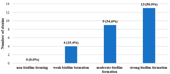
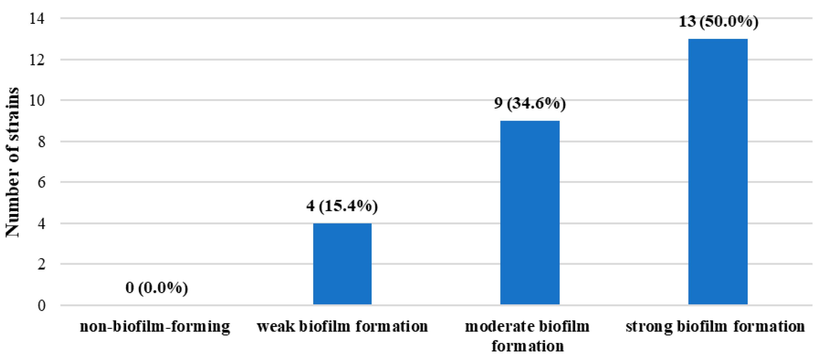
All tested isolates were classified as biofilm-forming under the conditions of the 24-h crystal violet assay. Among the analyzed isolates, 13 (50.0%) strains of L. monocytogenes were classified as strains with strong biofilm-forming ability, 9 (34.6%) with moderate biofilm-forming ability, and 4 (15.4%) with weak biofilm-forming ability. No non-adherent (non-biofilm-forming) strains were identified (Figure 1).
Figure 1.
Biofilm-forming ability after 24 h under the applied assay conditions of the tested L. monocytogenes strains.
3.3. Genotypic Characterization
3.3.1. Assessment of Serogroup Affiliation
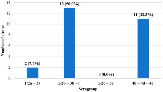
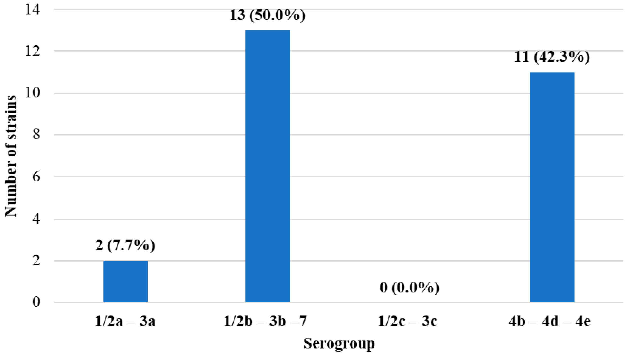
Among the analyzed isolates, 2 (7.7%) belonged to serogroup 1/2a–3a, 13 (50.0%) to serogroup 1/2b–3b–7, and 11 (42.3%) to serogroup 4b–4d–4e. No isolates belonging to serogroup 1/2c–3c were identified (Figure 2).
Figure 2.
Serogroup affiliation of the analyzed L. monocytogenes isolates.
3.3.2. Detection of Genes Encoding Virulence Factors
Four virulence gene profiles were identified among the 26 isolates. Profile A, detected in 12 isolates (46.2%), comprised all 10 analyzed genes (fbpA, plcA, hly, plcB, inlB, actA, iap, inlA, mpl, and prfA). Profile B, identified in 2 isolates (7.7%), comprised fbpA, plcA, hly, plcB, inlB, actA, iap, inlA, and prfA, with mpl not detected. Profile C, found in 1 isolate (3.8%), comprised fbpA, plcA, hly, plcB, inlB, actA, inlA, mpl, and prfA, with iap not detected. Profile D, observed in 11 isolates (42.3%), comprised plcA, hly, plcB, inlB, actA, iap, inlA, and prfA, with both fbpA and mpl not detected (Table 7). The hly gene was detected in all tested strains, which is consistent with the β-hemolytic phenotype observed on blood agar.
Table 7.
Virulence gene profiles of the analyzed L. monocytogenes strains.
3.3.3. Detection of Stress Survival Islets
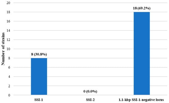
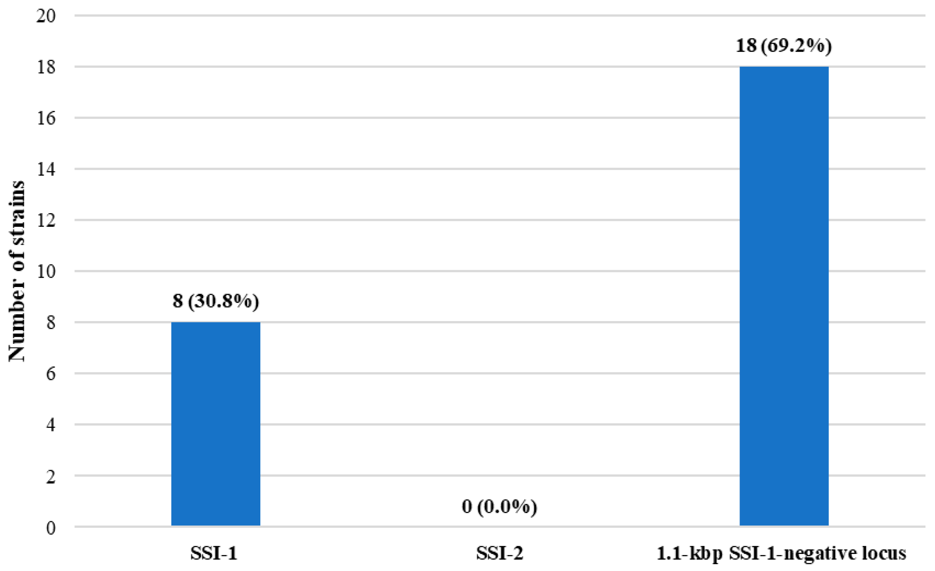
SSI-1 was detected in 8 isolates (30.8%), SSI-2 was not detected, and the 1.1 kbp SSI-1-negative locus configuration was detected in 18 isolates (69.2%) (Figure 3).
Figure 3.
Distribution of SSI-1, SSI-2, and the 1.1 kbp SSI-1-negative locus configuration among the analyzed L. monocytogenes isolates.
3.4. Relationships Between Phenotypic and Genotypic Characteristics of the Studied Strains
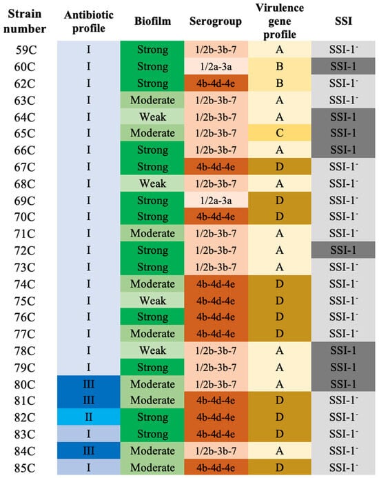
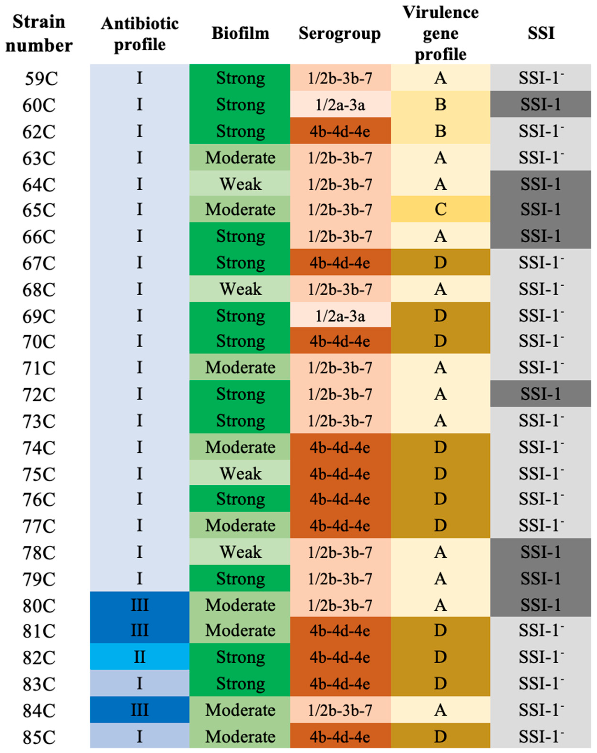
A summary of the phenotypic and genotypic characteristics of the studied strains is presented in Figure 4. Two isolates belonging to serogroup 1/2a–3a showed strong biofilm-forming ability. Among isolates belonging to serogroup 1/2b–3b–7, strong biofilm formation was observed in 5 strains, while among isolates belonging to serogroup 4b–4d–4e, it was observed in 6 strains. No statistically significant association was found between serogroup affiliation and biofilm-forming ability (padj = 1).
Figure 4.
Heatmap illustrating the distribution of phenotypic and targeted genotypic characteristics among the analyzed L. monocytogenes isolates. Antibiotic profile: I (susceptible to all tested antibiotics), II (resistant to trimethoprim–sulfamethoxazole but susceptible to penicillin, ampicillin, meropenem, and erythromycin), and III (resistant to erythromycin and trimethoprim–sulfamethoxazole but susceptible to penicillin, ampicillin, and meropenem). Virulence gene profiles: A (all 10 analyzed genes detected: fbpA, plcA, hly, plcB, inlB, actA, iap, inlA, mpl, and prfA), B (mpl gene not detected but fbpA, plcA, hly, plcB, inlB, actA, iap, inlA, and prfA detected), C (iap gene not detected but fbpA, plcA, hly, plcB, inlB, actA, inlA, mpl, and prfA detected), and D (fbpA and mpl genes not detected but plcA, hly, plcB, inlB, actA, iap, inlA, and prfA detected).
SSI-1 was detected in 7 isolates belonging to serogroup 1/2b–3b–7 and in 1 isolate belonging to serogroup 1/2a–3a, whereas it was not detected in any of the 11 isolates belonging to serogroup 4b–4d–4e. These results indicate potential variation in the prevalence of SSI-1 among the analyzed serogroups. A statistically significant association was demonstrated between serogroup affiliation and the presence of the SSI-1 stress survival islet (padj = 0.0196).
No statistically significant association was found between the presence of the fbpA gene and biofilm-forming ability (padj = 1). Similarly, the analysis provided no evidence of a statistically significant relationship between the presence of the mpl gene and biofilm-forming ability (padj = 1).
4. Discussion
Foodborne diseases remain a substantial public health burden in the United States, causing an estimated 48 million illnesses, 128,000 hospitalizations, and 3000 deaths annually; among the leading foodborne pathogens, L. monocytogenes ranks fifth as a cause of hospitalization and fourth as a cause of death, underscoring the disproportionate severity of listeriosis despite its relatively low incidence [23]. Therefore, the characterization of clinical isolates is particularly important, as it enables assessment of their virulence potential and adaptive capacity.
In the present study, all analyzed isolates were initially identified as L. monocytogenes using MALDI-TOF MS. This identification was subsequently confirmed by multiplex PCR targeting the rrs gene, which encodes 16S rRNA, and the hly gene, which encodes listeriolysin O (LLO), a major virulence factor of L. monocytogenes [13,14]. The molecular identification was based on the amplification of specific DNA fragments corresponding to the target sequences within both genes using appropriate primers. The detection of the expected PCR amplicons for both rrs and hly provided additional molecular confirmation of the MALDI-TOF MS identification, supporting the assignment of all tested isolates to the species L. monocytogenes.
The first characteristic examined was antimicrobial susceptibility, which is of particular clinical importance because the emergence of resistance may limit therapeutic options for invasive listeriosis and compromise the effectiveness of both first-line and alternative antibiotics. All tested strains were susceptible to ampicillin, penicillin, and meropenem, confirming preserved susceptibility to β-lactam antibiotics used in the treatment of listeriosis. Ampicillin and penicillin remain first-line therapeutic options, whereas meropenem may be used as an alternative agent. Similar findings were reported by Moura et al. [24], who analyzed 5339 clinical and food isolates in France and found no resistance to ampicillin or amoxicillin. Kawacka et al. [25] likewise reported susceptibility to all nine antibiotics tested, including penicillin and erythromycin, among isolates recovered from raw and processed meat and food-production environments. Susceptibility to antibiotics used for listeriosis treatment was also observed by Noll et al. [26] among 259 isolates from food production environments and clinical cases in Germany. Likewise, Jashari et al. [27] reported susceptibility of 114 food isolates to ampicillin and trimethoprim–sulfamethoxazole. In the present study, the clinical isolates were susceptible to penicillin. In contrast, Abdollahzadeh et al. [28] reported a high proportion (71.4%) of penicillin-resistant clinical isolates. Resistance to trimethoprim–sulfamethoxazole was detected in four isolates in the present study, three of which were also resistant to erythromycin. Similarly, Rugna et al. [29] identified two erythromycin-resistant L. monocytogenes isolates from pig-processing facilities and raw ham. This is consistent with the findings of Moura et al. [24], who reported that acquired antimicrobial resistance is relatively rare in L. monocytogenes. Such resistance may develop through the acquisition of mobile genetic elements carrying antimicrobial resistance genes or through mutations in chromosomal genes, potentially altering bacterial susceptibility to specific antimicrobial agents, particularly trimethoprim and macrolides such as erythromycin [24]. The detection of these resistant isolates supports the need for continued monitoring of antimicrobial resistance in L. monocytogenes. Systematic antimicrobial susceptibility testing of clinical isolates is therefore essential for the early detection of emerging resistance patterns and for supporting the selection of effective therapy in patients with listeriosis.
The ability to form biofilm is one of the key traits promoting the persistence of L. monocytogenes in the environment and increasing its tolerance to disinfectants. In the present study, most isolates were classified as strong or moderate biofilm producers, whereas weak biofilm formation was observed in 15% of isolates. The variability of this phenotype has also been reported by Doijad et al. [30], who, among 98 isolates, observed a predominance of weak and moderate biofilm producers and classified only nine strains as strong producers. Similarly, Di Ciccio et al. [31] showed that biofilm-forming ability differed among L. monocytogenes strains, with 58% of isolates producing weak biofilms, 38.5% moderate biofilms, and only 3.5% strong biofilms. In turn, Barbosa et al. [32] reported that, after incubation at 37 °C for 24 h, most food isolates were classified as weak or moderate biofilm producers, whereas all clinical isolates formed biofilms, although the majority were weak biofilm producers. In the present study, no significant association was found between biofilm formation intensity and serogroup affiliation. This finding is consistent with Doijad et al. [30], who described biofilm-forming ability as a strain-dependent trait, meaning that the capacity to produce biofilm varies among individual strains and is determined by their specific genetic and phenotypic characteristics rather than solely by their serogroup affiliation. In turn, Boscher et al. [33] demonstrated that strains belonging to serogroup IVb exhibited significantly lower biofilm formation compared to other serogroups. Although the association between SSI-1 carriage and biofilm-forming ability was not assessed in the present study, Keeney et al. [34] and Burdová et al. [35] reported enhanced biofilm formation in certain SSI-1-positive strains. Assessing biofilm-forming ability among clinical isolates is therefore important for identifying strain-specific persistence-related phenotypes that may facilitate survival in healthcare environments and contribute to recurrent contamination or transmission.
The studied strains were further subjected to genotypic analysis, including determination of their serogroup affiliation. Serogroups 1/2b–3b–7 and 4b–4d–4e predominated, whereas serogroup 1/2a–3a was detected least frequently. These findings are partly consistent with previous studies showing that serotypes 1/2a, 1/2b, and 4b are most commonly associated with human listeriosis [36]. Kuch et al. [37] also demonstrated that serogroup 4b-4d-4e was the most prevalent among L. monocytogenes isolates from invasive infections in Poland (55.8%), followed by serogroups 1/2a-3a (34.3%), 1/2b-3b (8.1%), and 1/2c-3c (1.8%). However, Wiktorczyk-Kapischke et al. [38] reported that serogroup 1/2a-3a predominated among clinical isolates, whereas serogroup 4b-4d-4e was most frequently identified among isolates obtained from frozen food. Pyz-Łusiak et al. [39] reported varying results among L. monocytogenes strains isolated from artisanal cheeses in Poland, confirming the presence of serogroups 1/2a-3a and 4b-4d-4e in 46.9% and 31.2% of the tested strains, respectively. In contrast, Kalekar et al. [40] reported a predominance of serogroup 4b–4d–4e (88.24%) among 17 clinical isolates from India, with 11.76% belonging to serogroup 1/2b–3b–7. Overall, these data highlight variability in serogroup distribution among clinical isolates and emphasize the particular importance of serogroup 4b-4d-4e, which is frequently associated with listeriosis outbreaks.
This study assessed the prevalence of 10 genes encoding virulence factors using multiplex PCR. The analysis of virulence gene occurrence revealed that most of the investigated determinants were present in all tested strains. The genes actA, hly, inlA, inlB, plcA, plcB, and prfA were detected in 100% of the analyzed isolates. This finding is consistent with previous reports indicating that these genes are essential for adhesion to host cells, invasion, intracellular survival, and cell-to-cell spread [1,4]. Particular importance is attributed to prfA, which acts as the major regulator of the expression of numerous L. monocytogenes virulence factors [4]. In contrast, fbpA was detected in 15/26 isolates (57.7%), whereas mpl was detected in 13/26 isolates (50.0%). Differences in the prevalence of these determinants have also been reported in other studies [41,42] and may reflect genetic diversity within the L. monocytogenes population. Similarly, Wiktorczyk-Kapischke et al. [38] demonstrated the absence of mpl in five strains and fbpA in eight strains. The presence of the fbpA gene in all strains studied, isolated from fish and the fish-processing environment in Poland, was confirmed by Zakrzewski et al. [43]. Das et al. [44] and Montero et al. [45] also confirmed the presence of the mpl gene in all of the strains studied. Investigating virulence genes in clinical L. monocytogenes isolates is essential for assessing their pathogenic potential and understanding the genetic factors contributing to infection. Combining virulence gene profiling with serogroup determination may provide valuable insights into the genetic diversity and epidemiological significance of clinical isolates.
Stress survival islets: SSI-1 and SSI-2 are genomic elements associated with the adaptation of L. monocytogenes to unfavorable environmental conditions. In the present study, SSI-1 was detected in 8 of 26 isolates (30.8%), whereas SSI-2 was not detected; the remaining 18 isolates (69.2%) carried the 1.1 kbp SSI-1-negative locus configuration. These findings are consistent with the PCR survey conducted by Hein et al. [21], who detected SSI-1 in 32.5% of 117 L. monocytogenes isolates and the 1.1 kbp SSI-1-negative fragment in 61.5% of isolates, demonstrating substantial variation at this locus among strains. Chen et al. [46] detected the presence of SSI-1 in 34.7% of line I isolates, 80.4% of line II isolates, and 2 line III isolates; the presence of SSI-2 was detected only in isolate CC121. Determining the presence of the SSI in clinical L. monocytogenes isolates is important for understanding their ability to adapt to stressful environmental conditions and potentially persist in the host or food-processing environments.
This study has several limitations. First, all clinical isolates were obtained from a single clinical center, which may limit the generalizability of the findings to other settings. Second, the molecular analysis was restricted to PCR-based detection of 10 selected virulence-associated genes and predefined genetic markers, which limited the assessment of the broader virulence gene repertoire. Antimicrobial resistance was assessed exclusively phenotypically, without analysis of the underlying genetic determinants of resistance. Furthermore, the lack of whole-genome sequencing and MLST/cgMLST analysis precluded comprehensive characterization of the genetic diversity, sequence types, and clonal relationships among the isolates. Nevertheless, this study provides valuable insights into selected phenotypic and genotypic characteristics of clinical L. monocytogenes isolates.
Overall, the analyzed clinical isolates combined preserved susceptibility to key β-lactam antibiotics with phenotypic traits associated with persistence, including biofilm formation, and a broad repertoire of virulence-associated genes. This combination indicates that the studied strains retained features important both for clinical infection and for survival under unfavorable conditions.
5. Conclusions
The present study demonstrated phenotypic and genotypic diversity among clinical L. monocytogenes isolates. Most strains remained susceptible to antibiotics commonly used in the treatment of listeriosis, although resistance to trimethoprim–sulfamethoxazole and erythromycin in a subset of isolates indicates the need for continued antimicrobial susceptibility monitoring. All isolates were able to form biofilm, with half classified as strong biofilm producers in the applied assay, suggesting a considerable potential for persistence and survival. The predominance of PCR serogroups 1/2b–3b–7 and 4b–4d–4e, together with frequent detection of virulence-associated genetic markers, demonstrates genotypic diversity within the analyzed clinical isolate collection. Variation in virulence gene profiles, the serogroup-dependent distribution of SSI-1, and the absence of SSI-2 further underscore the heterogeneity of the local clinical L. monocytogenes isolate collection. Collectively, these findings demonstrate the value of integrating phenotypic analyses with genotypic characterization to provide a more comprehensive description of local clinical L. monocytogenes populations.
Author Contributions
Conceptualization, N.W.-K. and K.S.; methodology, K.G.-B., N.W.-K. and A.B.; validation, A.B. and J.C.; formal analysis, N.W.-K., K.W. and A.B.; investigation, A.M.; resources, N.W.-K., and K.G.-B.; data curation, A.M. and J.C.; writing—original draft preparation, N.W.-K., A.B. and J.C.; writing—review and editing, K.G.-B., N.W.-K., K.W. and K.S.; visualization, N.W.-K. and A.B.; supervision, N.W.-K.; project administration, K.S. and N.W.-K.; funding acquisition, K.S. and N.W.-K. All authors have read and agreed to the published version of the manuscript.
Funding
This research received no external funding.
Institutional Review Board Statement
The study used archived Listeria monocytogenes isolates obtained during clinical diagnostic procedures. Ethics committee approval: KB 461/2025 (Ludwik Rydygier Collegium Medicum in Bydgoszcz, Nicolaus Copernicus University in Toruń, Poland).
Informed Consent Statement
Not applicable.
Data Availability Statement
The data presented in this study are available upon request from the corresponding authors.
Conflicts of Interest
The authors declare no conflicts of interest.
References
- Gandhi, M.; Chikindas, L.M. Listeria: A foodborne pathogen that knows how to survive. Int. J. Food Microbiol. 2007, 113, 1–15. [Google Scholar] [CrossRef] [Scilit] [PubMed]
- Matereke, L.T.; Okoh, A.I. Listeria monocytogenes Virulence, Antimicrobial Resistance and Environmental Persistence: A Review. Pathogens 2020, 9, 528. [Google Scholar] [CrossRef] [Scilit] [PubMed]
- Ravindhiran, R.; Sivarajan, K.; Sekar, J.N.; Murugesan, R.; Dhandapani, K. Listeria monocytogenes an Emerging Pathogen: A Comprehensive Overview on Listeriosis, Virulence Determinants, Detection, and Anti-Listerial Interventions. Microb. Ecol. 2023, 86, 2231–2251. [Google Scholar] [CrossRef] [Scilit] [PubMed]
- Camejo, A.; Carvalho, F.; Reis, O.; Leitão, E.; Sousa, S.; Cabanes, D. The arsenal of virulence factors deployed by Listeria monocytogenes to promote its cell infection cycle. Virulence 2011, 2, 379–394. [Google Scholar] [CrossRef] [Scilit] [PubMed]
- World Health Organization (WHO). Listeriosis. Available online: https://www.who.int/news-room/fact-sheets/detail/listeriosis (accessed on 4 July 2026).
- Koopmans, M.M.; Brouwer, M.C.; Vázquez-Boland, J.A.; Van De Beek, D. Human Listeriosis. Clin. Microbiol. Rev. 2023, 36, e00060-19. [Google Scholar] [CrossRef] [Scilit] [PubMed]
- Osek, J.; Wieczorek, K. Listeria monocytogenes—How This Pathogen Uses Its Virulence Mechanisms to Infect the Hosts. Pathogens 2022, 11, 1491. [Google Scholar] [CrossRef] [Scilit] [PubMed]
- Choi, M.H.; Park, Y.J.; Kim, M.; Seo, Y.H.; Kim, Y.A.; Choi, J.Y.; Yong, D.; Jeong, S.K.; Lee, K. Increasing Incidence of Listeriosis and Infection-associated Clinical Outcomes. Ann. Lab. Med. 2018, 38, 102–109. [Google Scholar] [CrossRef] [Scilit] [PubMed]
- European Food Safety Authority (EFSA); European Centre for Disease Prevention and Control (ECDC). The European Union One Health 2024 Zoonoses report. EFSA J. 2025, 23, e9759. [Google Scholar] [CrossRef] [Scilit] [PubMed]
- Romeo, M.; Lasagabaster, A.; Lavilla, M.; Amárita, F. Genetic Diversity, Biofilm Formation, and Antibiotic Resistance in Listeria monocytogenes Isolated from Meat-Processing Plants. Foods 2025, 14, 1580. [Google Scholar] [CrossRef] [Scilit] [PubMed]
- Luque-Sastre, L.; Arroyo, C.; Fox, E.M.; McMahon, B.J.; Bai, L.; Li, F.; Fanning, S. Antimicrobial Resistance in Listeria Species. Microbiol. Spectr. 2018, 6. [Google Scholar] [CrossRef] [Scilit] [PubMed]
- Żurawik, A.; Kasperski, T.; Olechowska-Jarząb, A.; Szczesiul-Paszkiewicz, P.; Żak, I.; Wójcicki, M.; Maćkiw, E.; Chmielarczyk, A. Genetic Diversity, Virulence Factors and Antibiotic Resistance of Listeria monocytogenes from Food and Clinical Samples in Southern Poland. Pathogens 2024, 13, 725. [Google Scholar] [CrossRef] [Scilit] [PubMed]
- Border, P.M.; Howard, J.J.; Plastow, G.S.; Siggens, K.W. Detection of Listeria species and Listeria monocytogenes using polymerase chain reaction. Lett. Appl. Microbiol. 1990, 11, 158–162. [Google Scholar] [CrossRef] [Scilit] [PubMed]
- Bansal, N.S.; McDonell, F.H.Y.; Smith, A.; Arnold, G.; Ibrahim, G.F. Multiplex PCR assay for the routine detection of Listeria in food. Int. J. Food Microbiol. 1996, 33, 292–300. [Google Scholar] [CrossRef] [Scilit] [PubMed]
- Skowron, K.; Wiktorczyk, N.; Grudlewska, K.; Wałecka-Zacharska, E.; Paluszak, Z.; Kruszewski, S.; Gospodarek-Komkowska, E. Phenotypic and genotypic evaluation of Listeria monocytogenes strains isolated from fish and fish processing plants. Ann. Microbiol. 2019, 69, 469–482. [Google Scholar] [CrossRef] [Scilit]
- The European Committee on Antimicrobial Susceptibility Testing. Breakpoint Tables for Interpretation of MICs and Zone Diameters. Available online: https://www.eucast.org (accessed on 18 February 2026).
- Wiktorczyk-Kapischke, N.; Wałecka-Zacharska, E.; Korkus, J.; Grudlewska-Buda, K.; Budzyńska, A.; Wnuk, K.; Gospodarek-Komkowska, E.; Skowron, K. The influence of stress factors on selected phenotypic and genotypic features of Listeria monocytogenes—A pilot study. BMC Microbiol. 2023, 23, 259. [Google Scholar] [CrossRef] [Scilit] [PubMed]
- Stepanović, S.; Cirković, I.; Ranin, L.; Svabić-Vlahović, M. Biofilm formation by Salmonella spp. and Listeria monocytogenes on plastic surface. Lett. Appl. Microbiol. 2004, 38, 428–432. [Google Scholar] [CrossRef] [Scilit] [PubMed]
- Doumith, M.; Buchrieser, C.; Glaser, P.; Jacquet, C.; Martin, P. Differentiation of the Major Listeria monocytogenes Serovars by Multiplex PCR. J. Clin. Microbiol. 2004, 42, 3819–3822. [Google Scholar] [CrossRef] [Scilit] [PubMed]
- Wałecka-Zacharska, E.; Kosek-Paszkowska, K.; Bania, J.; Karpíšková, R.; Stefaniak, T. Salt stress-induced invasiveness of major Listeria monocytogenes serotypes. Lett. Appl. Microbiol. 2013, 56, 216–221. [Google Scholar] [CrossRef] [Scilit] [PubMed]
- Hein, I.; Klinger, S.; Dooms, M.; Flekna, G.; Stessl, B.; Leclercq, A.; Hill, C.; Allerberger, F.; Wagner, M. Stress survival islet 1 (SSI-1) survey in Listeria monocytogenes reveals an insert common to Listeria innocua in sequence type 121 L. monocytogenes strains. Appl. Environ. Microbiol. 2011, 77, 2169–2173. [Google Scholar] [CrossRef] [Scilit] [PubMed]
- Harter, E.; Wagner, E.M.; Zaiser, A.; Halecker, S.; Wagner, M.; Rychli, K. Stress Survival Islet 2, Predominantly Present in Listeria monocytogenes Strains of Sequence Type 121, Is Involved in the Alkaline and Oxidative Stress Responses. Appl. Environ. Microbiol. 2017, 83, e00827-17. [Google Scholar] [CrossRef] [Scilit] [PubMed]
- Centers for Disease Control and Prevention (CDC). Facts About Food Poisoning. CDC Food Safety facts and statistics. Available online: https://www.cdc.gov/food-safety/data-research/facts-stats/index.html (accessed on 15 October 2026).
- Moura, A.; Leclercq, A.; Vales, G.; Tessaud-Rita, N.; Bracq-Dieye, H.; Thouvenot, P.; Madec, Y.; Charlier, C.; Lecuit, M. Phenotypic and genotypic antimicrobial resistance of Listeria monocytogenes: An observational study in France. Lancet Reg. Health Eur. 2024, 37, 100800. [Google Scholar] [CrossRef] [Scilit] [PubMed]
- Kawacka, I.; Olejnik-Schmidt, A.; Schmidt, M. Nonhemolytic Listeria monocytogenes—Prevalence rate, reasons underlying atypical phenotype, and methods for accurate hemolysis assessment. Microorganisms 2022, 10, 483. [Google Scholar] [CrossRef] [Scilit] [PubMed]
- Noll, M.; Kleta, S.; Al Dahouk, S. Antibiotic susceptibility of 259 Listeria monocytogenes strains isolated from food, food-processing plants and human samples in Germany. J. Infect. Public Health 2018, 11, 572–577. [Google Scholar] [CrossRef] [Scilit] [PubMed]
- Jashari, B.; Stessl, B.; Félix, B.; Cana, A.; Bisha, B.; Jankuloski, D.; Blagoevska, K.; Kayode, A.J. Multilocus sequence typing and antimicrobial susceptibility of Listeria monocytogenes isolated from foods surveyed in Kosovo. Microorganisms 2024, 12, 2441. [Google Scholar] [CrossRef] [Scilit] [PubMed]
- Abdollahzadeh, E.; Ojagh, S.M.; Hosseini, H.; Ghaemi, E.A.; Irajian, G.; Naghizadeh Heidarlo, M. Antimicrobial resistance of Listeria monocytogenes isolated from seafood and humans in Iran. Microb. Pathog. 2016, 100, 70–74. [Google Scholar] [CrossRef] [Scilit] [PubMed]
- Rugna, G.; Carra, E.; Bergamini, F.; Franzini, G.; Faccini, S.; Gattuso, A.; Morganti, M.; Baldi, D.; Naldi, S.; Serraino, A.; et al. Distribution, virulence, genotypic characteristics and antibiotic resistance of Listeria monocytogenes isolated over one-year monitoring from two pig slaughterhouses and processing plants and their fresh hams. Int. J. Food Microbiol. 2021, 336, 108912. [Google Scholar] [CrossRef] [Scilit] [PubMed]
- Doijad, S.P.; Barbuddhe, S.B.; Garg, S.; Poharkar, K.V.; Kalorey, D.R.; Kurkure, N.V.; Rawool, D.B.; Chakraborty, T. Biofilm-forming abilities of Listeria monocytogenes serotypes isolated from different sources. PLoS ONE 2015, 10, e0137046. [Google Scholar] [CrossRef] [Scilit] [PubMed]
- Di Ciccio, P.; Rubiola, S.; Panebianco, F.; Lomonaco, S.; Allard, M.; Bianchi, D.M.; Civera, T.; Chiesa, F. Biofilm formation and genomic features of Listeria monocytogenes strains isolated from meat and dairy industries located in Piedmont (Italy). Int. J. Food Microbiol. 2022, 378, 109784. [Google Scholar] [CrossRef] [Scilit] [PubMed]
- Barbosa, J.; Borges, S.; Camilo, R.; Magalhães, R.; Ferreira, V.; Santos, I.; Silva, J.; Almeida, G.; Teixeira, P. Biofilm Formation among Clinical and Food Isolates of Listeria monocytogenes. Int. J. Microbiol. 2013, 2013, 524975. [Google Scholar] [CrossRef] [Scilit] [PubMed]
- Boscher, E.; Félix, B.; Rincé, A.; Legrandois, P.; Sajan, L.; Soumet, C.; Denis, M. Genomic and phenotypic characterisation of Listeria monocytogenes strains isolated from pig feces on farm and from pork meat at retail in France. PLoS ONE 2026, 21, e0341663. [Google Scholar] [CrossRef] [Scilit] [PubMed]
- Keeney, K.; Trmcic, A.; Zhu, Z.; Delaquis, P.; Wang, S. Stress survival islet 1 contributes to serotype-specific differences in biofilm formation in Listeria monocytogenes. Lett. Appl. Microbiol. 2018, 67, 530–536. [Google Scholar] [CrossRef] [Scilit] [PubMed]
- Burdová, A.; Véghová, A.; Minarovičová, J.; Drahovská, H.; Kaclíková, E. The relationship between biofilm phenotypes and biofilm-associated genes in food-related Listeria monocytogenes strains. Microorganisms 2024, 12, 1297. [Google Scholar] [CrossRef] [Scilit] [PubMed]
- Brown, P.; Kucerova, Z.; Gorski, L.; Chen, Y.; Ivanova, M.; Leekitcharoenphon, P.; Parsons, C.; Niedermeyer, J.; Jackson, J.; Kathariou, S. Horizontal Gene Transfer and Loss of Serotype-Specific Genes in Listeria monocytogenes Can Lead to Incorrect Serotype Designations with a Commonly-Employed Molecular Serotyping Scheme. Microbiol. Spectr. 2023, 11, e0274522. [Google Scholar] [CrossRef] [Scilit] [PubMed]
- Kuch, A.; Goc, A.; Belkiewicz, K.; Filipello, V.; Ronkiewicz, P.; Gołębiewska, A.; Wróbel, I.; Kiedrowska, M.; Waśko, I.; Hryniewicz, W.; et al. Molecular diversity and antimicrobial susceptibility of Listeria monocytogenes isolates from invasive infections in Poland (1997–2013). Sci. Rep. 2018, 8, 14562. [Google Scholar] [CrossRef] [Scilit] [PubMed]
- Wiktorczyk-Kapischke, N.; Skowron, K.; Wałecka-Zacharska, E.; Grudlewska-Buda, K.; Wnuk, K.; Buszko, K.; Gospodarek-Komkowska, E. Assessment of the influence of selected stress factors on the growth and survival of Listeria monocytogenes. BMC Microbiol. 2023, 23, 27. [Google Scholar] [CrossRef] [Scilit] [PubMed]
- Pyz-Łukasik, R.; Paszkiewicz, W.; Kiełbus, M.; Ziomek, M.; Gondek, M.; Domaradzki, P.; Michalak, K.; Pietras-Ożga, D. Genetic diversity and potential virulence of Listeria monocytogenes isolates originating from polish artisanal cheeses. Foods 2022, 11, 2805. [Google Scholar] [CrossRef] [Scilit] [PubMed]
- Kalekar, S.; Rodrigues, J.; D’Costa, D.; Doijad, S.; Ashok Kumar, J.; Malik, S.V.S.; Kalorey, D.R.; Rawool, D.B.; Hain, T.; Chakraborty, T.; et al. Genotypic characterization of Listeria monocytogenes isolated from humans in India. Ann. Trop. Med. Parasitol. 2011, 105, 351–358. [Google Scholar] [CrossRef] [Scilit] [PubMed]
- Iwu, C.D.; Okoh, A.I. Characterization of antibiogram fingerprints in Listeria monocytogenes recovered from irrigation water and agricultural soil samples. PLoS ONE 2020, 15, e0228956. [Google Scholar] [CrossRef] [Scilit] [PubMed]
- Olaniran, A.O.; Nzimande, S.B.; Mkize, N.G. Antimicrobial resistance and virulence signatures of Listeria and Aeromonas species recovered from treated wastewater effluent and receiving surface water in Durban, South Africa. BMC Microbiol. 2015, 15, 234. [Google Scholar] [CrossRef] [Scilit] [PubMed]
- Zakrzewski, A.J.; Kurpas, M.; Zadernowska, A.; Chajęcka-Wierzchowska, W.; Fraqueza, M.J. A Comprehensive virulence and resistance characteristics of Listeria monocytogenes isolated from fish and the fish industry environment. Int. J. Mol. Sci. 2023, 24, 3581. [Google Scholar] [CrossRef] [Scilit] [PubMed]
- Das, S.; Lalitha, K.V.; Thampuran, N.; Surendran, P.L. Isolation and characterization of Listeria monocytogenes from tropical seafood of Kerala, India. Ann. Microbiol. 2013, 63, 1093–1098. [Google Scholar] [CrossRef] [Scilit]
- Montero, D.; Bodero, M.; Riveros, G.; Lapierre, L.; Gaggero, A.; Vidal, R.M.; Vidal, M. Molecular epidemiology and genetic diversity of Listeria monocytogenes isolates from a wide variety of ready-to-eat foods and their relationship to clinical strains from listeriosis outbreaks in Chile. Front. Microbiol. 2015, 6, 384. [Google Scholar] [CrossRef] [Scilit] [PubMed]
- Chen, Y.; Chen, Y.; Pouillot, R.; Dennis, S.; Xian, Z.; Luchansky, J.B.; Porto-Fett, A.C.S.; Lindsay, J.A.; Hammack, T.S.; Allard, M.; et al. Genetic diversity and profiles of genes associated with virulence and stress resistance among isolates from the 2010–2013 interagency Listeria monocytogenes market basket survey. PLoS ONE 2020, 15, e0231393. [Google Scholar] [CrossRef] [Scilit] [PubMed]

Disclaimer/Publisher’s Note: The statements, opinions and data contained in all publications are solely those of the individual author(s) and contributor(s) and not of MDPI and/or the editor(s). MDPI and/or the editor(s) disclaim responsibility for any injury to people or property resulting from any ideas, methods, instructions or products referred to in the content. |
© 2026 by the authors. Licensee MDPI, Basel, Switzerland. This article is an open access article distributed under the terms and conditions of the Creative Commons Attribution (CC BY) license.