


Simple Summary
Mussels stick firmly to rocks and ship hulls underwater, and the proteins responsible for this adhesion are valuable for medical glues and wound dressings. However, these proteins are hard to obtain in large amounts, and they need a special chemical modification—the conversion of tyrosine into 3,4-dihydroxyphenylalanine (DOPA)—to work properly. In this study, we produced a recombinant mussel adhesive protein in the yeast Komagataella phaffii and used a second enzyme, tyrosinase, to carry out this modification during the fermentation process. The modified protein carried substantially more catechol groups than the unmodified version and promoted the closure of scratched cell layers more effectively. These results show that it is possible to produce a chemically modified mussel adhesive protein in yeast, which may help make these proteins more available for medical and industrial uses in the future. More work is still needed to confirm how strongly the protein sticks to surfaces.
Abstract
Mussel adhesive proteins (MAPs) are promising bio-adhesives, but recombinant production faces challenges in yield and post-translational DOPA modification. Here, a fusion protein Pvfp-5P, comprising Perna viridis foot protein 5β and a preCol-P fragment, was expressed in Komagataella phaffii GS115 with and without co-expression of Bacillus megaterium tyrosinase, generating a catechol-modified form (Pvfp-5P(D)) and an unmodified tyrosine form (Pvfp-5P(Y)), respectively. Pvfp-5P(D) was secreted in shake flasks (yield ~353 mg/L determined by A280 quantification) and purified to >85% purity by one-step Ni-NTA chromatography. NBT staining and colorimetric quantification indicated a catechol content of 3.64 mol% (expressed as DOPA equivalents) in Pvfp-5P(D), compared with 0.32 mol% in Pvfp-5P(Y), consistent with tyrosinase-dependent catechol formation. In a scratch assay, Pvfp-5P(D) enhanced closure of L929 fibroblasts more effectively than Pvfp-5P(Y), and it also showed stronger adsorption to glass and polystyrene surfaces. This study provides evidence that co-expression of a bacterial tyrosinase in K. phaffii enables catechol incorporation into a recombinant mussel adhesive protein and that this modification enhances its in vitro functional properties. Definitive conclusions regarding the contribution of the preCol-P fusion and the adhesive strength of the protein require further controls and direct measurements.
1. Introduction
Mussels are marine bivalves that attach firmly to various submerged surfaces, including rocks, ship hulls, and piers, withstanding constant wave action and tidal currents. This remarkable underwater adhesion is mediated by the byssus, a bundle of proteinaceous threads secreted by the mussel foot [1]. At the terminal end of each thread is a flat adhesive plaque that forms strong, durable bonds with the substrate. The proteins responsible for this adhesion are collectively known as mussel adhesive proteins (MAPs) or mussel foot proteins (MFPs). To date, at least eight distinct MAPs have been identified and characterized, including Mfp-1, Mfp-2, Mfp-3F, Mfp-3S, Mfp-4, Mfp-5, Mfp-6, and Mfp-7. In addition, five byssal thread matrix proteins (preCOL-D, preCOL-P, preCOL-NG, PTMP-1, and TMP-1) have been reported [2,3].
The unique adhesive properties of MAPs are largely attributed to a post-translationally modified amino acid, 3,4-dihydroxyphenylalanine (DOPA), which is derived from tyrosine. DOPA contains a catechol side chain that can form strong bidentate hydrogen bonds, coordinate with metal ions, and undergo oxidation to reactive quinones that cross-link with surfaces or other proteins [4,5]. Among all MAPs, Mfp-5 (known as Pvfp-5 in Perna viridis) is, together with Mfp-3, one of the smallest MAPs and possesses the highest DOPA content (up to 25.5 mol% in the Mytilus genus), making it a key component for interfacial adhesion [6]. Natural MAPs also exhibit excellent biocompatibility, low immunogenicity, and biodegradability, which have led to their application in wound dressings, surgical adhesives, cell culture coatings, and drug delivery systems [7,8,9].
Despite their enormous potential, the practical use of natural MAPs is severely limited by the inefficiency of extraction from natural sources. Consequently, recombinant DNA technology has become a major focus for MAP production. Among various expression systems, the methylotrophic yeast Pichia pastoris (syn. Komagataella phaffii) is particularly attractive because it combines high-density fermentation, a strong methanol-inducible AOX1 promoter, efficient secretion, and the ability to perform eukaryotic post-translational modifications such as glycosylation and disulfide bond formation [10,11,12].
However, recombinant production of functional MAPs faces two major challenges. First, the critical DOPA modification is not efficiently performed by the yeast’s endogenous enzymes; therefore, co-expression of a heterologous tyrosinase is required [13]. Second, the expression levels of some MAPs in heterologous hosts remain modest, and the factors governing efficient secretion are not fully understood [14]. To address the DOPA modification challenge, several studies have explored the co-expression of tyrosinase with MAPs in bacterial hosts (e.g., Escherichia coli). For example, Choi et al. pioneered the in vivo modification of tyrosine residues in recombinant MAPs by co-expressing tyrosinase, demonstrating that this strategy could significantly improve the adhesion properties of the recombinant protein [15]. Despite these advances, bacterial systems present inherent limitations: they lack the eukaryotic post-translational machinery (such as proper disulfide bond formation and glycosylation), and highly repetitive MAPs often form insoluble inclusion bodies that require tedious denaturation and refolding processes. Therefore, whether this co-expression strategy can be effectively transferred to a eukaryotic secretion host, such as K. phaffii, to achieve functional, soluble, and modified MAP production has not yet been established.
Interestingly, the byssal thread matrix protein preCol-P contains a collagen-like domain with typical Gly-X-Y repeats [16]. It should be noted that without co-expression of prolyl-4-hydroxylase (P4H), the Gly-X-Y repeats cannot be hydroxylated, and the preCol-P fragment is unlikely to form a stable triple helix at 37 °C, likely existing as an unstructured, flexible extension [17]. Despite this, collagen-like sequences are often produced efficiently in K. phaffii, with recombinant human collagen reaching yields exceeding 4 g/L in fed-batch fermentations [18,19]. However, whether fusing such a sequence to a target protein can predictably enhance its expression has not been demonstrated. From a biomaterials perspective, a fusion between two native byssus components—Pvfp-5 and preCol-P—is conceptually attractive, even in the absence of triple-helix folding, as both are intrinsic protein building blocks of the mussel byssus and may confer complementary functional properties.
In this study, we constructed a fusion protein named Pvfp-5P, consisting of the full-length Pvfp-5β from P. viridis and a partial peptide of preCol-P. The synthetic gene was codon-optimized for K. phaffii and placed under the control of the AOX1 promoter in the pPICZαA vector. A second plasmid carrying the B. megaterium tyrosinase gene under the same promoter was introduced to generate a co-expression strain. Two protein preparations were compared: the catechol-modified form produced by the co-expression strain (Pvfp-5P(D)) and the unmodified tyrosine form produced by the strain lacking tyrosinase (Pvfp-5P(Y)). The recombinant proteins were produced in shake flasks, purified by one-step Ni-NTA affinity chromatography, and characterized for catechol content, scratch-closure activity, and surface adsorption. Our results provide evidence that co-expression of a bacterial tyrosinase in K. phaffii enables catechol incorporation into a recombinant mussel adhesive protein and that this modification enhances its in vitro functional properties. The limitations of the current dataset, including the absence of a Pvfp-5β-only control and of direct adhesion measurements, are explicitly acknowledged, and the conclusions are framed accordingly.
2. Materials and Methods
2.1. Strains, Vectors, and Chemicals
Komagataella phaffii strain GS115 (his4) and the expression vectors pPICZαA and pPIC9K were purchased from Invitrogen (Carlsbad, CA, USA). Restriction enzymes (XhoI, NotI, SacI, SalI), T4 DNA ligase, and PCR reagents were obtained from Thermo Fisher Scientific (Waltham, MA, USA). L-DOPA standard (≥99% purity), commercial mussel adhesive protein (MAP, ≥90% purity; Cat. No. HY-NP161, Batch No. 857745; recombinantly produced in K. phaffii, molecular weight ~100 kDa, DL-DOPA content 5.6 wt%), nitroblue tetrazolium (NBT), bovine serum albumin (BSA), and Coomassie Brilliant Blue R-250 were purchased from MedChemExpress (Monmouth Junction, NJ, USA). The integrity of the commercial MAP as received was verified by 12% SDS-PAGE (Figure S1). Ni-NTA agarose (6FF) was purchased from GE Healthcare (Chicago, IL, USA). All other reagents were of analytical grade and purchased from domestic suppliers.
2.2. Construction of Expression Vectors
The amino acid sequence of the fusion protein Pvfp-5P (Figure S2a) was designed by linking the full-length Pvfp-5β from P. viridis with a 171-amino-acid fragment from preCol-P. A (GGGGS)3 flexible linker was inserted between the two domains. The nucleotide sequence encoding Pvfp-5P (Figure S2b) was codon-optimized for K. phaffii using the web-based GeneArt GeneOptimizer tool (Thermo Fisher Scientific, Waltham, MA, USA; accessed on 15 May 2025) and synthesized by Sangon Biotech (Shanghai, China). The codon-optimized sequence has been deposited in GenBank under accession number QB197299. The synthesized fragment was digested with XhoI and NotI and ligated into the pPICZαA vector to generate pPICZαA-Pvfp5P. The construct was verified by DNA sequencing.
To convert tyrosine to DOPA in recombinant proteins, the gene encoding B. megaterium tyrosinase was codon-optimized and synthesized (Figure S3a,b). Its sequence is available in GenBank under accession number QB197300. This well-characterized bacterial tyrosinase exhibits high catalytic efficiency and robust activity, making it highly suitable for heterologous expression and L-DOPA production in microbial hosts [20]. To enable extracellular secretion, the synthetic sequence was fused with an N-terminal α-factor secretion signal and inserted into pPIC9K via XhoI and NotI to create pPIC9K-TYR, which was confirmed by sequencing.
2.3. Construction of the Pvfp-5P Single-Expression Strain
The pPICZαA-Pvfp5P plasmid (10 μg) was linearized with SacI and purified by phenol-chloroform extraction. The linearized DNA (2 μg) was electroporated into GS115 competent cells (1.5 kV, 25 μF, 200 Ω). After recovery in 1 M sorbitol for 1 h, cells were plated on YPD agar containing 100 μg/mL Zeocin and incubated at 30 °C for 2–3 days. Colony PCR with primers 9654-F (5′-TATCATGGTGCTCCAGGTAGAC-3′) and 3′AOX (5′-GCAAATGGCATTCTGACATCC-3′) confirmed positive integrants by a ~500 bp amplicon.
Ten PCR-positive clones were cultured in BMGY at 28.5 °C with shaking to OD600 2–6 and then induced in BMMY with 1% methanol for 70 h. Because this strain does not express tyrosinase, no CuSO4 was added during this stage. Culture supernatants were analyzed by SDS-PAGE and Western blotting using an anti-His antibody. The clone showing the highest expression and fewest contaminant bands was designated GS115/Pvfp5P and used to produce the unmodified Tyr-form (Pvfp-5P(Y)) in subsequent experiments.
2.4. Construction of the Co-Expression Strain
pPIC9K-TYR (10 μg) was linearized with SalI, purified, and electroporated into GS115/Pvfp5P. Transformants were selected on MD plates (1.34% YNB, 4 × 10⁻⁵% biotin, 2% dextrose, 1.5% agar) containing 100 μg/mL Zeocin. Colony PCR was performed using two primer pairs: (i) TYR-F (5′-GACGAGCAAGGTAACCCAT-3′)/3′AOX to amplify the tyrosinase gene, and (ii) Pvfp-5P-F (5′-TATCATGGTGCTCCAGGTAGAC-3′)/3′AOX to amplify the Pvfp-5P gene. Twenty clones were screened, and those positive for both genes were subjected to small-scale co-expression in BMMY containing 100 μM CuSO4 for 70 h, using the same culture conditions (temperature, shaking speed, and methanol supplementation) as described in Section 2.3. The best co-expressing clone was named GS115/Pvfp5P/TYR and used to produce the catechol-modified form (Pvfp-5P(D)).
2.5. Shake-Flask Production and Ni-NTA Purification
For Pvfp-5P(D), a single colony of GS115/Pvfp5P/TYR was inoculated into 10 mL BMGY and grown overnight at 28.5 °C with shaking. The overnight culture was diluted 1:1000 into 100 mL fresh BMGY and incubated under the same conditions until the OD600 reached approximately 5. Cells were harvested by centrifugation, resuspended in BMMY containing 100 μM CuSO4 to an OD600 of 1.0, and induced for 48 h (optimized for protein production) at 28.5 °C with 1% methanol, which was replenished at 24 h together with CuSO4. For Pvfp-5P(Y), GS115/Pvfp5P was cultured and induced in parallel under identical conditions, but without the addition of CuSO4 and without tyrosinase co-expression.
Both culture supernatants were collected and loaded onto separate Ni-NTA gravity columns pre-equilibrated with binding buffer (20 mM sodium phosphate, 500 mM NaCl, 30 mM imidazole, pH 7.8). After washing with 10 column volumes of the same buffer, the bound protein was eluted with elution buffer containing 300 mM imidazole. Fractions containing the target protein were pooled (total elution volume: 10 mL), desalted using a 3 kDa ultrafiltration device to remove small molecules and unbound metal ions, and then lyophilized. Protein concentration was determined by measuring the absorbance at 280 nm (A280). The samples were diluted 10-fold with deionized water prior to measurement to ensure the absorbance fell within the linear range. The extinction coefficient was calculated as 27,570 M−1cm−1 based on the 27.76 kDa polypeptide backbone using the ExPASy ProtParam tool (SIB Swiss Institute of Bioinformatics, https://web.expasy.org/protparam/, accessed on 25 October 2026). The final yield was calculated as: (A280 concentration × dilution factor × elution volume)/culture volume.
To confirm the removal of free L-DOPA and oxidation products, the UV–Vis absorption spectra (200–600 nm) of the final purified retentate and the 3 kDa ultrafiltration permeate were recorded using a UV–Vis spectrophotometer (MAPADA, Shanghai, China).
It should be noted that two batches of Pvfp-5P(Y) were prepared. The original batch (without CuSO4 supplementation) was used for the scratch, adsorption, and mechanical assays. A second, copper-matched batch (cultured and induced with 100 μM CuSO4, identical to the D-form conditions) was prepared exclusively for the final comparative catechol quantification.
2.6. Mass Spectrometry Identification
The purified Pvfp-5P band was excised from SDS-PAGE and subjected to in-gel digestion with trypsin. The resulting peptides were analyzed by LC-MS/MS using an UltiMate 3000 RSLCnano system coupled to a Q Exactive HF mass spectrometer (Thermo Fisher Scientific, Waltham, MA, USA), with separation on a C18 analytical column at 300 nL/min. Data-dependent acquisition was employed: full MS1 scans were acquired at 60,000 resolution, and the top-20 precursors were fragmented by HCD with 28% collision energy. Raw data were searched with MaxQuant (version 2.2.0.0). The search was performed against a custom database comprising the UniProt proteome of K. phaffii (strain GS115) and the specific sequences of the recombinant Pvfp-5P and B. megaterium tyrosinase. Fixed modification included carbamidomethylation of Cys, and variable modifications included oxidation of Met and acetylation of protein N-termini. Trypsin/P was used allowing up to two missed cleavages. Mass tolerances were 20 ppm for the first search and 4.5 ppm for the main search for MS1, and 20 ppm for MS2. Results were filtered at 1% FDR at both the peptide and protein levels.
To specifically search for catechol/DOPA modifications, a subsequent re-search of the raw data was performed with the addition of tyrosine hydroxylation (Tyr +15.9949 Da) as a variable modification. The site-localization probability threshold was set to >0.75 (Class I). However, this targeted re-search yielded no high-confidence Tyr + 16 modified peptides in either the Pvfp-5P(D) or Pvfp-5P(Y) samples, which is further discussed in Section 4.
2.7. Catechol Identification by NBT Staining
The presence of catechol was detected qualitatively by the nitroblue tetrazolium (NBT) method adapted from the Chinese pharmaceutical industry standard YY/T 1293.6-2020 [21]. Specifically, whereas the standard protocol is designed for complex dressing matrices requiring extraction steps, the assay was directly applied to the purified protein solutions. Purified Pvfp-5P(D), Pvfp-5P(Y), and commercial MAP (positive control) were dissolved in deionized water to 1 mg/mL. Two microliters of each solution were spotted onto a 0.2 μm nitrocellulose membrane and air-dried. The membrane was sonicated in deionized water for 10 min, then incubated in NBT staining solution (0.2 mg/mL NBT in 0.1 M glycine-NaOH, pH 10.0) in the dark for 45 min. After staining, the membrane was washed with boric acid solution, soaked in borate buffer overnight, and rinsed with deionized water. The appearance of a purple spot indicated catechol positivity. It should be noted that this procedure was adapted for purified protein solutions but was not formally validated for this specific sample type.
2.8. Catechol Quantification by Colorimetric Assay
Catechol content was determined by the nitrite–molybdate method adapted from YY/T 1293.6-2020 [21]. Unlike the standard protocol which involves complex extraction from dressing matrices, the purified lyophilized proteins were directly dissolved in 0.012 M HCl at a concentration of 0.5 mg/mL. Furthermore, relative quantification was performed using an L-DOPA standard curve rather than absolute quantification within a dressing matrix. L-DOPA standards (0–60 μg/mL) were prepared in 0.012 M HCl. One milliliter of each standard or sample was mixed with 0.5 mL of 0.516 M HCl and 1.5 mL of nitrite–molybdate reagent (1.45 M NaNO2, 0.41 M Na2MoO4·2H2O). After 5 min, 2 mL of 1 M NaOH was added, and the absorbance at 500 nm was measured immediately. A standard curve was plotted using L-DOPA as the standard, and catechol content in samples was calculated and expressed as DOPA equivalents. It should be noted that this method detects catechols generally rather than DOPA specifically; therefore, the reported values represent an estimate of total catechol content rather than a direct measurement of DOPA. A purified tyrosinase preparation was processed identically and used as a blank control to assess potential background catechol interference. It should be noted that this procedure was adapted for purified protein solutions but was not formally validated for this specific sample type.
2.9. Scratch Wound Healing Assay
L929 mouse fibroblasts were maintained in DMEM supplemented with 10% fetal bovine serum (FBS), 100 U/mL penicillin, and 100 μg/mL streptomycin at 37 °C in a 5% CO2 humidified incubator. For the scratch assay, cells were seeded into 6-well plates at 2 × 10⁶ cells per well and cultured to a confluent monolayer. A straight scratch was created using a sterile 200 μL pipette tip. Detached cells were removed by washing three times with PBS. To ensure matched vehicle conditions, all protein stocks were desalted into identical buffers and lyophilized. Prior to the assay, they were reconstituted in serum-free DMEM to a final concentration of 100 μg/mL, ensuring that the residual imidazole concentration from purification was uniformly below 1 mM across all groups. Then, serum-free DMEM containing 100 μg/mL of Pvfp-5P(D), Pvfp-5P(Y), or commercial MAP was added. Wells with serum-free DMEM containing 100 μg/mL of BSA served as the control. Phase-contrast images of the scratch were taken at 0, 24, 48, and 72 h using an inverted microscope (Nikon Eclipse Ti-U, Tokyo, Japan). Wound areas were measured using the ImageJ software (version 1.53t, National Institutes of Health, Bethesda, MD, USA). The cell-free area was manually segmented using the polygon tool, and the area was measured. The analysis was performed by a blinded operator to ensure consistency. The scratch closure rate was calculated as [(initial area − area at time t)/initial area] × 100%. The entire experiment was independently repeated three times using cells from different passages. For each independent repeat, each condition was tested in triplicate wells.
Cell viability was evaluated using the Cell Counting Kit-8 (CCK-8; Dojindo, Japan) according to the manufacturer’s instructions. Briefly, L929 cells were seeded into 96-well plates at a density of 1 × 10⁴ cells/well and cultured overnight. The culture medium was then replaced with serum-free DMEM containing 100 µg/mL of MAP, Pvfp-5P(D), Pvfp-5P(Y), or BSA. After incubation at 37 °C for 72 h, 10 µL of CCK-8 solution was added to each well and incubated for another 4 h in the dark. The optical density (OD) was measured at 450 nm using a microplate reader (Thermo Fisher Scientific, USA). The relative cell viability was calculated as (ODsample/ODBSA) × 100%. All experiments were performed in triplicate.
2.10. Surface Adsorption Assay
Glass (hydrophilic) and polystyrene (moderately hydrophobic) were used as substrates. Glass slides were cleaned by sequential sonication in acetone, ethanol, and water for 15 min each; polystyrene dishes were rinsed with water. Protein solutions (1.0 mg/mL) of Pvfp-5P(D), Pvfp-5P(Y), commercial MAP (positive control), and BSA (negative control) were prepared in deionized water. Ten microliters of each protein were spread over a 1 cm × 1 cm area on each substrate, incubated for 30 min, then washed three times with PBS (pH 7.4) and once with water. Coomassie Brilliant Blue R-250 (CBB) staining solution was applied for 30 min. Destaining solution (acetic acid:ethanol:water = 1:2:7, v/v) was added and changed until the background became colorless.
Digital images were captured using a fixed camera setup with locked exposure, gain, and lighting angles across all samples, without any post-acquisition manipulation. The images were converted to 8-bit grayscale. The mean gray value (MGV) of each stained spot was measured using ImageJ (version 1.53). To account for any potential minor field-of-view illumination variations, an internal background normalization was applied: the MGV of an uncoated area on the same substrate within the identical image was measured as the local reference. The net gray value (NGV) was calculated as: NGV = MGVBackground − MGVSample. The relative coating activity (RCA) was then calculated as: RCA = NGVsample/NGVBSA. Three independent biological replicates (performed with different purified protein batches) were evaluated, with technical triplicates for each batch.
To validate the CBB staining results with a mass-based method, an elution and BCA assay was performed. Protein solutions (1.0 mg/mL) were adsorbed onto identical substrates, washed, and eluted with 1% SDS buffer. The adsorbed protein mass was quantified using a BCA Protein Assay Kit (Beyotime, China), with standard curves generated for MAP, Pvfp-5P(D), Pvfp-5P(Y), and BSA, respectively. The surface density was calculated as µg/cm2.
2.11. Statistical Analysis
All quantitative data are presented as mean ± standard deviation (SD) from at least three independent biological replicates. For each biological replicate, technical triplicates were performed, and the mean was used for statistical analysis. Statistical comparisons were performed using one-way analysis of variance (ANOVA) followed by Tukey’s post hoc test in GraphPad Prism 8.0 (GraphPad Software, San Diego, CA, USA). A p-value < 0.05 was considered statistically significant.
2.12. Language Assistance
Language polishing and grammar checking of the manuscript were assisted by a generative AI tool. All AI-assisted output was subsequently reviewed, edited, and verified by the authors, who take full responsibility for the content of this publication.
3. Results
3.1. Construction and Screening of the Pvfp-5P Single-Expression Strain
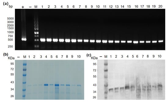
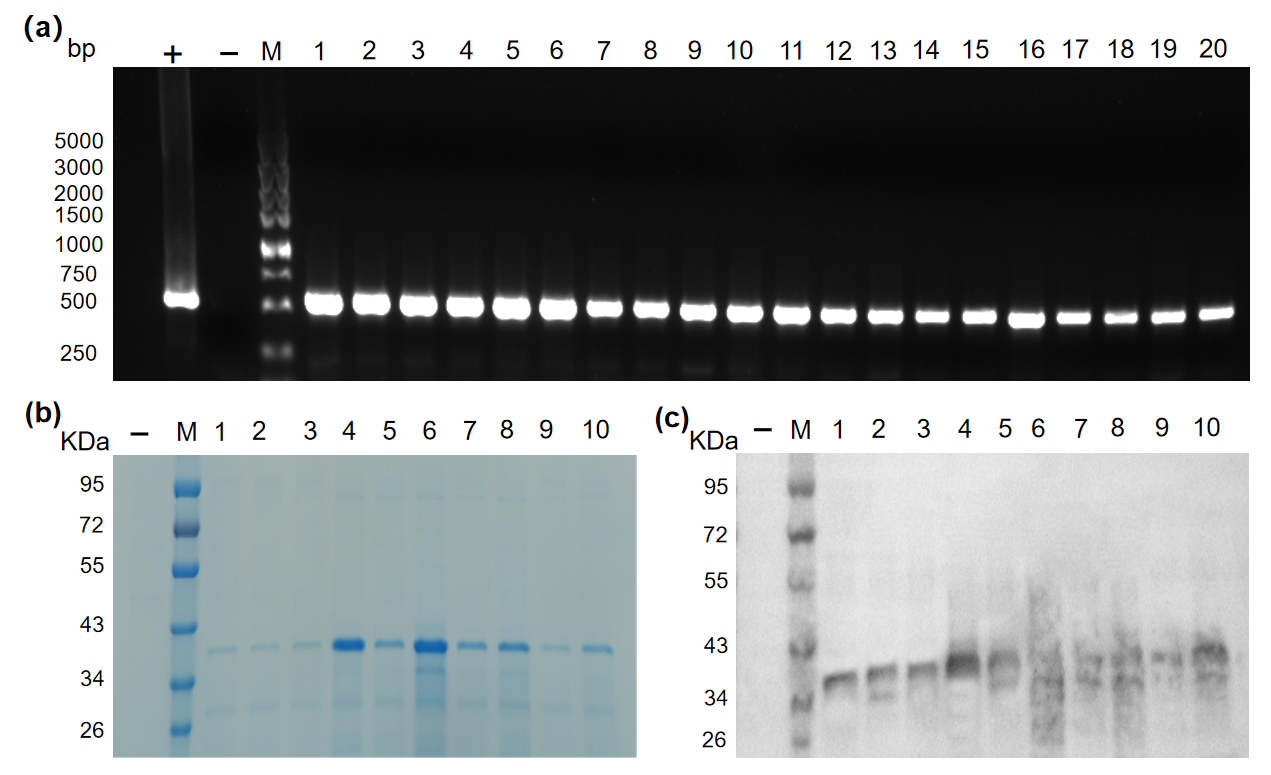
We first expressed the non-hydroxylated form of Pvfp-5P (Pvfp-5P(Y)) in K. phaffii. After electroporation of linearized pPICZαA-Pvfp5P into GS115, approximately 80 Zeocin-resistant colonies appeared on YPD plates. Twenty randomly selected clones were screened by colony PCR using primers specific for the Pvfp-5P gene. All 20 clones yielded an amplified product of the expected size (~500 bp) (Figure 1a). Ten positive clones were cultured in BMGY, induced with methanol for 70 h, and the supernatants were analyzed by SDS-PAGE. As shown in Figure 1b, all clones exhibited a major protein band with an apparent molecular mass of 34–43 kDa, which is higher than the theoretical molecular mass of 27.76 kDa. This discrepancy may be due to N-glycosylation (the sequence contains predicted sites) or anomalous migration of the highly hydrophilic fusion protein; however, this was not experimentally verified in the current study. Clone #4 showed the highest expression level and the fewest contaminant bands and was selected for further experiments. Western blotting with an anti-His antibody confirmed that the 34–43 kDa band represented the His-tagged Pvfp-5P (Figure 1c). This strain was named GS115/Pvfp5P.
Figure 1.
Screening of the Pvfp-5P single-expression strain. (a) PCR identification of Pvfp-5P integrants. Lane M: DL5000 DNA marker; lanes 1–20: individual clones; −: blank control; +: positive control (recombinant plasmid pPICZαA-Pvfp5P). (b) SDS-PAGE of culture supernatants after 70 h induction. Lane M: protein marker; lanes 1–10: positive clones; −: uninduced control. (c) Western blot using an anti-His antibody confirming expression. Note: The gel and blot images were cropped from larger raw images for clarity; no lanes were reordered.
3.2. Construction and Screening of the Co-Expression Strain
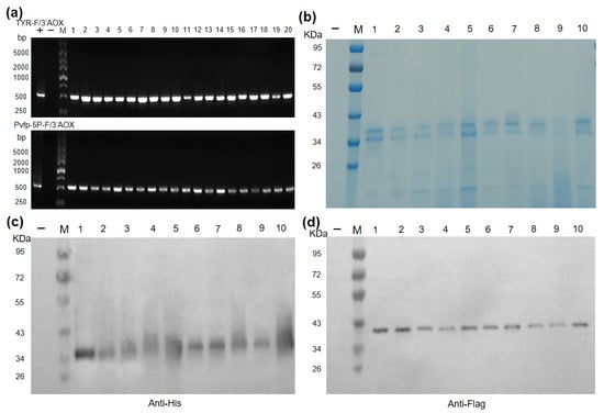
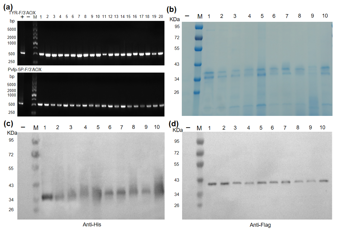
To enable catalytic modification, the unmodified adhesive protein was co-expressed in K. phaffii with tyrosinase from B. megaterium, a well-characterized enzyme with high catalytic efficiency [20]. The pPIC9K-TYR plasmid was electroporated into GS115/Pvfp5P. Transformants were selected on MD plates containing Zeocin. Twenty colonies were picked, and PCR was performed with two primer pairs. All 20 clones were positive for both genes (Figure 2a). Ten of these double-positive clones were induced in BMMY containing 100 μM CuSO4. SDS-PAGE analysis of culture supernatants from co-expression clones showed two clearly resolved bands within the 34–43 kDa region (Figure 2b). Specific anti-Flag Western blotting (Figure 2d) unambiguously confirmed the production of recombinant tyrosinase in positive clones, while anti-His Western blotting (Figure 2c) verified Pvfp-5P expression. Clone #1 exhibited the highest expression of both proteins and the lowest number of contaminating bands; it was designated GS115/Pvfp5P/TYR and used to produce Pvfp-5P(D) in all subsequent experiments.
Figure 2.
Screening of the co-expression strain. (a) PCR verification of co-integration. Lane M: marker; lanes 1–20: clones; −: blank control; +: positive control (recombinant plasmid). Upper panel: tyrosinase gene; lower panel: Pvfp-5P gene. (b) SDS-PAGE of culture supernatants from co-expression clones after 70 h induction for clone screening. Two distinct bands are observed within 34–43 kDa, in contrast to the single major band of the Pvfp-5P single-expression strain (Figure 1b). Lane M: marker; lanes 1–10: clones; −: uninduced control. (c) Western blot (anti-His) for Pvfp-5P. (d) Western blot (anti-Flag) for tyrosinase. Note: The gel and blot images were cropped from larger raw images for clarity; no lanes were reordered. In (d), the anti-Flag band (theoretical mass 35.35 kDa) migrates near 43 kDa, which is likely due to N-glycosylation or a partially retained alpha-factor propeptide.
Although the exact gene copy number of the co-expression strain was not experimentally determined in this study, the two plasmids were integrated at different loci (pPICZαA-Pvfp5P at the 5′ AOX1 locus and pPIC9K-TYR at the HIS4 locus). The strong simultaneous expression of both Pvfp-5P and tyrosinase observed at the 70 h time point (Figure 2) indicates that the engineered strains can efficiently co-express both proteins under our specific induction conditions. However, this single time-point observation does not definitively prove long-term mitotic stability in the absence of selection pressure, nor does it exclude the possibility of transcriptional competition. A single-cassette comparison and extended stability tests would be required to fully address these factors.
3.3. Production and Purification of Recombinant Pvfp-5P
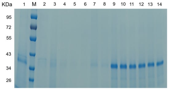
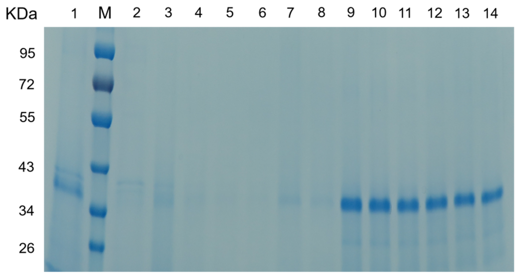
To produce sufficient quantities of Pvfp-5P for functional assays, we performed 48 h inductions in shake flasks (100 mL scale). The culture supernatant was collected and subjected to one-step Ni-NTA affinity chromatography under native conditions. SDS-PAGE analysis of the purification fractions is shown in Figure 3. The culture supernatant (lane 1) contained a major band at 34–43 kDa. Most of this protein bound to the Ni-NTA resin, as evidenced by its near-complete absence in the flow-through (lane 2). After washing with 30 mM imidazole (lane 3), most contaminating proteins were removed. Elution with 300 mM imidazole (lanes 4–14) yielded a single major band at the expected size, with an estimated purity >85% by densitometry. The yield of purified Pvfp-5P(D), determined by A280 quantification, was 353.1 ± 6.9 mg/L of culture (mean ± SD of three independent biological replicates). Since the extinction coefficient was calculated based on the 27.76 kDa polypeptide sequence, this yield reflects the mass of the polypeptide backbone, excluding the contribution of glycans.
Figure 3.
SDS-PAGE analysis of Pvfp-5P purified by Ni-NTA affinity chromatography. Lane M: protein marker; lane 1: culture supernatant; lane 2: flow-through fraction; lane 3: wash fraction; lanes 4–14: elution fractions.
3.4. Mass Spectrometry Confirmation of Pvfp-5P Identity
The Ni-NTA-purified Pvfp-5P band was excised and analyzed by LC-MS/MS. The MaxQuant search results (Table 1) identified a total of 16 proteins with 90 unique peptides. Besides the co-purified tyrosinase and several endogenous host cell proteins (HCPs) from K. phaffii, 27 unique peptides matched Pvfp-5P, covering 72.6% of its full-length amino acid sequence (60.4% unique coverage). Representative peptides included GVYYPNPCSPYPCR from the Pvfp-5 core region, GDEGPMGMIGMSGLK from the preCol-P fusion region, and KYGGGGSGGGGSGGGGSGEPGYHGAPGRPGQK from the linker region (Table S1). The protein score for Pvfp-5P was 323.31, confirming the reliable sequence match of the recombinantly expressed Pvfp-5P.
Table 1.
Summary of mass spectrometry identification results for the recombinantly expressed proteins.
To reconcile the >85% bulk purity observed by SDS-PAGE with the 16 proteins identified by LC-MS/MS, it is important to note that these two methods evaluate sample purity based on fundamentally different principles. SDS-PAGE densitometry measures bulk mass, meaning trace proteins below the Coomassie staining limit remain invisible. In contrast, LC-MS/MS is highly sensitive and can detect trace host cell proteins (HCPs). Based on MaxQuant iBAQ quantification of the excised band (Table 1), the target Pvfp-5P(D) overwhelmingly dominated the sample, accounting for approximately 98.0% of the total protein abundance. Importantly, the co-purified tyrosinase accounted for only ~1.3% of the total abundance, while all other HCPs combined accounted for less than 1%. This extremely low abundance of tyrosinase, together with its near-zero catechol background (as demonstrated by the blank control in Section 3.5), confirms that it does not significantly interfere with the subsequent catechol quantification.
3.5. Identification and Quantification of Catechol Content in Pvfp-5P
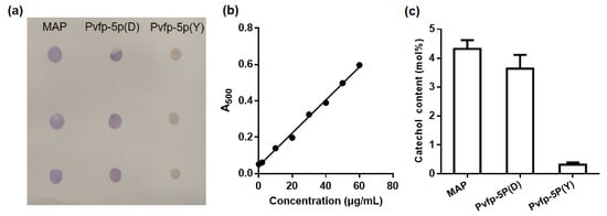
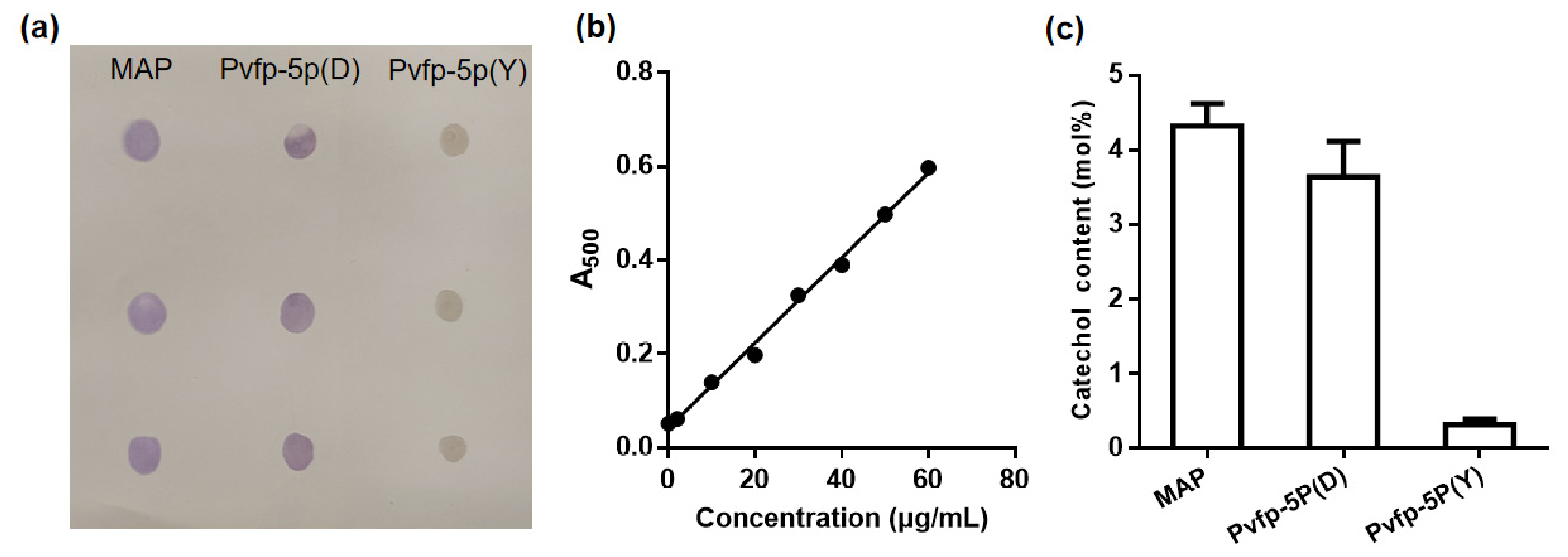
As the physiological activity of the adhesive protein is associated with its DOPA content [4], we assessed the presence of catechol groups in the purified recombinant protein. Catechol-containing species were assessed by NBT staining in both Pvfp-5P(D) and Pvfp-5P(Y). As shown in Figure 4a, commercial MAP (positive control) and Pvfp-5P(D) produced intense purple spots on the nitrocellulose membrane, whereas Pvfp-5P(Y) showed no purple spots. This qualitative result indicates that catechol formation in Pvfp-5P is dependent on tyrosinase co-expression.
Figure 4.
Identification and quantification of catechol in the recombinant Pvfp-5P. (a) NBT staining of proteins spotted on a nitrocellulose membrane. MAP: commercial mussel adhesive protein; Pvfp-5P(D): catechol-modified form; Pvfp-5P(Y): unmodified Tyr-form. (b) Standard curve of L-DOPA. A500: absorbance at 500 nm. Standard curve equation: y = 0.009073x + 0.04201, R2 = 0.9955. (c) Catechol contents (expressed as DOPA equivalents) in MAP, Pvfp-5P(D), and Pvfp-5P(Y) (mean ± SD of three independent biological replicates).
Quantitative analysis using the nitrite–molybdate colorimetric method (Figure 4b,c) revealed that commercial MAP contained 4.32 mol% catechol (expressed as DOPA equivalents), Pvfp-5P(D) contained 3.64 mol%, and the copper-matched Pvfp-5P(Y) contained only 0.32 mol% (which is consistent with the 0.28 mol% measured in the original batch used for functional assays). This method was cross-validated using the commercial MAP as an external standard. The manufacturer’s certificate of analysis (COA) specifies a DL-DOPA content of 5.6 wt%. Assuming an average residue mass of 110 Da and a molecular weight of ~100 kDa for the commercial MAP, this 5.6 wt% DL-DOPA corresponds to a theoretical DOPA content of approximately 3.4 mol%. Our colorimetric assay measured 4.32 mol% for the same commercial MAP. This ~25% higher value is consistent with the nature of the assay, which detects total catechols rather than exclusively DOPA. Furthermore, to rule out interference from the co-purified tyrosinase, a purified tyrosinase blank was assayed under identical conditions. It yielded an A500 of 0.046, corresponding to < 0.05 mol% catechol. This confirms that tyrosinase does not generate a measurable catechol background, ensuring that the catechol detected in Pvfp-5P(D) is specifically derived from the target protein.
Based on the Pvfp-5P sequence (277 total residues with 18 Tyrosine residues), the theoretical maximum catechol content is 6.50 mol%. Therefore, the 3.64 mol% measured in Pvfp-5P(D) represents a 56.0% conversion efficiency of the available tyrosine residues. Because the mol% was calculated based on the 277 amino acid residues of the polypeptide backbone, the contribution of N-glycans is not included in the denominator, meaning no further mass correction is required for this molar calculation.
Overall, the pronounced difference in catechol content between Pvfp-5P(D) and both Pvfp-5P(Y) batches (original and copper-matched) provides evidence that catechol formation is strictly dependent on the co-expressed tyrosinase, rather than being an artifact of copper supplementation. It should be emphasized that this colorimetric method detects catechols generally rather than DOPA specifically; the reported values therefore represent total catechol content rather than direct measurements of DOPA.
Additionally, to rule out the possibility that the measured catechol signal was derived from residual free L-DOPA or its oxidation products in the culture medium, we analyzed the UV–Vis absorption spectra of both the 3 kDa ultrafiltration permeate and the final purified protein retentate (Figure S4). The permeate exhibited strong absorption at 280 nm (indicating free L-DOPA and small aromatic peptides) and a characteristic broad peak at 475 nm (indicating dopachrome, an oxidation product of DOPA). In contrast, the final purified retentate displayed a typical protein absorption peak at 280 nm but lacked the 475 nm peak (A475 ≈ 0.005). This spectral evidence indicates that the free phenolic compounds and melanin precursors were removed during the ultrafiltration step, minimizing potential background interference in the colorimetric assay.
Notably, upon lyophilization, the purified Pvfp-5P(D) powder exhibited a light brownish color, whereas the Pvfp-5P(Y) powder remained white. This browning is characteristic of DOPA oxidation products (e.g., dopachrome/melanin), further supporting the presence of oxidized catechol groups. To assess whether the catechol residues undergo quinone-mediated covalent crosslinking, we performed non-reducing SDS-PAGE (Figure S5). While the unmodified Pvfp-5P(Y) migrated exclusively as a monomer (~34–43 kDa) under non-reducing conditions, Pvfp-5P(D) exhibited the main monomeric band accompanied by a faint ladder of high-molecular-weight oligomers (>95 kDa). Densitometric analysis indicated that these crosslinked oligomers accounted for less than 10% of the total protein, indicating that the majority of Pvfp-5P(D) remains monomeric.
3.6. Promotion of Scratch Closure by Pvfp-5P
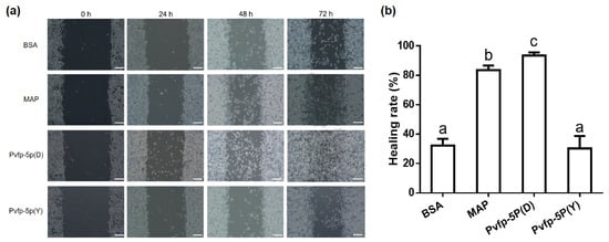
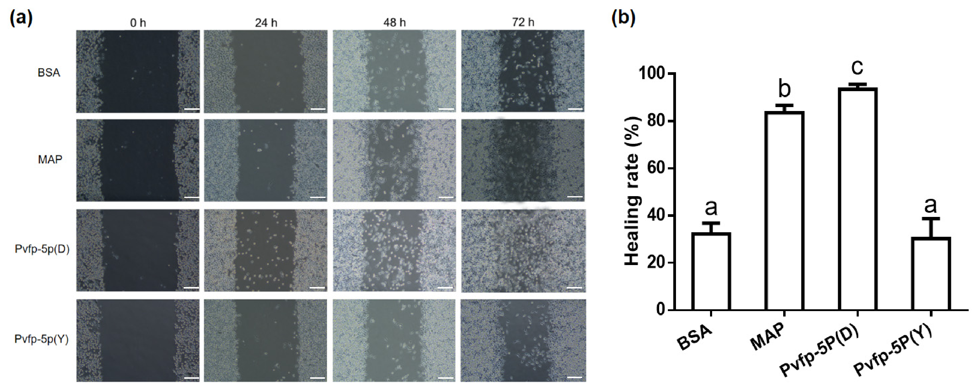
To evaluate the effect of catechol modification on scratch closure, we performed a scratch wound healing assay on L929 fibroblasts using Pvfp-5P(D), Pvfp-5P(Y), commercial MAP, and BSA. Representative images at 0, 24, 48, and 72 h are shown in Figure 5a. At 0 h, the scratch widths were similar across all groups. After 24 h, the BSA group showed only a few cells migrating into the scratch, whereas Pvfp-5P(D) and commercial MAP exhibited cell migration into the scratched area. At 48 h, the scratch in the Pvfp-5P(D) group was almost closed, while the BSA and the Pvfp-5P(Y) group still had a wide gap. By 72 h, the scratch closure rate in the Pvfp-5P(D) group reached 93.5 ± 2.1%, higher than that of the commercial MAP group (83.5 ± 3.2%), the Pvfp-5P(Y) group (30.3 ± 8.5%), and the BSA group (32.3 ± 4.5%). The difference between Pvfp-5P(D) and Pvfp-5P(Y) indicates that catechol modification contributes substantially to the scratch-closure activity of Pvfp-5P.
Figure 5.
Promotion of scratch closure by Pvfp-5P. (a) Representative micrographs at 0, 24, 48, and 72 h for BSA, commercial MAP, Pvfp-5P(D), and Pvfp-5P(Y). Scale bar: 200 μm. (b) Quantitative scratch closure rates at 72 h (mean ± SD of three independent biological replicates). Different letters above the bars denote significant differences among treatments (p < 0.05, Tukey’s test).
Additionally, CCK-8 assays performed under identical conditions (72 h, 100 µg/mL) showed no significant cytotoxicity for either Pvfp-5P(D) or Pvfp-5P(Y) compared to the BSA control (Figure S6), ruling out cell death as the cause of the low closure rate in the Y-form.
3.7. Surface Adsorption of Pvfp-5P
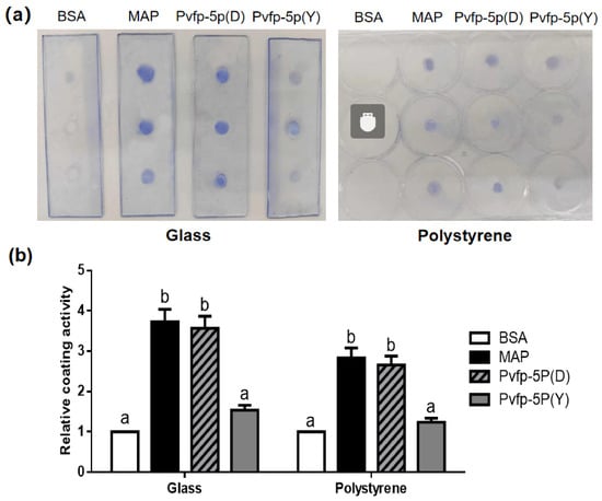
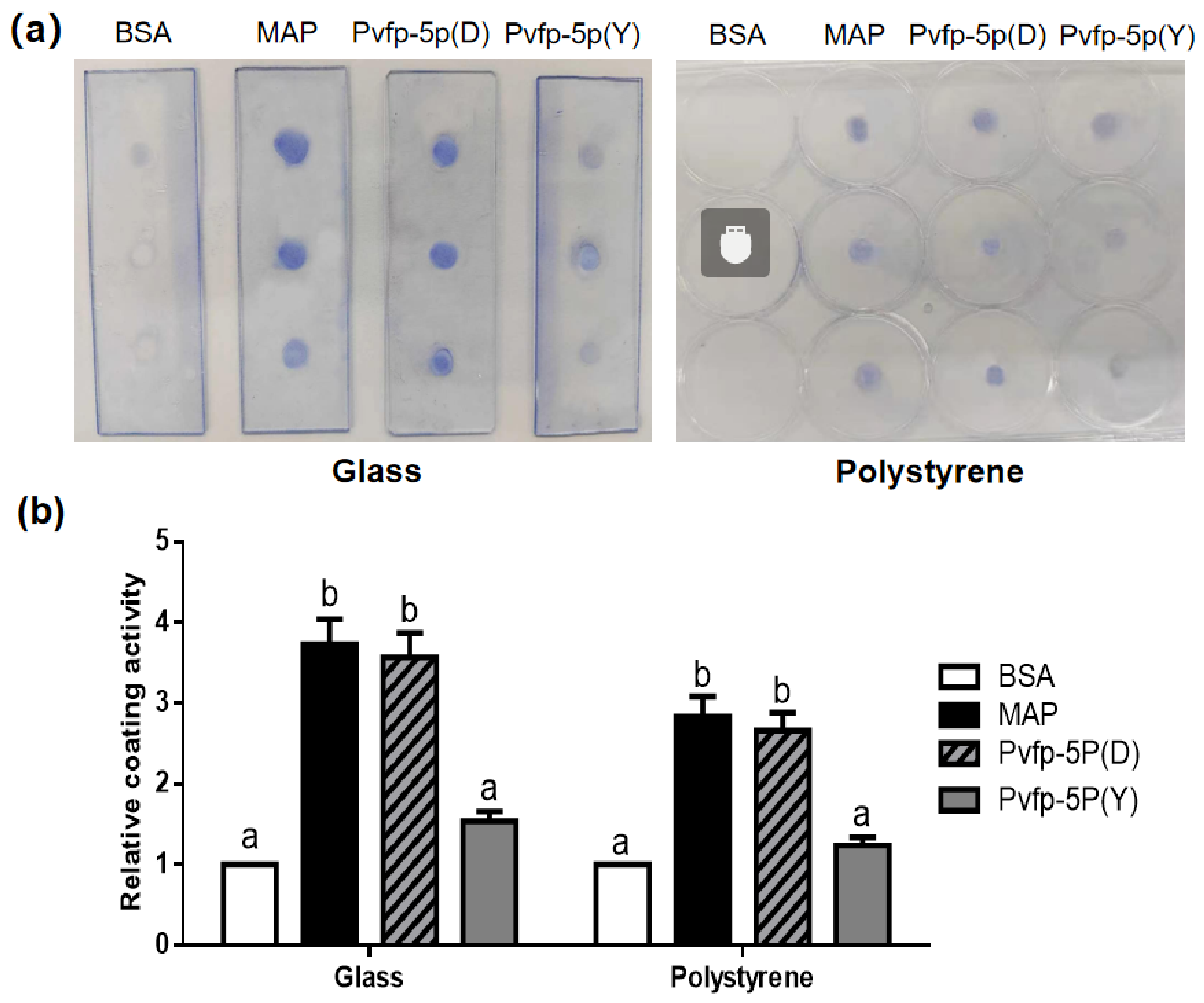
To evaluate the effect of catechol modification on surface adsorption, we performed a coating assay on glass (hydrophilic) and polystyrene (moderately hydrophobic) surfaces using Pvfp-5P(D), Pvfp-5P(Y), commercial MAP, and BSA. After washing and Coomassie Blue staining, Pvfp-5P(D) and commercial MAP produced visible blue spots on both substrates, whereas Pvfp-5P(Y) showed weaker staining and BSA (negative control) yielded only faint or no staining (Figure 6a). Quantitative analysis (Figure 6b) showed that the relative coating activities (RCA) for Pvfp-5P(D) were 3.6 (glass) and 2.7 (polystyrene), whereas the RCA for Pvfp-5P(Y) were only 1.5 and 1.2, respectively. Both forms were higher than BSA (p < 0.05), and Pvfp-5P(D) was significantly higher than Pvfp-5P(Y) (p < 0.01). The detailed raw mean gray values (MGV) and background values subtracted for all samples are provided in Supplementary Table S2.
Figure 6.
Surface adsorption assay. (a) Coomassie Blue-stained protein spots on glass and polystyrene surfaces for BSA, Pvfp-5P(Y), Pvfp-5P(D), and commercial MAP. (b) Relative coating activity for each protein on glass and polystyrene (mean ± SD of three independent biological replicates). Different letters above the bars denote significant differences among treatments (p < 0.05, Tukey’s test).
To validate this semi-quantitative staining and rule out the potential color interference from DOPA/quinones, an elution and BCA assay was performed to quantify the absolute adsorbed protein mass (Figure S7). As expected, BSA exhibited the lowest adsorption on both substrates (0.42 ± 0.06 μg/cm2 on glass; 0.35 ± 0.05 μg/cm2 on polystyrene). Pvfp-5P(Y) showed moderate adsorption (1.18 ± 0.15 μg/cm2 on glass; 0.86 ± 0.11 μg/cm2 on polystyrene). Notably, Pvfp-5P(D) exhibited a high level of adsorbed mass, reaching 2.65 ± 0.24 μg/cm2 on glass and 1.92 ± 0.18 μg/cm2 on polystyrene, which were significantly higher than those of Pvfp-5P(Y) (p < 0.01). These mass-based results are consistent with the CBB staining trend, confirming that the enhanced staining intensity of Pvfp-5P(D) is indeed due to a significantly higher amount of adsorbed protein, rather than the intrinsic color of the catechol groups.
4. Discussion
Mussel adhesive proteins have attracted considerable interest for biomedical applications, but their recombinant production faces challenges in yield and post-translational DOPA modification. In this study, we expressed a fusion protein Pvfp-5P in K. phaffii GS115 in two forms: a catechol-modified form generated by co-expression with B. megaterium tyrosinase (Pvfp-5P(D)), and an unmodified Tyr-form generated without tyrosinase co-expression (Pvfp-5P(Y)). The key findings are: (i) Pvfp-5P(D) was secreted at approximately 353 mg/L in shake flasks and purified to >85% purity by one-step Ni-NTA chromatography; (ii) catechol groups were detected at 3.64 mol% in Pvfp-5P(D) versus 0.32 mol% in Pvfp-5P(Y), demonstrating tyrosinase-dependent catechol formation; and (iii) Pvfp-5P(D) enhanced scratch closure and surface adsorption more effectively than Pvfp-5P(Y), indicating that catechol modification contributes to these functional properties.
We acknowledge that several conclusions cannot be definitively drawn from the current data. First, the contribution of the preCol-P fusion to the observed expression level remains unverified, as no matched Pvfp-5β-only control was included. Although previous reports on recombinant MAPs often describe yields below 100 mg/L in shake flasks [22,23], direct comparison with our results is limited by differences in expression host. Therefore, the 353 mg/L yield should be regarded as a description of this specific engineered system rather than evidence for a generalizable fusion strategy.
Second, although the Y-form control confirms that catechol formation is tyrosinase-dependent, the colorimetric method detects total catechols rather than specific DOPA. Re-searching our LC-MS/MS data with Tyr + 16 yielded no high-confidence DOPA peptides, a recognized challenge due to DOPA’s instability and in-source fragmentation during analysis [24]. Nevertheless, the parallel processing of D and Y samples (ruling out artifactual oxidation) and the strong chemical evidence from NBT and colorimetric assays collectively support successful catechol incorporation. Future work will employ targeted multiple reaction monitoring or amino acid analysis for direct residue-level verification. Furthermore, the in situ co-expression strategy used here offers a technically feasible alternative to harsh post-purification chemical modifications [15,25], though further comparative studies are needed.
Third, the scratch wound healing assay revealed that Pvfp-5P(D) promotes fibroblast migration (93.5%), which was even slightly higher than the commercial MAP (83.5%), despite its slightly lower catechol content (3.64 vs. 4.32 mol%). While catechol groups are essential for this bioactivity, this observation suggests that total catechol content is not the sole determinant. We hypothesize that the superior spatial accessibility of catechol groups in the smaller fusion protein (34–43 kDa) compared to the commercial MAP (~100 kDa) may allow for more efficient interaction with cell surface receptors. Additionally, the unstructured, flexible preCol-P extension may provide a favorable physical scaffold that synergizes with the catechol groups to promote cell migration.
However, the interpretation of this result requires caution. Although CCK-8 assays confirmed cell viability, we must acknowledge that Pvfp-5P(D) strongly adsorbs to the polystyrene surface, potentially providing a pre-coated substrate for cell migration, which Pvfp-5P(Y) does not. Future pre-coating experiments are required to fully decouple the biological stimulation of cell migration from the physical cues of the protein coating. Furthermore, although all protein stocks (BSA, Pvfp-5P(D), Pvfp-5P(Y), and commercial MAP) were desalted into identical buffers and reconstituted under the same conditions to ensure matched vehicle conditions, a true buffer-only control (without any protein) was not included in the original experimental design. Future assays should incorporate a buffer-only control to fully exclude any potential vehicle effects. The significant difference between Pvfp-5P(D) and Pvfp-5P(Y), together with the similar values for Pvfp-5P(Y) and BSA, suggests that catechol modification enhances scratch closure, but the precise contributions of migration versus proliferation remain unresolved. A proliferation inhibitor such as mitomycin C, or an independent migration assay, would help distinguish these contributions in future work.
Fourth, the surface adsorption measures distinct properties from true adhesive strength. The coating assay demonstrated that Pvfp-5P(D) adsorbs more strongly than Pvfp-5P(Y) to both hydrophilic and hydrophobic surfaces, consistent with the known surface-binding properties of DOPA-containing MAPs [3,26]. The mass-based quantification further confirmed that Pvfp-5P(D) achieved a significantly higher adsorbed protein mass compared to Pvfp-5P(Y). However, it should be emphasized that this coating assay primarily reflects the protein’s adsorption and retention on the substrate after washing, which is not a direct measure of its actual adhesive strength. Therefore, direct adhesion measurements (e.g., lap-shear or pull-off testing) would be required to substantiate the actual adhesive performance.
We also acknowledge that residual Cu2+ could theoretically influence the functional assays. However, a 3 kDa ultrafiltration step was applied during purification, which is expected to remove unbound free Cu2+ ions and small molecules. Importantly, while the functional assays used the original Pvfp-5P(Y) batch (cultured without additional CuSO4), a newly prepared copper-matched Pvfp-5P(Y) batch yielded a nearly identical catechol content (0.32 mol% vs. 0.28 mol%). This demonstrates their chemical equivalence, ensuring that the functional comparisons are not confounded by the copper concentration. While ICP-MS quantification was beyond our current instrumental capabilities, future studies will include this validation.
Finally, a major limitation of this study is that we did not perform enzymatic deglycosylation (e.g., PNGase F treatment) to verify the N-glycosylation status of Pvfp-5P. The apparent mass shift from the theoretical 27.76 kDa to 34–43 kDa was attributed to N-glycosylation based on sequence prediction, but anomalous migration cannot be entirely excluded. Future studies must include deglycosylation assays to determine the exact glycan mass fraction and to evaluate the impact of glycosylation on the immunogenicity and biomedical application of this recombinant protein.
Nevertheless, this work provides evidence that co-expression of B. megaterium tyrosinase in K. phaffii enables catechol incorporation into a recombinant mussel adhesive protein and that this modification enhances scratch closure and surface adsorption. Future work should include a Pvfp-5β-only variant, peptide-level DOPA verification, and quantitative adhesion measurements to fully establish the potential of this system.
5. Conclusions
We constructed a K. phaffii co-expression system for the production of a catechol-modified fusion mussel adhesive protein, Pvfp-5P, by fusing Pvfp-5β with a preCol-P peptide and co-expressing B. megaterium tyrosinase. The recombinant protein was secreted at approximately 353 mg/L in shake flasks, purified to >85% purity by one-step Ni-NTA chromatography, and contained catechol groups at 3.64 mol%, compared with only 0.32 mol% in the unmodified Tyr-form. Functional assays showed that Pvfp-5P(D) enhanced scratch closure in L929 fibroblasts (93.5% vs. 30.3% for Pvfp-5P(Y)) without cytotoxicity. It also exhibited stronger mass-based adsorption. These results demonstrate that tyrosinase co-expression enables catechol incorporation into a recombinant MAP and that this modification enhances its in vitro bioactivity. However, the contribution of the preCol-P fusion, the residue-level verification of DOPA groups, and the precise mechanism of the observed activity require further investigation. Future work should address these limitations to fully establish the potential of this system for biomedical applications.
Supplementary Materials
The following supporting information can be downloaded at: https://www.mdpi.com/article/10.3390/biology15222127/s1, Figure S1: SDS PAGE for the as received commercial MAP sample; Figure S2: Amino acid sequence and nucleotide sequence of the recombinant fusion protein; Figure S3: Amino acid sequence and nucleotide sequence of B. megaterium tyrosinase; Figure S4: UV-Vis absorption spectra of the purified Pvfp-5P(D) retentate and the 3 kDa ultrafiltration permeate; Figure S5: Non-reducing and reducing SDS-PAGE analysis of Pvfp-5P(Y) and Pvfp-5P(D); Figure S6: Cell viability of L929 fibroblasts treated with different proteins; Figure S7: Mass-based quantification of adsorbed proteins on different substrates; Table S1: List of unique peptides identified by mass spectrometry; Table S2: Raw mean gray values (MGV), background values, net gray values (NGV), and relative coating activ-ities (RCA) of different proteins on glass and polystyrene surfaces. Supplementary Data Files: MaxQuant output tables (proteinGroups.txt, peptides.txt, and evidence.txt), Tyr16_Re-search (peptides.txt, and evidence.txt), and README.txt are provided as a compressed .zip archive.
Author Contributions
Conceptualization, Z.W. (Zilong Wang) and L.Z.; methodology, J.H.; software, Y.C.; validation, Z.W. (Zejun Wang); formal analysis, T.L.; investigation, J.H.; resources, J.H. and Z.W. (Zilong Wang); data curation, J.H.; writing—original draft preparation, J.H.; writing—review and editing, Z.W. (Zilong Wang) and L.Z.; visualization, T.L.; supervision, Z.W. (Zilong Wang) and L.Z.; project administration, Z.W. (Zilong Wang); funding acquisition, J.H. and Z.W. (Zilong Wang). All authors have read and agreed to the published version of the manuscript.
Funding
This work was supported by the Science and Technology Planning Project of Fuzhou (Grant No. 2024-R-004) and the Science and Technology Project of Fuzhou Institute of Oceanography (Grant No. 2025F08).
Institutional Review Board Statement
Not applicable.
Informed Consent Statement
Not applicable.
Data Availability Statement
The data that support the findings of this study are available within the article and its Supplementary Materials. The nucleotide sequences have been deposited in GenBank under accession numbers QB197299 and QB197300. These records will be publicly available upon publication. The mass spectrometry proteomics data (including the MaxQuant output tables proteinGroups.txt, peptides.txt, and evidence.txt for both the standard and Tyr + 16 re-searches) are provided as Supplementary Data Files. Further inquiries regarding the raw mass spectrometry data can be directed to the corresponding authors.
Acknowledgments
We thank Sangon Biotech (Shanghai, China) for the mass spectrometry identification. During the preparation of this manuscript, the authors used Deepseek-V4.1-Flash (DeepSeek, Hangzhou, China) for language polishing and grammar checking. The authors have reviewed and edited the output and take full responsibility for the content of this publication.
Conflicts of Interest
The authors declare no conflicts of interest.
References
- Narayanan, A.; Dhinojwala, A.; Joy, A. Design principles for creating synthetic underwater adhesives. Chem. Soc. Rev. 2021, 50, 13321–13345. [Google Scholar] [CrossRef] [Scilit] [PubMed]
- Silverman, H.G.; Roberto, F.F. Understanding marine mussel adhesion. Mar. Biotechnol. 2007, 9, 661–681. [Google Scholar] [CrossRef] [Scilit] [PubMed]
- Petrone, L.; Kumar, A.; Sutanto, C.N.; Patil, N.J.; Kannan, S.; Palaniappan, A.; Amini, S.; Zappone, B.; Verma, C.; Miserez, A. Mussel adhesion is dictated by time-regulated secretion and molecular conformation of mussel adhesive proteins. Nat. Commun. 2015, 6, 8737. [Google Scholar] [CrossRef] [Scilit] [PubMed]
- Waite, J.H. Mussel adhesion—Essential footwork. J. Exp. Biol. 2017, 220, 517–530. [Google Scholar] [CrossRef] [Scilit] [PubMed]
- Pandurangan, S.; Easwaramoorthi, S.; Ayyadurai, N. Engineering proteins with catechol chemistry for biotechnological applications. Crit. Rev. Biotechnol. 2025, 45, 606–624. [Google Scholar] [CrossRef] [Scilit] [PubMed]
- Lu, Q.; Danner, E.; Waite, J.H.; Israelachvili, J.N.; Zeng, H.; Hwang, D.S. Adhesion of mussel foot proteins to different substrate surfaces. J. R. Soc. Interface 2013, 10, 20120759. [Google Scholar] [CrossRef] [Scilit] [PubMed]
- Dai, Q.; Liu, H.; Gao, C.; Sun, W.; Lu, C.; Zhang, Y.; Cai, W.; Qiao, H.; Jin, A.; Wang, Y.; et al. Advances in mussel adhesion proteins and mussel-inspired material electrospun nanofibers for their application in wound repair. ACS Biomater. Sci. Eng. 2024, 10, 6097–6119. [Google Scholar] [CrossRef] [Scilit] [PubMed]
- Yang, J.W.; Yoon, T.; Kim, H.; Joo, K.I.; Cha, H.J. Acrylated bioengineered mussel protein-based adhesive nanoparticles for locoregional and sustained drug delivery. ACS Biomater. Sci. Eng. 2025, 11, 3523–3532. [Google Scholar] [CrossRef] [Scilit] [PubMed]
- Sun, J.; Han, J.; Wang, F.; Liu, K.; Zhang, H. Bioengineered protein-based adhesives for biomedical applications. Chem. Eur. J. 2022, 28, e202102902. [Google Scholar] [CrossRef] [Scilit] [PubMed]
- Ohara, A.; Pengyue, Z.; Cao, X.; Donini, R.; Haslam, S.M.; Polizzi, K.M. The impact of methanol concentration on recombinant protein glycosylation in Pichia pastoris SuperMan5. Microb. Biotechnol. 2025, 18, e70272. [Google Scholar] [CrossRef] [Scilit] [PubMed]
- Wang, J.; Xiang, Z.-X.; Luan, M.-F.; Gong, J.-S.; Su, C.; Li, H.; Xu, Z.-H.; Shi, J.-S. High-level secretory expression of recombinant type III human-like collagen α1 in Pichia pastoris via multilevel systematic optimization. Int. J. Biol. Macromol. 2025, 313, 144270. [Google Scholar] [CrossRef] [Scilit] [PubMed]
- Tao, L.; Zhang, C.; Zhang, G.; Zhou, J. High-level production of patatin in Pichia pastoris and characterization of N-glycosylation modification in food processing properties. Food Res. Int. 2024, 196, 115111. [Google Scholar] [CrossRef] [Scilit] [PubMed]
- Yao, L.; Wang, X.; Xue, R.; Xu, H.; Wang, R.; Zhang, L.; Li, S. Comparative analysis of mussel foot protein 3B co-expressed with tyrosinases provides a potential adhesive biomaterial. Int. J. Biol. Macromol. 2022, 195, 229–236. [Google Scholar] [CrossRef] [Scilit] [PubMed]
- Baskaran, N.; Wang, Y.C.; Tan, R.J.; Chung, R.J.; Wei, Y. Overcoming the yield challenge of mussel foot proteins: Enhancing adhesion through metal ion-incorporated nanoparticles. Colloids Surf. B 2023, 229, 113479. [Google Scholar] [CrossRef] [Scilit] [PubMed]
- Choi, Y.S.; Yang, Y.J.; Yang, B.; Cha, H.J. In vivo modification of tyrosine residues in recombinant mussel adhesive protein by tyrosinase co-expression in Escherichia coli. Microb. Cell Fact. 2012, 11, 139. [Google Scholar] [CrossRef] [Scilit] [PubMed]
- Qi, Y.; Zhou, D.; Kessler, J.L.; Qiu, R.; Yu, S.M.; Li, G.; Qin, Z.; Li, Y. Terminal repeats impact collagen triple-helix stability through hydrogen bonding. Chem. Sci. 2022, 13, 12567–12576. [Google Scholar] [CrossRef] [Scilit] [PubMed]
- Salo, A.M.; Rappu, P.; Koski, M.; Karjalainen, E.; Izzi, V.; Drushinin, K.; Miinalainen, I.; Käpylä, J.; Heino, J.; Myllyharju, J. Collagen prolyl 4-hydroxylase isoenzymes I and II have sequence specificity towards different X-Pro-Gly triplets. Matrix Biol. 2024, 125, 73–87. [Google Scholar] [CrossRef] [Scilit] [PubMed]
- Xiang, Z.-X.; Gong, J.-S.; Shi, J.-H.; Liu, C.-F.; Li, H.; Su, C.; Jiang, M.; Xu, Z.-H.; Shi, J.-S. High-efficiency secretory expression and characterization of the recombinant type III human-like collagen in Pichia pastoris. Bioresour. Bioprocess. 2022, 9, 117. [Google Scholar] [CrossRef] [Scilit] [PubMed]
- Gao, Q.; Zhang, Z.; Fu, R.; Zhu, C.; Yuwen, W.; Xu, R.; Duan, Z.; Fan, D. Expression, optimization and biological activity analysis of recombinant type XII collagen in Pichia pastoris. Int. J. Biol. Macromol. 2025, 311, 143720. [Google Scholar] [CrossRef] [Scilit] [PubMed]
- Cha, G.S.; Mok, J.A.; Yun, C.H.; Park, C.M. Production of 3,4-dihydroxy-L-phenylalanine using novel tyrosinases from Bacillus megaterium. Enzym. Microb. Technol. 2022, 160, 110069. [Google Scholar] [CrossRef] [Scilit] [PubMed]
- YY/T 1293.6-2020; Contact Wound Dressing—Part 6: Mussel Adhesive Protein Dressing. National Medical Products Administration: Beijing, China, 2020.
- Xue, R.; Wang, J.; Huang, W.; Qiu, Y.; Xu, H.; Wang, R.; Li, S. Efficient production of recombinant hybrid mussel proteins with improved adhesion. Int. J. Biol. Macromol. 2025, 289, 139937. [Google Scholar] [CrossRef] [Scilit] [PubMed]
- Hu, L.; Chen, L.; Wang, Y.; Yu, S.; Xiao, S.; Chen, W.; Cheng, J.; Kang, Z. Condensate-based sequestration enables high-efficiency synthesis of mussel foot protein Mcofp-3. Appl. Environ. Microbiol. 2026, 92, e0078626. [Google Scholar] [CrossRef] [Scilit] [PubMed]
- Poojary, M.M.; Tiwari, B.K.; Lund, M.N. Selective and sensitive UHPLC-ESI-Orbitrap MS method to quantify protein oxidation markers. Talanta 2021, 234, 122700. [Google Scholar] [CrossRef] [Scilit] [PubMed]
- Yoon, T.; Cha, H.J. Amino acid chemistry and post-translational modifications underlying marine adhesive proteins: Biochemical insights for designing underwater adhesives. Protein Expr. Purif. 2026, 239, 107029. [Google Scholar] [CrossRef] [Scilit] [PubMed]
- Guo, Q.; Chen, J.; Wang, J.; Zeng, H.; Yu, J. Recent progress in synthesis and application of mussel-inspired adhesives. Nanoscale 2020, 12, 1307–1324. [Google Scholar] [CrossRef] [Scilit] [PubMed]

Disclaimer/Publisher’s Note: The statements, opinions and data contained in all publications are solely those of the individual author(s) and contributor(s) and not of MDPI and/or the editor(s). MDPI and/or the editor(s) disclaim responsibility for any injury to people or property resulting from any ideas, methods, instructions or products referred to in the content. |
© 2026 by the authors. Licensee MDPI, Basel, Switzerland. This article is an open access article distributed under the terms and conditions of the Creative Commons Attribution (CC BY) license.