


Abstract
Lianxi Academy in Zhuhai, Guangdong, is a rare surviving rammed-earth academy ruin representative of Lingnan architectural tradition, yet its rammed-earth walls suffer persistent weathering under the subtropical marine monsoon climate, with no site-specific material baseline available. This study aims to characterize the microstructural, mineralogical and particle-size features of wall surfaces, clarify internal compositional differentiation, and provide quantitative baseline data for earthen heritage conservation. Four surface crust specimens from wall facades were analyzed via multiscale scanning electron microscopy with energy-dispersive X-ray spectroscopy (SEM–EDS), X-ray diffraction (XRD), and laser diffraction granulometry. Results show all samples share an O–Si-rich background with notable local contrasts. Local EDS analyses record 12.93 wt% Ca in Sample 2 (accompanied by a unique intersecting rod-shaped network), 6.92 wt% Al and 4.97 wt% K in Sample 3, and 11.33 wt% Fe in Sample 4. Quartz dominates all XRD patterns with strongest reflections at 26.70–26.78° 2θ, accompanied by features consistent with subordinate carbonate-, aluminosilicate-, and Fe-bearing components. Particle-size distributions are highly heterogeneous: Sample 1 is the coarsest (D50 = 34.97 μm; 41.69% ≥ 63 μm), while Samples 3 and 4 are fine-fraction-dominated (D50 = 8.93 and 8.52 μm). These findings define three distinct surface material subgroups within a universal quartz-rich matrix, and are consistent with a heterogeneous wetting and selective particulate detachment weathering model. This work fills the data gap for Lingnan rammed-earth academy heritage, and the quantitative parameters serve as preliminary benchmarks for wall condition mapping, rainwater erosion regulation, and compatible repair material trial formulation.
1. Introduction
1.1. Research Background
In the context of the revival of excellent traditional Chinese culture, the protection and inheritance of cultural heritage have become an important part of the national cultural development strategy [1,2]. The rediscovery and appreciation of traditional academies, the core carriers of ancient Chinese education and cultural dissemination [3,4], have revealed their historical value and cultural connotation. Traditional academies are not only the material embodiment of the ancient education system but also an important medium for the integration of regional cultures and the inheritance of national spirit. They carry the national sentiment of “carrying the Way with literature” and the humanistic spirit of “educating people with virtue” [5] and play an irreplaceable role in the construction of contemporary cultural confidence and the development of moral education. However, with the continuous acceleration of urbanization in my country, modern industrial development and urban renewal have caused serious impacts on traditional historical relics. A large number of traditional academies with historical and cultural value are facing the dilemma of damage to the building’s body, destruction of spatial texture, weakening of memory connotation, and even extinction [6]. Statistics show that of the more than 7000 academies that have existed in China throughout history, only over 1000 remain today, and most of these have significant building damage and experience frequent construction destruction [7]. We need to improve the current status of academy heritage protection.
As a type of academy dedicated to Zhou Dunyi, the founder of Neo-Confucianism in the Song and Ming Dynasties, Lianxi Academy (濂溪書院), with its diversified regional forms across China, directly reflects the long-term evolution of rammed earth construction techniques for Lingnan academies, with historical records tracing the academy’s origin back to the Southern Song Dynasty. It has become an important component of regional cultural systems such as Hunan culture and Lingnan culture [8]. As an important remnant of Lianxi Academy in Lingnan, Zhuhai Lianxi Academy not only carries the spiritual core of Zhou Dunyi’s Neo-Confucian culture but also integrates the regional architectural characteristics of the coastal area of Lingnan. Its rammed earth walls, as the core material remains of the academy’s architecture, are a direct manifestation of the adaptation of the academy’s architectural techniques and regional materials. However, similar to most traditional academies in China, the Zhuhai Lianxi Academy site has been affected by natural erosion and human factors, and the original style of the academy’s main building is no longer present. The rammed earth walls of the academy’s remains have weathered, peeled, collapsed, and experienced other forms of damage, posing a severe challenge to the protection of these material remains. In the context of the cultural revival era, researching the material of the academy’s core architectural remains has become a crucial prerequisite for protecting and preserving its heritage. Against this backdrop, it is particularly important to analyze the microstructure of the only remaining rammed earth wall structure of the Lianxi Academy in Zhuhai.
1.2. Literature Review
At present, the Chinese academic community has formed a multi-dimensional research system for the study of traditional academies, among which the protection of academy heritage is one of the core research directions. Scholars have explored the protection of academies from different perspectives. Hao Bing [9] analyzed the material, spiritual, and social elements of traditional academies in the Hunan and Guangdong regions from the perspective of spatial heritage and proposed a protection strategy of material space integration and spiritual space revitalization. Yang Luling [6] pointed out from the perspective of urban memory that academies, as material carriers of urban cultural connotation, need to continue their historical and cultural heritage by creating cultural tourism brands and establishing protection mechanisms. Zhao Juqin et al. [10] proposed targeted reinforcement and repair design schemes for the damaged walls and structures of academies from the perspective of architectural repair technology, taking Xinchang Gushan Academy as an example. Deng Hongbo et al. [7] sorted out the current situation of protection and utilization of ancient academies in my country through a nationwide survey and pointed out that the current protection of academies has problems such as emphasizing utilization over research and difficulty in achieving coordinated development of protection and utilization. The study of academy heritage protection in the Lingnan region integrates digital technology and regional architectural features. Cai and Jiang [11] explored innovative application paths of digital technology in the protection of Lingnan architectural cultural heritage based on the multi-dimensional framework of “technology–object–function” and constructed a digital protection system adapted to Lingnan architecture. Zhang and Dechsubha [12] took Foshan Academy as the research object, systematically analyzed the architectural space art features of Lingnan Academy, and revealed the adaptability of Lingnan Academy in terms of layout, decoration, and material selection to Lingnan climate and regional culture, providing a regional reference for the protection of the material remains of Lingnan Academy.
However, research on Lianxi Academy in various regions has focused on cultural connotation, historical evolution, and dissemination, resulting in research findings with distinct regional characteristics. Peng Zuowei et al. [5] explored the integration path of Lianxi Academy culture and moral education in colleges and universities in the digital age, pointing out the value of academy culture as a moral education resource. Zhao et al. [13] took Lianxi Academy in Chenzhou City as the research object, analyzed its current dilemma in cultural dissemination, and proposed a strategy to promote the protection and dissemination of Lianxi Academy culture with digital technology. Yao Xianlin [14] explained the integration connotation of Lianxi culture and Hunan culture from the perspective of Hunan education development. Wu Yue [8] systematically examined the historical evolution of Lianxi Academy in Hechuan, Chongqing; clarified its development path; and corrected the errors in the literature. These studies have enriched the cultural and historical research system of Lianxi Academy. However, most existing research results focus on Lianxi Academy in Hunan, Guangdong, Sichuan, and Chongqing. There are currently no research results on Lianxi Academy in Zhuhai, and there has been no study on the materials and techniques related to the architectural remains of Lianxi Academy.
As an important branch of Lingnan architectural research, the study of traditional Lingnan academies has formed a research system from macro space to micro decoration. Chen and Zuo [15] analyzed the symmetrical beauty of Lingnan architecture and its cultural connotation, pointing out that Lingnan academies follow the principle of axial symmetry in layout, reflecting the humanistic pragmatism of Lingnan culture. Tang [16] proposed that traditional Lingnan Han Chinese vernacular architecture has three major adaptabilities: natural, social, and humanistic. Among them, natural adaptability determines the selection and application of building materials. Lingnan building materials need to be adapted to the local high-temperature and high-humidity coastal climate. Zhang and Dechsubha [12] further found that the Foshan academy, as a typical representative of Lingnan academies, uses wood carving, brick carving, and plastering techniques in building materials. Moreover, the material selection takes into account both aesthetics and practicality, adapting to the climate environment of Lingnan. Existing research on traditional Lingnan academies has paid attention to the regional adaptability of materials, but it has focused more on decorative materials and techniques. Research on the composition and proportion of core structural materials such as rammed earth walls is still insufficient, and systematic research results on rammed earth wall materials in Lingnan academies have not yet been formed.
Material diagnosis of earthen heritage requires complementary measurements at several scales [17,18,19,20,21]. SEM records particle habits, aggregate contacts, and voids; EDS maps local elemental differentiation; XRD identifies crystalline mineral assemblages; and particle-size analysis quantifies the granular framework. Integrated applications of these methods have distinguished material sources [17,18,19], wall sectors, construction stages, and weathering products in historic rammed earth [17,18,22,23,24,25,26,27,28,29]. Regional studies in Guangdong and southern China further document substantial variation in Si–Al-rich matrices, Ca- or Fe-bearing domains, microcracks, salts, and climatic exposure across earthen wall surfaces [17,20,21,22]. Their combined use has distinguished mineral assemblages, local enrichment, particle packing, and exposure-related deterioration in historic rammed earth in Europe, North Africa, Guangdong, Fujian, Macau, and other Chinese sites [17,18,27,28,29,30,31,32]. The contribution here is the integrated site-specific application of these methods, not a new analytical technique.
Particle grading controls packing density, pore continuity, capillary suction, shrinkage potential, and the balance between a load-bearing granular skeleton and a cohesive fine matrix in rammed earth [23,24,25,26]. Broad grading can improve packing, while mineral activity, compaction history, and ambient humidity govern the resulting performance. The strength of unstabilised earth is closely coupled to suction and water content, and sustained wetting promotes particle migration, local swelling, and surface softening. The grading contrasts among the four Lianxi Academy samples therefore carry direct implications for deterioration and repair. Water-salt migration, alternating wet–dry cycles and uneven solar irradiation are core driving factors triggering spalling, infiltration and structural failure of rammed earthen heritage, and existing studies have clarified the spatial distribution and differential deterioration characteristics of wall diseases from micro and macroscopic perspectives [33,34,35]. Various reinforcing materials and rammed earth reinforcement schemes have been developed to improve the mechanical stability of earthen ruins, while relevant research also points out that restoration materials must satisfy multi-dimensional constraints including mechanical performance, visual compatibility and vapour permeability [33,36,37]. Most previous assessments of earthen consolidation materials rely on single physical indicators and lack unified multi-index quantitative evaluation systems that balance conflicting performance trade-offs, which limits the objective screening of heritage protection reagents [33,37]. To fill this gap, Liu et al. proposed a PLS-MCDM integrated evaluation framework to comprehensively quantify the moisture regulation, mechanical, appearance and permeability performance of SAP consolidation materials, providing a standardized multi-criteria screening method for rammed earth conservation agents [38].
Material compatibility is equally important at the intervention stage. Historic rammed-earth studies demonstrate that repair performance depends on similarity in mineralogical skeleton, particle grading, water transport, drying, stiffness, and shrinkage rather than on maximum strength alone [28,39]. Long-term exposure studies likewise identify architectural rain control and local shelter as primary determinants of erosion rate [19,21,40]. A site-specific material baseline can therefore translate laboratory characterization into zoning, trial-mix design, and preventive conservation.
No site-specific analytical baseline has previously been established for the Lianxi Academy rammed-earth wall surfaces. Although SEM–EDS, XRD and laser diffraction are well-established characterization techniques, their coordinated multiscale application to establish the first site-specific material baseline for this academy ruin constitutes the principal contribution of this study. The present study addresses this gap through a coordinated multiscale comparison of four samples, linking nanometre- to micrometre-scale morphology and elemental distribution with crystalline phases and volume-based particle grading. The approach positions the academy walls within the wider material history of Lingnan earthen construction while generating parameters that can be used directly in conservation design.
1.3. Problem Statement and Objectives
The Lianxi Academy in Zhuhai, an important remnant of the Lingnan coastal region commemorating Zhou Dunyi, embodies the spiritual core of Song–Ming Neo-Confucianism and Hunan culture, while also incorporating the regional characteristics of Lingnan architecture. Its rammed earth walls, as the core structural remains of the academy, directly reflect the adaptation of Lingnan academy architectural techniques and regional materials since the Southern Song Dynasty, possessing significant historical, technical, and cultural value. However, affected by the high temperature and humidity of the Lingnan coastal region, typhoon erosion, and other natural environmental factors, as well as the long-term lack of systematic protection, the rammed earth walls of the Lianxi Academy site in Zhuhai have suffered severe weathering, peeling, and efflorescence. The integrity of the academy’s main structure has been compromised, and current academic research on the Lianxi Academy in Zhuhai is limited, with no analysis or testing of the composition and proportions of its rammed earth walls. Existing research on Lingnan academies predominantly examines spatial layout, decorative art, and conservation models [12,13], while lacking detailed microscopic analysis of core structural materials such as rammed earth walls. This deficiency fails to elucidate the temporal and regional characteristics of the rammed earth wall materials utilized in Lingnan academies. Research on Lianxi academies in various regions also tends to concentrate on cultural dissemination and historical verification [8,14], lacking technical research on architectural material remains, making it difficult to provide a reference for the conservation of the Zhuhai Lianxi Academy site. Furthermore, the climate characteristics of the coastal areas of Lingnan differ from those of the inland areas, inevitably leading to regional differences in the choice of materials for the rammed-earth walls of the Zhuhai Lianxi Academy compared to those of inland Lianxi academies. Currently, no research has explored the regional adaptability of the building materials used in Lianxi academies, nor is there an analysis of the temporal characteristics of the material ratios for rammed earth walls used in Lingnan academies during the Southern Song Dynasty.
Based on the above research status and the actual needs of site protection, this study takes the rammed earth wall of the Lianxi Academy site in Zhuhai as the research object, aiming to test whether the exposed wall contains compositionally differentiated material classes expressed consistently in particle habit, local elemental association, mineral signatures, and grading. Resolving these relationships provides a material basis for explaining differential weathering and for matching repair formulations to the local substrate.
Accordingly, the objectives are to: (1) document the multiscale morphology of the four materials; (2) compare local O, Si, Al, K, Ca, and Fe distributions; (3) identify the dominant crystalline phase and resolve the principal diffraction features; (4) quantify D10, D50, D90, operational size fractions, and replicate variability; and (5) integrate these results into a material model and conservation strategy for the Lianxi Academy walls. The resulting analytical record connects the site to the material history of humid-region academy architecture and supplies quantitative parameters for conservation design.
2. Materials and Methods
2.1. Study Area: Lianxi Academy Site in Guangdong
The Lianxi Academy in Zhuhai is located in Liqi New Village (also known as Liqi Xincun), Doumen Town, Doumen District, Zhuhai City, Guangdong Province, China. Today, the Lianxi Academy in Zhuhai has two locations (Figure 1). One is a modern, antique-style building constructed by later generations to commemorate Zhou Dunyi (whose pen name was Lianxi), a Neo-Confucian scholar of the Song Dynasty. Surrounded by ancient wells, bamboo groves, and vast fields, the academy enjoys a tranquil environment and has become an important cultural landmark for local rural revitalization and youth study tours. The other location is next to this antique-style building, where the rammed earth foundation of the original Lianxi Academy remains. This study analyzes the latter (Figure 2a,b). Doumen District has a subtropical monsoon climate. The climate is mild year-round, with abundant heat, sunshine, and rainfall. It is characterized by long summers and short winters, with rainfall and heat occurring simultaneously [17]. The average annual temperature is around 22 °C. However, due to frequent typhoons, concentrated heavy rainfall, and prolonged hot and humid conditions in the summer, these climatic features easily lead to dampness, cracking, and collapse of rammed earth walls.
Figure 1.
Research Objective: Location map of Lianxi Academy. (a) Guangdong’s location in China; (b) Doumen District of Zhuhai in Guangdong; (c) location of Lianxi Academy in Liqi New Village. (Image source: drawn by the author).
Figure 2.
The remaining rammed earth walls and sampling points at Zhuhai Lianxi Academy. (a,b) Photo of the Existing Site of Lianxi Academy. (c) Sample 1 Sampling Point Diagram. (d) Sample 2 Sampling Point Diagram. (e) Sample 3 Sampling Point Diagram. (f) Sample 4 Sampling Point Diagram (image source: taken by the author).
2.2. Field Investigation and Sample Collection
This study conducted a field survey and sample collection at the rammed earth wall building in Lianxi Academy, Doumen Town, Zhuhai City, from 9:00 a.m. to 2:00 p.m. on 29 June 2025. Four disaggregated surface-material samples (Samples 1–4) were collected from exposed rammed-earth wall areas using clean tools, sealed immediately in labelled containers, and stored dry before analysis. Each sample was homogenized gently and divided into representative aliquots for SEM-EDS, XRD, and laser diffraction. XRD aliquots were ground to powder; laser-diffraction aliquots were dispersed in water. The four-sample design was selected to capture material contrasts visible across the exposed wall fabric and to permit direct cross-method comparison.
All four surface samples were collected from decomposed crust layers at a depth of 0–5 mm on differently coloured sections of the wall. Sample 1 was yellow, Sample 2 green, Sample 3 white, and Sample 4 black rammed earth, taken at positions 0.8–2.0 metres above ground level. Samples were selected to cover distinct visible surface conditions: Sample 1 from a coarse eroded zone, Sample 2 from a locally calcified crust, Sample 3 from a fine-grained zone with microcracks, and Sample 4 from an Fe-stained discoloured zone. All were taken from sections without obvious modern repair traces and stored in sealed dry containers (Figure 2c–f).
2.3. Analytical Techniques
This study combined laser-diffraction particle-size analysis and X-ray diffraction (XRD) with scanning electron microscopy–energy-dispersive spectroscopy (SEM-EDS) to analyze the structural corrosion resistance mechanism of the Lianxi Academy.
2.3.1. SEM-EDS
Powdered material was mounted on conductive adhesive tape, sputter-coated with a thin gold layer, and examined with a ZEISS Sigma 360 (Carl Zeiss AG Co., Ltd., Oberkochen, Germany) field-emission SEM in secondary-electron/InLens mode. A systematic magnification series from 50× to 100,000× was recorded at an accelerating voltage of 3.0 kV, a 30 µm aperture, and working distances of 5.3–5.5 mm. The common high-resolution comparison in Figure 3 was acquired at 100,000× with a 100 nm scale bar. EDS mapping and total spectra were acquired from selected representative fields at 15.0 kV using a detector elevation angle of 33.9° and 2048 spectral channels. Elemental maps were evaluated together with normalized weight and atomic percentages, with emphasis on the spatial associations among O, Si, Al, K, Ca, and Fe. The Au coating signal was excluded from the reported normalization; C was retained in the analytical totals but was not used for mineral-phase assignment because the mounting medium can contribute to the measured response. EDS mapping was acquired on relatively flat areas to minimize topographic effects. The Au coating signal was excluded from quantitative normalization, and carbon tape contribution was considered in spectral interpretation. Cross-sample comparisons focus on relative compositional differences rather than absolute bulk concentrations.
Figure 3.
Comparison of 100K× SEM images of samples from four different locations on a rammed earth wall. (a) SEM morphology of particles in sample 1. (b) SEM morphology of particles in sample 2. (c) SEM morphology of particles in sample 3. (d) SEM morphology of particles in sample 4. All images were acquired at 100,000× magnification; scale bar = 100 nm (image source: compiled by the author based on SEM analysis results).
For each sample, at least two independent fields of view were examined across 50× to 100,000× magnification to confirm microtexture reproducibility. Quantitative image analysis was not performed; morphological descriptions are based on qualitative comparison of representative fields.
2.3.2. X-Ray Diffraction
Powder aliquots were ground, homogenized, and analyzed by XRD using Cu Kα radiation (λ = 1.5406 Å). Patterns were collected over 2θ = 10–80° at 2° min−1 with a recorded angular interval of approximately 0.0205°. Experimental traces were compared with RRUFF reference patterns for quartz (R040031-1; SiO2), orthoclase (R040055-1; KAlSi3O8), and goethite (R120086-9; FeOOH). Compatibility with a subordinate carbonate-bearing component was evaluated from the recurring 29.46–29.54° reflection together with the local Ca association and published mineralogical evidence from historic rammed earth [29]. Phase interpretation prioritised peak position and full-pattern agreement; the strongest observed reflections were extracted from the measured intensity series for cross-sample comparison.
2.3.3. Laser-Diffraction Particle-Size Analysis
Particle-size distributions were measured by wet laser diffraction with a Malvern Instruments Mastersizer system (Malvern Instruments Ltd., Malvern, UK) operated using Mastersizer software v3.81 and the HydroEV wet-dispersion procedure (v3.x). Each aliquot was dispersed in water (refractive index = 1.330) and ultrasonicated for 10 min to break weak agglomerates. The Mie scattering model was applied with a particle refractive index of 1.550 and an absorption index of 0.100. Mean obscuration ranged from 10.99% to 13.35%, within the prescribed 8%–15% measurement window. Three independent measurements were recorded for each sample. The instrument-averaged differential volume distribution and its reported Dv(10), Dv(50), and Dv(90) values were used for cross-sample comparison; these values denote the diameters below which 10%, 50%, and 90% of cumulative particle volume occur. Operational fractions were calculated as d < 2 µm, 2 µm ≤ d < 63 µm, and d ≥ 63 µm. D50 and D90 coefficients of variation used the sample standard deviation of the three run-specific percentiles. The fixed 10 min protocol defines an operationally dispersed state. Repeatability under this protocol does not establish whether weak historical aggregates remain intact, and no sonication-duration or dispersant-sensitivity series was performed.
2.3.4. Data Analysis
SEM morphology was compared at common magnifications, and EDS concentrations were evaluated as local mapped-field measurements together with their spatial distributions. XRD peak positions were matched to the reference patterns, while laser-diffraction curves were compared through instrument-reported percentiles, modal features, operational fractions, and replicate variability. Particle-distribution span was calculated as (D90 − D10)/D50. The combined analysis separates the shared mineral framework from sample-specific microtexture, local elemental enrichment, and bulk grading.
3. Results
3.1. SEM Morphology
Figure 3 provides a common 100,000× view of the four materials and resolves contrasting particle habits at the 100 nm scale. Detailed SEM images at different magnifications are provided in Appendix A. Sample 1 contains broad, irregular plates and blocky fragments whose surfaces carry clusters of much smaller particles. Sample 2 is fundamentally different: straight to slightly curved rods intersect in a dense, three-dimensional network, with individual features extending for several hundred nanometres across the field. These rod-shaped structures coexist with local Ca enrichment. Their exact mineralogical origin cannot be determined by SEM alone; potential explanations include carbonate crystals, clay aggregates, biological filaments, or secondary weathering precipitates. Sample 3 consists of overlapping plates with stepped edges and subparallel stacking, whereas Sample 4 combines thicker plates, short prismatic fragments, and fine surface particles. The contrast is repeated across neighbouring fields and establishes four distinct microtextural signatures.
In Sample 1 (Figure 3a), fractured margins and fine particles lodged between larger plates create a hierarchical aggregate with local point and face contacts. The interlaced rods of Sample 2 (Figure 3b) span dark interstitial spaces and form an open, intersecting network. Sample 3 (Figure 3c) shows face-to-face and edge-to-face contacts among thin lamellae, producing a compact layered fabric. Sample 4 (Figure 3d) retains wider dark gaps between plates and abundant small clusters at particle edges. These contact geometries document distinct local packing arrangements within materials that retain a common quartz-rich crystalline framework.
The high-resolution panels and the lower-magnification sequence in Appendix A show that the nanoscale plates and rods occur within rough micrometre-scale agglomerates. Sample 2 preserves its elongated fabric from 20,000× to 100,000×, while Samples 1, 3, and 4 progressively resolve from irregular aggregates into platy subunits. Recurrence across successive magnifications establishes scale continuity and links the high-resolution particle habits to the aggregate-scale morphology. The four contact patterns provide a microstructural counterpart to the particle-size and local elemental contrasts reported below.
3.2. EDS Elemental Characterization
All EDS results in this section reflect local compositional features of selected mapped fields, and do not represent the bulk chemical composition of the entire sample or wall.
3.2.1. Sample 1
Figure 4 presents the Sample 1 morphology image, composite overlay, and elemental maps at a 1 µm scale. The mapped area contains irregularly shaped particles and fine fragments consistent with the aggregate texture observed in Figure 3a. O and Si occur across most of the field, while Al is also broadly distributed. K, Ca, and Fe appear at lower signal density, and Na and Mg are comparatively sparse. The composite overlay confirms that the field contains a mixed elemental assemblage. Colour intensity indicates relative signal distribution within the mapped area and is not used as a concentration scale.
Figure 4.
EDS mapping results of Sample 1. (a) Investigated area; (b) imaging results; (c) mapping results. Scale bar = 1 µm (image source: compiled by the author based on SEM-EDS analysis results).
The widespread Si and O signals in Figure 4 are consistent with a silicate-rich material and complement the XRD identification of quartz as the dominant crystalline phase. The more discontinuous K, Ca, and Fe signals show that these elements occur locally within the mapped area. Together, the maps document micro-scale compositional heterogeneity in Sample 1.
The total spectrum for Sample 1 (Figure 5) is dominated by a strong Si peak and a pronounced O peak, with additional labelled peaks for Al, Mg, K, Ca, Fe, Na, and C. This peak pattern agrees with the map-based observation that Si and O are the most pervasive components. The K, Ca, and Fe peaks are much smaller than the dominant Si signal, consistent with their lower values in Table 1. The measured C response may include a contribution from the mounting medium and is therefore excluded from mineral-phase interpretation.
Figure 5.
EDS spectrum of Sample 1. In the energy spectrum, the X-axis represents the energy (in keV) or wavelength of the X-rays; the Y-axis represents the number of X-rays detected at a specific energy/wavelength, i.e., the intensity or count (image source: compiled by the author based on SEM-EDS analysis results).
Table 1.
Normalized EDS composition of Sample 1 (wt% and at%).
The spectral peaks, distribution maps, and Table 1 provide a consistent description of Sample 1: O (53.05 wt%) and Si (25.03 wt%) are the largest measured components, followed by C (18.43 wt%), while Al, Ca, Fe, K, and Mg occur at lower levels. Mineral phases were identified from the XRD results rather than from EDS alone.
Sample 1 therefore represents the most strongly Si-rich mapped field: O and Si together account for 78.08 wt%, and the low Ca and Fe values indicate only minor secondary contributions. The irregular plates and attached fines form a quartz-dominated microaggregate whose compositional pattern contrasts with the Ca-rich, Al–K-rich, and Fe-rich fields in Samples 2–4.
3.2.2. Sample 2
Figure 6 shows the Sample 2 EDS mapping area at a 5 µm scale. The morphology panel contains an irregular aggregate with flaky fragments and elongated features, providing a lower-magnification counterpart to the fibrous or rod-like texture in Figure 3b. O and Si are distributed across much of the mapped area, whereas K and Ca are clearly represented and Fe occurs at lower signal density. The composite overlay shows several overlapping elemental signals within the aggregate.
Figure 6.
EDS mapping results of Sample 2. (a) Investigated area; (b) imaging results; (c) mapping results. Scale bar = 5 µm (image source: compiled by the author based on SEM-EDS analysis results).
Compared with Figure 4, Figure 6 shows a more conspicuous Ca signal, consistent with the higher Ca value in Sample 2. O and Si remain widespread, whereas the Fe map is sparse and discontinuous. The pattern indicates a heterogeneous Si–O-rich material with Ca- and Fe-bearing contributions.
The Sample 2 spectrum (Figure 7) has pronounced O and Si peaks together with labelled Ca, K, Fe, Mg, Na, and C signals. The Ca feature is more evident than in Samples 1 and 3, in agreement with Table 2. Fe peaks occur at higher energy but remain much smaller than the main O and Si peaks. The spectrum confirms the multi-elemental character of the mapped area.
Figure 7.
EDS spectrum of Sample 2. In the energy spectrum, the X-axis represents the energy (in keV) or wavelength of the X-rays; the Y-axis represents the number of X-rays detected at a specific energy/wavelength, i.e., the intensity or count (image source: compiled by the author based on SEM-EDS analysis results).
Table 2.
Normalized EDS composition of Sample 2 (wt% and at%).
Table 2 shows O 48.80 wt%, Si 20.28 wt%, C 11.37 wt%, Ca 12.93 wt%, and Fe 4.45 wt% for Sample 2. The Ca value is the highest among the four samples. Despite this local Ca enrichment, O and Si remain the principal elemental signals.
The elevated Ca and Fe values distinguish the analysed Sample 2 field from the other mapped fields. Ca reaches 12.93 wt%, nearly an order of magnitude above the next-highest sample. The same sample also displays the rod-rich microtexture, establishing a robust co-occurrence of chemical and morphological differentiation without assigning the rods to a specific Ca-bearing phase.
3.2.3. Sample 3
Figure 8 documents the Sample 3 mapping area at a 1 µm scale. The morphology image contains overlapping plate-like particles and fine fragments, consistent with the layered texture in Figure 3c. O, Si, Al, and K are well represented across the mapped area, whereas Ca and Fe occur as lower-density, scattered signals. The composite image shows that the main Si–O–Al–K response is distributed through the particle assemblage.
Figure 8.
EDS mapping results of Sample 3. Scale bar = 1 µm. (a) Investigated area; (b) imaging results; (c) mapping results (image source: compiled by the author based on SEM-EDS analysis results).
The Sample 3 maps explain the difference between its composition and those of Samples 1 and 2. Al and K display denser and more continuous signal populations than Ca and Fe. This pattern is consistent with the relatively high Al and K values in Table 3, while O and Si remain widespread throughout the mapped area.
Table 3.
Normalized EDS composition of Sample 3 (wt% and at%).
The Sample 3 spectrum (Figure 9) contains strong O and Si peaks and a clearly labelled Al peak, with K, Fe, Ca, Mg, Na, and C also detected. The prominent Al and K signals agree with the maps in Figure 8 and with the values in Table 3. Sample 3 therefore shows a Si–O-rich field with notable Al and K contributions and lower Ca and Fe responses.
Figure 9.
EDS spectrum of Sample 3. In the energy spectrum, the X-axis represents the energy (in keV) or wavelength of the X-rays; the Y-axis represents the number of X-rays detected at a specific energy/wavelength, i.e., the intensity or count (image source: compiled by the author based on SEM-EDS analysis results).
Table 3 shows O 51.69 wt%, C 22.04 wt%, Si 13.41 wt%, Al 6.92 wt%, and K 4.97 wt% for Sample 3. The Al and K values are the highest among the four samples. The agreement between the table, spectrum, and maps identifies Sample 3 as locally enriched in Al and K relative to the other mapped areas.
The paired enrichment of Al (6.92 wt%) and K (4.97 wt%), together with widespread Si and O, is consistent with a greater feldspathic or aluminosilicate contribution in Sample 3. Its stacked lamellar fabric and high fine fraction define a compositionally and texturally coherent fine matrix distinct from the Ca-rich Sample 2 and Fe-rich Sample 4 domains.
3.2.4. Sample 4
Figure 10 shows the Sample 4 EDS mapping area at a 1 µm scale. The morphology image contains broad platy surfaces, smaller attached fragments, and a bright edge region associated with local topographic or compositional contrast. O, Si, and Al are distributed through much of the map. Fe is also clearly present, whereas K, Ca, Mg, and Na occur at lower signal densities. The composite overlay confirms a heterogeneous, multi-elemental particle field.
Figure 10.
EDS mapping results of Sample 4. (a) Investigated area; (b) imaging results; (c) mapping results. Scale bar = 1 µm (image source: compiled by the author based on SEM-EDS analysis results).
The Fe map in Figure 10 is more densely populated than the corresponding maps of Samples 1 and 3, consistent with the high Fe value in Sample 4. O and Si remain widespread across the field, indicating local Fe enrichment within a mixed Si–O–Al-bearing material.
The Sample 4 spectrum (Figure 11) has a dominant O peak, substantial Al and Si peaks, and clearly labelled Fe peaks at higher energy. The Fe response is more prominent than in the other three spectra, consistent with Table 4. C, Ca, K, Mg, and Na are also detected at lower intensity. Together, the spectrum and mapping results identify Fe as an important local component of Sample 4.
Figure 11.
EDS spectrum of Sample 4. In the energy spectrum, the X-axis represents the energy (in keV) or wavelength of the X-rays; the Y-axis represents the number of X-rays detected at a specific energy/wavelength, i.e., the intensity or count (image source: compiled by the author based on SEM-EDS analysis results).
Table 4.
Normalized EDS composition of Sample 4 (wt% and at%).
Table 4 shows O 50.54 wt%, C 29.07 wt%, Fe 11.33 wt%, Al 4.68 wt%, and Si 3.67 wt% for the analysed Sample 4 field. Fe and C are highest in Sample 4 among the four EDS tables. The Fe value agrees with the prominent Fe peaks in Figure 11. The measured C response may include a contribution from the mounting medium and is not used for mineral-phase assignment.
Sample 4 contains the strongest local Fe enrichment: Fe reaches 11.33 wt%, more than twice the value in Sample 2 and more than an order of magnitude above Samples 1 and 3. Its broad Fe distribution, prominent Fe spectral peaks, and comparison with the goethite reference pattern are consistent with a subordinate Fe-bearing oxide/oxyhydroxide component within a platy Si–O–Al matrix.
3.2.5. Cross-Sample EDS Comparison
The four EDS map–spectrum pairs share an O–Si-rich foundation and display three marked secondary-element contrasts. The mapped Sample 2 field is Ca-rich (12.93 wt% Ca), Sample 3 is Al–K-rich (6.92 wt% Al; 4.97 wt% K), and Sample 4 is Fe-rich (11.33 wt% Fe), whereas Sample 1 has the highest local Si value (25.03 wt%). Agreement among the maps, spectra, and normalized tables documents micro-scale compositional differentiation among the sampled surface materials.
The elemental associations complement morphological observations. The Si-rich Sample 1 field contains irregular plates and fragments; Ca enrichment in Sample 2 co-occurs with the rod-rich field; the Al–K-rich Sample 3 field is strongly lamellar; and the Fe-rich Sample 4 field contains platy particles with dispersed Fe signals. Because EDS represents selected mapped fields, these associations define local material contrasts and provide testable markers for a provisional condition-mapping framework.
3.3. Qualitative XRD Phase Identification
The present XRD interpretation is qualitative and based on peak-position matching of major reflections, combined with independent EDS elemental evidence and published mineralogical data of regional historic rammed earth. Full-pattern Rietveld refinement was not performed, and peak overlap for low-intensity subordinate phases cannot be fully excluded. All phase assignments are therefore presented as compatible mineral assemblages rather than definitive identifications.
The four XRD patterns share a dominant quartz signature (Figure 12, Figure 13, Figure 14 and Figure 15). Their strongest reflections occur at 26.70–26.78° 2θ, coincident with the principal quartz line, while recurring peaks near 20.9°, 36.5°, 39.5°, 50.2°, 60.0°, and 68.2° complete the quartz-rich full-pattern match. A recurrent feature at 29.46–29.54° is compatible with the principal reflection of calcite. Since full-pattern refinement was not conducted, alternative phases with overlapping diffraction peaks cannot be rigorously excluded. Nevertheless, this carbonate interpretation is reinforced by the locally elevated Ca content in Sample 2 and aligns with carbonate occurrences widely documented in regional historic rammed earth [29]. Lower-intensity features overlap with the orthoclase reference, and the Fe enrichment of Sample 4 is consistent with a subordinate Fe-bearing oxide/oxyhydroxide component. The combined evidence identifies quartz as the crystalline skeleton with locally differentiated secondary constituents.
Figure 12.
XRD pattern of Sample 1 compared with quartz, orthoclase, and goethite reference patterns (image source: compiled by the author based on XRD analysis results).
Figure 13.
XRD pattern of Sample 2 compared with quartz, orthoclase, and goethite reference patterns (image source: compiled by the author based on XRD analysis results).
Figure 14.
XRD pattern of Sample 3 compared with quartz, orthoclase, and goethite reference patterns (image source: compiled by the author based on XRD analysis results).
Figure 15.
XRD pattern of Sample 4 compared with quartz, orthoclase, and goethite reference patterns (image source: compiled by the author based on XRD analysis results).
The strongest reflections are located at approximately 26.74°, 26.70°, 26.78°, and 26.74° 2θ for Samples 1–4, respectively; the maximum inter-sample offset is only 0.08°. This reproducibility confirms that the same quartz-dominated crystalline framework persists across compositionally different surface domains. Differences in subsidiary peaks and background provide discriminating information for the individual samples.
Sample 1 (Figure 12) has a sharp principal quartz reflection at 26.74° 2θ and subsidiary peaks near 20.97°, 29.46°, 36.56°, 39.51°, 45.89°, 50.19°, and 60.03°. The narrow principal peak records a well-crystallised quartz contribution, while the subordinate 29.46° feature and lower-intensity reflections suggest minor secondary crystalline components. This mineralogical profile agrees with the high local Si content (25.03 wt%) and sparse K, Ca, and Fe signals in the mapped field.
Sample 2 (Figure 13) retains the dominant quartz reflection at 26.70° 2θ and resolved peaks near 21.05°, 29.54°, 36.15°, 39.53°, 42.60°, and 50.23°. The 29.54° reflection, together with 12.93 wt% Ca in the analysed EDS field, is consistent with a subordinate carbonate-bearing component locally superimposed on the quartz-rich crystalline framework. The bulk diffraction pattern remains quartz-dominated.
Sample 3 (Figure 14) displays strong reflections at 20.91° and 26.78° 2θ, followed by resolved peaks near 29.54°, 36.64°, 39.65°, 60.11°, and 67.89°. The paired low-angle quartz reflections and the series of subsidiary peaks give Sample 3 the most structured secondary profile of the four patterns. This is consistent with its elevated Al and K contents and supports a greater feldspathic or aluminosilicate contribution within the fine, lamellar matrix.
Sample 4 (Figure 15) has its maximum at 26.74° 2θ and distinct peaks near 39.51°, 45.91°, 50.23°, 60.05°, and 68.30°. These reflections primarily reinforce the quartz assignment. The high Fe concentration and overlap with the goethite reference are consistent with a subordinate Fe-bearing oxide/oxyhydroxide component within the quartz-rich material.
3.4. Particle-Size Distribution
The mean laser-diffraction curves separate the samples into three grading groups (Figure 16; Table 5). Sample 1 is the coarsest and broadest, with D10 = 1.88 µm, D50 = 34.97 µm, D90 = 352.98 µm, and a distribution span of 10.04. Sample 2 is intermediate (D10 = 1.59 µm; D50 = 10.98 µm; D90 = 90.04 µm; span = 8.06), whereas Samples 3 and 4 have closely matched fine medians (D50 = 8.93 and 8.52 µm) and narrower upper tails (D90 = 52.37 and 56.42 µm; spans = 5.75 and 6.48). D50 coefficients of variation range from 0.33% to 4.74%, and D90 coefficients remain at or below 7.94%, showing that the separation of the three grading groups is repeatable across replicate measurements.
Figure 16.
Mean differential volume particle-size distributions of Samples 1–4 (three wet-dispersion measurements per sample). Shaded intervals correspond to the operational fractions used in Table 6 (image source: compiled by the author based on particle-Size distribution analysis results).
Table 5.
Instrument-reported particle-size percentiles for the averaged distributions and replicate coefficients of variation.
All four curves contain a submicrometre shoulder centred near 0.77 µm, but their larger-size modes diverge. Sample 1 has a broad silt-size population near 4 µm and a dominant coarse mode at approximately 309.5 µm, which accounts for its high D90 and d ≥ 63 µm fraction. Sample 2 has a broad principal mode near 5.21 µm and a gradual tail into the coarse interval. Sample 3 combines modes near 4.03 and 27.37 µm, while Sample 4 forms a broad plateau from approximately 4.58 to 11.20 µm. The multimodal curves indicate mixtures of fine matrix particles and larger aggregates rather than narrowly graded soils.
The operational fractions in Table 6 quantify the same contrast. Sample 1 contains 41.69% material at or above 63 µm, approximately three times the proportion in Sample 2 and five to seven times those in Samples 3 and 4. Samples 2–4 are dominated by the 2 µm ≤ d < 63 µm interval (72.15%–75.32%), while Samples 3 and 4 contain the highest d < 2 µm fractions (18.65% and 17.75%). Sample 1 therefore represents a coarse skeletal material with a substantial fine component, Sample 2 a transitional grading, and Samples 3–4 fine-matrix materials. These measured distributions establish quantitative benchmarks for conservation trial mixes.
Table 6.
Operational particle-size fractions calculated from the mean laser-diffraction distributions.
4. Discussion
4.1. Multiscale Material Architecture and Internal Heterogeneity
The measurements resolve a common quartz-rich framework and several distinct material classes at the exposed wall surface. Replicated laser diffraction separates a coarse Sample 1 class, a transitional Sample 2 class, and closely related fine Sample 3–4 classes, with D50 CVs below 4.8% and D90 CVs below 8.0%. XRD independently shows that all four retain the same dominant quartz skeleton, with principal-peak positions confined to 26.70–26.78° 2θ. Within this shared framework, SEM and the analysed EDS fields reveal recurring local contrasts in particle habit and secondary-element association.
A quartz-rich crystalline skeleton unites the four materials, while their fine matrices and particle contacts differ. Sample 1 combines the highest local Si value with the coarsest distribution and irregular plate-like aggregates. In Sample 2, the rod-rich microtexture co-occurs with strong local Ca enrichment. Sample 3 links a fine grading and stacked lamellae to Al–K enrichment, while Sample 4 combines a similarly fine grading with the highest local Fe content. The magnification sequence in Appendix A traces these textures from whole agglomerates to their constituent particles. The resulting fabric is hierarchical: coarse grains supply skeletal volume, fine particles occupy intergranular space, and locally differentiated secondary constituents alter surface chemistry and particle contacts.
Comparable heterogeneity is characteristic of historic rammed earth, where differences in material source or construction batch establish the initial fabric and repeated wetting, drying, and selective particle loss subsequently amplify surface contrasts [27,28,29]. At Lianxi Academy, the coarse, transitional, and fine classes are consistent with this combined construction and weathering history. Their boundaries are therefore important targets for condition mapping and compatible repair.
4.2. Mineralogical and Elemental Differentiation
Quartz is the dominant crystalline phase in every sample and forms the most stable part of the wall’s mineral framework. The repeated reflections near 20.9°, 26.7°, 36.5°, 39.5°, 50.2°, and 60.0° 2θ, together with pervasive O and Si in the EDS maps, show that this framework extends across all four surface domains. Quartz-rich skeletons are also reported in historic earthen walls from Guangdong, Fujian, Iberia, and North Africa [17,20,21,22,28,29]. The shared framework explains why the samples retain a common material identity despite large differences in secondary elements and grading.
Sample 1 provides the clearest quartz-dominated reference. In its analysed field, Si reaches 25.03 wt% and O plus Si accounts for 78.08 wt%, while Ca, Fe, and K are minor. The broad grading and angular plate-like particles define a granular skeleton containing large fragments and attached fines. This material supplies a site-specific benchmark for the siliceous aggregate fraction of repair earth.
Sample 3 shows the strongest local Al–K enrichment detected by EDS. Al and K reach 6.92 and 4.97 wt% in the mapped field, and both elements are broadly distributed across the imaged area. Their association with Si and O overlaps with the orthoclase reference pattern, and stacked lamellar morphology is consistent with a greater feldspathic or aluminosilicate contribution. The 18.65% < 2 µm fraction and dominant 2 µm ≤ d < 63 µm population increase the surface area available for interparticle bonding and moisture interaction, giving this material a pronounced fine-matrix character.
Sample 2 shows the strongest local Ca enrichment detected by EDS. Ca reaches 12.93 wt% in the analysed field, compared with 0.14–0.75 wt% in the other mapped fields, and the recurring 29.54° XRD reflection is tentatively attributed to a subordinate carbonate-bearing component within the quartz-rich material. The rod-rich microtexture occurs in the same sample but is not assigned to the Ca-bearing phase. This combination of chemical and textural differentiation marks Sample 2 as a transitional material with distinct dissolution–precipitation and repair-compatibility requirements. No direct phase identification of the rod-shaped features was performed in this study; their association with Ca-rich domains is a correlative observation, not a confirmed mineral assignment. Plausible origins for these rod-shaped features include authigenic carbonate precipitates, clay mineral aggregates, microbial filament remains, and secondary weathering products formed by repeated dissolution–reprecipitation cycles. None of these hypotheses can be definitively verified with the present dataset, so the structures remain morphologically characterized but mineralogically and chemically unresolved.
Sample 4 shows the strongest local Fe enrichment detected by EDS. Its local Fe concentration of 11.33 wt%, prominent Fe spectral peaks, broad spatial distribution, and overlap with the goethite reference pattern are consistent with a subordinate Fe-bearing oxide/oxyhydroxide component in the platy matrix. Such phases commonly influence colour and local cementation in earth materials, while prolonged wetting can redistribute Fe at particle surfaces [22,29]. The coexistence of Fe-bearing material with fine grading makes Sample 4 particularly relevant to colour matching and monitoring of moisture-related surface change.
4.3. Particle-Size Architecture and Moisture-Sensitive Behaviour
The particle-size data converts the visual heterogeneity into a quantitative material architecture. Sample 1 contains 41.69% material ≥ 63 µm and a pronounced mode near 309.5 µm, but also 58.35% finer material; it is therefore a broadly graded skeletal composite rather than a uniformly coarse soil. Samples 3 and 4 contain only 6.01% and 7.75% material ≥ 63 µm, while approximately three quarters of each distribution lies between 2 and 63 µm. Sample 2 occupies a transitional position. These three grading groups provide a practical explanation for the different aggregate forms seen in SEM.
Grading governs water movement through competing pore-scale effects. Coarse skeletal zones tend to contain larger and more continuous interparticle pathways, allowing rapid local ingress or drainage under direct wetting, whereas fine-rich zones generate smaller pores, stronger capillary suction, higher water retention, and a larger reactive surface. Particle-size distribution is consequently a primary control on capillary absorption in rammed earth [24,26], and grading can influence strength and stiffness as strongly as compaction effort when humidity changes [23,25]. At Lianxi Academy, Sample 1-like zones are expected to transmit short-duration rainwater differently from Sample 3- and Sample 4-like zones, which retain a larger fine-matrix fraction. The inferred deterioration behaviour of Samples 3 and 4 is based on grading and morphological observations. Oriented clay fraction analysis with glycolation and thermal treatment was not conducted, so specific clay mineral composition and its swelling-shrinkage potential remain to be verified.
The multimodal curves further indicate that the surface materials contain both primary particles and persistent aggregates. The submicrometre shoulder common to all samples records a shared very fine component; the 4–30 µm populations dominate Samples 2–4; and the large Sample 1 mode records a separate coarse aggregate population. Based on established hydric behaviour of rammed earth [21,24,25,26], repeated wetting is expected to first drive water into connected voids and then into the fine matrix. Local swelling, softening, and particle migration may reduce contact strength, while subsequent drying may induce differential shrinkage and suction recovery. Repetition of such cycles may promote granular disaggregation and progressive exposure of the quartz-rich skeleton.
Interfaces between grading groups are especially important. A coarse domain adjacent to a fine-rich domain will not wet, store water, or dry at the same rate. The resulting moisture gradient concentrates strain at the transition and can initiate microcracking, edge loss, or detachment of fine surface material. The mapped heterogeneity may therefore explain why apparently similar areas of the wall can exhibit different recession rates under the same rainfall event. Material boundaries, including transitions that do not appear visually weake, are priority locations for condition monitoring.
4.4. Regional Significance and Deterioration Model
The Lianxi Academy results extend the regional material record of rammed earth in humid southeastern China. A nearby Guangdong study documented orientation-dependent Si–Al matrices, Ca-rich domains, and microcracking under high humidity, heavy rain, typhoons, and coastal exposure [17]. Studies of Fujian earthen walls similarly connect wind-driven rain, microclimatic differences, particle redistribution, quartz enrichment, and Fe-related surface change [20,21,22]. The academy walls suggest that Lingnan educational heritage preserves the same complex interaction between local earthen materials and monsoon exposure.
A four-stage mechanistic deterioration model is inferred from the combined material evidence. First, wind-driven rain, roof runoff, and splash water wet exposed particle contacts and enter larger interaggregate voids. Second, capillary transfer draws water into fine-rich matrices, increasing retention and local softening. Third, drying generates unequal shrinkage and suction recovery across coarse–fine interfaces and compositionally differentiated Ca-, Al–K-, and Fe-bearing areas. Fourth, repeated cycles detach fine particles, enlarge voids, and progressively expose a coarser quartz-rich residual surface. This sequence is consistent with the granular loss and surface recession observed in the field, and connects the measured grading and microtexture to a plausible deterioration pathway. This model is interpretive and derived from static material characterization; direct hydraulic and erosion testing would be required for full formal validation.
The model also suggests why mineral composition and grading should be interpreted together. Quartz provides a durable skeleton but little intrinsic cohesion; the fine aluminosilicate matrix supplies bonding and moisture buffering; Ca-bearing domains can alter local precipitation and dissolution; and Fe oxyhydroxides can contribute to colour and secondary cementation. Weathering selectively modifies these roles. Loss of fine matrix weakens the skeleton even when XRD continues to show abundant quartz, while local chemical redistribution changes surface appearance before major geometric loss becomes visible.
The analytical methods resolve different parts of the same material system. SEM traces aggregate and particle morphology across ten magnification levels; EDS identifies local secondary-element enrichments; XRD establishes the common quartz-rich crystalline framework; and replicated laser diffraction quantifies the three grading classes. Their integration distinguishes shared material identity from conservation-relevant local heterogeneity and provides a practical protocol for comparable humid-region earthen heritage studies [27].
4.5. Conservation Strategy and Material-Matching Criteria
Conservation should begin with water-path control because repeated wetting drives the deterioration sequence identified above. Priority measures are secure wall-top protection, controlled roof discharge, reduction in splash-back at the base, drainage that prevents ponding, and inspection after typhoon rainfall. Long-term exposure studies show that local shelter and rain detailing can reduce erosion more effectively than indiscriminate increases in material strength [19,21,40]. Moisture management preserves the existing particle contacts and also improves the durability of later repairs.
Material intervention should follow three compositionally differentiated classes used as a provisional condition-mapping framework: coarse Sample 1-like material, transitional Sample 2-like material, and fine Sample 3/4-like material. Applying one repair mix to all three would impose uniform shrinkage, pore structure, and drying behaviour on dissimilar substrates. Condition mapping should record surface texture, colour, recession, cracks, exposure, and the nearest analytical class so that trial repairs are placed on the corresponding substrate.
The measured distributions provide preliminary compositional and grading benchmarks for conservation trial formulations. Sample 1-like coarse material has D50 = 34.97 µm, 41.69% ≥ 63 µm, and 11.46% < 2 µm. Sample 2-like transitional material has D50 = 10.98 µm, 13.87% ≥ 63 µm, and 13.96% < 2 µm. The fine Sample 3 and Sample 4 materials have D50 = 8.93 and 8.52 µm, 75.32% and 74.53% in the 2 µm ≤ d < 63 µm interval, and 18.65% and 17.75% < 2 µm, respectively. Trial formulations should reproduce the appropriate complete curve and modal structure, retain quartz as the dominant crystalline skeleton, and match the local Al–K-, Ca-, or Fe-bearing signature. Full performance validation covering shrinkage, density, porosity, capillary uptake, vapour permeability, adhesion, mechanical compatibility, colour stability, erosion resistance and wet-season durability should be conducted via monitored trial panels prior to formal application; such functional testing lies beyond the scope of this baseline material characterization work.
Earth-based repair mortars made from material close to the substrate have outperformed lightly stabilised alternatives under capillary wetting and wet–dry cycling [30]. This evidence supports an unstabilised or minimally modified local-earth approach for Lianxi Academy. Portland-cement-rich or otherwise high-stiffness repairs would create abrupt contrasts in suction, vapour transport, shrinkage, and modulus. Binder addition should be reserved for formulations that demonstrate compatible drying, adhesion, colour, and sacrificial weathering in monitored trial panels.
Performance assessment should measure surface recession, crack development, edge detachment, colour change, moisture response, and repair–substrate adhesion through at least one wet season. Each trial panel should be compared with its corresponding particle-size class and local elemental/mineralogical signature. These paired baseline and exposure measurements provide objective criteria for selecting compatible repair materials and scheduling preventive maintenance.
4.6. Limitations
First, only four surface samples were analysed. The findings reflect characteristics of the surface weathering zone and cannot fully represent the overall material composition of the entire wall across all layers and orientations.
Second, all specimens are surface disaggregated materials, so results cannot directly reflect properties of unweathered original rammed earth deep in the wall. Stratified sampling from surface to interior is required in future work to distinguish primary construction features from secondary weathering products.
Third, no chronological or stratigraphic dating was performed, so the analysed materials cannot be confirmed to belong exclusively to the original construction phase and may include materials from later maintenance.
Fourth, XRD phase identification relies on qualitative peak matching rather than quantitative Rietveld refinement. Subordinate phases including carbonate and Fe-bearing oxyhydroxide are inferred from integrated diffraction and elemental evidence, and potential overlapping mineral phases cannot be definitively ruled out.
Fifth, the proposed wetting-detachment deterioration framework is drawn from compositional and textural observations rather than direct mechanical, moisture-transport, pore-structure, or weathering erosion experiments, and thus requires targeted laboratory testing for definitive validation.
Sixth, this study only provides compositional and grading baseline parameters for preliminary repair material design, and does not include performance verification of trial mortars against the original wall substrates. Comprehensive functional assessment of candidate formulations remains to be carried out in follow-up research.
Seventh, the rod-shaped microstructures observed in Sample 2 are only documented via SEM morphological observation. Their exact mineralogical identity and chemical composition remain undetermined, and targeted microscale characterization such as micro-Raman spectroscopy or transmission electron microscopy is required for conclusive phase assignment.
Eighth, all EDS elemental measurements reflect only the composition of selected local mapped fields and do not represent the bulk chemical composition of the full samples or wall sections.
5. Conclusions
This study provides the first integrated microstructural, elemental, mineralogical, and particle-size characterization of the exposed rammed-earth wall surfaces at Lianxi Academy. The coordinated use of SEM-EDS, XRD, and replicated laser diffraction resolves a quartz-rich material system that is unified at the crystalline level but strongly differentiated in microtexture, secondary-element association, and grading.
Four sample-specific signatures were resolved. Sample 1 is locally Si-rich and coarse (D50 = 34.97 μm; 41.69% ≥ 63 μm); Sample 2 combines the highest EDS-measured local Ca content (12.93 wt%) with a distinctive rod-rich microtexture, tentatively suggesting a subordinate carbonate-bearing component; Sample 3 is a fine, lamellar material with locally elevated Al and K (6.92 wt% Al; 4.97 wt% K by EDS), consistent with an enhanced aluminosilicate contribution; and Sample 4 is a fine material with 11.33 wt% Fe in the analysed EDS field, consistent with a subordinate Fe-bearing oxide/oxyhydroxide component. Quartz remains dominant in every sample, with principal reflections at 26.70–26.78° 2θ.
Replicated particle-size measurements separate the coarse, transitional, and fine grading classes; XRD establishes their common quartz skeleton; and multiscale SEM-EDS resolves local textural and elemental differentiation. Coarse and fine materials generate different pore architectures and moisture regimes, while Ca-, Al–K-, and Fe-bearing constituents modify local surface chemistry. The measured grading and compositional contrasts across material boundaries are consistent with granular disaggregation, fine-particle loss, void enlargement, and progressive quartz exposure driven by repeated wetting–drying cycles.
For conservation, the results support a provisional three-class condition-mapping framework and strict rainwater and splash exposure control, and provide compositional and grading baseline parameters as preliminary benchmarks for compatible repair earth trials rather than fully validated final formulations. Unstabilised or minimally modified local-earth mortars require comprehensive performance validation in monitored trial panels before broader application. The principal novelty of this work is the first integrated site-specific material baseline for Lianxi Academy rammed-earth wall surfaces. The findings also provide a reproducible multiscale characterization framework for comparable humid-region earthen heritage and demonstrate the scientific value of academy ruins as archives of regional construction technology.
Author Contributions
Conceptualization, J.L., Q.D., Y.C. and L.Z.; methodology, Y.C. and L.Z.; software, J.L. and Q.D.; validation, J.L. and Q.D.; formal analysis, J.L. and Q.D.; investigation, Y.C. and L.Z.; resources, J.L. and Q.D.; data curation, J.L. and Q.D.; writing—original draft preparation, J.L., Q.D., Y.C. and L.Z.; writing—review and editing, Y.C. and L.Z.; visualization, Y.C. and L.Z.; supervision, Y.C. and L.Z.; project administration, Y.C. and L.Z.; funding acquisition, Y.C. and L.Z. All authors have read and agreed to the published version of the manuscript.
Funding
This research was funded by the (1) Faculty Research Grants funded by Macau University of Science and Technology (FRG-MUST) (grant number: FRG-25-041-FA; FRG-25-067-FA); (2) Guangdong Provincial Department of Education’s key scientific research platforms and projects for general universities in 2023: Guangdong, Hong Kong, and Macau Cultural Heritage Protection and Innovation Design Team (grant number: 2023WCXTD042); (3) Guangdong Provincial Philosophy and Social Sciences Planning 2025 Lingnan Cultural Project (grant number: GD25LN30). The funders had no role in study conceptualization, data curation, formal analysis, methodology, software, decision to publish, or preparation of the manuscript. There was no additional external funding received for this study.
Institutional Review Board Statement
Not applicable.
Informed Consent Statement
Not applicable.
Data Availability Statement
The original contributions presented in this study are included in the article. Further inquiries can be directed at the corresponding author.
Conflicts of Interest
The authors declare no conflicts of interest.
Appendix A. Supplementary Multiscale SEM Observations
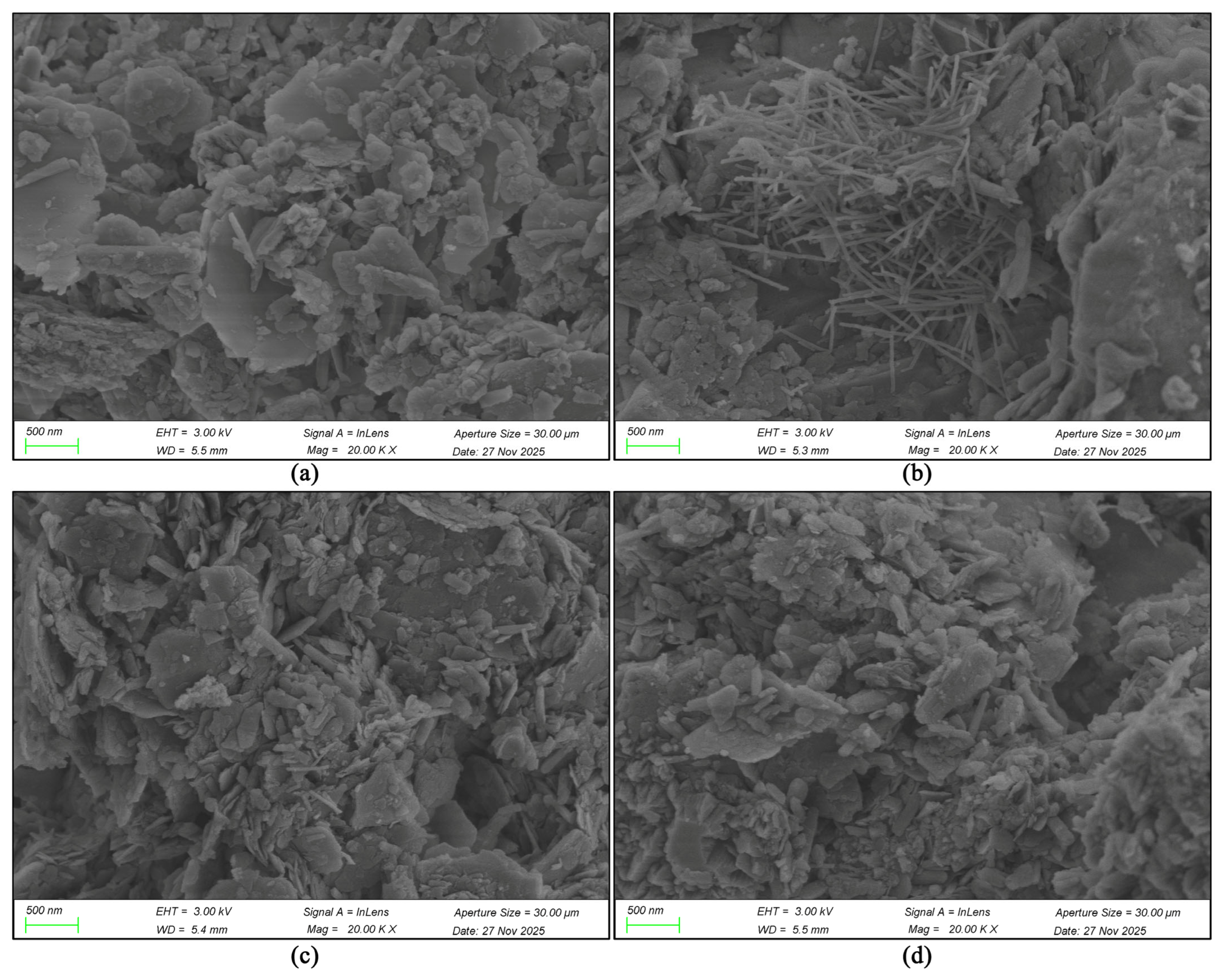
Appendix A records the complete SEM magnification sequence used to trace the four materials from dispersed aggregates to nanoscale particle habits (correspond to the content in Section 3.1). Each montage compares Samples 1–4 under the same accelerating voltage and magnification, allowing morphology, aggregation, and contact geometry to be followed consistently across scale.
At 50×, the deposited powders already display contrasting aggregate habits (Figure A1). Sample 1 contains scattered angular and platy clasts with a broad apparent size range. Sample 2 includes fewer blocky particles and a conspicuous elongated fragment. Sample 3 contains numerous irregular fine agglomerates distributed around larger clasts, whereas Sample 4 is dominated by more isolated compact particles. The low-magnification fields establish the broad heterogeneity later quantified by laser diffraction, particularly the coexistence of large and small material in Sample 1.
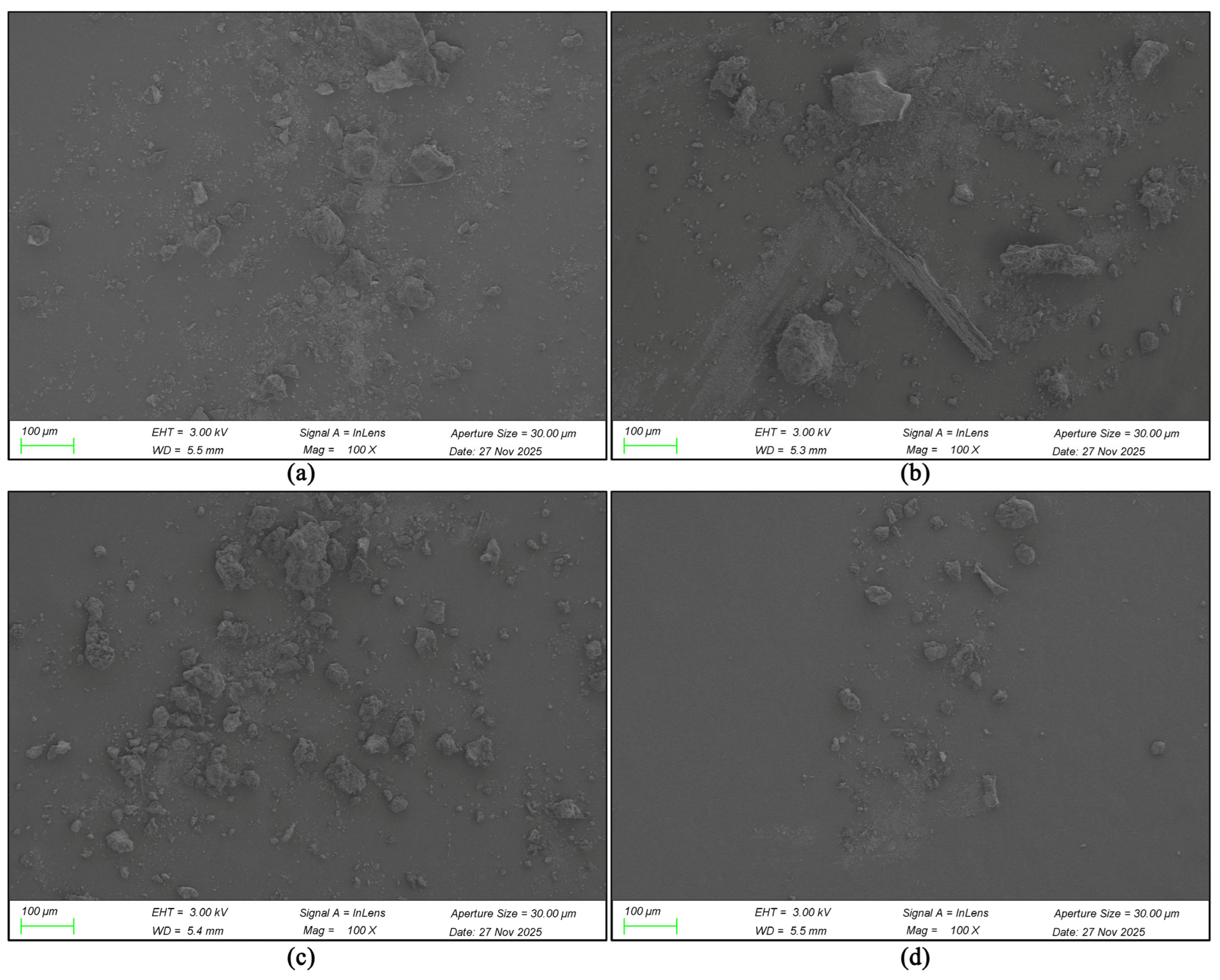
At 100×, particle outlines become clearer (Figure A2). Sample 1 contains blocky fragments accompanied by fine debris; Sample 2 preserves elongated splinter-like particles among irregular grains; Sample 3 forms a dense population of rough agglomerates; and Sample 4 contains relatively discrete clusters separated by wide gaps. The image sequence begins to separate the coarse skeletal character of Sample 1 from the more fine-aggregate character of Samples 3 and 4.
Figure A1.
Comparison of SEM images of four rammed earth wall samples at 50× magnification. (a) SEM morphology of particles in sample 1 (WD is 5.5 mm); (b) SEM morphology of particles in sample 2 (WD is 5.3 mm); (c) SEM morphology of particles in sample 3 (WD is 5.4 mm); (d) SEM morphology of particles in sample 4 (WD is 5.5 mm). Accelerating voltage = 3.0 kV; InLens detector (image source: compiled by the author based on SEM analysis results).
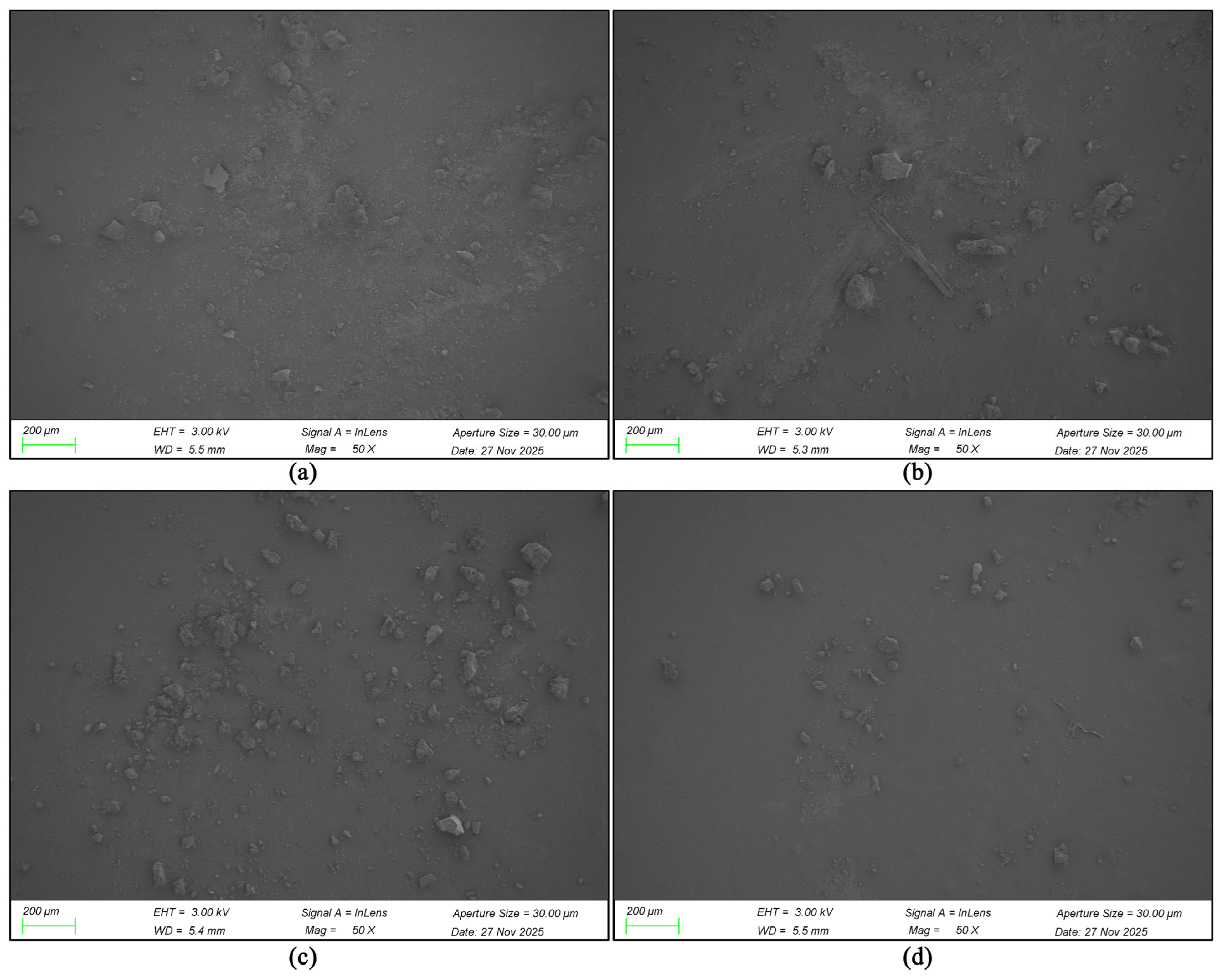
Figure A2.
Comparison of SEM images of four rammed earth wall samples at 100× magnification. (a) SEM morphology of particles in sample 1 (WD is 5.5 mm); (b) SEM morphology of particles in sample 2 (WD is 5.3 mm); (c) SEM morphology of particles in sample 3 (WD is 5.4 mm); (d) SEM morphology of particles in sample 4 (WD is 5.5 mm). Accelerating voltage = 3.0 kV; InLens detector (image source: compiled by the author based on SEM analysis results).
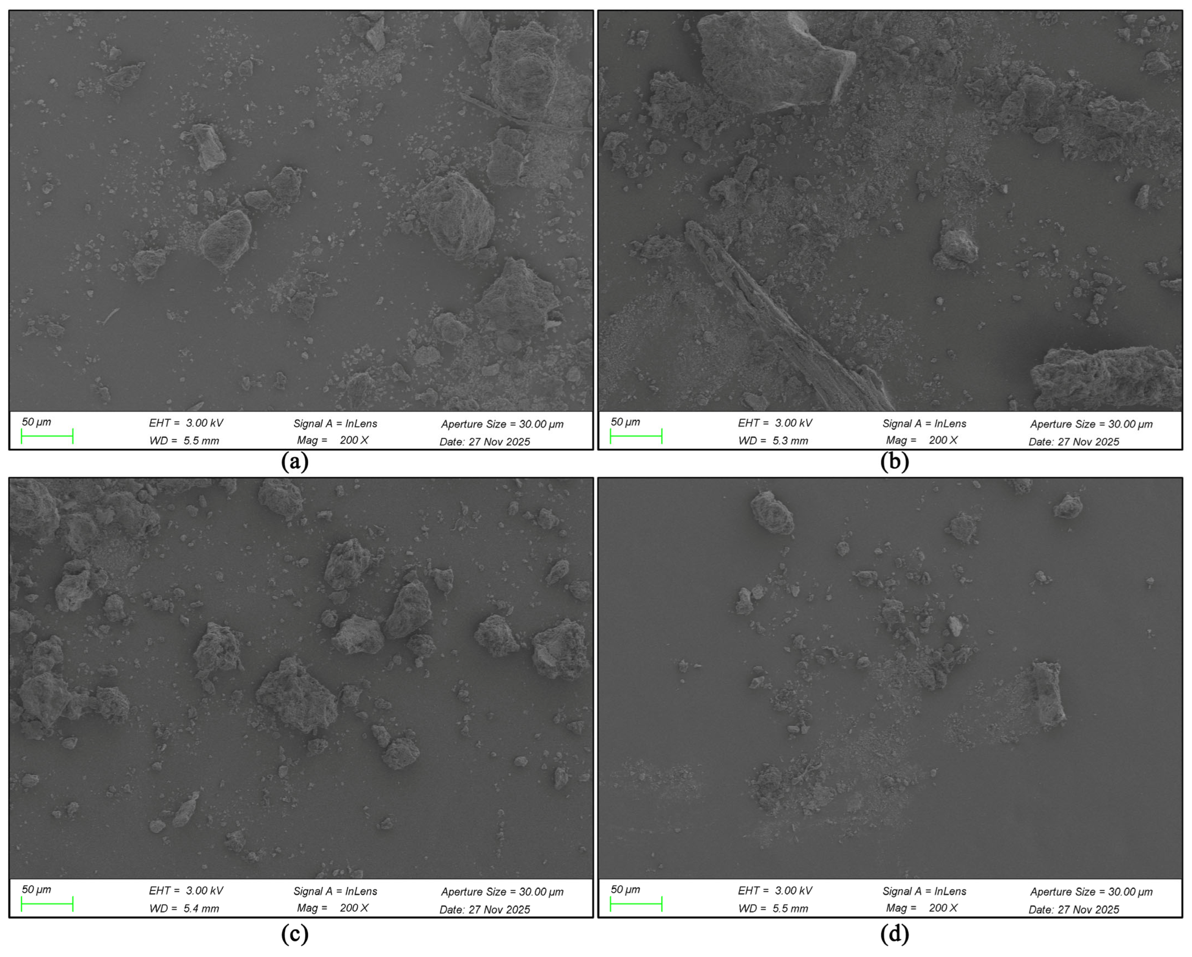
At 200×, individual aggregate architecture is resolved (Figure A3). Sample 1 contains subangular composite grains with smaller particles adhering to their surfaces. Sample 2 combines flaky clasts with a long fibre- or rod-like body, foreshadowing the elongated network visible at high magnification. Sample 3 is dominated by rough, multi-particle agglomerates, while Sample 4 contains smaller, more dispersed clusters. These fields show that the high-resolution particle habits occur within morphologically distinct parent aggregates.
Figure A3.
Comparison of SEM images of four rammed earth wall samples at 200× magnification. (a) SEM morphology of particles in sample 1 (WD is 5.5 mm); (b) SEM morphology of particles in sample 2 (WD is 5.3 mm); (c) SEM morphology of particles in sample 3 (WD is 5.4 mm); (d) SEM morphology of particles in sample 4 (WD is 5.5 mm). Accelerating voltage = 3.0 kV; InLens detector (image source: compiled by the author based on SEM analysis results).
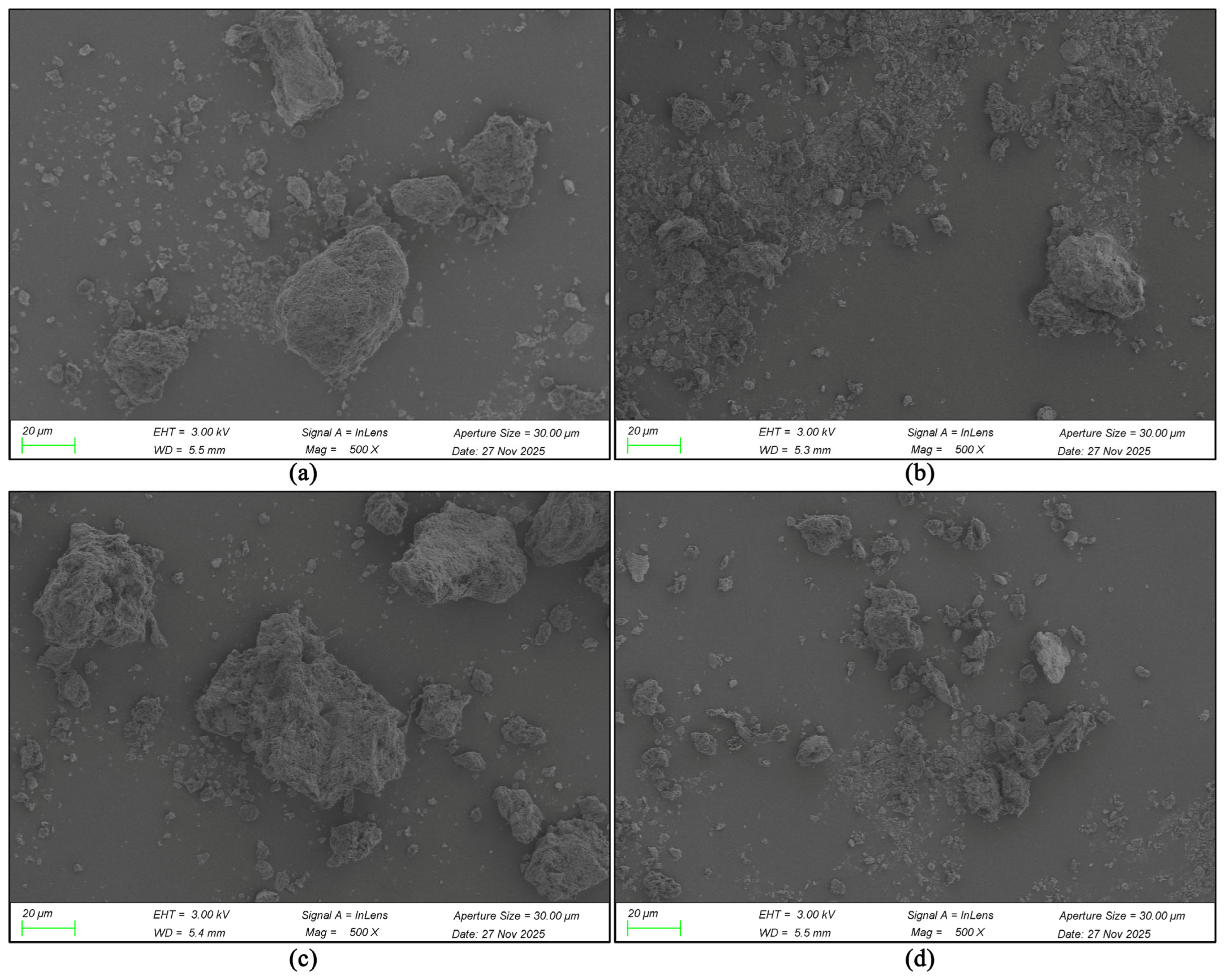
At 500×, surface roughness and intraparticle voids become prominent (Figure A4). Sample 1 contains an angular grain embedded in abundant fine fragments; Sample 2 contains a larger loose aggregate with irregular protrusions; Sample 3 forms massive porous agglomerates whose edges are composed of much finer units; and Sample 4 contains mixed granular and platy clusters. The fields distinguish the fragment-rich Sample 1 fabric from the more open-textured Sample 3 agglomerates.
Figure A4.
Comparison of SEM images of four rammed earth wall samples at 500× magnification. (a) SEM morphology of particles in sample 1 (WD is 5.5 mm); (b) SEM morphology of particles in sample 2 (WD is 5.3 mm); (c) SEM morphology of particles in sample 3 (WD is 5.4 mm); (d) SEM morphology of particles in sample 4 (WD is 5.5 mm). Accelerating voltage = 3.0 kV; InLens detector (image source: compiled by the author based on SEM analysis results).
At 1000×, all samples reveal hierarchical roughness, but their aggregate density and particle organisation diverge (Figure A5). Samples 1 and 2 contain relatively compact individual agglomerates with corrugated surfaces. Sample 3 shows a larger, open-textured mass composed of overlapping fine units, whereas Sample 4 contains smaller, partly separated agglomerates. This scale is the transition from whole grains to the platy, fibrous, and granular substructures that dominate the subsequent images.
Figure A5.
Comparison of SEM images of four rammed earth wall samples at 1000× magnification. (a) SEM morphology of particles in sample 1 (WD is 5.5 mm); (b) SEM morphology of particles in sample 2 (WD is 5.3 mm); (c) SEM morphology of particles in sample 3 (WD is 5.4 mm); (d) SEM morphology of particles in sample 4 (WD is 5.5 mm). Accelerating voltage = 3.0 kV; InLens detector (image source: compiled by the author based on SEM analysis results).
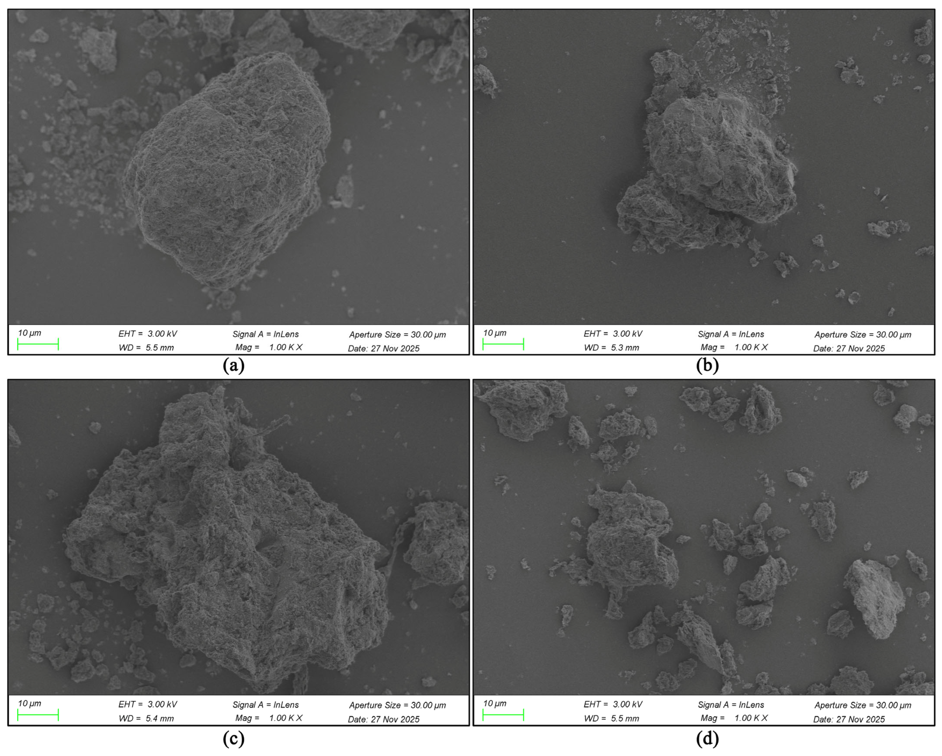
At 2000×, Sample 1 resolves into a compact layered aggregate with parallel surface ridges (Figure A6). Sample 2 shows a fractured, irregular body with flakes and projecting fragments. Sample 3 contains a rough composite of densely interlocked plates, while Sample 4 displays a looser aggregate with curled or fibrous margins. Dark interparticle recesses are most apparent around the rougher Sample 3 and Sample 4 surfaces, indicating more tortuous local contact networks.
Figure A6.
Comparison of SEM images of four rammed earth wall samples at 2000× magnification. (a) SEM morphology of particles in sample 1 (WD is 5.5 mm); (b) SEM morphology of particles in sample 2 (WD is 5.3 mm); (c) SEM morphology of particles in sample 3 (WD is 5.4 mm); (d) SEM morphology of particles in sample 4 (WD is 5.5 mm). Accelerating voltage = 3.0 kV; InLens detector (image source: compiled by the author based on SEM analysis results).
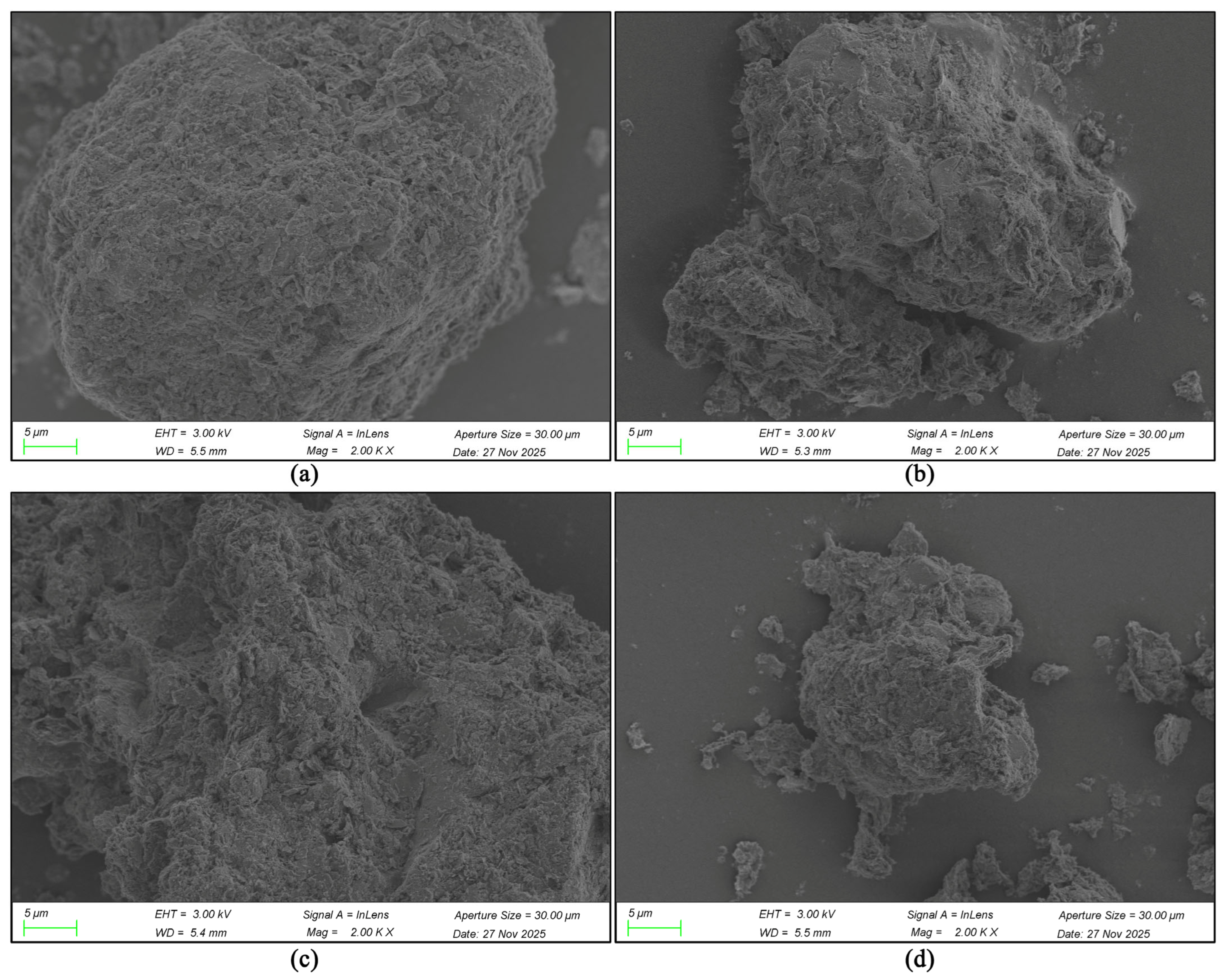
At 5000×, the constituent microtextures become diagnostic (Figure A7). Sample 1 combines small plates and granular fragments in a compact matrix. Sample 2 contains numerous elongated needle- or rod-like features emerging between plates. Sample 3 consists of interlocking, wrinkled lamellae, and Sample 4 forms an open assembly of curled plates and short elongated units. The persistence of these habits across the field confirms that the contrasts are structural characteristics of the sampled aggregates.
Figure A7.
Comparison of SEM images of four rammed earth wall samples at 5000× magnification. (a) SEM morphology of particles in sample 1 (WD is 5.5 mm); (b) SEM morphology of particles in sample 2 (WD is 5.3 mm); (c) SEM morphology of particles in sample 3 (WD is 5.4 mm); (d) SEM morphology of particles in sample 4 (WD is 5.5 mm). Accelerating voltage = 3.0 kV; InLens detector (image source: compiled by the author based on SEM analysis results).
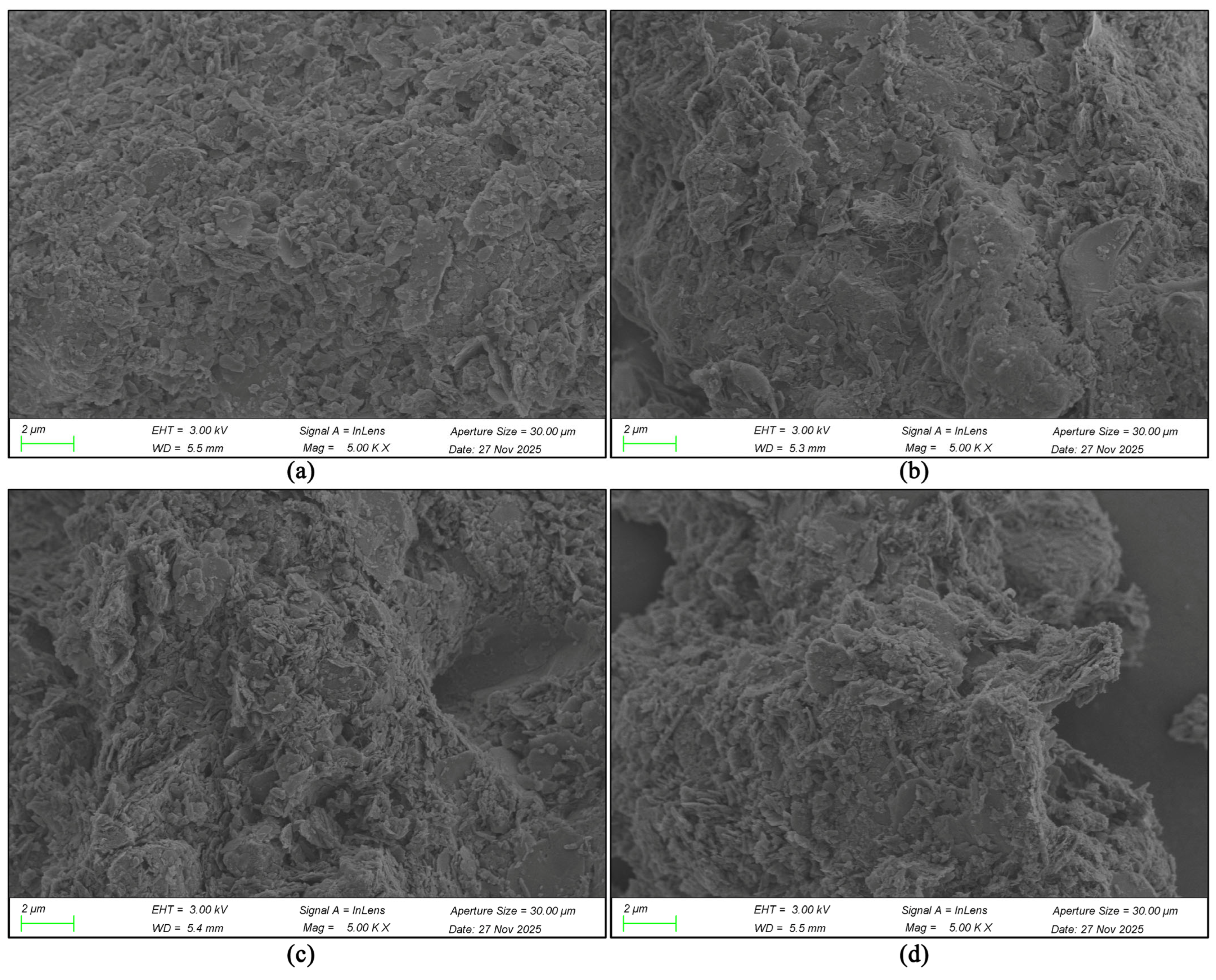
At 10,000×, Sample 1 displays stacked plates coated by fine equant particles (Figure A8). Sample 2 shows a mixed plate-and-rod fabric with rods crossing voids and particle faces. Sample 3 forms dense rosette-like clusters of thin lamellae, whereas Sample 4 contains strongly folded plates and open edge contacts. The plates in Samples 1, 3, and 4 differ in thickness and packing, while the elongated component makes Sample 2 immediately distinguishable.
Figure A8.
Comparison of SEM images of four rammed earth wall samples at 10,000× magnification. (a) SEM morphology of particles in sample 1 (WD is 5.5 mm); (b) SEM morphology of particles in sample 2 (WD is 5.3 mm); (c) SEM morphology of particles in sample 3 (WD is 5.4 mm); (d) SEM morphology of particles in sample 4 (WD is 5.5 mm). Accelerating voltage = 3.0 kV; InLens detector (image source: compiled by the author based on SEM analysis results).
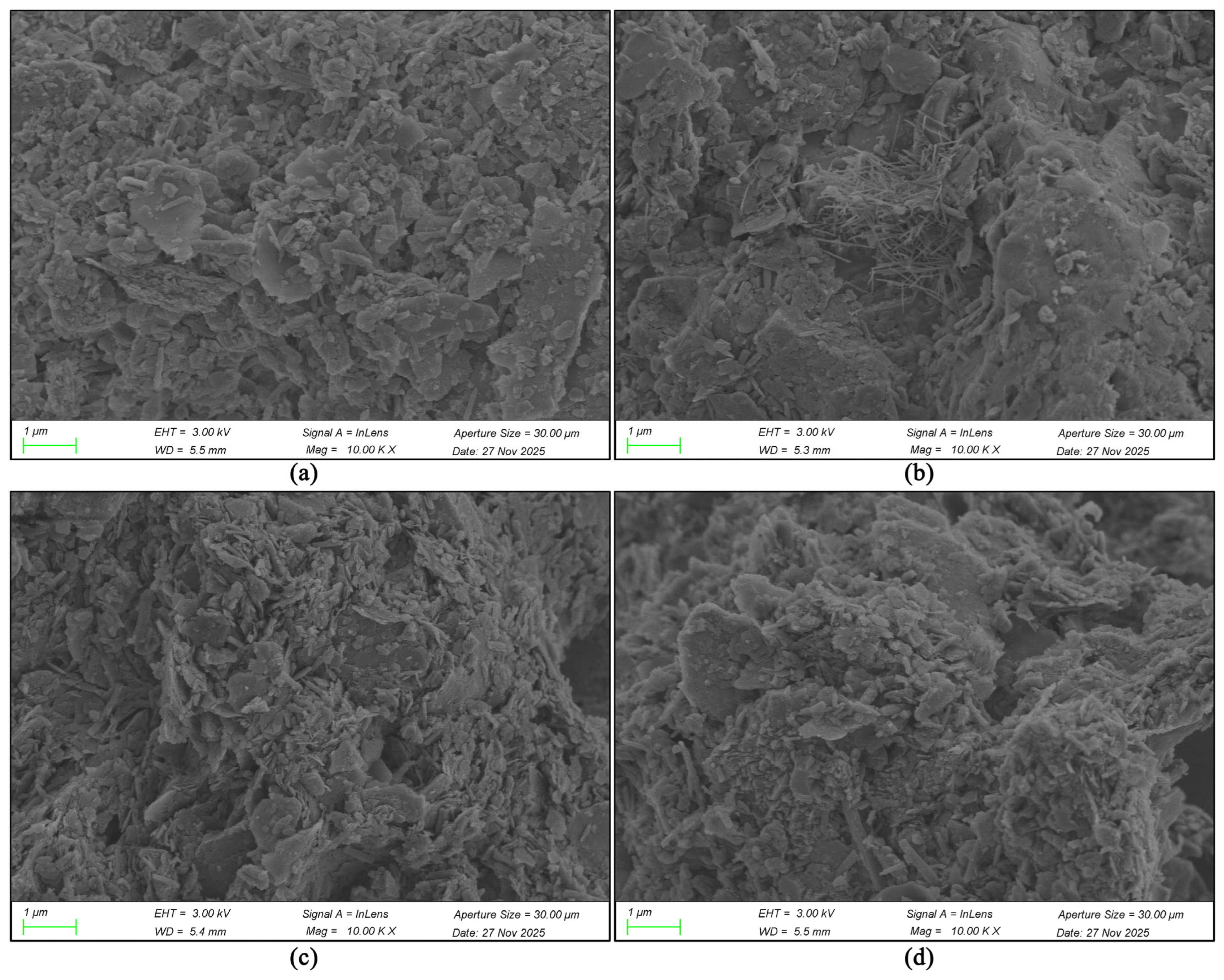
At 20,000×, the four microstructural signatures are fully resolved (Figure A9). Sample 1 is dominated by broad plates and granular attachments; Sample 2 by a dense mat of intersecting rods; Sample 3 by compact intergrowths of short plates; and Sample 4 by a mixed population of short rods, flakes, and rough clusters. Rods in Sample 2 span interparticle gaps, whereas Sample 3 is dominated by overlapping face and edge contacts. These observations document distinct local packing geometries.
Figure A9.
Comparison of SEM images of four rammed earth wall samples at 20,000× magnification. (a) SEM morphology of particles in sample 1 (WD is 5.5 mm); (b) SEM morphology of particles in sample 2 (WD is 5.3 mm); (c) SEM morphology of particles in sample 3 (WD is 5.4 mm); (d) SEM morphology of particles in sample 4 (WD is 5.5 mm). Accelerating voltage = 3.0 kV; InLens detector (image source: compiled by the author based on SEM analysis results).
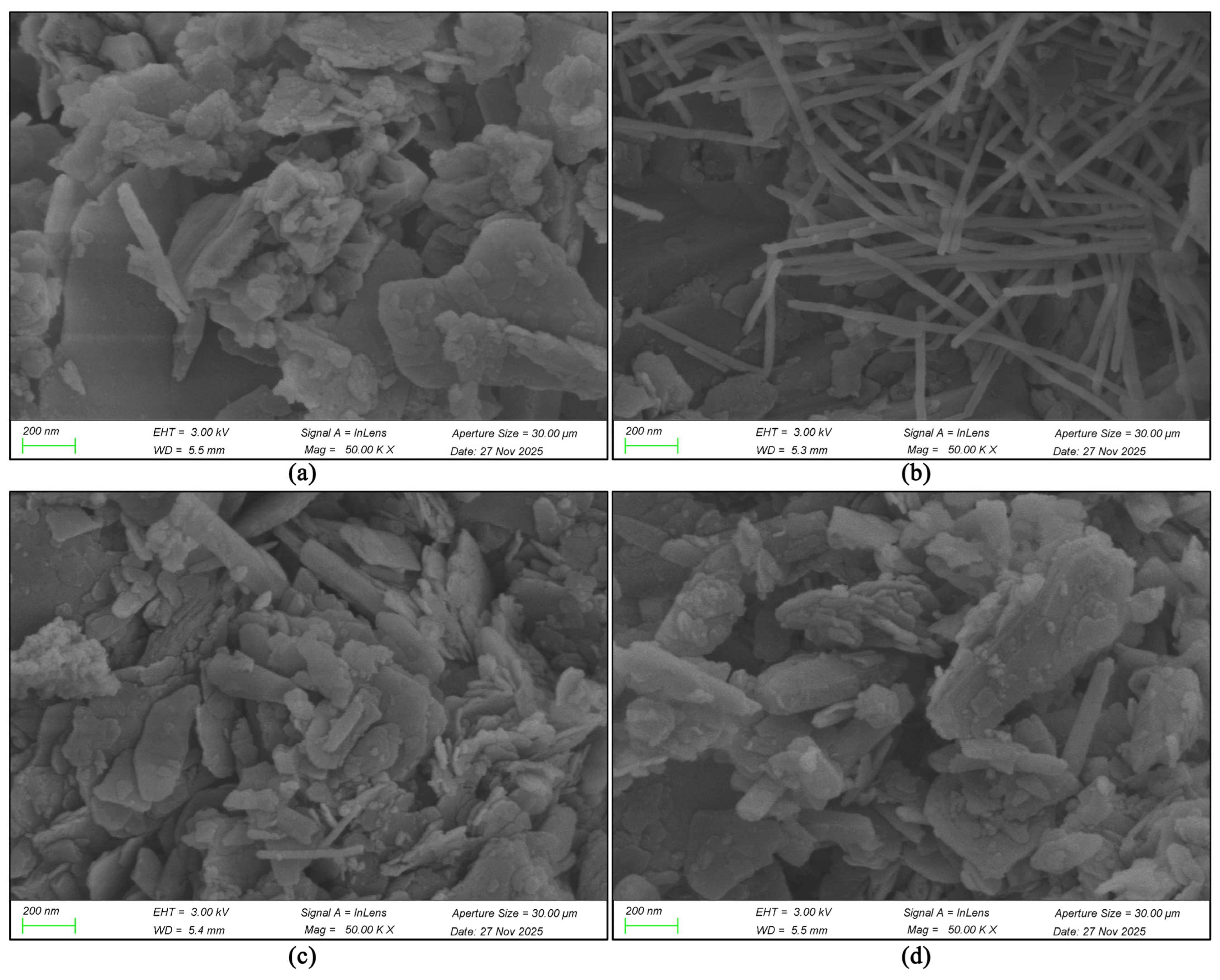
At 50,000×, Sample 1 contains thin stacked plates with abundant nanoscale fragments at their margins (Figure A10). Sample 2 retains a highly connected rod network with open spaces between crossing elements. Sample 3 contains short prismatic and platy units arranged in compact clusters, while Sample 4 combines interleaved plates, short rods, and fine surface particles. The montage connects the aggregate-scale sequence to the 100,000× comparison in Figure 3 and documents the continuity of the four characteristic fabrics.
Figure A10.
Comparison of SEM images of four rammed earth wall samples at 50,000× magnification. (a) SEM morphology of particles in sample 1 (WD is 5.5 mm); (b) SEM morphology of particles in sample 2 (WD is 5.3 mm); (c) SEM morphology of particles in sample 3 (WD is 5.4 mm); (d) SEM morphology of particles in sample 4 (WD is 5.5 mm). Accelerating voltage = 3.0 kV; InLens detector. Image source: Compiled by the author based on SEM analysis results.
References
- Chan, W.-Y.; Ma, S.-Y. Heritage preservation and sustainability of China’s development. Sustain. Dev. 2004, 12, 15–31. [Google Scholar] [CrossRef] [Scilit]
- Iossifova, D. Cultural Heritage: Preservation and Development with Chinese Characteristics. (Short Term Policy Briefs); Europe China Research and Advice Network: London, UK, 2014; Available online: https://research.manchester.ac.uk/en/publications/cultural-heritage-preservation-and-development-with-chinese-chara/ (accessed on 4 July 2026).
- Lee, T.H. Education in traditional China. In The Routledge Encyclopedia of Traditional Chinese Culture; Routledge: Beijing, China, 2019; pp. 238–253. [Google Scholar]
- Li, W.; Chen, S.; Cui, Y.T.; Lin, H.; Lv, H.; Ao, J. Academy, private school, Confucius Institute: Analyzing heritage art of educational architectural paintings via event coding. npj Herit. Sci. 2025, 13, 340. [Google Scholar] [CrossRef] [Scilit]
- Peng, Z.W.; Peng, H.; Peng, H.L. Integration and Practice of Lianxi Academy Culture and Moral Education in Colleges and Universities in the Digital Age. Cult. Relics Apprais. Apprec. 2025, 95–98. Available online: https://kns.cnki.net/kcms2/article/abstract?v=pX0ZfcSLBkXwjMaiSkG7ZIKVWA6zyOEK7he_DiEe0vLlt9gyUczpu2Kvq7gUWdho6u8PDDUZj2p8ujSkTRxAtX51wsYpZf8yrh27HHnGt0d5pPsQRFYZaZzeAue4SYw_eQCnRDRMTncZ7JF-vONtPyM6X9_r5YAMdCrtW2LLzJNbv4rzAb2jbg==&uniplatform=NZKPT&language=CHS (accessed on 14 August 2026). (In Chinese)
- Yang, L.L. A Study on the Development Path of Chinese Academies from the Perspective of Urban Memory. Master’s Thesis, Fujian Normal University, Fuzhou, China, 2022. Available online: https://kns.cnki.net/kcms2/article/abstract?v=pX0ZfcSLBkU3rgglqz7R0DarO1rqON62tkWqo3PpY9x3WckovI6SnL7W0fxwvPx4eAkeXkv7ybzyBixsUCUjjzbQ7EZ0QPrIKeKcfnYrql0Plz9l9ba2ZwvhVJTWeBy8Dh4W7_qpKwF_pXBKKcdAts0BespJCkO7mMpjqdQ1A-PxBfgAxtpFuIrylnLybtMn&uniplatform=NZKPT&language=CHS (accessed on 14 August 2026). (In Chinese)
- Deng, H.B.; Zheng, M.X.; Lou, Z.Y. A Survey on the Current Status of Protection and Utilization of Ancient Chinese Academies. Chin. Cult. Herit. 2014, 50–54. Available online: https://kns.cnki.net/kcms2/article/abstract?v=pX0ZfcSLBkWxu5ArKMWJkOA06PU7GVEjjgxxjhLOKOtVkBzk0KIPxeEzAsXruJmHLDWTnEqIlH4ZkcMhwM9PNNBviUwhk5aifDLXgVR7vJ0QXNKa22K5DINMHDwXw7sOU4sCl3KD8BwXcX-fNooFC4uwirayDV7nb3I44p237ReLKHRnNmRH-w==&uniplatform=NZKPT&language=CHS (accessed on 14 August 2026). (In Chinese)
- Wu, Y. A Brief Study on the History of Lianxi Academy in Hechuan, Chongqing. J. Hunan Univ. Sci. Technol. 2021, 42, 6–9. Available online: https://kns.cnki.net/kcms2/article/abstract?v=pX0ZfcSLBkVmK2YIYwS_s1GVEQt2x4zLe1TE7t25MuZI433opTkgxdsNxc_ghxJoKSwa_CU5DF_e3MVAl7llPb6CMp8H_jJtb4LagoCzflPiTkad5rvuvBkGqxc9Am7fyN3tXG4OMLnXWAjfDkiL5Pdu3poVyzTSmVM0N7mQSE5OpwtImlp-KQ==&uniplatform=NZKPT&language=CHS (accessed on 14 August 2026). (In Chinese)
- Hao, B. A Study on the Protection and Reuse of Traditional Academies in Hunan and Guangdong Regions from the Perspective of Spatial Heritage. Master’s Thesis, Huazhong University of Science and Technology, Wuhan, China, 2021. Available online: https://kns.cnki.net/kcms2/article/abstract?v=pX0ZfcSLBkVATSxj4BjJyrf2Uyt6FJOnUauMaWEfxQWbfVJLP-1MTLxGmALa0AQNq5Aa1Z53s9XItHT2LQBQNPlvI5dBedwNl_LX0QW70Txcm2va13KZaPkG-uOexE0zDBXBiTiGg-hTSizpMzzpECi9CB6cPJGmmc0C9MR21K_q15S7tDbI0b5HRuuaPYpq&uniplatform=NZKPT&language=CHS (accessed on 14 August 2026). (In Chinese)
- Zhao, J.; Ye, L. Design for the Reinforcement and Restoration of Gushan Academy in Xinchang. Archit. Shanxi 2018, 44, 4–5. (In Chinese) [Google Scholar]
- Cai, X.; Jiang, N. Research on the Protection of Lingnan Architectural Cultural Heritage Based on Digital Technology. In Proceedings of the 2nd Guangdong-Hong Kong-Macao Greater Bay Area International Conference on Digital Economy and Artificial Intelligence, Dongguan, China, 28–30 March 2025; pp. 1253–1259. [Google Scholar] [CrossRef] [Scilit]
- Zhang, Y.Q.; Dechsubha, T. The Architectural Space Art Characteristics of Foshan Academies. Int. J. Sociol. Anthropol. Sci. Rev. 2025, 5, 111–122. [Google Scholar] [CrossRef] [Scilit]
- Zhao, Y.Z.; Peng, H.; Xiang, Q. A Study on the Digital Dissemination of Lianxi Academy Culture—Taking Chenzhou City as an Example. Cult. Relics Apprais. Apprec. 2024, 150–153. Available online: https://kns.cnki.net/kcms2/article/abstract?v=pX0ZfcSLBkWuo4C9sKqLUMSC07vLwyB3lymRSS2s3Szkt1AiRodw6kqrtt2CrgT9uJ7AqcETjbOg9Vms4MnL8LOVBamNWOJ1ZG3U3GgDvAMU4-6l5EK4PFvS6BdqMGNwQxNAm5tcGSjBRP3RaSkdZEdKKDZC8kXx25EjczCjfmZ8Jd_ScCi5Hg==&uniplatform=NZKPT&language=CHS (accessed on 14 August 2026). (In Chinese)
- Yao, X.L. Drawing Wisdom from Lianxi Culture to Realize the Dream of Hunan Education. Market Information Daily, 18 December 2024; p. 006. Available online: https://kns.cnki.net/kcms2/article/abstract?v=pX0ZfcSLBkUe7qx82qEQVprZngysZRIE0wuzZ_sJkwBkSUPMrvFzCvCjbhA0d9K--WpanOY75hlpC0XwrpCUzIh1rb3Gm2MI0Ko5Bt_VMogjv61kCbeso8evUFzAQmqw3Xns_SNhguFhB0WUUzNv1yKXyL_DHLyYA7OndQTDis0UW0eAMIhOB6JAHgW6bQWO&uniplatform=NZKPT&language=CHS (accessed on 14 August 2026). (In Chinese)
- Chen, C.; Zuo, J. The symmetrical beauty and its cultural connotation of lingnan architecture. In Proceedings of the 2018 International Conference on Management, Economics, Education, Arts and Humanities (MEEAH 2018); Atlantis Press: Dordrecht, The Netherlands, 2018; pp. 143–149. [Google Scholar] [CrossRef] [Scilit]
- Tang, X.X. Three adaptabilities of the traditional vernacular architecture of the Han nationality in Lingnan. Appl. Mech. Mater. 2014, 644, 5109–5112. [Google Scholar] [CrossRef] [Scilit]
- Zheng, L.; Deng, Q.; Liang, J.; Guo, Z.; Zhu, Y.; Liu, W.; Chen, Y. Microchemical Analysis of Rammed Earth Residential Walls Surface in Xiaochikan Village, Guangdong. Coatings 2025, 15, 1351. [Google Scholar] [CrossRef] [Scilit]
- Martín-del-Rio, J.J.; Canivell, J.; Torres-González, M.; Mascort-Albea, E.J.; Romero-Hernández, R.; Alducin-Ochoa, J.M.; Alejan-dre-Sánchez, F.J. Analysis of the Materials and State of Conservation of the Medieval Rammed Earth Walls of Seville (Spain). J. Build. Eng. 2021, 44, 103381. [Google Scholar] [CrossRef] [Scilit]
- Beckett, C.T.S.; Jaquin, P.A.; Morel, J.-C. Weathering the Storm: A Framework to Evaluate the Resistance of Earthen Walls to Water Damage. Constr. Build. Mater. 2020, 242, 118098. [Google Scholar] [CrossRef] [Scilit]
- Lo, C.K.N.; Song, J. Weathering and Restoration of Traditional Rammed-Earth Walls in Fujian, China. Coatings 2025, 15, 1491. [Google Scholar] [CrossRef] [Scilit]
- Luo, Y.; Yang, M.; Ni, P.; Peng, X.; Yuan, X. Degradation of Rammed Earth under Wind-Driven Rain: The Case of Fujian Tulou, China. Constr. Build. Mater. 2020, 261, 119989. [Google Scholar] [CrossRef] [Scilit]
- Yan, L.; Zeng, H.; Yin, J.; Zhang, Y.; Jia, X. Microclimatic Effects and Durability of Surface Soil Materials in Fujian Tulou Rammed-Earth Wall. Coatings 2026, 16, 301. [Google Scholar] [CrossRef] [Scilit]
- Ciancio, D.; Jaquin, P.; Walker, P. Advances on the Assessment of Soil Suitability for Rammed Earth. Constr. Build. Mater. 2013, 42, 40–47. [Google Scholar] [CrossRef] [Scilit]
- Hall, M.; Djerbib, Y. Moisture Ingress in Rammed Earth: Part 1—The Effect of Soil Particle-Size Distribution on the Rate of Capillary Suction. Constr. Build. Mater. 2004, 18, 269–280. [Google Scholar] [CrossRef] [Scilit]
- Cuccurullo, A.; Gallipoli, D.; Bruno, A.W.; Augarde, C.; Hughes, P.; La Borderie, C. A Comparative Study of the Effects of Particle Grading and Compaction Effort on the Strength and Stiffness of Earth Building Materials at Different Humidity Levels. Constr. Build. Mater. 2021, 306, 124770. [Google Scholar] [CrossRef] [Scilit]
- Indekeu, M.L.; Feng, C.; Janssen, H.; Woloszyn, M. Experimental Study on the Capillary Absorption Characteristics of Rammed Earth. Constr. Build. Mater. 2021, 283, 122689. [Google Scholar] [CrossRef] [Scilit]
- Martín-del-Rio, J.J.; Flores-Alés, V.; Alejandre-Sánchez, F.J.; Blasco-López, F.J. New Method for Historic Rammed-Earth Wall Charac-terization: The Almohade Ramparts of Malaga and Seville. Stud. Conserv. 2019, 64, 363–372. [Google Scholar] [CrossRef] [Scilit]
- Daoudi, L.; Rocha, F.; Costa, C.; Arrebei, N.; Fagel, N. Characterization of Rammed-Earth Materials from the XVIth Century Badii Palace in Marrakech, Morocco to Ensure Authentic and Reliable Restoration. Geoarchaeology 2018, 33, 529–541. [Google Scholar] [CrossRef] [Scilit]
- Gomes, R.I.; Silva, A.S.; Gomes, L.; Faria, P. Fernandina Wall of Lisbon: Mineralogical and Chemical Characterization of Rammed Earth and Masonry Mortars. Minerals 2022, 12, 241. [Google Scholar] [CrossRef] [Scilit]
- Bai, X.; He, B.; Feng, Y.; Wang, Y.; Li, H.; Bai, X.; Ma, F.; Han, P.; Lu, Y.; Ren, Y. Basal Scouring in Historical Rammed Earth Sites: Formation and Height Prediction. npj Herit. Sci. 2025, 13, 6. [Google Scholar] [CrossRef] [Scilit]
- Zhai, Z.-H.; Li, X.-Y.; Ip, K.-H. Comparative Analysis of Physical Characteristics of Traditional Rammed Earth Dwellings in Macau. npj Herit. Sci. 2025, 13, 469. [Google Scholar] [CrossRef] [Scilit]
- Chu, X.; Ji, X.; Han, W. Mechanism of Herbaceous Plant Root Disturbance on Yongning Fortress Rammed Earth Heritage: A Case Study. Buildings 2025, 15, 3491. [Google Scholar] [CrossRef] [Scilit]
- De Leo, F.; Dominguez-Moñino, I.; Jurado, V.; Bruno, L.; Saiz-Jimenez, C.; Urzì, C. Fungal Outbreak in the Catacombs of SS. Marcellino and Pietro, Rome (Italy): From Diagnosis to an Emergency Treatment. Front. Microbiol. 2022, 13, 982933. [Google Scholar] [CrossRef] [Scilit] [PubMed]
- Du, Y.; Cui, K.; Chen, S.; Dong, W.; Chen, W. Quantitative Research on the Development Difference of Scaling Off on the Sunward Side and Nightside of Earthen Sites. J. Cult. Herit. 2022, 57, 107–117. [Google Scholar] [CrossRef] [Scilit]
- Bai, X.; Guo, T.; An, Y.; Wang, Y.; Han, P.; Ren, Y.; Wang, X. Unraveling the Dry–Wet-Induced Infiltration Characteristics and Failure Mechanisms of Rammed Earth. Eng. Geol. 2026, 368, 108757. [Google Scholar] [CrossRef] [Scilit]
- Romanazzi, A.; Oliveira, D.V.; Silva, R.A.; Barontini, A. Effectiveness of a TRM Solution for Rammed Earth under In-Plane Cyclic Loads. Constr. Build. Mater. 2023, 407, 133551. [Google Scholar] [CrossRef] [Scilit]
- Lingle, A.; Seifert, J. Stratagems and Back Spoils: Utilizing Backdirt in the Management of Archaeological Earthen Heritage. J. Field Archaeol. 2024, 49, 192–203. [Google Scholar] [CrossRef] [Scilit]
- Liu, S.; Zhang, Y.; Li, M.; Qin, Y.; Li, X.; He, J.; Zhao, X.; Li, J.; Zhu, J. PLS-MCDM-Based Evaluation of Consolidation Materials for Earthen Heritage Sites: Moisture Regulation and Overall Consolidation Performance of SAP. npj Herit. Sci. 2026, 14, 408. [Google Scholar] [CrossRef] [Scilit]
- Gomes, M.I.; Faria, P.; Gonçalves, T.D. Rammed Earth Walls Repair by Earth-Based Mortars: The Adequacy to Assess Effectiveness. Constr. Build. Mater. 2019, 205, 213–231. [Google Scholar] [CrossRef] [Scilit]
- Umubyeyi, C.; Wenger, K.; Dahmen, J.; Ochsendorf, J. Durability of Unstabilized Rammed Earth in Temperate Climates: A Long-Term Study. Constr. Build. Mater. 2023, 409, 133953. [Google Scholar] [CrossRef] [Scilit]
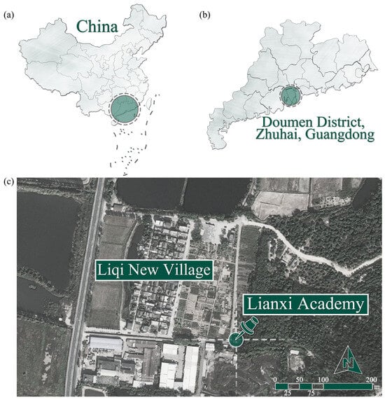
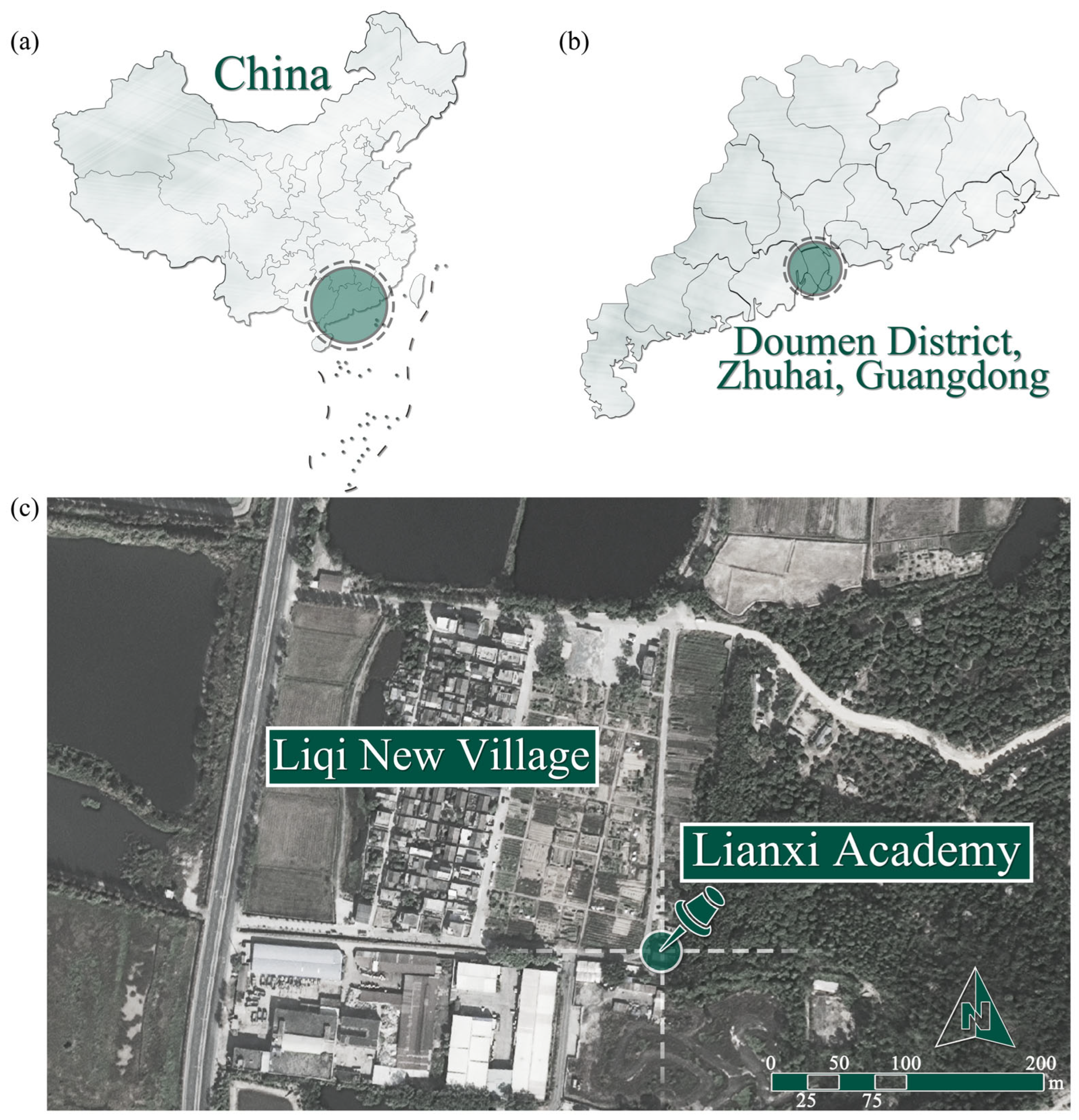
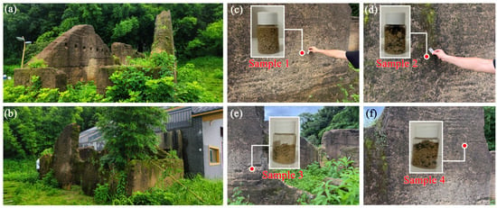
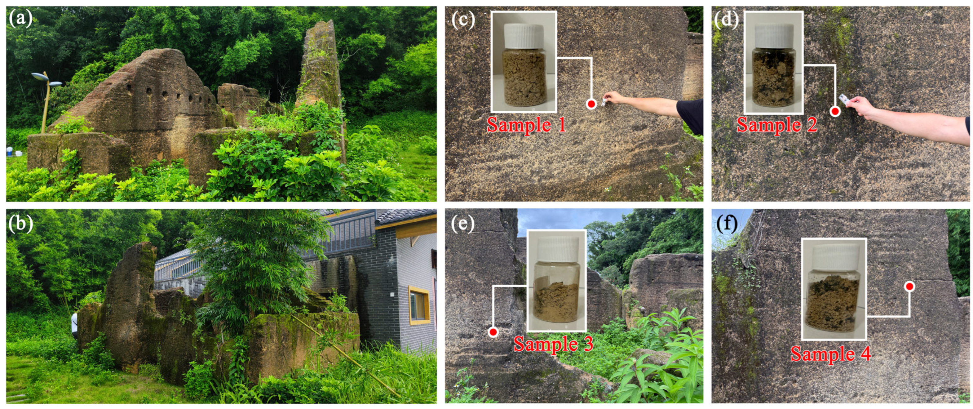
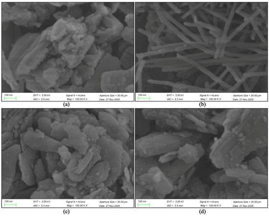
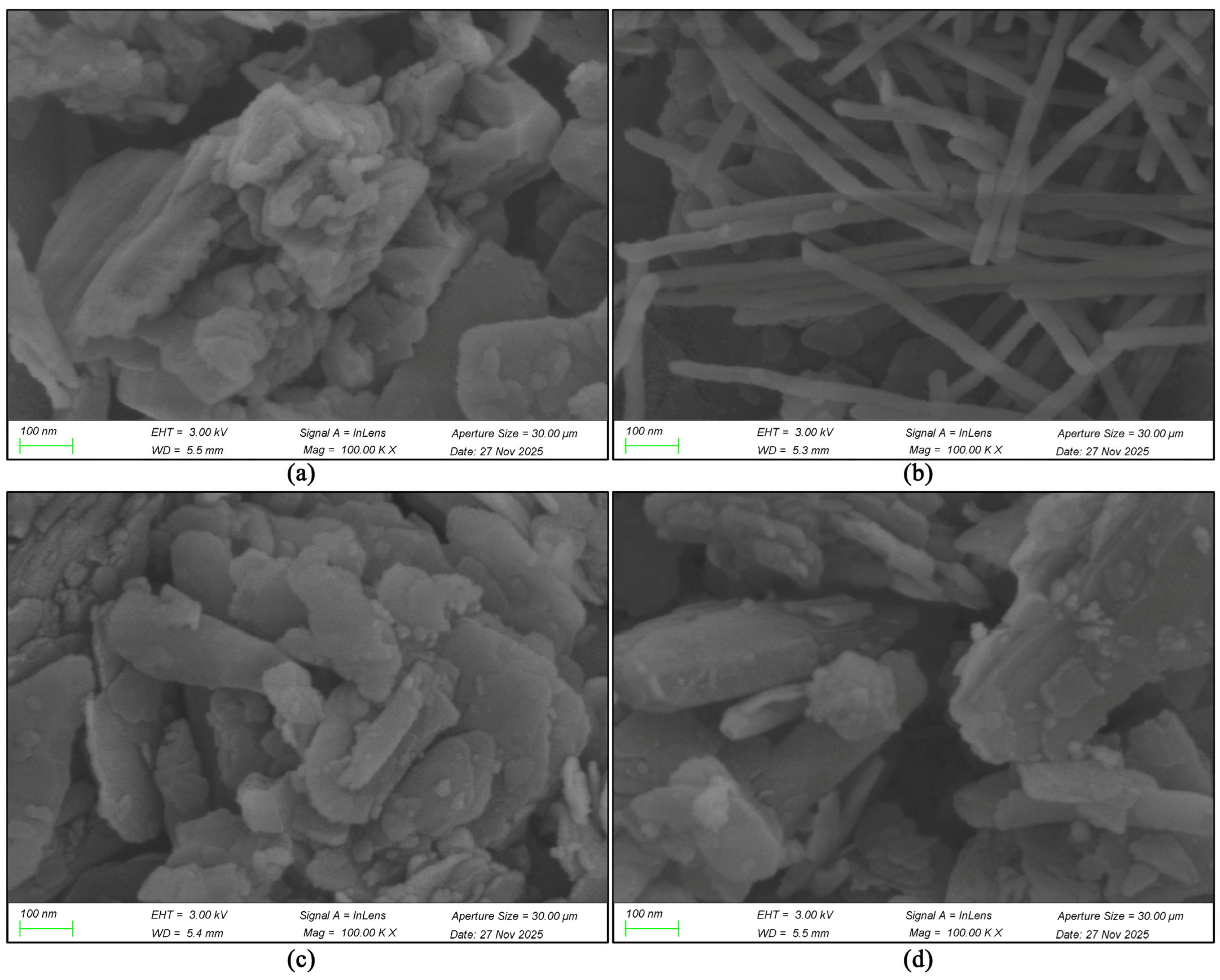
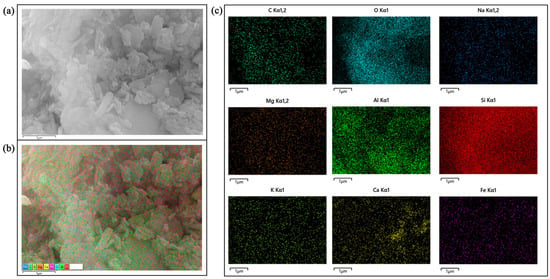
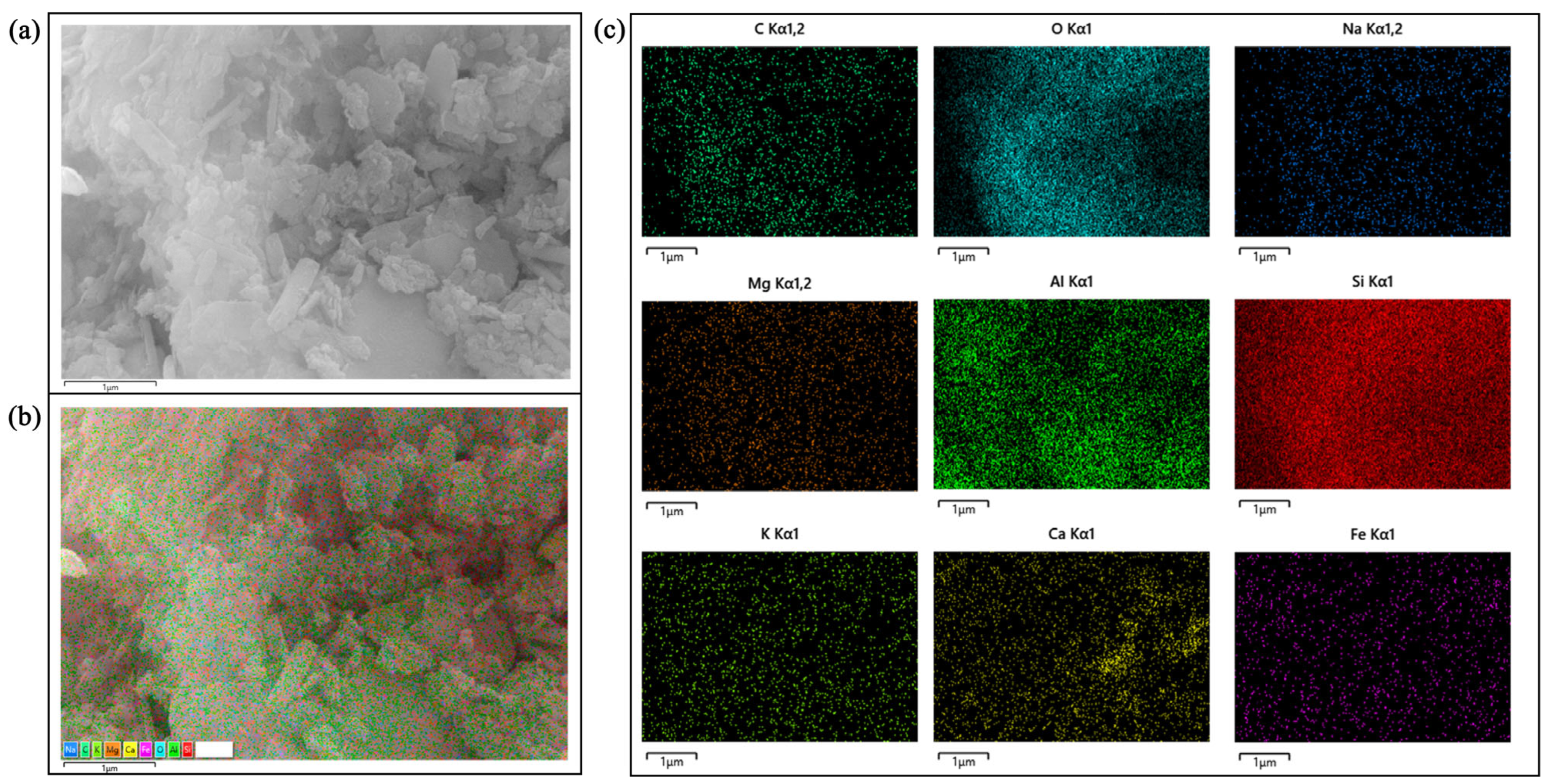
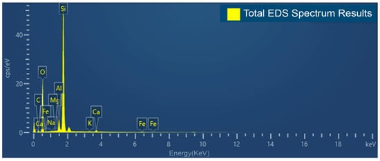
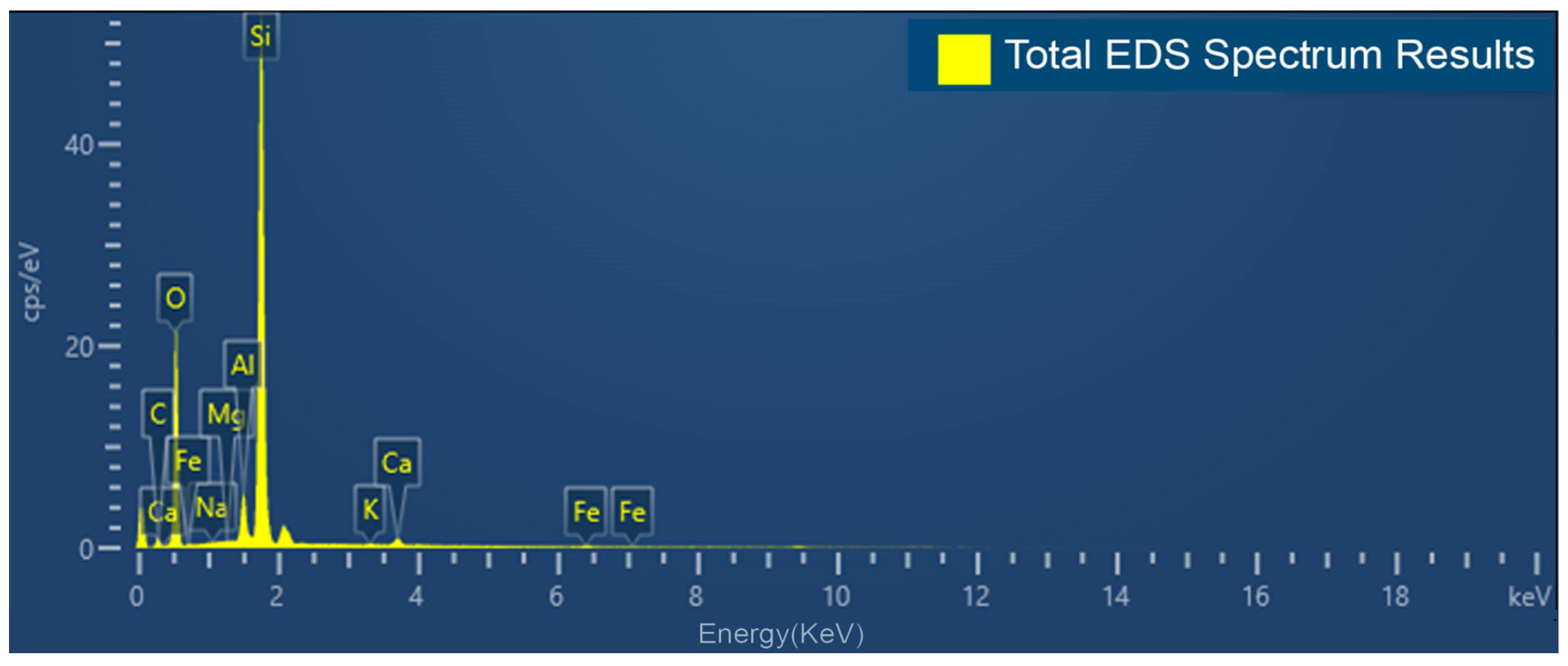
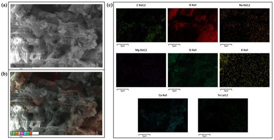
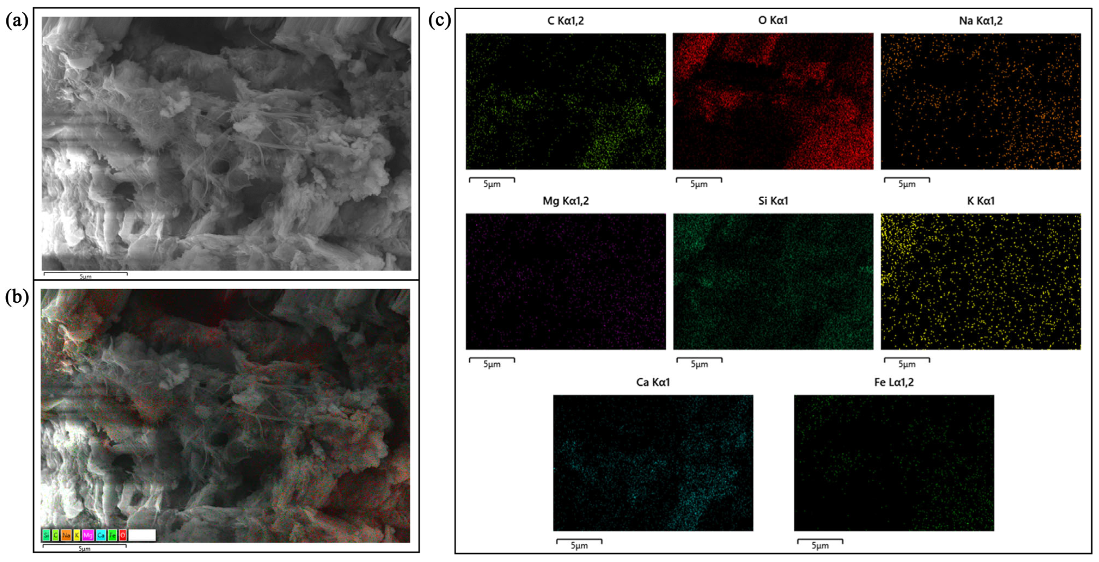
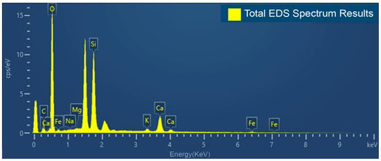
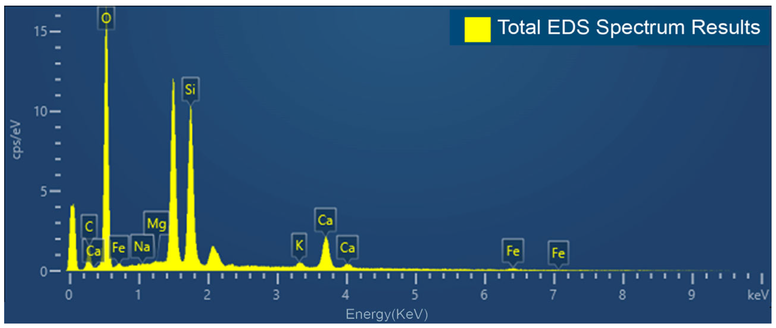
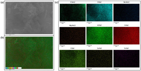
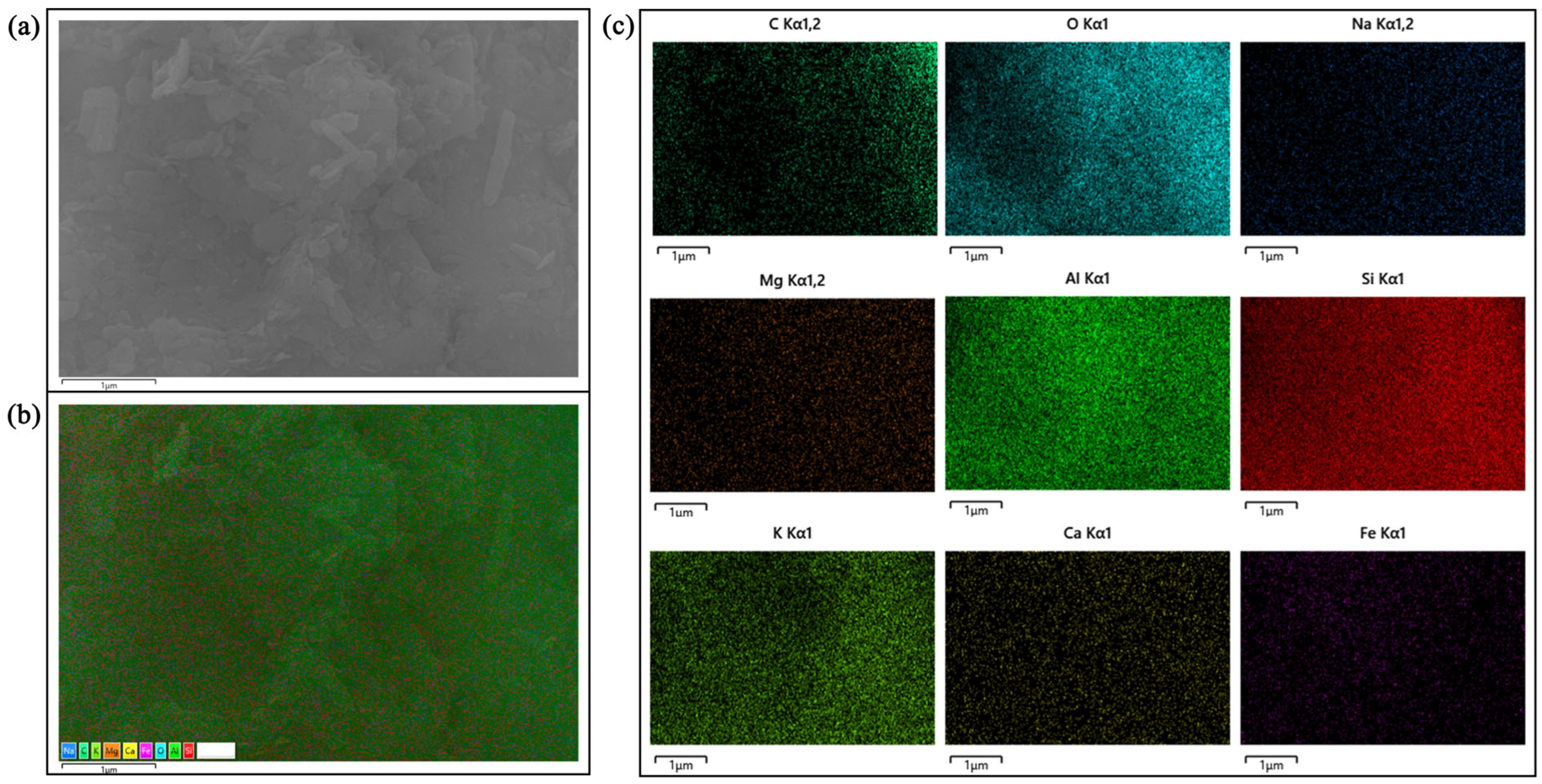
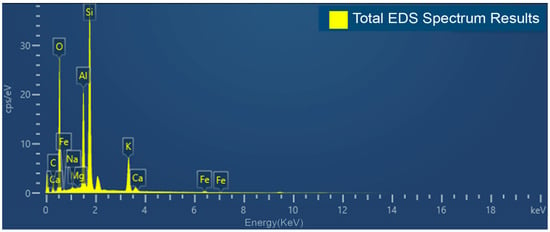
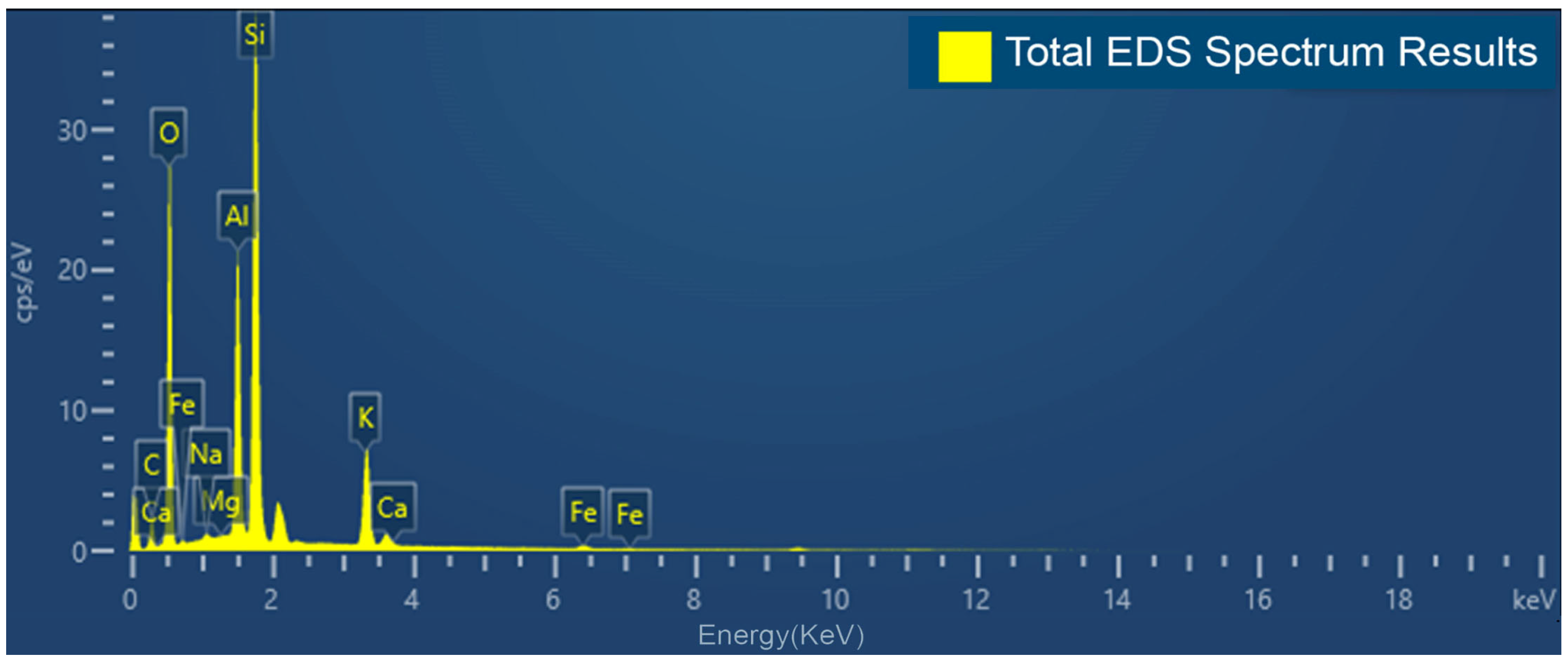
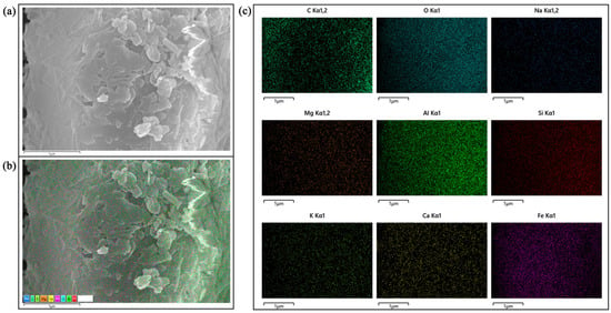
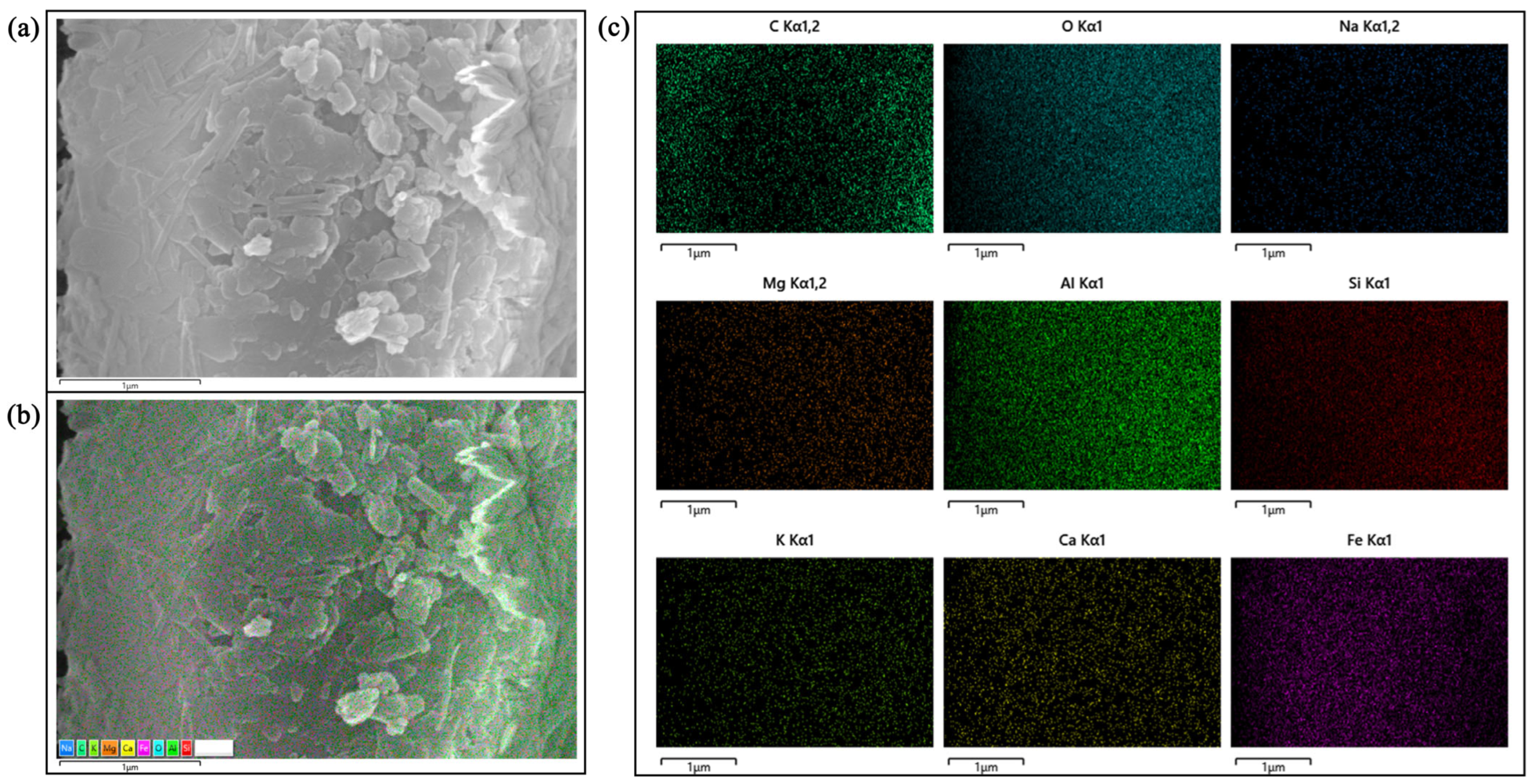
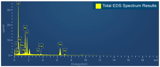
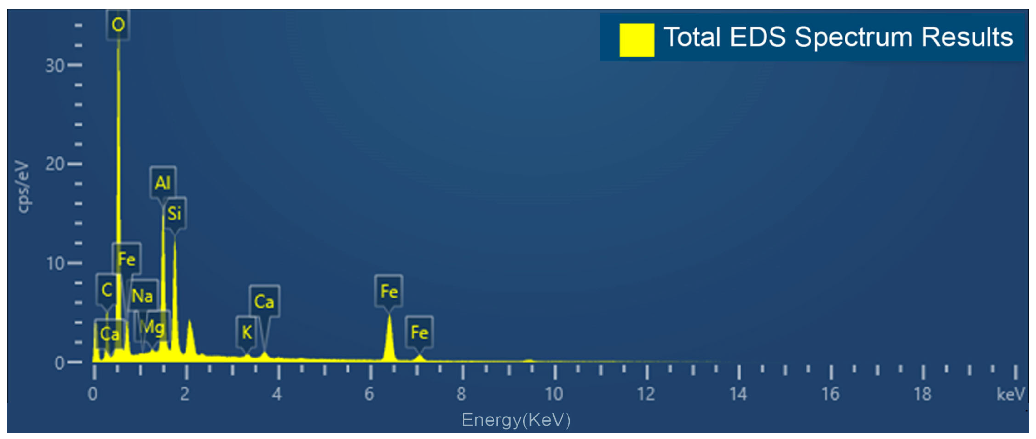
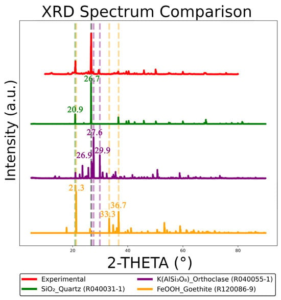
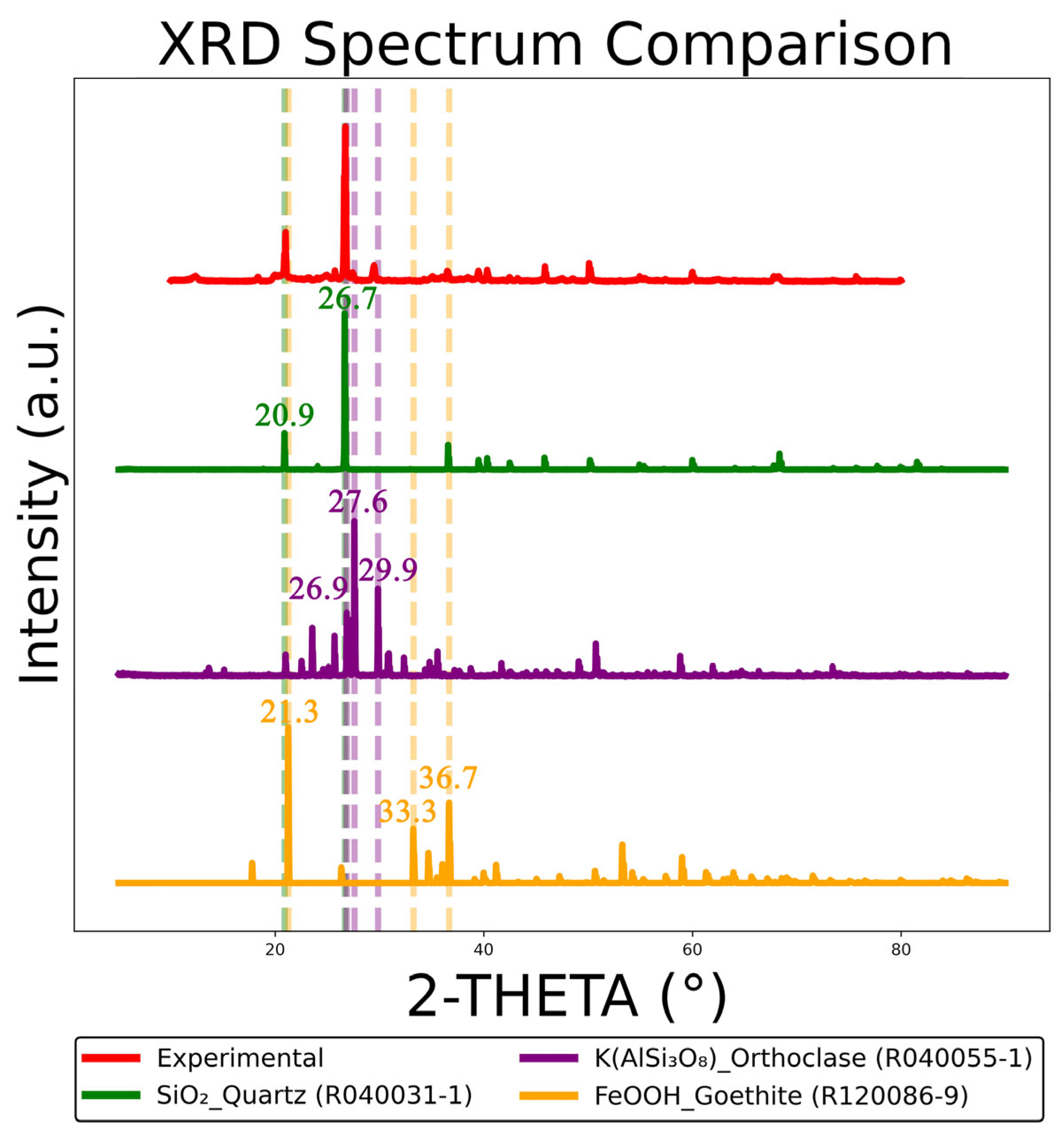
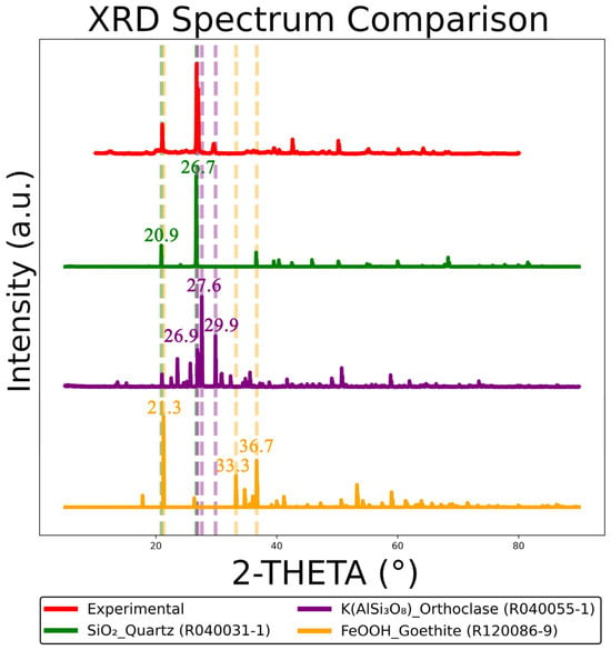
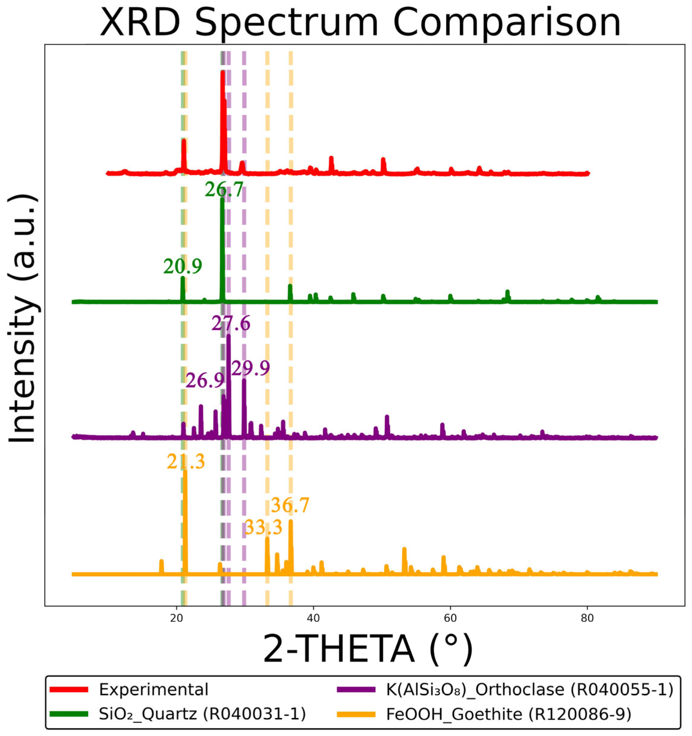
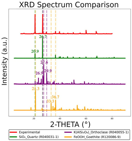
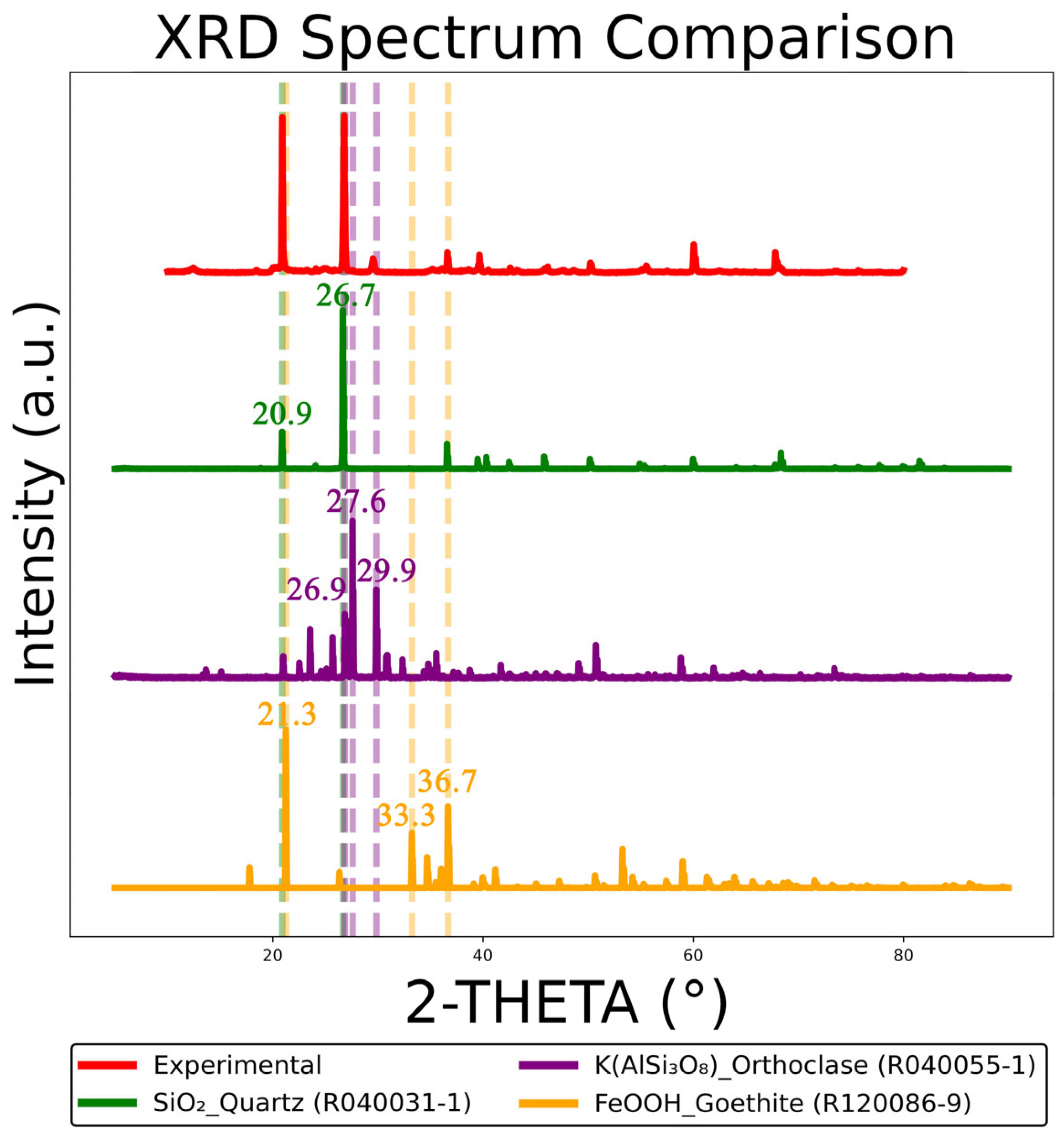
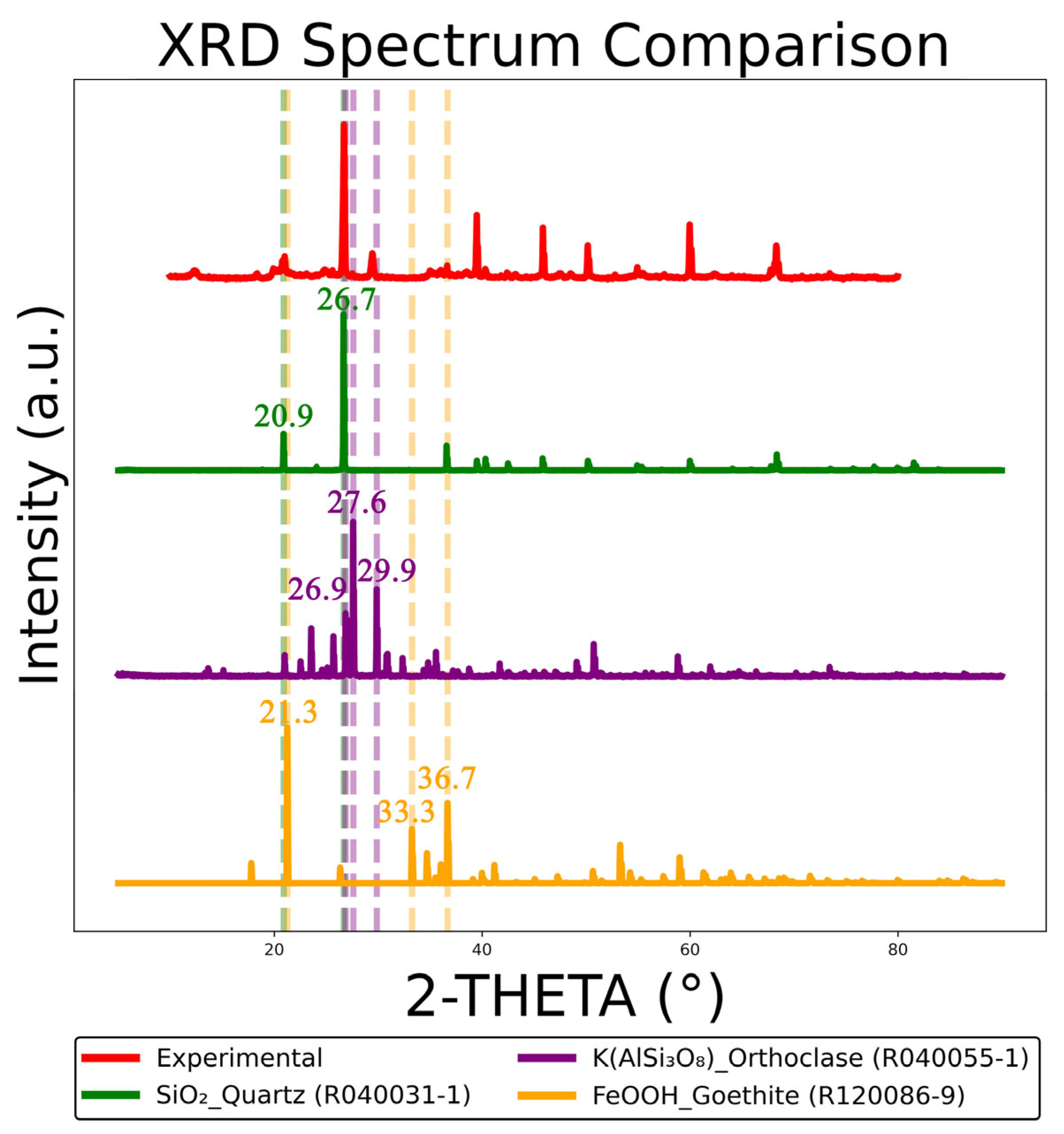
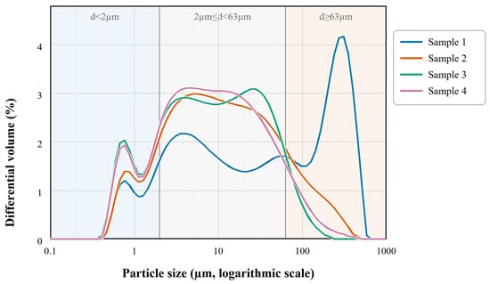
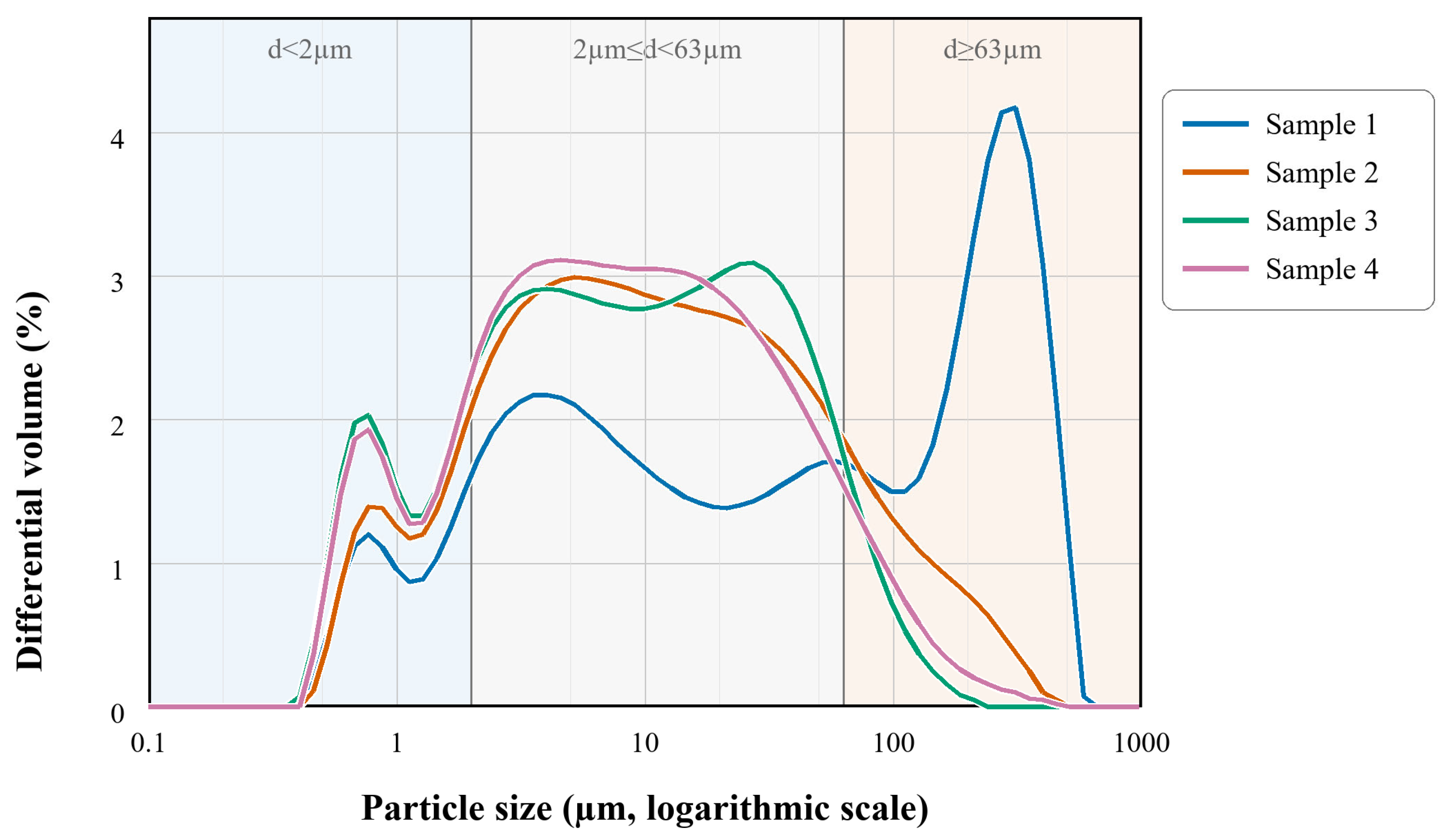

Disclaimer/Publisher’s Note: The statements, opinions and data contained in all publications are solely those of the individual author(s) and contributor(s) and not of MDPI and/or the editor(s). MDPI and/or the editor(s) disclaim responsibility for any injury to people or property resulting from any ideas, methods, instructions or products referred to in the content. |
© 2026 by the authors. Licensee MDPI, Basel, Switzerland. This article is an open access article distributed under the terms and conditions of the Creative Commons Attribution (CC BY) license.