


Abstract
Ti–Nb alloys are widely recognized as highly promising materials for orthodontic applications due to their excellent functional properties, including superior biocompatibility and a relatively low Young’s modulus that better matches that of human bone. The formation of Ti–Nb surface alloys or surface-modified layers is particularly attractive, as the surface properties of biomedical materials strongly influence their mechanical and biological interactions during service. In the present study, Ti-Nb coatings were fabricated on commercially pure titanium substrates using an electron beam surface treatment (EBST) technique. Initially, a 1 μm thick Nb layer was deposited onto the Ti substrates by direct current (DC) magnetron sputtering. Subsequently, the samples were modified through scanning electron beam irradiation, with beam power varied between 1000 and 2000 W to promote Ti-Nb alloyed layers. The phase composition of the resulting structures was analyzed by X-ray diffraction (XRD). Microstructural characteristics and chemical composition were examined using scanning electron microscopy (SEM) coupled with energy-dispersive X-ray spectroscopy (EDS). Mechanical properties were evaluated in terms of hardness and Young’s modulus. The findings of this study demonstrate the feasibility of tailoring the structural and mechanical properties of Ti–Nb surface alloys through controlled electron-beam processing and support their further investigation for potential orthodontic and dental applications.
1. Introduction
Titanium and its alloys have long been a preferred choice for biomedical applications, especially orthodontic and dental implants, due to their excellent biocompatibility, corrosion resistance, and favorable mechanical properties [1,2,3]. However, the elastic modulus of titanium (cp-Ti), although lower than many structural metals, remains significantly higher than that of human bone. This mismatch in mechanical properties can lead to implant failure over time [4,5]. The surface properties of biomaterials are crucial in determining their interaction with the surrounding biological environment [6]. For this reason, the development of surface modification techniques that improve the functionality and durability of implants is of significant scientific and practical interest. Among these techniques, electron beam surface treatment (EBST) has gained attention due to its high precision, high energy density, and ability to locally modify the composition and structure of the surface without affecting the bulk material [7]. The scanning electron-beam treatment technology offers a powerful approach for producing composite coatings with tailored properties [8,9]. During the treatment procedure, the kinetic energy of the electrons is transformed into heat. The conditions of the process can be precisely controlled, and the modified surface can reach molten state, i.e., the formation of a melt pool. Alloying elements can be introduced within the molten material, and once the melt pool solidifies, a coating with greatly improved functional properties is obtained [10].
In recent years, titanium–niobium (Ti–Nb) alloys have emerged as a promising class of biomaterials because of their lower elastic modulus, excellent biocompatibility, and good corrosion resistance [11]. Niobium is known for its ability to stabilize the β-phase of titanium, which is associated with a reduced elastic modulus and improved mechanical characteristics suitable for load-bearing implants [12,13]. The authors of [14] investigates the synthesis of Ti–Nb alloy films by depositing Nb onto Ti via magnetron sputtering, followed by a treatment by low-energy high-current pulsed electron beam (HCPEB). Two regimes were explored: varying Nb thickness at lower voltages with few pulses, and fixed thickness at higher voltages with more pulses. The alloys were characterized for phase composition, morphology, and mechanical properties using multiple techniques. The results showed that the modification induced metastable phases and grain refinement, leading to improved mechanical performance. Similarly, previous authors synthesized a ~1.5 μm Ti–Nb–Si surface alloy on a TiNi substrate using a low-energy high-current electron beam [15]. The resulting material has a composite structure with crystalline phases embedded in a glassy matrix. It exhibits higher corrosion rates than untreated TiNi but improved hardness and a gradient decrease in Young’s modulus. Despite lower near-surface plasticity, the alloy shows full recovery of 6% torsional strain, indicating potential for drug-delivery applications. Du et al. [8] used high-current pulsed electron beam (HCPEB) irradiation to produce a 3.6 μm thick Nb-alloyed layer on a TC4 titanium alloy, consisting mainly of α′-Ti martensite, β-Ti equiaxed crystals, and NbTi4 particles. This surface modification significantly improved the alloy’s hardness, wear resistance, and corrosion resistance compared with the untreated TC4 alloy. From a biomedical perspective, these results should be considered in the context of the established performance of commercially pure Ti. Commercially pure Ti already provides an excellent combination of biocompatibility, corrosion resistance, and long-term clinical reliability; therefore, improvement of hardness or wear resistance alone does not necessarily constitute a sufficient biomedical advantage. The significance of the electron-beam surface modification approaches reported in refs. [8,15] is rather the possibility of tailoring the local mechanical and structural properties of Ti-based implant surfaces while retaining the favorable bulk characteristics of the substrate. In the study of Ref. [15], the Ti–Nb–Si surface alloy exhibited increased hardness and a gradient decrease in Young’s modulus, which may be advantageous where a reduction in the mechanical mismatch between an implant surface and surrounding tissue is desirable. However, its higher corrosion rate compared with untreated TiNi also demonstrates that improvement of one property does not necessarily translate into an overall biomedical advantage. Similarly, the Nb-alloyed TC4 surface reported by Du et al. [8] exhibited substantially improved hardness, wear resistance, and corrosion resistance compared with untreated TC4. These improvements are particularly relevant for implant surfaces subjected to mechanical contact and wear, although they represent improvements relative to the Ti alloy substrate rather than direct evidence of superiority over commercially pure Ti. Thus, the main biomedical relevance of these studies lies in demonstrating that electron-beam processing can selectively tailor surface properties that are difficult to optimize simultaneously in the bulk material.
However, these investigations on the fabrication of Ti-Nb surface alloys and coatings by means of electron beam modification primarily focus on surface treatments using pulsed electron beams. To the best of the authors’ knowledge, studies on Ti–Nb surface alloying using continuous electron beam processing remain very limited. Therefore, the present study focuses on the fabrication of Ti–Nb surface alloyed layers on commercially pure titanium (cp-Ti) substrates using a two-step approach: initial deposition of a thin niobium layer by DC magnetron sputtering, followed by electron beam surface treatment using a continuous electron beam, to form coatings in the Ti–Nb system. The aim is to investigate the influence of the electron beam’s power on the phase composition, microstructure, and mechanical properties of the resulting coatings. Mechanical properties are of critical importance for metallic implants because these materials must withstand complex and repetitive physiological loads while maintaining structural integrity throughout their service life. High strength and hardness are required to prevent plastic deformation, wear, and premature failure, whereas an appropriately tailored elastic modulus is desirable to minimize stress shielding and promote favorable load transfer to the surrounding bone. In biomedical surface-engineered implants, mechanical properties of the modified surface are particularly important because they govern resistance to deformation, wear, and mechanical damage during service.
2. Materials and Methods
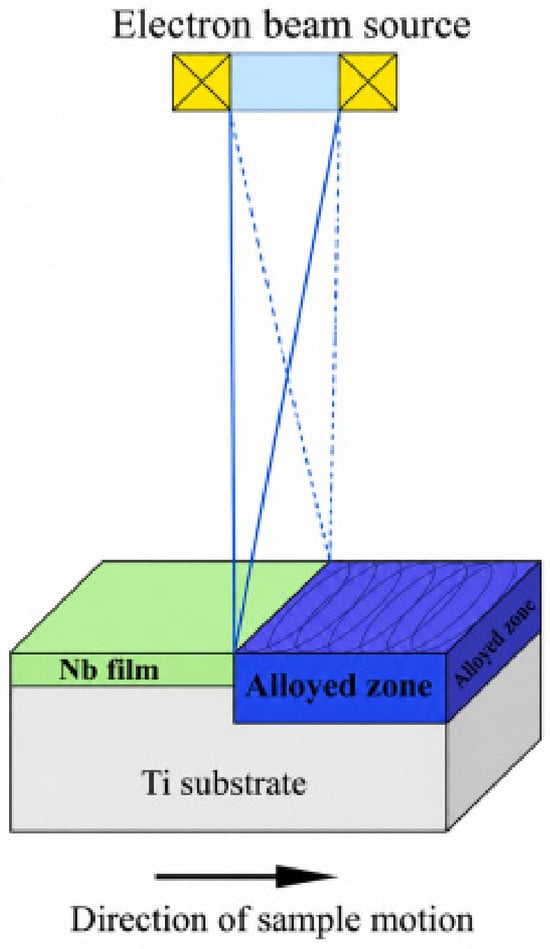
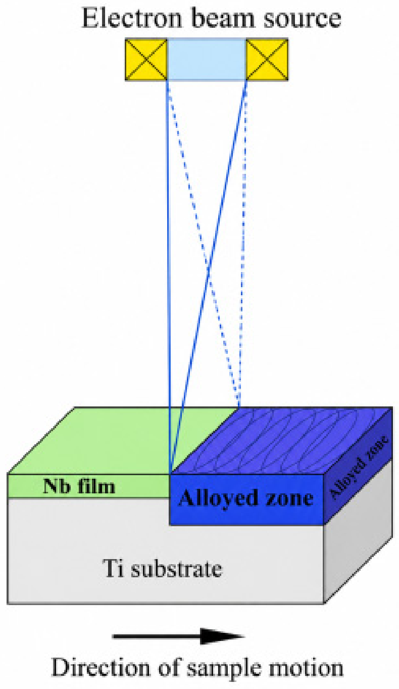
For the purpose of the experiments commercially pure titanium substrates (cp-Ti) were used with a thickness of 4 mm and a diameter of 20 mm. The substrates were preliminarily sputter-cleaned for 10 min, followed by a deposition of a Nb film with a thickness of 1 μm. The process was realized in an Ar+ medium. It was carried out at a discharge voltage of 440 V, discharge current 1 A, and the time for the deposition was 1 h. The specimens were subsequently treated with a scanning electron beam to promote mixing of the deposited Nb layer with the Ti substrate and form Ti–Nb surface-alloyed layers. During the experiments, the accelerating voltage was 60 kV; the electron beam scanning frequency was 1 kHz; the speed of the sample’s motion was 5 mm/s. The electron beam currents during the surface treatment were 20, 25, and 30 mA, corresponding to power ratings of 1200, 1500, and 1800 W. It is important to note that the experiments were carried out using a circular geometry of scanning. Using this approach, the trajectory of the electron beam overlaps, resulting in a longer lifetime of the melt pool, favoring the introduction of the alloying elements (Nb in the present particular case) and the formation of a much more homogeneous microstructure. The process is schematically represented in Figure 1.
Figure 1.
A sketch of the electron beam surface alloying of Nb film with Ti substrate [16].
A total of 12 cp-Ti specimens (Angel Stoilov 96, Plovdiv, Bulgaria) were prepared and divided into four experimental groups: a control group and three groups subjected to electron-beam surface alloying at beam powers of 1200, 1500, and 1800 W. Each experimental group consisted of three independent specimens. The specimens from each group were used for X-Ray diffraction analysis, SEM/EDS characterization, and nanoindentation measurements to evaluate the consistency and reproducibility of the surface alloying process and the resulting material properties.
The phase composition was assessed by X-ray diffraction (Empyrean X-ray diffractometer, Malvern Panalytical, Almelo, The Netherlands). The experiments were realized in a symmetrical Bragg–Brentano (B-B) mode using CuKα characteristic radiation (1.54 Å). The measurements were performed within the range of 30–80° at 2θ scale with a step of 0.05° and counting time of 1 s per step.
The microstructure and chemical composition were studied by scanning electron microscopy (SEM Zeiss EVO 10, Carl Zeiss Microscopy GmbH, Oberkochen, Germany) and energy-dispersive X-ray spectroscopy (EDX, INCA X-act, Oxford Instruments, Abingdon, Oxfordshire, UK), respectively. During the experiments, back-scattered electrons were used.
Nanoindentation measurements were conducted to evaluate the hardness and Young’s modulus of the investigated samples. Measurements were performed using a Bruker nanomechanical tester (Billerica, MA, USA) with 48 indentations carried out on each sample.
3. Results and Discussion
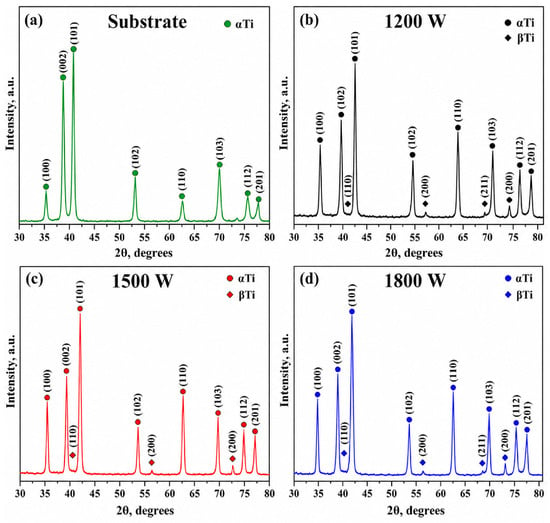
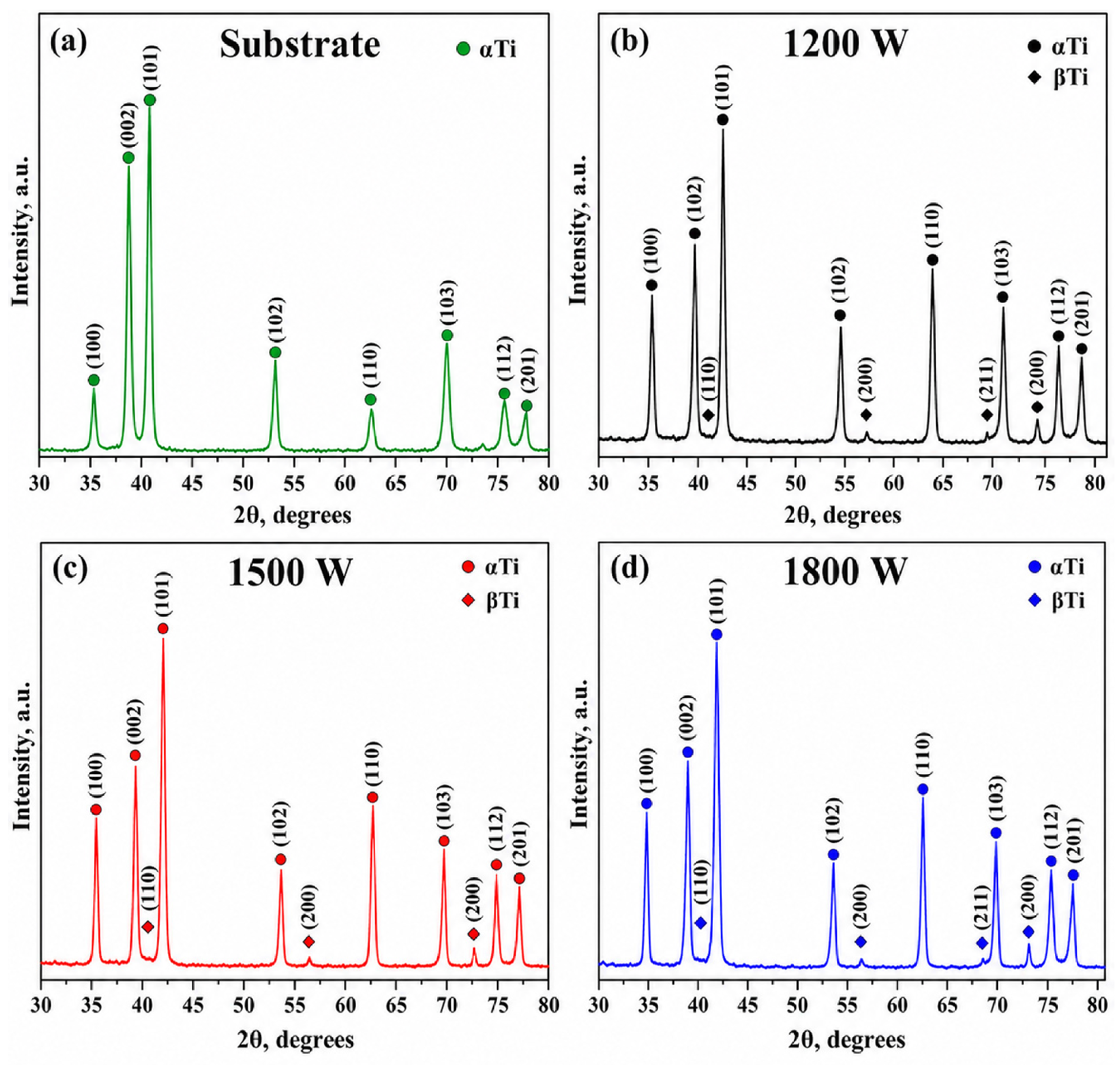
Figure 2 shows the experimentally obtained X-ray diffraction patterns for the base Ti-substrate, as well as electron-beam alloyed specimens. Figure 2a shows the diffractogram of the base titanium substrate; Figure 2b exhibits the pattern of sample alloyed using a beam power of 1200 W; Figure 2c—the diffractogram of the surface alloy obtained at a beam power of 1500 W; and Figure 2d—the diffractogram corresponding to the surface alloy fabricated by a power of 1800 W. The diffraction peaks are indexed according to the ICDD (International Center for Diffraction Data). In all cases, a low background is observed. The diffractogram corresponding to the base titanium substrate exhibits only diffraction maxima of α-Ti phase, characterized by a hexagonal close-packed (HCP) structure. This means that the phase composition of the base material is in the form of a single phase alpha structure. In addition to the peaks of the alpha phase, considering the patterns of the surface alloys formed at beam powers of 1200, 1500, and 1800 W, diffraction maxima corresponding to the β-Ti phase is detected. The beta phase is characterized by a body-centered cubic structure and is known as the high-temperature model of Ti material. Based on a qualitative comparison of the experimentally observed diffraction peaks with the ICDD reference patterns, the electron-beam-treated specimens were identified as containing α-Ti and β-Ti phases. In contrast, the untreated cp-Ti substrate exhibited only reflections corresponding to α-Ti.
Figure 2.
Experimentally obtained X-ray diffraction patterns of the (a) untreated Ti substrate, and of the Ti–Nb composite layers formed using a beam power of (b) 1200 W, (c) 1500 W, (d) 1800 W.
It should be noted that the experimentally observed positions of the β-phase peaks are shifted to higher Bragg angles compared to the reference crystallographic data. This is because the database values correspond to the high-temperature stable β-phase (above 1153 K), while in the present work, the β-phase is stabilized at room temperature by the presence of Nb. Thus, the lattice parameters and peak positions differ from the standard ICDD values. Such peak shifts due to atomic-radius effects of stabilizing elements have been reported in previous studies [17]. No diffraction maxima corresponding to the Nb phase were detected, meaning that in all considered cases, the Nb element was successfully introduced into the titanium substrate and the whole amount of the discussed alloying element reacted with the base material.
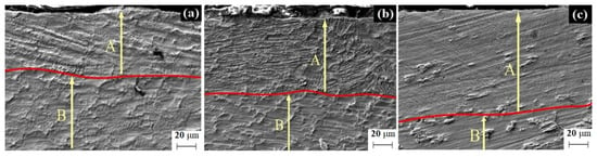
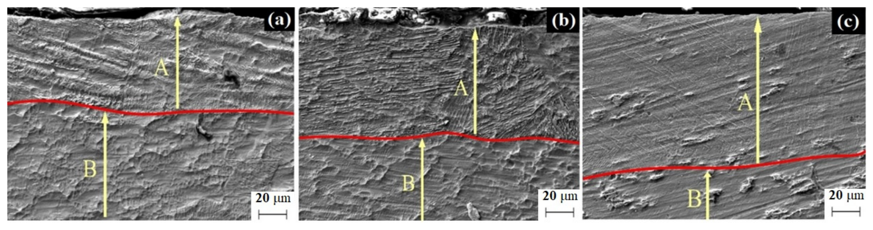
Figure 3 presents cross-sectional SEM images of the modified zones formed after EBST using a beam power of 1200 W (Figure 3a), 1500 W (Figure 3b), and 1800 W (Figure 3c). During the SEM investigations, back-scattered electrons were used. In the three considered cases, the fabricated surface structure is indicated as zone A, while the base titanium substrate is B. It is clearly visible that increasing the electron-beam power increases the thickness of the modified zone. Cross-sectional SEM observations revealed a continuous modified zone whose thickness increased from approximately 50 μm at 1200 W to 70 μm at 1500 W and 90 μm at 1800 W. At the investigated SEM resolution, no distinct residual Nb layer was evident after electron-beam treatment, consistent with remelting and redistribution of the deposited Nb within the Ti substrate. These results are consistent with previous research findings [18,19,20], where numerical simulations were performed in order to analyze the influence of the beam power on the thickness and structure of electron beam modified surfaces, which were further confirmed experimentally. The results published by Angelov et al. [18] indicated that increasing the power of the electron beam leads to an increase in the thickness of the modified zone, which is fully in agreement with the results of the current investigation. Cross-sectional SEM observations, presented in Figure 3, reveal relatively continuous and compact electron-beam modified layers for all investigated processing conditions. No obvious large pores, interconnected porosity, extended voids, or through-thickness cracks are observed within the examined cross-sections. The absence of pronounced macroscopic defects indicates good structural integrity of the modified layers. The performed analysis of the microstructure of the fabricated surface alloys shows that in all cases, along with the base Ti material, fine needle-like microstructural features were observed, particularly in the samples treated at 1500 and 1800 W. Their morphology is consistent with martensitic-type structures commonly reported in rapidly cooled titanium alloys. The formation of this type of microstructures is typical for materials subjected to high energy fluxes (electron beam surface modification in the present particular case) where α + β surface structure is fabricated. During the process of the electron beam modification, the heating and cooling rates are extremely high. In this case, during the heating, the temperature of the modified surface significantly exceeds the beta-transus temperature and the beta phase is formed. It is important to note that the introduction of the Nb alloying element, which is known as beta stabilizer, further promotes the formation of the beta structure. However, during the electron beam surface modifications procedure, as mentioned already, the cooling rates are also very high and can reach values of 103–105 K/s in continuous mode. At sufficiently high cooling rates, the equilibrium diffusion-controlled transformation of the high-temperature β phase into the α + β microstructure is suppressed in titanium alloys because there is insufficient time for the redistribution of alloying elements [21,22]. Under such rapid cooling conditions, diffusion-controlled transformation may be suppressed, and martensitic transformation can occur in titanium alloys. The needle-like morphology observed in the present samples is consistent with this interpretation. This transformation occurs because martensitic formation requires only short-range atomic displacements and can proceed rapidly without long-range diffusion. As a result, rapidly quenched titanium alloys exhibit a martensitic microstructure characterized by increased strength and hardness, reduced ductility, and elevated internal stresses compared with the equilibrium α + β microstructure [22,23]. These statements are in accordance with those of XRD. It should be noted that the identification of the α′-martensitic phase by conventional XRD is challenging because α′-Ti has a hexagonal close-packed structure similar to that of α-Ti, resulting in substantial overlap of their characteristic diffraction peaks. The lattice parameters of α′-Ti are also relatively close to those of α-Ti, and therefore the formation of α′ martensite may not produce clearly distinguishable additional reflections in the present XRD patterns. Consequently, the α-Ti reflections observed in Figure 2 may represent α-type structures that cannot be unambiguously differentiated between equilibrium α-Ti and α′-Ti solely by conventional XRD. Considering the rapid heating and subsequent cooling associated with electron-beam surface alloying, a martensitic transformation of the high-temperature β phase to α′ is possible. However, the present XRD data alone do not provide direct evidence to conclusively identify α′ martensite.
Figure 3.
SEM cross-sectional images of Ti–Nb layers formed at different electron-beam powers: (a) 1200 W; (b) 1500 W; (c) 1800 W.
The chemical composition of the fabricated surface structures was investigated by energy-dispersive X-ray spectroscopy (EDX). Table 1 shows the elemental ratio of the Ti–Nb surface layers formed using a beam power of 1200 W, 1500 W, and 1800 W. A clear decrease in the niobium content with the increase in the beam power is observed. The Nb concentration is the highest with a value of 7.95 wt% at 1200 W, decreases to 3.65 wt% at 1500 W, and is the lowest with a value of 2.33 wt% at 1800 W. This trend can be primarily attributed to the increased thermal energy input associated with the modulation of the electron beam power. The increase in the latter promotes deeper melt pool penetration and consequently results in a thicker modified zone. Since the thickness of the pre-deposited Nb film remained constant for all processing conditions, the total amount of alloying element introduced into the system was unchanged. However, the larger volume of the modified zone generated at higher beam powers led to a greater dilution of Nb within the titanium substrate. As a result, the same quantity of Nb is distributed throughout a larger re-melted volume, causing a decrease in the local Nb concentration in the surface-modified region.
Table 1.
Chemical composition of the Ti-Nb coatings formed by electron beam surface alloying using a beam power of 1200 W, 1500 W, and 1800 W.
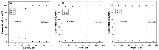
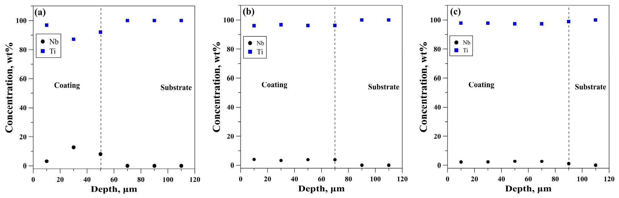
Figure 4 presents the EDS depth profiles of Ti and Nb across the modified layer produced by electron beam surface alloying at beam powers of 1200 W (Figure 4a), 1500 W (Figure 4b), and 1800 W (Figure 4c). The vertical dashed line in each profile denotes the interface between the formed surface-alloyed coating and the Ti substrate. In all specimens, Nb is concentrated within the near-surface modified layer, while the Ti concentration increases toward the substrate, confirming the successful incorporation of the alloying element into the titanium surface. The incorporation and redistribution of Nb are governed by Marangoni convection within the molten pool, which is driven by the large surface-tension gradients generated by the high thermal gradients during electron beam irradiation. These convection flows promote efficient mixing of the deposited Nb film with the molten Ti substrate, resulting in the formation of a surface-alloyed layer. At the lowest beam power (1200 W), the Nb distribution is less homogeneous, with a higher Nb concentration in the near-surface region, indicating that the melt pool is relatively shallow and the Marangoni-driven convection is less intense, leading to incomplete mixing. As the beam power increases to 1500 W and 1800 W, the Nb distribution becomes more uniform throughout the modified layer, owing to the stronger convection and enhanced melt pool mixing. However, the overall Nb concentration decreases with increasing beam power because the higher energy input produces a deeper and larger molten zone, diluting the fixed amount of deposited Nb over a greater volume of titanium. Consequently, although higher beam powers improve the homogeneity of the alloyed layer, they simultaneously reduce the average Nb content due to the increased thickness of the modified zone.
Figure 4.
Elemental distribution from the surface to the depth of the specimens alloyed using a beam power of (a) 1200 W; (b) 1500 W; (c) 1800 W.
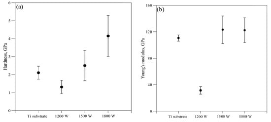
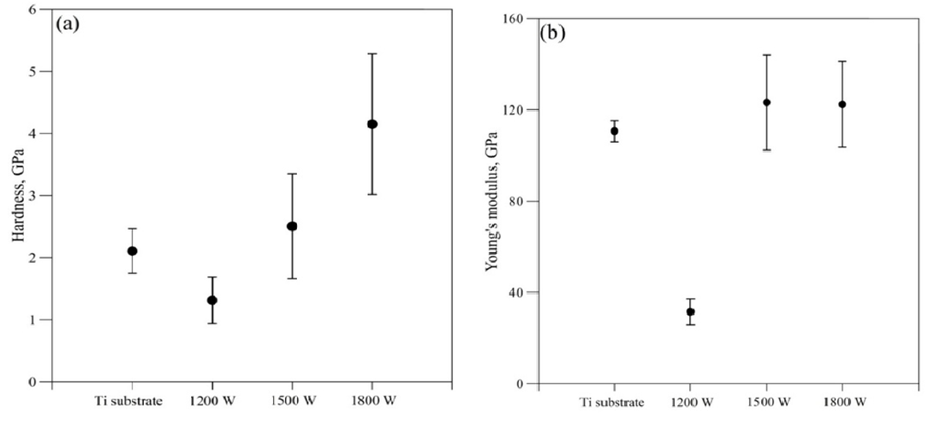
Figure 5 illustrates the variation in hardness (Figure 5a) and Young’s modulus (Figure 5b) for Ti-Nb coatings produced at beam powers of 1200 W, 1500 W, and 1800 W, with untreated cp-Ti included as a reference. At the lowest beam power (1200 W), the hardness of the treated surface decreases compared to the untreated titanium substrate. The comparatively lower hardness obtained at 1200 W may be associated with the phase and microstructural constitution developed at the lower electron-beam power. The XRD pattern indicates the coexistence of α-Ti and β-Ti phases, while the corresponding SEM microstructure does not show an obvious formation of the martensitic-type microstructure observed at higher beam powers. The presence of the relatively softer β phase may contribute to the lower resistance to plastic deformation. Thus, the lower hardness at 1200 W can plausibly be related to the retained β phase and the absence of a pronounced fine martensitic-type microstructure [24]. As the beam power increases to 1500 and 1800 W, a pronounced increase in hardness is observed, reaching nearly 2.5 and 4 GPa, respectively. This enhancement is associated with higher cooling rates and stronger thermal gradients during electron-beam processing, leading to the formation of finer martensitic structures and increased defect density. Finer martensitic microstructures lead to increased hardness because they provide a higher density of microstructural barriers that impede dislocation motion, which governs plastic deformation in metals. As martensite becomes more refined, the number of interfaces such as lath, block, packet, grain boundaries increase significantly, reducing the effective slip distance available for dislocations. In addition, martensite is inherently a metastable, defect-rich phase containing high dislocation densities and internal stresses formed during the diffusionless transformation, which further restricts plastic flow [21]. The combined effect of interface strengthening and defect hardening results in greater resistance to deformation, thereby increasing hardness. Grain refinement and residual stresses further contribute to the observed hardening effect. In parallel, Young’s modulus shows a moderate increase with beam power but remains lower than that of conventional titanium alloys. The moderate increase in Young’s modulus with increasing beam power is consistent particularly with the formation of α′ martensite, together with microstructural refinement and crystallographic texture evolution [21]. The relatively reduced elastic modulus, together with increased hardness at higher beam powers, is particularly advantageous for biomedical and orthodontic applications, where improved wear resistance must be balanced against mechanical compatibility with bone and surrounding tissues. Overall, Figure 5 demonstrates that electron-beam power is an effective parameter for tailoring both the hardness and elastic modulus, enabling optimization of mechanical performance through controlled surface alloying. Microstructural and hardness analyses provide key insights into the mechanical response of Ti–Nb surface alloys and their suitability for dental and orthodontic applications. An increase in hardness generally reflects microstructural strengthening associated with phase transformations, particularly the β → α′ martensitic transformation. However, excessive hardness may be accompanied by reduced ductility and elastic recoverability, both of which are critical for delivering controlled and continuous orthodontic forces. Accordingly, careful control of chemical composition and thermal processing is required to achieve an optimal balance between hardness, strength, and flexibility. In this context, hardness characterization serves as a reliable indicator for evaluating whether Ti–Nb alloys meet the mechanical requirements for safe, efficient, and durable orthodontic applications.
Figure 5.
Hardness (a) and Young’s modulus (b) of the specimens obtained at 1200 W, 1500 W, and 1800 W.
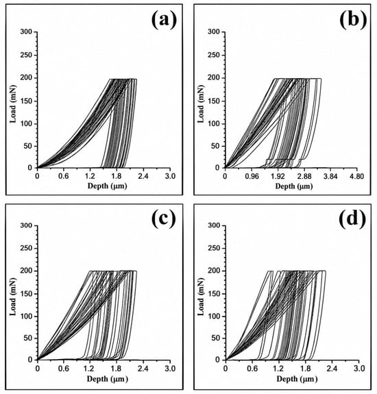
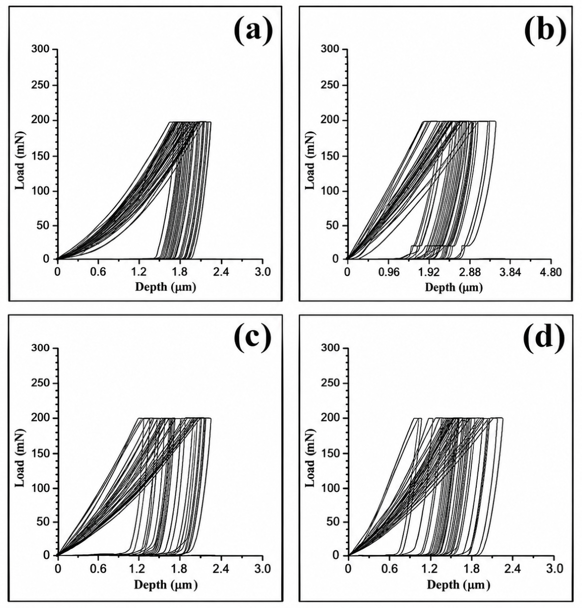
Figure 6 presents the load–displacement curves obtained during nanoindentation testing of the Ti substrate (Figure 6a) and the Ti–Nb coatings formed at beam powers of (b) 1200W, (c) 1500 W, and (d) 1800 W. These curves provide additional insight into the elastic–plastic deformation behavior of the modified surface layers. The specimen treated at 1200 W exhibits the highest penetration depth under the same applied load, indicating lower resistance to deformation. This observation is consistent with the reduced hardness values shown in Figure 5 and reflects the combined effect of higher Nb content and a less refined micro-structure. With increasing beam power, a progressive reduction in maximum penetration depth is observed. The coating formed at 1800 W shows the shallowest indentation depth, confirming its superior hardness and load-bearing capacity. The steeper unloading slopes at higher beam powers also indicate increased stiffness, in agreement with the measured Young’s modulus values. Figure 6 complements the hardness and elastic modulus results by confirming that higher electron-beam power produces mechanically stronger and more deformation-resistant Ti–Nb surface layers.
Figure 6.
Load–displacement curves of the indenter measured at the (a) substrate and at the Ti-Nb coating interface obtained at a beam power of (b) 1200W, (c) 1500 W, and (d) 1800 W.
The present study is based on the assumption that electron-beam power represents a critical parameter for tailoring the mechanical response of Ti–Nb surface alloys by controlling the thermal conditions and consequent microstructural evolution during surface alloying, thereby influencing hardness and Young’s modulus.
Among the numerous biomedical applications of Ti–Nb alloys, orthodontics has attracted particular attention, especially for the fabrication of orthodontic archwires, due to their favorable combination of high strength, reduced Young’s modulus, excellent corrosion resistance, and superior biocompatibility [25,26,27]. This interest originates from the specific mechanical requirements of orthodontic applications, where an appropriate balance between hardness, elasticity, strength, and biological performance is essential. Hardness is a critical parameter for orthodontic components, as it determines wear resistance, surface durability, and friction behavior during interaction with brackets and exposure to the oral environment. At the same time, a relatively low elastic modulus is required to ensure biomechanical compatibility and controlled force delivery during tooth movement. Ti–Nb alloys are considered promising candidates for orthodontic archwires, particularly for intermediate and final treatment stages, where higher stiffness, dimensional stability, and resistance to permanent deformation are required for accurate tooth positioning and finishing procedures. Compared with conventional titanium alloys and stainless-steel materials, Ti–Nb alloys offer improved mechanical compatibility with biological tissues while maintaining sufficient strength. Therefore, tailoring the hardness and elastic modulus of Ti–Nb surface alloys is essential for optimizing their long-term clinical performance.
In the present study, electron-beam surface alloying enabled the fabrication of Ti–Nb layers with controlled microstructural and mechanical characteristics depending on the processing conditions. The variation in electron-beam power modified the thermal field, melt-pool characteristics, and Nb distribution, which consequently affected the phase composition and mechanical response of the developed surface alloys. The formation of martensitic structures, together with Nb-induced β-phase stabilization, played a key role in controlling the hardness and elastic behavior. Increasing electron-beam power resulted in enhanced hardness and improved resistance to mechanical deformation, which are desirable characteristics for orthodontic applications requiring high structural stability. At the same time, the obtained Young’s modulus values remained within a moderate range, providing a favorable combination of stiffness and elasticity. These results are consistent with previous studies demonstrating that Ti–Nb alloys with optimized hardness and reduced elastic modulus exhibit promising performance for potential orthodontic archwire applications [11,28]. Based on their hardness values, the Ti–Nb surface alloys produced at higher electron-beam powers can be considered relatively hard materials and are particularly suitable for later stages of orthodontic treatment, where increased stiffness and mechanical stability are required for precise tooth positioning and finishing. Overall, the results demonstrate that electron-beam power is an effective parameter for tailoring the composition, microstructure, and mechanical response of Ti–Nb surface alloys. The controlled modification of the phase constitution and microstructural characteristics enables optimization of hardness and Young’s modulus, resulting in surface layers with a favorable combination of strength-related properties and elastic behavior. The combination of enhanced hardness, controlled elastic modulus, and metallurgical integration with the titanium substrate may highlight their potential for orthodontic, dental, and other similar applications. The ability to tailor the mechanical properties of the Ti–Nb surface layer is particularly relevant to dental and orthodontic applications, where the controlling stiffness and elastic response can be important for achieving an appropriate mechanical performance of Ti-based components. Such mechanical tailoring may provide advantages in applications where reduced elastic modulus and improved mechanical compatibility are desirable.
It should be noted that the present study is limited to the structural, compositional, and mechanical characterization of the Ti–Nb surface layers. Although Ti–Nb alloys are widely reported to exhibit favorable corrosion resistance and biocompatibility [28], these properties were not experimentally evaluated in the present work. Future studies will therefore include corrosion and biological assessments to further evaluate the applicability of the developed surface layers for dental and orthodontic applications.
4. Conclusions
Ti–Nb surface layers were successfully fabricated on commercially pure titanium by combining DC magnetron sputtering with electron-beam surface alloying. Electron-beam power was shown to be a key processing parameter controlling the microstructure, composition, and mechanical response of the modified layers. All treated specimens exhibited a mixed α/β-Ti structure, with Nb promoting β-phase stabilization. Increasing beam power produced deeper and more homogeneous modified zones while reducing the local Nb concentration due to enhanced dilution within the molten Ti substrate. The rapid thermal cycle additionally promoted martensitic microstructures, particularly at higher beam powers. Higher beam powers resulted in increased hardness and improved resistance to deformation while maintaining a relatively reduced elastic modulus. These findings demonstrate that controlled electron-beam processing enables effective tailoring of the structure–property relationship in Ti–Nb surface alloys. These findings suggest that the developed Ti–Nb surface layers by EBST could be promising candidates for further investigation in orthodontic, dental, and other biomedical applications. It is proposed that electron-beam-induced alloying of Nb with commercially pure Ti can overcome the limitations of the untreated Ti surface by providing localized control of its mechanical properties, thereby creating a Ti–Nb surface layer with a mechanical response that is more suitable for demanding orthodontic and dental applications.
Author Contributions
Conceptualization, I.I. and S.V.; methodology, I.I., F.P., G.K., E.L., B.S., L.V., A.A., M.O., and S.V.; software, I.I., F.P., G.K., and E.L.; validation, I.I., F.P., L.V., M.O., and S.V.; formal analysis, I.I., F.P., G.K., E.L., B.S., L.V., A.A., M.O., and S.V.; investigation, I.I., F.P., G.K., E.L., B.S., L.V., A.A., M.O., and S.V.; writing—original draft preparation, I.I., and S.V.; writing—review and editing, S.V.; visualization, I.I., F.P., L.V., A.A., and M.O.; supervision, S.V.; project administration, S.V.; funding acquisition, S.V. All authors have read and agreed to the published version of the manuscript.
Funding
This research was funded by the European Regional Development Fund under the Operational Program “Research, Innovation, and Digitalization for Smart Transformation 2021–2027,” project No. BG16RFPR002-1.014-0005, Center of Competence “Smart Mechatronic, Eco- and Energy-Saving Systems and Technologies”.
Institutional Review Board Statement
Not applicable.
Informed Consent Statement
Not applicable.
Data Availability Statement
The data presented in this study are available upon request from the corresponding author.
Conflicts of Interest
The authors declare no conflicts of interest.
References
- Niinomi, M.; Nakai, M.; Hieda, J. Development of new metallic alloys for biomedical applications. Acta Biomater. 2012, 8, 3888–3903. [Google Scholar] [CrossRef] [Scilit] [PubMed]
- Gao, Y.; Jiang, W.; Zeng, D.; Liang, X.; Ma, C.; Xiao, W. Additive manufacturing of titanium alloys for biomedical applications: A systematic review. Rev. Mater. Res. 2025, 1, 100011. [Google Scholar] [CrossRef] [Scilit]
- Jia, M.; Zhang, D.; Liang, J.; Gabbitas, B. Porosity, Microstructure, and Mechanical Properties of Ti-6Al-4V Alloy Parts Fabricated by Powder Compact Forging. Met. Mater. Trans. A 2017, 48, 2015–2029. [Google Scholar] [CrossRef] [Scilit]
- Liu, K.-Y.; Yin, L.-X.; Lin, X.; Liang, S.-X. Development of Low Elastic Modulus Titanium Alloys as Implant Biomaterials. Recent Prog. Mater. 2022, 04, 1–14. [Google Scholar] [CrossRef] [Scilit]
- Tardelli, J.D.C.; Alves, B.M.d.B.C.; Valente, M.L.d.C.; dos Reis, A.C. Influence of the modulus of elasticity of dental implants on the distribution of stresses in the alveolar bone by the finite element method: A systematic review. J. Oral Maxillofac. Surg. Med. Pathol. 2023, 35, 383–389. [Google Scholar] [CrossRef] [Scilit]
- Yi, P.; Peng, L.; Huang, J. Multilayered TiAlN films on Ti6Al4V alloy for biomedical applications by closed field unbalanced magnetron sputter ion plating process. Mater. Sci. Eng. C 2016, 59, 669–676. [Google Scholar] [CrossRef] [Scilit] [PubMed]
- Valkov, S.; Ormanova, M.; Petrov, P. Electron-Beam Surface Treatment of Metals and Alloys: Techniques and Trends. Metals 2020, 10, 1219. [Google Scholar] [CrossRef] [Scilit]
- Du, X.; Tian, N.; Zhang, C.; Lyu, P.; Cai, J.; Guan, Q. Surface Alloying and Improved Property of Nb on TC4 Induced by High Current Pulsed Electron Beam. Nanomaterials 2021, 11, 2906. [Google Scholar] [CrossRef] [Scilit] [PubMed]
- Li, X.; Liu, H.; Tian, N.; Zhang, C.; Lyu, P.; Guan, Q. Microstructure and Properties of Mechanical Alloying Al-Zr Coating by High Current Pulsed Electron Beam Irradiation. Nanomaterials 2020, 10, 2398. [Google Scholar] [CrossRef] [Scilit] [PubMed]
- Petrov, P. Electron beam surface remelting and alloying of aluminium alloys. Vacuum 1997, 48, 49–50. [Google Scholar] [CrossRef] [Scilit]
- Gabriel, S.B.; Dille, J.; Nunes, C.A.; Soares, G.d.A. THe effect of niobium content on the hardness and elastic modulus of heat-treated ti-10mo-xnb alloys. Mater. Res. 2010, 13, 333–337. [Google Scholar] [CrossRef] [Scilit]
- Eisenbarth, E.; Velten, D.; Müller, M.; Thull, R.; Breme, J. Biocompatibility of β-stabilizing elements of titanium alloys. Biomaterials 2004, 25, 5705–5713. [Google Scholar] [CrossRef] [PubMed]
- Rani, M.; Sharma, V. Medical Implants: An Overview, Innovation and Future Prospects. Asian J. Pharm. Technol. 2025, 15, 157–165. [Google Scholar] [CrossRef] [Scilit]
- Morini, F.; Bestetti, M.; Franz, S.; Vicenzo, A.; Markov, A.; Yakovlev, E. Synthesis and Characterization of Ti–Nb Alloy Films Obtained by Magnetron Sputtering and Low-Energy High-Current Electron Beam Treatment. Materials 2021, 14, 3238. [Google Scholar] [CrossRef] [Scilit] [PubMed]
- Semin, V.O.; Ostapenko, M.G.; Meisner, L.L.; D’yAchenko, F.A.; Neiman, A.A. Ti–Nb–Si-based surface alloy fabricated on TiNi SMA substrate through additive electron beam method: Structure and perspectives. Appl. Phys. A 2022, 128, 1–10. [Google Scholar] [CrossRef] [Scilit]
- Valkov, S.; Dechev, D.; Ivanov, N.; Bezdushnyi, R.; Ormanova, M.; Petrov, P. Influence of Beam Power on Young’s Modulus and Friction Coefficient of Ti–Ta Alloys Formed by Electron-Beam Surface Alloying. Metals 2021, 11, 1246. [Google Scholar] [CrossRef] [Scilit]
- Panda, K.B.; Chandran, K.S.R. Synthesis of ductile titanium-titanium boride (Ti-TiB) composites with a beta-titanium matrix: The nature of TiB formation and composite properties. Met. Mater. Trans. A 2003, 34, 1371–1385. [Google Scholar] [CrossRef] [Scilit]
- Angelov, V.; Ormanova, M.; Kaisheva, D.; Lazarova, R.; Dimitrova, R.; Petrov, P. Selective electron beam surface alloying of aluminum with TiCN nanoparticles. Nucl. Instrum. Methods Phys. Res. Sect. B Beam Interact. Mater. At. 2019, 440, 88–94. [Google Scholar] [CrossRef] [Scilit]
- Kaisheva, D.; Angelov, V.; Petrov, P. Simulation of heat transfer at welding with oscillating electron beam. Can. J. Phys. 2019, 97, 1140–1146. [Google Scholar] [CrossRef] [Scilit]
- Proskurovsky, D.I.; Rotshtein, V.P.; Ozur, G.E.; Markov, A.B.; Nazarov, D.S.; Shulov, V.A.; Ivanov, Y.F.; Buchheit, R.G. Pulsed electron-beam technology for surface modification of metallic materials. J. Vac. Sci. Technol. A 1998, 16, 2480–2488. [Google Scholar] [CrossRef] [Scilit]
- Banerjee, D.; Williams, J.C. Perspectives on Titanium Science and Technology. Acta Mater. 2013, 61, 844–879. [Google Scholar] [CrossRef] [Scilit]
- Ahmed, T.; Rack, H.J. Phase transformations during cooling in α+β titanium alloys. Mater. Sci. Eng. A 1998, 243, 206–211. [Google Scholar] [CrossRef] [Scilit]
- Liu, C.-C.; Li, Y.-H.; Gu, J.; Song, M. Phase transformation in titanium alloys: A review. Trans. Nonferrous Met. Soc. China 2024, 34, 3093–3117. [Google Scholar] [CrossRef] [Scilit]
- Gupta, A.; Khatirkar, R.; Singh, J. A review of microstructure and texture evolution during plastic deformation and heat treatment of β-Ti alloys. J. Alloys Compd. 2022, 899, 163242. [Google Scholar] [CrossRef] [Scilit]
- Niinomi, M. Mechanical properties of biomedical titanium alloys. Mater. Sci. Eng. A 1998, 243, 231–236. [Google Scholar] [CrossRef] [Scilit]
- Niinomi, M. Recent metallic materials for biomedical applications. Metall. Mater. Trans. A 2002, 33, 477. [Google Scholar] [CrossRef] [Scilit]
- Elias, C.N.; Lima, J.H.C.; Valiev, R.; Meyers, M.A. Biomedical applications of titanium and its alloys. JOM 2008, 60, 46–49. [Google Scholar] [CrossRef] [Scilit]
- Stoyanova-Ivanova, A.; Cherneva, S.; Petrunov, V.; Petrova, V.; Ilievska, I.; Mikli, V.; Iankov, R. Investigation of mechanical and physicochemical propertiesof clinically retrieved titanium-niobium orthodontic archwires. Acta Bioeng. Biomech. 2020, 22, 31–39. [Google Scholar] [CrossRef] [Scilit]

Disclaimer/Publisher’s Note: The statements, opinions and data contained in all publications are solely those of the individual author(s) and contributor(s) and not of MDPI and/or the editor(s). MDPI and/or the editor(s) disclaim responsibility for any injury to people or property resulting from any ideas, methods, instructions or products referred to in the content. |
© 2026 by the authors. Licensee MDPI, Basel, Switzerland. This article is an open access article distributed under the terms and conditions of the Creative Commons Attribution (CC BY) license.