


Abstract
Polymers and their composites have introduced significant advancements in engineering and technology. The primary advantages of polymeric materials include their lightweight nature, ease of manufacturing, anti-corrosion properties, reduced power consumption during assembly and integration, as well as enhanced stiffness, durability, and fatigue resistance. Polymer coatings with conductive polymers allow efficient charge transfer and make electrodes more flexible, helping them better match the mechanical properties of soft tissues. In addition, polymer coatings can protect electrodes from corrosion, reduce biofouling, and provide sites for attaching biomolecules, making them essential for reliable and long-term bioelectrode and biosensor performance. Polymer coatings for electrochemical bioelectrodes play a crucial role in enhancing sensor performance and stability in biological environments as they improve the interaction between electronic devices and biological tissues. These coatings enhance biocompatibility by reducing inflammation and tissue damage while also lowering electrode impedance to improve signal quality. The present review focuses on the most recent developments in polymer coatings for electrochemical biosensors and respective applications. The manuscript provides an overview of polymer materials, emerging strategies, coating approaches, and the resulting enhancements in bioelectrochemical applications.
1. Introduction
At present, the majority of commercial bioelectrochemical devices are engineered for single-use applications or are not suitable for extended storage times. The significant challenge lies in the production of commercial products designed for long-term and/or multiple use and/or prolonged shelf-life functionality. This can be achieved by deploying functionalized and/or protective coatings [1]. Coatings have been always of significant value as a vital subject within the scientific community, primarily due to their ability to integrate the bulk properties of a substrate with the customizable functionalities of a surface layer, as well as the range of available deposition techniques. Coatings can offer improved functionality through the incorporation of functional additives at minimal concentrations. By incorporating these components, the properties of the coatings are substantially enhanced, rendering them versatile and appropriate for a broad spectrum of applications. Furthermore, coatings diminish maintenance expenses and prolong the durability of products [2].
Electrochemical biosensors consist of two primary components, a biochemically sensitive layer that facilitates biochemical detection, and a signal converter that transforms the chemical output into an electrical signal. When the chemically responsive component is substituted with a biological one, the apparatus is referred to as a biosensor (Figure 1). The integration of biochemical processes with physicochemical sensors allows biosensors to deliver analytical results based on a robust technology platform [3].
Figure 1.
Elements and selected components of a typical biosensor [4].
Electrochemical biosensors are used for the determination of analytes (biological or chemical) by deploying biological components, which are sensitive to changes in ambient conditions and parameters like high temperatures, low or no humidity, and chemical components that could lead to their inactivation [5]. The main benefit of using electrochemical biosensors relies on their fast response from seconds to a few minutes [6]. Electrochemical, electro-biochemical, and biosensor devices demand robust and repeatable signals, especially regarding the immobilization of biological entities, including microorganisms, cells, proteins, coenzymes, vitamins, DNA (Deoxyribonucleic Acid), and peptides onto the electrode’s substrate [5,7]. The application of polymer-based nanocomposites has garnered considerable interest as a strategy for improving biosensor performance [8].
The stability, repeatability, shelf-life, and operational lifetime of a biosensor are operational parameters that define its device function. The capacity of a device to maintain its analytical and structural properties during a testing procedure is a measure of its stability. The integration of the biological component with the transducer and the sensor operational mode are crucial factors. Degradation of the biological constituent or sensor fouling are additional potential causes of sensor instability. Further, it is of utmost importance to protect the biosensing electrodes to prevent them from damage and to ensure stable responses even after long shelf-life. The protective layer should not interact with the active electrode in such a way that its performance is diminished or totally deactivated.
The shelf-life denotes the duration during which the sensor maintains its analytical properties while in storage, with bioelement stability being the critical constraint. The operational lifespan of an enzymatic electrode often relies on the preservation of the enzyme’s biological activity. The duration may range from days to months, contingent upon the enzyme’s stability, the manufacturing process, and the operational and storage circumstances [9]. Besides increasing the shelf-life, it is important to be able to predict it. Panjan and coworkers [10] presented a mathematical model derived from a series of laboratory experiments that analyze the effects of accelerated aging at elevated temperatures on glucose-oxidase-modified screen-printed electrodes, serving as a framework for electrochemical biosensors. Operational stability, also known as ruggedness, pertains to a device’s capability to function effectively under specified working conditions. This concept is characterized by a design and validation that are fit for purpose, along with considerations for device assembly and packaging [11]. The stability during storage and the stability during operation are essential attributes of a biosensor, particularly when aiming for commercialization [12].
Specialized enzyme and aptamer-based biosensors can be designed to detect target analyte concentrations in biofluids at low levels. Electrochemical sensors perform a redox reaction resulting in a signal by direct or indirect detection of the analyte of interest. In enzymatic electrochemical sensors, the enzyme facilitates the chemical conversion of the target analyte, while the electrode detects the resulting redox-active species. Consequently, enzymatic electrochemical sensors are especially sensitive to redox-active interferences, which can generate a signal enhancement unrelated to the targeted analyte [13,14]. Enzymes serve as biochemical catalysts, facilitating the transformation of substrates into products by reducing the activation energy required for the reaction. Enzymatic electrochemical sensors are especially vulnerable to redox-active interferences, which elevate analyte detection limits and complicate physiological measurements. Consequently, a comprehensive study has been conducted to devise multiple techniques to alleviate such interferences [14]. The idea behind the present review regarding extension of shelf-life of bioelectrodes is to be able to protect bioelectrodes based on enzymes, bacteria, aptamers, etc., and thus extend their shelf-life by covering them with an easily removable protective coating by either chemical or electrochemical means.
2. Application Fields of Polymer Coatings
Electrochemical biosensors are optimal for point-of-care (POC), a quantitative assessment of catalytic and affinity biorecognition phenomena, since they effectively convert biological processes into electrical data and provide seamless integration with embedded electronic devices [15].
Nanocomposites derived from polymers have garnered considerable interest as sensor materials, presenting potential applications across various domains, such as clinical diagnostics, biomedical research, pharmaceutical sectors, and environmental monitoring. Especially, they have attracted considerable interest in biological sensing because they combine the mechanical flexibility of polymers with the high electrical conductivity, large surface area and porosity, improved enzymatic/catalytic efficiency, and outstanding biological compatibility of nanometric structures (Table 1).
Table 1.
Overview of polymer coating strategies used in electrochemical bioelectrodes.
The integration of biomolecules into such nanocomposites results in improved polymer–bio interfaces that boost electrochemical signal transduction, identification of molecules, and surface stability, which are essential for attaining high sensitivity and selectivity in applications across various domains, such as clinical diagnostics, biomedical research, pharmaceutical industries, and environmental monitoring [7,34,35]. In Table 2 the suitability and application of various polymer classes for enzymatic, nucleic acid, and implantable electrochemical biosensors are presented.
There is a lot of interest in technologies that use capture probes, which are stuck to three-dimensional (3D) surfaces as they have more binding sites than regular 2D surface coatings, which makes them more sensitive and specific [36]. Combining three-dimensional (3D) structured materials can make biosensors work better by giving biorecognition probes more surface area to attach to and making signal transduction mechanisms work better. Biosensor technologies have used a variety of probes as each type of probe has its own advantages in terms of binding efficiency and signal amplification [37,38]. When these probes are fixed to advanced three-dimensional structures, e.g., graphene, hydrogel, and porous silica, they make the sensors work much better. For example, 3D graphene oxide frameworks improve electrochemical performance by making it easier for electrons to move, whereas hydrogels are the best biocompatible matrix for capturing biomolecules [39].
Table 2.
Suitability and application of various polymer classes for enzymatic, nucleic acid, and implantable electrochemical biosensors.
2.1. Anti-Biofouling
Fouling of bioelectrodes refers to the passivation of an electrode surface by a compound that creates a progressively impermeable coating leading to lowered sensitivity of the biosensor because the contact of the analyte with the surface electrode becomes hindered and the electron transfer less possible. Fouling is a widespread problem with consequences spanning various domains, including daily life, healthcare, and the environment. It has the capacity to undermine the sensing performance and normal operational condition of the involved interfaces, thereby reducing the operational lifetime of the devices. Electrode fouling, frequently occurring in electrochemical studies, leads to a deterioration in signal responsiveness over successive experiments [67,68].
The fouling can result from, for example, the development of a passive polymeric coating on the electrode surface. During anodic oxidation processes of potential polymer-building blocks, radicals are generated, which subsequently pair to produce dimers or oligomers, ultimately resulting in the deposition of a polymeric coating on the electrode surface. This polymeric film is robust, thermally stable, and chemically inert, resulting in minimal oxidation or hydrolysis in both acidic and basic environments (Figure 2). The films exhibit low permeability and significant adherence to the electrode, obstructing the surface and resulting in electrode fouling [64,69]. Anti-fouling agents can be applied to change the properties of the electrode-surface and thus to avoid fouling. For bioelectrochemical applications the used surface modifying agents must be biocompatible. There are several mechanisms involved in how the agents are acting in order to minimize or eliminate bioelectrode fouling. Such mechanisms include (a) formation of a hydration layer that increases hydrophilicity, (b) steric repulsion, (c) electrostatic repulsion, or (d) changes in the surface topography, which leads to changes in hydrophilicity or hydrophobicity of the bioelectrode surface [50]. Wang et al. [70] have successfully prepared and applied poly[N-(2-hydroxypropyl) methacrylamide] (poly(HPMA)) brushes prepared using solution and surface-initiated RAFT polymerization as anti-fouling agents. Chan and colleagues [51] have applied polyacrylamide hydrogels in implantable biosensors and increased successfully the operational lifetime of the devices. According to Zinggeler et al. [71], polymer-based composite electrodes, e.g., with CNTs, can be manufactured by deploying the ink-printing method.
Figure 2.
Antifouling biomaterials. Reproduced from [64] with permission.
One must distinguish between anti-biofouling materials and actions for implantable devices and externally used bioelectrochemical sensors. Biological contamination significantly hinders the advancement and utilization of medical equipment, resulting in substantial health risks and high costs. Biocontamination of surgical wounds [72], medical devices [73], and drug delivery systems [74] can result in significant infections, body responses, or even device failure. The implantation of a foreign body triggers adverse tissue reactions termed the foreign body response (FBR), which physically separates the biosensor from relevant analytes, undermines device functionality, and promotes biofilm formation, thereby increasing the risk of infection and implant failure [75].
Biological contamination is chiefly attributed to the non-specific adhesion of biomacromolecules, predominantly proteins. Non-specific protein adhesion is typically regarded as the initial phase of biological contamination, and it is widely recognized that hydrophilic surfaces can significantly diminish protein adsorption. Research indicates that surfaces exhibiting elevated surface-free energy comparable to the one of water, can inhibit protein adsorption owing to the formation of a hydrated layer. The surface prefers to be in contact with water instead of other species because of the reduced interfacial energy [76,77]. Many anti-biofouling materials have been developed based on this principle. Coatings utilizing contact-killing mechanisms can be efficacious without negatively impacting adjacent tissue; yet, they are susceptible to biofouling due to the accumulation of deceased microbes on their surface [40]. Zwitterionic-based coatings (see also Section 3.5) are the most efficient and attractive solution for addressing biological contamination. Recently, zwitterionic polymers exhibiting ultra-low protein adsorption and superior biocompatibility have emerged as a focal point of study for next-generation antifouling materials, garnering significant interest in biomedical applications. The repeating unit possesses a pair of opposing charges, yet the entire molecule remains electrically neutral, exhibiting super-hydrophilic characteristics.
A successful antifouling electrochemical monitoring system must exhibit essential characteristics, including cost-effectiveness, high sensitivity, long-term stability, and excellent biocompatibility. Antifouling coatings encounter difficulties in achieving economical competitiveness; although they provide good resistance to biofouling, they frequently result in electrode passivation, particularly at the high densities required to inhibit biomolecular attachment. The incorporation of conducting polymers into the system—whether in conjunction with other materials or as the principal layer for antifouling molecules—augments electron transfer efficiency, and thus enhances electrochemical sensitivity [1].
2.2. Enhanced Biocompatibility/Bioelectronics
Bioelectronic treatment has advanced significantly in the past decade. Devices that manage cardiac arrhythmia with electrical stimulation are referred to as cardiac pacemakers, representing the first wave of electronic devices [42]. Despite the potential of biomedical implants to enhance quality of life, post-implantation complications remain a concern. Numerous challenges remain to be addressed before technology can achieve its full potential, including restricted biocompatibility, potential corrosion of the interface material, and mechanical mismatch at the tissue/electrode interface, all of which significantly affect the device’s performance, often resulting in persistent inflammation and scarring [78,79]. Organic monolayer-based coatings are a viable solution to the significant constraints of implantable biomaterials, such as restricted biocompatibility, susceptibility to biofouling, bacterial colonization, and stability in in vivo environments [80].
2.3. Polymers for Immobilization of Active Bioelectrode Components
Electrochemical biosensor systems necessitate robustness and consistency in responses, especially regarding the immobilization of biological entities, including microorganisms, cells, proteins, coenzymes, vitamins, DNA, and polypeptides onto the transducer surface by physical adsorption, hydrogels, membranes, or covalent bonds [8,81]. This immobilization is typically achieved through techniques like physical adsorption, crosslinking, covalent binding, and embedding inside gels, membranes, and sol-gels. The utilization of electropolymerized polymeric nanocomposites has garnered considerable interest as a strategy for improving biosensor efficiency [7]. In order to achieve short response times and high sensitivity, mainly electrically conductive polymers and composites are used for immobilization. Because of the large diameter of biomolecules, it is of utmost importance to achieve (a) confinement of the molecule very close to the electrode support so that electrons can be transferred from the electrode to the biomolecule and vice versa and (b) the stereochemistry of the confined biomolecule should allow the reaction (not hide the active centers for attaching of the educts) meaning that it is important that the electrode binds the protein in an orientation suitable for fast electron transfer.
There are several examples of polymers widely employed for immobilization of bioelectrodes. These include naturally derived polysaccharides such as chitosan, alginate, agar agar, etc., which will be discussed briefly in further chapters. Due to chitosan’s excellent film-forming properties, presence of the primary amino groups, and its biocompatible character, it has been extensively studied as an immobilization matrix for enzymes. These features enable chitosan, facilitate immobilization via electrostatic interactions, physical absorption, or covalent bonding to the electrode surfaces [19,82]. There are successful cases of immobilization of enzymes such as glucose oxidase and laccase resulting in improvement of the electron transfer, using chitosan-based films [19]. In addition, crosslinking alginate with calcium cations, Ca2+, is utilized for entrapment of enzymes in hydrogel beads or thin films, to act as a biologically active immobilization environment [26,83,84]. Similarly, agar and agarose matrices have been employed for entrapment and immobilization of biomolecules on electrode surfaces resulting mechanical stability and controlling the diffusion of substrates and products [85]. Furthermore, there have been reports of combining these biopolymers into a hybrid immobilization system such as chitosan–alginate, which results in a multilayer film or a composite of hydrogels, which enhance the performance, stability, and mass transport of the immobilization [26,86].
In Table 3, polymer materials used for enzyme immobilization and their application fields are tabulated.
Table 3.
Polymer materials used for enzyme immobilization and their application fields.
2.4. Removable Bioelectrode Protective Layers
Packaging of bioelectrodes with polymer coatings is mainly required for the protection and increasing of the shelf-life of the biologically active elements that could bind target pollutants. Enzymes or bacteria able to undergo redox manipulations (possess redox-active centers) are directly used as active electrode material (confined on the electrode substrate) for performing reactions bioelectrochemically and evaluate the degradation condition of biologically active elements. To maintain the quality of electrochemical biosensors throughout time, both during usage and storage, it is essential to examine the protection of the active electrode surface to prevent the active electrode material from being influenced by ambient factors. Given that enzymes, proteins, or other organic ligands, particularly bacteria containing active electrode materials, necessitate storage in moist environments, a primary objective is the creation of sensors capable of being stored in dry conditions and subsequently activated through a specific wetting protocol, which can be achieved through these defined protocols, or more efficiently, by merely submerging them in water. The initial phase involves assessing the stability of the active biomaterial when immobilized on conductive or semiconductive glassy carbon supports, which will be exclusively influenced by the interaction of the immobilized enzyme with the free species in solution. Standard electrochemical methods, including cyclic voltammetry and impedance spectroscopy, are mostly employed for the electrochemical testing and characterization of the sensors within a single compartment electrochemical cell. The performed electrochemical analysis seeks to determine if the target species adhere to the active electrode surface after removing a protective polymer membrane and, if so, to assess the electrochemical response, which involves the reduction or oxidation of the species via electron transfer through the bioelectrode. The response is evaluated compared to the one of the pristine, non-protected bioelectrode.
Polymers that can be used as removable coatings are easily soluble polymers with the most prominent representatives being chitosan [20,21], alginate [22,23,27], pullulan [124,125,126].
2.5. Core Technical Challenges and Coating Requirements
Taking electrochemical biosensors from the laboratory to real-world applications, whether inside the human body or in environmental field stations, introduces a unique set of hurdles. In this context, the polymer coating is not merely a passive layer. It must actively resolve specific conflicts that arise between the sensing element and its surroundings. These requirements shift significantly depending on the intended use.
2.5.1. Biocompatibility and Biofouling in Physiological Environments
When it comes to implantable devices and continuous health monitoring, the biggest obstacle is the body’s natural defense mechanism. The implantation of a sensor almost immediately triggers a foreign body response (FBR), which can wall off the device from the target environment or lead to infection. To counter this, the coating has a dual responsibility. First, it must prevent the non-specific adhesion of proteins and cells, a task often handled by zwitterionic materials (such as phosphorylcholine-based polymers) that create a tightly bound hydration layer [43]. Second, the coating must bridge the mechanical gap between the rigid electrode and soft tissues. As demonstrated by Wang et al., hydrogel-based coatings (such as chitosan blends or electrospun coaxial PU-gelatin fibers) can mimic the mechanical properties of the tissue, thereby reducing inflammation and scarring while keeping the electrode electrically connected [32].
2.5.2. Selectivity and Interference Rejection in Complex Matrices
In the fields of environmental monitoring and food safety, the challenge changes from tissue rejection to signal clarity. Real-world samples, such as wastewater or biological fluids, are full of “noise” in the form of redox-active interferences that can distort the sensor’s reading. Consequently, the coating acts as a gatekeeper. Perm-selective polymers or anionic copolymer layers, as described by Figueiredo et al., are essential here; they use electrostatic repulsion to block interfering anions while allowing the target analyte to pass through [43].
For even more challenging scenarios that require high specificity, Molecularly Imprinted Polymers (MIPs) provide a solution by creating artificial recognition cavities that physically filter out structurally similar compounds [127].
2.5.3. Operational Stability and Shelf-Life
Finally, a universal challenge for commercial viability is time. Sensors must retain their analytical performance after storage and during prolonged use. Yet, biological components like enzymes are prone to denaturation. Understanding and predicting this degradation is critical. Accelerated aging models have been developed for that purpose [10]. To extend shelf-life, protective coatings must stabilize the active elements without suffocating them. This is often achieved through entrapment in conducting polymer networks or silica matrices, which secure the biomolecules and maintain the necessary electron transfer pathways over repeated operational cycles.
These scenarios make it clear that polymer coatings are not one-size-fits-all. Instead, they need to be designed for the specific application, in a way that balances biocompatibility, selectivity, mechanical compliance, and long-term stability depending on where and how the sensor will operate. In the following sections of the review, we outline the materials and fabrication strategies that can be combined to meet these scenario-driven requirements.
2.6. Health Monitoring
Conducting polymers have similar electrical and optical properties as metals and semi-conductors and at the same time have mechanical properties of the polymers. Conventional conducting polymers are stiff and friable, therefore, the application of conducting polymers in biomedical fields is limited as stand-alone materials. This issue has driven the develοpment and design of new hybrid or composite conducting polymer-based materials with more robust mechanics [128] including composites of conducting polymers and hydrogels [51]. Electrochemically synthesized conducting polymers are typically fabricated as coatings on the electrode surface, which can be employed for cell culture applications [129].
G. Yang and colleagues presented an alternative glucose biosensor based on different conductive polymers [130] whereas years ago Groenendaal et al. [131] already utilized poly(3,4-ethylenedioxythiophene) (PEDOT) owing to its exceptional chemical stability and enhanced conductivity. PEDOT:PSS has also been extensively applied in biosensors for real-time monitoring of various physiological parameters, providing valuable insights for disease diagnosis and treatment [132]. T. Yang et al. [133] fabricated a PEDOT:PSS hydrogel-based in situ electrochemical sensor designed for direct interaction with living cells. When used for electrochemical detection of dopamine secreted by PC-12 cells, the PEDOT:PSS hydrogel demonstrated biocompatibility, further confirming its potential for use in cellular monitoring and biomedical applications.
2.7. Environmental Monitoring
Pollution results in the discharge of hazardous compounds into the environment, which accumulate due to their persistence. The primary categories of pollutants that pose challenges due to their persistence are polyphenols, nitriles, polycyclic aromatic hydrocarbons (PAHs), cyanides, and heavy metals. The immobilization of enzymes has proven advantageous, facilitating enzyme reusability and stability; hence, remediation of polluted locations can be accomplished directly at the contaminated site or ex situ. In this respect, enzyme-based in situ approaches are economically advantageous compared to other established physicochemical techniques. A variety of polymer matrices exist for enzyme immobilization; eco-friendly matrices are frequently favored, particularly for enzymes intended for environmental applications. The enzymes that play a role in environmental remediation may be proteins or glycoproteins, consisting of one or more polypeptide chains [134]. Umapathi et al. give an excellent overview on recent advances on portable electrochemical sensors for detection of pesticides in fruits and vegetables [35]. Eyvazi and colleagues [62] reported on the recent advances in portable biosensors for the detection of bacterial and fungal contaminations as well as toxins in foods while Sohrabi et al. [63] reviewed the detection of pollutants in water using portable biosensing tests.
2.8. Food Quality
The use of food additives requires the establishment of a stringent food safety policy to protect consumer health. Research indicates that the progressive intake of fatty acids can precipitate obesity, diabetes, metabolic problems, and allergies.
To guarantee the quality and safety of commodities like food, it is imperative to establish precise and dependable procedures for detecting and quantifying fatty acids. One technique highlights electrochemical sensors, which have been developed as effective instruments for this purpose. Biosensors utilizing biopolymers are exceptional instruments for the detection of fatty acids. Their many capabilities, including enrichment, basic signal detection, and target binding, provide highly efficient probes for identifying food additives and pollutants [28,44]. Another example is the detection of azo-compounds that are used as artificial colorants because of chromophoric azo bonds (-N=N-). The electrochemical response of Ponceau 4R (E124) on a biopolymer-modified sensor was examined in a study by Cyriac et al. [29], and the limits of detection (LOD) and quantification (LOQ) were established. The catalytic efficiency of poly(L-Cys)-modified GCE was examined, revealing enhanced electron transfer capability and repeatability. The sensor was verified through the analysis of P4R content in soft drink samples, ensuring food quality and public health safety.
A third application of electrochemical biosensors lies in the field of the analysis of total phenolic antioxidants in wines. Biosensors serve as a highly effective testing tool for identifying polyphenolic compounds, offering numerous benefits such as simple sample preparation, selectivity, sensitivity, reproducibility, short response times, and ease of everyday use. Electrochemical biosensors utilized in the analysis of polyphenols in wines have relied on the activity of polyphenol oxidases or peroxidases [30].
Several molecularly imprinted electrochemical sensors (MIECS) have been created for the specific identification of food additives [31,44,47]. Molecularly imprinted polymers (MIPs), referred to as “plastic antibodies,” are specifically engineered biomimetic supramolecular receptors. These molecules exhibit the ability to recognize and bind to target molecules with high affinity and selectivity, akin to antibodies. They exhibit several distinctive properties, such as ease of preparation, cost-effectiveness, stability, and reusability [44,48]. The integration of molecularly imprinted conductive polymers with molecular imprinted polymer (MIP) identification, alongside their electrical and optical properties, presents novel potential applications.
Wang et al. [28] examined the attributes of five categories of food pollutants: heavy metals, pesticide residues, pathogenic microorganisms, allergies, and antibiotics, along with their detrimental impacts on human health and theoretical considerations on polymer-based biosensors and their recent applications in identifying five types of food pollutants in real food samples. Notwithstanding some obstacles, biopolymer-based biosensors in food technology continue to represent a potentially sustainable technology.
2.9. Drug Discovery/Delivery
Wang et al. [32] have developed a MEA (microelectrode array) based on a crosslinked hydrogel coating by deploying calcium alginate (CA) and chitosan (CS), which has been loaded with dexamethasone sodium phosphate, an anti-inflammatory drug. The coating improved the biocompatibility and the electrochemical properties of the composite microelectrodes. Further, the developed CA/CS-hydrogel layer aligns better with the mechanical characteristics of brain tissue and, through the active release of anti-inflammatory agents, markedly diminishes post-implantation inflammatory processes, prolongs the lifespan of electrodes, and improves the precision of neural activity monitoring. This modification guarantees elevated sensitivity and specificity in dopamine (DA) detection, demonstrating superior dual-mode neural activity during in vivo assessments and highlighting notable functional disparities among neuron types across different physiological conditions (anesthetized and awake).
2.10. Enzymatic Biofuel Cells
In enzymatic biofuel cells (ECBs), enzymes facilitate the oxidation of fuel and typically the reduction of oxygen. In such an ECB bigger molecules are broken down and produce smaller components in a series of redox reactions. Electrons are set free in oxidative reaction(s) and can be used in a closed circuit between anode and cathode as generated electrical energy. Finally, the electrons are consumed by the reductive reaction(s) [49]. In an electrochemical system, oxidoreductases facilitate the conversion of substrates while simultaneously regenerating the active site at the electrode surface. The mechanism requires the occurrence of both interfacial and intramolecular electron transfer processes [135,136].
Recent studies in enzymatic biofuel cells (BFCs) primarily concentrate on enhancing longevity and energy density using enzyme cascades. This approach elevates the oxidation level of the fuel, optimizes electron transfer pathways, and explores innovative enzyme immobilization methods. Mishra et al. [137] reported their findings on the development and performance of a biofuel cell utilizing a unique gold nanoparticle-structured polyaniline network, aimed at enhancing electrical connectivity within the bioelectrode interfaces, specifically the interconnected active redox centers. The resulting BFC is composed of strategically organized glucose oxidase and laccase within a gold (Au) embedded polyaniline (PANI) nano-network, achieved through the in situ interfacial polymerization (Au@PANI) process for the creation of a bioanode and biocathode, respectively. The nanostructured PANI and Au@PANI provide outstanding diffusive mass transport, enabling the development of innovative, highly efficient membrane-free bioenergy devices.
In ECBs, biosensors can be integrated and thus to for self-powered biosensors that do not need external energy devices to run. The appeal of these sensors lies in their straightforward design, which comprises only two electrodes. Their ability to serve as both a power supply and a sensor allows for seamless integration with various electronics, eliminating the need for challenging electronic circuitry [49]. These self-powered biosensors can be applied for enzyme substrate detection [138], enzyme [33,139], and/or allosteric effector detection [140,141].
3. Materials
3.1. Polysaccharide Polymer Coatings
Polysaccharides are widely available and renewable biopolymers that have garnered significant interest for their potential applications in flexible electronics, owing to their sustainability, flexibility, and multifunctionality. Natural polymers, such as cellulose, chitosan, alginate, starch, and hemicellulose, possess essential attributes including biodegradability, biocompatibility, and adjustable mechanical properties, rendering them suitable for advanced technological applications such as biosensors [142,143].
3.1.1. Chitosan
Chitosan is a natural, linear polysaccharide that is obtained by the partial deacetylation of chitin, the second most abundant natural biopolymer after cellulose and it is composed of randomly distributed β-(1→4)-linked D-glucosamine and N-acetyl-D-glucosamine units. The presence of free amino groups on its backbone imparts unique physicochemical and biological properties, including pH responsiveness, solubility in acidic media, and bioactivity [144]. Chitosan is derived from chitin, a naturally occurring polysaccharide found abundantly in the exoskeletons of crustaceans such as shrimp, crabs, and lobsters, as well as in the cell walls of fungi and insects, where it serves as a structural component that provides mechanical strength and protection. Chitin is composed primarily of N-acetyl-D-glucosamine units, and its tightly packed, crystalline nature contributes to its limited solubility and reactivity [145]. To convert chitin into chitosan, deacetylation is performed to remove the acetyl groups, thereby revealing the primary amine functional groups [146], as shown in Figure 3.
Figure 3.
Chitin deacetylation reaction to produce chitosan (a). Representation of the exoskeleton structure of crab (b). (Reproduced from [146] with permission).
Chitosan, rather, represents a collection of molecules than a particular polymer with a fixed structure, characterized by variations in composition, size, and monomer distribution. The characteristics of these properties significantly influence the biological and technological efficacy of the polymer [20].
Chitosan produces flat membranes, constituting a category of chitosan structures and films, with applications spanning wastewater treatment, food packaging, biomedical purposes, and innovative energy conversion and storage technologies. The capacity of chitosan to undergo a change from soluble to insoluble states by altering pH levels is essential for the development of composite chitosan structures with functions designed for specific applications. The predominant and most simple approach for synthesizing flat chitosan films is the solvent evaporation or solution-cast method [58].
Key reactive sites in chitosan include primary amine groups as well as primary and secondary hydroxyl groups. Because of that, chitosan demonstrates excellent film-forming capability because of the facile creation of intra- and intermolecular hydrogen bonds [45]. Additionally, glycosidic linkages and residual acetamide groups may also participate in chemical modifications. The presence of these reactive sites offers versatile opportunities for structural tailoring through targeted derivatization reactions, enabling the development of chitosan-based materials with tunable physicochemical and functional properties [147]. Because of these properties and as a biocompatible polymeric membrane, chitosan is frequently employed as a permselective membrane and binder to immobilize enzymes on electrode surfaces [14,148]. In a recent publication, Michna et al. gave an excellent overview of the experimental results of the properties of chitosan both as a solid as well as dissolved in water along with theoretical considerations and modelling [149].
Chitosan is recognized as an exceptional substrate for the immobilization of many enzymes, demonstrating enhanced thermostability relative to the free enzyme. Urease has been covalently immobilized onto a glutaraldehyde crosslinked chitosan membrane to enhance resistance against inhibitors, including boric acid, thioglycolic acid, sodium fluoride, and acetohydroxamic acid [16]. Chitosan can be combined with several components like CNTs [17,150], graphene and MWCNTs [18], MWCNTs, reduced GO and Au NPs [53], metals and graphene [151], carbon black [152], or conductive polymers (e.g., refs. [54,153]) to form composite bioelectrodes [145], hydrogels (Figure 4) [154], or for drug delivery (e.g., ref. [155]).
Figure 4.
Schematic illustration of chitosan-based bioelectronic sensors. Reproduced from [154] with permission.
3.1.2. Alginate
Alginate is a naturally occurring, linear polysaccharide that is extracted from the cell walls of algae, e.g., Laminaria, Macrocystis, and Ascophyllum, serving as the structural component providing mechanical strength and flexibility to the algal tissue [156]. Alginate’s polymer chains consist of two uronic acid monomers, α-l-guluronic acid (G) and β-d-mannuronic acid (M), which are arranged in different block sequences (M–M, G–G, M–G) along the polymer backbone [157] Several physiochemical properties of alginate such as viscosity, gel strength, porosity and ion-binding capacity are influenced by content and distribution of these M and G blocks [55]. In the presence of divalent cations, mostly Ca2+, alginate can undergo Ionotropic gelation through cooperative interactions between the charged calcium ions and the carboxylate groups of G blocks of alginate through the “egg-box” coordination model. Throughout this process, stable three-dimensional hydrogels are formed under mild and aqueous conditions that prevent the encapsulated molecules from further activity [158,159]. Alginate has been widely exploited for biomedical and biotechnological purposes due to its high biocompatibility, biodegradability, low toxicity, and facile processing. Furthermore, it has been effectively used for biosensing matrices, drug delivery systems, tissue engineering scaffolds and drug delivery systems [160].
The characteristic features of alginate hydrogels are adjustable pore size, high water content, and permeability which result in efficient mass transfer and maintaining the structure of entrapped biological entities such as enzymes and nanoparticles [161]. Alginate is in particular advantageous due to its ability to form hydrogels with ionic crosslinking without covalent bonds, mostly in enzymes, antibodies, and whole cells, in immobilization systems where preserving function is essential [162]. As mentioned before, alginate has been used both as a physical entrapment matrix and as a protective membrane directly integrated on electrode surfaces. Márquez et al. reported electrodeposition of calcium alginate hydrogels as biocompatible matrix for enzymatic amperometric glucose biosensors. The formed biosensors demonstrated higher enzyme stability and better analytical performance for glucose detection in whole blood samples [163]. Despite alginate’s advantages, its limited electrical conductivity and lack of specific cell adhesion motifs restrict direct electron transfer and cellular interactions in some applications. To overcome these limitations, alginate is used in hybrid immobilization systems, which is blended with conductive polymers, carbonaceous fillers, or multilayered with cationic biopolymers (e.g., chitosan). This not only further improves the stability but also increases the bifunctionality, and transduction efficiency [164].
To enable the regeneration and reuse of alginate-coated surfaces, it is essential to remove the ionically cross-linked alginate layer in a controlled manner, which only removes the alginate and the surface remains unaffected. For this purpose, phosphate-buffered saline (PBS) solution, a biocompatible aqueous buffer composed of sodium and potassium chlorides and phosphate salts, with an approximate pH of 7.3, is utilized [65]. By ion exchange between the Ca2+ in the Ca-crosslinked alginate films and the monovalent ions in PBS, the stabilized linkages in the egg-box junctions are disrupted, which results in dissolution of the alginate layer [159]. This effect can be considered as a non-destructive approach for removing alginate layers, to regenerate electrode surfaces that have been utilized for electrodeposition of alginate.
3.1.3. Pullulan
Pullulan is a natural, water-soluble linear polysaccharide that is produced through the fermentation process by various strains of the Aureobasidium pullulans fungus. The forming units of pullulan are repeating maltotriose units that are formed by of α-(1,6) and α-(1,4) glycosidic bonds. Pullulan production is affected by multiple parameters such as nutrients, minerals, pH, temperature, surfactants, light intensity, and the presence of melanin intermediates [66]. As an environmentally safe microbial biopolymer, it has attracted considerable attention [165]. Pullulan can form thin, transparent, waterproof films that are highly impermeable to oxygen. Pullulan’s unique properties resulted in its broad range of applications in various fields such as food, pharmaceuticals, cosmetics, and packaging industries [56,166]. Even though pullulan lacks the intrinsic gelation under mild conditions, it is possible to chemically crosslink or blend it with other polymers to form hydrogels and functional biomaterials [167]. Pullulan and related systems have been utilized in enzyme immobilization processes to enhance catalytic stability, reusability, and performance. For example, by complexation of pullulan with enzymes such as α-amylase and glucoamylase, in addition to co-entrapment with alginate, it is possible to achieve higher entrapment efficiency and enhanced thermal and pH stability, which highlights pullulan’s role in stabilizing enzymes within polymer matrices [168].
3.1.4. Starch
Starch is a naturally abundant plant-based polysaccharide that is primarily consisted of two glucose polymers—linear amylose with α-(1→4) linkages and highly branched amylopectin containing both α-(1→4) and α-(1→6) linkages, which results in semi-crystalline granules, which are not soluble in cold water but possible to form its gelatines by heating [24]. Due to its features such as biodegradability, biocompatibility, and low cost, it is extensively present in food and pharmaceutical industries and material science applications such as film and hydrogel formation [25]. In addition, in immobilization systems, starch and starch-based hydrogels have attracted increasing attention as matrices for enzymes entrapment, due to their ability of gel formation under mild conditions [24]. For instance, to improve long-term activity and operational stability of trypsin and bi-enzymatic systems, starch gels have been utilized [169]. In addition, modification or combination of starch with other polymers will result in the possibility to tailor mechanical strength, porosity, and diffusion properties. This makes starch a better candidate as a support in immobilization enzyme applications [25].
3.1.5. Cellulose and Derivatives
Cellulose, a fundamental constituent of plant cell walls, is made of (1-4)-β linked glucose units [170], and possesses notable tensile strength and biodegradability [171,172]. Cellulose and its derivatives (nanostructured or not) have numerous application fields such as food packaging [173], medicine [170], wearable sensors [171], etc. The chemical behavior of carbon nanostructure surfaces is crucial for their application in various fields. The surface of carbon nanomaterials (CNs) contains a high density of hydroxyl groups, prompting investigations into various chemical modifications such as esterification, oxidation, and polymer grafting. Furthermore, the study of non-covalent surface modification through the electrostatic adsorption of surfactants and polymer coatings has been conducted [174]. Cellulosic materials can be structurally modified by addition of magnetic Fe2O3 nanoparticles [175].
Arakawa et al. [176] employed a cellulose acetate membrane in a biosensor for the determination of glucose directly in the mouth, that was able to eliminate interferences from contaminants like ascorbic acid and uric acid in saliva.
3.1.6. Agar-Agar
Agar-agar or simply agar, a composite of polysaccharides consisting of linear arrangements of dextro and levo galactoses, is characterized as a robust gel-forming hydrocolloid derived from sea algae. The primary structure is chemically defined by repeated units of D-galactose and 3,6-anhydro-L-galactose, exhibiting little changes and a low concentration of sulfate esters. The remarkable gelling capacity of agar is solely attributed to the hydrogen bonds established between its linear galactan chains, which offer exceptional reversibility, with gelling and melting temperatures often differing by approximately 45 °C. As “physical gels”, they endow agar with distinctive and valuable characteristics in numerous applications, particularly in the formulation of microbiological culture media for bacteria, yeast, and molds, where these features are essential [177]. Ziegler et al. [178] have produced bilayer membranes made of lipids (BLM), which use an agar gel as substrate. The as-BLM (agar-supported BLM) exhibits identical electrical, mechanical, and dynamic qualities to those for which conventional BLM is renowned, in addition to offering enhanced long-term stability and significantly increased breakdown voltages.
3.1.7. Hydrogels
With the growing prevalence of wearable devices like bioelectronics in everyday life, there is an elevated demand for high performing flexible wearable electronic devices. Conductive hydrogels have garnered increasing interest in the domain of wearable sensors due to their excellent conductivity, biocompatibility, tunable flexibility, and diverse stimuli responsiveness. Conductive hydrogels are categorized as ionic conductive hydrogels and electronic conductive hydrogels. Despite the physiological suitability of ionic conductive hydrogels, their conductivity and mechanical durability fail in daily use. Conversely, the electronic conductive hydrogel establishes a continuous conducting network through the use of conductive fillers improving the electronic conductivity. In real biofluids, electrochemical biosensors are limited as much by surface chemistry as by transduction: non-specific protein adsorption and matrix deposition distort baselines, slow electron/ion transfer, and shorten lifetime. Hydrophilic coatings such as polymer brushes and hydrogels mitigate these effects by forming solid, moist interphases that resist fouling while remaining permeable to analytes. Recent reviews discuss design rules, including the choice of neutral or charged monomers, grafting density, thickness or mesh size, robust anchoring, and performance improvements in undiluted plasma or serum and wearable formats [1,50]. Electrically conductive hydrogels are utilized in various sensor applications. These involve physical activity tracking [179], health monitoring and blood analysis [180,181], tissue regeneration [182], environmental monitoring [183], and adaptable electronic skin [184].
Qin et al. [185] created implantable hydrogel probes utilizing amylopectin and polyacrylamide as structural matrices, with PEDOT:PSS serving as the electrically conducting component. Essential characteristics of implantable electrodes, including electrical conductivity, optical transparency, mechanical compliance, biocompatibility, and long-term stability, are paramount in this context for the dependable monitoring of physiological signs. Similar composites based on PEDOT:PSS/PNIPAM have been reported by Jia et al. [186].
A polysaccharide often used in hydrogels is dextran, which is characterized as an α-d-(1,6) linked polymer of glucose, featuring approximately 5% branching through α-d-(1,3) glycosidic bonds. The dextran polymer contains ring-oxygen and bridge-oxygen atoms in each repeating unit, in addition to hydroxyl groups within the ring structure [187]. Dextran is capable of effectively forming hydrogel layers that exhibit an extended 3D structure on bioelectrode materials. Due to their elevated water content and adaptable structure, dextran coatings exhibit enhanced antifouling properties. These characteristics can be utilized in traditional bioanalytical measurements and in the advancement of cell-on-a-chip biosensors [188]. Dang et al. [189] highlight the suitability of starch-based conductive materials in hydrogels for application with biological tissues, detailing their respective advantages and disadvantages.
Most hydrogel sensing devices that are currently available provide only one detection option. In addition to stress and strain indicators, fluctuations in physiological factors such as temperature, humidity, and pH can accurately reflect alterations in health status. Consequently, the development of hydrogel biosensors exhibiting rapid response times, enhanced sensitivity, and extensive detection ranges for temperature, humidity, and pH signals demonstrates considerable promise for advanced wearable and implantable technology. Although 3D printing provides versatility and personalization, the scalability of conductive hydrogel sensor manufacture for commercial applications is a significant hurdle. Continuous attempts must concentrate on enhancing superior sensing characteristics and establishing high-precision, high-throughput, and economical manufacturing procedures that preserve requisite attributes and performance [190].
3.2. Electrically Conductive Polymers
Conductive polymers mark a significant advancement in materials science, merging electronic conductivity, typically linked to metals, with the inherent benefits of polymers, such as structural flexibility, lightweight characteristics, and ease of processing. The importance of the conductive polymers has been awarded with a Noble prize for Shirakawa, MacDiarmid and Heeger in 2000 [191]. The conductivity of such polymers is attributed to their π-conjugated backbone, where double and single bonds that alternate allow for the delocalization of π-electrons. By introducing appropriate electron donors or acceptors, charge carriers like polarons, bipolarons, or solitons are generated, facilitating effective charge transport [192,193]. These characteristics have established conductive polymers as essential elements in contemporary biomedical engineering [193,194,195,196]. In this respect, biodegradable conducting polymers have garnered significant interest over the past decades as a distinct category of biomaterials, combining the electrical conductivity of conducting polymers with the biodegradation characteristics of biocompatible polymers [197]. Numerous conducting polymers like polyaniline (PANI), polythiophene (PTh), polypyrrole (PPy), polyindole, polyrhodanine, poly(phenylenediamine), poly(carbazole), and many others are easily synthesized and widely applicable in biosensor electrodes [198]. One distinguishes electronically conductive polymer materials (the majority) and ionic conductors like Nafion® (Figure 5). Multifunctional conducting polymer-based composites exhibit significant potential for in vivo treatments and implanted electronics, encompassing drug administration, brain interfacing, and implantable electronics [153]. In the following, some examples of applications of conductive polymers and their derivatives are briefly presented.
Figure 5.
Conductive hydrogel fabrication and conductive mechanisms. (a) Direct gelation of conductive materials as cross-linking monomers, (b) suspending conductive materials within the hydrogel network, (c) in situ polymerization within a prepolymer hydrogel matrix, and (d) introduction of conductive ionic compounds. Reproduced from [199] with permission.
3.2.1. Polypyrrole
Polypyrrole (PPy) possesses distinctive characteristics, including facile synthesis, durability under environmental conditions, and elevated electrical conductivity (about 105 S/cm and exceeding 380 S/cm) for both bulk and thin-film materials. Consequently, PPy is utilized in several known applications, including sensors [200], microelectronic devices [201], supercapacitors [202], and biomedicine [203]. These applications leverage the distinctive characteristics conferred by the structure and properties of PPy [60,196]. Pyrrole is a readily oxidizable, water-soluble, and commercially accessible substance that polymerizes to form polypyrrole (PPy), exhibiting strong electrical conductivity, exceptional redox characteristics, elevated affinity for electrons, low oxidation potential, and remarkable stability under adverse conditions. Some restrictions in its applicability arise as PPY is brittle, mechanically inflexible, and insoluble, rendering it difficult to process further and unsuitable for most uses, but its processing potential can be markedly enhanced through copolymerization with other nanomaterials [204,205]. This way, Şenel [206] synthesized a film composed of chitosan, polypyrrole, and gold nanoparticles, utilizing it in a glucose biosensor that demonstrated high accuracy, a brief response time, a linear dynamic range of 1 to 20 mM, and a low limit of detection (LOD) of 0.068 mM. Olea et al. [207] have fabricated a novel PPy-based amperometric, enzymatic biosensor by encapsulating glycol oxidase in multilamellar vesicles. Several metal/PPy [208,209] as well as metal oxide/PPy [210,211,212,213,214] composites have been prepared and applied in biosensors.
3.2.2. Polyindole
The polymerization of indole leads to formation of a conductive polymer (Polyindole–PIN) that is thermally stable and exhibits low hydrolytic degradation and better redox potential than PPy [204]. Polyindole (PIN) is considered an exceptional material for several uses. It integrates the characteristics of polyphenylene and polypyrrole and can be synthesized effortlessly by both chemical and electrochemical polymerization methods. Nonetheless, the conductivity of PIN is worse relative to other conducting polymers [215]. The conductivity of PIN can be significantly enhanced in a composite containing metal oxides or sulfides. Several PIN-based composites like metal/PIN [216,217,218], metal oxide/PIN [215,219,220,221] or MWCNTs/PIN [222] etc., have been prepared and applied in biosensors.
3.2.3. Polythiophene (PT) and Derivatives
Polythiophenes are a distinct category of conducting polymers that have experienced rapid advancement during the past decades. They have superior thermal and environmental stability as well as biocompatibility [223]. Applications primarily focus on antistatic coatings [224], molecularly imprinted polymer coatings [225], solar cells [226,227], supercapacitors [228], biomedicine [229] etc. Polythiophenes have been utilized in biological sensors for the immobilization of different enzymes [230,231,232]. Polythiophene and its derivatives are highly regarded in technological uses due to its substantial stability in both doped and undoped states, simplicity with regard to structural changes, and solution processability. Consequently, polythiophene has been thoroughly investigated through both theoretical [233] and experimental methodologies [234,235]. To enhance processing potential and environmental durability in general and for polythiophene in particular, one approach involves the fabrication of composites that consist of conducting polymers and processable conducting or non-conducting polymers [236].
Poly(3,4-ethylenedioxythiophene) (PEDOT) is a conducting polymer (CP) that is characterized by exceptional redox reversibility, biocompatibility, and electrochemical properties. It demonstrates significant potential as an interfacing material in biosensing, effectively connecting the realms of organic electronics and bioelectronics. PEDOT’s chemical character possesses considerable similarities with biological compounds, thereby promoting the development of all-polymer biosensors [237,238].
The electrically conducting polymer composite material, poly(3,4-ethylenedioxythiophene):poly(styrene sulfonate) (PEDOT:PSS), has garnered significant research interest owing to its distinctive characteristics, including flexibility, high electrical conductivity, favorable thermoelectric properties, and excellent water processability [239,240,241] so that in the past decades, an enormous increase in publications on PEDOT:PSS-based devices for various sensing applications, covering gas, biological, bioelectrochemical, and physical devices has been observed [242,243,244,245,246,247]. Because of its unique properties PEDOT:PSS has been considered as a replacement of indium tin oxide in optoelectronics. Carter et al. [248] have investigated the best conditions for the preparation of PEDOT:PSS films with and without Tween 80 and prepared films with reduced resistivity. PEDOT:PSS has been widely utilized in biosensors for the real-time monitoring of diverse health indicators, offering critical insights for illness diagnosis and therapy. Yang et al. [133] developed an in situ electrochemical sensor based on a PEDOT:PSS hydrogel for the electrochemical detection of dopamine released by PC-12 cells. PEDOT:PSS is an excellent composite material for bioelectrochemical electrodes, attributed to its ion–electron coupling diffusion, structural flexibility, and efficient charge-transfer properties when subjected to external electric fields [249]. Improving the performance of sensors often requires the incorporation of catalytic electrode materials, like Au [250] or Pd-Fe3O4 [251], into PEDOT:PSS-based composites, thereby optimizing both catalytic efficiency and detection sensitivity. Huang et al. [252] developed a sandwich composite film consisting of PEDOT:PSS, MXene-PdAu, and PEDOT:PSS through a one-pot synthesis method, where the deposition of PdAu nanoparticles onto MXene nanosheets markedly enhanced the electrocatalytic oxidation of shikonin.
Cyclic polythiophene oligomers have distinctive features regarding their analogs, underscoring the importance in developing novel polythiophene derivatives [253,254]. Poly(3-decylthiophene) is a polymer that demonstrates conductivity due to its conjugated chain architecture. The exploration of innovative applications and improvements of these materials in sensors and organic photovoltaics requires a comprehensive understanding of them. Riga Junior et al. [255] have investigated the conductivity of thin films and discovered a dependence of conductivity on film thickness. The conductivity measured was 3.50 × 10⁻⁶ S/m for the film produced from a 1.5 mg/mL solution and 2.57 × 10⁻⁶ S/m for films derived from a 2.0 mg/mL solution. Moreover, photoconductivity studies indicated a positive reaction of the material to light exposure.
3.2.4. Polyaniline (PANI)
Polyaniline-based polymers exhibit significant potential as materials for electrochemical sensors, attributed to their distinctive physical and chemical characteristics, including effective gas absorption, minimal dielectric loss, and robust chemical and thermal stabilities [196]. The performance of sensing is significantly influenced by the structure and dimensions of polyaniline-based conductive polymers [256,257]. Hui et al. [258] synthesized composite nanofibers by grafting polyethylene glycol (PEG) onto polyaniline (PANI) nanofibers and utilized them in antifouling electrochemical biosensors. The nanowires were deposited onto glassy carbon (GC) using electropolymerization. Subsequently, this interface was altered with PEG to which the amino-functionalized methylene blue-modified DNA capture markers were attached. The PEGylated PANI (PANI/PEG) nanofibers exhibited a substantial surface area, maintained conductivity, and revealed exceptional antifouling properties in both single protein solutions and complicated human serum samples. The interaction of complementary target DNA and DNA probe dislodged the MB from the duplex structure, resulting in a decrease in current with a limit of detection of 0.01 pM [259]. 1D PANI microwire arrays on silicon wafers have been fabricated and evaluated for SO2 gas sensing at ambient temperature. The response time was 20.82 ± 0.16 s, and the sensor could detect SO2 at a concentration as low as 1 ppm [256]. Recently, Setiawan et al. [260] prepared conductive polymers that replicate the acid–base interaction framework of PEDOT:PSS by in situ polymerization of polyaniline in sulfonated polyaniline (PANI:SPAN). They suggested them as an economical substitute for both PEDOT:PSS and PANI:PSS in their role as a conductive layer for holes in optoelectronic devices.
3.2.5. Polyacetylene (PA)
Polyacetylene, a semiconductive polymer, was the first discovered conductive polymer possessing an electrical conductivity of ca. 100–1000 S/cm [7]. It has been doped in the group of Shirakawa, MacDiarmid, and Heeger in the seventies of the last century with iodine becoming a highly conductive material already at room temperature [261]. This facilitated the production and examination of the features of various materials characterized by the π-conjugated polymeric chain, which governs their electrical and optical attributes [262,263]. The conductivity of PA can be adjusted across the entire spectrum, ranging from semiconductor to metal, via doping. At elevated dopant levels, calculations indicate that the energy gap may close due to the suppression of bond alternation in pristine trans-PA [264,265,266].
3.2.6. Nafion
Nafion® (Chemours, Wilmington, Delaware) consists of a hydrophobic PTFE backbone and perfluoroether side chains terminating in sulfonic acid groups. The resulting phase-separated structure provides high resistance to chemicals and high temperatures, high proton conductivity, and high cation exchange as well as perm-selectivity. The fixed sulfonic acid functional groups in Nafion® repel anionic interferents through electrostatic forces, while allowing diffusion of neutral substrates. For that reason, Nafion® has become a benchmark polymer coating for electrochemical biosensors, functioning as both a protective diffusion barrier and a functional immobilization/binding matrix. Nafion® was originally used as an outer sphere perm-selective barrier in the development of glucose biosensors. In multi-layer needle biosensor designs, which commonly incorporate an internal interferent rejection barrier, an enzymatic diffusion barrier, and an outer Nafion® barrier, it attenuates currents generated by anionic interferents such as ascorbate and urate. Moreover, heat-cured Nafion® barriers have been instrumental in improving mechanical durability and in vivo lifespan, enabling monitoring of glucose concentrations over several days in animal models, with minimal tissue reaction [267,268]. Conductometric GOx sensors were further supplemented with a Nafion® overlayer, which reduced buffer capacity and extended the linear detection range, primarily by modulating proton and substrate diffusion [269]. In addition to serving as an external barrier, Nafion® membranes are commonly used to create an enzyme microenvironment. The hydrated ionic clusters in Nafion® membranes can retain enzymatic activity when deposited from mixed solvents, while the thickness and treatment conditions of the resulting films regulate water content and diffusion. Optimizing the component composition in GOx-entrapped Nafion® has enabled the retention of enzymatic activity and a significant increase in biosensor sensitivity [270]. The robustness of Nafion® membranes has also supported the development of enzymatic biosensors on heated electrodes by providing resistance to repeated temperature cycling in Nafion®/GOx films, thereby enhancing perceived enzymatic kinetics and extending the linear range with minimal deactivation [271]. The role in biosensing, however, could be non-neutral. The most recent application of cathodic bioelectrocatalysis, for example, shows that Nafion® has a slightly negative effect on the activity of bilirubin oxidase-modified CNT surfaces compared with unfunctionalized CNT interfaces [272]. One important application of Nafion® in the modern era is its use as a nanomaterial dispersant and binder, enabling the formation of stable, high-surface-area nanocomposite films. Nafion’s ability to disperse CNTs has led to the development of uniform CNT/Nafion® inks, which reduce overpotentials for H2O2 and NADH, and support the optimization of oxidase biosensors [273]. These nanocomposite materials have since been used in the development of metal and oxide nanoparticles, such as Pd NP/GOx sensors on Nafion®-CNT supporting structures, which improve glucose detection and enhance biosensor sensitivity and specificity for glucose concentrations [274], and Fe3O4/chitosan-Nafion® films, which facilitate biologically compatible film formation, aided by Nafion’s perm-selectivity and optimized electron transport [275]. Recent works have also widened the scope of using Nafion® in glucose sensing, especially in areas where the distinct properties offered by Nafion® and its composites could be utilized. Graphene–Nafion® composites, for instance, offered a mechanically stable and conductive platform in the development of impedimetric DNA sensors with much lower detection limits [276]. Additionally, an ultrathin Nafion® topcoat has been effective in securing the nanofiber immunointerface, thereby enhancing stability and signal retention in cytokine sensors such as those for TNF-α [277]. Nafion® has also been used as an antifouling protective overlay in DNA assays involving complex beverages and biological samples [278], and in cell-based biosensing, where the incorporation of cation-exchange Nafion® interlayers in conductive polymers has promoted cell adhesion and functionality [279]. In summary, Nafion® remains one of the most flexible and adaptable polymer films in electrochemical biosensing. The complex set of properties offered by Nafion®—namely, perm-selectivity, environmental robustness, biocompatibility, and stability in the form of mercerized nanocomposites—strongly supports the extension of the biosensor’s range and lifetime in enzyme, DNA, immuno-, and cellular biosensors. However, special attention must be paid to optimizing the thickness and curing conditions, as well as thorough evaluation of the biomolecule–ionomer and nanomaterial–ionomer interfaces.
3.2.7. Polysulfone (PSF)
Numerous important developments have occurred in the design and creation of various polymers as substitutes for the traditional perfluorosulfonic acid (PFSA) type membranes, with Nafion® (Chemours, Wilmington, Delaware) being the most prominent, extensively researched, and utilized. Several inexpensive synthetic polymers, including poly(aryl ether ketone), polysulfone (PSF), and polybenzimidazole, can serve as alternatives to Nafion® because of their favorable stability characteristics. The sulfonation process applied to these polymers significantly improves their proton conductivity [280].
An amperometric immunosensor for the identification and quantification of antirabbit IgG, is being manufactured utilizing a permeable graphite–polysulfone electrode [281]. Polysulfone has been utilized for the immobilization of enzymes [282], such as glutamate dehydrogenase, in ammonium biosensors [283]. Costa et al. [284] investigated the biocatalytic efficacy of immobilized laccase in degrading a mixture of phenolic compounds, including phenol, resorcinol, 4-methoxyphenol, and 4-chlorophenol, in aqueous solutions. Laccase was immobilized on polysulfone membranes blended with functionalized carbon nanotubes to enhance cost efficiency in enzyme reutilization, and its effectiveness in degrading 4-methoxyphenol was investigated. The biocompatibility of polysulfone can be enhanced by grafting with sulfonated hydroxypropyl chitosan (SHPCS) [285].
3.2.8. Poly(o-phenylenediamine)
Poly(o-phenylenediamine) (PoPD) is typically introduced to electrochemical biosensors as a thin, electropolymerized coating. In practical sensor fabrication, PoPD is attractive because it can be grown directly on the electrode with good conformity (including carbon and screen-printed substrates), while its thickness can be tuned electrochemically. In recent studies, PoPD is not used merely as a passive barrier, but as an electroactive “host” matrix that can be engineered to balance analyte enrichment/recognition and acceptable interfacial electron transfer.
A strong use-case is molecularly imprinted polymer (MIP) sensing, where PoPD is electropolymerized in the presence of a template molecule and then “emptied” to leave recognition cavities. Ting et al. reported a PoPD-based MIP on an oxygen-functionalized screen-printed carbon electrode (with Au nanoparticle integration) for interleukin-6 (IL-6), achieving pg mL−1-level detection in serum, while also acknowledging that the PoPD matrix can adsorb other electroactive species in real samples, which is an important reminder that PoPD films may require careful surface chemistry and thickness control when moving from buffer to biofluids [286]. A different biomedical application is the PoPD/hydroquinone MIP architecture for Aβ42 (amyloid-β 1–42) detection, where PoPD serves as the electropolymerized imprinting scaffold (on a conductivity-boosting underlayer), enabling selective measurements in complex matrices such as serum and artificial cerebrospinal fluid [287]. Beyond clinical targets, PoPD-MIPs are also appearing in demanding environmental assays. Chen et al. demonstrated a conjugated-polymer-nanoparticle-enhanced PoPD-MIP platform for perfluorooctanesulfonate (PFOS), emphasizing rapid response and trace-level quantification in real water samples [288].
3.3. Polyurethanes (PUs)
Polyurethanes (PUs) are segmented block copolymers that result from the reaction of diisocyanates with diols (polyols) and short-chain extenders. The result is a microphase-separated morphology consisting of soft (typically polyether or polyester) and hard (urethane/urea) segments. This structure makes PUs highly tunable, as elasticity, toughness, hydrophilicity, gas and solute permeability, and degradation rate can be independently adjusted by varying the soft-to-hard segment ratio, the polyol chemistry, and the NCO:OH stoichiometry [289]. Additionally, PUs can be processed into dense films, porous foams, sponges, and electrospun fibers, a feature particularly attractive for the coating of electrochemical biosensors, for which mechanical robustness, controlled mass transport, and long-term stability are essential. Recent reviews highlight that the same design strategies used in PU dressings, vascular grafts, and soft implants—such as bio-based polyols, hydrophilic or degradable segments, and antibacterial fillers—are increasingly being applied in sensing platforms [290].
A substantial body of research has used PU as the outer mass-transport limiting and protective membrane in implantable glucose sensors. In early minimally invasive needle-type sensors, epoxy-enhanced PU blends were introduced, consisting of 50%–70% PU combined with 30%–40% epoxy resin as an outer membrane to enhance in vivo durability without compromising adequate glucose permeability, thereby enabling long-term continuous monitoring in subcutaneous tissue [291]. However, later studies on electrospun fibro-porous PU coatings for coil-type glucose electrodes demonstrated that highly interconnected subcellular pores can be used to finely tune diffusion while mimicking the extracellular matrix, with minimal impact on sensitivity [292]. These studies established PU as a “workhorse” neutral polymer for balancing analyte flux, oxygen transport, enzyme retention, and mechanical integrity at the tissue–sensor interface [289].
Building on this, Wang et al. electrospun coaxial PU–gelatin fibers to impart bioactivity to the PU coating of a miniaturized implantable glucose biosensor. The membrane comprised a sturdy, elastic PU core enveloped by a hydrophilic GE shell that enhanced glucose transport. They then adjusted fiber diameter and shell thickness by varying PU concentration in the core solution: thin fibers with thick GE shells retained initial sensitivity but compromised structural integrity and linearity over time; thicker fibers with a thinner GE shell acted as effective mass-transport limiting membranes, maintaining sensor sensitivity and linearity for at least 12 weeks in vitro. This work demonstrates how the processability of PU into coaxial nanofibers enables independent control of mechanics, permeability, and biointerface chemistry within the same coating [292].
Another active approach to modulate the foreign-body response uses porous, drug-eluting PU coatings. In this context, tubular PU sleeves with well-controlled porosity (~85%), pore size (~75 µm), and thickness (~80 µm) were fabricated by a gas-foaming/salt-leaching process and loaded with dexamethasone (Dex) by Vallejo-Heligon et al. [293]. In vitro, these coatings released Dex over approximately two weeks while only minimally affecting sensor response time and sensitivity. Follow-up in vivo studies on Medtronic MiniMed SOF-SENSOR™ glucose sensors (Medtronic MiniMed, Inc., Northridge, CA, USA) implanted subcutaneously in rats showed that Dex-loaded porous PU coatings reduced macrophage density and enhanced vascularization around the implant, and extended sensor sensitivity over a 21-day period compared with bare or Dex-free coated controls, at the cost of increased signal lag. Along with nitric-oxide-releasing and growth-factor-loaded PU systems, these data support the concept of “pharmacologically active” PU membranes, which marry topographical cues and localized drug delivery to engineer the tissue–sensor microenvironment [1,289].
PU is also an appealing structural scaffold in non-enzymatic electrochemical sensors. Guo et al. utilized one-pot hydrothermal deposition of binderless nickel hydroxide onto a 3D substrate provided by an inexpensive, flexible PU sponge to create a Ni(OH)2/PU electrode for glucose detection. The open-cell PU network allowed for homogeneous growth of Ni(OH)2 nanosheets and ensured a large electroactive surface area and efficient diffusional pathways, resulting in very high sensitivity of 2845 mA mM−1 cm−2, a low detection limit of 0.32 µM, and fast response (<5 s), along with good stability and selectivity in the presence of common interferents and successful measurements in fetal bovine serum. Here, PU plays the primary role of a mechanically strong yet flexible 3D template; in principle, however, its chemistry can be further tailored to enhance wettability and analyte accessibility [294].
Further testimony to the versatility of PU membranes is their use in microfluidic and cell culture platforms. Lwin et al. fabricated an automated microfluidic platform for real-time lactate monitoring in the cell culture medium of breast cancer cells, using an integrated needle microelectrode coated with an inner m-phenylenediamine exclusion layer, a chitosan/lactate oxidase hydrogel, and an outer PU film. The linear range of the biosensor was extended up to 6 mM with the PU over-layer, optimized by limiting the lactate flux, while a ~95 nA mM−1 mm−2 sensitivity with stable performance over more than 18 h at room temperature was maintained. The robust PU coating allowed the microelectrode to be removed and replaced following fouling, thus illustrating the utility of PU in dynamic automated sensing setups where mechanical handling and long-term stability are critical [54].
Nanostructured transducer coatings can be prepared by combining PU with conductive polymers. Gokce and co-workers electrospun nanofiber mats from blends of PU and the carboxylated conductive polymer poly(m-anthranilic acid) (P3ANA); amino-modified DNA probes were then covalently immobilized on the P3ANA carboxyl groups to give an impedimetric DNA biosensor for Salmonella spp. The PU component supplied excellent fiber-forming ability and mechanical strength, whereas the P3ANA phase provided conductivity and abundant functional groups that allowed for stable probe attachment. The final PU/P3ANA nanofibers possessed a high surface-to-volume ratio and exhibited a wide linear range (0.1–10 µM), high mismatch discrimination, and retained ~93% of their initial response after 30 days; this represents an improvement upon the operational stability of several earlier DNA sensor architectures [295].
State-of-the-art PU biosensor literature places particular emphasis on structural and chemical tailoring of the polymer to match it with the required task from a materials-design perspective. Specific strategies include the use of hyperbranched or waterborne PUs for higher functional group density and better processing, respectively; the incorporation of nanofillers, such as carbon nanomaterials or metal nanoparticles, to enhance conductivity and stability; blending with other polymers; and surface functionalization to reduce biofouling or immobilize biorecognition elements. These exemplify emerging amino-acid-based poly(ester urea)s that, besides ureido linkages, offer tunable degradation profiles and are another example of how urethane/urea-containing backbones can be engineered to provide controllable mechanical properties, degradability, and protein-repellent behavior in biomedical devices [296]. In terms of electrochemical biosensors, these developments represent a toolbox of PU-family materials that can serve simultaneously as mass-transport regulators, mechanically resilient encapsulants, and active modulators of the host response.
3.4. Hydrophilic Hydrogel and Antifouling Polymers
In real biofluids, electrochemical biosensors are limited as much by surface chemistry as by transduction: non-specific protein adsorption and matrix deposition distort baselines, slow electron/ion transfer, and shorten lifetime. Hydrophilic coatings such as polymer brushes and hydrogels mitigate these effects by forming solid, moist interphases that resist fouling while remaining permeable to analytes. Recent reviews synthesize design rules, including the choice of neutral or charged monomers, grafting density, thickness or mesh size, robust anchoring, and performance improvements in undiluted plasma or serum and wearable formats [1,50].
3.4.1. PEG and PEG-like Brushes (POEGMA)
Poly(ethylene glycol) (PEG) remains the canonical low-fouling chemistry. Flexible chains and dense hydration shells suppress protein adsorption and cell adhesion, resulting in cleaner voltammograms and improved specificity in complex media [1,50]. On metal electrodes, surface-initiated atom transfer radical polymerization (ATRP), including electrochemically mediated surface-initiated electrochemically mediated ATRP (SI-eATRP), produces poly(oligo(ethylene glycol) methacrylate) (POEGMA) brushes with controllable thickness and grafting density. Both parameters determine the antifouling threshold and diffusion penalty by electrochemical means [297]. For manufacturability, printed antifouling layers, coatings containing a photoreactive antifouling copolymer with conductive carbon nanotubes (CNTs) on screen-printed electrodes, enable easy access to low-fouling, disposable transducers without lengthy wet chemistry steps [71].
In PEG systems, high grafting density and moderate thickness is critical, since too thin leaves “defect windows” for proteins, whereas too thick increases ohmic loss and slows mass transport [1,71].
3.4.2. PHEMA (Poly(2-hydroxyethyl methacrylate))
PHEMA forms neutral, water-rich hydrogels with tunable mesh size and good optical and mechanical stability, making it suitable for use as diffusion moderator and protective skins over enzyme or aptamer layers [50].
Plasmapolymerised 2-hydroxyethyl methacrylate (HEMA)/2-(diethylamino)ethyl methacrylate (DEAEMA) films can be firmly deposited on screen-printed electrodes and provide solid functional hydrogel coatings with suitable wettability and biointeraction, while retaining stable electrochemical readout [298]. In flexible or sweat sensing, Cu-modified PHEMA hydrogels laminated on screen printed electrodes (SPEs) have served as mass transport layers for glucose or ascorbate, illustrating that catalytic inclusions can be hosted without sacrificing antifouling behavior [299]. Where abrasion and long wear times are expected, NSCC PHEMA (nanosilica covalently coated PHEMA) provides superhydrophilicity and unusually high durability, thanks to Si–O covalent bonding at the hydrogel surface [300].
During design, crosslinking light should be kept to maintain high water content. Catalytic or conductive domains should be introduced as dispersed nanoinclusions or interpenetrating networks to avoid blocking aqueous channels [298,299,300].
3.4.3. Methacrylamide
Poly(N-(2-hydroxypropyl) methacrylamide) (HPMA) is a PEG alternative for antifouling brushes, with excellent protein resistance and greater chemical stability. Two brush attributes dominate performance: grafting density and thickness [70,301].
Reversible addition–fragmentation chain transfer (RAFT) “grafting-from” protocols produce dense HPMA brushes directly on electrodes. Comparative studies show that higher grafting densities (typical for graft-from versus graft-to) correlate with lower adsorption and improved brush characteristics [70,301] PLL–g–HPMA bottlebrushes, in which a poly-L-lysine backbone anchors to oxide or gold and HPMA side chains form the hydration corona, provide strong antifouling across three scalable synthesis routes, offering practical options for different fabrication lines [301]. Recent surface-initiated RAFT (including PET–RAFT) protocols further streamline well-defined HPMA brush growth and are attractive for patterning and multiplexing on electrode arrays [301].
The choosing between linear and bottlebrush architectures should be based on the available space at the interface: bottlebrushes maximize hydration but increase diffusion resistance, whereas thinner linear brushes facilitate mass transport [70,301].
3.4.4. Poly(N-isopropylacrylamide) (PNIPAm)
PNIPAm introduces thermal gating: below its lower critical solution temperature (LCST) of ~32 °C it is hydrated and permeable. Above this temperature, it collapses, expels water, and releases weakly bound foulants. This reversible volume change can enable self-cleaning cycles and adaptive flux control during operation [302].
To overcome the mechanical softness of neat PNIPAm, ionogels and hybrid networks maintain ionic conductivity and integrity while preserving thermoresponsiveness, which makes it promising for wearables and soft bioelectronics [303]. Coupling PNIPAm to a soft conductor, like PEDOT:PSS/PNIPAm hydrogels, yields electrodes that remain electronically percolated through the thermal cycle and have been integrated as gates in organic electrochemical transistors and wearable devices [186].
When using PNIPAm, one should take into account the LCST shifts in high-ionic-strength biofluids, as co-monomer or zwitterionic segments can stabilize hydration near physiological temperatures [186,302,303].
3.4.5. Poly(vinyl alcohol) (PVA)
PVA forms highly hydrated, mechanically resilient networks through either physical freeze–thaw crystallization or chemical crosslinking, which makes it a reliable diffusion-moderating, antifouling cushion over delicate biointerfaces. Its dense hydrogen-bond network and tunable water content provide dimensional stability under flow or motion, while the hydroxyl-rich backbone readily hosts conductive fillers (conducting polymers, carbon, metals) or ionic dopants to build electronically or ionically conductive hydrogels without sacrificing hydration. These traits explain why recent surveys highlight PVA as a “workhorse” matrix for wearable and on-skin electrochemical sensors in sweat and interstitial fluid, and why classic freeze–thaw design rules (cycle number, peak/low temperature, hold time, PVA molecular weight and degree of hydrolysis) remain the first levers for balancing permeability with toughness [304,305,306].
In electron-conducting PVA composites, embedding polyaniline (PANI) and Pt nanoparticles into a PVA hydrogel built a porous, conductive channel for enzymatic glucose detection with a µM-level LOD and a wide (1 µM–30 mM) linear range, illustrating how noble-metal catalysis and conducting-polymer percolation can be combined inside a hydrated PVA network [307]. For antifouling and conductivity at once, a double-conductive hydrogel interface (MXene and PEDOT within a hydrophilic network) enabled ultrasensitive carcinoembryonic antigen detection in serum while maintaining a clean baseline, and a PVA/PDA/PEDOT ternary conductive-antifouling coating supported direct GFAP detection in complex clinical matrices. Both these studies exemplify how PVA’s hydration (antifouling), PDA’s adhesion, and a soft conductor can be co-optimized for real-sample electrochemistry [308,309]. On the ion-conducting side, purely physically crosslinked PVA hydrogels activated by simple salt soaking deliver high ionic conductivity and elasticity, retaining performance even near sub-zero conditions, which is useful when stable signal and comfort are required in wearable formats [306].
In practice, a rational workflow begins by fixing the PVA network architecture through freeze–thaw cycling, which determines mesh size and toughness. Functionality can then be added with minimal loading of conductive fillers (e.g., PANI, PEDOT, MXene, or metal nanoparticles) at approximately the percolation threshold required for signal transduction, so as not to block water channels. For sustained operation in undiluted biofluids, pairing PVA with an explicit antifouling component (e.g., polydopamine or zwitterionic segments) helps preserve the hydration layer while maintaining electronic and ionic pathways. In wearable formats, ionically conductive PVA gels produced by salt or plasticizer soaking mitigate dehydration and low temperature brittleness. Finally, permeability to the intended redox mediator and analyte should be verified early, as tightening of the network (through additional freeze–thaw cycles or increased crystallinity) can inadvertently restrict faradaic access [304,306,308,309].
3.5. Zwitterionic Polymers
Zwitterionic (ZW) polymers consist of repeat units with balanced positive and negative charges that form tightly hydrated net-neutral interfacial layers that provide significant barrier protection against non-specific absorption of complex media. This is why ZW coatings often exhibit “ultralow fouling” behavior and longer operating lifetimes in serum, plasma, milk, and other biofluids [1,77,259]. The practical challenge is that even though the coatings have to remain clean, they still have to permit redox mediators, targets, and electrons to reach the transducer. Therefore, recent research has focused on the relationship between antifouling strength and interfacial transport or bioconjugation and it is apparent that it can be achieved in real devices [77,259,310].
Performance on electrodes across sulfobetaines (SB), carboxybetaines (CB), and phosphorylcholines (PC) is largely determined by two control factors. The first one is the architecture (dense brush, ultrathin film, or hydrogel). The second is the electrostatic microenvironment at the interface. Multiscale studies, from atomistic modeling to brush-level electrochemistry, show that very dense brushes maximize hydration but can increase charge transfer resistance and hinder the diffusion of charged probes. Each effect can be tuned through brush thickness, grafting density, and zwitterion selection [77]. In practice, ZW overlayers have enabled aptamer and immunosensor formats to operate in undiluted matrices with stable baselines and preserved sensitivity, showing that antifouling and transport do not need to be mutually exclusive [310].
3.5.1. Poly(carboxybetaine)
What makes Poly(carboxybetaine) (pCB) special is its ability to combine strong fouling resistance with convenient carboxyl groups for 1-ethyl-3-(3-dimethylaminopropyl)carbodiimide (EDC)/N-hydroxysuccinimide (NHS) coupling. Electrochemical studies of pCB methacrylamide (pCBMAA) brushes that have been grown from alkanethiolate self-assembled monolayers (SAMs) have shown that longer chain SAMs yield denser, more protein-resistant brushes, which may result in higher interfacial charge transfer resistance unless thickness and morphology are tuned [311]. After functionalization, the zwitterionic balance can be perturbed. By adjusting post-activation deactivation chemistry, like rebalancing charge after coupling, low fouling can be achieved while keeping high capture–probe loading in undiluted media [312]. Storage tests have reinforced the practical appeal that pCB acrylamide (pCBAA) brushes retain antifouling performance over months with just minor changes in swelling or their binding capacity [313]. Finally, hybrid/terpolymer brushes that combine CB with other units like SB or HPMA have delivered label-free detection of viral targets in crude clinical samples. In that way, it has been shown how pCB scaffolds can be extended for demanding real-sample assays [314].
3.5.2. 2-Methacryloyloxyethyl Phosphorylcholine (MPC)–Lipidure
Phosphorylcholine (PC/MPC) polymers mimic the headgroup of phosphatidylcholine, giving exceptionally low protein adsorption on metals, carbons, and conducting polymers. Device-level data on mediated glucose electrodes have shown that tethered ZW coatings (including commercial Lipidure-type films) reduced foreign-body response without compromising glucose sensitivity. That suggests that antifouling protection can coexist with mediator access and electron hopping [315]. In food and clinical matrices, a thiolated PC monomer (PC-SH) assembled on PEDOT/Au produced sub-percent signal suppression in milk and pg mL−1 detection of tetracycline, demonstrating strong antifouling performance under protein-rich conditions [316]. On carbon electrodes, poly(2-methacryloyloxyethyl phosphorylcholine) (PMPC) brushes were grown by surface-initiated ATRP, and they offered durable protein resistance in serum, while maintaining electron-transfer kinetics for small redox probes, illustrating that PC brushes can be both bioinert and electrochemically permissive [317]. Recent reviews have summarized how to integrate MPC chemistry with coupling handles and when to choose brush, thin-film, or hydrogel architectures for miniaturized biodevices [318].
3.6. Polyelectrolyte Coatings
Polyelectrolyte coatings are a convenient way to “engineer” the immediate environment of an electrochemical biosensor. By introducing a charged polymer layer—often as part of a multilayer structure—one can tune adhesion, mass transport, charge screening, and in some cases even antifouling and antimicrobial behavior. Cationic systems such as poly(diallyldimethylammonium chloride) (PDDA), poly-L-lysine (PLL), and N-halamine-based polymers (PMPQ-type), together with anionic copolymers like poly(vinylimidazole-co-sodium styrenesulfonate) (P(VI-SS)), are among the most commonly used families in this context.
Below, four representative examples are discussed: PDDA and PLL, where recent electrochemical sensor work is available; PMPQ-type N-halamine polyelectrolytes, mainly in antimicrobial coatings; and P(VI-SS), which has reappeared in modern, protected redox-polymer biosensors.
3.6.1. P(VI-SS) as a Protective Anionic Polyelectrolyte
Poly(1-vinylimidazole-co-styrene sulfonate) (P(VI-SS)), is a 1:1 copolymer of poly(vinylimidazole) and polysulfostyrene. It carries a high density of sulfonate groups and is strongly anionic under physiological conditions. It has been recently revisited as an intermediate protective layer in redox-polymer enzyme electrodes.
Figueiredo et al. used P(VI-SS) in combination with a zwitterionic MPC copolymer as a protective coating in an amperometric galactose biosensor designed for human plasma. The inner sensing layer consisted of galactose oxidase embedded in an osmium-modified poly(vinylimidazole) redox polymer (PVI–Os). On top of this, a thin P(VI-SS) coating was applied to repel anionic interferents such as ascorbic and uric acids, and finally an outer MPC film was added to mitigate protein fouling. Compared with uncoated electrodes, the P(VI-SS)/MPC-protected sensors showed strongly suppressed interference currents and preserved approximately one-third of their catalytic response when switching from buffer to plasma. The authors attributed this improvement to the strong electrostatic exclusion of small anions by P(VI-SS), combined with the protein-repellent nature of MPC [43].
Cross-linkable multilayer architectures reported by Lielpetere et al. reinforce this picture, where P(VI-SS) acts as the charged “filter” layer, while MPC or other hydrophilic polymers define the antifouling interface [319]. In that sense, P(VI-SS) is a good example of how polyelectrolytes can be used not only to build or stabilize redox-polymer films, but also to introduce a defined charge-based selectivity into the overall coating.
3.6.2. Poly(diallyldimethylammonium Chloride) (PDDA)
PDDA is a strong cationic polyelectrolyte with permanent quaternary ammonium groups. In sensing applications, it is usually not used alone, but as a “glue” that binds and disperses nanomaterials or builds layer-by-layer (LbL) structures.
Liu et al. demonstrated a simple example: they assembled PDDA and β-cyclodextrin (β-CD) alternately on a glassy carbon electrode to create a multilayer film for paracetamol detection. The PDDA/β-CD coating provided a porous, hydrophilic network that improved electron transfer and enhanced preconcentration of paracetamol, delivering a low detection limit (~30 nM) and good stability over weeks [320]. Zhang et al. took a similar approach but with a different target, using a gold nanoparticle–PDDA-reduced graphene oxide (AuNP–PDDA–rGO) nanocomposite to detect bromate. Here, PDDA acted as the cationic dispersant for rGO and as a soft support for in situ AuNP formation. The resulting electrode showed high sensitivity and a detection limit in the 10−8 mol·L−1 range, benefiting from both the high surface area of rGO and the preconcentration of anionic bromate by the PDDA phase [321].
Across such examples, PDDA consistently plays three roles: stabilizing nanomaterials in water, providing a positively charged environment that preconcentrates anionic analytes, and enabling straightforward construction of LbL architectures through electrostatic assembly.
3.6.3. Polylisine (PLL)
PLL is another cationic polyelectrolyte, but with a polypeptide backbone. Its dense array of primary amines makes it attractive for immobilizing biomolecules and for building redox-active polymers.
Estrada-Osorio et al. modified PLL with ferrocene carboxylate to obtain a redox-active polymer (Fc-PLL) for mediated glucose biosensing. The Fc-PLL film, co-immobilized with glucose oxidase, provided both a high density of redox centers and a cationic scaffold that promoted enzyme entrapment. The sensor showed a wide linear range (up to 10 mM glucose) and micromolar-level detection limits, illustrating how a polyelectrolyte backbone can be “upgraded” into a redox polymer while retaining its electrostatic and adhesive advantages [322]. In a different direction, Santana et al. used PLL as a structural and functional matrix for a DNA biosensor for hepatitis C virus. They built a three-dimensional PLL/carbon nanotube (CNT) film on the electrode, taking advantage of electrostatic interactions between cationic PLL and carboxylated CNTs. The resulting 3D network provided a high surface area and abundant amine groups to covalently attach DNA probes, leading to enhanced hybridization signals and improved analytical performance [323].
These studies highlight PLL’s versatility. Τhe same polymer can act as a benign, biocompatible matrix for biorecognition layers, or as the backbone for redox-polymer architectures, depending on how it is chemically modified.
3.6.4. PMPQ and Related N-Halamine Polyelectrolytes
Poly(2,2,6,6-tetramethyl-4-piperidyl methacrylate-co-trimethyl-2-methacryloxyethylammonium chloride) (PMPQ)-type polymers belong to the broader class of cationic N-halamine polyelectrolytes. They are best known not from electrochemical sensing, but from antimicrobial coatings that combine a charged backbone with reversible N–Cl (or N–Br) functionalities.
Chen et al. synthesized cationic polymeric N-halamines that bind to bacterial biofilms and inactivate adherent bacteria after chlorination. The cationic character drives association with negatively charged cell surfaces, while the N-halamine groups provide oxidative killing. Importantly, the coatings can be “recharged” by brief exposure to dilute bleach, restoring their antimicrobial capacity [324]. Li et al. showed a similar concept on cotton, where layer-by-layer assembled N-halamine copolymer coatings imparted strong antibacterial and antifungal properties to fabrics, with good washing durability. While Li et al. do not use PMPQ specifically, they illustrate the broader applicability of N-halamine-containing polyelectrolytes as robust, regenerable antimicrobial coatings [325].
Although these works do not involve electrochemical readout, they are relevant for sensor coatings in situations where long-term exposure to biofilms or contaminated fluids is expected. In such cases, PMPQ-like polyelectrolytes could act as outer antimicrobial layers in a multilayer stack, provided that their oxidative activity is spatially separated from sensitive enzyme or aptamer layers.
3.7. Engineering Thermoplastics and Hydrophobic Membranes
Hydrophobic engineering thermoplastics and fluoropolymers are widely used in electrochemical biosensors as mechanically robust, chemically inert supports and as diffusion-controlling membranes. Their low surface energy and controlled porosity allow them to act as size- and mass-transport selectors, often physically separating the biological recognition element from the external medium while still allowing small analytes or ionic currents to pass. In parallel, the same materials are increasingly used in housings, connectors, and packaging, which strongly influences the practical stability of point-of-care and wearable devices. This section focuses on polycarbonate, styrene/vinylbenzene polymers, polylactic acid (PLA), poly(vinyl chloride) (PVC), and polytetrafluoroethylene (PTFE) in such roles.
3.7.1. Polycarbonate
Track-etched polycarbonate (PC) membranes are a workhorse support for nano- and micro-structured biosensors. Their well-defined cylindrical pores (typically tens to hundreds of nanometres in diameter) provide a reproducible nanochannel geometry that can be coated with conductive or bioactive layers.
A representative example is the perforated PC membrane modified with single-walled carbon nanotubes and graphene oxide (PCM/SWCNT/GO) or reduced graphene oxide (PCM/SWCNT/rGO), on which glucose oxidase (GOx) was immobilized to form an amperometric glucose biosensor. Scanning electrochemical microscopy (SECM) and electrochemical impedance spectroscopy (EIS) were used to map local activity and pore blocking, demonstrating how the PC scaffold supports electronically active carbon coatings while maintaining through-pore mass transport [326]. Complementary work using scanning electrochemical impedance microscopy to estimate the effective thickness of Cyclopore® PC membranes highlights how thickness uniformity and pore architecture can be quantified and related to ionic transport [327].
More recently, PC track-etched membranes have been exploited as nano-sieve platforms in highly sensitive electrochemical nano-biosensors for viral detection. Priyadarshini et al. designed a GO-functionalized PC track-etch membrane with integrated silver electrodes, on which SARS-CoV-2 spike protein was detected via covalently immobilized antibodies. Binding events partially blocked the nano-sieve, causing a pronounced decrease in ionic current; femtomolar detection limits were achieved when Protein-G mediated antibody immobilization was used, underscoring how the combination of nanostructured PC and tailored surface chemistry can convert subtle binding into large changes in transport [328].
Taken together, these studies illustrate a shared design logic. Polycarbonate provides a mechanically stable, easily handled template with tunable pore size and thickness, while the actual electronic and biochemical functionality is supplied by thin composite films (carbon nanomaterials, graphene derivatives, metal layers, or biointerfaces) deposited on the pore walls. For protective coatings, PC thus acts less as a “barrier” and more as a structural skeleton that defines the geometry of the electrochemical interface and the balance between sensitivity and robustness.
3.7.2. Styrene/Vinylbenzene
Styrene-based thermoplastics (polystyrene and styrene–butadiene–styrene block copolymers) offer a combination of hydrophobicity, processability, and mechanical toughness that is attractive for membranes and flexible supports. Their aromatic backbones provide a low-permeability and solvent-resistant matrix, while electrospinning or blow-spinning can introduce hierarchical porosity. For example, ultra-high-molecular-weight polystyrene porous fiber membranes prepared by microfluidic blow-spinning show high tensile strength, good elongation at break and water contact angles > 110°, making them promising candidates for hydrophobic membrane supports in sensing and separation [329].
In parallel, styrene–butadiene–styrene (SBS) and related styrenic elastomers have been used as flexible matrices for strain and pressure sensors. SBS composites with segregated cellulose nanocrystal/carbon black networks exhibit high gauge factors and mechanical durability, enabling reproducible strain sensing under large deformations [330]. While these devices are not “biosensors” in the strict sense, they demonstrate the capacity of styrenic matrices to host conductive fillers while retaining elasticity and environmental stability properties that are directly relevant to wearable biosensing formats.
Beyond purely hydrophobic roles, sulfonated styrene–ethylene–butylene–styrene (SEBS) triblock copolymers show how styrenic frameworks can be chemically modified into ion-conducting membranes. Soft-block sulfonated SBS/SEBS membranes with crosslinking exhibit high proton conductivity in water and improved dimensional stability compared to conventional polystyrene-block-sulfonated systems. Although developed for fuel-cell proton-exchange membranes, this work is instructive for biosensors, since it demonstrates that one can start from a mechanically robust styrenic thermoplastic and introduce controlled ion-conducting channels, effectively decoupling mechanical integrity from ionic transport [331].
For electrochemical biosensors, styrene/vinylbenzene polymers therefore mainly provide hydrophobic mechanical scaffolds and, when sulfonated or blended, tunable ionic pathways. Their role as protective coatings is particularly relevant where long-term dimensional stability and resistance to swelling or solvent attack are required.
3.7.3. Poly(lactic acid) (PLA)
PLA is an engineering thermoplastic that brings an additional dimension to sensor design: biodegradability and bio-origin. PLA nanocomposites have been extensively reviewed as sensor materials, covering moisture, piezo/strain, chemical, thermal, and biosensing applications. Incorporation of nanofillers such as carbon nanotubes, graphene, quantum dots, or metal oxides enhances electrical and thermal properties while preserving the overall degradable character of the matrix [332].
Traditionally, PLA has been used a lot in sensor packaging and system-level components [333]. However, several studies illustrate how PLA can move from a “structural plastic” to an active component in sensor architectures. Disposable sensors based on PLA piezoelectrets have been demonstrated, where a porous, charge-trapped PLA film acts as a biodegradable transducer for compressive and tensile forces [334]. Bifunctional PLA/PEG nanofiber mats produced by solution-blow spinning have been used as degradable supports for electrochemical biosensors, with Prussian Blue nanoparticles electrodeposited to detect hydrogen peroxide in undiluted human urine. The PLA/PEG scaffolds provide a porous, wettable, and ultimately waste-free support, while the inorganic phase supplies the electrochemical activity [335].
PLA is also being used directly in electrochemical sensing by combining it with conductive fillers. For instance, polyaniline/PLA/ZnO films prepared by solution blow spraying show thermoresistive behavior suitable for temperature sensing, with the PLA matrix providing mechanical support and processability, and the PANI/ZnO network ensuring conductivity and temperature sensitivity [336]. Likewise, 3D-printed graphite/PLA biocomposite electrodes have been employed for voltammetric determination of TNT, demonstrating that relatively simple fused-deposition modeling can yield low-cost, disposable sensors based on PLA composites [337].
Overall, PLA and PLA-based composites are emerging as multifunctional materials for biosensing. Mainly, they have been used as (i) degradable supports for electrochemical or piezoelectric transducers, (ii) porous membranes when electrospun or solution-blow-spun, and (iii) structural elements in additive-manufactured device platforms.
3.7.4. Poly(vinyl chloride) (PVC)
PVC remains the dominant matrix for potentiometric ion-selective electrodes and is increasingly engineered for improved hydrophilicity, antifouling behavior, and mechanical robustness. Conventional PVC membranes plasticized with appropriate phthalates or citrate esters provide a hydrophobic environment in which lipophilic ionophores and ion-exchangers can be dissolved, defining the selectivity of ion-selective electrodes. Recent work on PVC plasticized membranes modified with Fe3O4 nanoparticles has demonstrated improved potentiometric sensing of highly hydrated anions such as sulfate, which are otherwise difficult to extract into non-polar polymer matrices. The magnetic nanoparticles not only modify microstructure and dielectric properties but also appear to facilitate ion exchange at the membrane/solution interface, extending the linear range and lowering the detection limit [338].
The hydrophobicity and fouling tendency of PVC membranes have motivated the incorporation of hydrophilic or bio-based modifiers. For example, Fe3O4@Gum Arabic nanocomposites have been embedded into PVC nanofiltration membranes to increase hydrophilicity, water permeability, and antifouling behavior in water treatment [339]. Although this work targets filtration rather than biosensing, the same strategy, decorating a PVC matrix with hydrophilic nanodomains, could be translated into ion-selective membranes operating in complex biofluids, where reduced protein adsorption and stable baseline are required.
In the context of protective coatings, PVC thus acts primarily as a hydrophobic, ionophore-compatible host that can be systematically modified with nanoparticles or hydrophilic additives to balance permselectivity, fouling resistance, and mechanical stability.
3.7.5. Polytetrafluoroethylene (PTFE)
PTFE is arguably the archetypal hydrophobic, chemically inert polymer. Its extremely low surface energy and high crystallinity make it an excellent barrier layer, but also a challenging substrate for biofunctionalization. Porous PTFE membranes, however, are widely used as supports and selective barriers in electrochemical devices. A notable example in biosensing is the efficient portable urea biosensor where urease is immobilized on parylene-A coated PTFE membranes. The parylene coating improves adhesion and provides reactive groups for crosslinkers, while the underlying porous PTFE maintains mechanical integrity and defines the mass-transport characteristics of the membrane [340].
More recently, PTFE has been integrated into electrochemical devices as part of all-polymer conductive architectures. PEDOT:PF6 electropolymerized onto commercial porous PTFE membranes forms a thin, porous, electrically conductive layer, yielding a self-conductive PTFE-based gas diffusion layer (GDL) for CO2 electroreduction. The PEDOT coating renders the otherwise insulating PTFE electroactive while preserving gas permeability and mechanical robustness under industrially relevant operating conditions [341]. This concept of adding a conformal conducting polymer layer onto a porous PTFE scaffold, could be readily adapted to electrochemical biosensors, where gas or analyte transport through a hydrophobic, mechanically stable support must coexist with efficient electron transfer.
In summary, PTFE and porous PTFE membranes are best viewed as chemically inert, hydrophobic backbones that can be tailored via thin functional coatings (parylene, conducting polymers, bio-interfaces) to provide mass-transport control and robust encapsulation, particularly in aggressive or long-term applications [340].
3.8. Protein/Bio-Derived and Inorganic Hybrid Coatings
3.8.1. Bovin Serum Alboumin (BSA)
Bovine serum albumin (BSA) is probably the most widely used “quick-and-dirty” antifouling layer in electrochemical biosensors. In its simplest form, a BSA blocking step is applied after immobilizing the capture layer, so that any remaining bare sites on the electrode or linker film are covered by a hydrated protein monolayer. This protein “cushion” screens hydrophobic patches and charged sites that would otherwise attract non-specific proteins and cells, and it is therefore routinely recommended in antifouling overviews alongside PEG and zwitterionic polymers [1].
From an electrochemical standpoint, BSA is a double-edged sword. Monolayer-protected gold electrodes illustrate the trade-off nicely. Increasing BSA coverage improves resistance to non-specific adsorption, but it also increases the charge-transfer resistance for outer-sphere redox probes and can attenuate faradaic signals from the actual biorecognition event if the layer becomes too compact [342,343,344]. In other words, the same hydrated, globular structure that makes BSA anti-adhesive also makes it a relatively poor electronic conductor.
Recent work has moved beyond simple physisorbed monolayers towards protein hydrogels and hybrid BSA interfaces designed explicitly for antifouling electrochemical sensing. Denatured BSA hydrogels, formed by controlled unfolding and crosslinking of the protein, create a three-dimensional, highly hydrated network that can host recognition elements while strongly suppressing non-specific adsorption. When combined with a conductive substrate, such as a carbon or metal nanostructure, these hydrogels support sensitive IgG detection directly in serum with minimal baseline drift [345]. A related concept uses amyloid-like BSA (AL-BSA) fibrils co-assembled with a conducting polymer to form a soft, percolating film. This architecture has enabled ultrasensitive protein detection in human serum by decoupling the antifouling function (hydrated AL-BSA network) from charge transport (π-conjugated polymer) [342].
Phase-transited BSA (PTB) is another variant where rapid reduction of disulfide bonds triggers BSA aggregation into a robust membrane. PTB coatings have been used as ultralow-fouling interlayers in uric acid sensors, enabling direct measurements in undiluted serum with only minor signal suppression, while maintaining high sensitivity thanks to an underlying conducting polymer layer [346]. Similarly, BSA hydrogels doped with conductive carbon black provide a practical route to low-fouling cortisol biosensors, where the carbon particles form an electronic pathway, while the BSA matrix preserves a hydrated, protein-repellent surface that tolerates repeated measurements in complex matrices [181].
Taken together, these developments reframe BSA from a purely “sacrificial” blocking agent into a structural material for antifouling interfaces. Design choices revolve around (i) how thick and crosslinked the BSA layer should be to balance fouling resistance with acceptable charge-transfer resistance; (ii) whether to maintain BSA as a monolayer (minimal diffusion penalty) or as a gel (maximal fouling resistance); and (iii) how to integrate conductive phases so that electrons or ions can percolate through an otherwise insulating protein network. In practice, many successful devices use BSA as a hydrated intermediate layer between the hard electrode (often nano-structured or coated with a conducting polymer) and the fragile biorecognition film, acting simultaneously as a mechanical cushion, an antifouling barrier, and in newer designs, a component of a composite conductive hydrogel.
3.8.2. Silica Sol–Gel and Inorganic Protective Layers
Silica is a very different kind of protective coating. It is an inorganic, usually insulating network that can be deposited as thin films or mesoporous layers by sol–gel chemistry, electrochemically assisted self-assembly or related routes. Despite its poor intrinsic conductivity, silica has become a standard platform in electrochemical sensing because its composition, porosity, thickness and surface chemistry are highly tunable [347,348]. For protective coatings, this tunability is exploited to create size- and charge-selective barriers that allow small redox mediators or analytes to diffuse while excluding proteins, cells, and particulates.
Current reviews emphasize two broad classes of silica architectures for electrochemical biosensors: (i) mesoporous or nanoporous films with well-defined pore sizes and orientations and (ii) organic–inorganic hybrids and composite silica films, where organosilanes, polymers, or nanoparticles are co-condensed with the silica matrix [347,348,349]. Vertically aligned mesoporous silica films on electrodes, for instance, offer fast, one-dimensional transport of small ions through aligned channels while physically blocking macromolecules, leading to high sensitivity and good fouling resistance in biological samples [347,349]. More recently, composite silica films have been developed that incorporate carbon nanomaterials or metal nanoparticles into sol–gel derived coatings, combining the mechanical and chemical robustness of silica with improved conductivity and catalytic activity [348,350].
Enzyme entrapment in silica sol–gel matrices remains a classic but still very active strategy. Sol–gel encapsulation of glucose oxidase (GOx) within mesoporous silica can preserve enzyme activity and create a confined microenvironment that protects against denaturation while ensuring access for glucose and the redox mediator [347,348,351]. In flow-type devices, glucose oxidase–loaded mesoporous silica particles have even been packed between a nanohole array electrode and a hydrogel layer to build a compact flow biosensor unit, where the silica provides both high surface area for enzyme loading and a mechanically stable scaffold that withstands repeated flow cycles [351]. Mesoporous silica films have also been used directly as antifouling coatings on graphene-modified electrodes, where the pores are small enough to exclude serum proteins but large enough to allow the target analyte and mediator to diffuse, sustaining stable calibration slopes in undiluted serum [350].
From a design perspective, silica coatings are most effective when the pore size and connectivity are matched to the relevant length scales. Pore diameters on the order of a few nanometers exclude most proteins but allow small metabolites, while thicker films improve mechanical robustness at the cost of increased diffusion times [347,348,350]. The surface charge of the silica can also be tuned to preconcentrate or repel charged species. For electrochemical biosensors, hybrid architectures where a conductive underlayer or co-embedded nanostructures handle charge transport and the silica phase mainly governs mass transport and fouling, have emerged as a practical compromise between protection and performance.
3.8.3. Eggshell Membrane
Eggshell membrane (ESM) is an intriguing example of a waste-derived biopolymer that can be repurposed as a protective or functional layer in biosensors. Structurally, ESM is a thin, fibrous protein network that is rich in collagens and glycoproteins and is located between the calcified shell and the egg white. It combines high porosity, micrometer-scale thickness and good mechanical integrity. It is produced at industrial scale as a low-value by-product. Recent engineering and materials science reviews have described how these hierarchical protein fibers can be cleaned, decalcified, and processed into membranes, powders or dissolved fractions for biomedical and materials applications [352,353].
For electrochemical biosensors, the most direct approach has been to use native ESM as an immobilization and protective matrix on the electrode surface. A classic example is the reusable glucose sensor where glucose oxidase was immobilized within the eggshell membrane and the membrane itself was mounted on the electrode as a semi-permeable, enzyme-bearing film. The ESM provided a mechanically robust scaffold that could be repeatedly mounted and removed, while its porous structure allowed rapid diffusion of glucose and mediator, resulting in stable responses over many measurement cycles [354]. More recent work has taken a broader biocatalysis perspective, showing that ESM can serve as a generic feedstock for enzyme immobilization, with multiple lipases and oxidases being immobilized by simple adsorption or covalent coupling, while retaining good activity and reusability. These studies highlight that the native chemistry and rough, fibrous topology of ESM are naturally well suited to anchoring proteins without extensive surface modification [355].
Beyond using intact membranes, ESM is increasingly processed into composite films and hydrogels. ESM-derived proteins and fibers have been combined with PVA to form electron-beam-crosslinked hydrogels with interconnected porous networks and high water uptake [356]. Although primarily explored for wound-healing applications, the same features (soft, hydrated networks reinforced by ESM fibers) are attractive for protective skins over flexible electrodes, especially when combined with conductive fillers. Similarly, soluble eggshell membrane fractions have been blended with chitosan to prepare antibacterial, biodegradable films in which the ESM component contributes both mechanical reinforcement and bioactivity [357]. From the perspective of sensor coatings, these ESM-based biopolymer films show that eggshell derivatives can be cast, crosslinked and composited in ways that are compatible with thin-film processing rather than relying solely on the original membrane geometry.
Recent biopolymer overviews place ESM alongside cellulose, chitosan, and silk fibroin as a promising processable natural polymer, emphasizing routes to convert it into value-added membranes, fibers, and hydrogels for biomedical devices, biosensors and environmental applications [352]. In parallel, work on bone regeneration and tissue engineering underlines that ESM and ESM-derived collagen matrices can act as mechanically competent, bioactive scaffolds, which further supports the idea that they can withstand long-term deployment as protective interlayers on implanted or wearable electrodes [358].
In practical terms, eggshell-based coatings for electrochemical biosensors are still at an exploratory stage, but a few design lessons are beginning to emerge. First, thickness control is crucial, since native membranes are tens of micrometers thick, so partial thinning, selective decalcification or incorporation into thinner polymer blends may be needed to avoid excessive diffusion barriers. Second, because ESM is electrically insulating, conductive pathways must come from the underlying electrode or from added fillers. ESM is best thought of as a structural and antifouling scaffold, not as a conductor. Finally, its origin as a food-industry waste stream means that ESM-based films sit naturally within a sustainability narrative, which may be attractive for future sensor platforms that aim to couple high performance with low environmental footprint.
4. Coating Fabrication Methods
The electrode converts biochemical into electrical signals. The immobilization of the recognizing element, while maintaining its capability, is essential for the performance of bioelectrodes. In addition to the biorecognition element, a range of other materials is incorporated onto the electrode surface to enhance the efficiency of the biosensor. The coating fabrication is an effective and simple method for integration of soluble sensitive materials together with other key components (Figure 6) into a thin film on the top layer of a substrate [359]. The field of research dealing with advancement in nano-biosensors (nBioSensors) is vast and comprises of a variety of multidisciplinary aspects. The primary applications of nano-biosensors can be broadly divided into the following fields: monitoring of air, food, and water quality, medical research, biosecurity, and public health and safety. These fields of research can be further subdivided into additional categories [360]. The fabrication process of polymer coatings does not necessitate costly equipment or extreme fabrication conditions, rendering it appropriate for a majority of adaptable substrates, particularly when there are no stringent specifications regarding the boundary range of the sensitive material film [361,362]. This section introduces specific methods for manufacturing polymer coatings.
Figure 6.
Research and applications fields of nBioSensors (Reproduced from [360] with permission).
4.1. Drop-Casting
Sharma et al. [363] have used drop-casting for the preparation of polyvinyl alcohol (PVA), polyethylene glycol (PEG), and optimized 50:50 blends of the two polymers. They characterized the resulting coatings and found that the polyblends exhibit an increased disorder due to interaction of functional groups in the respective single components. Bravo-Sánchez et al. [364] utilized an automated drop-casting coating technique to fabricate polymeric sensing films of poly(methyl methacrylate) (PMMA), Apiezon®, and ethyl cellulose on a quartz crystal microbalance, resulting in reduced film surface roughness and uniformity in layer thickness. Suganthi et al. [365] used drop-casting to develop a chitosan-based nanocomposite with functionalized ZnS nanoparticles with cholesterol oxidase (ChOx) for the detection of cholesterol.
4.2. Dip-Coating
Dip-coating is a technique wherein the substrate is submerged in a coating solution for a specified duration and subsequently, under avoidance of vibrations, removed vertically at a constant velocity, allowing for solvent evaporation. The rate of withdrawal and the conditions of evaporation significantly influence the film creation process, as the film is mostly created through solvent evaporation during the typically brief withdrawal period [362]. The polymer solution can also contain dispersed nanoparticles that can be co-deposited and form nanocomposite coatings with improved properties [366]. Not only standard electrode materials like metals, glassy carbon or ITO can be deployed as substrate but also paper. In this respect Kumar et al. have prepared PEDOT:PSS-based nanocomposites with iron oxide (Fe3O4) nanoparticles [367] or graphene [368] and used them in immunosensors for the detection of carcinoembryonic antigen (CEA), with superior performance of the Fe3O4 containing electrode coating. Fe3O4-and-ZnO-based nanocomposites on an F-doped tin oxide (SnO2) electrode have been also applied by dip-coating and used as an urea biosensor [369].
A major problem of the otherwise simple dip-coating method is the need for large volumes of stable coating solutions or suspensions, which often contain expensive or dangerous substances. Further, the dip-coating solution or dispersion may be contaminated with components already applied on the electrode to be coated [362].
4.3. Spin-Coating
Among various fabrication methods for polymer coatings, spin coating stands out as particularly straightforward and rapid, enabling the production of ultrathin membranes with a thickness of less than 1 µm [370]. A small quantity of the material is applied at the center of the substrate, which is subsequently rotated at an increased speed to distribute the coating materials across its surface through the application of centrifugal force. The solvent employed is mostly volatile and evaporates concurrently. The film thickness is influenced by the angular speed of spinning, the type of solvent used, speed, and the viscosity of the solution. This method presents multiple advantages over conventional methods due to the formation of a uniform and thin layer film, which is additionally more suitable for membrane applications [371]. In case of composite membranes, the ratio of the filler strongly affects the properties of the resulting coating and thus attention must be paid to that [372]. As an example, spin-coating can be used for the preparation of surface-mounted metal–organic frameworks (MOFs) for the preparation of surface-active electrodes with high (electro)catalytic activity [373]. The spin-coating technique facilitates the swift production of pNIPAm substrates, ensuring excellent repeatability and homogeneity. Dzhoyashvili et al. [374] examined the fluctuations of cell attachment, development, and functioning on non-cross-linked spin-coated pNIPAm films of varying thicknesses. The analysis of advancing contact angle indicated that contact angles increased as film thickness increased. The findings indicate that the more hydrophilic 50 and 80 nm thin pNIPAm films are advantageous for cell sheet fabrication, while the more hydrophobic 300 and 900 nm thick spin-coated pNIPAm films hinder cell attachment. The observed alterations in cell behavior were associated with variations in the thickness and moisture content of pNIPAm films.
4.4. Electrodeposition–Electropolymerization
The cathodic electrochemical deposition (CED) of chitosan is a mild, cost-effective, and environmentally friendly technique for synthesizing hybrid or functionalized chitosan materials. This process occurs as the local pH increase, resulting from the electrochemical cathodic half-reaction, facilitates the chemical precipitation of chitosan, which incorporates relevant species from the electrolytic solution into its structure [144]. A separate anodic electrodeposition mechanism was reported by Kim et al. [375] allowing chitosan hydrogel films to be assembled at an electrode address [376].
Luo and coworkers [377] developed chitosan-CNTs nanocomposites through a straightforward and controllable approach involving cathodic electrochemical deposition exhibiting strong electrocatalytic capabilities for the reduction and oxidation of H2O2. Other compounds that have been detected using chitosan based voltametric biosensors are catechol, bis-phenol, amitrole, neotame, luteolin, hydrochinone, and different organophosphates. Amperometric biosensors are used to detect ascorbic acid, phenols, acetaminophen, hydrogen peroxide, ethanol, and glucose [378].
Electropolymerization can be combined with a molecular imprinting technique for the preparation of an electrochemical sensor with gold nanoparticles functionalized with p-aminothiophenol (AuNPs-pATP) for caffeine quantification (Figure 7) [379].
Figure 7.
Schematic illustration of the preparation of electropolymerized MIP film and its application for the quantification of caffeine. Reproduced from [379] with permission.
4.5. Electrospinning
The electrospinning technique employs a high voltage of 10–25 kV to extract fibers from a liquid at the micro or nano scale. The setup consists of four primary elements: a reservoir holding the dispersion solution, a high-voltage power source for either alternating or direct current, a spinneret (needle), and a grounded collector. The polymer solution is forced through the metallic nozzle by an external pump while the specified voltage is applied to this nozzle. When voltage is applied, the formed droplet transforms its shape into what is known as a “Taylor cone.” In general, when subjected to an electric field, a charged jet is expelled in a linear trajectory, gradually narrowing and oscillating resulting in the continuous formation of the fiber. As the jet narrows, it undergoes solidification, leading to its deposition on the grounded collector [380]. The formation of electrospun fibers and the adjustment of their diameters are primarily influenced by the applied voltage, the flow rate of the liquid, and the distance between the needle tip and the collector. The solution jet creates a fibrous film on the supporting material (often an aluminum foil) through the process of solvent evaporation. The concentration of the polymer solution significantly influences the morphology of the electrospinning products [366,381,382]. Recently, Rujiralai et al. [382] synthesized novel polyvinyl alcohol/gum tragacanth molecularly imprinted electrospun nanofibers out of a polymer mixture of polyvinyl alcohol (PVA) and gum tragacanth (GT), under a voltage of 25 kV. This material has been utilized for the preparation of screen-printed electrodes (SPEs) aimed at the removal of bisphenol A (BPA). Wang et al. [383] have deployed electrospinning for the preparation of polyurethane-core and gelatin-shell coaxial fiber coatings for miniature implantable biosensors.
4.6. Screen Printing
As an increasing portfolio of electrochemical and biosensors is being developed annually, there is a necessity for dependable and effective approaches to large-scale production in order to satisfy this demand. Screen printing is an accessible, cost-effective, and adaptable method. This additive manufacturing technique is esteemed for these reasons. The essential configuration necessitates the use of a squeegee for vertical printing, a mesh structure to contain the design, and printing paste(s). Screen-printed electrodes (SPEs) have been utilized for several decades as disposable point-of-care diagnostic tools for the development of electrochemical sensors that meet the necessary criteria. The process of screen printing the electrode onto a substrate enables miniaturization, enhances cost-effectiveness, and improves the portability of the sensor through the integration of the working, reference, and counter electrodes (Figure 8). SPEs facilitate mass manufacturing due to their straightforward fabrication process and the streamlined nature of the sensor design [384,385,386]. Shaikh et al. [387] report on the development of an inexpensive and disposable immunosensor for the sensitive, specific, and label-free detection of HSA using electrochemical impedance spectroscopy (EIS). In this respect, a straightforward one-step screen-printing protocol has been employed to create a carbon-based three-electrode system on flexible substrates. In order to achieve efficient antibody immobilization and enhance sensitivity, the carbon working electrode underwent a sequential modification process involving electropolymerized polyaniline (PANI) followed by the electrodeposition of gold nanocrystals (AuNCs).
Figure 8.
Schematic diagram of (a) nylon fabric-based electrochemical sweat sensor electrode prepared by screen printing process. The formation of Ti3C2TX/PANI nanocomposite includes (b) the stripping process of Ti3AlC2 and (c) the polymerization process of ANI. Schematic illustration of (d) electrochemical sensor electrode structure based on nylon fabric and (e) non-invasive wearable sweat biosensor for sweat glucose detection using Ti3C2TX/PANI nanocomposite layer. Reproduced from [388] with permission.
Haroon and Stine [384] give an overview on the preparation of screen-printed carbon electrodes (SPCEs) that integrate reference, working, and counter electrode on a polymer or ceramic substrate. The screen-printed materials can be modified with catalytically active components such as metallic nanoparticles (e.g., Au) or graphene oxide and carbon nanotubes or modify their surface by plasma treatment [389] or application of polymer coatings.
4.7. Layer-by-Layer (LbL)
In comparison to alternative deposition methods, the layer-by-layer (LBL) assembly technique facilitates the creation of a three-dimensional film architecture that allows for precise control over composition, layer count, and film thickness at the molecular or nanometer scale. Moreover, the LbL assembly technique is straightforward and applicable to diverse substrate matrices with varying characteristics, dimensions, and topologies by selecting the kind of polyelectrolytes as they enable the fabrication of multilayer films that mostly depends on the electrostatic attraction between positively and negatively charged substances. The formation of multilayer LbL films is predominantly governed by Coulombic interactions between the supporting material and polyelectrolytes; however, other interactions, such as hydrogen bonding, van der Waals forces, and dipole–dipole interactions, also affect the stability of the LbL films [390]. The thickness of the layers and other microstructural characteristics of these assemblies were shown to be influenced by the kind of charged species, their concentration, molecular weight, charge, and pH [391,392]. The layer-by-layer method is straightforward and cost-effective, frequently yielding a durable film. Additionally, the integration of metal nanoparticles and carbon-based nanomaterials is feasible, which may improve the performance of the biosensor [393]. This method was utilized to create the biosensor electrode for the immunosensing of PSA [394]. The deposition method starts with the formation of a self-assembled monolayer (SAM) on a gold electrode, which was subsequently used for layer-by-layer film construction. The SAM-modified electrode was immersed in a cationic polymer solution of poly(ethylenimine-PEI), followed by immersion in an anionic solution of poly(vinylsulfonic acid, sodium salt-PVS), and then returned to PEI. The outer PEI creates positively charged groups on the surface, which were employed for the immobilization of an AuNP-conjugated PSA antigen. This proposed sensing method demonstrated a reduction in analysis time compared to traditional sandwich-assay-based PSA detection and eliminated the need for secondary antibodies.
LbL proved to be a universal method for the automated preparation of aptamer-based electrochemical biosensors that enable for example the identification and quantification of carcinogenic pesticide residues such as glyphosate in groundwater at 0.3 µM levels with high selectivity [391]. Recently, Qin et al. [395] have developed a cell separation technique to capture tumor cells from peripheral blood samples utilizing a flexible multilayered antibody network. This design incorporates a gold-plated iron mesh characterized by a pore diameter of 20 μm, which has been functionally modified through a layer-by-layer (LbL) assembly of streptavidin and biotin-labeled antibodies. This method differs from traditional sensors that utilize a monolayer of antibodies, as it creates a soft and flexible layer of detecting molecules on an inflexible mesh substrate. This adaptable structure not only improves the identification of molecules but also successfully decreases the functional pore size during filtration. The device demonstrates an enhanced equilibrium between elevated sensitivity and specificity in the detection of circulating tumor cells (CTCs). Liu et al. [396] have developed a method for label-free detection of heparin using a Fabry–Perot interferometer biosensor with surface functionalized fiber. Functionalized graphene oxide was used for increased and specific interaction with the heparin. Ram et al. [397] used LbL deposition for the detection of cholesterol with negatively charged proteins (COX) and cholesterol esterase (CE) on polyethyleneimine (PEI). Ferreira and coworkers [398] prepared enzyme-mediated amperometric sensors using LbL assembly of glucose oxidase and MW-CNTs on a polymer substrate. Glucose biosensors have been prepared as well by Yan et al. [399] by deploying LbL assembly.
Molecular imprinting technology (MIT) is an approach for producing polymer coatings that possess customized binding sites specifically designed to complement template molecules in terms of shape, size, and functional groups (Figure 9). There are five primary categories of molecular imprinting: (i) non-covalent, (ii) electrostatic/ionic, (iii) covalent, (iv) semicovalent, and (v) metal center coordination [400]. They are intricate materials designed for the specific detection and binding of particular compounds [381]. Due to their distinctive characteristics of structural predictability, identification specificity, and broad applicability, MIPs have been utilized across diverse fields [401,402,403,404,405]. Typically, to achieve elevated sensitivity and selectivity, sensor surfaces are altered with various chemical compounds including molecularly imprinted polymers (MIP) [406,407]. While MIPs are highly efficient, they exhibit certain limitations, including laborious grinding procedures, sluggish mass transfer, and diminished analyte accessibility to their selective cavities, attributable to the uneven particle shape and heterogeneous distribution of binding sites. Moreover, challenges include incomplete removal [401] or leakage of the template and contradiction with water-based environments [408]. An example of their specificity is their combination with nanozymes, which are designed nanomaterials exhibiting enzyme-like catalytic activity, and have arisen as economical and stable substitutes for traditional enzymes. Nonetheless, their extensive substrate range and non-specificity constrain their effectiveness in highly specific biosensing. To address this issue, molecularly imprinted polymers (MIP) have been combined with nanozymes, creating hybrid nanozyme@MIP systems that couples catalytic performance with molecular detection [409].
Figure 9.
Schematic representation of the MIP sensor for glucose detection and different modifications on the working electrode. (a) After cleaning the surface; (b) electrodeposition of AuNP; (c) electropolymerization of the MIP sensing layer; (d) removal of the glucose by incubation in water. Reproduced from [410] with permission.
According to BelBruno [127] one can distinguish MIP-based sensors in chemiresistors, capacitance sensors, field effect transistors, bioelectrochemical quartz crystal microbalance (QCM) sensors, electrochemical sensors, or some less common sensors like MIP-coated surface plasmon resonance crystals. MIP-based sensors can be used for detection of both gaseous components and/or components dissolved in water. Ibrahim et al. [411] have recently applied a thin layer of electrochemical molecularly imprinted polymers (e-MIPs) onto the surface of gold electrodes to enable detection of benzo(a)pyrene (BaP) traces.
5. Coating Characterization Methods
5.1. Electrochemical Methods
Electrochemical assessments represent the state of the art in evaluating polymer coatings for bioelectrodes. Traditional methods are regarded as effective instruments for monitoring the deterioration of the electrochemical processes occurring on the surface/coating of the working electrode. Among many others, key methodologies in coatings assessment encompass open-circuit potential (OCP) and electrochemical impedance spectroscopy (EIS) [412].
5.1.1. Open-Circuit Potential (OCP)
The initial phase of every electrochemical corrosion experiment involves the determination of the Open Circuit Potential (OCP), a method that assesses the potential of a sample without current flow. In corroding samples, this potential typically diminishes and stabilizes with time. A steady or constant open circuit potential (OCP) signifies that the system being examined (e.g., coated metal) has attained equilibrium, with all electrochemical reactions occurring at a uniform rate. At this juncture, subsequent electrochemical tests (elaborated in the ensuing sections) may be conducted [412].
5.1.2. Electrochemical Impedance Spectroscopy (EIS)
EIS is a technology that has considerably advanced during the past decades. The scope of its applications has expanded, and enhanced instruments and software have been created to facilitate the investigation of corrosion in metallic substrates and characterization of coatings. Given that the kinetic equations are linearized in the analysis, it is consistently feasible to resolve the equations to determine the equivalent circuit, hence enabling the derivation of numerical values for parameters in most cases. During EIS testing, several factors such as voltage amplitude, frequency ranges, immersion duration, and the type and concentration of the solution (salt or acid) can be adjusted.
The electrochemical properties of a polymeric sample are refined, yielding more accurate results due to the extended signal interval of the apparatus [413,414]. EIS is a highly effective method for assessing and tracking the deterioration and swelling of polymeric coatings subjected to electrolytes, as well as for examining electrochemical cathodic and anodic processes during corrosion events. The primary advantages of EIS in the analysis of polymeric coatings include its non-destructive nature, the ability to evaluate film deterioration or regeneration, and, crucially, the provision of quantitative data regarding the underlying electrochemical processes. However, the outcomes reflect a surface average, and the modeling and interpretation of EIS results can be intricate. In Figure 10, typical Bode and Nyquist plots along with corresponding electrical circuits are presented [415].
Figure 10.
Typical Bode plots for a polymer-coated metal (a) without any electrolyte diffusion, (b) with electrolyte diffusion but showing no coating delamination, and (c) showing both diffusion and coating delamination and typical Nyquist plots (with corresponding EECs) showing (d) a capacitive vertical line, (e) a depressed semicircle, (f) two depressed semicircles, and (g) a depressed semicircle with a Warburg diffusion tail (reproduced from [415] with permission).
5.2. Surface Sensitive Methods
Numerous efficient surface analysis techniques are available for the characterization of polymer coatings. The subsequent subsections will concisely outline some typical methods.
5.2.1. Ellipsometry
Ellipsometry is an optical, non-contact, and non-destructive measurement technique used to determine the properties of thin films. This includes determining the thickness of these films and optical constants such as the refractive index n and the extinction coefficient k, as well as characterizing various properties such as composition, crystallinity, surface texture, doping concentration, and other material properties related to changes in optical interaction. Since ellipsometry is an indirect measurement technique, modeling and parameter adjustment (see below) for the actual layer structure of the sample are necessary to determine the values for thickness and refractive index [416,417]. Ellipsometry is an effective and very straightforward method for measuring the thickness (h) of thin layers. A polarized light beam shines onto the surface of the coating and quantifies the alteration in polarization of the reflected beam. The precise alteration in polarization is attributable to certain characteristics of the surface, especially thickness and refractive index. Ellipsometry is capable of measuring thicknesses of films from a few angstroms to several micrometers [418].
5.2.2. Drop Contour Analysis
The measurement of contact angle and analysis of drop contour are essential methods for characterizing the wettability, surface energy, and interfacial interactions of solid surfaces. Wettability is a fundamental characteristic of polymer coatings influenced by their chemical composition and material structure. Hydrophilicity or hydrophobicity of a material can be obtained by examining the morphology of a liquid droplet on a surface and application of algebraic models based on the Young–Laplace equation. The results allow insights into adhesion, coating efficacy, and the impacts of surface treatments [419,420,421].
5.2.3. Atomic Force Microscopy (AFM)
Atomic force microscopy (AFM) enables the acquisition of surface features of diverse substances by scanning the surface with a small tip. The force between the tip and the surface is then quantified. This method’s value is in its ability to measure practically all samples, irrespective of their hardness, including very hard surfaces, nanoparticle suspensions, or extremely soft materials. AFM is a widely utilized instrument for examining polymer surfaces. The AFM is utilized to determine among others surface roughness, dimensions of structures on the surface, and pore dimensions in polymer coatings [422,423,424].
5.2.4. Scanning Electron Microscopy (SEM)
SEM images illustrate the sample’s shape, offer its chemical composition, indicate homogeneity, and provide insights into pore structure and particle dimensions [425]. Scanning Electron Microscopy (SEM) is employed to investigate the morphology of a membrane through detailed examination of its surface layers and cross sections. It is a widely utilized instrument for membrane characterization [426,427].
5.2.5. X-Ray Photoelectron Spectroscopy (XPS)
X-ray photoelectron spectroscopy (XPS) is a very sensitive surface analytical method used for the chemical characterization of materials and is utilized to analyze the elemental compositions of coatings at the surface level. When an X-ray is directed at a sample, it induces the emission of core-level electrons from the atoms close to the surface. This phenomenon is referred to as the photoelectric effect, which serves as the foundation for X-ray photoelectron spectroscopy (XPS) and electron spectroscopy for chemical analysis (ESCA). Measuring the kinetic energies of released electrons and subtracting these values from the known energy of the used X-rays leads to the binding energies of the electrons to their respective atoms. The number of electrons per energy interval, as a function of their kinetic energy, yields a spectrum, with each element having its own unique spectrum [418,428,429].
5.2.6. Attenuated Total Reflection-FTIR (ATR-FTIR)
ATR-FTIR is an infrared spectroscopic measuring technique utilized for the surface analysis of non-transparent materials, including paint layers and polymer films, as well as liquid samples like solvent mixtures [430,431].
Combining ATR-FTIR with in situ conductance measurements provide insights into structural, electronic, and conductance alterations during the doping and electropolymerization of conducting polymers and thus a deeper comprehension of the electrical properties and the characteristics of charge carriers, e.g., in PNMA films formed in acidic aqueous solutions [432].
5.3. Mechanical Properties
Polymer coatings must possess sufficient mechanical strength to prevent breaking or fracture during processing, manufacturing, packaging, shipping, or storage. Tensile strength, stress analysis, compression testing, puncture strength, and dynamic mechanical analysis are conducted to assess the mechanical properties of the polymeric material utilized for coating. Various methods of mechanical characterization exist, and the choice mostly depends on the material’s condition. The most prevalent types of stress are tension, in which the material is elongated along a single axis; compression, where the material is constricted along an axis; and shear, which can occur as either simple shear (two plates sliding against one another) or rotating shear between parallel plates (Figure 11).
To acquire comprehensive information on material properties, mechanical measurements must be conducted across a spectrum of temperatures, as qualities and phase transitions can significantly alter mechanical characteristics [433,434].
Figure 11.
Typical modes of mechanical deformation and testing including tension, compression simple shear, and rotational shear. Green arrow shows the direction of the force (F) or torque (T) applied to deform the material and blue highlights the pertinent dimensional parameter. A refers to cross sectional area, r refers to radius, H0 refers to initial height. Displacements are given as either ΔH in tension or compression, Δx in simple shear or θ in rotational shear (reproduced from [434] with permission).
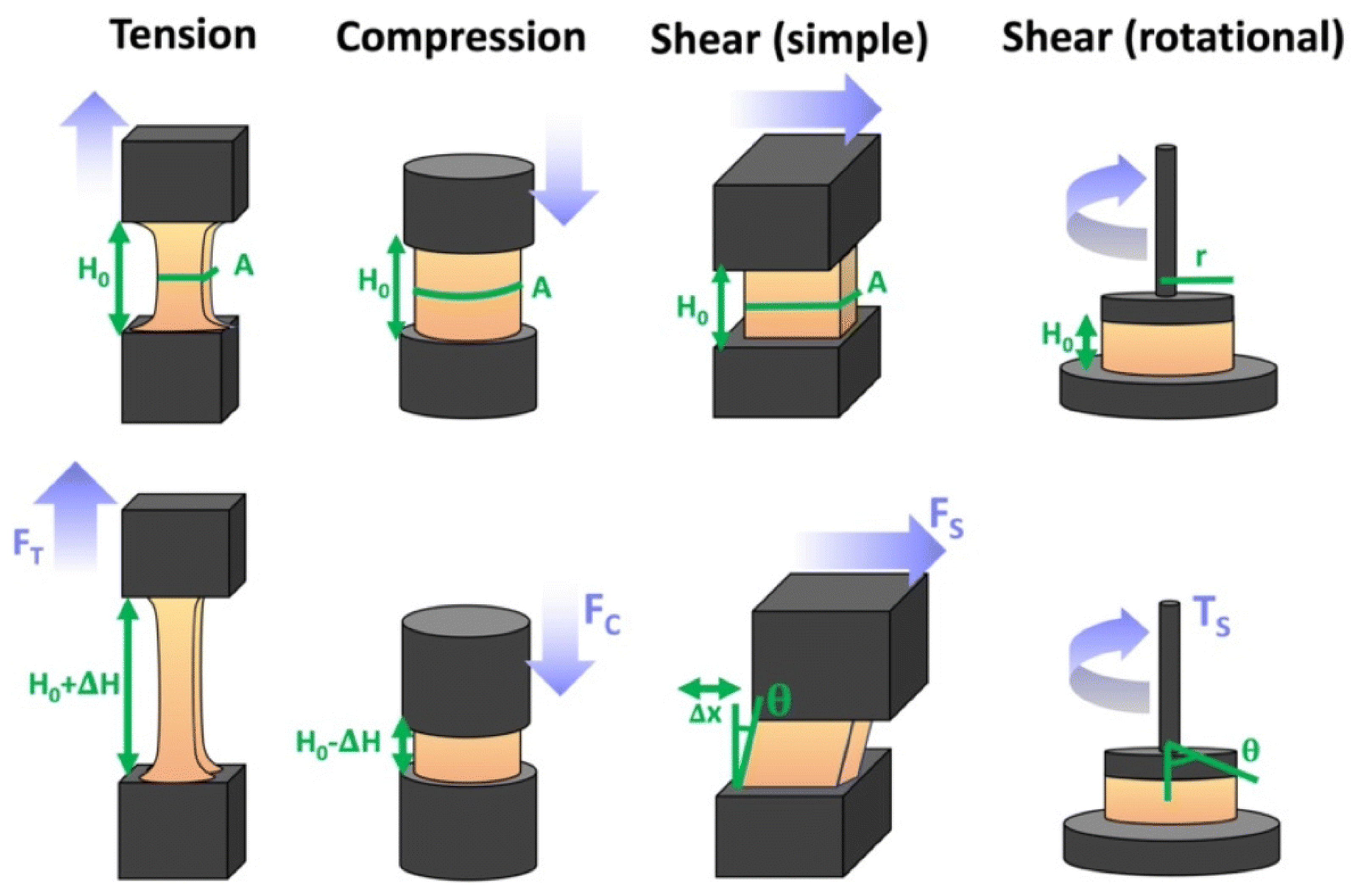
6. Summary and Outlook
The present manuscript provides a comprehensive review of polymer coatings for bioelectrodes and sensors, covering materials, their synthesis and properties, processing techniques, and practical applications.
Polymer coatings on bioelectrodes (e.g., biosensors, implantable devices, and wearable interfaces) are essential because they can reduce the impedance and improve signal quality as conducting polymers increase charge transfer and increase the signal to noise ratio. Further, especially for implantable probes and biosensors, they provide flexibility as mechanically compliant coatings match soft biological tissues and also improve the biocompatibility by reducing foreign-body responses and inflammation at the tissue–device interface.
Despite significant advancements, several key challenges persist in the complete realization of polymer coatings for bioelectrodes and biosensors. Along with numerous benefits, problems persist, encompassing dissolution, biochemical and thermal stability, and structural constraints of biopolymers. Furthermore, there is a need for standardization of protocols of deposition methods with focus on electrodeposition [435] and surface functionalization.
Major challenges are the need to further improve biocompatibility and long-term stability as even biocompatible polymers can evoke immune responses over chronic implantation periods, and polymer degradation products may have undesired effects.
In future bioelectrodes polymer coatings are increasingly used in neuromodulation implants, brain–computer interfaces, and chronic biosensors—areas where interfacing reliability and longevity are crucial [436]. Rejection of implantable bioelectrodes is an issue, and measures must be taken to avoid it.
A trade-off frequently exists between sensitivity and rapid response against long-term stability, requiring the evaluation of suitable biomarker immobilization techniques for accomplishing these objectives. Considering that in such devices the polymer itself serves both as supporting structure and mediator, the structural characteristics of the materials, such as flexibility and resistance, are pertinent and essential for the fabrication of non-invasive sensor devices, along with the ease of chemical modification of their surfaces to immobilize the biomolecules responsible for sensing [437]. The objective is to create highly sensitive, stable, flexible, and cost-effective biosensors suitable for easy and general use.
Polymers with increased conductivity are not always mechanically stable. Additionally, stability in conductivity is crucial for stable long-term applications. On the other hand, high conductivity can result in high currents endangering disruption of living cells. If metallic electrodes or simply metal containing electrodes are used, then corrosion can be an issue especially in physiological environment. These issues need attention in future applications.
An essential concern in academic research is the dependence on commercially available formulations instead of the creation of innovative structures and materials, which constrains progress in the field. Regarding manufacturing and scalability, lab-scale deposition techniques do not always scale to industrial production, as control of uniformity, throughput, and quality is non-trivial.
In many cases, the coating can no longer be treated as a purely passive barrier. Responsive (smart) coatings are now being explored as active interfaces that can adapt to the local environment and help tackle very practical problems such as biofouling, baseline drift, and transport limitations. A common approach is to use stimuli-responsive hydrogels or polymer brushes whose swelling, permeability, or surface chemistry can change reversibly (for example with temperature or pH), effectively allowing the interface to be tuned during operation [374]. Related concepts include electroactive coatings that can be switched to modulate transport or enable partial self-cleaning cycles. In parallel, self-healing coatings, which are often built on dynamic covalent bonds or supramolecular interactions, are drawing attention for wearables and long-term implants, where repeated bending, micromotion, or small defects can gradually lead to cracking, delamination, and unstable electrical contact. At the same time, there is a clear shift toward greener coating strategies, including bio-based and biodegradable polymers (such as polysaccharides and PLA-based systems) and even waste-derived biomaterials [336]. These materials are attractive not only because they can be biocompatible, but also because they support sustainability and end-of-life considerations, especially for disposable sensor platforms [352]. Progress here will also depend on scalable, lower-impact processing routes, such as water-based formulations, mild crosslinking, and electrodeposition-based coating methods. The objective is to create highly sensitive, stable, flexible, and cost-effective biosensors suitable for easy and general use.
Author Contributions
Conceptualization, N.A. (Niyaz Alizadeh) and N.A. (Nikolaos Argirusis); methodology, C.A. and N.A. (Nikolaos Argirusis); validation, C.A., N.A. (Niyaz Alizadeh) and N.A. (Nikolaos Argirusis); formal analysis, N.A. (Nikolaos Argirusis); investigation, N.A. (Niyaz Alizadeh), C.A., G.S. and A.G.; resources, N.A. (Nikolaos Argirusis); data curation, N.A. (Niyaz Alizadeh), C.A. and A.G.; writing—original draft preparation, N.A. (Niyaz Alizadeh), N.A. (Nikolaos Argirusis), A.G. and C.A.; writing—review and editing, N.A. (Niyaz Alizadeh), N.A. (Nikolaos Argirusis), A.G., G.S. and C.A.; supervision, N.A. (Nikolaos Argirusis) and C.A.; project administration, G.S.; funding, N.A. (Nikolaos Argirusis) and G.S. All authors have read and agreed to the published version of the manuscript.
Funding
This research was funded by the European Union under Project: 101135402—Mobiles—HORIZON-CL6-2023-ZEROPOLLUTION-01. Views and opinions expressed are, however, those of the author(s) only and not necessarily reflect those of the European Union or European Research Executive Agency. Neither the European Union nor the European Research Executive Agency can be responsible for them.
Institutional Review Board Statement
Not applicable.
Informed Consent Statement
Not applicable.
Data Availability Statement
No new data was created in the preparation of this review article.
Conflicts of Interest
Authors Niyaz Alizadeh and Nikolaos Argirusis were employed by the company mat4nrg GmbH. The remaining authors declare that the research was conducted in the absence of any commercial or financial relationships that could be construed as a potential conflict of interest. The funders had no role in the design of the study; in the collection, analyses, or interpretation of data; in the writing of the manuscript; or in the decision to publish the results.
Abbreviations
The following abbreviations appear in this manuscript:
| AL-BSA | amyloid-like BSA | OTFT | organic thin-film transistor |
| as-BLM | agar-supported bilayer lipid membrane | P(VI-SS) | poly(1-vinylimidazole-co-styrene sulfonate) |
| ATRP | atom transfer radical polymerization | P3ANA | poly(m-anthranilic acid) |
| Au NPs | gold nanoparticles | P4R | ponceau 4R |
| Au@PANI | gold decorated PANI | PA | polyacetylene |
| AuNCs | gold nanocrystals | PAHs | polycyclic aromatic hydrocarbons |
| AuNP | gold nanoparticles | PANI | polyaniline |
| BaP | benzo(a)pyrene | PBS | phosphate-buffered saline |
| BFC | biofuel cell | PC | phosphorylcholines |
| bHJ | bulk heterojunction | pCB | poly(carboxybetain |
| BLM | bilayer membranes made of lipids | pCBAA | pCB acrylamide |
| BPA | bisphenol A | pCBMAA | pCB methacrylamide |
| BSA | bovine serum albumin | PCM | perforated PC membrane |
| CA | calcium alginate | PDDA | poly(diallyldimethylammonium chloride) |
| CB | carboxybetaines | PEDOT | poly(3,4-ethylenedioxythiophene) |
| CE | Cholesterol esterase | PEDOT:PSS | poly(3,4-ethylenedioxythiophene) polystyrene sulfonate |
| CEA | carcinoembryonic antigen | PEG | polyethylene glycol |
| CED | cathodic electrochemical deposition | PEI | poly(ethylenimine) |
| ChOx/COX | cholesterol oxidase | PFSA | perfluorosulfonic acid |
| CNs | carbon nanomaterials | PHEMA | poly(2-hydroxyethyl methacrylate) |
| CNTs | carbon nanotubes | PIN | polyindole |
| CP | conducting polymer | PLA | poly(lactic acid) |
| CS | chitosan | PLL | poly-L-lysine |
| CTC | circulating tumor cell | PMMA | poly(methyl methacrylate) |
| DA | dopamine | PMPC | poly(2-methacryloyloxyethyl phosphorylcholine) |
| DEAEMA | 2-(diethylamino)ethyl methacrylate | PMPQ | poly(2,2,6,6-tetramethyl-4-piperidyl methacrylate-co-trimethyl-2-methacryloxyethylammonium chloride |
| Dex | dexamethasone | PNIPAm | poly(N-isopropylacrylamide) |
| DNA | deoxyribonucleic acid | POC | point-of-care |
| ECBs | enzymatic biofuel cells | POEGMA | poly(oligo(ethylene glycol) methacrylate) |
| EDC | 1-ethyl-3-(3-dimethylaminopropyl)carbodiimide | poly(HPMA) | poly[N-(2-hydroxypropyl) methacrylamide] |
| EIS | electrochemical impedance spectroscopy | poly(L-Cys) | poly(L-cysteine) |
| e-MIPs | electrochemical molecularly imprinted polymers | PoPD | poly(o-phenylenediamine) |
| ESM | eggshell membrane | PPy | polypyrrole |
| Fc-PLL | redox-active polymer | PSA | prostate-specific antigen |
| GA | α-l-guluronic acid | PF | polysulfone |
| GCE | glassy carbon electrode | PSS | poly(styrene sulfonate) |
| GDL | gas diffusion layer | PT | polythiophene |
| GFAP | glial fibrillary acidic protein | PTB | phase-transited BSA |
| GO | graphene oxide | PTFE | polytetrafluoroethylene |
| GT | gum tragacanth | PTh | polythiophene |
| HEMA | 2-hydroxyethyl methacrylate | PU | polyurethane |
| HPMA | hydroxypropylmethacrylat | PVA | poly(vinyl alcohol) |
| HSA | human serum albumin | PVC | poly(vinyl chloride) |
| IgG | immunoglobulin G | PVI–Os | poly(vinylimidazole) redox polymer |
| ITO | indium tin oxide | PVS | poly(vinylsulfonic acid), sodium salt |
| LbL | layer-by-layer | QCM | quartz crystal microbalance |
| LCST | lower critical solution temperature | RAFT | reversible addition–fragmentation chain transfer (polymerization) |
| LOD | limit of detection | SAMs | self-assembled monolayers |
| LOQ | limit of quantification | SARS-CoV-2 | COVID-19 virus |
| M | β-d-mannuronic acid | SB | sulfobetaines |
| MEA | microelectrode array | SBS | styrene–butadiene–styrene |
| MIECS | molecularly imprinted electrochemical sensors | SECM | scanning electrochemical microscopy |
| MIPs | molecularly imprinted polymers | SHPCS | sulfonated hydroxypropyl chitosan |
| MIT | molecular imprinting technology | SI-eATRP | surface-initiated electrochemically mediated ATRP |
| MOFs | metal-organic frameworks | SPCEs | screen-printed carbon electrodes |
| MPC | 2-methacryloyloxyethyl phosphorylcholine | SPEs | screen printed electrodes |
| MWCNTs | multiwall carbon nanotubes | SWCNT | single-walled carbon nanotubes |
| NADH | nicotinamide-Adenine-Dinucleotide-Hydride | TFT | thin-film transistor |
| NHS | N-hydroxysuccinimide | TNT | tri-nitro-toluol |
| NP | nanoparticles | ZW | zwitterionic |
| NSCC | nanosilica covalently coated | β-CD | β-cyclodextrin |
| OLED | organic light-emitting diode |
References
- Song, Z.; Han, R.; Yu, K.; Rong Li, R.; Luo, X. Antifouling strategies for electrochemical sensing in complex biological media. Microchim. Acta 2024, 191, 138. [Google Scholar] [CrossRef] [Scilit]
- Pillai, V.V.; Ramasubramanian, B.; Sequerth, O.; Pilla, S.; Wang, T.; Mohanty, A.K.; Govindaraj, P.; Alhassan, S.M.; Salim, N.; Kingshott, P.; et al. Nanomaterial advanced smart coatings: Emerging trends shaping the future. Appl. Mater. Today 2025, 42, 102574. [Google Scholar] [CrossRef] [Scilit]
- Siontorou, C.; Georgopoulos, K.N. Boosting the advantages of biosensors: Niche applicability and fitness for environmental purpose. Trends Environ. Anal. Chem. 2021, 32, e00146. [Google Scholar] [CrossRef] [Scilit]
- Grieshaber, D.; MacKenzie, R.; Vörös, J.; Reimhult, E. Electrochemical Biosensors—Sensor Principles and Architectures. Sensors 2008, 8, 1400–1458. [Google Scholar] [CrossRef] [Scilit]
- Shanbhag, M.M.; Manasa, G.; Mascarenhas, R.J.; Mondal, K.; Shetti, N.P. Fundamentals of bio-electrochemical sensing. Chem. Eng. J. Adv. 2023, 16, 100516. [Google Scholar] [CrossRef] [Scilit]
- Srisomwat, C.; Yakoh, A.; Chuaypen, N.; Tangkijvanich, P.; Vilaivan, T.; Chailapakul, O. Amplification-free DNA sensor for the one-step detection of the hepatitis B virus using an automated paper-based lateral flow electrochemical device. Anal. Chem. 2021, 93, 2879–2887. [Google Scholar] [CrossRef] [Scilit]
- Natarajan, B.; Kannan, P.; Maduraiveeran, G.; Alnaser, A.S. Polymer nanocomposite-based biomolecular sensor for healthcare monitoring. Adv. Colloid Interface Sci. 2025, 343, 103557. [Google Scholar] [CrossRef] [Scilit] [PubMed]
- Chen, X.-C.; Zhang, H.; Liu, S.-H.; Zhou, Y.; Jiang, L. Engineering polymeric nanofluidic membranes for efficient ionic transport: Biomimetic design, material construction, and advanced functionalities. ACS Nano 2022, 16, 17613–17640. [Google Scholar] [CrossRef] [Scilit] [PubMed]
- Khan, G.F.; Wernet, W. Design of Enzyme Electrodes for Extended Use and Storage Life. Anal. Chem. 1997, 69, 2682–2687. [Google Scholar] [CrossRef] [Scilit]
- Panjan, P.; Ohtonen, E.; Tervo, P.; Virtanen, V.; Sesay, A.M. Shelf life of enzymatic electrochemical sensors. Procedia Technol. 2017, 27, 306–308. [Google Scholar] [CrossRef] [Scilit]
- Sezgintürk, M.K. (Ed.) Commercial Biosensors and Their Applications: Clinical, Food, and Beyond; Elsevier: Amsterdam, The Netherlands, 2020. [Google Scholar] [CrossRef] [Scilit]
- Chaniotakis, N.A. Enzyme stabilization strategies based on electrolytes and polyelectrolytes for biosensor applications. Anal. Bioanal. Chem. 2004, 378, 89–95. [Google Scholar] [CrossRef] [Scilit]
- Singh, A.; Sharma, A.; Ahmed, A.; Sundramoorthy, A.K.; Furukawa, H.; Arya, S.; Khosla, A. Recent advances in electrochemical biosensors: Applications, challenges, and future scope. Biosensors 2021, 11, 336. [Google Scholar] [CrossRef] [Scilit]
- DeBrosse, M.C.; Suresh, S.; Karajic, A.; Kim, S.; Hussain, S.; Heikenfeld, J. A novel conductive membrane sensor protection technique to mitigate redox-active interferences. Biosens. Bioelectron. X 2023, 14, 100366. [Google Scholar] [CrossRef] [Scilit]
- Rahmati, Z.; Roushani, M.; Hosseini, H.; Choobin, H. An electrochemical immunosensor using SARS-CoV-2 spike protein-nickel hydroxide nanoparticles bio-conjugate modified SPCE for ultrasensitive detection of SARS-CoV-2 antibodies. Microchem. J. 2021, 170, 106718. [Google Scholar] [CrossRef] [Scilit] [PubMed]
- Zaborska, W. Competitive inhibitors of free and chitosan immobilized urease. Acta Biochim. Pol. 1995, 42, 115–118. [Google Scholar] [CrossRef] [Scilit] [PubMed]
- Soyalp, S.; Ece Eksin, E.; Erdem, A. Chitosan and carbon nanotube modified DNA biosensor for determination of drug-DNA interaction. J. Pharm. Biomed. Anal. Open 2025, 5, 100050. [Google Scholar] [CrossRef] [Scilit]
- Zou, J.; Yuan, M.-M.; Huang, Z.-N.; Chen, X.-Q.; Jiang, X.-Y.; Jiao, F.-P.; Zhou, N.; Zhou, Z.; Yu, J.-G. Highly-sensitive and selective determination of bisphenol A in milk samples based on self-assembled graphene nanoplatelets-multiwalled carbon nanotube-chitosan nanostructure. Mater. Sci. Eng. C 2019, 103, 109848. [Google Scholar] [CrossRef] [Scilit]
- Susanto, H.; Samsudin, A.M.; Rokhati, N.; Widiasa, I.N. Immobilization of glucose oxidase on chitosan-based porous composite membranes and their potential use in biosensors. Enzym. Microb. Technol. 2013, 52, 386–392. [Google Scholar] [CrossRef] [Scilit]
- Aranaz, I.; Alcántara, A.R.; Civera, M.C.; Arias, C.; Elorza, B.; Heras Caballero, A.; Acosta, N. Chitosan: An overview of its properties and applications. Polymers 2021, 13, 3256. [Google Scholar] [CrossRef] [Scilit]
- Harugade, A.; Sherje, A.P.; Pethe, A. Chitosan: A review on properties, biological activities and recent progress in biomedical applications. React. Funct. Polym. 2023, 191, 105634. [Google Scholar] [CrossRef] [Scilit]
- Ahmad, A.; Mubarak, N.; Jannat, F.T.; Ashfaq, T.; Santulli, C.; Rizwan, M.; Najda, A.; Bin-Jumah, M.; Abdel-Daim, M.M.; Hussain, S.; et al. A critical review on the synthesis of natural sodium alginate based composite materials: An innovative biological polymer for biomedical delivery applications. Processes 2021, 9, 137. [Google Scholar] [CrossRef] [Scilit]
- Cao, Y.; Cong, H.; Yu, B.; Shen, Y. A review on the synthesis and development of alginate hydrogels for wound therapy. J. Mater. Chem. B 2023, 11, 2801–2829. [Google Scholar] [CrossRef] [Scilit]
- Esimbekova, E.N.; Torgashina, I.G.; Nemtseva, E.V.; Kratasyuk, V.A. Enzymes immobilized into starch-and gelatin-based hydrogels: Properties and application in inhibition assay. Micromachines 2023, 14, 2217. [Google Scholar] [CrossRef] [Scilit]
- Versino, F.; Lopez, O.V.; Garcia, M.A.; Zaritzky, N.E. Starch-based films and food coatings: An overview. Starch-Stärke 2016, 68, 1026–1037. [Google Scholar] [CrossRef] [Scilit]
- Taqieddin, E.; Amiji, M. Enzyme immobilization in novel alginate–chitosan core-shell microcapsules. Biomaterials 2004, 25, 1937–1945. [Google Scholar] [CrossRef] [Scilit]
- Adamiak, K.; Sionkowska, A. State of innovation in alginate-based materials. Mar. Drugs 2023, 21, 353. [Google Scholar] [CrossRef] [Scilit]
- Wang, B.; Huang, D.; Weng, Z. Recent advances in polymer-based biosensors for food safety detection. Polymers 2023, 15, 3253. [Google Scholar] [CrossRef] [Scilit]
- Cyriac, S.T.; Sivasankaran, U.; Kumar, K.G. Biopolymer based electrochemical sensor for ponceau 4R: An insight into electrochemical kinetics. J. Electrochem. Soc. 2018, 165, B746–B752. [Google Scholar] [CrossRef] [Scilit]
- Sánchez Arribas, A.; Martínez-Fernández, M.; Chicharro, M. The role of electroanalytical techniques in analysis of polyphenols in wine. Trends Anal. Chem. 2012, 34, 78–96. [Google Scholar] [CrossRef] [Scilit]
- Rizzotto, F.; Khalife, M.; Hou, Y.; Chaix, C.; Lagarde, F.; Scaramozzino, N.; Vidic, J. Recent advances in electrochemical biosensors for food control. Micromachines 2023, 14, 1412. [Google Scholar] [CrossRef] [Scilit] [PubMed]
- Wang, Y.; Han, M.; Jing, L.; Jia, Q.; Lv, S.; Xu, Z.; Liu, J.; Cai, X. Enhanced neural activity detection with microelectrode arrays modified by drug-loaded calcium alginate/chitosan hydrogel. Biosens. Bioelectron. 2025, 267, 116837. [Google Scholar] [CrossRef] [Scilit]
- Ghica, M.E.; Brett, C.M.A. Glucose oxidase inhibition in poly(neutral red) mediated enzyme biosensors for heavy metal determination. Microchim. Acta 2008, 163, 185–193. [Google Scholar] [CrossRef] [Scilit]
- Wang, W.; Wang, X.; Cheng, N.; Luo, Y.; Lin, Y.; Xu, W.; Du, D. Recent advances in nanomaterials-based electrochemical (bio)sensors for pesticides detection. TrAC Trends Anal. Chem. 2020, 132, 116041. [Google Scholar] [CrossRef] [Scilit]
- Umapathi, R.; Ghoreishian, S.M.; Sonwal, S.; Rani, G.M.; Huh, Y.S. Portable electrochemical sensing methodologies for on-site detection of pesticide residues in fruits and vegetables. Coord. Chem. Rev. 2022, 453, 214305. [Google Scholar] [CrossRef] [Scilit]
- Verma, C.; Berdimurodov, E.; Verma, D.K.; Berdimuradov, K.; Alfantazi, A.; Hussain, C.M. 3D Nanomaterials: The future of industrial, biological, and environmental applications. Inorg. Chem. Commun. 2023, 156, 111163. [Google Scholar] [CrossRef] [Scilit]
- Patil, T.P.; Parkhe, V.S.; Kundale, S.S.; Kamat, R.K.; Dongale, T.D.; Patil, R.S.; Tiwari, A.P. Antisense oligonucleotide conjugated gold nanoconstructs-based electrochemical biosensor for detection of SARS-CoV-2. Appl. Surf. Sci. Adv. 2024, 22, 100618. [Google Scholar] [CrossRef] [Scilit]
- Bao, Q.; Li, G.; Yang, Z.; Liu, J.; Wang, H.; Pang, G.; Guo, Q.; Wei, J.; Cheng, W.; Lin, L. Electrochemical biosensor based on antibody-modified Au nanoparticles for rapid and sensitive analysis of influenza A virus. Ionics 2023, 29, 2021–2029. [Google Scholar] [CrossRef] [Scilit]
- Kim, H.W.; Lee, A.S.; Kim, C.S. Electrochemical biosensors based on the 3D immobilization of capture probes for influenza virus detection. RSC Adv. 2025, 15, 28565–28580. [Google Scholar] [CrossRef] [Scilit]
- Olmo, J.A.D.; Ruiz-Rubio, L.; Pérez-Alvarez, L.; Sáez-Martínez, V.; Vilas-Vilela, J.L. Antibacterial Coatings for Improving the Performance of Biomaterials. Coatings 2020, 10, 139. [Google Scholar] [CrossRef] [Scilit]
- Rehman, H.U.; Nawaz, M.A.; Pervez, S.; Jamal, M.; Attaullah, M.; Aman, A.; Qader, S.A.U. Encapsulation of pectinase within polyacrylamide gel: Characterization of its catalytic properties for continuous industrial uses. Heliyon 2020, 6, e04578. [Google Scholar] [CrossRef] [Scilit]
- Feiner, R.; Dvir, T. Tissue-electronics interfaces: From implantable devices to engineered tissues. Nat. Rev. Mater. 2018, 3, 17076. [Google Scholar] [CrossRef] [Scilit]
- Figueiredo, C.; Psotta, C.; Jayakumar, K.; Lielpetere, A.; Mandal, T.; Schuhmann, W.; Leech, D.; Falk, M.; Pita, M.; Shleev, S.; et al. Effect of Protection Polymer Coatings on the Performance of an Amperometric Galactose Biosensor in Human Plasma. Biosensors 2024, 14, 167. [Google Scholar] [CrossRef] [Scilit]
- Tığ, G.A.; Marrazza, G.; Turan, K.; Erdoğan, N.Ö.; Şimşek, N. Recent developments in electrochemical sensors and biosensors for food additives determination: Principle and application. Trends Anal. Chem. 2025, 183, 118127. [Google Scholar] [CrossRef] [Scilit]
- Harish Prashanth, K.V.; Tharanathan, R.N. Chitin/chitosan: Modifications and their unlimited application potential—An overview. Trends Food Sci. Technol. 2007, 18, 117–131. [Google Scholar] [CrossRef] [Scilit]
- Prokopijević, M. Natural polymers: Suitable carriers for enzyme immobilization. Biol. Serbica 2021, 43, 43–49. [Google Scholar] [CrossRef] [Scilit]
- Arshad, F.; Mohd-Naim, N.F.; Chandrawati, R.; Cozzolino, D.; Ahmed, M.U. Nanozyme-based sensors for detection of food biomarkers: A review. RSC Adv. 2022, 12, 26160–26175. [Google Scholar] [CrossRef] [Scilit] [PubMed]
- Kong, J.; Xu, X.; Ma, Y.; Miao, J.; Bian, X. Rapid and sensitive detection of sulfamethizole using a reusable molecularly imprinted electrochemical sensor. Foods 2023, 12, 1693. [Google Scholar] [CrossRef] [Scilit]
- Cai, R. Fabrication and applications of enzymatic biofuel cells. In Biofuel Cells and Energy Generation; Savasiduni, K.K., Geetha, M., Eds.; Woodhead Series in Bioenergy; Elsevier: Amsterdam, The Netherlands, 2025; pp. 109–181. [Google Scholar] [CrossRef] [Scilit]
- Lin, P.-H.; Li, B.-R. Antifouling strategies in advanced electrochemical sensors and biosensors. Analyst 2020, 145, 1110. [Google Scholar] [CrossRef] [Scilit] [PubMed]
- Chan, D.; Chien, J.-C.; Axpe, E.; Blankemeier, L.; Baker, S.W.; Swaminathan, S.; Piunova, V.A.; Zubarev, D.Y.; Maikawa, C.L.; Grosskopf, A.K.; et al. Combinatorial Polyacrylamide Hydrogels for Preventing Biofouling on Implantable Biosensors. Adv. Mater. 2022, 34, 2109764. [Google Scholar] [CrossRef] [Scilit]
- Munarin, F.; Tanzi, M.C.; Petrini, P. Advances in biomedical applications of pectin gels. Int. J. Biol. Macromol. 2012, 51, 681–689. [Google Scholar] [CrossRef] [Scilit]
- Beilinson, R.M.; Yavisheva, A.A.; Lopatko, N.P.; Medyantseva, E.P. Amperometric biosensors for the determination of tetracycline. Inorg. Mater. 2023, 59, 1462–1469. [Google Scholar] [CrossRef] [Scilit]
- Lwin, H.T.; Phonklam, K.; Khunpitak, T.; Khongkow, P.; Phairatana, T. Automated microfluidic-based polyurethane/lactate oxidase-chitosan/m-phenylenediamine biosensing platform for lactate monitoring in breast cancer cell culture. Anal. Chim. Acta 2025, 1356, 344049. [Google Scholar] [CrossRef] [Scilit]
- Pawar, S.N.; Edgar, K.J. Alginate derivatization: A review of chemistry, properties and applications. Biomaterials 2012, 33, 3279–3305. [Google Scholar] [CrossRef] [Scilit] [PubMed]
- Cheng, K.C.; Demirci, A.; Catchmark, J.M. Pullulan: Biosynthesis, production, and applications. Appl. Microbiol. Biotechnol. 2011, 92, 29–44. [Google Scholar] [CrossRef] [Scilit] [PubMed]
- Wan, Y.; Zhou, J.; Ni, J.; Cai, Y.; Cohen Stuart, M.; Wang, J. Electrostatically mediated in situ polymerization for enzyme immobilization and activation. Biomacromolecules 2024, 25, 809–818. [Google Scholar] [CrossRef] [Scilit]
- Sacco, P.; Pedroso-Santana, S.; Kumar, Y.; Joly, N.; Martin, P.; Bocchetta, P. Ionotropic Gelation of Chitosan Flat Structures and Potential Applications. Molecules 2021, 26, 660. [Google Scholar] [CrossRef] [Scilit] [PubMed]
- Guajardo, N. Immobilization of lipases using poly (vinyl) alcohol. Polymers 2023, 15, 2021. [Google Scholar] [CrossRef] [Scilit]
- Pang, A.L.; Arsad, A.; Ahmadipour, M. Synthesis and factor affecting on the conductivity of polypyrrole: A short review. Polym. Adv. Technol. 2021, 32, 1428–1454. [Google Scholar] [CrossRef] [Scilit]
- Selloum, D.; Abou Chaaya, A.; Bechelany, M.; Rouessac, V.; Miele, P.; Tingry, S. A highly efficient gold/electrospun PAN fiber material for improved laccase biocathodes for biofuel cell applications. J. Mater. Chem. A 2014, 2, 2794–2800. [Google Scholar] [CrossRef] [Scilit]
- Eyvazi, S.; Baradaran, B.; Mokhtarzadeh, A.; de la Guardia, M. Recent advances on development of portable biosensors for monitoring of biological contaminants in foods. Trends Food Sci. Technol. 2021, 114, 712–721. [Google Scholar] [CrossRef] [Scilit]
- Sohrabi, H.; Hemmati, A.; Majidi, M.R.; Eyvazi, S.; Jahanban-Esfahlan, A.; Baradaran, B.; Adlpour-Azar, R.; Mokhtarzadeh, A.; de la Guardia, M. Recent advances on portable sensing and biosensing assays applied for detection of main chemical and biological pollutant agents in water samples: A critical review. TrAC Trends Anal. Chem. 2021, 143, 116344. [Google Scholar] [CrossRef] [Scilit]
- Campuzano, S.; Pedrero, M.; Yáñez-Sedeño, P.; Pingarrón, J.M. Antifouling (Bio)materials for Electrochemical (Bio)sensing. Int. J. Mol. Sci. 2019, 20, 423. [Google Scholar] [CrossRef] [Scilit]
- Piras, C.C.; Smith, D.K. Multicomponent polysaccharide alginate-based bioinks. J. Mater. Chem. B 2020, 8, 8171–8188. [Google Scholar] [CrossRef] [Scilit]
- Flórez, M.; Cazón, P.; Vázquez, M. Selected biopolymers’ processing and their applications: A review. Polymers 2023, 15, 641. [Google Scholar] [CrossRef] [Scilit]
- Hanssen, B.L.; Siraj, S.; Wong, D.K.Y. Recent strategies to minimise fouling in electrochemical detection systems. Rev. Anal. Chem. 2016, 35, 1–28. [Google Scholar] [CrossRef] [Scilit]
- Yang, X.; Chen, P.; Zhang, X.; Zhou, H.; Song, Z.; Yang, W.; Luo, X. An electrochemical biosensor for HER2 detection in complex biological media based on two antifouling materials of designed recognizing peptide and PEG. Anal. Chim. Acta 2023, 1252, 341075. [Google Scholar] [CrossRef] [Scilit]
- Yang, X.; Kirsch, J.; Olsen, E.V.; Fergus, J.W.; Simonian, A.L. Anti-fouling PEDOT:PSS modification on glassy carbon electrodes for continuous monitoring of tricresyl phosphate. Sens. Actuators B Chem. 2013, 177, 659–667. [Google Scholar] [CrossRef] [Scilit]
- Wang, Y.-M.; Kálosi, A.; Halahovets, Y.; Romanenko, I.; Slabý, J.; Homola, J.; Svoboda, J.; de los Santos Pereira, A.; Pop-Georgievski, O. Grafting Density and Antifouling Properties of Poly [N-(2-Hydroxypropyl) Methacrylamide] Brushes Prepared by “Grafting to” and “Grafting From”. Polym. Chem. 2022, 13, 3815–3826. [Google Scholar] [CrossRef] [Scilit]
- Zinggeler, M.; Schär, S.; Kurth, F. Printed Antifouling Electrodes for Biosensing Applications. ACS Appl. Mater. Interfaces 2022, 14, 56578–56584. [Google Scholar] [CrossRef] [Scilit]
- Yang, W.; Chengkai Xuan, C.; Liu, X.; Zhang, Q.; Wu, K.; Bian, L.; Shi, X. A sandwiched patch toward leakage-free and anti-postoperative tissue adhesion sealing of intestinal injuries. Bioact. Mater. 2023, 24, 112–123. [Google Scholar] [CrossRef] [Scilit]
- Seget, S.; Rusak, E.; Partyka, M.; Samulska, E.; Pyziak-Skupień, A.; Kamińska, H.; Skała-Zamorowska, E.; Jarosz-Chobot, P. Bacterial Strains Colonizing the Sensor Electrodes of a Continuous Glucose Monitoring System in Children with Diabetes. Acta Diabetol. 2021, 58, 191–195. [Google Scholar] [CrossRef] [Scilit]
- Kutner, N.; Kunduru, K.R.; Rizik, L.; Farah, S. Recent advances for improving functionality, biocompatibility, and longevity of implantable medical devices and deliverable drug delivery systems. Adv. Funct. Mater. 2021, 31, 2010929. [Google Scholar] [CrossRef] [Scilit]
- Wareham-Mathiassen, S.; Jolly, P.; Radha Shanmugam, N.; Jagannath, B.; Prabhala, P.; Zhai, Y.; Ozkan, A.; Naziripour, A.; Singh, R.; Bengtsson, H.; et al. An Antimicrobial and Antifibrotic Coating for Implantable Biosensors. Biosensors 2025, 15, 171. [Google Scholar] [CrossRef] [Scilit]
- Leonardi, A.K.; Ober, C.K. Polymer-based marine antifouling and fouling release surfaces: Strategies for synthesis and modification. Annu. Rev. Chem. Biomol. Eng. 2019, 10, 241–264. [Google Scholar] [CrossRef] [Scilit] [PubMed]
- Wen, J.; Huang, S.; Hu, Q.; He, W.; Wei, Z.; Wang, L.; Lu, J.; Yue, X.; Men, S.; Miao, C.; et al. Recent Advances in Zwitterionic Polymers-Based Non-Fouling Coating Strategies for Biomedical Applications. Mater. Today Chem. 2024, 40, 102232. [Google Scholar] [CrossRef] [Scilit]
- Vallejo-Giraldo, C.; Krukiewicz, K.; Biggs, M.J.P. Understanding the mechanobiology of gliosis may be the key to unlocking sustained chronic performance of bioelectronic neural interfaces. Adv. NanoBiomed Res. 2022, 2, 2100098. [Google Scholar] [CrossRef] [Scilit]
- Gray, M.E.; Meehan, J.; Blair, E.O.; Ward, C.; Langdon, S.P.; Morrison, L.R.; Marland, J.R.K.; Tsiamis, A.; Kunkler, I.H.; Murray, A.; et al. Biocompatibility of common implantable sensor materials in a tumor xenograft model. J. Biomed. Mater. Res. Part B Appl. Biomater. 2019, 107, 1620–1633. [Google Scholar] [CrossRef] [Scilit]
- Patel, T.; Huang, J.; Krukiewicz, K. Multifunctional organic monolayer-based coatings for implantable biosensors and bioelectronic devices: Review and perspectives. Biosens. Bioelectron. X 2023, 14, 100349. [Google Scholar] [CrossRef] [Scilit]
- Meng, X.; O’Hare, D.; Ladame, S. Surface immobilization strategies for the development of electrochemical nucleic acid sensors. Biosens. Bioelectron. 2023, 237, 115440. [Google Scholar] [CrossRef] [Scilit] [PubMed]
- Zhang, Y.; Li, Y.; Wu, W.; Jiang, Y.; Hu, B. Chitosan coated on the layers’ glucose oxidase immobilized on cysteamine/Au electrode for use as glucose biosensor. Biosens. Bioelectron. 2014, 60, 271–276. [Google Scholar] [CrossRef] [Scilit]
- Zhu, H.; Srivastava, R.; Brown, J.Q.; McShane, M.J. Combined physical and chemical immobilization of glucose oxidase in alginate microspheres improves stability of encapsulation and activity. Bioconjug. Chem. 2005, 16, 1451–1458. [Google Scholar] [CrossRef] [Scilit]
- Wang, X.; Zhu, K.X.; Zhou, H.M. Immobilization of glucose oxidase in alginate-chitosan microcapsules. Int. J. Mol. Sci. 2011, 12, 3042–3054. [Google Scholar] [CrossRef] [Scilit]
- Anchidin-Norocel, L.; Savage, W.K.; Gheorghita, R.; Amariei, S. Biopolymers Used for Receptor Immobilization for Nickel-Detection Biosensors in Food. Micromachines 2023, 14, 1529. [Google Scholar] [CrossRef] [Scilit]
- Song, M.; Lin, X.; Peng, Z.; Xu, S.; Jin, L.; Zheng, X.; Luo, H. Materials and methods of biosensor interfaces with stability. Front. Mater. 2021, 7, 583739. [Google Scholar] [CrossRef] [Scilit]
- Tadesse, M.; Liu, Y. Recent Advances in Enzyme Immobilization: The Role of Artificial Intelligence, Novel Nanomaterials, and Dynamic Carrier Systems. Catalysts 2025, 15, 571. [Google Scholar] [CrossRef] [Scilit]
- Mohammad Karimi, S.; Ershad-Langroudi, A.; Alizadegan, F. Surface analysis and water contact angle of modified natural biopolymers. In Handbook Natural Polymers; Elsevier: Amsterdam, The Netherlands, 2024; pp. 473–500. [Google Scholar] [CrossRef] [Scilit]
- Aziz, T.; Farid, A.; Haq, F.; Kiran, M.; Ullah, A.; Zhang, K.; Al Jaouni, S.K. A review on the modification of cellulose and its applications. Polymers 2022, 14, 3206. [Google Scholar] [CrossRef] [Scilit]
- Intisar, A.; Hedar, M.; Sharif, A.; Ahmed, E.; Hussain, N.; Hadibarata, T.; Shariati, M.A.; Smaoui, S. Enzyme immobilization on alginate biopolymer for biotechnological applications. In Microbial Biomolecules; Elsevier: Amsterdam, The Netherlands, 2023; pp. 471–488. [Google Scholar] [CrossRef] [Scilit]
- Wang, J.; Zhuang, S. Chitosan-based materials: Preparation, modification and application. J. Clean. Prod. 2022, 355, 131825. [Google Scholar] [CrossRef] [Scilit]
- Bisht, M.; Thayallath, S.K.; Bharadwaj, P.; Franklin, G.; Mondal, D. Biomass-derived functional materials as carriers for enzymes: Towards sustainable and robust biocatalysts. Green Chem. 2023, 25, 4591–4624. [Google Scholar] [CrossRef] [Scilit]
- Prokopijevic, M.; Prodanovic, O.; Spasojevic, D.; Kovacevic, G.; Polovic, N.; Radotic, K.; Prodanovic, R. Tyramine-modified pectins via periodate oxidation for soybean hull peroxidase induced hydrogel formation and immobilization. Appl. Microbiol. Biotechnol. 2017, 101, 2281–2290. [Google Scholar] [CrossRef] [Scilit]
- Karim, A.A.; Bhat, R. Gelatin alternatives for the food industry: Recent developments, challenges and prospects. Trends Food Sci. Technol. 2008, 19, 644–656. [Google Scholar] [CrossRef] [Scilit]
- Xin, M.; Zhang, X.; Yang, J.; Chen, M.; Ma, X.; Liu, J. Preparation and electrochemical sensing application of electrospun porous organosilica fibers. Electrochem. Commun. 2014, 48, 123–126. [Google Scholar] [CrossRef] [Scilit]
- Mehrasa, M.; Asadollahi, M.A.; Ghaedi, K.; Salehi, H.; Arpanaei, A. Electrospun aligned PLGA and PLGA/gelatin nanofibers embedded with silica nanoparticles for tissue engineering. Int. J. Biol. Macromol. 2015, 79, 687–695. [Google Scholar] [CrossRef] [Scilit]
- Shen, Q.; Yang, R.; Hua, X.; Ye, F.; Zhang, W.; Zhao, W. Gelatin-templated biomimetic calcification for β-galactosidase immobilization. Process Biochem. 2011, 46, 1565–1571. [Google Scholar] [CrossRef] [Scilit]
- Rather, A.H.; Khan, R.S.; Wani, T.U.; Beigh, M.A.; Sheikh, F.A. Overview on immobilization of enzymes on synthetic polymeric nanofibers fabricated by electrospinning. Biotechnol. Bioeng. 2022, 119, 9–33. [Google Scholar] [CrossRef] [Scilit]
- Fromager, B.; Marhuenda, E.; Louis, B.; Bakalara, N.; Cambedouzou, J.; Cornu, D. Recent Advances in Electrospun Fibers for Biological Applications. Macromol 2023, 3, 569–613. [Google Scholar] [CrossRef] [Scilit]
- Vorvi, S.; Tsougeni, K.; Tserepi, A.; Kakabakos, S.; Petrou, P.; Gogolides, E. Enhanced Immobilization of Enzymes on Plasma Micro-Nanotextured Surfaces and Microfluidics: Application to HRP. Molecules 2024, 29, 4736. [Google Scholar] [CrossRef] [Scilit]
- Du, X.; Wang, L.; Liang, H.; Chen, G.; Wu, J.; Xia, W.; Gao, D. Removal of benzo [a] pyrene from the soil by adsorption coupled with degradation on saponin-modified bentonite immobilized crude enzymes. Environ. Res. 2024, 261, 119716. [Google Scholar] [CrossRef] [Scilit]
- Terzopoulou, Z.; Zamboulis, A.; Koumentakou, I.; Michailidou, G.; Noordam, M.J.; Bikiaris, D.N. Biocompatible synthetic polymers for tissue engineering purposes. Biomacromolecules 2022, 23, 1841–1863. [Google Scholar] [CrossRef] [Scilit]
- Xu, X.; Pose-Boirazian, T.; Eibes, G.; McCoubrey, L.E.; Martínez-Costas, J.; Gaisford, S.; Goyanes, A.; Basit, A.W. A customizable 3D printed device for enzymatic removal of drugs in water. Water Res. 2022, 208, 117861. [Google Scholar] [CrossRef] [Scilit] [PubMed]
- Li, G.; Nandgaonkar, A.G.; Lu, K.; Krause, W.E.; Lucia, L.A.; Wei, Q. Laccase immobilized on PAN/O-MMT composite nanofibers support for substrate bioremediation: A de novo adsorption and biocatalytic synergy. RSC Adv. 2016, 6, 41420–41427. [Google Scholar] [CrossRef] [Scilit]
- Muthukumar, J.; Kandukuri, V.A.; Chidambaram, R. A critical review on various treatment, conversion, and disposal approaches of commonly used polystyrene. Polym. Bull. 2024, 81, 2819–2845. [Google Scholar] [CrossRef]
- Sánchez-Otero, M.G.; Quintana-Castro, R.; Rojas-Vázquez, A.S.; Badillo-Zeferino, G.L.; Mondragón-Vázquez, K.; Espinosa-Luna, G.; Kumar Nadda, A.; Oliart-Ros, R.M. Polypropylene as a selective support for the immobilization of lipolytic enzymes: Hyper-activation, purification and biotechnological applications. J. Chem. Technol. Biotechnol. 2022, 97, 436–445. [Google Scholar] [CrossRef] [Scilit]
- Singthong, J.; Thongkaew, C. Using hydrocolloids to decrease oil absorption in banana chips. LWT-Food Sci. Technol. 2009, 42, 1199–1203. [Google Scholar] [CrossRef] [Scilit]
- Mai, T.H.A. Biochemical studies on the immobilized lactase in the combined alginate–carboxymethyl cellulose gel. Biochem. Eng. J. 2013, 74, 81–87. [Google Scholar] [CrossRef] [Scilit]
- Holyavka, M.; Redko, Y.; Goncharova, S.; Lavlinskaya, M.; Sorokin, A.; Kondratyev, M.; Artyukhov, V. Novel Hybrid Catalysts of Cysteine Proteases Enhanced by Chitosan and Carboxymethyl Chitosan Micro-and Nanoparticles. Polymers 2024, 16, 3111. [Google Scholar] [CrossRef] [Scilit]
- Chen, N.; Chang, B.; Shi, N.; Yan, W.; Lu, F.; Liu, F. Cross-linked enzyme aggregates immobilization: Preparation, characterization, and applications. Crit. Rev. Biotechnol. 2023, 43, 369–383. [Google Scholar] [CrossRef] [Scilit] [PubMed]
- Knop, K.; Hoogenboom, R.; Fischer, D.; Schubert, U.S. Poly (ethylene glycol) in drug delivery: Pros and cons as well as potential alternatives. Angew. Chem. Int. Ed. 2010, 49, 6288–6308. [Google Scholar] [CrossRef] [Scilit]
- Li, X.Y.; Yu, S.Y.; Park, H.J.; Zhao, M. Polyethyleneglycol diacrylate microspheres: A novel carrier for laccase immobilisation. J. Microencapsul. 2015, 32, 22–28. [Google Scholar] [CrossRef] [Scilit]
- Choi, D.; Lee, W.; Park, J.; Koh, W. Preparation of poly (ethylene glycol) hydrogels with different network structures for the application of enzyme immobilization. Bio-Med. Mater. Eng. 2008, 18, 345–356. [Google Scholar] [CrossRef] [Scilit]
- Crulhas, B.P.; Recco, L.C.; Delella, F.K.; Pedrosa, V.A. A novel superoxide anion biosensor for monitoring reactive species of oxygen released by cancer cells. Electroanalysis 2017, 29, 1252–1257. [Google Scholar] [CrossRef] [Scilit]
- Shakeri, F.; Ariaeenejad, S.; Ghollasi, M.; Motamedi, E. Synthesis of two novel bio-based hydrogels using sodium alginate and chitosan and their proficiency in physical immobilization of enzymes. Sci. Rep. 2022, 12, 2072. [Google Scholar] [CrossRef] [Scilit]
- Mohammadi, N.S.; Khiabani, M.S.; Ghanbarzadeh, B.; Mokarram, R.R. Enhancement of biochemical aspects of lipase adsorbed on halloysite nanotubes and entrapped in a polyvinyl alcohol/alginate hydrogel: Strategies to reuse the most stable lipase. World J. Microbiol. Biotechnol. 2020, 36, 45. [Google Scholar] [CrossRef] [Scilit] [PubMed]
- Bilal, M.; Rasheed, T.; Zhao, Y.; Iqbal, H.M. Agarose-chitosan hydrogel-immobilized horseradish peroxidase with sustainable bio-catalytic and dye degradation properties. Int. J. Biol. Macromol. 2019, 124, 742–749. [Google Scholar] [CrossRef] [Scilit] [PubMed]
- Horn, C.; Pospiech, D.; Allertz, P.J.; Müller, M.; Salchert, K.; Hommel, R. Chemical design of hydrogels with immobilized laccase for the reduction of persistent trace compounds in wastewater. ACS Appl. Polym. Mater. 2021, 3, 2823–2834. [Google Scholar] [CrossRef] [Scilit]
- Chen, H.Y.; Cheng, K.C.; Hsu, R.J.; Hsieh, C.W.; Wang, H.T.; Ting, Y. Enzymatic degradation of ginkgolic acid by laccase immobilized on novel electrospun nanofiber mat. J. Sci. Food Agric. 2020, 100, 2705–2712. [Google Scholar] [CrossRef] [Scilit]
- Jankowska, K.; Su, Z.; Sigurdardóttir, S.B.; Staszak, M.; Pinelo, M.; Zdarta, J.; Jesionowski, T. Tailor-made novel electrospun polystyrene/poly (d, l-lactide-co-glycolide) for oxidoreductases immobilization: Improvement of catalytic properties under extreme reaction conditions. Bioorg. Chem. 2021, 114, 105036. [Google Scholar] [CrossRef] [Scilit]
- Li, J.; Wu, H.; Liang, Y.; Jiang, Z.; Jiang, Y.; Zhang, L. Facile fabrication of organic–inorganic hybrid beads by aminated alginate enabled gelation and biomimetic mineralization. J. Biomater. Sci. Polym. Ed. 2013, 24, 119–134. [Google Scholar] [CrossRef] [Scilit]
- Vatsyayan, P.; Bordoloi, S.; Goswami, P. Large catalase based bioelectrode for biosensor application. Biophys. Chem. 2010, 153, 36–42. [Google Scholar] [CrossRef] [Scilit]
- Jiang, W.; Pei, R.; Zhou, S.-F. 3D-printed xylanase within biocompatible polymers as excellent catalyst for lignocellulose degradation. Chem. Eng. J. 2020, 400, 125920. [Google Scholar] [CrossRef] [Scilit]
- Aquinas, N.; Chithra, C.H.; Bhat, M.R. Progress in bioproduction, characterization and applications of pullulan: A review. Polym. Bull. 2024, 81, 12347–12382. [Google Scholar] [CrossRef] [Scilit]
- Singh, R.S.; Kaur, N.; Hassan, M.; Kennedy, J.F. Pullulan in biomedical research and development—A review. Int. J. Biol. Macromol. 2021, 166, 694–706. [Google Scholar] [CrossRef] [Scilit]
- de Souza, C.K.; Ghosh, T.; Lukhmana, N.; Tahiliani, S.; Priyadarshi, R.; Hoffmann, T.G.; Han, S.S. Pullulan as a sustainable biopolymer for versatile applications: A review. Mater. Today Commun. 2023, 36, 106477. [Google Scholar] [CrossRef] [Scilit]
- BelBruno, J.J. Molecularly Imprinted Polymers. Chem. Rev. 2019, 119, 94–119. [Google Scholar] [CrossRef] [Scilit]
- Guo, B.; Ma, P.X. Conducting Polymers for Tissue Engineering. Biomacromolecules 2018, 19, 1764–1782. [Google Scholar] [CrossRef] [Scilit]
- Guimard, N.K.; Gomez, N.; Schmidt, C.E. Conducting polymers in biomedical engineering. Prog. Polym. Sci. 2007, 32, 876–921. [Google Scholar] [CrossRef] [Scilit]
- Yang, G.; Kampstra, K.L.; Abidian, M.R. High performance conducting polymer nano-fiber biosensors for detection of biomolecules. Adv. Mater. 2014, 26, 4954–4960. [Google Scholar] [CrossRef] [Scilit]
- Groenendaal, L.; Zotti, G.; Aubert, P.-H.; Waybright, S.M.; Reynolds, J.R. Electrochemistry of poly(3,4-alkylenedioxythiophene) derivatives. Adv. Mater. 2003, 15, 855–879. [Google Scholar] [CrossRef] [Scilit]
- Shahini, A.; Yazdimamaghani, M.; Walker, K.J.; Eastman, M.A.; Hatami-Marbini, H.; Smith, B.J.; Ricci, J.L.; Madihally, S.V.; Vashaee, D.; Tayebi, L. 3D conductive nanocomposite scaffold for bone tissue engineering. Int. J. Nanomed. 2014, 9, 167. [Google Scholar] [CrossRef] [Scilit]
- Yang, T.; Yang, M.; Xu, C.; Yang, K.; Su, Y.; Ye, Y.; Dou, L.; Yang, Q.; Wenbo Ke, W.; Wang, B.; et al. PEDOT:PSS hydrogels with high conductivity and biocompatibility for in situ cell sensing. J. Mater. Chem. B 2023, 11, 3226–3235. [Google Scholar] [CrossRef] [Scilit]
- Dwewedi, A. Chapter 4 Polymer-based immobilized enzymes in environmental remediation. In Polymeric Supports for Enzyme Immobilization—Opportunities and Applications; Elsevier Inc.: Amsterdam, The Netherlands, 2021; pp. 105–166. [Google Scholar] [CrossRef] [Scilit]
- Nasar, A.; Perveen, R. Applications of enzymatic biofuel cells in bioelectronic devices—A review. Int. J. Hydrogen Energy 2019, 44, 15287–15312. [Google Scholar] [CrossRef] [Scilit]
- Wu, F.; Yu, P.; Mao, L. Bioelectrochemistry for in vivo analysis: Interface engineering toward implantable electrochemical biosensors. Curr. Opin. Electrochem. 2017, 5, 152–157. [Google Scholar] [CrossRef] [Scilit]
- Mishra, P.; Lakshmi, G.B.V.S.; Mishra, S.; Avasthi, D.K.; Swart, H.C.; Turner, A.P.F.; Mishra, Y.K.; Tiwari, A. Electrocatalytic biofuel cell based on highly efficient metal-polymer nano-architectured bioelectrodes. Nano Energy 2017, 39, 601–607. [Google Scholar] [CrossRef] [Scilit]
- Katz, E.; Bückmann, A.F.; Willner, I. Self-powered enzyme-based biosensors. J. Am. Chem. Soc. 2001, 123, 10752–10753. [Google Scholar] [CrossRef] [Scilit] [PubMed]
- Moore, C.M.; Minteer, S.B.; Martin, R.S. Microchip-based ethanol/oxygen biofuel cell. Lab Chip 2005, 5, 218–225. [Google Scholar] [CrossRef] [Scilit]
- Guo, Z.; Smutok, O.; Johnston, W.A.; Ayva, C.E.; Walden, P.; McWhinney, B.; Ungerer, J.J.P.; Melman, A.; Katz, E.; Alexandrov, K. Circular permutated PQQ-glucose dehydrogenase as an ultrasensitive electrochemical biosensor. Angew. Chem. Int. Ed. 2022, 61, e202109005. [Google Scholar] [CrossRef] [Scilit]
- Cai, R.; Ngwadom, C.; Saxena, R.; Soman, J.; Bruggeman, C.; Hickey, D.P.; Verduzco, R.; Ajo-Franklin, C.M. Creation of a point-of-care therapeutics sensor using protein engineering, electrochemical sensing and electronic integration. Nat. Commun. 2024, 15, 1689. [Google Scholar] [CrossRef] [Scilit]
- Jain, R.; Singhal, N.; Vardhan, H.; Vashistha, P.; Bist, Y.; Pandey, A.; Wagri, N.K.; Gaur, A. Polysaccharide-based functional materials for flexible electronics: A comprehensive review of synthesis strategies, functionalization, and applications. Carbohydr. Polym. Technol. Appl. 2025, 12, 101038. [Google Scholar] [CrossRef] [Scilit]
- Yang, L.; Yuan, Q.-Y.; Lou, C.-W.; Lin, J.-H.; Li, T.-T. Recent Research Progress of Polysaccharide Polymer Coatings for Improving Properties of Paper-Based Packaging Materials. Coatings 2025, 15, 326. [Google Scholar] [CrossRef] [Scilit]
- Boccheta, P.; Oltman, A.; Gupta, M.; Andriani, G.; Martin, P.; Kumar, Y.; Joly, N.; Sacco, P.; Javed, M.S. Chitosan in electrochemical (bio)sensors: Nanostructuring and methods of synthesis. Eur. Polym. J. 2024, 213, 113092. [Google Scholar] [CrossRef] [Scilit]
- Suginta, W.; Khunkaewla, P.; Schulte, A. Electrochemical Biosensor Applications of Polysaccharides Chitin and Chitosan. Chem. Rev. 2013, 113, 5458–5479. [Google Scholar] [CrossRef] [Scilit] [PubMed]
- Jin, T.; Liu, T.; Edmond Lam, E.; Moores, A. Chitin and chitosan on the nanoscale. Nanoscale Horiz. 2021, 6, 505. [Google Scholar] [CrossRef] [Scilit]
- Ashrafizadeh, M.; Hushmandi, K.; Mirzaei, S.; Bokaie, S.; Bigham, A.; Makvandi, P.; Rabiee, N.; Thakur, V.K.; Kumar, A.P.; Sharifi, E.; et al. Chitosan-based nanoscale systems for doxorubicin delivery: Exploring biomedical application in cancer therapy. Bioeng. Transl. Med. 2023, 8, e10325. [Google Scholar] [CrossRef] [Scilit] [PubMed]
- Ambrózy, A.; Hlavatá, L.; Labuda, J. Protective membranes at electrochemical biosensors. Acta Chim. Slovaca 2013, 6, 35–41. [Google Scholar] [CrossRef] [Scilit]
- Michna, A.; Lupa, D.; Płaziski, W.; Batys, P.; Adamczyk, Z. Physicochemical characteristics of chitosan molecules: Modeling and experiments. Adv. Colloid Interface Sci. 2025, 337, 103383. [Google Scholar] [CrossRef] [Scilit]
- Venkatesan, J.; Kim, S.K. Chitosan Composites for Bone Tissue Engineering—An Overview. Mar. Drugs 2010, 8, 2252–2266. [Google Scholar] [CrossRef] [Scilit]
- Barra, A.; Alves, Z.; Ferreira, N.M.; Martins, M.A.; Oliveira, H.; Ferreira, L.P.; Cruz, M.M.; Carvalho, M.; Neumayer, S.M.; Rodriguez, B.J.; et al. Biocompatible chitosan-based composites with properties suitable for hyperthermia therapy. J. Mater. Chem. B 2020, 8, 1256–1265. [Google Scholar] [CrossRef] [Scilit]
- Buaki-Sogó, M.; García-Carmona, L.; Gil-Agustí, M.; García-Pellicer, M.; Quijano-López, A. Flexible and Conductive Bioelectrodes Based on Chitosan-Carbon Black Membranes: Towards the Development of Wearable Bioelectrodes. Nanomaterials 2021, 11, 2052. [Google Scholar] [CrossRef] [Scilit]
- Liu, Y.; Yin, P.; Chen, J.; Cui, B.; Zhang, C.; Wu, F. Conducting Polymer-Based Composite Materials for Therapeutic Implantations: From Advanced Drug Delivery System to Minimally Invasive Electronics. Int. J. Polym. Sci. 2020, 2020, 5659682. [Google Scholar] [CrossRef] [Scilit]
- Wu, S.; Wu, S.; Zhang, X.; Feng, T.; Wu, L. Chitosan-Based Hydrogels for Bioelectronic Sensing: Recent Advances and Applications in Biomedicine and Food Safety. Biosensors 2023, 13, 93. [Google Scholar] [CrossRef] [Scilit]
- Hasannezhad, H.; Bakhshi, A.; Mozafari, M.R.; Naghib, S.M. A review of chitosan role in milk bioactive-based drug delivery, smart packaging and biosensors: Recent advances and developments. Int. J. Biol. Macromol. 2025, 294, 139248. [Google Scholar] [CrossRef] [Scilit] [PubMed]
- Lee, K.Y.; Mooney, D.J. Alginate: Properties and biomedical applications. Prog. Polym. Sci. 2012, 37, 106–126. [Google Scholar] [CrossRef] [Scilit]
- Draget, K.I.; Skjåk-Bræk, G.; Smidsrød, O. Alginate based new materials. Int. J. Biol. Macromol. 1997, 21, 47–55. [Google Scholar] [CrossRef] [Scilit]
- Grant, G.T.; Morris, E.R.; Rees, D.A.; Smith, P.J.; Thom, D. Biological interactions between polysaccharides and divalent cations: The egg-box model. FEBS Lett. 1973, 32, 195–198. [Google Scholar] [CrossRef] [Scilit]
- Augst, A.D.; Kong, H.J.; Mooney, D.J. Alginate hydrogels as biomaterials. Macromol. Biosci. 2006, 6, 623–633. [Google Scholar] [CrossRef] [Scilit]
- Roquero, D.M.; Katz, E. “Smart” alginate hydrogels in biosensing, bioactuation and biocomputing: State-of-the-art and perspectives. Sens. Actuators Rep. 2022, 4, 100095. [Google Scholar] [CrossRef] [Scilit]
- Datta, S.; Christena, L.R.; Rajaram, Y.R.S. Enzyme immobilization: An overview on techniques and support materials. 3 Biotech 2013, 3, 1–9. [Google Scholar] [CrossRef] [Scilit]
- Nazreen, A.; Rawat, D.; Malik, R.; Meena, J. Biopolymers-based Electrochemical Sensors: An Overview of Synthesis and Applications. Acta Biol. Forum 2024, 3, 9–22. [Google Scholar] [CrossRef] [Scilit]
- Márquez, A.; Jiménez-Jorquera, C.; Domínguez, C.; Muñoz-Berbel, X. Electrodepositable alginate membranes for enzymatic sensors: An amperometric glucose biosensor for whole blood analysis. Biosens. Bioelectron. 2017, 97, 136–142. [Google Scholar] [CrossRef] [Scilit]
- Benettayeb, A.; Ghosh, S.; Usman, M.; Seihoub, F.Z.; Sohoo, I.; Chia, C.H.; Sillanpää, M. Some well-known alginate and chitosan modifications used in adsorption: A review. Water 2022, 14, 1353. [Google Scholar] [CrossRef] [Scilit]
- Su, T.; Wu, L.; Pan, X.; Zhang, C.; Shi, M.; Gao, R.; Dong, W. Pullulan-derived nanocomposite hydrogels for wastewater remediation: Synthesis and characterization. J. Colloid Interface Sci. 2019, 542, 253–262. [Google Scholar] [CrossRef] [Scilit]
- Moscovici, M. Present and future medical applications of microbial exopolysaccharides. Front. Microbiol. 2015, 6, 1012. [Google Scholar] [CrossRef] [Scilit]
- Thomas, N.; Puluhulawa, L.E.; Cindana Mo’o, F.R.; Rusdin, A.; Gazzali, A.M.; Budiman, A. Potential of pullulan-based polymeric nanoparticles for improving drug physicochemical properties and effectiveness. Polymers 2024, 16, 2151. [Google Scholar] [CrossRef] [Scilit]
- Jadhav, S.B.; Singhal, R.S. Pullulan-complexed α-amylase and glucosidase in alginate beads: Enhanced entrapment and stability. Carbohydr. Polym. 2014, 105, 49–56. [Google Scholar] [CrossRef] [Scilit]
- Esimbekova, E.N.; Torgashina, I.G.; Nemtseva, E.V.; Antashkevich, A.A.; Sasova, P.Y.; Kratasyuk, V.A. Trypsin-based chemoenzymatic assay for detection of pollutants and safety assessment of food additives. Chemosensors 2023, 11, 237. [Google Scholar] [CrossRef] [Scilit]
- Sannino, A.; Demitri, C.; Madaghiele, M. Biodegradable Cellulose-based Hydrogels: Design and Applications. Materials 2009, 2, 353–373. [Google Scholar] [CrossRef] [Scilit]
- Chen, S.; Qi, J.; Fan, S.; Qiao, Z.; Yeo, J.C.; Lim, C.T. Flexible wearable sensors for cardiovascular health monitoring. Adv. Healthc. Mater. 2021, 10, 2100116. [Google Scholar] [CrossRef] [Scilit]
- Sun, S.; Sun, S.; Cao, X.; Sun, R. The role of pretreatment in improving the enzymatic hydrolysis of lignocellulosic materials. Bioresour. Technol. 2016, 199, 49–58. [Google Scholar] [CrossRef] [Scilit]
- Yaradoddi, J.S.; Banapurmath, N.R.; Ganachari, S.V.; Soudagar, M.E.M.; Mubarak, N.M.; Hallad, S.; Hugar, S.; Fayaz, H. Biodegradable carboxymethyl cellulose based material for sustainable packaging application. Nat. Res.—Sci. Rep. 2020, 10, 21960. [Google Scholar] [CrossRef] [Scilit]
- Phuong, H.T.; Thoa, N.K.; Tuyet, P.T.A.; Van, Q.N.; Hai, Y.D. Cellulose Nanomaterials as a Future, Sustainable and Renewable Material. Crystals 2022, 12, 106. [Google Scholar] [CrossRef] [Scilit]
- Evans, S.K.; Wesley, O.N.; Koech, L.; Nelana, S.M.; Rutto, H.L. Structural Features of Cellulose and Cellulose Nanocrystals via In Situ Incorporation of Magnetic Iron Oxide Nanoparticles: Modification and Characterization. Coatings 2023, 13, 39. [Google Scholar] [CrossRef] [Scilit]
- Arakawa, T.; Tomoto, K.; Nitta, H.; Toma, K.; Takeuchi, S.; Sekita, T.; Minakuchi, S.; Mitsubayashi, K. A Wearable Cellulose Acetate-Coated Mouthguard Biosensor for In Vivo Salivary Glucose Measurement. Anal. Chem. 2020, 92, 12201–12207. [Google Scholar] [CrossRef] [Scilit]
- Armisén, R.; Galatas, F. Chapter 4 Agar. In Handbook of Hydrocolloids, 2nd ed.; Phillips, G.O., Williams, P.A., Eds.; Woodhead Publishing Ltd.: Cambridge, UK, 2009; ISBN 978-1-84569-587-3 (e-book). [Google Scholar]
- Ziegler, W.; Gaburjáková, J.; Gaburjáková, M.; Sivák, B.; Řehaček, V.; Tvarožek, V.; Hianik, T. Agar-supported lipid bilayers—Basic structures for biosensor design. Electrical and mechanical properties. Eng. Asp. 1998, 140, 357–367. [Google Scholar] [CrossRef] [Scilit]
- Barhoum, A.; Sadak, O.; Ramirez, I.A.; Iverson, N. Stimuli-bioresponsive hydrogels as new generation materials for implantable, wearable, and disposable biosensors for medical diagnostics: Principles, opportunities, and challenges. Adv. Colloid Interface Sci. 2023, 317, 22. [Google Scholar] [CrossRef] [Scilit]
- Mo, F.; Zhou, P.C.; Lin, S.H.; Zhong, J.W.; Wang, Y. A review of conductive hydrogel-based wearable temperature sensors. Adv. Healthc. Mater. 2024, 13, 2401503. [Google Scholar] [CrossRef] [Scilit]
- Yu, X.H.; Meng, W.C.; Li, Y.; Luo, X.L. A low-fouling electrochemical biosensor based on BSA hydrogel doped with carbon black for the detection of cortisol in human serum. Anal. Chim. Acta 2024, 1307, 6. [Google Scholar] [CrossRef] [Scilit]
- Li, S.H.; Xu, J.J.; Qian, Y.Q.; Zhang, R.P. Hydrogel in the Treatment of Traumatic Brain Injury. Biomater. Res. 2024, 28, 0085. [Google Scholar] [CrossRef] [Scilit]
- Safavi-Mirmahalleh, S.A.; Yazdi, M.K.; Saeb, M.R.; Salami-Kalajahi, M. Conductive Hydrogels: Bioelectronics and Environmental Applications. Curr. Opin. Solid. State Mater. Sci. 2025, 34, 101213. [Google Scholar] [CrossRef] [Scilit]
- Zhou, Z.X.; Liu, K.H.; Ban, Z.Y.; Yuan, W.Z. Highly adhesive, self-healing, anti-freezing and anti-drying organohydrogel with self-power and mechanoluminescence for multifunctional flexible sensor. Compos. Part A Appl. Sci. Manuf. 2022, 154, 105806. [Google Scholar] [CrossRef] [Scilit]
- Qin, Y.X.; Zhao, H.; Chang, Q.; Liu, Y.; Jing, Z.; Yu, D.H.; Mugo, S.M.; Wang, H.; Zhang, Q. Amylopectin-based hydrogel probes for brain-machine interfaces. Adv. Mater. 2025, 37, 2416926. [Google Scholar] [CrossRef] [Scilit]
- Jia, M.; Zhang, J. Thermoresponsive PEDOT:PSS/PNIPAM Conductive Hydrogels as Wearable Resistive Sensors for Breathing Pattern Detection. Polym. J. 2022, 54, 793–801. [Google Scholar] [CrossRef] [Scilit]
- Saftics, A.; Kurunczi, S.; Szekrényes, Z.; Kamarás, K.; Khánh, N.Q.; Sulyok, A.; Bösze, S.; Horvath, R. Fabrication and characterization of ultrathin dextran layers: Time dependent nanostructure in aqueous environments revealed by OWLS. Colloids Surf. B Biointerfaces 2016, 146, 861–870. [Google Scholar] [CrossRef] [Scilit]
- Saftics, A.; Türk, B.; Sulyok, A.; Nagy, N.; Agócs, E.; Kalas, B.; Petrik, P.; Fried, M.; Khánh, N.Q.; Prósz, A.; et al. Chapter 8 Dextran-based Hydrogel Layers for Biosensors. In Nanobiomaterial Engineering; Chandra, P., Prakash, R., Eds.; Springer Nature: Berlin/Heidelberg, Germany, 2020. [Google Scholar] [CrossRef] [Scilit]
- Dang, X.; Han, S.; Tang, J.; Wang, X. Functional Starch-Based Conductive Hydrogel for Flexible Electronics: Design, Construction, and Applications. Aggregate 2025, 6, e70121. [Google Scholar] [CrossRef] [Scilit]
- Liang, X.; Zhang, M.; Chong, C.-M.; Lin, D.; Chen, S.; Zhen, Y.; Ding, H.; Zhong, H.-J. Recent Advances in the 3D Printing of Conductive Hydrogels for Sensor Applications: A Review. Polymers 2024, 16, 2131. [Google Scholar] [CrossRef] [Scilit]
- Guo, X.; Facchetti, A. The journey of conducting polymers from discovery to application. Nat. Mater. 2020, 19, 922–928. [Google Scholar] [CrossRef] [Scilit]
- Idumah, C.I. Recent advancements in conducting polymer bionanocomposites and hydrogels for biomedical applications. Int. J. Polym. Mater. Polym. Biomater. 2022, 71, 513–530. [Google Scholar] [CrossRef] [Scilit]
- Elsayed, R.; Teow, Y.H. Electrically conductive materials: Opportunities and challenges in biomedical engineering. Mater. Today Commun. 2025, 49, 113822. [Google Scholar] [CrossRef] [Scilit]
- Elsayed, R.; Teow, Y.H. Advanced functional polymer materials for biomedical applications. J. Appl. Polym. Sci. 2025, 142, e56391. [Google Scholar] [CrossRef] [Scilit]
- Dubey, N.; Kushwaha, C.S.; Shukla, S.K. A review on electrically conducting polymer bionanocomposites for biomedical and other applications. Int. J. Polym. Mater. Polym. Biomater. 2020, 69, 709–727. [Google Scholar] [CrossRef] [Scilit]
- Yi, N.; Abidian, M.R. Chapter 10 Conducting polymers and their biomedical applications. In Biosynthetic Polymers for Medical Applications; Poole-Warren, L., Martens, P., Green, R., Eds.; Elsevier: Amsterdam, The Netherlands, 2016. [Google Scholar] [CrossRef] [Scilit]
- Jadoun, S.; Riaz, U.; Budhiraja, V. Biodegradable conducting polymeric materials for biomedical applications: A review. Med. Devices Sens. 2021, 4, e10141. [Google Scholar] [CrossRef] [Scilit]
- Kaliyannan, G.V.; Karavalasu Velusamy, M.K.; Palaniappan, S.K.; Anandraj, M.K.; Rathanasamy, R. Chapter 17 Polymer Coatings for Corrosive Protection. In Polymer Coatings Technology and Application; Inamuddin, Boddula, R., Ahamed, M.I., Asiri, A.M., Eds.; Wiley: Hoboken, NJ, USA, 2020; ISBN 978-1-119-65499-5. [Google Scholar] [CrossRef] [Scilit]
- Thirumalai, D.; Santhamoorthy, M.; Kim, S.-C.; Lim, H.-R. Conductive Polymer-Based Hydrogels for Wearable Electrochemical Biosensors. Gels 2024, 10, 459. [Google Scholar] [CrossRef] [Scilit]
- Ramanavičius, A.; Ramanavičienė, A.; Malinauskas, A. Electrochemical sensors based on conducting polymer—Polypyrrole. Electrochim. Acta 2006, 51, 6025–6037. [Google Scholar] [CrossRef] [Scilit]
- Livache, T.; Bazin, H.; Mathis, G. Conducting polymers on microelectronic devices as tools for biological analyses. Clin. Chim. Acta 1998, 278, 171–176. [Google Scholar] [CrossRef] [Scilit]
- Huang, Y.; Li, H.; Wang, Z.; Zhu, M.; Pei, Z.; Xue, Q.; Huang, Y.; Zhi, C. Nanostructured Polypyrrole as a flexible electrode material of supercapacitor. Nano Energy 2016, 22, 422–438. [Google Scholar] [CrossRef] [Scilit]
- Aznar-Cervantes, S.; Roca, M.I.; Martinez, J.G.; Meseguer-Olmo, L.; Cenis, J.L.; Moraleda, J.M.; Otero, T.F. Fabrication of conductive electrospun silk fibroin scaffolds by coating with polypyrrole for biomedical applications. Bioelectrochemistry 2012, 85, 36–43. [Google Scholar] [CrossRef] [Scilit]
- Thadathil, A.; Pradeep, H.; Joshy, D.; Ismail, Y.A.; Periyat, P. Polyindole and polypyrrole as a sustainable platform for environmental remediation and sensor applications. Mater. Adv. 2022, 3, 2990. [Google Scholar] [CrossRef] [Scilit]
- Vernitskaya, T.V.; Efimov, O.N. Polypyrrole: A conducting polymer; its synthesis, properties and applications. Russ. Chem. Rev. 1997, 66, 443–457. [Google Scholar] [CrossRef] [Scilit]
- Şenel, M. Simple method for preparing glucose biosensor based on in-situ polypyrrole cross-linked chitosan/glucose oxidase/gold bionanocomposite film. Mater. Sci. Eng. C 2015, 48, 287–293. [Google Scholar] [CrossRef] [Scilit]
- Olea, D.; Viratelle, O.; Faur, C. Polypyrrole-glucose oxidase biosensor Effect of enzyme encapsulation in multilamellar vesicles on analytical properties. Biosens. Bioelectron. 2008, 23, 788–794. [Google Scholar] [CrossRef] [Scilit]
- German, N.; Ramanaviciene, A.; Ramanavicius, A. Formation of Polyaniline and Polypyrrole Nanocomposites with Embedded Glucose Oxidase and Gold Nanoparticles. Polymers 2019, 11, 377. [Google Scholar] [CrossRef] [Scilit]
- Babu, K.F.; Dhandapani, P.; Maruthamuthu, S.; Kulandainathan, M.A. One pot synthesis of polypyrrole silver nanocomposite on cotton fabrics for multifunctional property. Carbohydr. Polym. 2012, 90, 1557–1563. [Google Scholar] [CrossRef] [Scilit]
- Yang, Z.; Sheng, Q.; Zhang, S.; Zheng, X.; Zheng, J. One-pot synthesis of Fe3O4/polypyrrole/graphene oxide nanocomposites for electrochemical sensing of hydrazine. Microchim. Acta 2017, 184, 2219–2226. [Google Scholar] [CrossRef] [Scilit]
- Sun, L.; Shi, Y.; Li, X.; Ding, K.; He, Z.; Li, B. Synthesis and Characterization of SnO2/polypyrrole Nanocomposites by Hydrothermal Reverse Microemulsion. J. Inorg. Organomet. Polym. Mater. 2014, 24, 395–400. [Google Scholar] [CrossRef] [Scilit]
- Varshney, S.; Ohlan, A.; Jain, V.K.; Dutta, V.P.; Dhawan, S.K. In Situ Synthesis of Polypyrrole-γ-Fe2O3-Fly Ash Nanocomposites for Protection against EMI Pollution. Ind. Eng. Chem. Res. 2014, 53, 14282–14290. [Google Scholar] [CrossRef] [Scilit]
- Gao, L.; Cao, Y.; Wang, J.; Ren, H.; Wang, J.; Huang, J. Construction of polypyrrole coated hollow cobalt manganate nanocages as an effective sulfur host for lithium-sulfur batteries. Ceram. Int. 2020, 46, 18224–18233. [Google Scholar] [CrossRef] [Scilit]
- Peymanfar, R.; Norouzi, F.; Javanshir, S. A novel approach to prepare one-pot Fe/PPy nanocomposite and evaluation of its microwave, magnetic, and optical performance. Mater. Res. Express 2018, 6, 035024. [Google Scholar] [CrossRef] [Scilit]
- Anjitha, T.; Anilkumar, T.; Mathew, G.; Ramesan, M. Zinc ferrite@ polyindole nanocomposites: Synthesis, characterization and gas sensing applications. Polym. Compos. 2019, 40, 2802–2811. [Google Scholar] [CrossRef] [Scilit]
- Kumar, L.; Gupta, R.; Thakar, D.; Vibhu, V.; Annapoorni, S. A New Route to Glucose Sensing Based on Surface Plasmon Resonance Using Polyindole. Plasmonics 2013, 8, 487–494. [Google Scholar] [CrossRef] [Scilit]
- Divya, V.; Jeetika, Y.; Sangaranarayanan, M. Synthesis of silver-polyindole nanocomposite at water/dichloromethane interface: Nanomolar fluorescence detection of dopamine. Mater. Today Proc. 2020, 26, 97–103. [Google Scholar] [CrossRef] [Scilit]
- Tebyetekerwa, M.; Wang, X.; Marriam, I.; Dan, P.; Yang, S.; Zhu, M. Green approach to fabricate polyindole composite nanofibers for energy and sensor applications. Mater. Lett. 2017, 209, 400–403. [Google Scholar] [CrossRef] [Scilit]
- Sankar, S.; Naik, A.A.; Anilkumar, T.; Ramesan, M. Characterization, conductivity studies, dielectric properties, and gas sensing performance of in situ polymerized polyindole/copper alumina nanocomposites. J. Appl. Polym. Sci. 2020, 137, 49145. [Google Scholar] [CrossRef] [Scilit]
- Mayuri, P.; Huang, S.-T.; Mani, V.; Kumar, A.S. A new organic redox species-indole tetraone trapped MWCNT modified electrode prepared by in-situ electrochemical oxidation of indole for a bifunctional electrocatalysis and simultaneous flow injection electroanalysis of hydrazine and hydrogen peroxide. Electrochim. Acta 2018, 268, 150–162. [Google Scholar] [CrossRef] [Scilit]
- Chauhan, N.; Narang, J.; Jain, U. Amperometric acetylcholinesterase biosensor for pesticides monitoring utilising iron oxide nanoparticles and poly (indole-5-carboxylic acid). J. Exp. Nanosci. 2016, 11, 111–122. [Google Scholar] [CrossRef] [Scilit]
- Lata, S.; Batra, B.; Pundir, C. Construction of d-amino acid biosensor based on d-amino acid oxidase immobilized onto poly (indole-5-carboxylic acid)/zinc sulfide nanoparticles hybrid film. Process Biochem. 2012, 47, 2131–2138. [Google Scholar] [CrossRef] [Scilit]
- Fusco, G.; Göbel, G.; Zanoni, R.; Kornejew, E.; Favero, G.; Mazzei, F.; Lisdat, F. Polymer-supported electron transfer of PQQ-dependent glucose dehydrogenase at carbon nanotubes modified by electropolymerized polythiophene copolymers. Electrochim. Acta 2017, 248, 64–74. [Google Scholar] [CrossRef] [Scilit]
- An, S.Y.; Li, Z.Q.; Fang, J.; Chen, S.; Fu, C.Q.; Qiao, Y.L. One-pot preparation of water-borne PEDOT:PSS/epoxy dispersions for mechanically sound, water-resistant, and antistatic coatings. J. Coat. Technol. Res. 2025, 22, 663–673. [Google Scholar] [CrossRef] [Scilit]
- Dabrowski, M.; Sharma, P.S.; Iskierko, Z.; Noworyta, K.; Cieplak, M.; Lisowski, W.; Oborska, S.; Kuhn, A.; Kutner, W. Early diagnosis of fungal infections using piezomicrogravimetric and electric chemosensors based on polymers molecularly imprinted with d-arabitol. Biosens. Bioelectron. 2016, 79, 627–635. [Google Scholar] [CrossRef] [Scilit]
- Kundara, R.; Baghel, S. Performance optimization of lead-free KGeCl3 based perovskite solar cells using SCAPS-1D. Sol. Energy 2025, 287, 113253. [Google Scholar] [CrossRef] [Scilit]
- Truong, M.A.; Fukuta, S.; Koganezawa, T.; Shoji, Y.; Ueda, M.; Higashihara, T. Synthesis, characterization, and application to polymer solar cells of polythiophene derivatives with ester- or ketone-substituted phenyl side groups. J. Polym. Sci. Part A Polym. Chem. 2015, 53, 875–887. [Google Scholar] [CrossRef] [Scilit]
- Cheng, T.; Wang, F.; Zhang, Y.Z.; Li, L.; Gao, S.Y.; Yang, X.L.; Wang, S.; Chen, P.-F.; Lai, W.-Y. 3D printable conductive polymer hydrogels with ultra-high conductivity and superior stretchability for free-standing elastic all-gel supercapacitors. Chem. Eng. J. 2022, 450, 138311. [Google Scholar] [CrossRef] [Scilit]
- Li, G.; Huang, K.X.; Deng, J.; Guo, M.X.; Cai, M.K.; Zhang, Y.; Guo, C.F. Highly conducting and stretchable double-network hydrogel for soft bioelectronics. Adv. Mater. 2022, 34, 2200261. [Google Scholar] [CrossRef] [Scilit] [PubMed]
- Kumar Singh, A.; Singh, M. QCM sensing of melphalan via electropolymerized molecularly imprinted polythiophene films. Biosens. Bioelectron. 2015, 74, 711–717. [Google Scholar] [CrossRef] [Scilit] [PubMed]
- Krikstolaityte, V.; Kuliesius, J.; Ramanaviciene, A.; Mikoliunaite, L.; Kausaite-Minkstimiene, A.; Oztekin, Y.; Ramanavicius, A. Enzymatic polymerization of polythiophene by immobilized glucose oxidase. Polymer 2014, 55, 1613–1620. [Google Scholar] [CrossRef] [Scilit]
- Abasıyanık, M.F.; Şenel, M. Immobilization of glucose oxidase on reagentless ferrocene-containing polythiophene derivative and its glucose sensing application. J. Electroanal. Chem. 2010, 639, 21–26. [Google Scholar] [CrossRef] [Scilit]
- Kaloni, T.P.; Schreckenbach, G.; Freund, M.S. The Structural and Electronic Properties of Pristine and Doped Polythiophene: Periodic Versus Molecular Calculations. J. Phys. Chem. C 2015, 119, 3979–3989. [Google Scholar] [CrossRef] [Scilit]
- Kaloni, T.P.; Giesbrecht, P.K.; Schreckenbach, G.; Freund, M.S. Polythiophene: From fundamental perspectives to applications. Chem. Mater. 2017, 29, 10248–10283. [Google Scholar] [CrossRef] [Scilit]
- Heinze, J.; Frontana-Uribe, B.A.; Ludwigs, S. Electrochemistry of Conducting Polymers-Persistent Models and New Concepts. Chem. Rev. 2010, 110, 4724–4771. [Google Scholar] [CrossRef] [Scilit]
- Bobade, R.S. Polythiophene composites: A review of selected applications. J. Polym. Eng. 2011, 31, 209–215. [Google Scholar] [CrossRef] [Scilit]
- Meng, L.; Turner, A.P.F.; Mak, W.C. Tunable 3D nanofibrous and bio-functionalised PEDOT network explored as a conducting polymer-based biosensor. Biosens. Bioelectron. 2020, 159, 112181. [Google Scholar] [CrossRef] [Scilit]
- Inal, S.; Rivnay, J.; Suiu, A.-O.; Malliaras, G.G.; McCulloch, I. Conjugated Polymers in Bioelectronics. Acc. Chem. Res. 2018, 51, 1368–1376. [Google Scholar] [CrossRef] [Scilit]
- Zhang, X.; Yang, W.; Zhang, H.; Xie, M.; Duan, X. PEDOT:PSS: From conductive polymers to sensors. Nanotechnol. Precis. Eng. 2021, 4, 045004. [Google Scholar] [CrossRef] [Scilit]
- Girtana, M.; Mallet, R.; Socol, M.; Stanculescu, A. On the Physical Properties PEDOT:PSS Thin Films. Mater. Today Commun. 2020, 22, 100735. [Google Scholar] [CrossRef] [Scilit]
- Yang, Y.; Deng, H.; Fu, Q. Recent progress on PEDOT:PSS based polymer blends and composites for flexible electronics and thermoelectric devices. Mater. Chem. Front. 2020, 4, 3130. [Google Scholar] [CrossRef] [Scilit]
- Wang, X.; Sun, K.; Li, K.; Gogotsi, Y. Ti3C2Tx/PEDOT:PSS hybrid materials for room-temperature methanol sensor. Chin. Chem. Lett. 2020, 31, 1018–1021. [Google Scholar] [CrossRef] [Scilit]
- Gao, N.; Yu, J.; Tian, Q.; Shi, J.; Zhang, M.; Chen, S.; Zang, L. Application of PEDOT:PSS and Its Composites in Electrochemical and Electronic Chemosensors. Chemosensors 2021, 9, 79. [Google Scholar] [CrossRef] [Scilit]
- Zeng, R.; Wang, W.; Chen, M.; Wan, Q.; Wang, C.; Knopp, D.; Tang, D. CRISPR-Cas12a-driven MXene-PEDOT:PSS piezoresistive wireless biosensor. Nano Energy 2021, 82, 105711. [Google Scholar] [CrossRef] [Scilit]
- Shen, X.; Ju, F.; Li, G.; Ma, L. Smartphone-based electrochemical potentiostat detection system using PEDOT:PSS/chitosan/graphene modified screen-printed electrodes for dopamine detection. Sensors 2020, 20, 2781. [Google Scholar] [CrossRef] [Scilit] [PubMed]
- Adekoya, G.J.; Sadiku, R.E.; Ray, S.S. Nanocomposites of PEDOT:PSS with graphene and its derivatives for flexible electronic applications: A review. Macromol. Mater. Eng. 2021, 306, 2000716. [Google Scholar] [CrossRef] [Scilit]
- Khan, M.R.R.; An, T.K.; Lee, H.S. A battery-free, chipless, highly sensitive LC pressure sensor tag using PEDOT:PSS and melamine foam. IEEE Sens. J. 2021, 21, 2184–2193. [Google Scholar] [CrossRef] [Scilit]
- Carter, J.L.; Kelly, C.A.; Jenkins, M.J. Processing optimization of PEDOT:PSS and PEDOT:PSS/Tween 80 films. Polym. J. 2023, 55, 253–260. [Google Scholar] [CrossRef] [Scilit]
- Chen, S.; Liang, L.; Zhang, Y.; Lin, K.; Yang, M.; Zhu, L.; Yang, X.; Zang, L.; Lu, B. PEDOT:PSS-based electronic materials: Preparation, performance tuning, processing, applications, and future prospect. Prog. Polym. Sci. 2025, 166, 101990. [Google Scholar] [CrossRef] [Scilit]
- Baruah, S.; Mohanta, D.; Betty, C.A. Highly sensitive and label free on-site monitoring immunosensor for detection of Aflatoxin B1 from real samples. Anal. Biochem. 2024, 689, 115493. [Google Scholar] [CrossRef] [Scilit]
- Deng, L.; Yuan, J.; Xie, S.Q.; Huang, H.; Yue, R.R.; Xu, J.K. A novel Pd-Fe3O4/PEDOT:PSS/nitrogen and sulfur doped-Ti3C2Tx frameworks as highly sensitive sensing platform toward parathion-methyl residue in nature. Electrochim. Acta 2022, 407, 139897. [Google Scholar] [CrossRef] [Scilit]
- Huang, H.; Deng, L.; Xie, S.; Li, J.; You, X.; Yue, R.; Xu, J. Sandwich-structured PEDOT:PSS/MXene-PdAu/PEDOT:PSS film for highly sensitive detection of shikonin in lithospermum erythrorhizon. Anal. Chim. Acta 2022, 1221, 340127. [Google Scholar] [CrossRef] [Scilit] [PubMed]
- Sajid, H.; Ayub, K.; Mahmood, T. A comprehensive DFT study on the sensing abilities of cyclic oligothiophenes (n CTs). New J. Chem. 2019, 43, 14120–14133. [Google Scholar] [CrossRef] [Scilit]
- Simõis, A.V.S.; Ferreira, N.D.; Medina, M.E.R.S.; Junior, L.A.R.; Borro, M.S.; Olivati, C.A. Optical and Electrical Characterization of Poly(3-Decylthiophene)Thin Films: Chloroform Sensor. Mater. Res. 2024, 27, e20240183. [Google Scholar] [CrossRef] [Scilit]
- Riga Junior, L.A.; Riga, M.V.; Santosa, A.M.P.; Medina, M.E.R.S.; Borro, M.S.; Simõis, A.V.S.; Olivati, C.A. Manufacture and Photoelectrical Characterization of Poly(3-decylthiophene) Thin Films by Drop Casting Technique. Mater. Res. 2024, 27, e20240204. [Google Scholar] [CrossRef] [Scilit]
- Awoyemi, R.F.; Almtiri, M.; Giri, H.; Scott, C.N.; Wipf, D.O. Corrosion Protective Coatings from Poly(heterocyclic diphenylamine): Polyaniline Analogues. ACS Appl. Polym. Mater. 2024, 6, 3060–3072. [Google Scholar] [CrossRef] [Scilit]
- Zhao, Z.; Ma, C.; Xu, L.; Yu, Z.; Wang, D.; Jiang, L.; Jiang, X.; Gao, G. Conductive Polyaniline-Based Microwire Arrays for SO2 Gas Detection. ACS Appl. Mater. Interfaces 2023, 15, 38938–38945. [Google Scholar] [CrossRef] [Scilit]
- Hui, N.; Sun, X.T.; Niu, S.Y.; Luo, X.L. PEGylated Polyaniline Nanofibers: Antifouling and Conducting Biomaterial for Electrochemical DNA Sensing. ACS Appl. Mater. Interfaces 2017, 9, 2914–2923. [Google Scholar] [CrossRef] [Scilit]
- Jiang, C.; Wang, G.; Hein, R.; Liu, N.; Luo, X.; Davis, J.J. Antifouling Strategies for Selective In Vitro and In Vivo Sensing. Chem. Rev. 2020, 120, 3852–3889. [Google Scholar] [CrossRef] [Scilit]
- Setiawan, K.M.; Humaidi, A.R.; Hidayat, F.; Elsoraya, N.; Hanifah, A.; Alfiah, M.; Steky, F.V.; Milana, P.; Fukutani, K.; Rizal, C.; et al. Synthesis of Polyaniline/Sulfonated Polyaniline (PANI:SPAN) Acid–Base Pair Mimicking PEDOT:PSS Intermolecular Structure as a Hole-Transporting Layer. ACS Appl. Electron. Mater. 2025, 7, 9368–9379. [Google Scholar] [CrossRef] [Scilit]
- Shirakawa, H.; Lewis, E.J.; MacDiarmid, A.G.; Chiang, C.K.; Heeger, A.J. Synthesis of Electrically Conducting Organic Polymers: Halogen Derivatives of Polyacetylene, (CH)x. JCS Chem. Commun. 1977, 16, 578–580. [Google Scholar] [CrossRef] [Scilit]
- Kukhta, N.A.; Marks, A.; Luscombe, C.K. Molecular design strategies toward improvement of charge injection and ionic conduction in organic mixed ionicelectronic conductors for organic electrochemical transistors. Chem. Rev. 2022, 122, 4325–4355. [Google Scholar] [CrossRef] [Scilit]
- Jamal Mohamed, S.; Murugasenapathi, N.K.; Murugathas, T.; Gopinath, S.C.B.; Tamilarasan, P. Chapter 15 Organic electrochemical transistor-based advanced biosensor for clinical diagnosis. In Health and Environmental Applications of Biosensing Technologies; Elsevier: Amsterdam, The Netherlands, 2024; pp. 317–340. [Google Scholar] [CrossRef] [Scilit]
- Wang, S.; Sun, Q.; Gröning, O.; Widmer, R.; Pignedoli, C.A.; Cai, L.; Xin Yu, X.; Yuan, B.; Li, C.; Ju, H.; et al. On-surface synthesis and characterization of individual polyacetylene chains. Nat. Chem. 2019, 11, 924–930. [Google Scholar] [CrossRef] [Scilit] [PubMed]
- Park, Y.W.; Park, C.; Lee, Y.S.; Yoon, C.O.; Shirakawa, H.; Suezaki, Y.; Akagi, K. Electrical conductivity of highly-oriented-polyacetylene. Solid. State Commun. 1988, 65, 147–150. [Google Scholar] [CrossRef] [Scilit]
- Lavarda, F.C.; dos Santos, M.C.; Galvão, D.S.; Laks, B. Insulator-to-metal transition in polythiophene. Phys. Rev. B 1994, 49, 979–983. [Google Scholar] [CrossRef] [Scilit]
- Moussy, F.; Jakeway, S.; Harrison, D.J.; Rajotte, R.V. In Vitro and in Vivo Performance and Lifetime of Perfluorinated Ionomer-Coated Glucose Sensors after High-Temperature Curing. Anal. Chem. 1994, 66, 3882–3888. [Google Scholar] [CrossRef] [Scilit]
- Moussy, F.; Harrison, D.J.; Rajotte, R.V. A Miniaturized Nation-Based Glucose Sensor: In Vitro and in Vivo Evaluation in Dogs. Int. J. Artif. Organs I 1994, 17, 88–94. [Google Scholar] [CrossRef] [Scilit]
- Soldatkin, A.P.; El’Skaya, A.V.; Shul’Ga, A.A.; Jdanova, A.S.; Dzyadevich, S.V.; Jaffrezic-Renault, N.; Martelet, C.; Clechet, P. Glucose Sensitive Conductometric Biosensor with Additional Nafion Membrane: Reduction of Influence of Buffer Capacity on the Sensor Response and Extension of Its Dynamic Range. Anal. Chim. Acta 1994, 288, 197–203. [Google Scholar] [CrossRef] [Scilit]
- Karyakin, A.A.; Kotel’nikova, E.A.; Lukachova, L.V.; Karyakina, E.E. Optimal Environment for Glucose Oxidase in Perfluorosulfonated Ionomer Membranes: Improvement of First-Generation Biosensors. Anal. Chem. 2002, 74, 1597–1603. [Google Scholar] [CrossRef] [Scilit]
- Tseng, T.F.; Yang, Y.L.; Chuang, M.C.; Lou, S.L.; Galik, M.; Flechsig, G.U.; Wang, J. Thermally Stable Improved First-Generation Glucose Biosensors Based on Nafion/Glucose-Oxidase Modified Heated Electrodes. Electrochem. Commun. 2009, 11, 1819–1822. [Google Scholar] [CrossRef] [Scilit]
- Artner, C.; Bohrer, B.; Pasquini, L.; Mazurenko, I.; Lahrach, N.; Byrne, D.; de Poulpiquet, A.; Lojou, E. Effects of Interactions between SPEEK or Nafion Ionomers and Bilirubin Oxidase on O2 Enzymatic Reduction. Electrochim. Acta 2022, 426, 140787. [Google Scholar] [CrossRef] [Scilit]
- Wang, J.; Musameh, M.; Lin, Y. Solubilization of Carbon Nanotubes by Nafion toward the Preparation of Amperometric Biosensors. J. Am. Chem. Soc. 2003, 125, 2408–2409. [Google Scholar] [CrossRef] [Scilit]
- Lim, S.H.; Wei, J.; Lin, J.; Li, Q.; KuaYou, J. A Glucose Biosensor Based on Electrodeposition of Palladium Nanoparticles and Glucose Oxidase onto Nafion-Solubilized Carbon Nanotube Electrode. Biosens. Bioelectron. 2005, 20, 2341–2346. [Google Scholar] [CrossRef] [Scilit]
- Yang, L.; Ren, X.; Tang, F.; Zhang, L. A Practical Glucose Biosensor Based on Fe3O4 Nanoparticles and Chitosan/Nafion Composite Film. Biosens. Bioelectron. 2009, 25, 889–895. [Google Scholar] [CrossRef] [Scilit]
- Gong, Q.; Wang, Y.; Yang, H. A Sensitive Impedimetric DNA Biosensor for the Determination of the HIV Gene Based on Graphene-Nafion Composite Film. Biosens. Bioelectron. 2017, 89, 565–569. [Google Scholar] [CrossRef] [Scilit]
- Kumar, S.; Tripathy, S.; Singh, O.K.; Singh, S.G. Cerium Oxide Nanofiber Based Electroanalytical Sensor for TNF-α Detection: Improved Interfacial Stability with Nafion. Bioelectrochemistry 2021, 138, 107725. [Google Scholar] [CrossRef] [Scilit]
- Hlavatá, L.; Vyskočil, V.; Beníková, K.; Borbélyová, M.; Labuda, J. DNA-Based Biosensors with External Nafion and Chitosan Membranes for the Evaluation of the Antioxidant Activity of Beer, Coffee, and Tea. Cent. Eur. J. Chem. 2014, 12, 604–611. [Google Scholar] [CrossRef] [Scilit]
- Flampouri, E.; Kintzios, S. Nafion and Polylysine Treated PEDOT Mammalian Cell Biosensor. Procedia Eng. 2011, 25, 976–979. [Google Scholar] [CrossRef] [Scilit]
- Ahmad, H.; Kamarudin, S.K.; Hasran, U.A.; Daud, W.r.W. overview of hybrid membranes for direct-methanol fuel-cell applications. Int. J. Hydrogen Energy 2010, 35, 2160–2175. [Google Scholar] [CrossRef] [Scilit]
- Ciobanu, M.; Marin, L.; Cozan, V.; Bruma, M. Aromatic polysulfones used in sensor applications. Rev. Adv. Mater. Sci. 2009, 22, 89–96. [Google Scholar]
- Giorno, L.; Drioli, E.; Carvoli, G.; Cassano, A.; Donato, L. Study of an enzyme membrane reactor with immobilized fumarase for production of L-malic acid. Biotechnol. Bioeng. 2001, 72, 77. [Google Scholar] [CrossRef]
- Prieto-Simon, B.; Campas, M.; Marty, J.-L. Biomolecule Immobilization in Biosensor Development: Tailored Strategies Based on Affinity Interactions. Protein Pept. Lett. 2008, 15, 757–763. [Google Scholar] [CrossRef] [Scilit]
- Costa, J.B.; Lima, M.J.; Sampaio, M.J.; Neves, M.C.; Faria, J.L.; Morales-Torres, S.; Tavares, A.P.; Silva, C.G. Enhanced biocatalytic sustainability of laccase by immobilization on functionalized carbon nanotubes/polysulfone membranes. Chem. Eng. J. 2019, 355, 974–985. [Google Scholar] [CrossRef] [Scilit]
- Liu, T.-M.; Xu, J.-J.; Qiu, Y.-R. A novel kind of polysulfone material with excellent biocompatibility modified by the sulfonated hydroxypropyl chitosan. Mater. Sci. Eng. C 2017, 79, 570–580. [Google Scholar] [CrossRef] [Scilit] [PubMed]
- Ting, W.T.; Wang, M.J.; Howlader, M.M.R. Interleukin-6 Electrochemical Sensor Using Poly(o-Phenylenediamine)-Based Molecularly Imprinted Polymer. Sens. Actuators B Chem. 2024, 404, 135282. [Google Scholar] [CrossRef] [Scilit]
- Ding, M.; Niu, H.; Guan, P.; Hu, X. Molecularly Imprinted Sensor Based on Poly-o-Phenylenediamine-Hydroquinone Polymer for β-Amyloid-42 Detection. Anal. Bioanal. Chem. 2023, 415, 1545–1557. [Google Scholar] [CrossRef] [Scilit]
- Chen, G.; Zhou, X.; Wu, L.; Jia, S.; Shan, B.; Zheng, C.; Tian, Y.; Ma, J.; Gu, C. Conjugated Polymer Nanoparticle-Enhanced Molecularly Imprinted Poly(o-Phenylenediamine) Electrochemical Sensors for Selective, Sensitive and Rapid Detection of Trace Perfluorooctanesulfonate. J. Mater. Chem. A Mater. 2025, 13, 22365–22374. [Google Scholar] [CrossRef] [Scilit]
- Ahmadi, Y.; Kim, K.H. Functionalization and Customization of Polyurethanes for Biosensing Applications: A State-of-the-Art Review. TrAC—Trends Anal. Chem. 2020, 126, 115881. [Google Scholar] [CrossRef] [Scilit]
- Cui, M.; Chai, Z.; Lu, Y.; Zhu, J.; Chen, J. Developments of Polyurethane in Biomedical Applications: A Review. Resour. Chem. Mater. 2023, 2, 262–276. [Google Scholar] [CrossRef] [Scilit]
- Yu, B.; Long, N.; Moussy, Y.; Moussy, F. A Long-Term Flexible Minimally-Invasive Implantable Glucose Biosensor Based on an Epoxy-Enhanced Polyurethane Membrane. Biosens. Bioelectron. 2006, 21, 2275–2282. [Google Scholar] [CrossRef] [Scilit]
- Wang, N.; Burugapalli, K.; Song, W.; Halls, J.; Moussy, F.; Ray, A.; Zheng, Y. Electrospun Fibro-Porous Polyurethane Coatings for Implantable Glucose Biosensors. Biomaterials 2013, 34, 888–901. [Google Scholar] [CrossRef] [Scilit]
- Vallejo-Heligon, S.G.; Klitzman, B.; Reichert, W.M. Characterization of Porous, Dexamethasone-Releasing Polyurethane Coatings for Glucose Sensors. Acta Biomater. 2014, 10, 4629–4638. [Google Scholar] [CrossRef] [Scilit]
- Guo, S.; Zhang, C.; Yang, M.; Zhou, Y.; Bi, C.; Lv, Q.; Ma, N. A Facile and Sensitive Electrochemical Sensor for Non-Enzymatic Glucose Detection Based on Three-Dimensional Flexible Polyurethane Sponge Decorated with Nickel Hydroxide. Anal. Chim. Acta 2020, 1109, 130–139. [Google Scholar] [CrossRef] [Scilit]
- Guler Gokce, Z.; Akalın, P.; Kok, F.N.; Sarac, A.S. Impedimetric DNA Biosensor Based on Polyurethane/Poly(m-Anthranilic Acid) Nanofibers. Sens. Actuators B Chem. 2018, 254, 719–726. [Google Scholar] [CrossRef] [Scilit]
- Dziewior, C.S.; Godwin, K.; Judge, N.G.; Dreger, N.Z.; Becker, M.L. Poly(Ester Urea)s: Synthesis, Material Properties, and Biomedical Applications. Prog. Polym. Sci. 2024, 156, 101866. [Google Scholar] [CrossRef] [Scilit]
- Chávez, M.; Luo, J.; Sánchez-Obrero, G.; Fantin, M.; Madueño, R.; Isse, A.A.; Sevilla, J.M.; Blázquez, M.; Pineda, T. Efficient Formation of Poly(Ethylene Glycol) Polymer Brushes on Gold Electrodes via Surface-Initiated Electrochemically Mediated ATRP (SI-EATRP). J. Electroanal. Chem. 2023, 947, 117804. [Google Scholar] [CrossRef] [Scilit]
- Levien, M.; Farka, Z.; Pastucha, M.; Skládal, P.; Nasri, Z.; Weltmann, K.D.; Fricke, K. Functional Plasma-Polymerized Hydrogel Coatings for Electrochemical Biosensing. Appl. Surf. Sci. 2022, 584, 152511. [Google Scholar] [CrossRef] [Scilit]
- Li, J.; Liu, J.; Wang, J.; Li, W.; Wang, F. A Flexible Biosensor for Efficiently Glucose Monitoring in Sweat Based on Copper Nanoparticles Modified PHEMA Hydrogel. Eur. Polym. J. 2025, 222, 113630. [Google Scholar] [CrossRef] [Scilit]
- Ouyang, T.; Su, S.; Deng, H.; Liu, Y.; Cui, L.; Rong, J.; Zhao, J. Superhydrophilic Poly(2-Hydroxyethyl Methacrylate) Hydrogel with Nanosilica Covalent Coating: A Promising Contact Lens Material for Resisting Tear Protein Deposition and Bacterial Adhesion. ACS Biomater. Sci. Eng. 2023, 9, 5653–5665. [Google Scholar] [CrossRef] [Scilit]
- Kuzmyn, A.R.; de Beer, S. Polymer Brushes by SI-PET-RAFT: Synthesis and Applications. Polymer 2025, 323, 128155. [Google Scholar] [CrossRef] [Scilit]
- Tang, L.; Wang, L.; Yang, X.; Feng, Y.; Li, Y.; Feng, W. Poly(N-Isopropylacrylamide)-Based Smart Hydrogels: Design, Properties and Applications. Prog. Mater. Sci. 2021, 115, 100702. [Google Scholar] [CrossRef] [Scilit]
- Xu, S.; Wu, S.; Zhu, R.; Qiu, Z.; Yan, Y. Fully Physically Crosslinked PNIPAM Ionogels with High Mechanical Properties and Temperature-Managed Adhesion Achieved by H2O/Ionic Liquid Binary Solvents. Adv. Funct. Mater. 2024, 34, 2405965. [Google Scholar] [CrossRef] [Scilit]
- Ji, J.; Wu, S.; Su, H.; An, S.; Ruan, J.; Zeng, D. Research Progress of PVA Conductive Hydrogel-Based Wearable Biosensors in Sweat Detection. Chem. Eng. Sci. 2024, 300, 120620. [Google Scholar] [CrossRef] [Scilit]
- Adelnia, H.; Ensandoost, R.; Shebbrin Moonshi, S.; Gavgani, J.N.; Vasafi, E.I.; Ta, H.T. Freeze/Thawed Polyvinyl Alcohol Hydrogels: Present, Past and Future. Eur. Polym. J. 2022, 164, 110974. [Google Scholar] [CrossRef] [Scilit]
- Di, X.; Ma, Q.; Xu, Y.; Yang, M.; Wu, G.; Sun, P. High-Performance Ionic Conductive Poly(Vinyl Alcohol) Hydrogels for Flexible Strain Sensors Based on a Universal Soaking Strategy. Mater. Chem. Front. 2021, 5, 315–323. [Google Scholar] [CrossRef] [Scilit]
- Devi, L.S.; Paily, R.; Dasmahapatra, A.K. Platinum Embedded Conducting Polyaniline/Polyvinyl Alcohol Hydrogel for Enhanced Glucose Biomolecule Detection. Polymer 2025, 317, 127947. [Google Scholar] [CrossRef] [Scilit]
- Geng, F.; Li, Y.; Wu, Q.; Ding, C. An Efficient Electrochemical Biosensor Based on Double-Conductive Hydrogel as Antifouling Interface for Ultrasensitive Analysis of Biomarkers in Complex Serum Medium. Sens. Actuators B Chem. 2025, 422, 136625. [Google Scholar] [CrossRef] [Scilit]
- Guo, Z.; Wang, S.; Wang, L.; Wang, J.; Li, M.; Li, Y.; Liu, S.; Liu, D.; Gu, C.; Wu, H.; et al. An Electrochemical Immunobiosensor Based on PVA/PDA/PEDOT Conductive Antifouling Coating for Direct Detection of CNS Injury Biomarker in Forensic Clinical Sample. Anal. Chim. Acta 2025, 1380, 344762. [Google Scholar] [CrossRef] [Scilit]
- Chen, Z. Surface Hydration and Antifouling Activity of Zwitterionic Polymers. Langmuir 2022, 38, 4483–4489. [Google Scholar] [CrossRef] [Scilit]
- Anthi, J.; Vaněčková, E.; Spasovová, M.; Houska, M.; Vrabcová, M.; Vogelová, E.; Holubová, B.; Vaisocherová-Lísalová, H.; Kolivoška, V. Probing Charge Transfer through Antifouling Polymer Brushes by Electrochemical Methods: The Impact of Supporting Self-Assembled Monolayer Chain Length. Anal. Chim. Acta 2023, 1276, 341640. [Google Scholar] [CrossRef] [Scilit]
- Víšová, I.; Houska, M.; Spasovová, M.; Forinová, M.; Pilipenco, A.; Mezuláníková, K.; Tomandlová, M.; Mrkvová, K.; Vrabcová, M.; Dejneka, A.; et al. Tuning of Surface Charge of Functionalized Poly(Carboxybetaine) Brushes Can Significantly Improve Label-Free Biosensing in Complex Media. Adv. Mater. Interfaces 2022, 9, 2201210. [Google Scholar] [CrossRef] [Scilit]
- Vrabcová, M.; Spasovová, M.; Houska, M.; Mrkvová, K.; Lynn, N.S.; Fekete, L.; Romanyuk, O.; Dejneka, A.; Vaisocherová-Lísalová, H. Long-Term Stability of Antifouling Poly(Carboxybetaine Acrylamide) Brush Coatings. Prog. Org. Coat. 2024, 188, 108187. [Google Scholar] [CrossRef] [Scilit]
- Forinová, M.; Pilipenco, A.; Víšová, I.; Lynn, N.S.; Dostálek, J.; Mašková, H.; Hönig, V.; Palus, M.; Selinger, M.; Kočová, P.; et al. Functionalized Terpolymer-Brush-Based Biointerface with Improved Antifouling Properties for Ultra-Sensitive Direct Detection of Virus in Crude Clinical Samples. ACS Appl. Mater. Interfaces 2021, 13, 60612–60624. [Google Scholar] [CrossRef] [Scilit]
- Jayakumar, K.; Lielpetere, A.; Domingo-Lopez, D.A.; Levey, R.E.; Duffy, G.P.; Schuhmann, W.; Leech, D. Tethering Zwitterionic Polymer Coatings to Mediated Glucose Biosensor Enzyme Electrodes Can Decrease Sensor Foreign Body Response yet Retain Sensor Sensitivity to Glucose. Biosens. Bioelectron. 2023, 219, 114815. [Google Scholar] [CrossRef] [Scilit]
- Wu, Q.; Zhang, C.; Yi, L.; Fan, X.; Gu, Y.; Wang, S. Biomimetic Phosphorylcholine-Based Antifouling Electrochemical Biosensor for Tetracycline Analysis in Milk. Sens. Actuators B Chem. 2024, 419, 136411. [Google Scholar] [CrossRef] [Scilit]
- Niu, M.; Ge, K.; Yi, L.; Duan, F.; Fan, X.; Gu, Y.; Wang, S. A Zwitterionic Phosphorylcholine-Based Antifouling Electrochemical Aptasensor for Aflatoxin B1 Detection in Food. J. Food Compos. Anal. 2025, 139, 107178. [Google Scholar] [CrossRef] [Scilit]
- Pilipenco, A.; Forinová, M.; Černochová, Z.; Kolská, Z.; Fekete, L.; Vaisocherová-Lísalová, H.; Houska, M. The Effective Charge of Low-Fouling Polybetaine Brushes. Langmuir 2025, 41, 15307–15318. [Google Scholar] [CrossRef] [Scilit]
- Lielpetere, A.; Jayakumar, K.; Leech, D.; Schuhmann, W. Cross-Linkable Polymer-Based Multi-Layers for Protecting Electrochemical Glucose Biosensors against Uric Acid, Ascorbic Acid, and Biofouling Interferences. ACS Sens. 2023, 8, 1756–1765. [Google Scholar] [CrossRef] [Scilit]
- Liu, Y.; Yang, R.; Zhao, M.; Guo, H.; Liu, Y.; Yan, H.; Liu, Z. Layer-by-Layer Self-Assembled Poly(Diallyldimethylammonium Chloride)/Cyclodextrin Composite Materials for the Electrochemical Detection of Paracetamol. Int. J. Electrochem. Sci. 2024, 19, 100878. [Google Scholar] [CrossRef] [Scilit]
- Zhang, A.P.; Fan, Y.X.; Wang, N.; Yu, H. A Sensitive Bromate Sensor Based on a Gold Nanoparticle-Poly(Diallyldimethylammonium Chloride)–Reduced Graphene Oxide Nanocomposite Modified Glassy Carbon Electrode. Microchim. Acta 2025, 192, 43. [Google Scholar] [CrossRef] [Scilit]
- Estrada-Osorio, D.V.; Escalona-Villalpando, R.A.; Gutiérrez, A.; Arriaga, L.G.; Ledesma-García, J. Poly-L-Lysine-Modified with Ferrocene to Obtain a Redox Polymer for Mediated Glucose Biosensor Application. Bioelectrochemistry 2022, 146, 108147. [Google Scholar] [CrossRef] [Scilit]
- Santana, G.M.; Souza, A.P.O.; Trindade, E.K.G.; Benjamin, S.R.; Dutra, R.F. Electrochemical DNA Biosensor for Detection of Hepatitis C Virus Using a 3D Poly-L-Lysine/Carbon Nanotube Film. Chemosensors 2025, 13, 379. [Google Scholar] [CrossRef] [Scilit]
- Jing, Z.; Xiu, K.; Ren, X.; Sun, Y. Cationic Polymeric N-Halamines Bind onto Biofilms and Inactivate Adherent Bacteria. Colloids Surf. B Biointerfaces 2018, 166, 210–217. [Google Scholar] [CrossRef] [Scilit]
- Li, L.; Jung, J.; Ma, W.; Wen, J.; Ren, X.; Sun, Y. Enhanced Antimicrobial and Antifungal Property of Two-Dimensional Fibrous Material Assembled by N-Halamine Polymeric Electrolytes. Mater. Sci. Eng. C 2020, 115, 111122. [Google Scholar] [CrossRef] [Scilit]
- Ramanavicius, A.; Morkvenaite-Vilkonciene, I.; Samukaite-Bubniene, U.; Petroniene, J.J.; Barkauskas, J.; Genys, P.; Ratautaite, V.; Viter, R.; Iatsunskyi, I.; Ramanaviciene, A. Scanning Electrochemical Microscopy and Electrochemical Impedance Spectroscopy-Based Characterization of Perforated Polycarbonate Membrane Modified by Carbon-Nanomaterials and Glucose Oxidase. Colloids Surf. A Physicochem. Eng. Asp. 2021, 624, 126822. [Google Scholar] [CrossRef] [Scilit]
- Tao, W.; Lin, Z.; Yuan, Q.; Gong, P. Estimation of Effective Thickness of Cyclopore Polycarbonate Membrane by Scanning Electrochemical Impedance Microscopy. J. Electroanal. Chem. 2022, 905, 115974. [Google Scholar] [CrossRef] [Scilit]
- Priyadarshini, S.; Karki, K.; Kumar, S.; Bhandari, L.; Singh, K.P.; Sharma, N.; Nehra, A. Design and Fabrication of an Electrochemical Nano-Biosensor for the Quick Sensing of SARS CoV-2. Biosens. Bioelectron. X 2025, 27, 100695. [Google Scholar] [CrossRef] [Scilit]
- Li, J.; Yang, H.; Jiang, Q.; Jiang, B.; Jiang, L.; Xue, X.; Huang, W. Preparation and Properties of Ultra-High Molecular Weight Polystyrene Porous Fiber Membrane. J. Porous Mater. 2025, 32, 1683–1691. [Google Scholar] [CrossRef] [Scilit]
- Huang, J.; Li, Z.; Kang, T.; Wei, W.; Liu, F.; Xu, X.; Liu, Z. Fabrication of Styrene–Butadienestyrene (SBS) Matrix-Based Flexible Strain Sensors with Brittle Cellulose Nanocrystal (CNC)/Carbon Black (CB) Segregated Networks. Compos. Struct. 2023, 320, 117231. [Google Scholar] [CrossRef] [Scilit]
- Pagels, M.K.; Tian, D.; Turan, S.; Bae, C. Soft Block Sulfonated Styrene-Butadiene-Styrene (SBS) Triblock Copolymer Proton Exchange Membranes. Polym. Chem. 2025, 16, 3977–3985. [Google Scholar] [CrossRef] [Scilit]
- Qian, F.; Jia, R.; Cheng, M.; Chaudhary, A.; Melhi, S.; Mekkey, S.D.; Zhu, N.; Wang, C.; Razak, F.; Xu, X.; et al. An Overview of Polylactic Acid (PLA) Nanocomposites for Sensors. Adv. Compos. Hybrid Mater. 2024, 7, 75. [Google Scholar] [CrossRef] [Scilit]
- Georgas, A.; Nestoras, L.; Kanaris, A.I.; Angelopoulos, S.; Ferraro, A.; Hristoforou, E. Packaging and Optimization of a Capacitive Biosensor and Its Readout Circuit. Sensors 2023, 23, 765. [Google Scholar] [CrossRef] [Scilit]
- Ma, X.; Hu, Q.; Dai, Y.; He, P.; Zhang, X. Disposable Sensors Based on Biodegradable Polylactic Acid Piezoelectret Films and Their Application in Wearable Electronics. Sens. Actuators A Phys. 2022, 346, 113834. [Google Scholar] [CrossRef] [Scilit]
- Gomes, N.O.; Paschoalin, R.T.; Bilatto, S.; Sorigotti, A.R.; Farinas, C.S.; Mattoso, L.H.C.; Machado, S.A.S.; Oliveira, O.N.; Raymundo-Pereira, P.A. Biodegradable Mats for the Design of Bifunctional Biosensors for Glucose Detection in Urine. Eng. Proc. 2023, 35, 18. [Google Scholar] [CrossRef] [Scilit]
- Zárate, R.P.L.; Raimundo, R.A.; Medeiros, E.S.; Torquato, R.A. Structural, Morphological, Optical and Thermoresistive Study of the Polyaniline/Polylactic Acid/ZnO Films Produced by Solution Blow Spraying for Temperature Sensors. Ceram. Int. 2023, 49, 40695–40704. [Google Scholar] [CrossRef] [Scilit]
- Siqueira, G.P.; Araújo, D.A.G.; de Faria, L.V.; Ramos, D.L.O.; Matias, T.A.; Richter, E.M.; Paixão, T.R.L.C.; Muñoz, R.A.A. A Novel 3D-Printed Graphite/Polylactic Acid Sensor for the Electrochemical Determination of 2,4,6-Trinitrotoluene Residues in Environmental Waters. Chemosphere 2023, 340, 139796. [Google Scholar] [CrossRef] [Scilit]
- Rashitova, K.; Kirsanov, D.; Voznesenskiy, M.; Osmolovskaya, O. PVC Plasticized Membranes Modified with Fe3O4 Nanoparticles for Potentiometric Sensing of Sulfate. Surf. Interfaces 2024, 48, 104326. [Google Scholar] [CrossRef] [Scilit]
- Vatanpour, V.; Paziresh, S.; Behroozi, A.H.; Karimi, H.; Esmaeili, M.S.; Parvaz, S.; Imanian Ghazanlou, S.; Maleki, A. Fe3O4@Gum Arabic Modified Polyvinyl Chloride Membranes to Improve Antifouling Performance and Separation Efficiency of Organic Pollutants. Chemosphere 2023, 328, 138586. [Google Scholar] [CrossRef] [Scilit]
- Kim, J.; Sung, G.Y.; Park, M. Efficient Portable Urea Biosensor Based on Urease Immobilized Membrane for Monitoring of Physiological Fluids. Biomedicines 2020, 8, 596. [Google Scholar] [CrossRef] [Scilit]
- Noh, H.; Yeo, H.; Boudouris, B.W.; Tackett, B.M. Conducting Polymer Transforms Hydrophobic Porous Membranes into Robust Gas Diffusion Layers in Electrochemical Applications. Energy Environ. Sci. 2024, 18, 1272–1281. [Google Scholar] [CrossRef] [Scilit]
- Li, Y.; Han, R.; Chen, M.; Yang, X.; Zhan, Y.; Wang, L.; Luo, X. Electrochemical Biosensor with Enhanced Antifouling Capability Based on Amyloid-like Bovine Serum Albumin and a Conducting Polymer for Ultrasensitive Detection of Proteins in Human Serum. Anal. Chem. 2021, 93, 14351–14357. [Google Scholar] [CrossRef] [Scilit]
- Ozoemena, O.C.; Ehirim, T.J.; Khawula, T.; Makgopa, K.; Shai, L.J.; Ozoemena, K.I. Bovine Serum Albumin-Dependent Charge-Transfer Kinetics Controls the Electrochemical Immunosensitive Detection: Vibrio Cholerae as a Model Bioanalyte. Electrocatalysis 2021, 12, 595–604. [Google Scholar] [CrossRef] [Scilit]
- Sun, Y.S.; Zhu, X. Characterization of Bovine Serum Albumin Blocking Efficiency on Epoxy-Functionalized Substrates for Microarray Applications. J. Lab. Autom. 2016, 21, 625–631. [Google Scholar] [CrossRef] [Scilit]
- Yang, H.; Hou, Q.; Ding, C. Denatured Bovine Serum Albumin Hydrogel–Based Electrochemical Biosensors for Detection of IgG. Microchim. Acta 2022, 189, 400. [Google Scholar] [CrossRef] [Scilit]
- Song, Z.; Li, R.; Yang, X.; Ambrosi, A.; Luo, X. Ultralow Fouling Electrochemical Detection of Uric Acid Directly in Serum Based on Phase-Transited Bovine Serum Albumin and Conducting Polymer. Chin. Chem. Lett. 2023, 34, 108314. [Google Scholar] [CrossRef] [Scilit]
- Walcarius, A. Silica-Based Electrochemical Sensors and Biosensors: Recent Trends. Curr. Opin. Electrochem. 2018, 10, 88–97. [Google Scholar] [CrossRef] [Scilit]
- Tananaiko, O.; Walcarius, A. Composite Silica-Based Films as Platforms for Electrochemical Sensors. Chem. Rec. 2024, 24, e202300194. [Google Scholar] [CrossRef] [Scilit]
- Subramaniyam, V.; Pichumani, M. Nanoporous Silica Materials for Electrochemical Sensing and Bioimaging. In Handbook of Nanobioelectrochemistry: Application in Devices and Biomolecular Sensing; Springer: Berlin/Heidelberg, Germany, 2023; pp. 599–620. [Google Scholar]
- Guo, J.; Liu, X.; Wang, A.; Yu, X.; Ding, L. Antifouling Electrochemical Sensor-Based on Mesoporous Silica Film for Imidacloprid Detection in Traditional Chinese Medicine. Microchem. J. 2022, 183, 107964. [Google Scholar] [CrossRef] [Scilit]
- Inaba, D.; Yamaguchi, A. Integration of Enzyme-Encapsulated Mesoporous Silica between Nanohole Array Electrode and Hydrogel Film for Flow-Type Electrochemical Biosensor. Anal. Sci. 2023, 39, 153–161. [Google Scholar] [CrossRef] [Scilit]
- Liang, X.; Guo, S.; Kuang, X.; Wan, X.; Liu, L.; Zhang, F.; Jiang, G.; Cong, H.; He, H.; Tan, S.C. Recent Advancements and Perspectives on Processable Natural Biopolymers: Cellulose, Chitosan, Eggshell Membrane, and Silk Fibroin. Sci. Bull. 2024, 69, 3444–3466. [Google Scholar] [CrossRef] [Scilit]
- Park, S.; Choi, K.S.; Lee, D.; Kim, D.; Lim, K.T.; Lee, K.H.; Seonwoo, H.; Kim, J. Eggshell Membrane: Review and Impact on Engineering. Biosyst. Eng. 2016, 151, 446–463. [Google Scholar] [CrossRef] [Scilit]
- Singh, A.K.; Roychoudhury, A.; Jha, S.K. Reusable Glucose Sensor Based on Enzyme Immobilized Egg-Shell Membrane. Anal. Sci. 2016, 32, 1077–1082. [Google Scholar] [CrossRef] [Scilit]
- Girelli, A.M.; Scuto, F.R. Eggshell Membrane as Feedstock in Enzyme Immobilization. J. Biotechnol. 2021, 325, 241–249. [Google Scholar] [CrossRef] [Scilit]
- Choi, J.; Pant, B.; Lee, C.; Park, M.; Park, S.J.; Kim, H.Y. Preparation and Characterization of Eggshell Membrane/PVA Hydrogel via Electron Beam Irradiation Technique. J. Ind. Eng. Chem. 2017, 47, 41–45. [Google Scholar] [CrossRef] [Scilit]
- Webb, B.C.W.; Rafferty, S.; Vreugdenhil, A.J. Preparation and Characterization of Antibacterial Films with Eggshell-Membrane Biopolymers Incorporated with Chitosan and Plant Extracts. Polymers 2022, 14, 383. [Google Scholar] [CrossRef] [Scilit]
- Torres-Mansilla, A.; Hincke, M.; Voltes, A.; López-Ruiz, E.; Baldión, P.A.; Marchal, J.A.; Álvarez-Lloret, P.; Gómez-Morales, J. Eggshell Membrane as a Biomaterial for Bone Regeneration. Polymers 2023, 15, 1342. [Google Scholar] [CrossRef] [Scilit]
- Zheng, Y.; Li, H.; Shen, W.; Jian, J. Wearable electronic nose for human skin odor identification: A preliminary study. Sens. Actuators A 2019, 285, 395–405. [Google Scholar] [CrossRef] [Scilit]
- Banerjee, A.; Maity, S.; Mastrangelo, C.H. Nanostructures for Biosensing, with a Brief Overview on Cancer Detection, IoT, and the Role of Machine Learning in Smart Biosensors. Sensors 2021, 21, 1253. [Google Scholar] [CrossRef] [Scilit]
- Zhou, C.; Shi, N.; Jiang, X.; Chen, M.; Jiang, J.; Zheng, Y.; Wu, W.; Cui, D.; Haick, H.; Tang, N. Techniques for wearable gas sensors fabrication. Sens. Actuators B Chem. 2022, 353, 131133. [Google Scholar] [CrossRef] [Scilit]
- Ceratti, D.R.; Louis, B.; Paquez, X.; Faustini, M.; Grosso, D. A New Dip Coating Method to Obtain Large-Surface Coatings with a Minimum of Solution. Adv. Mater. 2015, 27, 4958–4962. [Google Scholar] [CrossRef] [Scilit]
- Sharma, J.P.; Kumar, P.; Sharma, K.; Kumar, M.; Arora, A.; Singh, P.K. Optical and structural properties of drop-cast PVA/PEG polyblends. Mater. Today Proc. 2021, 34, 705–709. [Google Scholar] [CrossRef] [Scilit]
- Bravo-Sánchez, A.I.; Castillo-Mixcóatl, J.; Moreno-Acosta, M.A.; Sosa-Ramos, O.; Rodríguez-Torres, M.; Beltrán-Pérez, G.; Altuzar, V.; Mendoza-Barrera, C.; Muñoz-Aguirre, S. Automation of the drop-casting deposition method for polymeric sensing films over a quartz crystal microbalance. Discov. Appl. Sci. 2025, 7, 676. [Google Scholar] [CrossRef] [Scilit]
- Suganthi, G.; Ramanathan, G.; Arockiadoss, T.; Sivagnanam, U.T. Facile synthesis of chitosan-capped ZnS nanoparticles as a soft biomimetic material in biosensing applications. Process Biochem. 2016, 51, 845–853. [Google Scholar] [CrossRef] [Scilit]
- Kalita, N.; Gogoi, S.; Minteer, S.D.; Goswami, P. Advances in Bioelectrode Design for Developing Electrochemical Biosensors. ACS Meas. Sci. Au 2023, 3, 404–433. [Google Scholar] [CrossRef] [Scilit]
- Kumar, S.; Umar, M.; Saifi, A.; Kumar, S.; Augustine, S.; Srivastava, S.; Malhotra, B.D. Electrochemical Paper Based Cancer Biosensor Using Iron Oxide Nanoparticles Decorated PEDOT:PSS. Anal. Chim. Acta 2019, 1056, 135–145. [Google Scholar] [CrossRef] [Scilit]
- Kumar, S.; Kumar, S.; Srivastava, S.; Yadav, B.K.; Lee, S.H.; Sharma, J.G.; Doval, D.C.; Malhotra, B.D. Reduced Graphene Oxide Modified Smart Conducting Paper for Cancer Biosensor. Biosens. Bioelectron. 2015, 73, 114–122. [Google Scholar] [CrossRef] [Scilit]
- Mikani, M.; Talaei, S.; Rahmanian, R.; Ahmadi, P.; Mahmoudi, A. Sensitive Electrochemical Sensor for Urea Determination Based on F-Doped SnO2 Electrode Modified with ZnO-Fe3O4 Nanoparticles Transducer: Application in Biological Fluids. J. Electroanal. Chem. 2019, 840, 285–294. [Google Scholar] [CrossRef] [Scilit]
- Kim, H.J.; Kim, M.; Neoh, K.C.; Han, G.D.; Bae, K.; Shin, J.M.; Kim, G.-T.; Shim, J.H. Slurry spin coating of thin film yttria stabilized zirconia/gadolinia doped ceria bi-layer electrolytes for solid oxide fuel cells. J. Power Sources 2016, 327, 401–407. [Google Scholar] [CrossRef] [Scilit]
- Sharif, S.; Ahmad, K.S.; Rehman, F.; Bhatti, Z.; Thebo, K.H. Two-dimensional graphene oxide based membranes for ionic and molecular separation: Current status and challenges. J. Environ. Chem. Eng. 2021, 9, 105605. [Google Scholar] [CrossRef] [Scilit]
- Cao, D.; Sun, X.; Zhao, Y.; Zhu, H. Solid-state batteries based on composite polymer electrolytes. In Green Sustainable Process for Chemical and Environmental Engineering and Science; Elsevier Inc.: Amsterdam, The Netherlands, 2023. [Google Scholar] [CrossRef] [Scilit]
- Shrivastav, V.; Mansi; Gupta, B.; Dubey, P.; Deep, A.; Nogala, W.; Shrivastav, V.; Shashank Sundriyal, S. Recent advances on surface mounted metal-organic frameworks for energy storage and conversion applications: Trends, challenges, and opportunities. Adv. Colloid Interface Sci. 2023, 318, 102967. [Google Scholar] [CrossRef] [Scilit]
- Dzhoyashvili, N.A.; Thompson, K.; Gorelov, A.V.; Rochev, Y.A. Film Thickness Determines Cell Growth and Cell Sheet Detachment from SPIN-COATED Poly(N-Isopropylacrylamide) Substrates. ACS Appl. Mater. Interfaces 2016, 8, 27564–27572. [Google Scholar] [CrossRef] [Scilit]
- Kim, E.; Xiong, Y.; Cheng, Y.; Wu, H.-C.; Liu, Y.; Morrow, B.H.; Ben-Yoav, H.; Ghodssi, R.; Rubloff, G.W.; Shen, J.; et al. Chitosan to Connect Biology to Electronics: Fabricating the Bio-Device Interface and Communicating Across This Interface. Polymers 2015, 7, 1–46. [Google Scholar] [CrossRef] [Scilit]
- Cheng, Y.; Luo, X.; Betz, J.; Buckhout-White, S.; Bekdash, O.; Payne, G.F.; Bentley, W.E.; Rubloff, G.W. In situ quantitative visualization and characterization of chitosan electrodeposition with paired sidewall electrodes. Soft Matter 2010, 6, 3177–3183. [Google Scholar] [CrossRef] [Scilit]
- Luo, X.-L.; Xu, J.-J.; Wang, J.-L.; Chen, H.-Y. Electrochemically deposited nanocomposite of chitosan and carbon nanotubes for biosensor application. Chem. Commun. 2005, 16, 2169–2171. [Google Scholar] [CrossRef] [Scilit]
- Chauhan, S.; Thakur, A. Chitosan-Based Biosensors-A Comprehensive Review. Mater. Today Proc. 2023; in press. [CrossRef] [Scilit]
- Espinoza-Torres, S.; Choquehuanca-Azaña, A.; Rufino, M.; da Silva, E.; Angnes, L. Development of an Electrochemical Sensor Based on Molecularly Imprinted Polymer Using Functionalized Gold Nanoparticles for Caffeine Quantification. Biosensors 2025, 15, 704. [Google Scholar] [CrossRef] [Scilit]
- Xue, J.; Wu, T.; Dai, Y.; Xia, Y. Electrospinning and Electrospun Nanofibers: Methods, Materials, and Applications. Chem. Rev. 2019, 119, 5298–5415. [Google Scholar] [CrossRef] [Scilit]
- Kapase, S.A.; Jadhav, S.A.; Peralta, M.E.; Carlos, L. Molecularly Imprinted Polymers (MIPs): Synthesis, Applications and Recent Advances in Water Remediation. ACS Appl. Polym. Mater. 2025, 7, 10358–10394. [Google Scholar] [CrossRef] [Scilit]
- Rujiralai, T.; Rungsawang, N.; Hama, N.; Sirimahachai, U.; Salea, A.; Putson, C. Novel Polyvinyl Alcohol/Gum Tragacanth Molecularly Imprinted-Electrospun Nanofibers as Adsorbent for Selective Solid Phase Extraction of Bisphenol A. Int. J. Biol. Macromol. 2024, 278, 134706. [Google Scholar] [CrossRef] [Scilit]
- Wang, N.; Burugapalli, K.; Wijesuriya, S.; Far, M.Y.; Song, W.; Moussy, F.; Zheng, Y.; Ma, Y.; Wu, Z.; Li, K. Electrospun polyurethane-core and gelatin-shell coaxial fibre coatings for miniature implantable biosensors. Biofabrication 2014, 6, 015002–015020. [Google Scholar] [CrossRef] [Scilit]
- Haroon, N.; Stine, K.J. Surface Modification of Screen-Printed Carbon Electrodes. Coatings 2025, 15, 1182. [Google Scholar] [CrossRef] [Scilit]
- Crapnell, R.D.; Ferrari, A.G.-M.; Dempsey, N.C.; Banks, C.E. Electroanalytical overview: Screen-printed electrochemical sensing platforms for the detection of vital cardiac, cancer and inflammatory biomarkers. Sens. Diagn. 2022, 1, 405–428. [Google Scholar] [CrossRef] [Scilit]
- Suresh, R.R.; Lakshmanakumar, M.; Arockia Jayalatha, J.; Rajan, K.; Sethuraman, S.; Krishnan, U.M.; Rayappan, J.B.B. Fabrication of screen-printed electrodes: Opportunities and challenges. J. Mater. Sci. 2021, 56, 8951–9006. [Google Scholar] [CrossRef] [Scilit]
- Shaikh, M.O.; Srikanth, B.; Zhu, P.-Y.; Chuang, C.-H. Impedimetric immunosensor utilizing polyaniline/gold nanocomposite-modified screen-printed electrodes for early detection of chronic kidney disease. Sensors 2019, 19, 3990. [Google Scholar] [CrossRef] [Scilit]
- Wang, L.; Li, M.; Ya, S.; Tian, H.; Li, K.; Zhang, Q.; Li, Y.; Wang, H.; Hou, C. Ti3C2TX MXene/Polyaniline-Modified Nylon Fabric Electrode for Wearable Non-Invasive Glucose Monitoring in Sweat. Biosensors 2025, 15, 531. [Google Scholar] [CrossRef] [Scilit]
- Sedaghat, S.; Kasi, V.; Nejati, S.; Krishnakumar, A.; Rahimi, R. Improved performance of printed electrochemical sensors via cold atmospheric plasma surface modification. J. Mater. Chem. C 2022, 10, 10562–10573. [Google Scholar] [CrossRef] [Scilit]
- Stolarczyk, K.; Rogalski, J.; Bilewicz, R. NAD(P)-dependent glucose dehydrogenase: Applications for biosensors, bioelectrodes, and biofuel cells. Bioelectrochemistry 2020, 135, 107574. [Google Scholar] [CrossRef] [Scilit]
- Rukhlyada, K.A.; Matytcina, V.V.; Baldina, A.A.; Volkova, O.; Kozodaev, D.A.; Barakova, N.Y.; Orlova, O.Y.; Smirnov, E.; Skorb, E.V. Universal Method Based on Layer-by-Layer Assembly for Aptamer-Based Sensors for Small-Molecule Detection. Langmuir 2023, 39, 10820–10827. [Google Scholar] [CrossRef] [Scilit]
- Zhao, W.; Xu, H.-J.; Chen, H.-Y. Electrochemical Biosensors Based on Layer-by-Layer Assemblies. Electroanalysis 2006, 18, 1737–1748. [Google Scholar] [CrossRef] [Scilit]
- Barsan, M.M.; Brett, C.M.A. Recent Advances in Layer-by-Layer Strategies for Biosensors Incorporating Metal Nanoparticles. TrAC—Trends Anal. Chem. 2016, 79, 286–296. [Google Scholar] [CrossRef] [Scilit]
- Camilo, D.E.; Miyazaki, C.M.; Shimizu, F.M.; Ferreira, M. Improving Direct Immunoassay Response by Layer-by-Layer Films of Gold Nanoparticles—Antibody Conjugate towards Label-Free Detection. Mater. Sci. Eng. C 2019, 102, 315–323. [Google Scholar] [CrossRef] [Scilit]
- Qin, R.; Ma, Z.; Song, C.; Huang, C.; Zhu, Z.; Yan, J.; Wang, J. LbL-Antibody Embedded Gold Mesh: An Effective Method for Early Detection of Circulating Tumor Cells. Int. J. Nanomed. 2025, 20, 13563–13571. [Google Scholar] [CrossRef] [Scilit]
- Liu, B.; Li, Y.; Wang, R.; Chen, X.; Li, J.; Chen, H.; Jiang, M. Label-free and selective heparin detection by surface functionalized fiber Fabry-Perot interferometer biosensor. Opt. Fiber Technol. 2024, 84, 103770. [Google Scholar] [CrossRef] [Scilit]
- Ram, M.K.; Bertoncello, P.; Ding, H.; Paddeu, S.; Nicolini, C. Cholesterol biosensors prepared by layer-by-layer technique. Biosens. Bioelectron. 2001, 16, 849–856. [Google Scholar] [CrossRef] [Scilit]
- Ferreira, M.; Fiorito, P.A.; Oliveira, O.N., Jr.; Córdoba de Torresi, S.I. Enzyme-mediated amperometric biosensors prepared with the Layer-by-Layer (LbL) adsorption technique. Biosens. Bioelectron. 2004, 19, 1611–1615. [Google Scholar] [CrossRef] [Scilit]
- Yan, X.B.; Chen, X.J.; Tay, B.K.; Khor, K.A. Transparent and flexible glucose biosensor via layer-by-layer assembly of multi-wall carbon nanotubes and glucose oxidase. Electrochem. Commun. 2007, 9, 1269–1275. [Google Scholar] [CrossRef] [Scilit]
- Lofgreen, J.E.; Ozin, G.A. Controlling morphology and porosity to improve performance of molecularly imprinted sol–gel silica. Chem. Soc. Rev. 2014, 43, 911–933. [Google Scholar] [CrossRef] [Scilit]
- Lamaoui, A.; Mani, V.; Durmus, C.; Salama, K.N.; Amine, A. Molecularly imprinted polymers: A closer look at the template removal and analyte binding. Biosens. Bioelectron. 2024, 243, 115774. [Google Scholar] [CrossRef] [Scilit]
- Chen, L.; Wang, X.; Lu, W.; Wu, X.; Li, J. Molecular imprinting: Perspectives and applications. Chem. Soc. Rev. 2016, 45, 2137. [Google Scholar] [CrossRef] [Scilit]
- Gavrilă, A.M.; Ionită, M.; Toader, G. Recent Advances in Molecularly Imprinted Polymers and Emerging Polymeric Technologies for Hazardous Compounds. Polymers 2025, 17, 1092. [Google Scholar] [CrossRef] [Scilit]
- Zubrytė, R.; Mavliutova, L.; García, Y.; Sullivan, M.V.; Turner, N.W.; Patitucci, F.; Polania, L.C.; Jiménez, V.A.; Porter, R.; Mattsson, A.; et al. Development of molecularly imprinted polymers for the detection of human chorionic gonadotropin. Nat. Portf.-Sci. Rep. 2025, 15, 10436. [Google Scholar] [CrossRef] [Scilit]
- Fu, L.; Zhang, X.; Zhao, X.; Wang, L.; Fu, Y. Advances in biomass-based molecularly imprinted polymers: From sustainable design to diverse applications. Green Chem. 2025, 27, 9055. [Google Scholar] [CrossRef] [Scilit]
- Maduraiveeran, G.; Sasidharan, M.; Ganesan, V. Electrochemical sensor and biosensor platforms based on advanced nanomaterials for biological and biomedical applications. Biosens. Bioelectron. 2018, 103, 113–129. [Google Scholar] [CrossRef] [Scilit]
- Ertuğrul Uygun, H.D.; Nalan Demir, M. The Role of Molecularly Imprinted Polymers in Sensor Technology: Electrochemical, Optical and Piezoelectric Sensor Applications. JOTCSA 2023, 10, 1081–1098. [Google Scholar] [CrossRef] [Scilit]
- Mortaria, B.; Zeba, S.; Pupina, R.R.; Khana, S.; Wong, A.; Taboada Sotomayor, M.D.P. Chapter 9, Molecularly imprinted polymer composites as sensor. In Molecularly Imprinted Polymer Composites—Synthesis, Characterisation and Applications; Sooraj, M.P., Nair, A.S., Mathew, B., Thomas, S., Eds.; Elsevier Ltd.: Amsterdam, The Netherlands, 2021; pp. 227–265. [Google Scholar] [CrossRef] [Scilit]
- Abdulsada, S.H.; Cruz, A.G.; Zaleski, C.; Piletska, E.; Piletsky, S.A. Molecularly imprinted polymer-integrated nanozymes for biosensing: Advances and prospects. J. Mater. Chem. B 2025, 13, 15213–15229. [Google Scholar] [CrossRef] [Scilit]
- Cardoso, A.R.A.; Barquinha, P.M.C.; Sales, M.G.F. Enzyme-Free Monitoring of Glucose Using Molecularly Imprinted Polymers and Gold Nanoparticles. Biosensors 2025, 15, 537. [Google Scholar] [CrossRef] [Scilit]
- Ibrahim, F.; Sala, A.; Fahs, A.; Morrin, A.; Nanteuil, C.; Laffite, G.; Nicholls, I.A.; Regan, F.; Brisset, H.; Catherine Branger, C. Investigation of the modification of gold electrodes by electrochemical molecularly imprinted polymers as a selective layer for the trace level electroanalysis of PAH. Electrochem. Commun. 2024, 169, 107837. [Google Scholar] [CrossRef] [Scilit]
- Trentin, A.; Pakseresht, A.; Duran, A.; Castro, Y.; Galusek, D. Electrochemical Characterization of Polymeric Coatings for Corrosion Protection: A Review of Advances and Perspectives. Polymers 2022, 14, 2306. [Google Scholar] [CrossRef] [Scilit]
- Lasia, A. Electrochemical Impedance Spectroscopy and its Applications. In Modern Aspects of Electrochemistry; Conway, B.E., Bockris, J.O., White, R.E., Eds.; Springer: Boston, MA, USA, 2002; Volume 32. [Google Scholar] [CrossRef] [Scilit]
- Koochaki, M.S.; Khorasani, S.N.; Neisiany, R.E.; Ashrafi, A.; Magni, M.; Trasatti, S.P. Facile strategy toward the development of a self-healing coating by electrospray method. Mater. Res. Express 2019, 6, 116444. [Google Scholar] [CrossRef] [Scilit]
- Panahi, P.; Khorasani, S.N.; Mensah, R.A.; Das, O.; Neisiany, R.E. A review of the characterization methods for self-healing assessment in polymeric coatings. Prog. Org. Coat. 2024, 186, 108055. [Google Scholar] [CrossRef] [Scilit]
- Handbook of Ellipsometry; Tompkins, H.G., Irene, E.A., Eds.; William Andrew Inc.: Norwich, NY, USA; Springer: Berlin/Heidelberg, Germany, 2005; ISBN 3-540-22293-6. [Google Scholar]
- Fujiwara, H. Spectroscopic Elllipsometry—Principles and Applications; John Wiley & Sons Ltd.: Hoboken, NJ, USA, 2007; ISBN 4 621 07253 6. [Google Scholar]
- Rufin, M.A.; Grunlan, M.A. Chapter 7 Surface-Grafted Polymer Coatings: Preparation, Characterization, and Antifouling Behavior. In Functional Polymer Coatings: Principles, Methods, and Applications, 1st ed.; Wu, L., Baghdachi, J., Eds.; John Wiley & Sons, Inc.: Hoboken, NJ, USA, 2015; pp. 218–238. [Google Scholar] [CrossRef] [Scilit]
- Butt, H.J.; Berger, R.; De Coninck, J.; Tadmor, R. Drop friction. Nat. Rev. Phys. 2025, 7, 425–438. [Google Scholar] [CrossRef] [Scilit]
- Melnik, A.; Bogoslovtseva, A.; Petrova, A.; Safonov, A.; Markides, C.N. Oil–Water Separation on Hydrophobic and Superhydrophobic Membranes Made of Stainless Steel Meshes with Fluoropolymer Coatings. Water 2023, 15, 1346. [Google Scholar] [CrossRef] [Scilit]
- Gupta, A.; Pandey, A.; Kesarwani, H.; Sharma, S.; Saxena, A. Automated determination of interfacial tension and contact angle using computer vision for oil field applications. J. Pet. Explor. Prod. Technol. 2022, 12, 1453–1461. [Google Scholar] [CrossRef] [Scilit]
- Joshi, J.; Homburg, S.V.; Ehrmann, A. Atomic Force Microscopy (AFM) on Biopolymers and Hydrogels for Biotechnological Applications—Possibilities and Limits. Polymers 2022, 14, 1267. [Google Scholar] [CrossRef] [Scilit]
- Werner, E.; Güth, U.; Brockhagen, B.; Döpke, C.; Ehrmann, A. Examination of Polymer Blends by AFM Phase Images. Technologies 2023, 11, 56. [Google Scholar] [CrossRef] [Scilit]
- Voigtländer, B. Atomic Force Microscopy; Springer Nature Switzerland AG: Cham, Switzerland, 2019; ISBN 978-3-030-13654-3 (e-Book). [Google Scholar] [CrossRef] [Scilit]
- Datye, A.; DeLaRiva, A. Scanning Electron Microscopy (SEM). In Springer Handbook of Advanced Catalyst Characterization; Wachs, I.E., Bañares, M.A., Eds.; Springer Handbooks; Springer: Cham, Switzerland, 2023. [Google Scholar] [CrossRef] [Scilit]
- Shenk, T.M.; Benjamin, K.M.; Winter, R.M. Atomic force microscopy and scanning electron microscopy characterization of the controlling of surface morphology of epoxy-amine-cured spin-coated films. J. Appl. Polym. Sci. 2024, 141, e54925. [Google Scholar] [CrossRef] [Scilit]
- Dery, B.; Zaixiang, L. Scanning Electron Microscopy (SEM) as an Effective Tool for Determining the Morphology and Mechanism of Action of Functional Ingredients. Food Rev. Int. 2023, 39, 2007–2026. [Google Scholar] [CrossRef] [Scilit]
- Stevie, F.A.; Donley, C.L. Introduction to X-ray photoelectron spectroscopy. J. Vac. Sci. Technol. A 2020, 38, 63204. [Google Scholar] [CrossRef] [Scilit]
- Greczynki, G.; Hultmann, L. A step-by-step guide to perform X-ray photoelctron spectroscopy. J. Appl. Phys. 2022, 132, 011101. [Google Scholar] [CrossRef] [Scilit]
- Portaccio, M.; Faramarzi, B.; Lepore, M. Probing Biochemical Differences in Lipid Components of Human Cells by Means of ATR-FTIR Spectroscopy. Biophysica 2023, 3, 524–538. [Google Scholar] [CrossRef] [Scilit]
- Geminiani, L.; Campione, F.P.; Corti, C.; Luraschi, M.; Motella, S.; Recchia, S.; Rampazzi, L. Differentiating between Natural and Modified Cellulosic Fibres Using ATR-FTIR Spectroscopy. Heritage 2022, 5, 4114–4139. [Google Scholar] [CrossRef] [Scilit]
- Wei, D.; Espindola, P.; Lindfors, T.; Kvarnström, C.; Heinze, J.; Ivaska, A. In situ conductance and in situ ATR-FTIR study of poly(N-methylaniline) in aqueous solution. J. Electroanal. Chem. 2007, 602, 203–209. [Google Scholar] [CrossRef] [Scilit]
- Sharif, H.; Rehman, K.; Irshad, K.; Akash, M.S.H. Chapter 16 Characterization Techniques for Polymer Coatings. In Polymer Coatings; Inamuddin, Boddula, R., Ahamed, M.I., Asiri, A.M., Eds.; John Wiley & Sons: Hoboken, NJ, USA, 2020; pp. 359–371. [Google Scholar] [CrossRef] [Scilit]
- Rajawasam, C.W.H.; Dodo, O.J.; Weerasinghe, M.A.S.N.; Raji, I.O.; Wanasinghe, S.V.; Konkolewicz, D.; Watuthanthrige, N.D.W. Educational series: Characterizing crosslinked polymer networks. Polym. Chem. 2024, 15, 219. [Google Scholar] [CrossRef] [Scilit]
- Li, L.; Jiang, C. Electrodeposited coatings for neural electrodes: A review. Biosens. Bioelectron. 2025, 282, 117492. [Google Scholar] [CrossRef] [Scilit]
- Duan, W.; Robles, U.A.; Poole-Warren, L.; Esrafilzadeh, D. Bioelectronic Neural Interfaces: Improving Neuromodulation Through Organic Conductive Coatings. Adv. Sci. 2024, 11, e2306275. [Google Scholar] [CrossRef] [Scilit]
- Lanzalaco, S.; Molina, B.G. Polymers and Plastics Modified Electrodes for Biosensors: A Review. Molecules 2020, 25, 2446. [Google Scholar] [CrossRef] [Scilit]
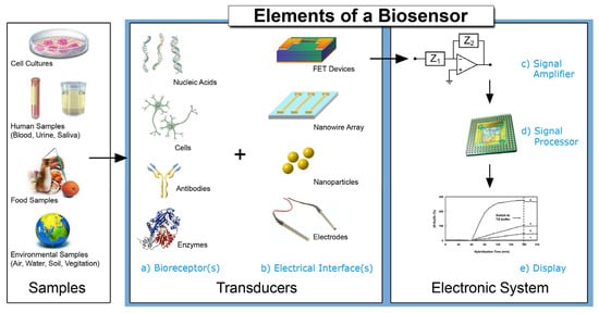
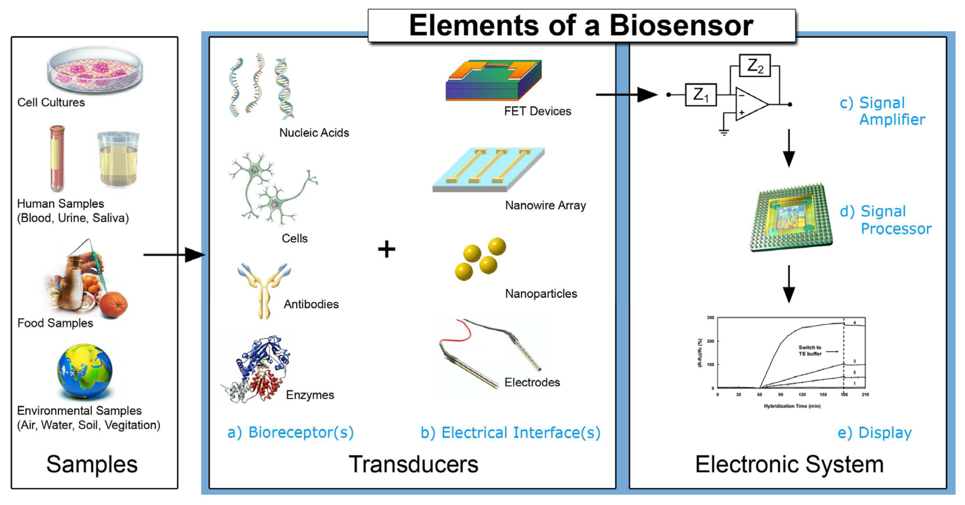
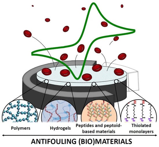
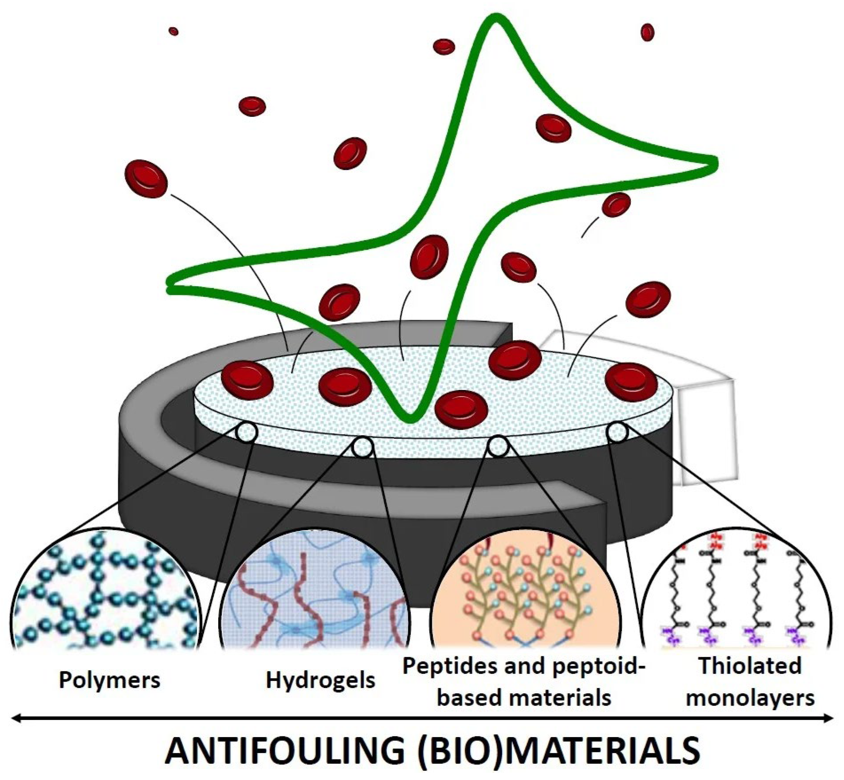
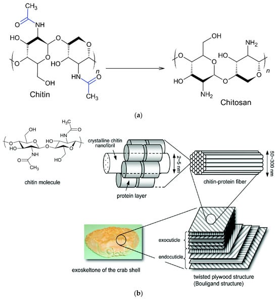
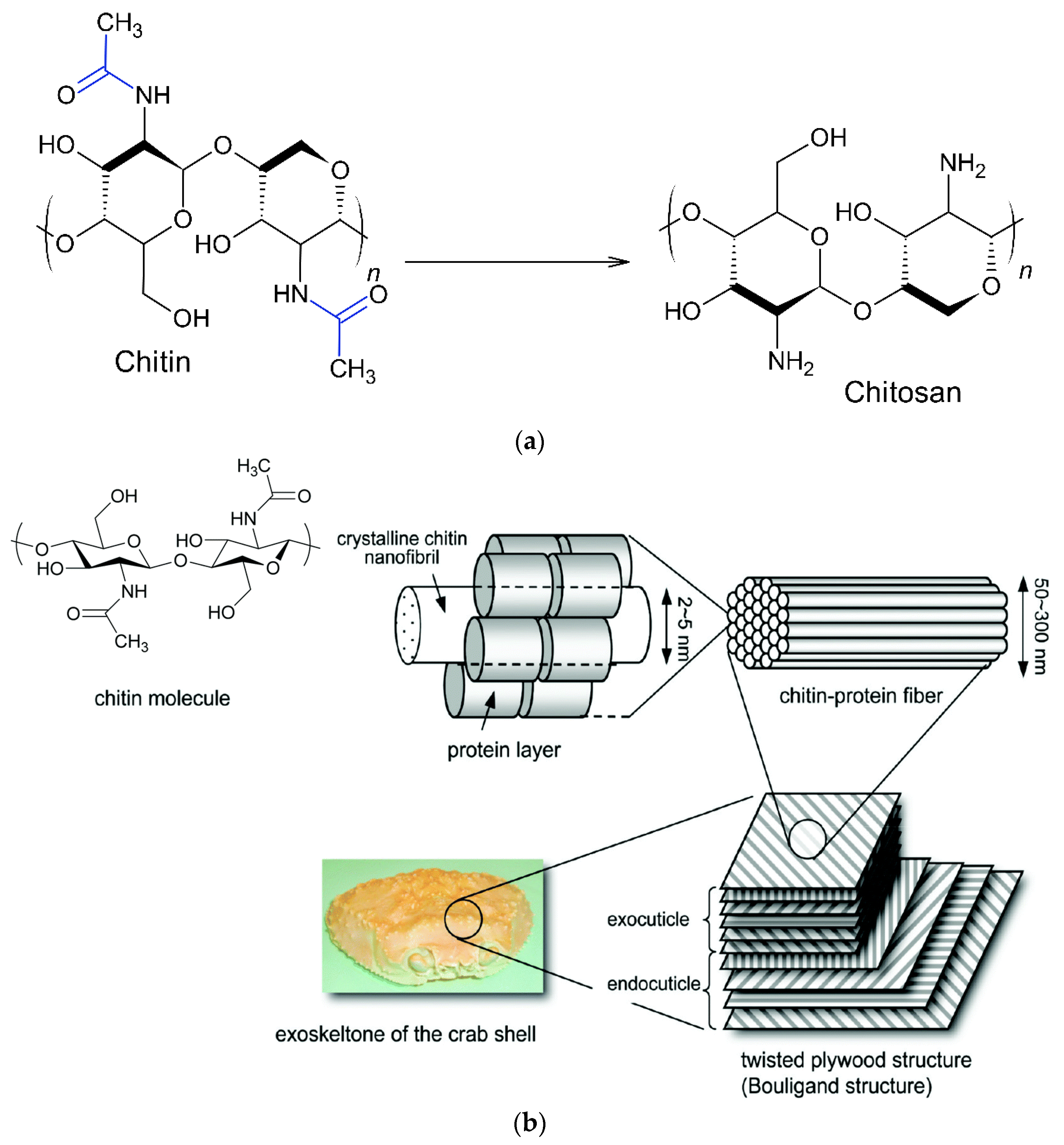
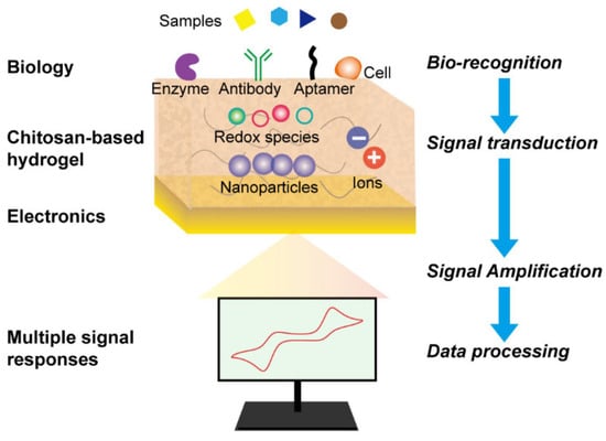
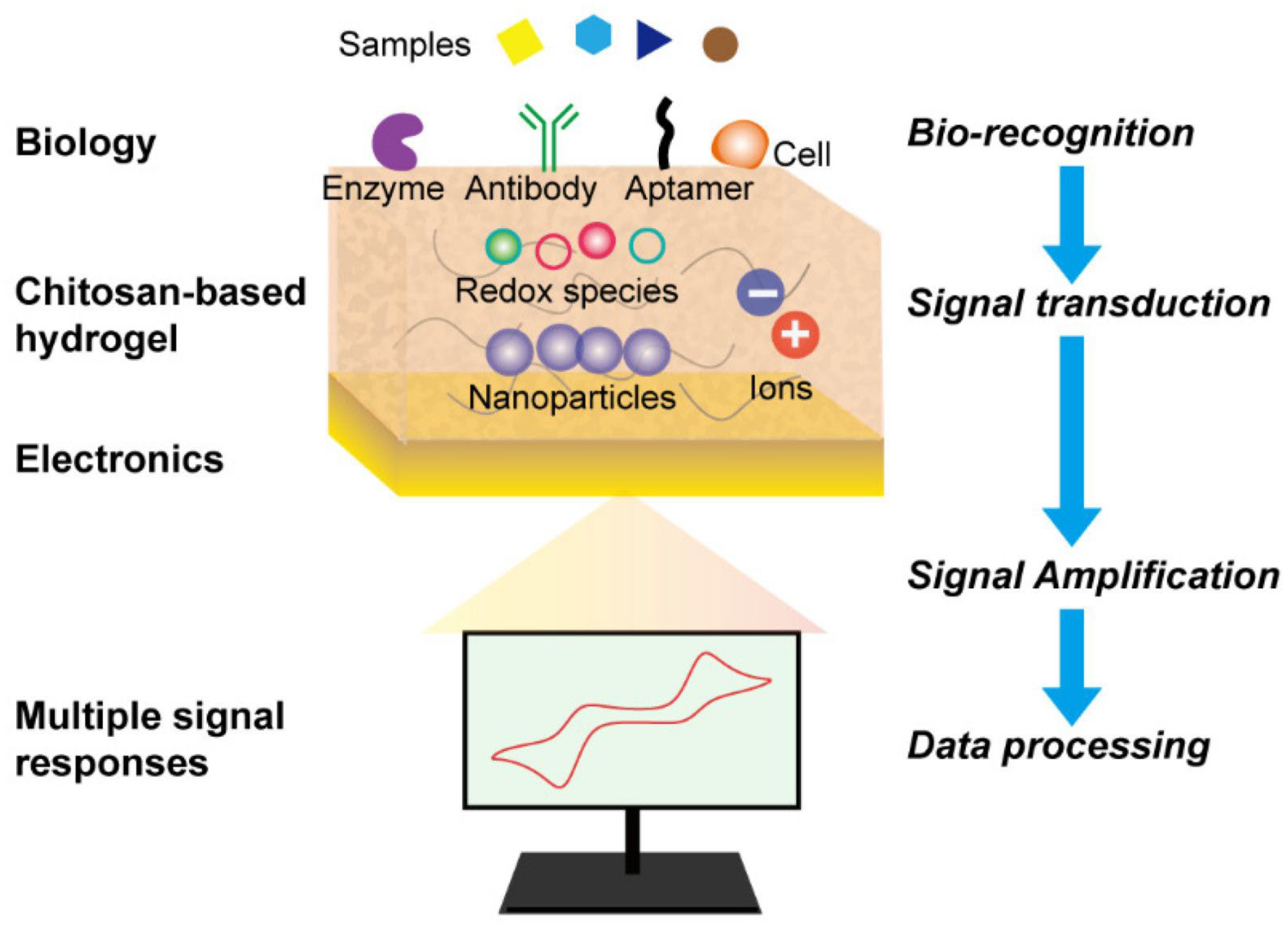
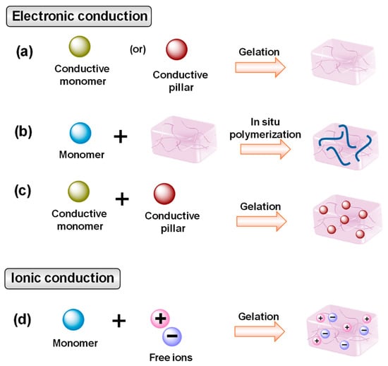
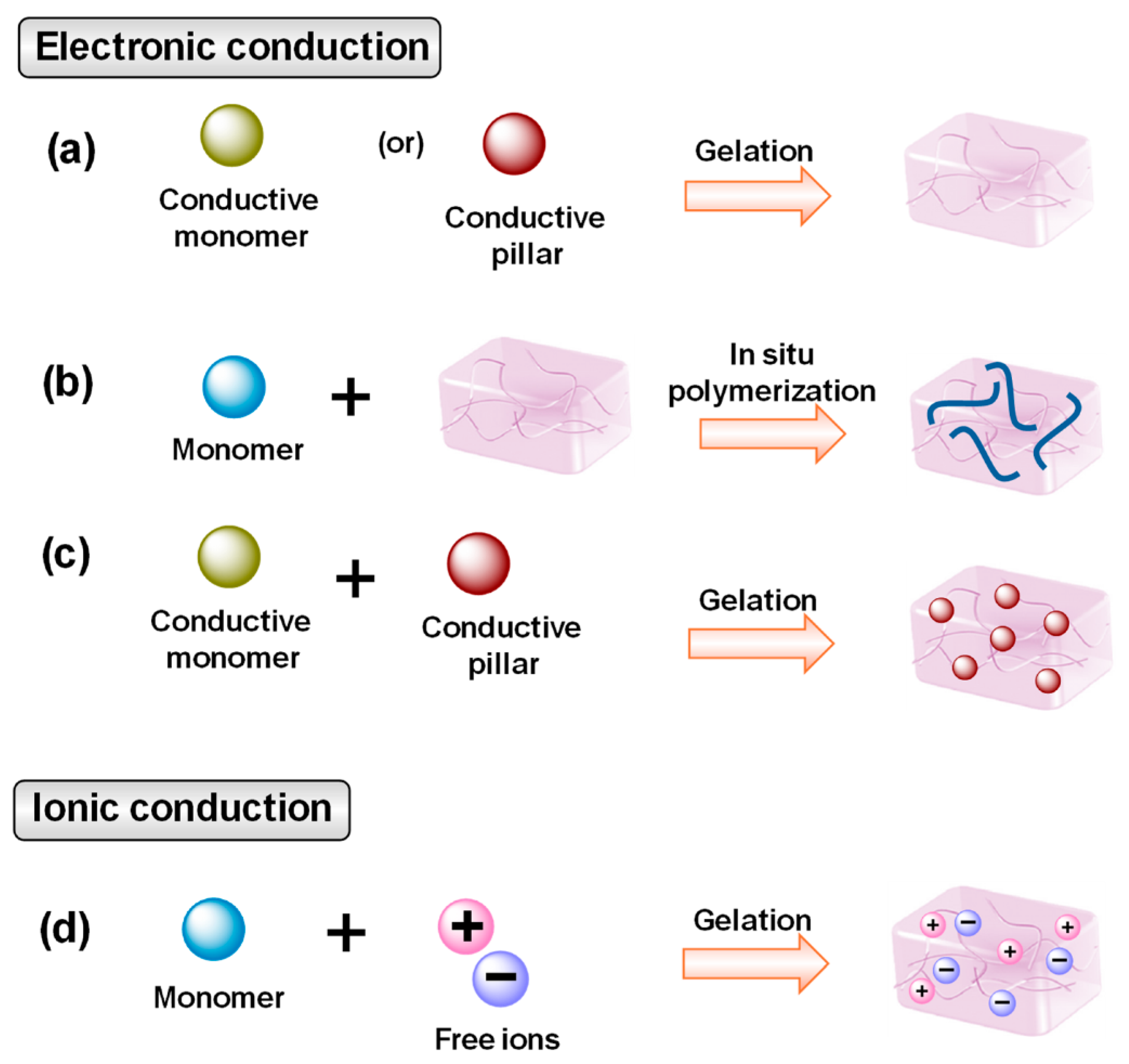
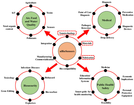
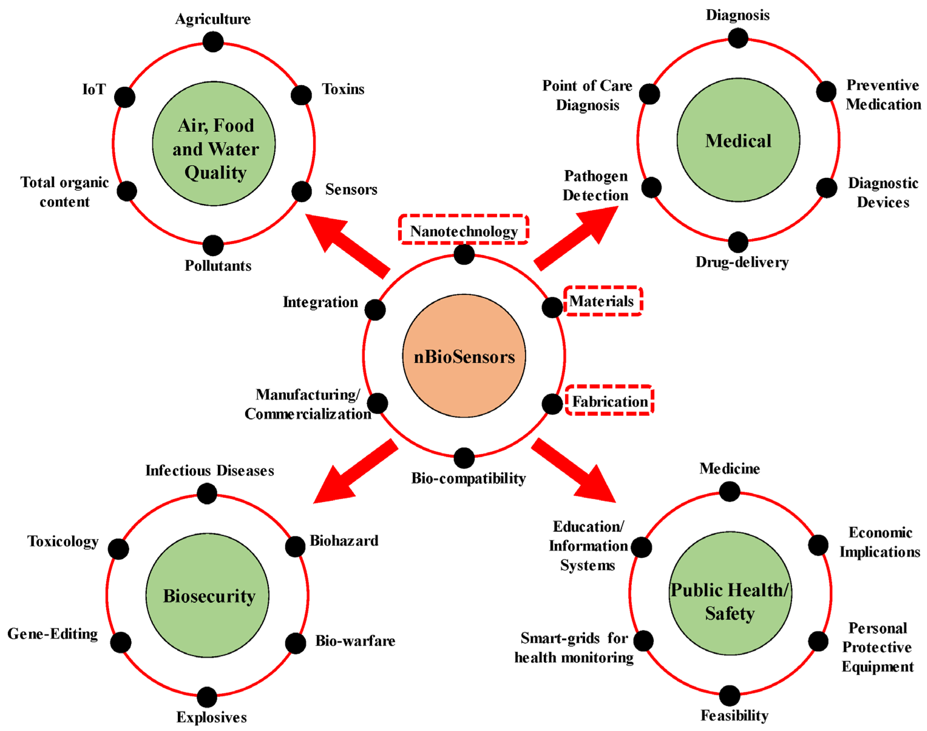
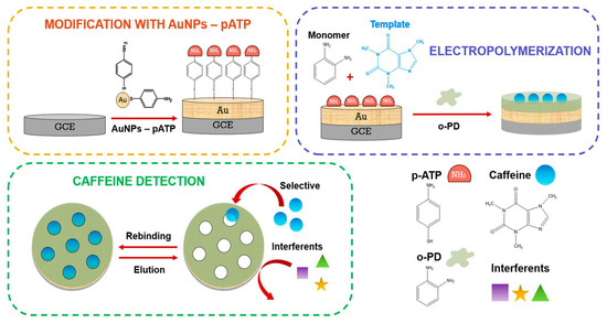
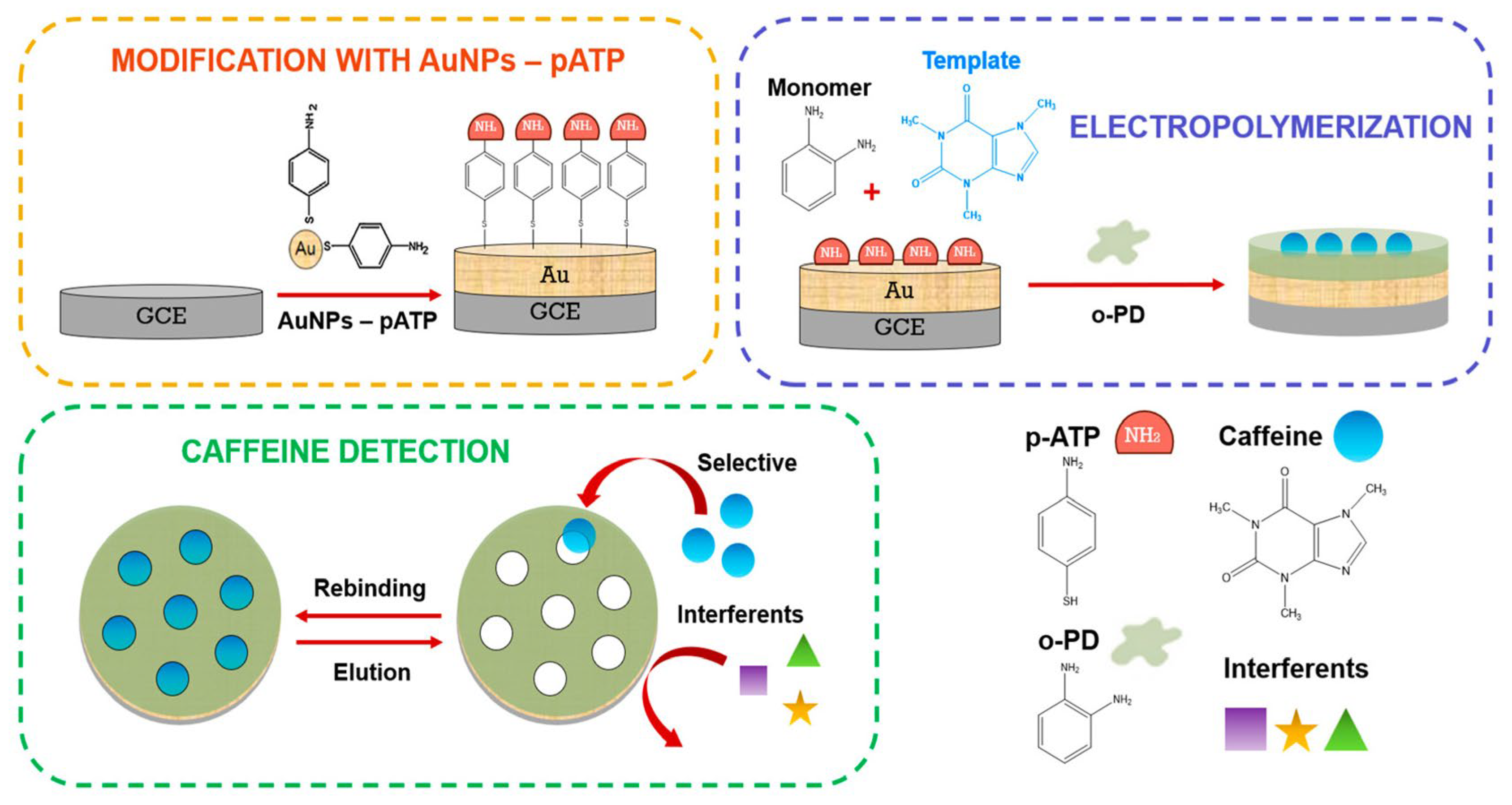
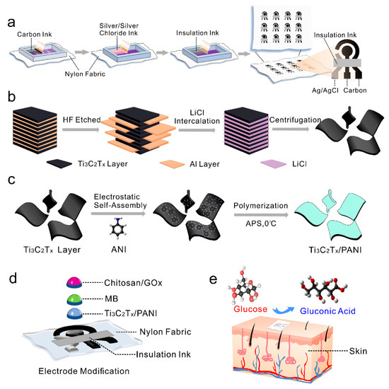
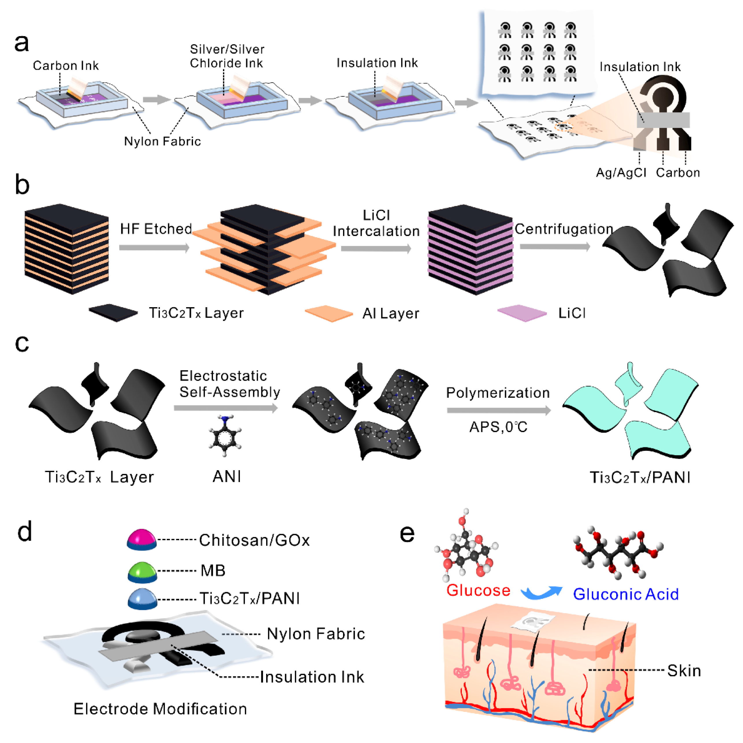
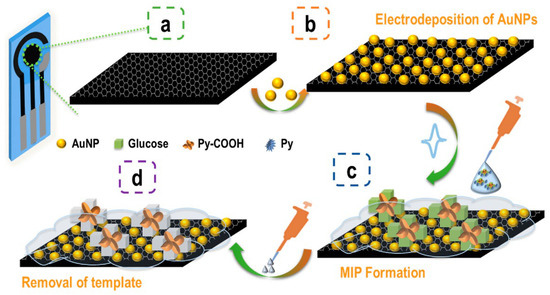
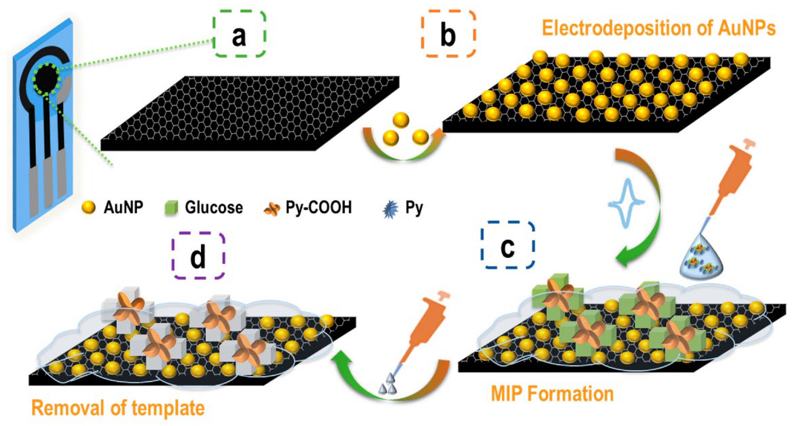
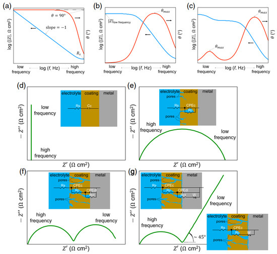
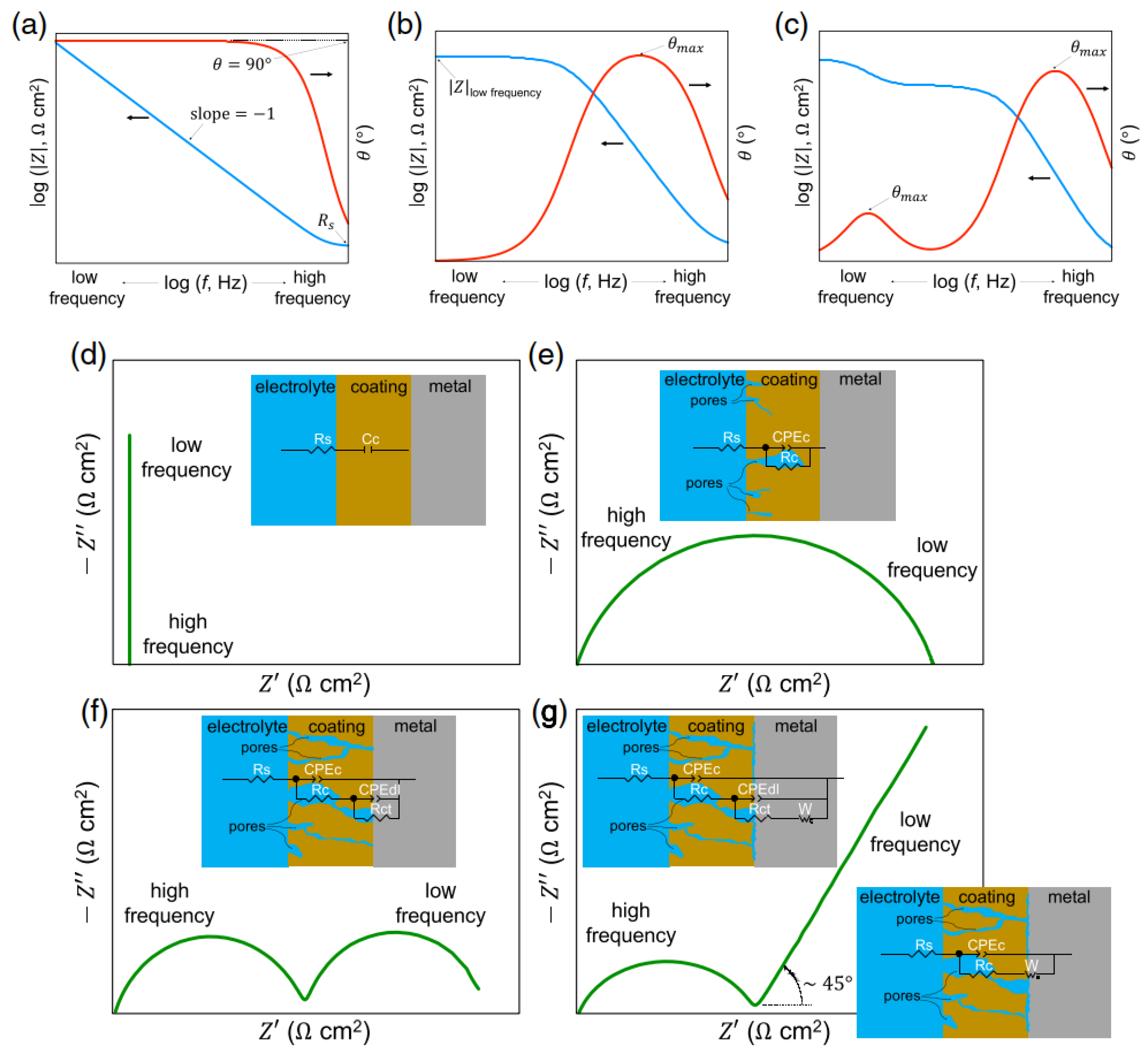
Disclaimer/Publisher’s Note: The statements, opinions and data contained in all publications are solely those of the individual author(s) and contributor(s) and not of MDPI and/or the editor(s). MDPI and/or the editor(s) disclaim responsibility for any injury to people or property resulting from any ideas, methods, instructions or products referred to in the content. |
© 2026 by the authors. Licensee MDPI, Basel, Switzerland. This article is an open access article distributed under the terms and conditions of the Creative Commons Attribution (CC BY) license.