


Highlights
What are the main findings?
- The drug-free 4:1 coating combined enhanced interfacial retention with low friction over 10 min.
- PVA/PAA-coated PEEK delivered diclofenac over 72 h in vitro.
- Drug-loaded constructs lowered COX-1, COX-2 and IL-6 mRNA relative to gPCH.
- Rabbit histology documented tissue contact and a retained hydrogel-bearing layer at 12 weeks.
What are the implications of the main findings?
- Bioinspired bilayers couple a load-supporting PEEK base with a functional hydrogel surface.
- Interface engineering supports the integration of lubrication and local drug delivery.
- The design offers a joint-contact biomaterial strategy with anti-inflammatory potential.
Abstract
Retaining a lubricating, drug-delivering hydrogel on a load-supporting implant requires coordinated scaffold and interface design. We developed an osteochondral-inspired poly(ether ether ketone) (PEEK)/hydrogel bilayer by continuous 3D printing and surface modification. A dense PEEK base supports a porous upper region containing a poly(vinyl alcohol)/poly(acrylic acid) (PVA/PAA) composite hydrogel. Sulfonation and ultraviolet-assisted acrylic acid (AA) treatment prepare the substrate for hydrogel attachment. At a 4:1 PVA:AA feed mass ratio, the drug-free composite construct (gPCH) exhibited a maximum detachment force per unit width of 304.60 ± 7.20 N m−1, 2.39 times that of PVA on sulfonated PEEK without AA/UV treatment. Its friction coefficient was 0.073 ± 0.005 during a 10 min screening test under 2 N in 25% fetal bovine serum at room temperature. The diclofenac sodium (DFN)-loaded counterpart (gPCHD) released 73.92 ± 3.24% of DFN over 72 h in PBS at 37 °C. In lipopolysaccharide-stimulated rabbit bone marrow-derived mesenchymal stem cells, gPCHD showed lower COX-1, COX-2, and IL-6 mRNA expression than composition-matched, drug-free gPCH. Twelve-week rabbit histology (n = 3 per group) provided descriptive observations of tissue contact and a retained hydrogel-bearing layer. This architecture–interface design integrates structural support, interfacial retention, short-duration lubrication, and drug delivery; long-term tribological performance and component-specific in vivo effects remain to be established.
1. Introduction
Repairing an articular cartilage defect requires more than filling a tissue void: the repair site must accommodate load while maintaining a low-friction contact surface. The limited intrinsic repair capacity of cartilage and the risk of progressive joint degeneration make this combination difficult to achieve [1,2]. Cell-based therapies and tissue-engineering approaches seek to restore the osteochondral unit [3], yet long-term outcomes remain challenging, particularly for larger defects treated by microfracture [4]. Scaffold-associated procedures can improve patient-reported outcomes at two years, although a consistent advantage over microfracture at five years has not been established [5]. For implant-based approaches, this clinical challenge translates into a materials-design problem: structural support and a compliant, hydrated surface must be combined without making the connection between them the weak point of the construct.
Hydrogels are attractive for this purpose because their water-rich networks offer a route to lubrication, mechanical compliance, and local therapeutic delivery within the same material [6,7,8]. Poly(vinyl alcohol) (PVA)-based hydrogels have been used to carry diclofenac [9] and reinforced to improve their suitability for cartilage replacement [10]. Meniscus-inspired, friction-responsive hydrogels further illustrate how a hydrated contact surface can be coupled to therapeutic function [11]. These capabilities suggest a functional division between a supportive framework and a hydrogel-based contact region. Adding a hydrogel to a rigid substrate also requires the coating to remain attached as the two components swell and deform differently. The lubricating formulation and coating attachment should therefore be designed together.
Additive manufacturing provides a means of arranging poly(ether ether ketone) (PEEK) into porous frameworks for cartilage-related applications [12], while surface modification can tailor its wettability and biological interactions [13,14]. Together, these capabilities allow the geometry of the support and the chemistry of its contact interface to be addressed at different design levels. Xu et al. demonstrated a multifunctional PEEK platform by combining 3D printing and sulfonation with a photocured acrylic acid/acrylamide hydrogel carrying simvastatin and cefepime for periodontal implant osseointegration [15]. This precedent establishes the feasibility of integrating drug-loaded hydrogels with printed PEEK. The present study builds on that concept for a different functional objective: coupling a load-supporting bilayer framework to a poly(vinyl alcohol)/poly(acrylic acid) (PVA/PAA) coating that provides lubrication and carries diclofenac sodium (DFN) at the joint-contact region.
Progress in PEEK surface functionalization has increasingly connected lubrication with coating architecture and attachment to the substrate. PVA coatings established a route to improved wettability and sliding behavior [16], while hydrogel–polymer-brush bilayers [17] and layered hydrogel coatings [18] introduced additional control over the lubricating interface. Hard–soft PEEK/hydrogel scaffolds extended this approach to osteochondral applications [19]. Mechanical–chemical anchoring [20], graft-modified cartilage-contact interfaces [21], and bioactive hydrogel modification [22] further show that coating retention and biological function are integral to this development. The design question addressed here is whether substrate modification and PVA/AA precursor composition can improve coating retention while maintaining low-friction performance on a continuously printed, load-supporting PEEK framework.
Here, we examine a coordinated architecture–interface strategy rather than introducing PVA, PAA, or drug loading on PEEK as new individual concepts. Prior studies have demonstrated porous PEEK frameworks [12], lubricating PVA coatings [16], layered hydrogel interfaces [17,18], and hard–soft osteochondral constructs [19]. In the present design, continuous printing joins a dense lower support to a porous upper region so that the PEEK framework can support, rather than replace, a deformable hydrogel-bearing contact surface. Substrate modification and the PVA:AA precursor feed ratio are evaluated together through wettability, interfacial retention, and short-duration friction measurements. DFN loading is subsequently evaluated as an additional function of the integrated hydrogel, alongside composition-matched drug-free and loaded cell treatments. Twelve-week rabbit histology provides exploratory morphological observations. This coordinated evaluation addresses how support, hydrogel accommodation, coating attachment, short-term lubrication, and drug delivery can be combined within one construct without assuming that each function or its long-term clinical performance has been independently established.
2. Materials and Methods
2.1. Materials and 3D Printing
Medical-grade PEEK filament (diameter, 1.75 mm) was purchased from Shanghai Sandi Tribe Technology Co., Ltd. (Shanghai, China). Acrylic acid (AA, >99%), poly(vinyl alcohol) (PVA, 99% degree of alcoholysis; degree of polymerization, 1799), diclofenac sodium (DFN, >99%), photoinitiator I2959 (98%), and concentrated sulfuric acid (approximately 98%) were obtained from Shanghai Aladdin Biochemical Technology Co., Ltd. (Shanghai, China).
The bilayer framework comprised a dense lower PEEK region for structural support and a grid-like porous upper region for hydrogel integration. Both regions were fabricated as a single, continuously printed framework rather than as separately printed components joined afterward. Fused-deposition modeling used a nozzle temperature of 445 °C, a bed temperature of 60 °C, a printing speed of 5 mm s−1, a layer height of 0.3 mm, and a nozzle diameter of 0.3 mm. The 60% software infill setting applied to the upper hydrogel-integration region; the lower support was designed as solid. Structures with designed pore sizes of 300, 500, and 800 µm were used for macroscopic characterization. An additional 1000 µm design was fabricated for compression screening. These pore sizes and the infill setting are fabrication-design parameters; an independently measured as-printed volumetric porosity is not available for this study.
2.2. Surface Modification, Hydrogel Integration, and DFN Loading
The printed PEEK samples were sequentially polished with 400, 1000, 1500, and 2000 grit abrasive paper, ultrasonically cleaned in acetone, ethanol, and deionized water, and dried at 80 °C for 1 h. Sulfonation was performed by immersing the samples in concentrated sulfuric acid (approximately 98%) at 25 °C for 30 s under ultrasonication and magnetic stirring. After washing, the samples were hydrothermally treated at 120 °C for 4 h to remove residual acid and labile sulfur-containing species. The resulting sulfonated PEEK was designated SPEEK.
Two UV steps were used. First, SPEEK was immersed in a 1 mol L−1 aqueous AA solution and irradiated for 30 min without an added photoinitiator, yielding the AA-treated substrate designated SPEEK-g. After this initial AA/UV treatment, the substrates were washed three times with fresh deionized water for 5 min per wash to reduce residual AA monomer and free PAA homopolymer before immersion in the hydrogel precursor. For hydrogel preparation, PVA and AA were combined from the beginning of precursor preparation and heated in a 96 °C water bath for 2 h. The final precursor contained 25 wt% total PVA and AA, with PVA:AA feed mass ratios of 19:1, 9:1, or 4:1. After AA addition and before heating, the pH of each formulation was measured at room temperature using a pH meter and adjusted to approximately 4.7 by adding 5 mol L−1 aqueous NaOH solution. The 25 wt% refers to the combined feed mass of PVA and AA relative to the final precursor mass after pH adjustment and deionized-water make-up, including the added NaOH solution. I2959 was used at 1 mol% relative to AA for the subsequent precursor-polymerization step. The treated PEEK substrates were immersed in the precursor and vacuum-infiltrated for 20 min before the second 30 min UV exposure. Both UV steps used the same source, with a spectral peak at 365 nm, an irradiation intensity of 5 mW cm−2, and a source-to-sample distance of 25 cm; neither step used nitrogen purging. The constructs then underwent five freeze–thaw cycles, each consisting of freezing at −20 °C for 6 h and thawing at room temperature for 2 h, followed by dialysis in deionized water for 2 weeks. The freeze–thaw treatment promotes physical gelation of PVA. The material is described here as a PVA/PAA composite hydrogel because the present evidence does not establish two independently identifiable networks. All formulation ratios refer to the precursor feed mass ratio rather than a measured final polymer composition.
PH denotes PVA on SPEEK without AA treatment, whereas gPH denotes PVA on the AA-treated SPEEK-g substrate. gPCH denotes the corresponding drug-free PVA/PAA composite-hydrogel construct, and gPCHD denotes its DFN-loaded counterpart. PEEK, SPEEK, and SPEEK-g retain their substrate meanings. Formulation labels are appended where more than one feed ratio is compared, for example gPCH(4:1). The nomenclature is consolidated in Table 1. After hydrogel integration, the constructs were dried at 80 °C for 12 h and immersed in a 2 mg mL−1 DFN solution for 48 h. Thus, the hydrogel-bearing PEEK construct, rather than an isolated gel later attached to PEEK, underwent drug loading.
Table 1.
Sample nomenclature used throughout the manuscript.
2.3. Morphological, Chemical, and Surface Characterization
Surface and hydrogel morphologies were examined by scanning electron microscopy (SEM; JSM-6701F, JEOL, Tokyo, Japan) after lyophilization and gold sputter-coating. Atomic force microscopy (AFM; MultiMode 8, Bruker Nano Surfaces Division, Santa Barbara, CA, USA) was used to characterize surface topography and roughness (Ra). Fourier-transform infrared (FTIR) spectra were collected over 4000–650 cm−1 using an IFS 66v/s spectrometer (Bruker Optics, Ettlingen, Germany). X-ray photoelectron spectroscopy (XPS; Escalab 250Xi, Thermo Fisher Scientific, Waltham, MA, USA) was used to acquire survey spectra and S 2p, N 1s, and Cl 2p narrow scans. X-ray diffraction (XRD) patterns were recorded over 2θ = 5–80° using a D/Max-2400 diffractometer (Rigaku, Tokyo, Japan). Static water contact angles were measured on dried specimens by the sessile-water-drop method in air. AFM and contact-angle measurements each included five independent specimens per group.
Gravimetric swelling was expressed as Q = 100 × (mw − md)/md, where md is the dry hydrogel mass and mw is the corresponding swollen hydrogel mass. The PEEK support is excluded from this hydrogel-mass normalization. For the selected composite hydrogel, the reported swelling ratio was 490%, corresponding to a swollen-to-dry hydrogel mass ratio of 5.90. The measurement used free-standing PVA/PAA hydrogel immersed in deionized water at room temperature (approximately 25 °C) for approximately 24 h until fully swollen. The gel was removed and surface water was gently blotted away before the swollen mass was measured; the dry-mass denominator was the mass of the fully dried gel.
Additional peak-shape analysis was performed on the exported N 1s, S 2p, and Cl 2p data using nonlinear least-squares fitting in Python (https://www.python.org/, accessed on 6 November 2026) and SciPy (https://scipy.org/, accessed on 6 November 2026). The exported intensity values were retained without additional smoothing or removal of points. A linear background and area-normalized pseudo-Voigt components were fitted jointly. N 1s was represented by one component. S 2p and Cl 2p were each represented by one spin–orbit doublet, with a 2:1 area ratio and common full width at half maximum (FWHM); the respective fixed separations were 1.16 and 1.60 eV. The supplied energy axes were retained without an additional charge-reference shift. These fits describe the measured peak envelopes; absolute grafting density and a complete set of atomic percentages were not derived from this analysis.
2.4. Ninety-Degree Peeling and Maximum Detachment Force per Unit Width
Interfacial retention was evaluated by 90° peeling using 30 × 100 mm rectangular specimens. One half of the hydrogel was attached to the PEEK-based substrate and the other half was pre-separated to form a free end. The substrate was fixed in the lower fixture of a universal testing machine, and the free hydrogel end was gripped and pulled upward at 2 mm min−1 while maintaining the 90° peeling geometry, until complete separation. Force–displacement curves were recorded for three independent specimens per group. The maximum force from each curve, Fmax, was divided by the specimen width b = 30 mm = 0.030 m to obtain the maximum detachment force per unit width, Fmax/b, in N m−1. The mean and sample SD were calculated from the three independently tested specimens.
2.5. Tribological Testing
Tribological behavior was evaluated using a UMT TriboLab system (Bruker Nano Surfaces and Metrology Division, San Jose, CA, USA) in reciprocating ball-on-disc mode. A stainless-steel sphere 6 mm in diameter was used as the counterface against the flat exposed surface of each 30 × 30 × 1 mm specimen. Tests were performed in 25% fetal bovine serum (FBS) at room temperature under a normal load of 2 N, a sliding speed of 2 mm s−1, and a stroke length of 2 mm for 10 min. Three independent friction curves were obtained per group; the specimen-level mean coefficient of friction was calculated from the final 5 min of each curve. The test was used for short-term friction screening. The counterface sphere had a surface roughness of ≤0.1 µm (≤100 nm).
2.6. Compression Testing
Compression testing was performed on separately fabricated hydrogel-bearing PEEK cylinders measuring 10 mm in diameter and 20 mm in height. These were composite specimens containing the PEEK support and integrated hydrogel, rather than bare PEEK or free-standing hydrogel specimens. They were tested in the hydrated state following DFN immersion and at room temperature using a universal testing machine (Shimadzu, Kyoto, Japan) at 2 mm min−1. The screening matrix included pore sizes of 300, 500, 800, and 1000 µm and total PVA/AA precursor contents of 15, 20, and 25 wt%. Engineering stress was calculated as F/A0, with A0 = πd02/4, and engineering strain as Δh/h0. The compressive elastic modulus was obtained from the slope of the initial approximately linear stress–strain segment, using the 0%–10% engineering-strain interval. Ultimate compressive strength was defined as the stress at actual structural failure. Three independent specimens were tested for each pore-size/formulation combination, with results summarized as mean ± SD. These separately prepared cylinders served formulation and structural screening; they were not the 3 mm-high rabbit implants described in Section 2.9.
2.7. Rabbit BMSC Culture and Cytocompatibility
Rabbit bone marrow-derived mesenchymal stem cells (BMSCs) were isolated from femoral marrow under protocol no. LZUKQ-2023-068 at the School of Stomatology, Lanzhou University. Primary isolation was performed using α-MEM supplemented with 30% FBS and 1% penicillin–streptomycin. Subsequent cultures were maintained in α-MEM containing 10% FBS and 1% penicillin–streptomycin at 37 °C in an atmosphere of 5% CO2.
For the CCK-8 assay, cells were prepared at 5 × 10⁴ cells mL−1 and co-cultured with equal masses of the respective materials in 96-well plates containing approximately 100 µL of complete culture medium per well. CCK-8 reagent was added on days 1, 3, 5, and 7, followed by incubation for 2 h and absorbance measurement at 490 nm. The signal was normalized to the control at each time point and summarized from three independent experiments. CCK-8 is reported here as relative metabolic activity, not as a direct percentage of viable or proliferating cells. For live/dead imaging, cells were co-cultured with the materials in 24-well plates containing approximately 1 mL of complete culture medium per well, stained with Calcein-AM/propidium iodide for 0.5 h, and examined by fluorescence microscopy. For both assays, each material-treated well contained one material piece approximately 1 × 1 mm in lateral dimensions; equal material masses were used across the material groups within each assay.
2.8. DFN Release and RT-qPCR
DFN was quantified by high-performance liquid chromatography (HPLC) at 276 nm using the corresponding five-point calibration. Drug loading was performed after hydrogel integration, by immersing the dried hydrogel-bearing PEEK constructs in a 2 mg mL−1 DFN solution for 48 h. Following loading, the residual loading bath, drainage liquid, and collected rinses were pooled and brought to a defined final volume. The amount associated with the batch was estimated by solution mass balance: Mbatch = C0V0 − CpoolDVf, where C0 and V0 are the initial loading-solution concentration and volume, Cpool is the calibrated concentration in the measured pooled aliquot, D is its dilution factor (D = 1 if the reported concentration already includes dilution correction), and Vf is the final pooled-solution volume. All terms were converted to µg and mL. The average amount per construct was Mbatch/N, where N is the number loaded together, and loading efficiency was 100Mbatch/(C0V0). The actual values were C0 = 2000 µg mL−1, V0 = 5 mL, Cpool = 18.9 µg mL−1 after 50-fold dilution, D = 50, Vf = 10 mL, and N = 10. The initial and recovered DFN masses were 10,000 and 9450 µg, respectively, giving an estimated batch-associated amount of 550 µg, a mean of 55 µg per construct, and a loading efficiency of 5.5%. When content is expressed per mg of dry hydrogel, the denominator is the corresponding dry hydrogel mass, excluding the PEEK support; a pooled-bath measurement gives a batch average rather than independent measurements for every construct.
For release testing, lyophilized gPCHD constructs were immersed in 2 mL of sterile PBS at 37 °C with agitation at 150 rpm. Samples were collected at 0.5, 1, 2, 4, 6, 8, 10, 12, 18, 24, 36, 48, 60, and 72 h. At each time, 1 mL of supernatant was collected after centrifugation and replaced with 1 mL of fresh PBS. Under the well-mixed, constant-volume sampling procedure, cumulative released mass was Mn = VCn + Σ(i < n) viCi, where V = 2 mL and vi = 1 mL. Release percentage was calculated as 100Mn/M0 using M0 = 55 µg, the batch-average loading amount per construct; each of the three release tests used one construct from this same ten-construct loading batch. Each release curve was calculated separately before summarizing the three independent curves as mean ± SD.
For inflammatory stimulation, BMSCs were exposed to 10 µg mL−1 lipopolysaccharide (LPS) in α-MEM for 12 h, followed by 24 h in the material-free Control condition, the drug-free PVA/PAA composite construct gPCH, or its DFN-loaded counterpart gPCHD. The gPCH and gPCHD treatments used the same PVA/PAA formulation and substrate preparation and differed in DFN-loading status. Cell culture and material exposure before RT-qPCR were performed in 6-well plates containing approximately 2 mL of complete culture medium per well. Each material-treated well contained one piece approximately 1 × 1 mm in lateral dimensions, with equal material masses across the material groups. In this assay, the group previously labelled HSPEEK was the drug-free composite group, not the PVA-only gPH comparator used in other tests. Total RNA was extracted using TRIzol, reverse-transcribed, and analyzed by SYBR Green RT-qPCR. GAPDH was the reference gene and relative expression was calculated using the 2−ΔΔCt method. Each group comprised three independent biological samples, with three technical wells per sample; biological samples defined n. Primer sequences are listed in Table 2.
Table 2.
Primer sequences used for RT-qPCR.
2.9. Rabbit Implantation and Histology
Animal procedures were approved by the Animal Ethics Committee of the School of Stomatology, Lanzhou University, on 8 June 2023 (approval no. LZUKQ-2023-068). Eighteen 2-month-old New Zealand White rabbits weighing 2.6–3.2 kg were allocated to six groups (n = 3 per group): BLANK, Control, PEEK, PH, gPCH, and gPCHD. BLANK animals had neither a defect nor an implant, whereas Control animals had a defect without an implant.
General anesthesia was induced by intravenous urethane administration. After exposure of the lateral femoral condyle, an osteochondral defect measuring 3 mm in diameter and 3 mm in depth was created under sterile saline irrigation in all groups except BLANK. The PEEK implant measured 3 × 3 mm, and the bilayer constructs comprised a 2 mm lower PEEK region and a 1 mm upper hydrogel-bearing region. The gPCHD construct used a pore size of 500 µm, a total PVA/AA precursor content of 25 wt%, and a PVA:AA feed mass ratio of 4:1. Incisions were closed in layers. Penicillin sodium was administered intramuscularly for 3 days postoperatively, after which the animals were allowed free movement in their cages.
Animals were euthanized at 12 weeks. Joint and opposing-meniscus specimens were collected from the BLANK, Control, PEEK, PH, and gPCHD groups, but not from the drug-free gPCH group. Organ sections from the Control, gPCH, and gPCHD groups were examined.
Heart, liver, spleen, lung, and kidney samples were fixed in 10% formalin, embedded, sectioned, and stained with hematoxylin and eosin (H&E). Joint specimens containing the implant and surrounding tissue were processed for hard-tissue H&E sectioning to assess tissue contact, fibrous encapsulation, hydrogel retention, and defect-site morphology. The opposing meniscus and tibial articular surface were examined by gross observation and H&E staining. All histological assessments were descriptive; quantitative tissue scores were not assigned.
2.10. Finite-Element Analyses
Two separate finite-element models were used for exploratory analysis. An idealized knee-joint model was analyzed in Abaqus (www.3ds.com/products/simulia/abaqus, accessed on 6 November 2026) to obtain stress and displacement contours for the PEEK, PH, and gPCH configurations; correspondence to rabbit-specific knee geometry was not established. A separate cylindrical contact model was analyzed in ANSYS 19.0 using N–mm–MPa units. The latter used a 5 mm-diameter, 1 mm-thick cylinder against an approximately 8 × 12 × 1 mm plate, with SOLID186 solids and CONTA174/TARGE170 contact/target elements. The contact-plane normal was parallel to Y. A pressure of 0.5 MPa was applied, and the actual tangential displacement constraint was UZ = 0 to −1 mm. A separately defined +1 mm table was not applied and is not described as a second imposed displacement. The static analysis-step endpoint was TIME = 1; this was not interpreted as a physiological cycle count or a 12-week exposure.
Wear was prescribed using the Archard implementation, dh/dt = (K/H)pm vn, with K = 1 × 10⁻⁵, H = 1 MPa, and m = n = 1, equivalently dh = (K/H)p ds. Thus K/H = 1 × 10⁻⁵ mm2 N−1 in the adopted units. The archived PH and gPCH cylinder scenarios both used E = 6.38 MPa and ν = 0.40; the PEEK scenario used E = 3600 MPa and ν = 0.38. The opposing plate used E = 20 MPa and ν = 0.40. The respective archived friction coefficients were 0.1228, 0.07879, and 0.13429 for PH, gPCH, and PEEK; these are model inputs, not replacements for the present experimental means. The two hydrogel-cylinder scenarios shared the simplified geometry and elastic properties and differed mainly in the prescribed friction coefficient. The analysis output CONTNMISC179 represents the Y-directed wear-correction component (mm); Y is parallel to the normal of this planar contact. The output is not a measured wear volume. The wear parameters were not independently calibrated or validated against material-specific wear measurements. The archived solver reported a small-sliding applicability warning whose numerical effect has not been resolved; therefore, the contours are treated as unvalidated illustrative model outputs, not quantitative predictions of long-term wear or service life.
2.11. Statistical Analysis
Data are presented as the mean ± standard deviation (SD) where independent measurements were available. AFM roughness and water contact angle were measured using five independent specimens per group. Interfacial detachment force and friction were evaluated using three independent specimens or curves per group. Compression testing included three independent specimens for each pore-size/formulation combination. DFN release and CCK-8 measurements were summarized from three independent release curves and three independent experiments, respectively. Each RT-qPCR group included three independent biological samples, each measured in three technical wells; the biological sample was the independent unit of analysis.
Group differences in specimen-level AFM roughness, contact angle, detachment force, and friction data were evaluated by Welch one-way analysis of variance (ANOVA), followed by two-sided Welch t tests with Holm adjustment for family-wise multiple comparisons. RT-qPCR data were analyzed in SPSS 25 using one-way ANOVA followed by Tukey’s multiple-comparison test. The compression-screening matrix, normalized CCK-8 metabolic activity, and histological observations were summarized descriptively without inferential p values. For CCK-8, error bars represent the sample SD across three independent experiments. A p value < 0.05 was considered statistically significant. Significance symbols denote * p < 0.05, ** p < 0.01, and *** p < 0.001 for the inferential comparisons indicated in the figures.
2.12. Use of Generative Artificial Intelligence
ChatGPT (OpenAI; https://chatgpt.com/, accessed on 6 November 2026) was used to develop prompts and to generate and revise the Graphical Abstract, and to assist with manuscript text revision and proofreading, including grammar correction. The Graphical Abstract is a schematic illustration, not an experimental image.
3. Results and Discussion
3.1. Bioinspired Bilayer Architecture and Hierarchical Surface Integration
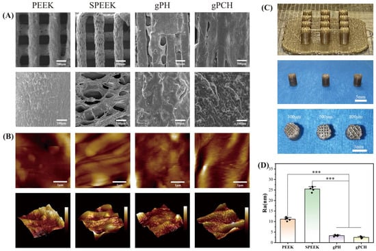
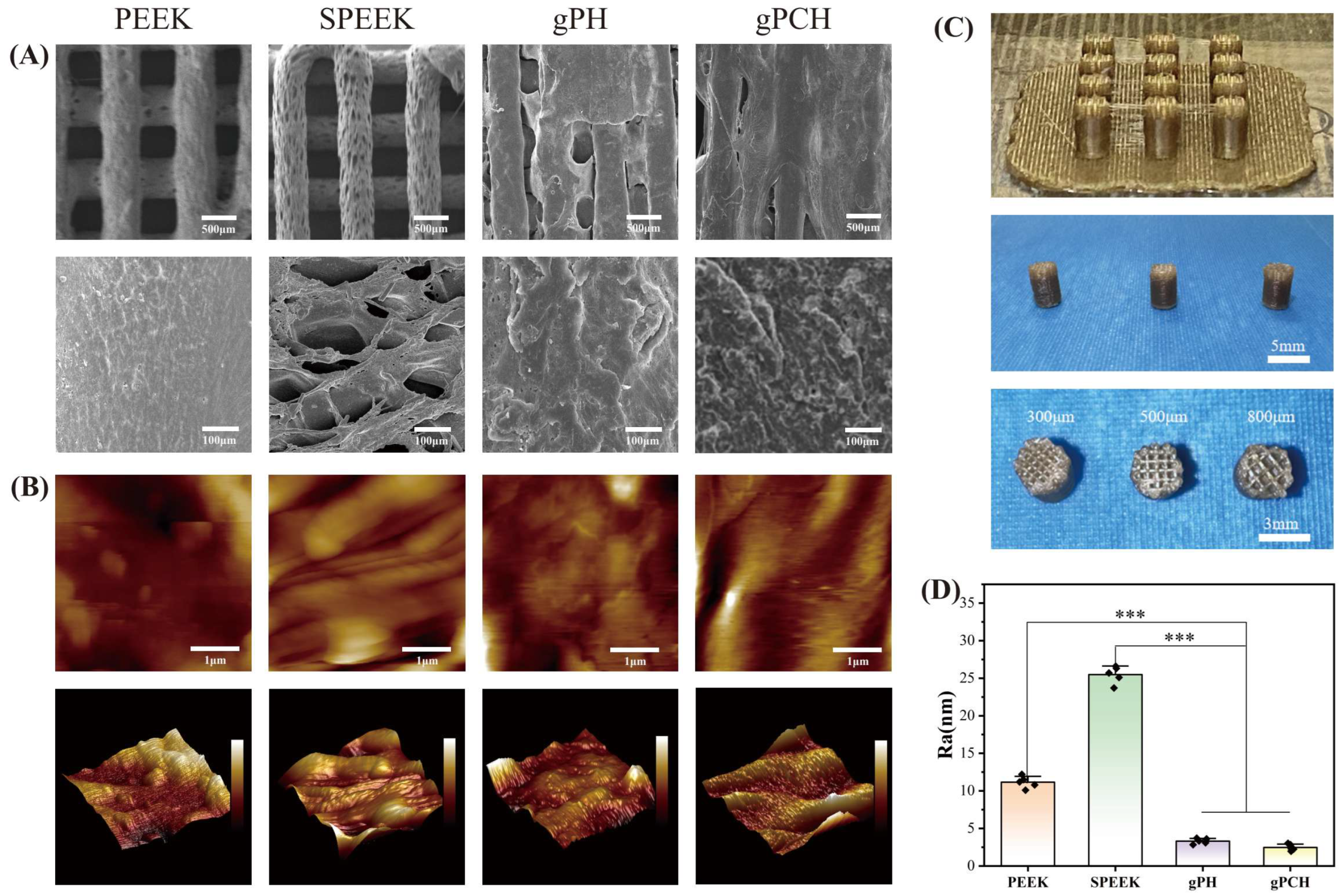
The scaffold combines the supporting role of PEEK with the surface function of the hydrogel in a continuously printed framework. A dense lower region provides support, while a porous upper region accommodates the hydrogel-bearing contact layer. This arrangement follows the functional division between a supportive foundation and a hydrated articulating surface. Photographs show the 300, 500, and 800 µm pore designs (Figure 1C); a 1000 µm design was additionally fabricated for compression screening. Continuous printing connects the two PEEK regions, while the subsequent surface treatments address hydrogel attachment.
Figure 1.
Architecture and surface morphology of printed and hydrogel-modified PEEK scaffolds. (A) SEM images of PEEK, SPEEK, gPH, and gPCH. (B) AFM height maps and corresponding three-dimensional surface topographies. (C) Photographs of printed structures with designed pore sizes of 300, 500, and 800 µm. (D) Surface roughness (Ra), presented as mean ± SD from five independent specimens per group. In (B), color indicates relative surface height; the bar colors in (D) distinguish the labelled groups. In (D), *** p < 0.001 for the bracketed comparisons; Welch one-way ANOVA followed by two-sided Welch t tests with Holm adjustment was used.
Surface modification created a roughened substrate for hydrogel incorporation and a smoother exposed coating after integration. SEM showed that sulfonation transformed the comparatively smooth as-printed PEEK into a rough, hierarchically porous surface. Hydrogel incorporation then produced a more continuous covering layer (Figure 1A). AFM captured the corresponding changes in roughness (Figure 1B,D): Ra increased from 11.16 ± 0.78 nm for PEEK to 25.48 ± 1.15 nm for SPEEK, then decreased to 3.32 ± 0.36 nm for gPH and 2.47 ± 0.44 nm for gPCH (n = 5). Roughness differed among groups (p < 0.001), and SPEEK was rougher than both hydrogel-covered groups (p < 0.001). Substrate roughening and surface smoothing therefore serve complementary roles: the modified PEEK accommodates the hydrogel, which forms the smoother outer contact surface.
3.2. Chemical Evidence for Surface Functionalization and DFN Incorporation
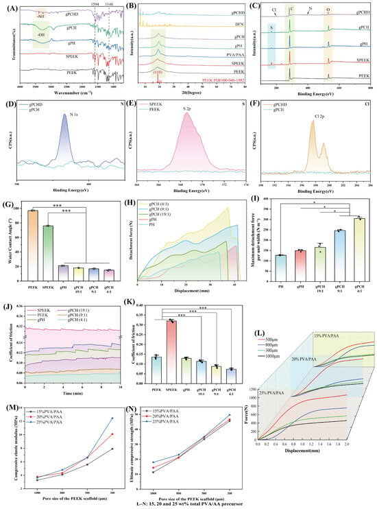
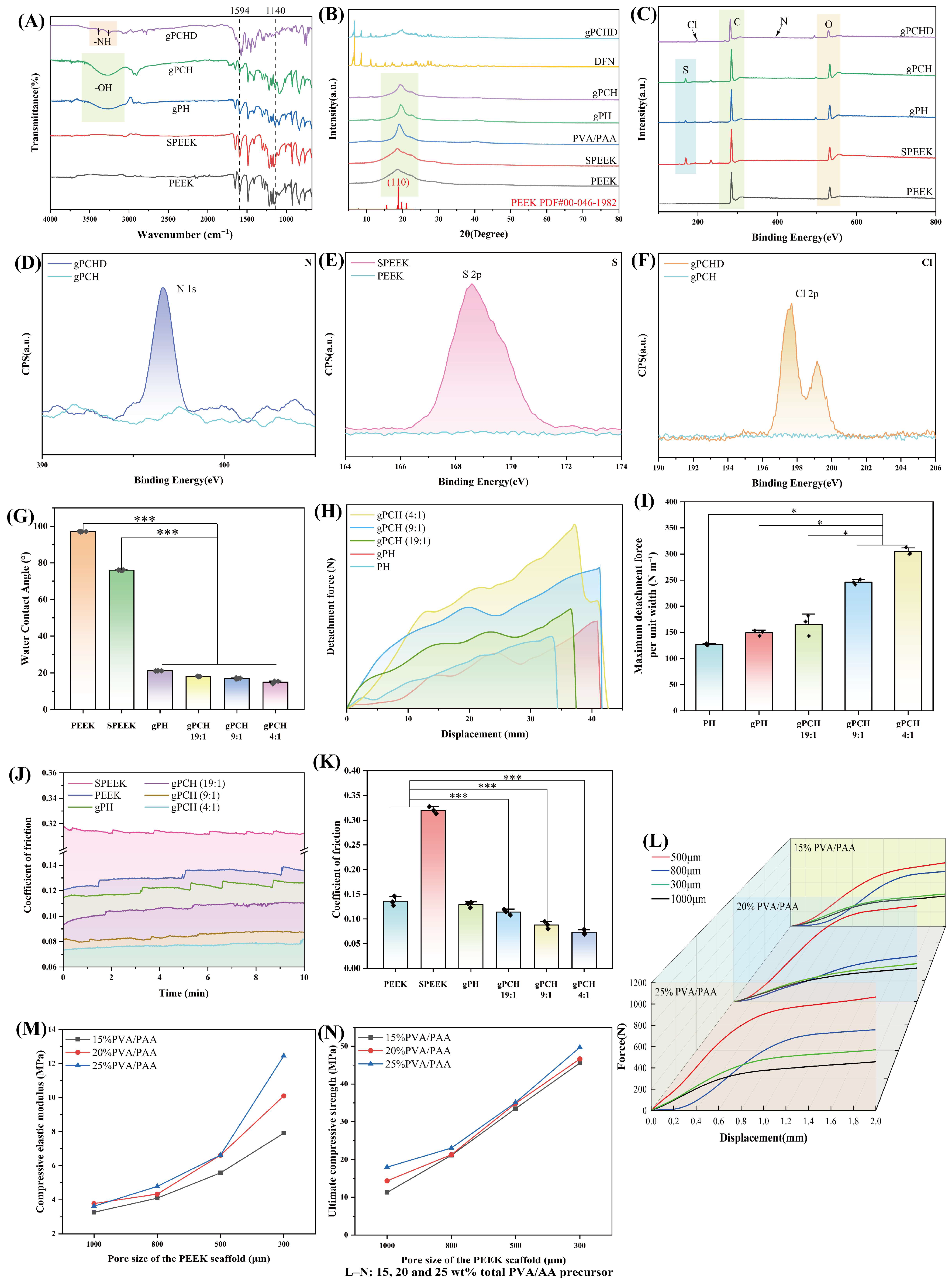
The chemical analyses showed changes associated with surface functionalization while the characteristic PEEK phase remained identifiable (Figure 2A–F). FTIR spectra showed the aromatic-ketone features of PEEK and an additional band at 1140 cm−1 after sulfonation, consistent with O=S=O stretching. Hydrogel-covered samples exhibited a broader contribution at 3200–3600 cm−1, consistent with hydroxyl- and carboxyl-containing species. XRD patterns showed the principal PEEK reflections alongside a broad contribution from the hydrogel phase. These observations support the formation of a chemically distinct coating while preserving the identifiable PEEK phase of the framework.
Figure 2.
Chemical characterization, wettability, interfacial retention, and mechanical performance. (A) FTIR spectra. (B) XRD patterns; DFN and PVA/PAA denote material reference patterns rather than PEEK constructs. (C) XPS survey spectra. (D–F) N 1s, S 2p, and Cl 2p narrow scans, respectively. (G) Static water contact angles measured on dried surfaces in air, mean ± SD, n = 5. (H) Representative 90° peeling force–displacement curves; force is retained in N. (I) Maximum detachment force divided by strip width (b = 0.030 m), in N m−1, mean ± SD from three independent specimens. PH and gPH contain PVA without and with AA treatment, respectively; gPCH is the drug-free PVA/PAA composite and gPCHD its DFN-loaded counterpart. Ratios identify PVA:AA precursor feed mass ratios. (J) Friction curves during 10 min reciprocating sliding against a 6 mm stainless-steel ball at room temperature under 2 N in 25% FBS. (K) Mean coefficients from the final 5 min of three independent curves, presented as mean ± SD. (L) Representative compression force–displacement responses. (M,N) Compressive elastic modulus and ultimate compressive strength across pore sizes and total PVA/AA precursor contents of 15, 20, and 25 wt%; each combination included three independent specimens. Compression-screening data are summarized descriptively. In (G,I,K), * p < 0.05 and *** p < 0.001 for bracketed comparisons, assessed using Welch one-way ANOVA followed by two-sided Welch t tests with Holm adjustment. In (A–C), shaded regions identify selected spectral bands or features. In (D–F,H,J), colored lines and fills distinguish the labelled groups or emphasize the corresponding curves; the fills are not uncertainty intervals.
The appearance of S 2p after sulfonation supports the introduction of sulfur-containing surface species. N 1s and Cl 2p signals in gPCHD, interpreted together with FTIR and XRD, are consistent with DFN incorporation (Figure 2C–F). Additional fitting describes the exported N 1s, S 2p, and Cl 2p peak envelopes. The original quantification project and report were unavailable, so a traceable full set of C/O/S/N/Cl atomic percentages was not reconstructed. The XPS observations are therefore used as qualitative surface-composition evidence, not as measurements of absolute grafting-chain density or total DFN mass per construct.
3.3. Coupling Wettability with Enhanced Coating Retention
Static contact-angle measurements showed that the surface-treatment sequence and precursor composition increased the wettability of the exposed coating (Figure 2G). The angle decreased from 97.06 ± 0.11° for PEEK to 76.02 ± 0.16° for SPEEK and 21.16 ± 0.05° for gPH. Increasing the AA fraction in the PVA/AA precursor further reduced the angle to 18.10 ± 0.07° at 19:1, 17.01 ± 0.28° at 9:1, and 15.00 ± 0.58° at 4:1 (n = 5; overall p < 0.001). The PVA:AA feed ratio therefore provides a formulation variable for adjusting the wetting response of the exposed coating. Detachment testing was then used to examine how the surface treatment and formulation affected coating retention.
The 90° peeling results showed contributions from both substrate treatment and hydrogel formulation (Figure 2H,I; n = 3). The maximum detachment force per unit width was 127.17 ± 2.03 N m−1 for PH and 149.00 ± 5.23 N m−1 for gPH. Because these groups both contained PVA and differed in AA treatment of the sulfonated substrate, their comparison supports an AA-treatment-associated increase in interfacial retention under the bench-test conditions. In the drug-free gPCH series, the values were 165.00 ± 19.93, 246.10 ± 4.83, and 304.60 ± 7.20 N m−1 at 19:1, 9:1, and 4:1, respectively. The 4:1 composite value was 2.39 times the PH value (p = 0.0016). This comparison supports improved resistance to separation within the tested material series; it combines the effects of substrate treatment and hydrogel formulation rather than isolating AA alone.
The treatment sequence offers complementary ways to retain the coating. Sulfonation creates a porous substrate, AA/UV treatment prepares the contact region, and the PVA/PAA composition adjusts the hydrogel network. Hydrogel retention may involve both physical accommodation within the modified surface and interactions between the AA/UV-treated surface and polymer network. This interpretation is consistent with interfacial interlinking in hydrogel-coated UHMWPE [23].
3.4. Combining Low-Friction Performance with Structural Load Support
Improved coating retention was accompanied by lower friction within the drug-free gPCH formulation series (Figure 2J,K). Under room-temperature reciprocating sliding against a 6 mm stainless-steel ball in 25% FBS, the coefficients were 0.114 ± 0.006 at 19:1, 0.088 ± 0.007 at 9:1, and 0.073 ± 0.005 at 4:1, based on three independent curves per group and the final 5 min of each 10 min test. The gPH value was 0.129 ± 0.006. The corresponding mean coefficients were 0.320 ± 0.007 for SPEEK and 0.136 ± 0.010 for PEEK. Thus, the 4:1 formulation combined the highest maximum detachment force per unit width with the lowest mean friction coefficient within the tested composite-hydrogel series. These measurements are short-term screening results under a 2 N normal load, not a simulation of long-term articulating service. Qualitative inspection after the short test did not identify obvious surface disruption or coating delamination. Wear volume was not quantified, so minor material loss cannot be excluded.
The PVA/PAA network may provide a hydrated, deformable surface that reduces direct contact between the counterface and PEEK. The hydroxyl- and carboxyl-containing chains, smoother coating surface, and increased wettability are consistent with this interpretation. Improved retention helps maintain the hydrogel layer on the rigid substrate. In the protein-containing test medium, adsorption may also influence interfacial shear, as reported for a tannic acid/PVA hydrogel system [24]. The surface and friction results together support combining stronger coating attachment with a lubricating contact layer.
The porous architecture was intended to tune the overall response of the relatively stiff PEEK framework while retaining space for hydrogel integration. A dense lower support and a porous hydrogel-bearing upper region meet different functional requirements: continuous load support on one hand, and a deformable, hydrated contact region on the other. Pore size affects both structural response and access of the precursor to the framework. The screening matrix therefore compared 300, 500, 800, and 1000 µm pores and 15, 20, and 25 wt% total PVA/AA precursor contents rather than selecting the stiffest specimen alone. The 500 µm/25 wt% combination had a mean compressive elastic modulus of 6.63 MPa and mean ultimate compressive strength of 35.10 MPa, and was selected to meet the cartilage-contact design objective of support with compliance. The 4:1 feed ratio was selected at the coating-formulation level because it combined the greatest maximum detachment force per unit width with the lowest short-term friction coefficient within the tested gPCH series; cell observations after DFN loading were considered alongside these material results. These linked considerations explain the selected 500 µm, 25 wt%, 4:1 construct. They do not establish this combination as a universal optimum across other geometries, loading rates, or physiological conditions. Each compression-screening combination was tested using three independent specimens and summarized as mean ± SD.
Combining a lubricating surface with a supporting substrate is a recurring strategy in cartilage-contact materials. Network reinforcement has been used to balance compliance and load support in hydrogel cartilage substitutes [25,26], while compliant coatings and hydrogel/metal interfaces offer related approaches [27,28]. In the present construct, the printed PEEK framework provides structural support and space for hydrogel integration. The PVA/PAA-bearing region supplies the contact surface and drug reservoir, allowing the framework and coating to meet complementary requirements.
3.5. Cytocompatibility, Diclofenac Delivery, and Anti-Inflammatory Potential
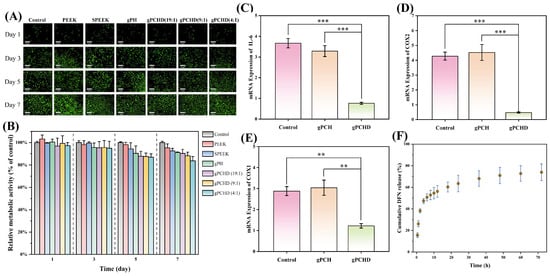
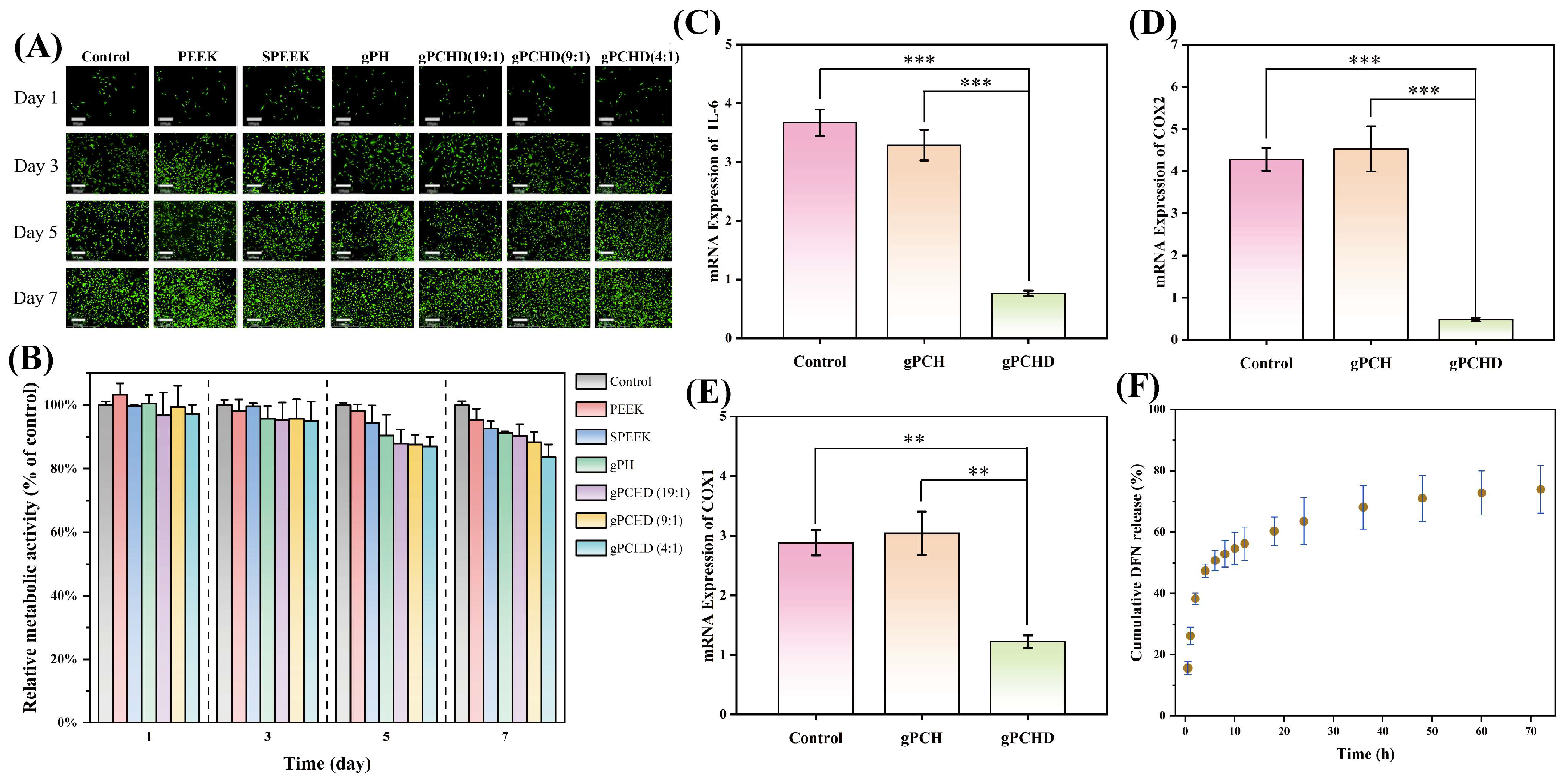
Live/dead imaging showed predominantly green-stained rabbit BMSCs over days 1, 3, 5, and 7 (Figure 3A). Control-normalized CCK-8 signals remained above 0.80, but the day-7 gPCHD(4:1) value of 0.837 ± 0.047 (n = 3 independent experiments) was approximately 16.3% below the control mean of 1.000. This reduction warrants attention and is not dismissed simply because the value exceeded 80% of control. CCK-8 reflects the combined influence of cell number and cellular metabolic state; the 16.3% decrease cannot be equated directly with a 16.3% cell-death rate or a measured change in proliferation. Predominantly green live/dead staining provides complementary qualitative evidence of viable cells but does not exclude a modest metabolic effect. The data support preliminary cytocompatibility under the tested exposure conditions while identifying a day-7 decrease that merits further examination. These observations do not isolate which material component accounts for the metabolic change. Figure 3B therefore reports relative metabolic activity descriptively, without inferential significance brackets.
Figure 3.
Cytocompatibility, inflammation-related gene expression, and DFN release. (A) Live/dead staining of rabbit BMSCs on days 1, 3, 5, and 7; columns show Control, PEEK, SPEEK, gPH, and gPCHD at 19:1, 9:1, and 4:1; scale bars, 200 µm. (B) Relative CCK-8 metabolic activity normalized to the control at each time point, presented as mean ± SD from three independent experiments and summarized descriptively without inferential significance symbols. (C–E) Relative mRNA expression of IL-6, COX-2, and COX-1, respectively, after LPS stimulation and 24 h of material exposure. gPCH and gPCHD are composition-matched drug-free and DFN-loaded composite constructs. Each group comprises three independent biological samples, each measured in three technical wells; comparisons used one-way ANOVA with Tukey’s test. Ratio labels denote the PVA:AA precursor feed mass ratio. (F) Cumulative DFN release into PBS at 37 °C over 72 h, mean ± SD from three independent release curves. In (A), green and red fluorescence indicate live and membrane-compromised cells, respectively. Colors in (B) follow the legend; in (C–E), pink, peach, and pale green identify Control, gPCH, and gPCHD, respectively. In (C–E), ** p < 0.01 and *** p < 0.001 for the bracketed comparisons. In (F), brown points show means and blue error bars show SD.
In PBS at 37 °C, cumulative DFN release reached 63.49 ± 3.68% at 24 h, 70.97 ± 3.84% at 48 h, and 73.92 ± 3.24% at 72 h (n = 3; Figure 3F). Release was faster during the first day and continued more slowly over the following two days. The independently established loading amount and cumulative released mass were 55 µg per construct as a loading-batch average and 40.66 ± 1.78 µg per construct released at 72 h, respectively. The latter was calculated by multiplying each release curve by the common 55 µg denominator; its SD reflects variation among the three release curves and does not include uncertainty in the pooled loading estimate. The arithmetic complement of the 72 h mean, 26.08%, is the fraction not recovered as released DFN during this observation window; it is not a direct assay of intact residual drug or proof of later therapeutic release. The PBS experiment demonstrates the release time course under the stated in vitro conditions, not an in vivo therapeutic concentration or pharmacokinetic profile.
In the composition-matched RT-qPCR comparison, gPCHD showed lower COX-1, COX-2, and IL-6 mRNA expression than drug-free gPCH following 12 h of LPS stimulation and 24 h of material exposure. Mean relative expression values for gPCHD versus gPCH were 0.479 versus 4.525 for COX-2, 1.225 versus 3.042 for COX-1, and 0.764 versus 3.287 for IL-6 (Figure 3C–E); all three comparisons had p < 0.05. The earlier HSPEEK label in this experiment referred to the drug-free composite, not to the PVA-only gPH comparator used elsewhere. The corrected naming makes the shared PVA/PAA formulation and different DFN-loading status explicit. These findings support a DFN-loading-associated transcriptional response under the tested conditions. They do not directly measure cyclooxygenase enzyme inhibition, demonstrate superiority over free DFN, or establish a quantitative release-rate–response relationship.
Surface-directed regulation of inflammatory responses has also been investigated using zwitterionic coatings on PEEK [29]. The present coating combines a lubricating interface with drug delivery. Its material performance and lower inflammation-related mRNA expression in the loaded group support complementary approaches to improving joint contact and modulating the cellular response.
3.6. Twelve-Week Histological Observations
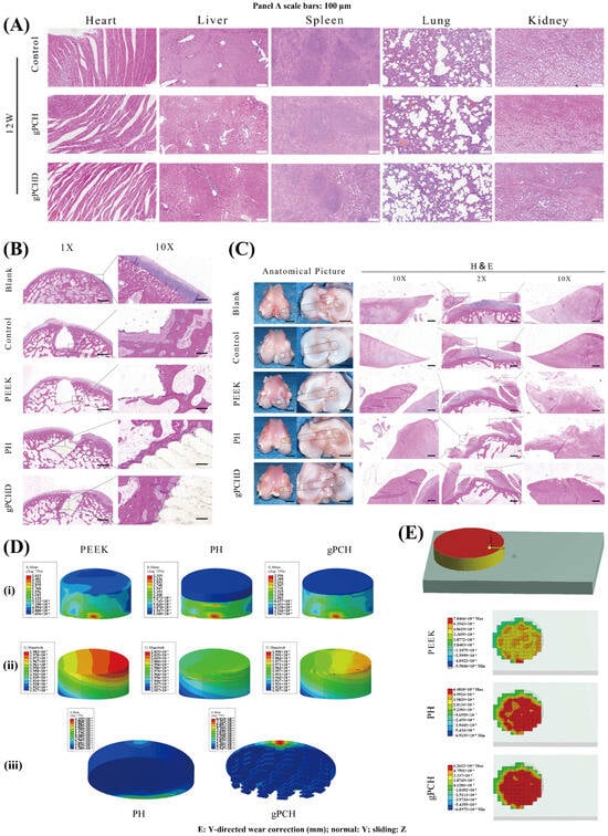
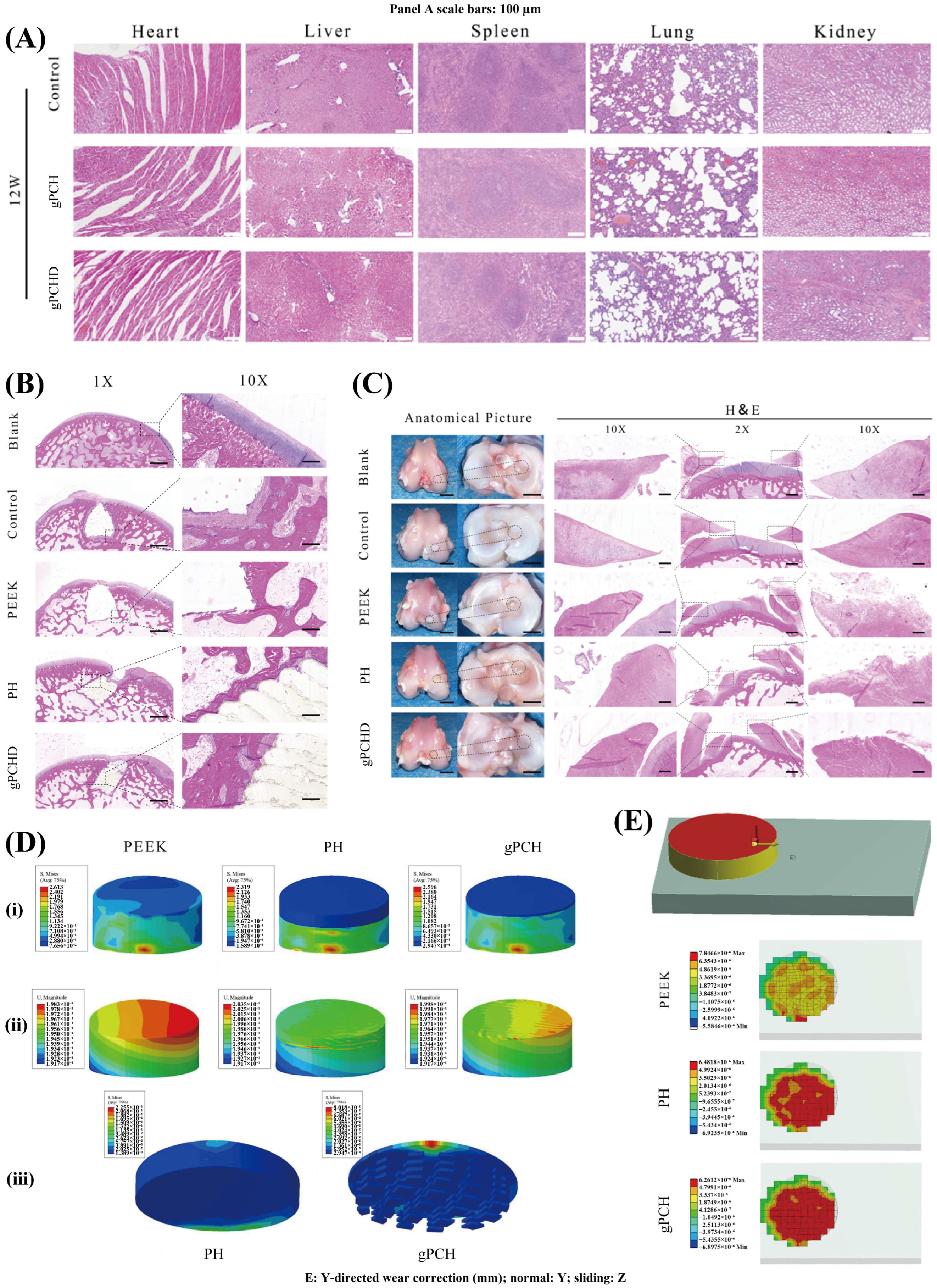
The rabbit study examined the implanted constructs over 12 weeks, and all 18 animals completed the observation period. H&E-stained sections of the heart, liver, spleen, lung, and kidney from the Control, gPCH, and gPCHD groups showed preserved tissue architecture without overt histopathological abnormalities in the examined fields (Figure 4A).
Figure 4.
Twelve-week rabbit histology and exploratory finite-element analyses. (A) H&E-stained heart, liver, spleen, lung, and kidney sections; scale bars, 100 µm; rows are Control, gPCH, and gPCHD. (B) Femoral-condyle and implant-interface H&E sections; scale bars, 500 µm. BLANK denotes no defect or implant, and Control a defect without an implant. (C) Gross appearance and H&E sections of the opposing meniscus and tibial articular surface; scale bars, 500 µm. Panels B and C show BLANK, Control, PEEK, PH, and gPCHD. Joint and meniscal sections were not collected from drug-free gPCH; all histology is descriptive. (D) Abaqus outputs for the idealized knee-joint configurations: (i) implant von Mises stress, MPa; (ii) displacement magnitude, mm; (iii) stress in the hydrogel-bearing region, MPa. (E) ANSYS cylindrical-contact outputs, showing the Y-directed wear correction (mm; CONTNMISC179). Y is parallel to the planar contact normal and the applied tangential displacement is 0 to −1 mm along Z, under 0.5 MPa pressure. The original individual contour scales and signs are retained. The finite-element outputs were not independently validated against material-specific wear measurements and are not rabbit-tissue measurements. In (A–C), H&E staining yields purple-blue nuclei and pink tissue; the blue background in the gross photographs is a photographic backdrop. In (D,E), colors correspond to the physical quantity and values on each individual color scale; identical colors across panels do not imply identical values.
Defect-site sections were examined for tissue contact and the presence of a hydrogel-bearing layer (Figure 4B). BLANK provided tissue without a defect or implant; the defect-only Control sections documented the sampled defect-site morphology. Fibrous tissue was visible along portions of the PEEK boundary. In the displayed PH sections, fibrous tissue occupied the superficial region and a distinct hydrogel layer was not identified. A hydrogel-bearing layer and tissue contact were visible in the displayed gPCHD implant region at 12 weeks. These findings describe the sampled sections. They provide morphological information alongside the bench-top peeling experiment but do not quantify an AA-specific in vivo retention effect or establish complete absence of detachment throughout the animal series.
Gross images and H&E sections documented the opposing meniscus and tibial articular surface (Figure 4C). Focal meniscal surface discontinuities were visible in the sampled PEEK sections, whereas portions of the surface contour in the displayed gPCHD sections were continuous. These are descriptive observations of the collected tissue fields, not a blinded or statistically scored comparison of damage severity. Joint and opposing-meniscus specimens were not collected from drug-free gPCH, and no in vivo pair differing only in AA treatment was available. The individual contributions of DFN, hydrogel composition, and AA treatment therefore cannot be separated from these tissue observations. Multilayer joint designs have also been studied in a distinct temporomandibular-joint setting [30]; this broader motivation does not substitute for controlled wear evaluation in the present model.
3.7. Exploratory Finite-Element Analyses
The Abaqus analysis describes stress and displacement within the idealized knee-joint configurations. The calculated peak hydrogel von Mises stress was 0.08018 MPa for gPCH and 0.2255 MPa for PH (Figure 4D(iii)). Maximum implant displacements were 0.1998, 0.2035, and 0.1983 mm for gPCH, PH, and PEEK, respectively (Figure 4D(ii)). These model-specific outputs illustrate how the supporting framework and hydrogel-bearing region distribute mechanical demand. They are not measurements from the implanted rabbits and do not independently validate the material’s clinical load-bearing or wear performance.
The separate ANSYS contact calculation produced spatial contours of the signed Y-directed wear correction (Figure 4E), in mm, under the stated 0.5 MPa pressure and 0 to −1 mm tangential displacement. The values depend on the prescribed Archard parameters and contact assumptions. The original solver reported a small-sliding applicability warning, and its influence was not quantified by an additional calculation. Consequently, the contours are retained only as illustrative outputs of the archived model, without interpreting differences in a signed component as validated total-wear rankings or predictions of service life.
3.8. Study Scope and Future Development
Tribology was evaluated over 10 min at room temperature under 2 N in 25% FBS against a stainless-steel sphere. This establishes a common short-term screening condition rather than long-term articulation against cartilage. Wear volume, prolonged cyclic durability, and performance at physiological temperature in synovial-fluid-like media remain to be established. Future group studies will apply longer durations, additional loads, 37 °C testing, and cartilage-relevant counterfaces and lubricating media.
The upper hydrogel-integration region was designed to be 1 mm high and contains both hydrogel and the reinforcing PEEK framework. The regional 60% software infill is not an independently measured as-printed pore-volume fraction. Structure–property interpretation is based on the designed geometry, available images, and composite-specimen tests rather than a measured three-dimensional porosity. Compression results from the separate cylindrical specimens at room temperature do not directly establish the behavior of the 3 mm-high implants at 37 °C.
The RT-qPCR comparison used composition-matched drug-free and DFN-loaded constructs. A free-DFN comparator at a matched delivered dose was not included, so superiority over free-drug exposure and a quantitative release-rate–response relationship are not established. The 72 h PBS release profile does not define drug exposure in cell wells or the joint. The animal study did not provide joint and meniscal sections for drug-free gPCH or an AA-only treatment comparison; tissue findings therefore cannot separate the individual in vivo contributions of DFN, PVA/PAA composition, and AA treatment. Histological findings remain descriptive rather than statistically scored damage comparisons.
The present chemical data support formation of a PVA/PAA composite coating but do not establish two independent networks or absolute grafting density. The archived XPS quantification metadata and a full atomic-percentage table were unavailable. The finite-element models are exploratory and were not independently calibrated and validated against matched wear measurements; the unresolved contact-approximation warning further limits quantitative interpretation. These issues motivate more complete surface-chemical, interface, tribological, and dose-controlled biological evaluations in subsequent studies.
The surface-modification strategy used in this study is supported by previous reports of UV-induced, self-initiated graft polymerization on PEEK, including studies using acrylic acid as the grafting monomer [31,32,33,34]. In the present work, the improved resistance to interfacial separation following AA/UV treatment is consistent with the intended role of this surface-modification step. However, the AA/UV-treated intermediate was not characterized independently before hydrogel integration. Therefore, although previous studies provide a mechanistic basis for the approach, the present study did not directly identify newly formed covalent bonds, such as interfacial C–C bonds, between AA-derived chains and the PEEK substrate. Accordingly, the washing procedure does not by itself establish the persistence or chemical nature of AA-derived surface modifications, and the present data do not distinguish AA adsorption, residual PAA, and covalent grafting.
4. Conclusions
This work presents an osteochondral-inspired approach to integrating a PVA/PAA composite hydrogel with a continuously printed PEEK bilayer. The dense support and porous upper region provide complementary structural and surface functions. In the bench-top material series, substrate treatment and hydrogel formulation increased resistance to separation. Drug-free gPCH(4:1) exhibited a maximum detachment force per unit width of 304.60 ± 7.20 N m−1, 2.39 times the PH comparator, and a friction coefficient of 0.073 ± 0.005 under the 2 N, 10 min, room-temperature screening conditions. Its DFN-loaded counterpart released the drug over the 72 h observation period and showed lower inflammation-related mRNA expression than composition-matched drug-free gPCH after LPS stimulation. Twelve-week histology documented tissue morphology and the presence of a hydrogel-bearing layer in the sampled implant region without establishing a quantitative comparison of meniscal damage. The study links framework design, hydrogel accommodation, interfacial retention, short-term friction, and drug delivery, while leaving long-term tribological performance and component-specific in vivo effects for further evaluation.
Author Contributions
H.S.: Conceptualization, Visualization, Formal analysis, Writing—original draft, Software. Y.Y.: Conceptualization, Methodology, Data curation, Validation. X.H.: Investigation, Methodology. J.M.: Investigation, Software. J.L.: Investigation, Data curation. P.W.: Investigation, Validation. Y.Z.: Writing—review & editing, Conceptualization, Supervision, Project administration, Funding acquisition, Resources. All authors have read and agreed to the published version of the manuscript.
Funding
This work was supported by the Clinical Research Center of Shaanxi Province for Dental and Maxillofacial Diseases, College of Stomatology, Xi’an Jiaotong University (Grant No. 2023YHJB02). The funder provided financial support only and had no role in the study design; data collection, analysis, or interpretation; manuscript preparation; or the decision to submit the manuscript for publication.
Institutional Review Board Statement
Rabbit BMSC isolation and animal procedures were approved by the Animal Ethics Committee of the School of Stomatology, Lanzhou University (approval no. LZUKQ-2023-068, 8 June 2023).
Informed Consent Statement
Not applicable.
Data Availability Statement
The data supporting the findings of this study are available from the corresponding author upon reasonable request, subject to institutional and ethical requirements.
Acknowledgments
During the preparation of this manuscript, the authors used ChatGPT (OpenAI) for assistance with the Graphical Abstract and manuscript text revision and proofreading. The authors reviewed and edited the output and take full responsibility for the content of this publication.
Conflicts of Interest
The authors declare no conflicts of interest.
References
- Nordberg, R.C.; Bielajew, B.J.; Takahashi, T.; Dai, S.; Hu, J.C.; Athanasiou, K.A. Recent advancements in cartilage tissue engineering innovation and translation. Nat. Rev. Rheumatol. 2024, 20, 323–346. [Google Scholar] [CrossRef] [Scilit] [PubMed]
- Muthu, S.; Korpershoek, J.V.; Novais, E.J.; Tawy, G.F.; Hollander, A.P.; Martin, I. Failure of cartilage regeneration: Emerging hypotheses and related therapeutic strategies. Nat. Rev. Rheumatol. 2023, 19, 403–416. [Google Scholar] [CrossRef] [Scilit] [PubMed]
- Zelinka, A.; Roelofs, A.J.; Kandel, R.A.; De Bari, C. Cellular therapy and tissue engineering for cartilage repair. Osteoarthr. Cartil. 2022, 30, 1547–1560. [Google Scholar] [CrossRef] [Scilit] [PubMed]
- Gopinatth, V.; Jackson, G.R.; Touhey, D.C.; Chahla, J.; Smith, M.V.; Matava, M.J.; Brophy, R.H.; Knapik, D.M. Microfracture for medium size to large knee chondral defects has limited long-term efficacy: A systematic review. J. Exp. Orthop. 2024, 11, e70060. [Google Scholar] [CrossRef] [Scilit] [PubMed]
- Seewoonarain, S.; Ganesh, D.; Perera, E.; Popat, R.; Jones, J.; Sugand, K.; Gupte, C. Scaffold-associated procedures are superior to microfracture in managing focal cartilage defects in the knee: A systematic review & meta-analysis. Knee 2023, 42, 320–338. [Google Scholar] [CrossRef] [Scilit] [PubMed]
- Qiu, F.; Fan, X.; Chen, W.; Xu, C.; Li, Y.; Xie, R. Recent Progress in Hydrogel-Based Synthetic Cartilage: Focus on Lubrication and Load-Bearing Capacities. Gels 2023, 9, 144. [Google Scholar] [CrossRef] [Scilit] [PubMed]
- Jiang, X.; Sun, Y.; Lyu, Y.; Kang, H.; Zhao, J.; Zhang, K.; Bian, L. Hydrogels for ameliorating osteoarthritis: Mechanical modulation, anti-inflammation, and regeneration. BMEMat 2024, 2, e12078. [Google Scholar] [CrossRef] [Scilit]
- Zhang, W.; Zha, K.; Hu, W.; Xiong, Y.; Knoedler, S.; Obed, D.; Panayi, A.C.; Lin, Z.; Cao, F.; Mi, B.; et al. Multifunctional hydrogels: Advanced therapeutic tools for osteochondral regeneration. Biomater. Res. 2023, 27, 76. [Google Scholar] [CrossRef] [Scilit] [PubMed]
- Branco, A.C.; Oliveira, A.S.; Monteiro, I.; Nolasco, P.; Silva, D.C.; Figueiredo-Pina, C.G.; Colaço, R.; Serro, A.P. PVA-Based Hydrogels Loaded with Diclofenac for Cartilage Replacement. Gels 2022, 8, 143. [Google Scholar] [CrossRef] [Scilit] [PubMed]
- Oliveira, A.S.; Silva, J.C.; Loureiro, M.V.; Marques, A.C.; Kotov, N.A.; Colaço, R.; Serro, A.P. Super-Strong Hydrogel Composites Reinforced with PBO Nanofibers for Cartilage Replacement. Macromol. Biosci. 2023, 23, 2200240. [Google Scholar] [CrossRef] [Scilit] [PubMed]
- Liu, L.; Xian, Y.; Wang, W.; Huang, L.; Fan, J.; Ma, W.; Li, Y.; Liu, H.; Yu, J.K.; Wu, D. Meniscus-Inspired Self-Lubricating and Friction-Responsive Hydrogels for Protecting Articular Cartilage and Improving Exercise. ACS Nano 2023, 17, 24308–24319. [Google Scholar] [CrossRef] [Scilit] [PubMed]
- Yuan, Z.; Long, T.; Zhang, J.; Lyu, Z.; Zhang, W.; Meng, X.; Qi, J.; Wang, Y. 3D printed porous sulfonated polyetheretherketone scaffold for cartilage repair: Potential and limitation. J. Orthop. Transl. 2022, 33, 90–106. [Google Scholar] [CrossRef] [Scilit] [PubMed]
- Pidhatika, B.; Widyaya, V.T.; Nalam, P.C.; Swasono, Y.A.; Ardhani, R. Surface Modifications of High-Performance Polymer Polyetheretherketone (PEEK) to Improve Its Biological Performance in Dentistry. Polymers 2022, 14, 5526. [Google Scholar] [CrossRef] [Scilit] [PubMed]
- Shi, Y.; Deng, T.; Peng, Y.; Qin, Z.; Ramalingam, M.; Pan, Y.; Chen, C.; Zhao, F.; Cheng, L.; Liu, J. Effect of Surface Modification of PEEK Artificial Phalanx by 3D Printing on its Biological Activity. Coatings 2023, 13, 400. [Google Scholar] [CrossRef] [Scilit]
- Xu, X.; Zuo, J.; Zeng, H.; Zhao, Y.; Fan, Z. Improving Osseointegration Potential of 3D Printed PEEK Implants with Biomimetic Periodontal Ligament Fiber Hydrogel Surface Modifications. Adv. Funct. Mater. 2024, 34, 2308811. [Google Scholar] [CrossRef] [Scilit]
- Zhao, X.; Xiong, D.; Liu, Y. Improving surface wettability and lubrication of polyetheretherketone (PEEK) by combining with polyvinyl alcohol (PVA) hydrogel. J. Mech. Behav. Biomed. Mater. 2018, 82, 27–34. [Google Scholar] [CrossRef] [Scilit] [PubMed]
- Wei, Q.; Liu, H.; Zhao, X.; Zhao, W.; Xu, R.; Ma, S.; Zhou, F. Bio-inspired hydrogel-polymer brush bi-layered coating dramatically boosting the lubrication and wear-resistance. Tribol. Int. 2023, 177, 108000. [Google Scholar] [CrossRef] [Scilit]
- Hu, X.; Zhao, Y.; Cheng, S.; Zhen, J.; Jia, Z.; Zhang, R. Biomimetic Layered Hydrogel Coating for Enhanced Lubrication and Load-Bearing Capacity. Coatings 2024, 14, 1229. [Google Scholar] [CrossRef] [Scilit]
- Hu, K.; Ma, Q.; Guo, W.; Zhao, W.; Zhao, Y.; Cai, X.; Zhang, H. Bioinspired Hard–Soft Composite Scaffold with Excellent Lubrication and Osteogenic Properties for the Treatment of Osteochondral Defect. Adv. Mater. Interfaces 2024, 11, 2300473. [Google Scholar] [CrossRef] [Scilit]
- Xu, R.; Liu, J.; Zhao, W.; Liu, B.; Li, R.; Zhao, X.; Wei, Q.; Ma, S. Bioinspired highly entangled hydrogel coating with integrated mechanical-chemical anchorage for enhanced tribological performance of PEEK. Tribol. Int. 2026, 223, 112249. [Google Scholar] [CrossRef] [Scilit]
- Elkington, R.J.; Pryce, G.M.; Keeling, D.; Hall, R.M.; Beadling, A.R.; Pandit, H.; Bryant, M.G. Performance parity in cartilage repair: SPMK-g-PEEK versus cartilage–cartilage interfaces. J. Mech. Behav. Biomed. Mater. 2025, 167, 106964. [Google Scholar] [CrossRef] [Scilit] [PubMed]
- Lin, H.; Zhang, L.; Ye, X.; Wang, X.; Han, C.; Yan, Z.; Wang, Q.; Yan, G. The surface modification of 3D-printed polyether ether ketone with bioactive hydrogel for bone repair. Colloids Surf. B Biointerfaces 2025, 254, 114846. [Google Scholar] [CrossRef] [Scilit] [PubMed]
- Huang, J.; Tang, Y.; Wang, P.; Zhou, H.; Li, H.; Cheng, Z.; Wu, Y.; Xie, Z.; Cai, Z.; Wu, D.; et al. One-Pot Construction of Articular Cartilage-Like Hydrogel Coating for Durable Aqueous Lubrication. Adv. Mater. 2024, 36, 2309141. [Google Scholar] [CrossRef] [Scilit] [PubMed]
- Li, Q.; Gong, Y.; Li, Y.; Li, S.; Liang, W.; Leng, Y.X. Study on the lubrication behavior of tannic acid/poly (vinyl alcohol) hydrogel enhanced by protein adsorption for articular cartilage applications. J. Mech. Behav. Biomed. Mater. 2025, 162, 106825. [Google Scholar] [CrossRef] [Scilit] [PubMed]
- Feng, H.; Wang, S.; Chen, K.; Zhang, X.; Feng, C.; Li, X.; Zhang, D. Dual-network nanocomposite robust hydrogel with excellent durability properties as cartilage replacement. Tribol. Int. 2024, 194, 109518. [Google Scholar] [CrossRef] [Scilit]
- Hu, D.; Liu, D.; Hu, Y.; Wang, Y.; Lu, Y.; Bai, C.; Hossain, K.R.; Jiang, P.; Wang, X. Dual-physical network PVA hydrogel commensurate with articular cartilage bearing lubrication enabled by harnessing nanoscale crystalline domains. Nano Res. 2024, 17, 9784–9795. [Google Scholar] [CrossRef] [Scilit]
- Zhao, W.; Zhang, Y.; Zhao, X.; Sheng, W.; Ma, S.; Zhou, F. Mechanically Robust Lubricating Hydrogels Beyond the Natural Cartilage as Compliant Artificial Joint Coating. Adv. Sci. 2024, 11, 2401000. [Google Scholar] [CrossRef] [Scilit] [PubMed]
- Cui, L.; Lu, Y.; Gong, C.; Li, H.; Huang, J.; Xiong, D. Metal ion reinforced hydrogel/Ti6Al4V bionic composite joint bearing interface with extraordinary mechanical and biotribological properties. Tribol. Int. 2023, 189, 109024. [Google Scholar] [CrossRef] [Scilit]
- Roventini, E.; Iacoponi, F.; Poliziani, A.; Canepa, P.; Cavalleri, O.; Cassa, M.A.; Parlanti, P.; Pucci, C.; Gemmi, M.; Tonda-Turo, C.; et al. Comparison between SBMA and MPC coatings on PEEK surface: Stability over time and anti-inflammatory effects in vitro. Sci. Rep. 2025, 15, 41263. [Google Scholar] [CrossRef] [Scilit] [PubMed]
- Lu, S.; Qin, L.; Zhao, Z.; Feng, Z.; Ma, Z.; Wu, Y.; Wang, Z.; Huang, X.; Liu, J.; Qiu, J.; et al. Bio-inspired Multi-layer structure artificial joint for In vivo wear resistant. Adv. Compos. Hybrid Mater. 2026, 9, 221. [Google Scholar] [CrossRef] [Scilit]
- Kyomoto, M.; Ishihara, K. Self-Initiated Surface Graft Polymerization of 2-Methacryloyloxyethyl Phosphorylcholine on Poly(ether ether ketone) by Photoirradiation. ACS Appl. Mater. Interfaces 2009, 1, 537–542. [Google Scholar] [CrossRef] [Scilit] [PubMed]
- Yousaf, A.; Farrukh, A.; Oluz, Z.; Tuncel, E.; Duran, H.; Doğan, S.Y.; Tekinay, T.; Rehman, H.u.; Yameen, B. UV-light assisted single step route to functional PEEK surfaces. React. Funct. Polym. 2014, 83, 70–75. [Google Scholar] [CrossRef] [Scilit]
- Zhao, X.; Xiong, D.; Wang, K.; Wang, N. Improved biotribological properties of PEEK by photo-induced graft polymerization of acrylic acid. Mater. Sci. Eng. C 2017, 75, 777–783. [Google Scholar] [CrossRef] [Scilit] [PubMed]
- Yang, F.; Sun, R.; Qi, H.; Sun, W.; Li, J.; Dong, W.; Zhang, M.; Pang, J.; Jiang, Z. Preparation and Properties of PEEK-g-PAA Separation Membranes with Extraordinary Solvent Resistance for Emulsion and Organic Liquid Separation. ACS Appl. Polym. Mater. 2023, 5, 8305–8314. [Google Scholar] [CrossRef] [Scilit]

Disclaimer/Publisher’s Note: The statements, opinions and data contained in all publications are solely those of the individual author(s) and contributor(s) and not of MDPI and/or the editor(s). MDPI and/or the editor(s) disclaim responsibility for any injury to people or property resulting from any ideas, methods, instructions or products referred to in the content. |
© 2026 by the authors. Licensee MDPI, Basel, Switzerland. This article is an open access article distributed under the terms and conditions of the Creative Commons Attribution (CC BY) license.