


Abstract
Resin infiltration of root cementum requires optimal surface preparation; however, traditional protocols using hydrochloric acid raise some concerns about potential cementum degradation. In this in vitro study the physicochemical and morphological impact of 36% H3PO4 on root cementum compared to natural demineralisation was evaluated to establish a candidate tissue-preserving conditioning agent. Twelve extracted teeth—incisors with exposed cementum over gum recession level and fully impacted third molars were analysed using scanning electron microscopy (SEM) and Energy Dispersive Spectrometry (EDS) in etched and not-etched specimens. Orthophosphoric acid significantly reduced calcium and phosphorus content across all specimens while maintaining overall morphological integrity. Notably, the elemental response varied based on tooth exposure history. Incisors exhibited a statistically significant reduction in the Ca/P atomic ratio, indicating complex stoichiometric shifts and preferential calcium loss. Conversely, impacted third molars maintained a stable Ca/P ratio, suggesting proportional mineral dissolution in unexposed cementum. These findings indicate that natural demineralisation is insufficient for adequate surface modification and that orthophosphoric acid demineralizes root cementum without compromising its structural integrity. The magnitude and pattern of this response depend on the tissue’s prior exposure to the oral environment; thus, structural heterogeneity of cementum, though tailored protocols, are necessary.
1. Introduction
Root caries (RC) involves either non-cavitated or cavitated lesions below cemento-enamel junction, excluding the adjacent enamel [1]. It is characterised by discolouration and softening of ill-defined cementum and dentin [2]. It might be associated with many potential issues, starting from proper diagnosis, as it might be occasionally mistaken for external cervical root resorption [3]. There are notable distinctions between RC and caries affecting the crown region of a tooth, both with regard to their development and their respective treatment strategies. Root tissues are more susceptible to demineralisation [4] than enamel, owing to the lower inorganic content, which is 45%–50% for cementum and 70% for dentine in contrast to 90% for enamel [5,6]. Furthermore, due to their higher magnesium and carbonate content, they are more soluble than enamel [7]. Additionally, bacterial colonisation of the roots may be initiated at an earlier stage than that of the crown, as it does not necessitate complete demineralisation. This is due to the fact that channels of Sharpey’s fibres can function as potential colonisation niches [8]. RC lesions usually have a wide and shallow shape and spread outwards [9], leading to an unfavourable width-to-depth ratio of the cavity. Other challenging factors, such as high mechanical stress in the cervical region or difficulties in moisture control [8] contribute to a low success rate of restorative treatment of such cavities. According to the literature, one-year survival rates of root restorations were reported to be 90%, but this was found to decrease to 65% in two-year periods [10,11,12,13], assessed as low to moderate certainty [14,15]. These facts highlight the importance of RC treatment in the early stages, prior to the necessity of an invasive approach.
Recent advancements in materials and technology have precipitated a transformation in prevailing trends towards minimally invasive dentistry (MID). The objective of this holistic approach is to establish a structured system for maintaining long-term oral health that is centred on the implementation of preventive care plans that are specifically tailored to the needs of the individual patient [16,17,18]. Over the years, it has been used with the aim of providing predictable results, whilst concomitantly minimising associated risks [19,20,21]. This is achieved based on a multidisciplinary strategy that incorporates active caries prevention, intervention at the early stages of its development, conservation of healthy tooth tissue and preserving pulp vitality. Digitalisation, fluorescence, microradiography and microcomputed tomography facilitate improved diagnosis, as they enable the detection of caries at an earlier stage [22,23,24]. Concurrently, the utilisation of magnification and enhanced lighting facilitates superior visualisation of anatomical structures within the treatment area [16,25,26]. The potential applications of such approach extend beyond the domain of cariology, encompassing other areas, e.g., tooth wear [27]. Such conceptual changes have led to a significant enhancement in the quality of care, yet sustained vigilance regarding the dangers of overdiagnosis and overtreatment is crucial to ensure optimal oral health [28].
Infiltration is one of minimally invasive treatment techniques used to treat dental caries and involves the use of low-viscosity composite resins [29,30]. In order to ensure long-term stability without nanoleakage detachment, it is essential that adhesives and resin monomers present superior permeability, rapid capillary penetration and enhanced surface tension in order to sufficiently infiltrate demineralised pores [31,32]. The obturation of compromised microporous regions of initial, non-cavitated lesions creates a protective barrier presenting the potential to suppress caries progression and preserve tooth anatomical structure [32,33,34]. Resin infiltration has been shown to be an effective treatment modality for white spot lesions and fluorosis, considered to be more effective than the established gold-standard treatment of fluoride varnish [35] or natural remineralisation [29]. The results achieved ensure a high standard of aesthetics [17]. Other fields might also make use of this method, including the treatment of dentine hypersensitivity [36] or developmental defects, e.g., molar incisor hypomineralisation [37]. It has also been found that resin infiltration serves as an effective additive in the process of teeth bleaching. A recent study has indicated that the process of infiltration prior to the application of bleaching agents can enhance the effectiveness of masking fluorotic lesions in the anterior teeth when compared with bleaching alone [38].
In order to guarantee effective resin-tooth bonding, it is imperative that the surface of the tooth is prepared thoroughly [37]. The duration of the etching process, in conjunction with appropriate time for drying, has been demonstrated to exert a significant influence on the homogeneity and depth of resin infiltration [39]. Extensive research has been dedicated to the study of appropriate procedures for enamel [40,41,42], in contrast to the paucity of research that has been conducted on root cementum. The systematic review and meta-analysis by Schmidt et al. led to a conclusion that it is currently impossible to make an evidence-based choice of restorative strategy for treating root caries lesions [15]. A study by Nowak et al. demonstrated that HCl in Icon Etch (Icon, DMG, Hamburg, Germany) caused damage to the cementum surface both in single and double etching procedures [43]. Similarly, Zhou et al. reported that 120 s of HCl pretreatment of demineralised root surfaces enabled good resin penetration but was associated with an additional risk of cervical enamel loss [44]. Given these findings, there is a crucial need to identify a more conservative, minimally invasive approach that facilitates resin bonding without compromising the structural integrity of the root surface. H3PO4, the most widely used and well-characterised etchant in adhesive dentistry, may be considered a candidate. Its effectiveness in the removal of the smear layer has been proven [45]. However, its effect on the composition and morphology of root cementum has not been widely described. Consequently, this study has been designed as a pilot study to address this literature gap.
Additionally, considering the susceptibility of cementum, it remains to be determined whether cementum exposed to the oral environment in recession areas undergoes sufficient natural demineralisation to allow effective infiltration without additional chemical preparation. If further conditioning is required, the use of a less aggressive agent would be preferable. Therefore, the aim of this in vitro study was to evaluate the composition and morphology of root cementum at various topographical sites to evaluate the impact of a conditioning process using H3PO4, compared to untreated naturally demineralised surfaces.
2. Materials and Methods
2.1. Sample and Design
2.1.1. Study Sample
The Bioethics Committee of the Silesian Medical University in Katowice granted approval for the study on 21 November 2024. The approval was documented under the reference number BNW/NWN/0052/KB/25. The research was conducted in accordance with the Checklist for Reporting In vitro Studies (CRIS) guidelines [46].
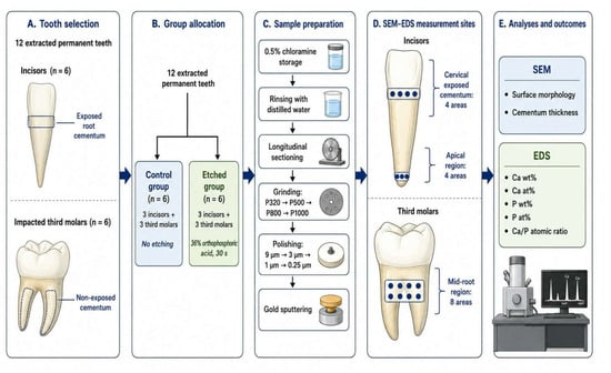
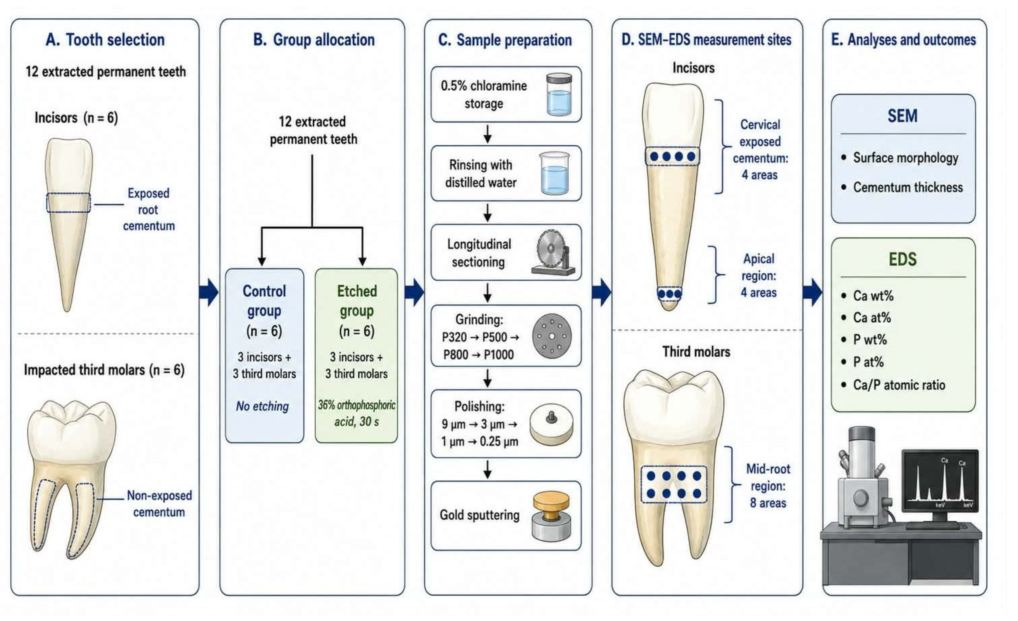
The basis of the methodology is presented in Figure 1.
Figure 1.
Diagram illustrating the basis of methodology.
The study material included 12 extracted permanent teeth meeting the inclusion criteria [Table 1]. These include 2 groups of teeth, i.e., incisors with exposed root cementum to a depth less than 6 mm (i.e., maximum gum recession of 6 mm), above which invasive, periodontal, surgical treatment is necessary, and impacted third molars, where the cementum was not exposed to the oral cavity environment. The teeth were free of caries or restorations with preserved anatomical crowns and roots.
Table 1.
Inclusion and exclusion criteria.
The extracted teeth were stored in a 0,5% chloramine solution until the commencement of the experiment. The experimental group consisted of 3 incisors and 3 third molars, etched according to the standard process, with Blue Etch (Cerkamed, Stalowa Wola, Poland), containing 36% H3PO4 for 30 s. The control group, consisting of 3 incisors and 3 molars as well, was not exposed to any etchant.
2.1.2. Preparation for Microscopic Observation
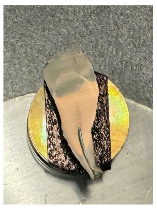
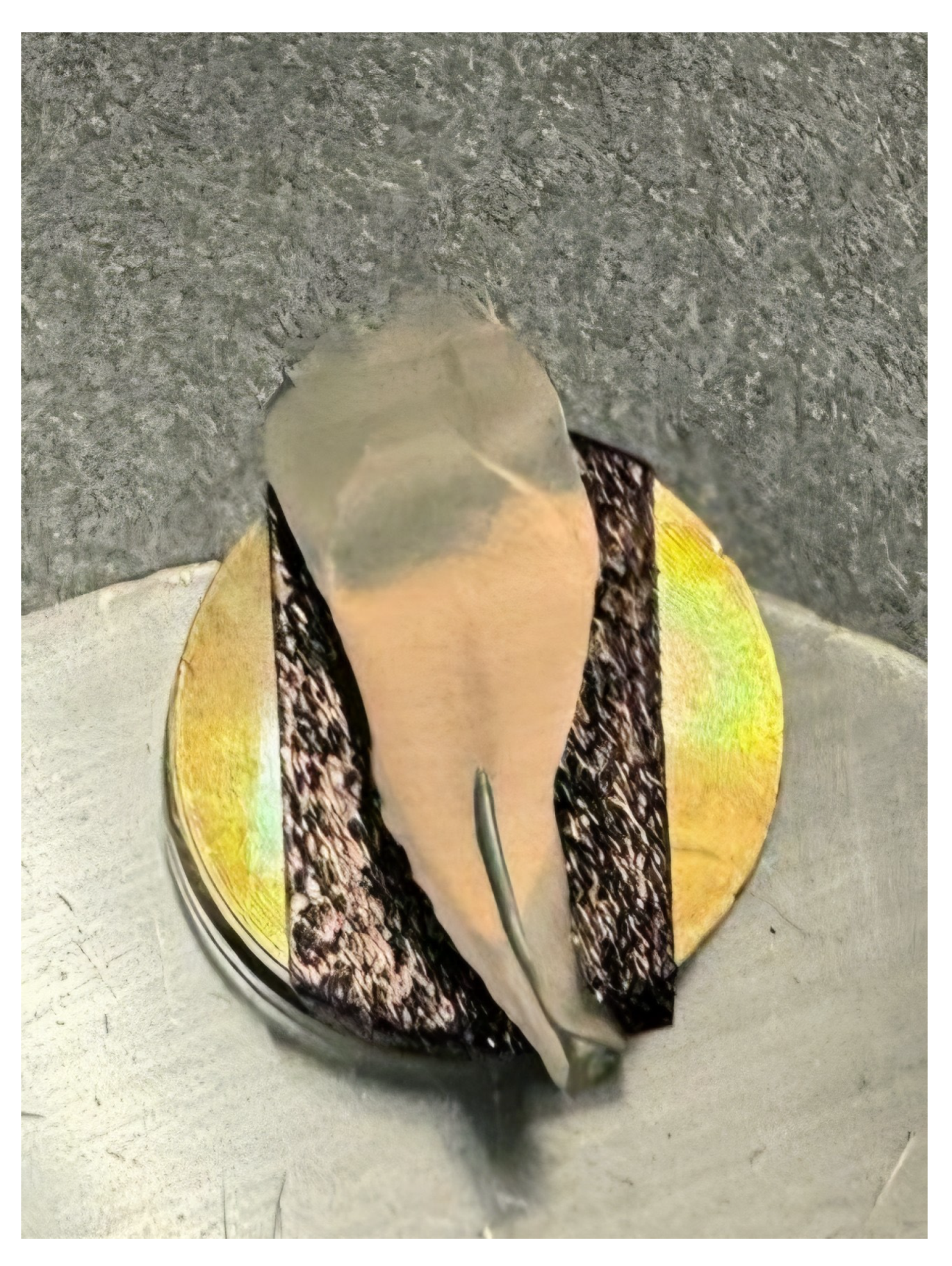
A duration of 24 h prior to microscopic observations the teeth were taken out of the chloramine solution, rinsed with distilled water and thereafter stored in it. After rinsing and initial drying the teeth were cut along the long axis using a diamond saw Struers Secotom 15 (Struers A/S, Ballerup, Denmark) and thoroughly dried. Subsequently, the surfaces of the cross-sections were sanded with Metalogis DEMPAX water-resistant sandpaper with decreasing gradations of P320, P500, P800 and P1000 (Metalogis s.c, Warsaw, Poland); and subsequently polished with Struers DP Suspension diamond slurries of 9 μm, 3 μm, 1 μm, 0.25 μm (Struers A/S, Ballerup, Denmark). The samples were then sputtered with gold (Figure 2). Gold sputtering was performed using a Leica EM SCD050 device with a 99.99% Au target. The current parameter was set to 40 mA, and time was set to 90 s following the sputtering time diagram for gold (d = 19.3 g/cm3) in accordance with the instructions provided in the user manual. After such preparation the samples were observed under the microscope.
Figure 2.
Specimen prepared for microscopic observation.
2.2. Microscopic Observation and Composition Analysis
Scanning electron microscopy (SEM) studies were performed in a scanning electron microscope Zeiss Supra 35 (Zeiss, Oberkochen, Germany), equipped with an Energy Dispersive Spectrometer (EDS) for the chemical composition analysis. The analysis was performed on longitudinal cross-sections of the roots, in which the cementum layer is visible in the profile. In the case of incisors, measurements of composition were taken in two regions: in the cervical part of the root, within the exposed cementum—4 areas, and in the apical part—4 areas. In the case of third molars, measurements were taken in one region at mid-root length—8 areas. The reported values represent the mean ± standard deviation (SD). Elemental composition was expressed as weight percentage (weight %), atomic percentage (atom %), and calcium-to-phosphorus (Ca/P) atomic ratio.
2.3. Statistical Analysis
Regarding the statistical analysis, for each tooth the EDS measurements obtained from the 8 predefined areas were averaged to obtain a tooth-level mean for each analysed parameter and anatomical region, as applicable. The individual tooth was treated as the independent experimental unit. The results were presented as mean and standard deviation (M ± SD), with the SD calculated from the tooth-level values within each group. Statistical differences were assessed using the Shapiro–Wilk test. Furthermore, a Student’s t-test was employed to compare the two variables. A p-value ≤ 0.05 was considered to indicate a statistically significant difference. The statistical analysis was performed using Statistica, v. 7.1 PL (StatSoft Poland, Krakow, Poland).
3. Results
3.1. Anatomical Structures Determination
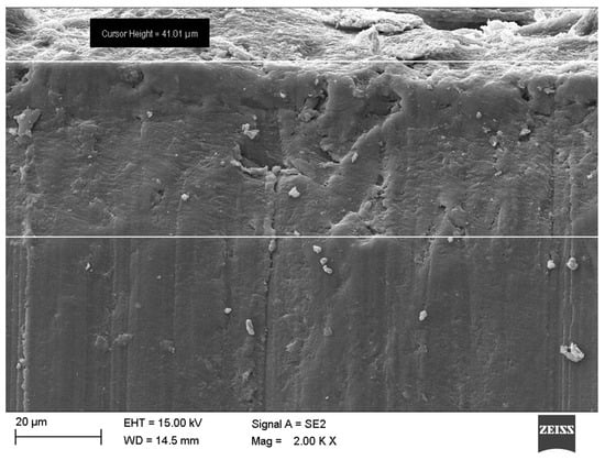
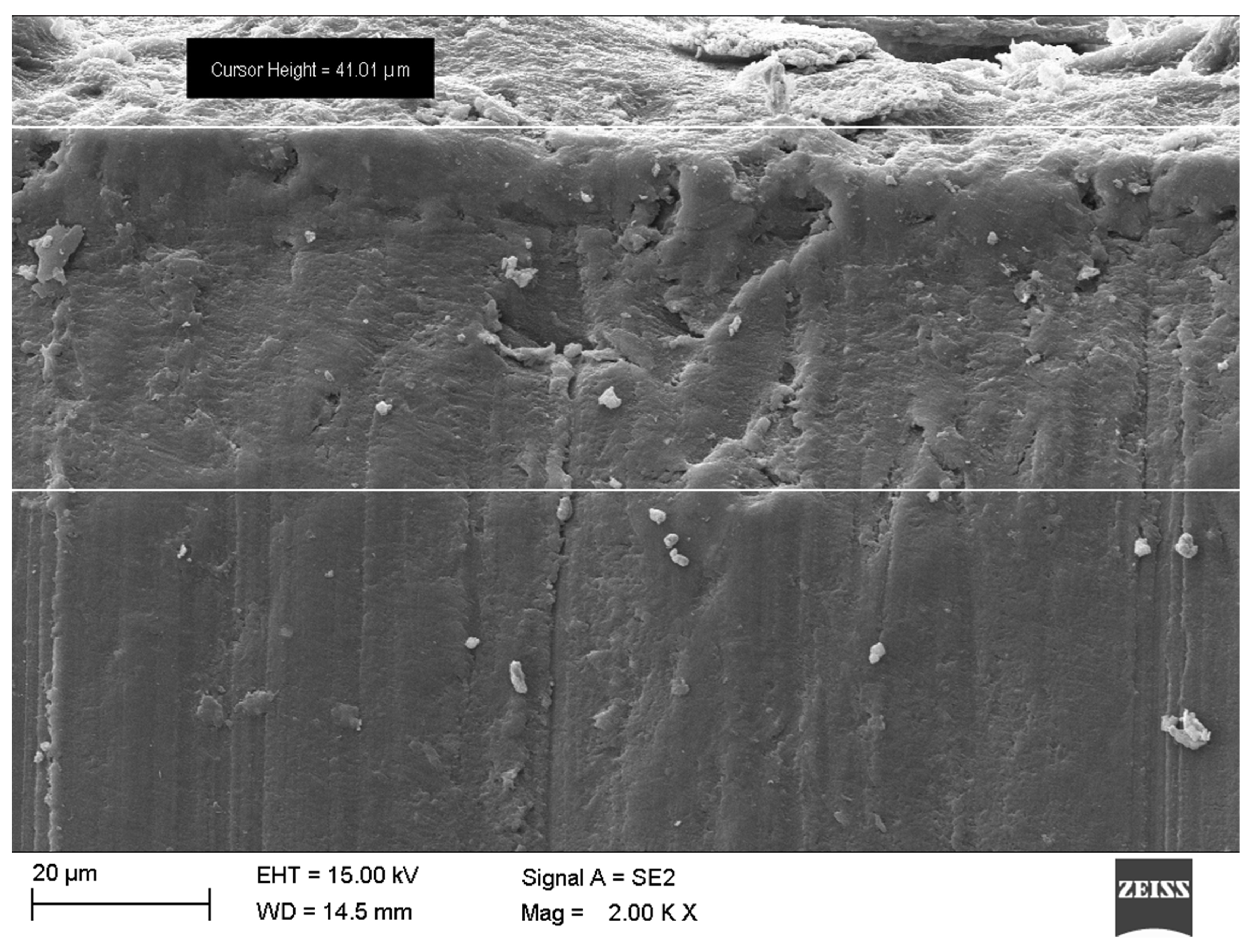
The initial phase of microscopic observations included anatomical structure identification. The thickness of root cementum among incisors varied between 21.81 μm in the cervical part of the tooth in the region of cementum exposure and 161.80 μm in the apical region. Mean cementum thickness among incisors was equal to 80.92 ± 51.05 μm. In the case of third molars, the values ranged from 28.04 μm in the cervical part to 680.92 μm in the apical part. Mean cementum thickness among the third molars was equal to 161.20 ± 65.13 μm (Figure 3).
Figure 3.
SEM image showing root cementum thickness.
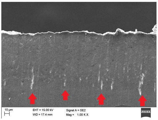
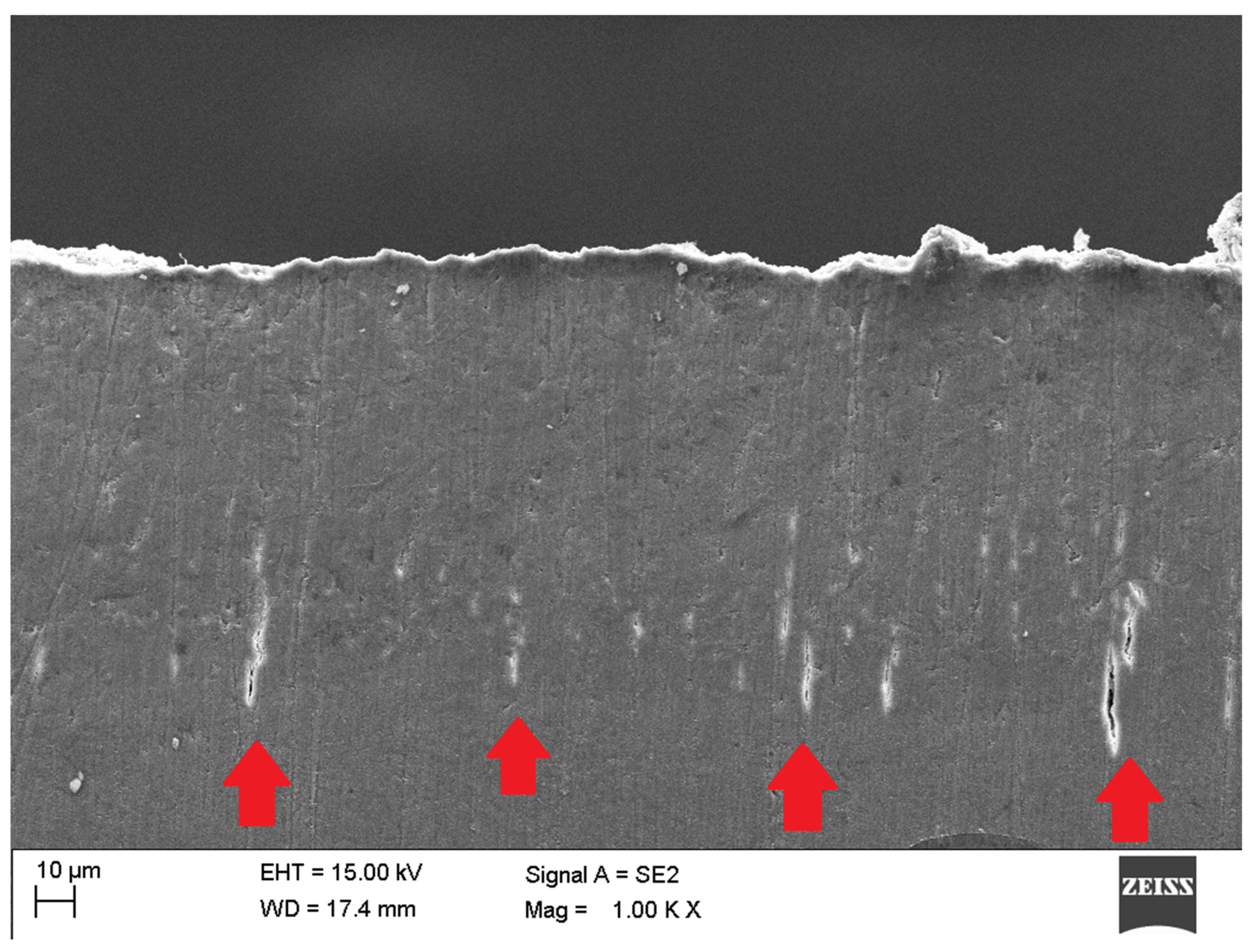
Further observations in the study revealed damage in the cemento-dentinal junction (CDJ) area, with no evident major damage to the cementum surface among few specimens (Figure 4). This was probably caused by the formation of the cross-section, when the pressure of the saw was applied from the external tooth surface, suggesting lower resistance to mechanical forces in the CDJ area in comparison to the external surface of cementum.
Figure 4.
SEM image showing mechanical damage (red arrows) in the region of cemento-dentinal junction (CDJ).
3.2. Chemical Composition Analysis
The results of chemical composition analysis obtained by EDS are included in Table 2. The results of Student’s t-test are included in Table 3. Reported values represent the average of measurements from eight different areas of each sample—for each incisor four in the cervical region and four in the apical region and for each third molar all measurements were taken in the middle part of the root. The mean calcium content in the control group was equal: 38.46 ± 4.18 weight % (wt%) and 21.81 ± 4.06 atom % (at%) for the exposed cementum area of incisors, 38.33 ± 4.50 wt% and 21.63 ± 4.27 at% for the apical area of incisors and 41.28 ± 7.08 wt% and 25.81 ± 5.46 at% for third molars. In the study group the results were equal as follows: 29.93 ± 2.05 wt% and 17.23 ± 3.47 at% for the exposed cementum area of incisors, 33.25 ± 1.04 wt% and 17.53 ± 0.88 at% for the apical area of incisors and 37.05 ± 1.97 wt% and 19.18 ± 2.02 at% for third molars. For phosphorus, the values in the control group were equal 14.34 ± 1.99 wt% and 10.89 ± 1.94 at% for the exposed cementum area of incisors, 14.05 ± 1.89 wt% and 10.53 ± 1.82 at% for the apical area of incisors and 16.08 ± 2.22 wt% and 12.83 ± 2.79 at% for third molars. In the study group the results were as follows: 13.33 ± 0.70 wt% and 8.38 ± 1.71 at% for the exposed cementum area of incisors, 13.11 ± 1.47 wt% and 8.30 ± 1.67 at% for the apical area of incisors and 12.81 ± 2.65 wt% and 9.21 ± 2.05 at% for third molars. In summary, both calcium and phosphorus levels decreased after etching among incisors as well as third molars; however further Student’s t-test analysis, comparing control and study groups, yielded variable outcomes. The differences between control and study groups in terms of calcium content were statistically significant (p ≤ 0.05) among all examined teeth. On the other hand, both cervical area and apical area of incisors showed no significant difference between study and control group in terms of weight percentage of phosphorus content.
Table 2.
Energy Dispersive Spectrometer (EDS) chemical composition analysis results.
Table 3.
Results of Student’s t-test.
The Ca/P ratio in the control group was equal to 2.10 ± 0.19 in the cervical area of incisors, 2.18 ± 0.22 in the apical area of incisors and 2.14 ± 0.36 among third molars. In the study group the results were as follows: 2.00 ± 0.13 in the exposed cementum area of incisors, 2.05 ± 0.11 in the apical area of incisors and 2.04 ± 0.30 in the third molars. The differences were statistically significant among both areas of incisors, conversely to the third molars, where no statistically significant difference in the Ca/P atomic ratio was detected between etched and non-etched specimens.
4. Discussion
4.1. General Results
The findings of the present study address a notable gap in clinical protocols regarding the efficient, yet not excessively destructive, preparation of root surface for interaction with materials, such as in an infiltration procedure. While the susceptibility of exposed cementum to the oral environment is well-documented [47,48,49], results of this study indicate that spontaneous natural demineralisation of exposed cementum in recession areas may not provide an appropriately high level of surface preparation for further effective interaction with materials, such as in resin infiltration. It is due to significant differences between the groups in the exposed cementum in the cervical area of incisors. However, further research involving larger study groups, along with various groups of teeth, is needed. Similarly, these results require confirmation by direct measurement of resin penetration in the future studies. Consequently, the clinical necessity for a proper tissue preparation remains, but the available data suggests a shift away from invasive processes. By utilising H3PO4, a balanced modification of the surface topography that preserves the structural framework of the cementum was observed, addressing the concerns raised by previous studies regarding tissue damage [43]. However, further studies comparing the impact of different surface conditioning agents are crucial to draw meaningful conclusions and help to create standardised, minimally invasive protocols.
In the present study the results of cementum thickness measurements appear as follows: mean thickness among incisors was 80.92 ± 51.05 μm and among molars 161.20 ± 65.13 μm. Such results are consistent with those of other related studies [50,51,52,53]. The minimal value observed for incisors was 21.81 μm and maximal 161.80 μm. The minimal and maximal values for molars were 28.04 μm and 680.92 μm, respectively. Sarna-Boś et al. noted similar results of 15 μm to 200 μm at the cervical section up to 600 μm at the root’s apical region [54]. However, a discrepancy in the mean values for incisors 30 μm (± 10) and molars 370 μm (± 30) was observed. This may be due to a reduced sample size in the present study (n = 12 vs. n = 20) and consequently a larger standard deviation. Beyond the limited sample size, these variations may stem from the heterogeneity of the specimens, particularly the age and gender of the donors, occlusal forces and the total time of the tooth’s exposure to the oral environment, which was not specified in the inclusion criteria of both studies. Furthermore, root cementum tissue exhibits considerable individual variability [55,56,57,58].
Regardless of the measurement area, the data demonstrate that thickness values for the molar teeth are higher than those recorded for the incisor teeth. The present findings are in accordance with the results of previous research [51,54,55]. In both groups the lowest values were observed in the cervical region and the highest in the apical region. The extant literature reports analogous findings for specific anatomical regions in the context of extreme values of cementum thickness [53,54].
The present study established that the average calcium content of the control group was between 38.33 ± 4.50 and 41.28% ± 7.08 as also reported in the control group in the study by Bukhary et al. 2026 [59]—between 41.75 ± 0.5 and 47.5% ± 4.5. Similar findings were observed for the experimental group exposed to etching in the present study and the periodontitis group in the aforementioned study—calcium content (cervical area) 29.93% ± 2.05 vs. 26.36% ± 0.5 and phosphorus content—between 12.81 ± 2.65 and 13.33% ± 0.70 vs between 13.67 ± 3.6 and 15.89% ± 3.5. Based on this, it can be hypothesised that orthophosphoric acid might exert comparable effects during the etching process to the resorptive changes and impaired mineral metabolism that are observed in periodontitis, in terms of demineralisation.
On the other hand, Sarna-Boś et al. [54] observed higher values for calcium content among incisors (15.47%) than molars (10.45%) in contrast to the present study. The differences may be due to the small sample size, which might not accurately represent the broader population, or discrepancies in the inclusion criteria between the studies. In this study, the inclusion criteria were more narrowly defined to cover selected groups of teeth, which may also account for these differences in chemical composition. Different measurement points between the studies might be another contributing factor, as various types of cementum present anatomical and histological differences, including uncalcified spaces. Cellular intrinsic fibre cementum contains lacunae; cellular mixed stratified cementum includes uncalcified core of extrinsic Sharpey’s fibres or alternating layers of cellular intrinsic fibre cementum and acellular extrinsic fibre cementum [60,61,62].
The most notable finding of the present study is the divergent behaviour of the Ca/P atomic ratio between the two dental groups. The Ca/P atomic ratio provides additional information regarding the relative changes in mineral composition. A significant reduction in the Ca/P ratio was observed in both cervical and apical regions of incisors, which might indicate that phosphoric acid etching induced changes in mineral stoichiometry in addition to overall mineral loss—a greater loss in calcium relative to phosphorus was observed. Conversely, although both calcium and phosphorus contents decreased significantly in impacted third molars, the Ca/P ratio remained unchanged, indicating that both elements were removed proportionally. This finding may suggest differences in the response of exposed and non-exposed cementum to acid conditioning. Thus, it may reflect differences in the baseline mineral maturity and crystallinity of cementum according to its exposure history. It has been shown that pathological or environmentally mediated surface alterations are associated with measurable shifts in calcium and phosphorus content [59,63,64,65], supporting the notion that cementum composition is not static but can be modulated by local environmental exposure over time and may present interindividual variation [61,62,66,67,68]. Unexposed cementum in impacted teeth, having developed without contact with saliva, dental plaque, or cyclical de-/remineralisation episodes, may possess a more homogeneous and stoichiometrically stable hydroxyapatite lattice, in which acid dissolution proceeds congruently for both Ca and P [69,70]. Previously exposed cementum, on the other hand, may have already undergone partial surface remodelling, potentially resulting in a mineral phase more susceptible to preferential calcium leaching upon acid challenge. This interpretation is consistent with reports of a physiological gradient in calcium and phosphorus concentration across the thickness and length of root cementum, suggesting that cementum mineralisation is a spatially and temporally heterogeneous process even under physiological conditions [63,66,71].
4.2. Limitations
It is crucial to recognise several limitations inherent in the present study before drawing the conclusions. Conducting research under in vitro conditions, while allowing precise control over the etching process and direct chemical analysis of the mineralised substrate, precludes the possibility to fully replicate complex conditions that exist within the oral cavity environment, including biological variables and healing dynamics, such as saliva, gingival crevicular fluid, fluctuations in pH, mechanical loading associated with mastication [72]. These factors may modify the tissue response to acid conditioning under clinical conditions. The relatively small sample size also represents a limitation of the present study. Although statistically significant differences were detected for several investigated parameters, larger studies are warranted to confirm the reproducibility and generalisability of the present findings. Furthermore, SEM–EDS provides information regarding elemental composition and does not identify the chemical phases or crystallographic structure of the mineral component. Moreover, the present study did not directly evaluate resin penetration, interfacial adaptation, or the mechanical performance of acid-treated cementum. Therefore, the observed changes in elemental composition cannot be interpreted as direct evidence of improved suitability for resin infiltration. Additionally, the accuracy of the Ca/P ratio obtained by EDS microanalysis can additionally be influenced by instrumental parameters such as the electron beam accelerating voltage, as well as by characteristics of the analysed sample itself [73]. Consequently, the present study could not determine whether the observed changes resulted from alterations in hydroxyapatite crystallinity, crystal size, or other structural modifications of the mineral phase. It also does not directly evaluate changes in the mechanical properties of cementum. Moreover, several donor-related biological variables that are known determinants of dental hard-tissue composition were not recorded or controlled for in this study. Donor age was not documented, despite evidence that cementum undergoes progressive age-related mineralisation and structural adaptation over time with increased mineralisation of cementum reported with advancing age [74]. Donor gender was likewise not recorded, although sex-related differences in systemic mineral metabolism could plausibly influence dental hard-tissue composition. The presence of occlusal trauma or excessive occlusal force was not assessed. Such mechanical loading is a recognised stimulus for reactive cementum apposition and altered cementum morphology with hyperfunctional mechanical stress identified as one of the probable aetiological factors underlying excessive cementum production [75] and could therefore have contributed to unmeasured variability in baseline mineral composition among the specimens.
4.3. Future Implications
From a clinical perspective, controlled demineralisation of root cementum may be advantageous before resin infiltration procedures. Removal of the smear layer and superficial mineral layer may increase surface permeability and facilitate penetration of low-viscosity resin into superficial porosities. At the same time, excessive mineral loss should be avoided, as the preservation of the structural integrity of root cementum remains essential for maintaining its biological and mechanical properties. The present findings indicate that the standard phosphoric acid etching process produces measurable alterations in cementum mineral composition while affecting exposed and non-exposed cementum differently and might be an efficient candidate conditioning agent for cementum preparation for infiltration procedure. Concurrently, such findings provide a material basis for further investigations aimed at optimising the surface preparation process prior to resin infiltration.
Mineral loss, obtained through etching, is one of the factors of proper surface conditioning, as it contributes to the creation of pores, subsequently penetrated by the resin [33]. The Ca/P ratio changes indicate whether mineral dissolution was proportional, as in the third molars group, or preferential for calcium, as in the incisors group, pointing to differences in the mineral phase of exposed and unexposed cementum. However further studies including resin penetration depth measurement remain vital in order to deepen the research on this topic. Additionally, including other factors, such as pore volume and size, collagen exposure or the permeability of the surface would be helpful to gain a deeper understanding of the demineralisation process.
In recent years, there has been a notable rise in life expectancy across many countries, primarily due to significant advancements in the fields of healthcare, nutrition, and public health initiatives. Such a demographic shift, characterised by an ageing population, poses a multifaceted challenge. As the human life expectancy continues to increase, so does the prevalence of chronic and degenerative diseases that have a direct impact on oral health. For this reason, a much greater emphasis is placed on prevention and the treatment of the early stages of oral diseases. It is widely known that oral health is associated with quality of life. According to Atanda et al., it has been demonstrated that older adults who retain a minimum of 20 teeth are less likely to exhibit signs of decline in health. On the other hand, the presence of fewer than 20 teeth is associated with an increased probability of functional dependence and the onset of disability, which may have a deleterious effect on successful ageing [76]. Many elderly people are care-dependent, which contributes to a higher risk of poor oral hygiene and caries progression, because of decreased manual skills and less frequent dental appointments. A cross-sectional study by Andersen et al. demonstrated that among 217 care dependent patients 54.5% had oral diseases, and 14.8% had poor oral hygiene [77]. A significant association between the existence of an oral disease and a detrimental impact on quality of life was shown, especially due to difficulties in chewing, as well as a non-aesthetic smile. Lipsky et al. point out that the relationship between oral health and chronic diseases is bidirectional and further posit that maintaining oral health in older people necessitates an interdisciplinary approach [78]. Preventive or therapeutic measures taken in the early stages of caries help to protect the patient from tooth loss and the subsequent complications. The importance of conducting research into effective methodologies for the preparation of teeth prior to the implementation of these procedures should not be underestimated.
Future research should extend the present findings by investigating whether the observed alterations in elemental composition are accompanied by changes in the mechanical properties of root cementum. In particular, microhardness measurements may provide valuable information on the relationship between acid-induced demineralisation and the structural integrity of cementum. Correlating elemental composition with microhardness could help identify an optimal conditioning process that sufficiently increases tissue permeability for resin infiltration while minimising undesirable weakening of the root surface. In addition, future studies should combine compositional, mechanical, and morphological analyses with direct evaluation of resin penetration depth and interfacial adaptation. Such a multimodal approach would provide a more comprehensive understanding of the mechanisms underlying resin infiltration of root cementum and facilitate the development of evidence-based protocols for the minimally invasive management of early root caries lesions. Ultimately, integrating elemental, mechanical, and ultrastructural analyses may contribute to the development of standardised protocols specifically dedicated to root cementum, thereby expanding the scientific basis for resin infiltration procedure as a minimally invasive treatment strategy for root caries management.
5. Conclusions
This study demonstrates that H3PO4 conditioning is associated with measurable changes in the elemental composition of human root cementum. Owing to the fact that successful material–tissue interaction requires proper tissue pretreatment, 36% H3PO4 might be a candidate conditioning agent for root surface preparation for potential bonding with materials. In this study, a successful modification of the elemental composition of the superficial layer was observed, along with no signs of severe degradation in overall morphological integrity of the cementum structure. However, further studies comparing various conditioning agents are needed. Notably, the physicochemical response to such acid etching is strongly dictated by the anatomical origin and the environmental exposure history of the tooth. A distinct response was observed between the exposed root cementum of incisors and cementum of impacted third molars. While both tissues exhibited a reduction in calcium and phosphorus content following etching, a significant decrease in the Ca/P atomic ratio was observed only in the incisors, suggesting differences in mineral stoichiometry associated with previous exposure of the root surface to the oral environment and emphasise the necessity for tailored, site-specific conditioning processes. While this pilot study suggests the compositional efficacy of orthophosphoric acid, further extensive research is warranted to evaluate the crystallographic alterations of the hydroxyapatite lattice and the subsequent clinical performance of the infiltration process following this preparation method in order to create standardised protocols for root cementum infiltration.
Author Contributions
Conceptualisation, K.J. and M.S.-N.; methodology, K.J., M.S.-N., A.K.-W. and K.G.; software, K.J., K.G. and A.Z.; validation, K.G. and W.N.; formal analysis, K.J., K.G. and A.Z.; investigation, K.J., K.G. and A.Z.; resources, K.J., K.G., A.Z. and A.K.-W.; data curation, K.J. and K.G.; writing—original draft preparation, K.J.; writing—review and editing, K.J.; visualisation, K.J., W.N. and K.G.; supervision, M.S.-N.; project administration, K.J. and M.S.-N.; funding acquisition, M.S.-N. All authors have read and agreed to the published version of the manuscript.
Funding
The work was financed by contracts BNW-1-075/N/4/K and BNW-1-129/N/6/0 at Medical University of Silesia in Katowice, Poland.
Institutional Review Board Statement
The study was conducted in accordance with the Declaration of Helsinki, and approved by the Institutional Ethics Committee of the Medical University of Silesia in Katowice (BNW/NWN/0052/KB/260/24).
Data Availability Statement
The data presented in this study are available on request from the corresponding author.
Conflicts of Interest
The authors declare no conflicts of interest.
Abbreviations
The following abbreviations are used in this manuscript:
| SEM | Scanning Electron Microscopy |
| EDS | Energy Dispersive Spectrometry |
| RC | Root caries |
| MID | Minimally invasive dentistry |
| CRIS | Checklist for Reporting In vitro Studies |
| CDJ | Cemento-dentinal junction |
| Wt% | Weight % |
| At% | Atom % |
References
- AlQranei, M.S.; Balhaddad, A.A.; Melo, M.A.S. The Burden of Root Caries: Updated Perspectives and Advances on Management Strategies. Gerodontology 2021, 38, 136–153. [Google Scholar] [CrossRef] [Scilit]
- Sumney, D.L.; Jordan, H.V.; Englander, H.R. The Prevalence of Root Surface Caries in Selected Populations. J. Periodontol. 1973, 44, 500–504. [Google Scholar] [CrossRef] [Scilit]
- Spielman, R.; Ameh, G.; Brandes, I.; Berkowitz, L.; Elson, N.; Blum, I.R. Challenges in Differential Diagnosis and Treatment of Cervical Root Resorption vs. Root Caries. Prim. Dent. J. 2024, 13, 65–70. [Google Scholar] [CrossRef] [Scilit]
- Paris, S.; Banerjee, A.; Bottenberg, P.; Breschi, L.; Campus, G.; Doméjean, S.; Ekstrand, K.; Giacaman, R.A.; Haak, R.; Hannig, M.; et al. How to Intervene in the Caries Process in Older Adults: A Joint ORCA and EFCD Expert Delphi Consensus Statement. Caries Res. 2020, 54, 459–465. [Google Scholar] [CrossRef] [Scilit]
- Abou Neel, E.; Aljabo, A.; Strange, A.; Ibrahim, S.; Coathup, M.; Young, A.; Bozec, L.; Mudera, V. Demineralization-Remineralization Dynamics in Teeth and Bone. Int. J. Nanomed. 2016, 11, 4743–4763. [Google Scholar] [CrossRef] [Scilit]
- Goldberg, M. Dentin Structure Composition and Mineralization. Front. Biosci. 2011, E3, 711–735. [Google Scholar] [CrossRef] [Scilit]
- Driessens, F.C. Mineral Aspects of Dentistry. Monogr. Oral Sci. 1982, 10, 1–215. [Google Scholar]
- Takahashi, N.; Nyvad, B. Ecological Hypothesis of Dentin and Root Caries. Caries Res. 2016, 50, 422–431. [Google Scholar] [CrossRef] [Scilit]
- Göstemeyer, G.; Da Mata, C.; McKenna, G.; Schwendicke, F. Atraumatic vs. Conventional Restorative Treatment for Root Caries Lesions in Older Patients: Meta- and Trial Sequential Analysis. Gerodontology 2019, 36, 285–293. [Google Scholar] [CrossRef] [Scilit]
- Gil-Montoya, J.A.; Mateos-Palacios, R.; Bravo, M.; González-Moles, M.A.; Pulgar, R. Atraumatic Restorative Treatment and Carisolv Use for Root Caries in the Elderly: 2-Year Follow-up Randomized Clinical Trial. Clin. Oral Investig. 2014, 18, 1089–1095. [Google Scholar] [CrossRef] [Scilit]
- Hiraishi, N.; Sayed, M.; Takahashi, M.; Nikaido, T.; Tagami, J. Clinical and Primary Evidence of Silver Diamine Fluoride on Root Caries Management. Jpn. Dent. Sci. Rev. 2022, 58, 1–8. [Google Scholar] [CrossRef] [Scilit]
- Hu, J.; Chen, X.; Li, Y.; Smales, R.; Yip, K. Radiation-Induced Root Surface Caries Restored with Glassionomer Cement Placed in Conventional and ART Cavity Preparations: Results at Two Years. Aust. Dent. J. 2005, 50, 186–190. [Google Scholar] [CrossRef] [Scilit]
- Lo, E.C.M.; Luo, Y.; Tan, H.P.; Dyson, J.E.; Corbet, E.F. ART and Conventional Root Restorations in Elders after 12 Months. J. Dent. Res. 2006, 85, 929–932. [Google Scholar] [CrossRef] [Scilit]
- Meyer-Lueckel, H.; Machiulskiene, V.; Giacaman, R.A. How to Intervene in the Root Caries Process? Systematic Review and Meta-Analyses. Caries Res. 2019, 53, 599–608. [Google Scholar] [CrossRef] [Scilit]
- Schmidt, J.; Proesl, S.; Schulz-Kornas, E.; Haak, R.; Meyer-Lueckel, H.; Campus, G.; Esteves-Oliveira, M. Systematic Review and Network Meta-Analysis of Restorative Therapy and Adhesive Strategies in Root Caries Lesions. J. Dent. 2024, 142, 104776. [Google Scholar] [CrossRef] [Scilit]
- Al-Helou, N.; Zaki, A.A.; Al Agha, M.; Moawad, E.; Jarad, F. Which Endodontic Access Cavity Is Best? A Literature Review. Br. Dent. J. 2023, 234, 335–339. [Google Scholar] [CrossRef] [Scilit]
- Alqahtani, A.M.; Alshihri, Y.D.; Alhumaid, A.E.; Al Nafaie, M.M.; Alnaim, A.A. Advancements in Minimally Invasive Techniques in Pediatric Dentistry: A Review. Cureus 2025, 17, e76929. [Google Scholar] [CrossRef] [Scilit]
- Lin, Y.; McColl, E.; Duncan, H.F. Top Tips for Minimally Invasive Dentistry in Primary Care. Br. Dent. J. 2023, 235, 84–86. [Google Scholar] [CrossRef] [Scilit]
- Amer, M. Intracoronal Tooth Bleaching—A Review and Treatment Guidelines. Aust. Dent. J. 2023, 68, S141–S152. [Google Scholar] [CrossRef] [Scilit]
- Hegde, D.; Suprabha, B.S.; Rao, A. Silver Modified Atraumatic Restorative Treatment: A Paradigm Shift in Dental Caries Management. Prim. Dent. J. 2024, 13, 29–35. [Google Scholar] [CrossRef] [Scilit]
- Torres, P.J.; Phan, H.T.; Bojorquez, A.K.; Garcia-Godoy, F.; Pinzon, L.M. Minimally Invasive Techniques Used for Caries Management in Dentistry. A Review. J. Clin. Pediatr. Dent. 2021, 45, 224–232. [Google Scholar] [CrossRef] [Scilit]
- Lopes, P.C.; Carvalho, T.; Gomes, A.T.P.C.; Veiga, N.; Blanco, L.; Correia, M.J.; Mello-Moura, A.C.V. White Spot Lesions: Diagnosis and Treatment—A Systematic Review. BMC Oral Health 2024, 24, 58. [Google Scholar] [CrossRef] [Scilit]
- Shaalan, O. DIAGNOdent versus International Caries Detection and Assessment System in Detection of Incipient Carious Lesions: A Diagnostic Accuracy Study. J. Conserv. Dent. 2023, 26, 199. [Google Scholar] [CrossRef] [Scilit]
- Yehua, D.; Yiyuan, Y.; Yihao, L.; Jianjun, Z.; Shanshan, L.; Rourong, C.; Han, J.; Baojun, T.; Minquan, D.; Chang, L. Evaluation of DIAGNOdent Pen for Initial Occlusal Caries Diagnosis in Permanent Teeth. BMC Oral Health 2024, 24, 1111. [Google Scholar] [CrossRef] [Scilit]
- Chauhan, S.; Chauhan, R.; Bhasin, P.; Bhasin, M. Magnification: The Game Changer in Dentistry. World J. Methodol. 2025, 15, 100937. [Google Scholar] [CrossRef] [Scilit]
- Wajngarten, D.; Botta, A.C.; Garcia, P.P.N.S. Magnification Loupes in Dentistry: A Qualitative Study of Dental Students’ Perspectives. Eur. J. Dent. Educ. 2021, 25, 305–309. [Google Scholar] [CrossRef] [Scilit]
- Fan, J.; Wang, B.; Wang, L.; Xu, B.; Wang, L.; Wang, C.; Fu, B. Clinical Performance of Minimally Invasive Full-Mouth Rehabilitation Using Different Materials and Techniques for Patients with Moderate to Severe Tooth Wear: A Systematic Review and Meta-Analysis. Clin. Oral Investig. 2025, 29, 96. [Google Scholar] [CrossRef] [Scilit]
- Cenci, M.S.; Laske, M.; Mendes, F.M. Minimally Invasive Caries Treatment. Ned. Tijdschr. Tandheelkd. 2024, 131, 421–428. [Google Scholar]
- Bourouni, S.; Dritsas, K.; Kloukos, D.; Wierichs, R.J. Efficacy of Resin Infiltration to Mask Post-Orthodontic or Non-Post-Orthodontic White Spot Lesions or Fluorosis—A Systematic Review and Meta-Analysis. Clin. Oral Investig. 2021, 25, 4711–4719. [Google Scholar] [CrossRef] [Scilit]
- Yang, Q.; Li, F.; Ye, Y.; Zhang, X. Antimicrobial, Remineralization, and Infiltration: Advanced Strategies for Interrupting Dental Caries. Med. Rev. 2025, 5, 87–116. [Google Scholar] [CrossRef] [Scilit]
- Cavalheiro, A.; Cruz, J.; Sousa, B.; Silva, A.; Coito, C.; Lopes, M.; Vargas, M. Dentin Adhesives Application Deviations: Effects on Permeability and Nanoleakage. Dent. Mater. J. 2021, 40, 1160–1168. [Google Scholar] [CrossRef] [Scilit]
- Chatzimarkou, S.; Koletsi, D.; Kavvadia, K. The Effect of Resin Infiltration on Proximal Caries Lesions in Primary and Permanent Teeth. A Syst. Rev. Meta-Anal. Clin. Trials. J. Dent. 2018, 77, 8–17. [Google Scholar] [CrossRef] [Scilit]
- Meyer-Lueckel, H.; Paris, S. Infiltration of Natural Caries Lesions with Experimental Resins Differing in Penetration Coefficients and Ethanol Addition. Caries Res. 2010, 44, 408–414. [Google Scholar] [CrossRef] [Scilit]
- Meyer-Lueckel, H.; Paris, S. Improved Resin Infiltration of Natural Caries Lesions. J. Dent. Res. 2008, 87, 1112–1116. [Google Scholar] [CrossRef] [Scilit]
- Xie, Z.; Yu, L.; Li, S.; Li, J.; Liu, Y. Comparison of Therapies of White Spot Lesions: A Systematic Review and Network Meta-Analysis. BMC Oral Health 2023, 23, 346. [Google Scholar] [CrossRef] [Scilit]
- Song, J.; Wang, H.; Yang, Y.; Xiao, Z.; Lin, H.; Jin, L.; Xue, Y.; Lin, M.; Chen, F.; Zhu, M.; et al. Nanogels of Carboxymethyl Chitosan and Lysozyme Encapsulated Amorphous Calcium Phosphate to Occlude Dentinal Tubules. J. Mater. Sci. Mater. Med. 2018, 29, 84. [Google Scholar] [CrossRef] [Scilit]
- Altan, H.; Yilmaz, R.E. Clinical Evaluation of Resin Infiltration Treatment Masking Effect on Hypomineralised Enamel Surfaces. BMC Oral Health 2023, 23, 444. [Google Scholar] [CrossRef] [Scilit]
- Wittich, F.K.; Cebula, M.; Effenberger, S.; Schoppmeier, C.M.; Schwendicke, F.; Barbe, A.G.; Wicht, M.J. Masking Efficacy of Bleaching and/ or Resin Infiltration of Fluorotic Spots on Anterior Teeth—A Systematic Review and Meta-Analysis. J. Dent. 2024, 149, 105276. [Google Scholar] [CrossRef] [Scilit]
- Kobbe, C.; Fritz, U.; Wierichs, R.J.; Meyer-Lueckel, H. Evaluation of the Value of Re-Wetting Prior to Resin Infiltration of Post-Orthodontic Caries Lesions. J. Dent. 2019, 91, 103243. [Google Scholar] [CrossRef] [Scilit]
- Ibrahim, D.F.A.; Hasmun, N.N.; Liew, Y.M.; Venkiteswaran, A. Repeated Etching Cycles of Resin Infiltration up to Nine Cycles on Demineralized Enamel: Surface Roughness and Esthetic Outcomes—In Vitro Study. Children 2023, 10, 1148. [Google Scholar] [CrossRef] [Scilit]
- Çiloğlu, M.; Keskin, G. Visualization of Etching Cycles Efficacy at the Resin Infiltration into Artificial Enamel Caries: In-Vitro Study on Bovine Teeth. J. Clin. Pediatr. Dent. 2024, 48, 191. [Google Scholar] [CrossRef] [Scilit]
- Ibrahim, D.F.A.; Venkiteswaran, A.; Hasmun, N.N. The Penetration Depth of Resin Infiltration Into Enamel: A Systematic Review. J. Int. Soc. Prev. Community Dent. 2023, 13, 194–207. [Google Scholar] [CrossRef] [Scilit]
- Nowak-Wachol, A.; Korytkowska-Wałach, A.; Chmiela, B.; Wachol, K.; Wyszyńska, M.; Łopaciński, M.; Mostafavi, S.; Morawiec, T.; Skucha-Nowak, M. Evaluation of Infiltrant Application in the Course of Root Cementum Caries with Different Methods of Surface Preparation—An In Vitro Study. Coatings 2022, 12, 675. [Google Scholar] [CrossRef] [Scilit]
- Zhou, Y.; Matin, K.; Shimada, Y.; Sumi, Y.; Tagami, J. Evaluation of Resin Infiltration on Demineralized Root Surface: An in Vitro Study. Dent. Mater. J. 2017, 36, 195–204. [Google Scholar] [CrossRef] [Scilit]
- AL-Helal, A.S.; Armstrong, S.R.; Xie, X.J.; Wefel, J.S. Effect of Smear Layer on Root Demineralization Adjacent to Resin-Modified Glass Ionomer. J. Dent. Res. 2003, 82, 146–150. [Google Scholar] [CrossRef] [Scilit]
- Krithikadatta, J.; Datta, M.; Gopikrishna, V. CRIS Guidelines (Checklist for Reporting In-Vitro Studies): A Concept Note on the Need for Standardized Guidelines for Improving Quality and Transparency in Reporting in-Vitro Studies in Experimental Dental Research. J. Conserv. Dent. 2014, 17, 301. [Google Scholar] [CrossRef] [Scilit] [PubMed]
- Janik, K.; Skucha-Nowak, M. Root Cementum Molecular Structure and Its Role in Maintaining Oral Health—Systematic Review. Int. J. Mol. Sci. 2025, 26, 11178. [Google Scholar] [CrossRef] [Scilit]
- Shetty, J.; Hegde, M.N.; D’Souza, N. Root Caries: Prevalence, Risk Indicators, and Clinical Characteristics—A Cross-Sectional Study. J. Conserv. Dent. Endod. 2024, 27, 1178–1186. [Google Scholar] [CrossRef] [Scilit]
- Singh, S.; Hegde, M. Root Caries a Challenge to the Restorative Dentist in the next Decade. J. Conserv. Dent. Endod. 2024, 27, 789. [Google Scholar] [CrossRef] [Scilit]
- Dastmalchi, R.; Poison, A.; Bouwsma, O.; Proskin, H. Cementum Thickness and Mesial Drift. J. Clin. Periodontol. 1990, 17, 709–713. [Google Scholar] [CrossRef]
- Gualdi-Russo, E.; Saguto, I.; Frisoni, P.; Neri, M.; Rinaldo, N. Tooth Cementum Thickness as a Method of Age Estimation in the Forensic Context. Biology 2022, 11, 784. [Google Scholar] [CrossRef] [Scilit]
- Gupta, R.; Galgali, S.; Bavle, R.; Chandavarkar, V. Evaluation of Thickness of Cementum of Periodontally Diseased Teeth of Non-Diabetic and Type 2 Diabetic Patients: A Scanning Electron Microscopy Study. J. Indian Soc. Periodontol. 2013, 17, 592. [Google Scholar] [CrossRef] [Scilit]
- Štamfelj, I.; Vidmar, G.; Cvetko, E.; Gašperšič, D. Cementum Thickness in Multirooted Human Molars: A Histometric Study by Light Microscopy. Ann. Anat.-Anat. Anz. 2008, 190, 129–139. [Google Scholar] [CrossRef] [Scilit]
- Sarna-Boś, K.; Skic, K.; Boguta, P.; Adamczuk, A.; Vodanovic, M.; Chałas, R. Elemental Mapping of Human Teeth Enamel, Dentine and Cementum in View of Their Microstructure. Micron 2023, 172, 103485. [Google Scholar] [CrossRef] [Scilit]
- Nicklisch, N.; Hinrichs, C.; Palaske, L.; Vach, W.; Alt, K.W. Variability in Human Tooth Cementum Thickness Reflecting Functional Processes. J. Periodontal Res. 2024, 59, 408–419. [Google Scholar] [CrossRef] [Scilit]
- Edinborough, M.; Chan, S.L.C.; Amery, K.; Ahwah, J.; Abbas, T.; Bucki-Smith, A.; Chan, V.; Edinborough, K. Interobserver Variation Affects Accuracy of Inference in Life History Studies Using Cementochronology. Heliyon 2024, 10, e39887. [Google Scholar] [CrossRef] [Scilit]
- Jang, A.T.; Lin, J.D.; Choi, R.M.; Choi, E.M.; Seto, M.L.; Ryder, M.I.; Gansky, S.A.; Curtis, D.A.; Ho, S.P. Adaptive Properties of Human Cementum and Cementum Dentin Junction with Age. J. Mech. Behav. Biomed. Mater. 2014, 39, 184–196. [Google Scholar] [CrossRef] [Scilit]
- Liang, Y.; Liu, H.; Xie, C.; Yu, Y.; Shao, J.; Lv, C.; Kang, W.; Yan, F.; Pan, Y.; Chen, F.; et al. Expert Consensus on the Diagnosis and Treatment of Cemental Tear. Int. J. Oral Sci. 2025, 17, 61. [Google Scholar] [CrossRef] [Scilit]
- Bukhary, S.M.N.; Othman, H.I.; Mansour, G.; Alkeheli, M.F. A Comparative Elemental and Surface Analysis of Root Cementum in Severe Periodontitis and Healthy Teeth. Eur. J. Dent. 2026, 20, 130–136. [Google Scholar] [CrossRef] [Scilit]
- Schroeder, H.E. Human Cellular Mixed Stratified Cementum: A Tissue with Alternating Layers of Acellular Extrinsic- and Cellular Intrinsic Fiber Cementum. Schweiz. Monatsschrift Zahnmed. Rev. Mens. Suisse Odonto-Stomatol. Riv. Mens. Svizz. Odontol. E Stomatol. 1993, 103, 550–560. [Google Scholar]
- Schroeder, H.E. Biological Problems of Regenerative Cementogenesis: Synthesis and Attachment of Collagenous Matrices on Growing and Established Root Surfaces. In International Review of Cytology; Elsevier: Amsterdam, The Netherlands, 1992; Volume 142, pp. 1–59. ISBN 978-0-12-364545-6. [Google Scholar]
- Bosshardt, D.D.; Selvig, K.A. Dental Cementum: The Dynamic Tissue Covering of the Root. Periodontol 2000 1997, 13, 41–75. [Google Scholar] [CrossRef] [Scilit]
- Alvarez-Pérez, M.A.; Alvarez-Fregoso, O.; Ortiz-López, J.; Arzate, H. X-Ray Microanalysis of Human Cementum. Microsc. Microanal. 2005, 11, 313–318. [Google Scholar] [CrossRef] [Scilit]
- Wang, W.; Chen, R.; Xie, Y.; Gao, Y.; Ma, L.; Li, B.; Yao, W. Research Progress of Bioactive Glass in the Remineralization of Dental Hard Tissue. Biomed. Phys. Eng. Express 2025, 11, 052001. [Google Scholar] [CrossRef] [Scilit]
- Yao-Umezawa, E.; Yamaguchi, M.; Shimizu, M.; Kikuta, J.; Suzuki, K.; Kasai, K. Relationship between Root Resorption and Individual Variation in the Calcium/Phosphorous Ratio of Cementum. Am. J. Orthod. Dentofac. Orthop. 2017, 152, 465–470. [Google Scholar] [CrossRef] [Scilit]
- Dietz, W.; Kraft, U.; Hoyer, I.; Klingberg, G. Influence of Cementum on the Demineralization and Remineralization Processes of Root Surface Caries in Vitro. Acta Odontol. Scand. 2002, 60, 241–247. [Google Scholar] [CrossRef] [Scilit]
- Mu, Y.; Gao, W.; Zhou, Y.; Xiao, L.; Xiao, Y. Physiological and Pathological/Ectopic Mineralization: From Composition to Microstructure. Microstructures 2023, 3, 2023030. [Google Scholar] [CrossRef] [Scilit]
- Yamamoto, T.; Hasegawa, T.; Yamamoto, T.; Hongo, H.; Amizuka, N. Histology of Human Cementum: Its Structure, Function, and Development. Jpn. Dent. Sci. Rev. 2016, 52, 63–74. [Google Scholar] [CrossRef] [Scilit]
- Arifa, M.K.; Ephraim, R.; Rajamani, T. Recent Advances in Dental Hard Tissue Remineralization: A Review of Literature. Int. J. Clin. Pediatr. Dent. 2019, 12, 139–144. [Google Scholar] [CrossRef] [Scilit]
- Sarna-Boś, K.; Boguta, P.; Skic, K.; Wiącek, D.; Maksymiuk, P.; Sobieszczański, J.; Chałas, R. Physicochemical Properties and Surface Characteristics of Ground Human Teeth. Molecules 2022, 27, 5852. [Google Scholar] [CrossRef] [Scilit]
- Rex, T.; Kharbanda, O.P.; Petocz, P.; Darendeliler, M.A. Physical Properties of Root Cementum: Part 4. Quantitative Analysis of the Mineral Composition of Human Premolar Cementum. Am. J. Orthod. Dentofac. Orthop. 2005, 127, 177–185. [Google Scholar] [CrossRef] [Scilit]
- Khabadze, Z.; Mordanov, O.; Magomedov, O. Comparative In Vitro Analysis of Root Cementum Surface Alterations Following Various Mechanical and Chemical Treatment Protocols in Gingival Surgery. J. Clin. Med. 2025, 14, 6174. [Google Scholar] [CrossRef] [Scilit]
- Miculescu, F.; Luță, C.; Constantinescu, A.E.; Maidaniuc, A.; Mocanu, A.-C.; Miculescu, M.; Voicu Ștefan, I.; Ciocan, L.T. Considerations and Influencing Parameters in EDS Microanalysis of Biogenic Hydroxyapatite. J. Funct. Biomater. 2020, 11, 82. [Google Scholar] [CrossRef] [Scilit]
- Huang, L.; Salmon, B.; Yin, X.; Helms, J.A. From Restoration to Regeneration: Periodontal Aging and Opportunities for Therapeutic Intervention. Periodontol 2000 2016, 72, 19–29. [Google Scholar] [CrossRef] [Scilit]
- Massé, L.; d’Incau, E.; Souron, A.; Vanderesse, N.; Santos, F.; Maureille, B.; Le Cabec, A. Unraveling the Life History of Past Populations through Hypercementosis: Insights into Cementum Apposition Patterns and Possible Etiologies Using Micro-CT and Confocal Microscopy. Biology 2024, 13, 43. [Google Scholar] [CrossRef] [Scilit]
- Atanda, A.J.; Livinski, A.A.; London, S.D.; Boroumand, S.; Weatherspoon, D.; Iafolla, T.J.; Dye, B.A. Tooth Retention, Health, and Quality of Life in Older Adults: A Scoping Review. BMC Oral Health 2022, 22, 185. [Google Scholar] [CrossRef] [Scilit] [PubMed]
- Andersen, C.; Jespersen, A.P.; Ekstrand, K.; Øzhayat, E.B. Prediction of Oral Diseases in Care Dependent Older People. BMC Oral Health 2025, 25, 60. [Google Scholar] [CrossRef] [Scilit]
- Lipsky, M.S.; Singh, T.; Zakeri, G.; Hung, M. Oral Health and Older Adults: A Narrative Review. Dent. J. 2024, 12, 30. [Google Scholar] [CrossRef] [Scilit]

Disclaimer/Publisher’s Note: The statements, opinions and data contained in all publications are solely those of the individual author(s) and contributor(s) and not of MDPI and/or the editor(s). MDPI and/or the editor(s) disclaim responsibility for any injury to people or property resulting from any ideas, methods, instructions or products referred to in the content. |
© 2026 by the authors. Licensee MDPI, Basel, Switzerland. This article is an open access article distributed under the terms and conditions of the Creative Commons Attribution (CC BY) license.