


Highlights
- Pecan nutshell extract provided phenolic and antioxidant functionality to bacterial cellulose films.
- Glycerol enhanced the antibacterial performance of PNSE-functionalized bacterial cellulose against S. aureus.
- PNSE and glycerol modified the FTIR profile, surface morphology, and thermal behavior of bacterial cellulose.
Abstract
Valorizing agro-industrial by-products as feedstocks for coatings and biomaterials offers a sustainable route to value-added materials. This study investigated the effects of pecan nutshell extract (PNSE) and glycerol on the physicochemical, bioactive, and antibacterial properties of functionalized bacterial cellulose (BC) films. PNSE showed a high phenolic and flavonoid content and substantial antioxidant capacity (DPPH, ABTS, and FRAP). BC-based films containing PNSE and glycerol were subsequently characterized by FTIR, SEM, TGA/DTG, and DSC, and their antibacterial activity was evaluated by disk diffusion against Staphylococcus aureus ATCC 25923, Escherichia coli ATCC 35218, and Salmonella typhimurium ATCC 14028. The extract contained 742.91 ± 4.04 mg GAE/g of total phenolic compounds, 82.42 ± 0.67 mg CE/g of flavonoids, and 475.13 ± 2.84 µmol TE/g in the DPPH assay. ATR-FTIR revealed spectral changes consistent with interactions among cellulose hydroxyl groups, glycerol, and phenolic constituents of PNSE. SEM revealed changes in surface morphology, and TGA/DTG recorded degradation temperatures of up to 386 °C. The BC-PNSE-GC-4 formulation exhibited the strongest antibacterial activity against S. aureus, demonstrating an inhibition zone of 18 mm. The developed films show promising potential for use in active food packaging applications.
1. Introduction
In recent decades, interest in renewable biopolymers has increased substantially as the demand for biodegradable materials to replace conventional petrochemical products intensifies. In this context, bacterial cellulose (BC) has emerged as a promising and environmentally friendly alternative to conventional plant cellulose. In contrast to bacterial cellulose, cellulose extraction from lignocellulosic biomass often requires chemical bleaching to remove lignin and hemicellulose, which may generate toxic and environmentally persistent chlorinated by-products, including dioxins and chlorinated lignin derivatives [1]. BC, however, is synthesized in a pure form via biotechnology, free of chlorinated by-products [2]. In addition to high intrinsic purity, BC exhibits physical properties such as high crystallinity, a three-dimensional network of interconnected nanofibers, and a high-water-retention capacity [1,3].
However, BC films are rigid, which limits their flexibility and elasticity and significantly restricts their application [4]. Therefore, incorporating plasticizing agents could help improve their physicochemical properties [5]. Glycerol (GC) is a simple polyol classified as generally recognized as safe (GRAS) and biodegradable, containing three hydroxyl groups in its molecular structure. It is distinguished by its molecular compatibility and hygroscopic character [6]. Glycerol promotes hydrogen bonding with cellulose hydroxyl groups, increasing interchain spacing and disrupting chain packing, which reduces film crystallinity and density [6]. The cellulose fibers become more mobile, lowering the glass transition temperature and notably decreasing film fragility [6,7,8].
Moreover, the incorporation of natural extracts derived from agro-industrial by-products can impart additional functional properties to bacterial cellulose films while contributing to the valorization of underutilized biomass. This approach provides an effective means of imparting functional properties to biopolymeric matrices while supporting circular-economy principles [9]. In this context, Sonora, Mexico, is one of the major pecan-producing regions, with an extensive area devoted to pecan cultivation and, consequently, a substantial availability of pecan nutshell as an agro-industrial by-product [9]. The Wichita cultivar has shown high yields in the coastal region of Hermosillo, Sonora, reaching 2.0 t ha−1 or higher in recent years, depending on orchard infrastructure and specialized technical management [10,11]. However, during its industrial processing, the shell accounts for approximately 40% to 50% of the fruit’s total weight [12]. The industry widely discards this lignocellulosic residue and routinely burns it uncontrollably, causing adverse environmental impacts through air pollution.
The pecan nutshell stands out for its significantly higher concentration of bioactive phytochemical compounds than the edible section or kernel [13]. It is naturally rich in polyphenols, flavonoids, and tannins, including specific phenolic acids such as gallic, ellagic, caffeic, vanillic, and p-hydroxybenzoic acids, as well as catechins, epigallocatechin, and condensed tannins or proanthocyanidins [13,14,15,16]. These complex molecules, with their abundant aromatic hydroxyl groups that stabilize free radicals and alter the cellular permeability of various microorganisms, confer potent natural antioxidant and antimicrobial properties on the shell extracts [17]. Therefore, valorizing and biotechnologically utilizing this agro-industrial by-product to formulate bioactive extracts not only mitigates waste generation but also opens the possibility of designing active polymeric films that protect foods and extend their shelf life against oxidative deterioration and microbiological contamination [18,19].
This study proposes a BC-based biomaterial containing glycerol as a plasticizer and pecan nutshell phenolic extract as an active additive. By integrating a biodegradable cellulose matrix with bioactive compounds recovered from a food-processing by-product, this approach aims to improve material flexibility while imparting antioxidant and antimicrobial functionality. The resulting biomaterial provides a promising strategy for valorizing pecan nutshell waste and developing functional materials for active food packaging. However, to date, no similar studies combining BC with PNSE and glycerol have been reported. Therefore, the main objective of the present study was to develop and characterize bioactive bacterial cellulose films functionalized with pecan nutshell extract (Carya illinoinensis [Wangenh.] K. Koch) and glycerol (GC), and to evaluate their physicochemical, structural, thermal, and antibacterial properties. This strategy contributes to the valorization of agro-industrial by-products and facilitates the development of bioactive films with potential applications in food packaging and biotechnology.
2. Materials and Methods
2.1. Raw Materials and Reagents
The bacterial strain Gluconacetobacter entanii was used to biosynthesize bacterial cellulose (BC) films. Pecan nutshells (Carya illinoinensis [Wangenh.] K. Koch) of the Wichita variety, harvested in 2024 in the coastal region of Hermosillo, Sonora, Mexico (geographical coordinates: 28°22′ N, 111° W; 29°05′ N, 112°25′ W), were used as the agro-industrial by-product to obtain the bioactive extract.
All chemical reagents used were of analytical grade. The solvents ethanol (C2H6O), methanol (CH4O), and acetone (C3H6O) were purchased from Meyer (Mexico City, Mexico). Granular sodium hydroxide (NaOH), 99% ethanol (C2H6O), sodium carbonate (Na2CO3), ferric chloride (FeCl3), and anhydrous glycerol were obtained from J.T. Baker (Phillipsburg, NJ, USA). Finally, 2,2-diphenyl-1-picrylhydrazyl (DPPH), 2,4,6-tris(2-pyridyl)-s-triazine (TPTZ), Folin–Ciocalteu reagent, 6-hydroxy-2,5,7,8-tetramethylchroman-2-carboxylic acid (Trolox), polyethylene glycol (MW 3350), and aluminum chloride (AlCl3) were supplied by Sigma-Aldrich (St. Louis, MO, USA).
2.2. Synthesis and Purification of Bacterial Cellulose (BC)
The bacterial cellulose (BC) was synthesized under static culture conditions. Erlenmeyer flasks (500 mL) were prepared with a nutrient culture medium containing acetic acid (7.97 g/L), ethanol (5.2 g/L), casein peptone (5.0 g/L), yeast extract (5.0 g/L), (NH4)2HPO4 (1.5 g/L), (NH4)2SO4 (0.15 g/L), and MgSO4·7H2O (0.05 g/L). The medium was supplemented with 20.0 g/L of non-centrifugal cane sugar (locally known as piloncillo) as the carbon source. The medium was autoclaved at 121 °C for 15 min and cooled to room temperature before inoculation with G. entanii at an initial concentration of 106 CFU/mL. The cultures were incubated at 30 °C for 14 days under static conditions [1].
Based on the BC production kinetics previously reported by our research group [1], a 14-day cultivation period was selected for the present study. BC production was evaluated using three independent cultures prepared in separate Erlenmeyer flasks (n = 3). At the end of the incubation period, the BC membranes formed at the air–liquid interface were harvested and purified by immersion in 0.5 M NaOH to remove bacterial cells and residual culture components. The membranes were subsequently washed repeatedly with distilled water until a neutral pH (7.0) was reached [1]. The purified BC membranes were dried in a convection oven (Thermo Fisher Scientific, Waltham, MA, USA) at 45 °C for 5 h and weighed. BC yield was expressed as grams of dry BC per liter of culture medium (g/L) and calculated as follows:
BC yield (g/L) = dry weight of BC (g)/volume of culture medium (L).
The dried BC membranes were then stored in sterile airtight containers until further use.
2.3. Preparation of the Pecan Nutshell Extract (PNSE)
The pecan nutshells were ground (Thomas-Wiley Laboratory Mill, Model 4, Thomas Scientific, Swedesboro, NJ, USA) to 50 µm and stored in amber bottles at −20 °C. For extract preparation, 1 g of powder was mixed with 30 mL of 60% (v/v) ethanol. The mixture was vortexed (Vari-Whirl Mixer, VWR Scientific, Radnor, PA, USA) for 1 min and sonicated using a Branson 1510 (Branson Ultrasonics Corporation, Danbury, CT, USA) for 21 min. Subsequently, it was centrifuged at 3000× g at 4 °C for 15 min. The supernatant was filtered (Whatman No. 4), and the ethanol was removed by rotary evaporation. Finally, the remaining aqueous solution was lyophilized, and the resulting powder extract (PNSE) was stored at −20 °C until use [20].
2.4. Phytochemical Characterization and Antioxidant Capacity of the Pecan Nutshell Extract (PNSE)
Quantification of Total Phenolic and Flavonoid Compounds in Pecan Nutshell Extract (PNSE).
Total phenolic content (TPC) was determined using the Folin–Ciocalteu method according to Singleton and Rossi (1965) and Chacón-Figueroa et al. (2025) [21,22], with modifications for a microplate format. PNSE (0.5 g) was reconstituted in 3 mL of 60% ethanol (166.67 mg/mL) and diluted 1:20 to 8.33 mg/mL. Aliquots (30 µL) were analyzed at 750 nm using a VarioskanTM LUX microplate reader (Thermo Scientific, Waltham, MA, USA). Quantification was based on a gallic acid calibration curve (0–0.4 mg/mL; R2 > 0.99), and results were expressed as mg GAE/g extract. Three independent experiments were performed, each in triplicate. Total flavonoid content was determined using the aluminum chloride colorimetric method described by Zhishen et al. (1999) [23]. PNSE was analyzed at a working concentration of 8.33 mg/mL. Absorbance was measured at 496 nm using the same microplate reader. Quantification was based on a catechin calibration curve (0–0.4 mg/mL; R2 > 0.99), and results were expressed as mg CE/g extract. Three independent experiments were performed, each in triplicate.
2.5. Antioxidant Capacity Evaluation (DPPH, ABTS, and FRAP)
2.5.1. DPPH Assay
DPPH radical scavenging activity was determined according to Palafox-Carlos et al. (2012) [24], with modifications. The PNSE working solution (8.33 mg/mL) was mixed with a DPPH solution (20 and 280 µL, respectively), previously adjusted to an initial absorbance of 0.70 ± 0.05 at 515 nm. The mixture was incubated for 30 min at 25 °C in the dark, and absorbance was measured at 515 nm using a Varioskan™ LUX multimode microplate reader (Thermo Fisher Scientific, Vantaa, Finland). Quantification was based on a Trolox calibration curve (0–0.4 mg/mL; R2 > 0.99), and results were expressed as µmol TE/g extract. Three independent experiments were performed, each in triplicate.
2.5.2. ABTS Assay
ABTS radical scavenging activity was determined according to Pellegrini et al. (1999) [25], with modifications for a microplate format. The PNSE working solution (8.33 mg/mL) was mixed with 280 µL of ABTS· + solution (20 and 280 µL, respectively) and incubated for 7 min at 25 °C in the dark. Absorbance was measured at 734 nm using a Varioskan™ LUX multimode microplate reader (Thermo Fisher Scientific, Vantaa, Finland). Quantification was based on a Trolox calibration curve (0–0.5 mg/mL; R2 > 0.99), and results were expressed as µmol TE/g extract. Three independent experiments were performed, each in triplicate.
2.5.3. FRAP (Ferric Reducing Antioxidant Power) Assay
FRAP was determined according to Chacón-Figueroa et al. (2025) [22,26], with a modification in the detection wavelength. PNSE was analyzed at a working concentration of 8.33 mg/mL, and absorbance was measured at 630 nm, corresponding to the maximum absorbance experimentally determined under the assay conditions. The wavelength was verified using the Trolox calibration curve and solvent controls and was subsequently applied to all samples, standards, and controls. Quantification was based on a Trolox calibration curve (0–0.4 mg/mL; R2 > 0.99), and results were expressed as µmol TE/g extract. Three independent experiments were performed, each in triplicate.
2.6. Formulation and Development of the Bioactive Composite Films
The PNSE and plasticizer were incorporated using an immersion method with pre-dehydrated BC discs [6]. For this purpose, 1 g of dry BC discs with a uniform diameter of 5 mm was cut. Also, stock solutions were prepared by combining different concentrations of the lyophilized PNSE and glycerol (GC), using 60% (v/v) ethanol as the solvent. The cellulose discs were completely immersed in their respective solutions for 24 h at 25 °C to facilitate the incorporation of PNSE and the plasticizer into the bacterial cellulose matrix. After the immersion period, the functionalized discs were removed from the solutions and dried in a vacuum oven at 40 °C for 14 h. Table 1 shows the treatment combinations for the formation of functionalized bioactive bacterial cellulose films. The discs were used for subsequent characterization.
Table 1.
Formulation of the bacterial cellulose films functionalized with pecan nutshell extracts and glycerol.
2.7. Physicochemical, Morphological, and Thermal Characterization
2.7.1. ATR-FTIR Spectroscopy
ATR-FTIR spectroscopy was used for the structural characterization and identification of the functional groups of PNSE, BC, and bacterial cellulose films functionalized with pecan nutshell extracts and glycerol. Samples were analyzed using an FTIR spectrometer (Nicolet™ iS™ 5 FTIR spectrometer equipped with a Smart iTX accessory, Thermo Fisher Scientific, Madison, WI, USA) equipped with an attenuated total reflectance (ATR) accessory with a diamond crystal. We performed the analyses by averaging 32 spectral scans per sample from 4000 to 650 cm−1, with a resolution of 4 cm−1.
2.7.2. Scanning Electron Microscopy (SEM)
The surface morphology of the films and their nanofiber networks were evaluated using a scanning electron microscope (SEM; Model JSM-5410LV, JEOL, Tokyo, Japan) operating at an acceleration voltage of 20 kV under high vacuum [1]. Samples were fixed onto aluminum stubs using double-sided carbon adhesive tape and coated with an ultrathin layer of gold before visualization.
2.7.3. Thermogravimetric Analysis (TGA)
The thermal decomposition properties and temperatures of the composite films were evaluated using a thermogravimetric analyzer (Pyris 1 TGA, PerkinElmer, Waltham, MA, USA). The samples were heated from 30 °C to 600 °C at a heating rate of 10 °C/min under an inert nitrogen atmosphere at a constant flow rate of 25 mL/min. The residual mass percentage curves (TGA) and their respective derivative curves (DTG) as a function of temperature were obtained [1].
2.7.4. Differential Scanning Calorimetry (DSC)
Differential scanning calorimetry (DSC 8500, PerkinElmer, Waltham, MA, USA) was used to determine the heat flow profile and phase transitions. Dehydrated samples of 5.3 ± 0.7 mg were placed in hermetically sealed aluminum pans and subjected to a heating scan from 30 °C to 200 °C at a constant rate of 10 °C/min, using an empty aluminum pan as a reference.
2.8. Evaluation of Antibacterial Activity
The MIC and MBC of PNSE were evaluated against Staphylococcus aureus ATCC 25923, Escherichia coli ATCC 35218, and Salmonella typhimurium ATCC 14028. PNSE was dissolved at 150 mg/mL in 60% (v/v) ethanol and serially diluted twofold in Mueller–Hinton broth to obtain 150, 75, 37.5, 18.75, 9.38, and 4.69 mg/mL. For each treatment, 150 µL of the PNSE dilution was mixed with 10 µL of bacterial suspension adjusted to 0.5 McFarland (approximately 1.5 × 10⁸ CFU/mL). The final ethanol concentrations were 56.25, 28.13, 14.06, 7.03, 3.52, and 1.76% (v/v), respectively. Gentamicin was used as the positive control, and 60% (v/v) ethanol as the solvent control. A growth control containing the bacterial inoculum without PNSE or ethanol was also included. The plates were incubated at 37 °C for 18–24 h. The MIC was defined as the lowest PNSE concentration showing no visible bacterial growth. For MBC determination, aliquots from wells without visible growth were streaked onto Mueller–Hinton agar and incubated at 37 °C for 24 h. The MBC was defined as the lowest PNSE concentration resulting in the complete absence of visible colony growth [26].
Disk Diffusion Assay of the Bioactive Films
The antibacterial efficacy of the composite films was quantitatively evaluated using the disk diffusion method described by Kowalska-Krochmal et al. (2021) [26] against the pathogenic strains. The bacteria were cultured individually in Brain Heart Infusion (BHI) broth for 24 h at 37 °C, and their turbidity was adjusted to 0.5 on the McFarland scale (1.5 × 10⁸ CFU/mL) using sterile saline solution [26].
The bacterial suspension was uniformly inoculated across the surface of Mueller–Hinton agar plates using a sterile swab. Using sterile forceps, the functionalized bacterial cellulose films of 5 mm diameter (BC-B, BC-GC-1, BC-PNSE-2, BC-PNSE-GC-3, BC-PNSE-GC-4, and BC-PNSE-GC-5) were placed onto the inoculated agar. Filter paper discs impregnated with pure extract (PNSE) and commercial 10 µg gentamicin discs were included as positive controls. The plates were incubated at 37 °C for 24 h in duplicate. Following incubation, the diameter of the inhibition zone (in mm) formed around each disc was measured using a digital caliper.
2.9. Statistical Analysis
A completely randomized design was used. The disk diffusion assay was performed in duplicate, while TPC, TFC, DPPH, ABTS, and FRAP assays were performed in triplicate. Statistical analyses for the disk diffusion assay were conducted using InfoStat, version 2020 (InfoStat Group, Córdoba, Argentina), and graphs were generated using Origin, version 12.0 (OriginLab Corporation, Northampton, MA, USA).
3. Results
3.1. Synthesis and Purification of Bacterial Cellulose (BC)
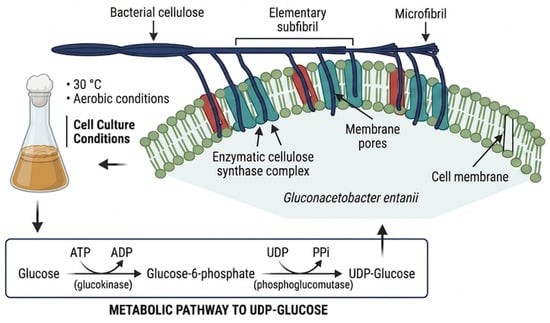
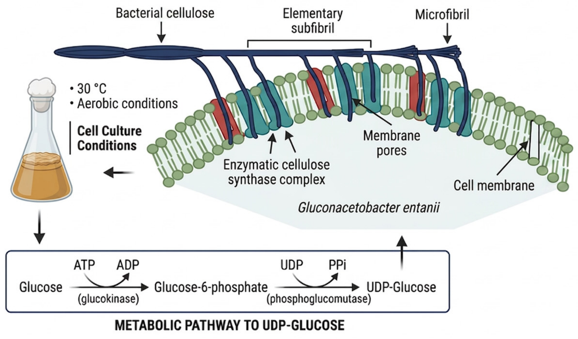
Based on the BC production kinetics previously reported by our research group [1], a 14-day cultivation period was selected for the present study. Under static culture conditions using piloncillo as the carbon source, BC production reached 1.50 ± 0.21 g/L after 14 days of incubation. The cellulose formed a compact membrane at the air–liquid interface, consistent with the characteristic morphology of BC produced under static cultivation [4]. Following cultivation, the membranes were subjected to thermo-alkaline treatment with 0.5 M NaOH to remove bacterial cells and residual culture components. After repeated washing to reach neutral pH and drying, the resulting BC membranes were translucent and free of visible cellular residues [5]. Figure 1 presents a schematic representation of the BC production process using G. entanii, including the main steps involved in its biosynthesis and recovery.
Figure 1.
Schematic diagram of bacterial cellulose production.
3.2. Phenolic and Flavonoid Compounds and Antioxidant Capacity Evaluation (DPPH, ABTS, and FRAP) in Pecan Nutshell Extract (PNSE)
The colorimetric analysis showed a high concentration of total phenolic compounds in PNSE (742.91 ± 4.04 mg GAE/g) and total flavonoids (82.42 ± 0.67 mg CE/g). Consistent with the high phenolic content, PNSE exhibited outstanding in vitro antioxidant capacity through different reaction mechanisms. In the DPPH radical assay, PNSE exhibited a value of 475.13 ± 2.84 µmol TE/g. In comparison, the ABTS assay yielded 15.55 ± 1.51 µmol TE/g, while FRAP revealed 22.49 ± 0.30 µmol TE/g.
3.3. Characterization of PNSE, BC and Bacterial Cellulose Films Functionalized with Pecan Nutshell Extract Using ATR-FTIR Spectroscopy
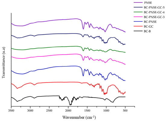
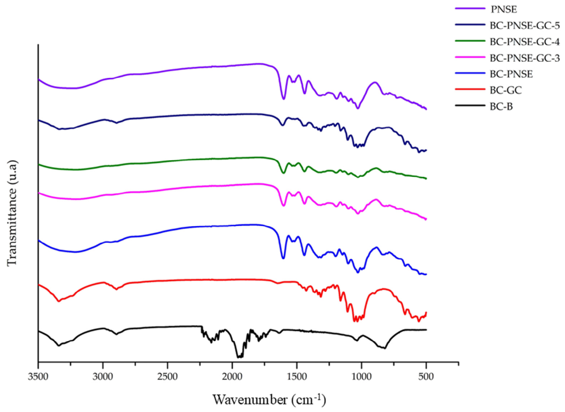
ATR-FTIR spectroscopic analysis confirmed the chemical integration of the components into the polymer matrix (Figure 2) [4]. The BC control film (BC-B) exhibited the characteristic spectrum of pure cellulose, with a broad, high-intensity band at 3300 cm−1, corresponding to the stretching of hydroxyl groups (–OH), indicating a dense network of hydrogen bonds. The peak at 2920 cm−1 was assigned to C–H stretching, while the bands between 1000 and 1150 cm−1 and the sharp signal at 895 cm−1 were associated with the glucosidic backbone and the deformation of the β-1,4-glucosidic bond, respectively [5]. The C–H stretching around ~2900 cm−1 and prominent C–O and C–O–C vibrations in the 1200–1000 cm−1 region were associated with the polysaccharide backbone. Also, the extract (PNSE) showed signals absent in the control, mainly at 1600 cm−1 (vibrations of the C=C bond in aromatic rings) and 1710 cm−1 (C=O carbonyl stretching). In addition, when evaluating the plasticized film (BC-GC-1), an intensification and slight shift in the O–H band to 3300 cm−1 were observed. In the bacterial cellulose films functionalized with pecan nutshell extract (BC-PNSE-2 and BC-PNSE-GC-3-5), bands at 1600 cm−1 and 1710 cm−1 were observed. In the 1000–1100 cm−1 region, there was a decrease in the peak at 895 cm−1.
Figure 2.
ATR-FTIR spectroscopic analysis of bacterial cellulose (BC-B), pecan nutshell extract (PNSE), bacterial cellulose with glycerol (BC-GC-1), bacterial cellulose with pecan nutshell extract (BC-PNSE-2), and bacterial cellulose with pecan nutshell extract and glycerol with extract concentrations of 150, 80, and 30 mg/mL (BC-PNSE-GC-3, BC-PNSE-GC-4, and BC-PNSE-GC-5).
3.4. Morphological Analysis by Scanning Electron Microscopy (SEM)
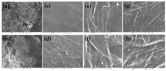
Figure 3 shows SEM images of bacterial cellulose (BC-B), pecan nutshell extract (PNSE), bacterial cellulose with glycerol (BC-GC-1), bacterial cellulose with pecan nutshell extract (BC-PNSE-2), and bacterial cellulose with pecan nutshell extract and glycerol with extract concentrations of 150, 80, and 30 mg/mL (BC-PNSE-GC-3, BC-PNSE-GC-4, and BC-PNSE-GC-5) at magnifications of 200X and 1000X. The control film (BC-B) showed a highly homogeneous, compact surface characterized by a continuous, well-defined three-dimensional fibrillar network of randomly interconnected nanofibers, with no evidence of disruptions, cracks, or structural impurities (Figure 3c,d). PNSE showed granular structures of various sizes, associated with its amorphous nature (Figure 3a,b).
Figure 3.
SEM images of (a) pecan nutshell extract (PNSE) at 200X magnification, (b) PNSE at 1000X, (c) bacterial cellulose (BC-B) at 200X, (d) BC-B at 1000X, (e) bacterial cellulose with glycerol (BC-GC-1) at 200X, (f) BC-GC-1 at 1000X, (g) bacterial cellulose with pecan nutshell extract (BC-PNSE-2) at 200X, and (h) BC-PNSE-2 at 1000X.
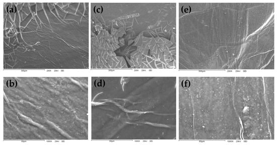
The plasticized film (BC-GC-1) exhibited a surface that was slightly denser and less porous compared with the control BC-B (Figure 3e,f). In the film with extract but without plasticizer (BC-PNSE-2) (Figure 3g,h), a rougher, more heterogeneous surface texture was observed with embedded phenolic particles. The formulations BC-PNSE-GC-3 (Figure 4a,b) and BC-PNSE-GC-4 (Figure 4c,d) exhibited a markedly rough morphology, with clear signs of fibrillar network disintegration and dense zones. Conversely, formulation BC-PNSE-GC-5 (Figure 4e,f) showed a more uniform microstructure characterized by a less interrupted fibrillar network (Figure 4a–f).
Figure 4.
SEM images of bacterial cellulose with pecan nutshell extract and glycerol with extract concentrations of 150, 80, and 30 mg/mL: (a) BC-PNSE-GC-3 at 200X, (b) BC-PNSE-GC-3 at 1000X, (c) BC-PNSE-GC-4 at 200X, (d) BC-PNSE-GC-4 at 1000X, (e) BC-PNSE-GC-5 at 200X, (f) BC-PNSE-GC-5 at 1000X.
3.5. Thermal Stability (TGA/DTG and DSC)
3.5.1. Analysis of TGA/DTG
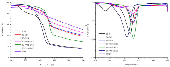
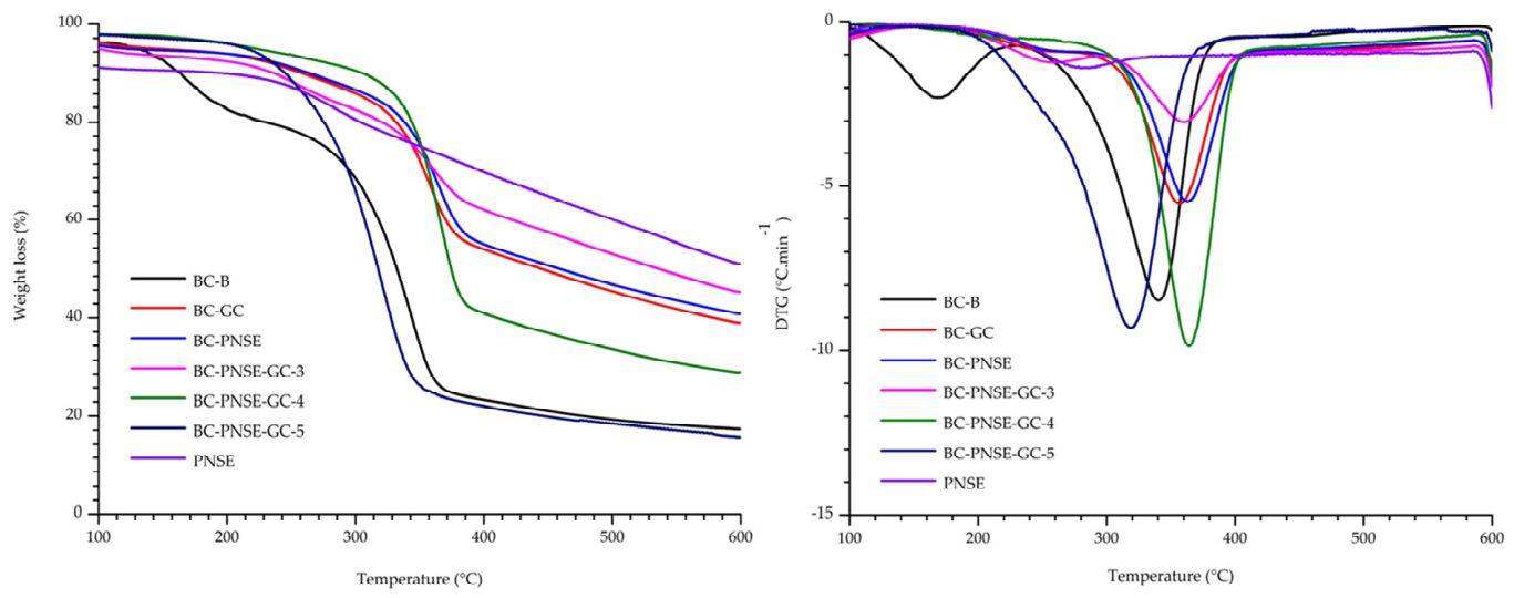
The thermal properties of BC films and BC films incorporated with glycerol and PNSE were evaluated using thermogravimetric analysis (TGA), its derivative (DTG), and differential scanning calorimetry (DSC) to understand mass-loss behavior and phase transitions during the heating ramp (Figure 5). Figure 5 of TGA and DTG curves revealed changes in the thermal stability of the composite materials depending on their formulation. The control film (BC-B) showed the lowest thermal stability at 357 °C, with a maximum degradation peak (DTG) at 342 °C. BC-GC-1 with glycerol shifted the maximum degradation temperature toward 380 °C. The thermal stability of the functional BC-PNSE-GC formulations varied proportionally with extract concentration. The BC-PNSE-GC-4 formulation exhibited the highest thermal stability recorded in this study, delaying maximum decomposition to 386 °C.
Figure 5.
Thermogravimetric analysis (TGA) of bacterial cellulose (BC-B), pecan nutshell extract (PNSE), bacterial cellulose with glycerol (BC-GC-1), bacterial cellulose with pecan nutshell extract (BC-PNSE-2), and bacterial cellulose with pecan nutshell extract and glycerol at extract concentrations of 150, 80, and 30 mg/mL (BC-PNSE-GC-3, BC-PNSE-GC-4, and BC-PNSE-GC-5).
3.5.2. Analysis of DSC
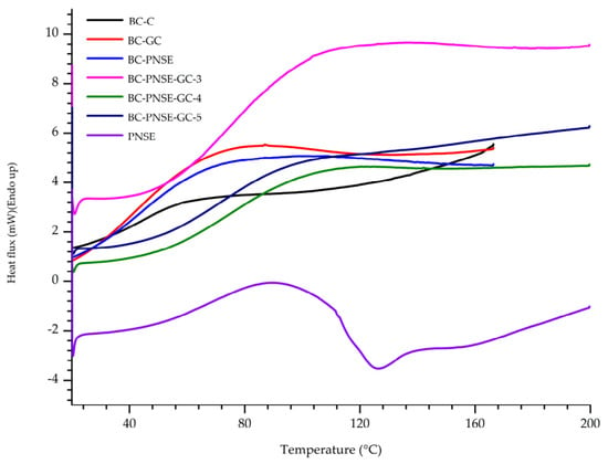
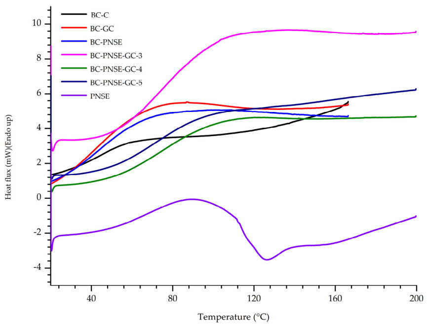
The DSC thermograms supported the TGA findings and showed heat flow transitions associated with each formulation (Figure 6). The BC-B control film showed an endothermic transition between 50 and 100 °C (Figure 6). The incorporation of glycerol in BC-GC-1 significantly broadened the transition to the range of 50 to 120 °C. Meanwhile, the functional composite films BC-PNSE-GC-4 and BC-PNSE-GC-5 showed smooth thermal transitions, with an integrated peak between 90 and 150 °C.
Figure 6.
DSC thermograms of bacterial cellulose (BC-B), pecan nutshell extract (PNSE), bacterial cellulose with glycerol (BC-GC-1), bacterial cellulose with pecan nutshell extract (BC-PNSE-2), and bacterial cellulose with pecan nutshell extract and glycerol with extract concentrations of 150, 80, and 30 mg/mL (BC-PNSE-GC-3, BC-PNSE-GC-4, and BC-PNSE-GC-5).
3.6. Antibacterial Efficacy of the Extract and Composite Films
3.6.1. Minimum Inhibitory Concentration (MIC) and Minimum Bactericidal Concentration (MBC) of PNSE
The free pecan nutshell extract (PNSE) showed inhibitory and bactericidal activity against the evaluated bacterial strains, with MIC and MBC values of 37.5 and 75 mg/mL, respectively (Table 2). These findings support the antibacterial potential of PNSE as a value-added extract derived from pecan nutshell under the conditions evaluated.
Table 2.
Minimum inhibitory concentration (MIC) and minimum bactericidal concentration (MBC) of PNSE against pathogenic bacteria.
3.6.2. Disk Diffusion Assay of the Composite Films
The inhibition zone diameters for the composite films indicated that the final antibacterial activity was strongly influenced by the polymeric matrix formulation and the cell wall structure of the microorganism evaluated (Table 3).
Table 3.
Inhibition zone diameters (mm) of bacterial cellulose (BC) films against target pathogens.
4. Discussion
4.1. Synthesis and Purification of Bacterial Cellulose (BC)
The BC yield obtained in the present study (1.50 ± 0.21 g/L) was higher than the yields previously reported for winery industry residues used as alternative carbon sources (0.3–0.6 g/L) and was within or above the range reported for Komagataeibacter xylinus cultivated in modified Hestrin–Schramm (HS) medium (0.6–1.2 g/L) [1,2]. These differences may be related, at least in part, to the composition and availability of fermentable carbohydrates in the culture medium.
The relatively high BC yield with piloncillo may reflect its carbohydrate composition, particularly the presence of readily available sugars. Free glucose can enter central metabolic pathways, leading to UDP-glucose formation, while sucrose may be hydrolyzed to glucose and fructose before further metabolism. UDP-glucose serves as the immediate glucose donor for cellulose biosynthesis, providing the substrate for the polymerization of β-1,4-glucan chains by the cellulose synthase complex of Gluconacetobacter entanii [3]. Thus, the availability of fermentable sugars in piloncillo may have contributed to the BC production observed under the conditions evaluated.
The use of bacterial cellulose obtained from low-cost and renewable substrates is of increasing interest in food and biotechnology because of its biodegradability, structural properties, and versatility. In this context, the use of piloncillo as a carbon source provides an alternative approach for BC production while supporting the development of value-added biomaterials. Depending on their functionalization and physicochemical properties, BC-based materials may be further explored for applications such as food coatings and bioactive films.
4.2. Phenolic and Flavonoid Compounds and Antioxidant Capacity Evaluation (DPPH, ABTS, and FRAP) in Pecan Nutshell Extract (PNSE)
The reported values significantly exceed previous reports of by-products from the same fruit. For example, earlier studies reported concentrations of 214.3 mg GAE/g in PNSE [15], while other studies indicated an approximate phenolic content of 500 mg GAE/g through conventional maceration extraction [20]. The marked superiority in phytochemical recovery observed in this study is directly attributed to the operational variables of the extraction process, including the type of solvent with intermediate polarity (60% v/v ethanol) and, especially, physical assistance via ultrasound, which promotes acoustic cavitation and facilitates the disintegration of the highly lignified cell walls of the shell tissue. Additionally, the variations observed compared with values reported by other authors are associated with the fruit’s specific varietal origin, as well as genotypic factors and the agroclimatic conditions of the cultivation region, which influence the biosynthesis and accumulation of phenolic metabolites in the plant [27,28].
The antioxidant capacity values obtained, compared with the literature, show that extracts from walnut seed or kernel without shell have been reported to show DPPH activity below 200 µmol TE/g and FRAP values ranging only between 8 and 14 µmol TE/g [5,29]. This empirically demonstrates that the external pericarp fraction (the shell) concentrates a substantially higher density of bioactive molecules capable of donating electrons or hydrogen atoms than the edible portion of the fruit [30]. The high measured antioxidant activity is closely linked to specific polyphenols such as catechins and phenolic acids (gallic and ellagic acids), whose aromatic rings contain multiple free hydroxyl groups that can neutralize reactive oxygen species through resonance. Furthermore, the quantitative disparity observed among the three assays (DPPH, ABTS, and FRAP) is explained by the chemical nature of the polyphenols present, which operate through different antioxidant mechanisms ranging from simple electron transfer (SET) to hydrogen atom transfer (HAT), in media with varying hydrophilic or lipophilic affinities [31]. This behavior underscores the need to use multiple simultaneous spectroscopic methods to obtain a comprehensive assessment of a natural extract’s antioxidant capacity [27].
4.3. Characterization of PNSE, BC and Bacterial Cellulose Films Functionalized with Pecan Nutshell Extract Using ATR-FTIR Spectroscopy
The BC control film (BC-B) presents the characteristic spectrum of pure cellulose, as reported by other authors [1,32,33,34,35]. These peaks reflect adequate synthesis and purification of bacterial cellulose. For PNSE, the peaks are characteristic bands of phenolic acids (such as gallic acid, ellagic acid, catechins, and proanthocyanidins) and abundant condensed tannins found in pecan nutshell [6,7]. Also, the extract (PNSE) showed signals absent in the control, mainly at 1600 cm−1 (vibrations of the C=C bond in aromatic rings) and 1710 cm−1 (C=O carbonyl stretching). These bands increased as the concentration of PNSE was raised. This could be attributed to effective bonding between the BC matrix and the extract (PNSE). In BC-GC-1, the peaks observed indicate the physical insertion of glycerol between the cellulose microfibrils and the formation of new intermolecular links through hydrogen bonds [8].
In BC-PNSE-GC-2, BC-PNSE-GC-3, and BC-PNSE-GC-4, the incorporation of the extract was confirmed by the appearance and proportional increase in aromatic and phenolic bands at 1600 cm−1 and 1710 cm−1, respectively. Additionally, the plasticized and functionalized formulations showed broadening in the 1000–1100 cm−1 region, indicating simultaneous interactions between the polyol, the active extract, and the cellulose fibers [9], as well as a decrease in the intensity of the peak at 895 cm−1. These changes reflect conformational reorganizations and a reduction in the original crystalline order of bacterial cellulose, promoted by the physical spacing induced by phenolic molecules and glycerol as they synergistically insert into the biopolymer [3,36]. These spectral features were retained or became more evident in the BC-PNSE-GC-3 films, particularly in the formulations containing higher PNSE levels, supporting the incorporation of extract-derived compounds into the cellulose matrix. In addition, modifications in the O–H stretching region suggest changes in the hydrogen-bonding environment resulting from interactions among cellulose hydroxyl groups, phenolic compounds from PNSE, and glycerol. Changes in the 1200–1000 cm−1 region may arise from overlapping C–O and C–O–C vibrations of cellulose with C–O vibrations contributed by glycerol and phenolic constituents. Overall, the FTIR spectra indicate that PNSE and glycerol were successfully incorporated into the bacterial cellulose matrix while preserving the main structural features of cellulose, with the observed spectral changes suggesting intermolecular interactions, particularly through hydrogen bonding.
4.4. Morphological Analysis by Scanning Electron Microscopy (SEM)
SEM analysis of the films’ microscopic morphology allowed the evaluation of how incorporating the plasticizer and the active extract affected the organization of the bacterial cellulose (BC) polymer network.
This microstructure indicates controlled biotechnological synthesis and efficient hydrogel drying. In contrast, the free extract (PNSE) showed a markedly rough and irregular surface, with dispersed granular structures of various sizes, associated with its amorphous nature and precipitation of phenolic compounds during dehydration [36].
The role of BC-GC-1 in this structural rearrangement is physically relevant and is attributed to glycerol acting as a plasticizer, positioning itself between the cellulose chains, reducing void spaces in the network, and diminishing the system’s apparent porosity [2,37]. In BC-PNSE-2, the bioactive compound was incorporated non-uniformly into the cellulose matrix in the absence of a plasticizer [3]. When evaluating the films simultaneously enriched with extract and glycerol, notable morphological variations were observed depending on the formulation studied.
BC-PNSE-GC-3 and BC-PNSE-GC-4 present a characteristic structure. This behavior suggests a supersaturation of the polymer system due to the extract concentration, leading to visible agglomerates within the matrix. However, BC-PNSE-GC-5 showed a smoother and less rough structure but still displayed isolated areas with bumps and surface accumulations, associated with local zones of conformational interaction between the phenolic extract and glycerol [6].
4.5. Thermal Stability (TGA/DTG and DSC)
4.5.1. Analysis of TGA/DTG
For BC-B, the main thermal degradation event occurred at approximately 357 °C, as indicated by the maximum mass-loss rate in the DTG curve. After this temperature, the sample continued to undergo gradual mass loss, with a measurable residual mass remaining at 600 °C, and a maximum degradation peak (DTG) at 342 °C due to pyrolysis of the glycosidic groups of highly crystalline cellulose [1,2]. Contrary to the conventional theory that plasticizers reduce thermal stability by decreasing intermolecular cohesion, the addition of glycerol in BC-GC-1 shifted the maximum degradation temperature toward 380 °C, a favorable behavior associated with the formation of a dense network of new ordered hydrogen bonds between the hydroxyl groups of glycerol and the microfibrils of the BC, which requires higher thermal energy for molecular dissociation [3,38,39]. This confirms that controlled dosing of phenolic compounds at 30 and 80 mg/mL, synergistically coupled with glycerol and cellulose, reinforces the compound’s molecular stability through complex chemical interactions [4,5]. Conversely, the formulation with the highest active-extract concentration, 150 mg/mL (BC-PNSE-GC-3), showed a marked decrease in thermal stability, with a degradation temperature of 354 °C and a DTG peak that dropped sharply to 317 °C. This loss of stability is due to the high density of free, thermally unstable phytochemical compounds suspended in the matrix, which undergo premature thermal degradation, starting with the decomposition of the pure extract, with an early mass-loss peak observed at 52 °C [6].
4.5.2. Analysis of DSC
The control film (BC-B) showed an initial endothermic transition between 50 and 100 °C, with a jump around 70 °C, corresponding to dehydration and residual moisture loss trapped in the polymer network, and maintained a stable heat flow above 150 °C due to the polymer’s structural purity [2,40]. Analysis of BC-GC-1 confirmed possible structural changes in the film, indicating structural compatibility and strong hydrogen bond-mediated interactions between cellulose, the plasticizer, and the polyphenols from the pecan extract [11], which is consistent with findings showing that at lower phenolic extract concentrations, the thermal stability of the matrix-additive system is maintained or even improved [40,41,42,43,44,45]. BC-PNSE-GC-3 exhibited a markedly broader and more intense thermal event compared with the unmodified BC, whereas BC-PNSE-GC-4 and BC-PNSE-GC-5 displayed differences in the position and intensity of the transition region. These changes suggest that PNSE concentration influences the molecular organization and hydrogen-bonding network within the films. The thermal behavior of the isolated PNSE was substantially different from that of the composite films, showing a pronounced exothermic event at approximately 125–130 °C. The attenuation or absence of this event after incorporation into the BC matrix suggests that the polymeric network modifies the thermal environment and mobility of PNSE constituents. Overall, the DSC results, together with the FTIR findings, support the occurrence of intermolecular interactions among bacterial cellulose, PNSE-derived compounds, and glycerol, while the main structural features of the cellulose matrix were retained.
4.5.3. Disk Diffusion Assay of the Composite Films
The control films (BC-B and BC-GC-1) exhibited no inhibition zones (0 mm), confirming the lack of intrinsic antibacterial activity of pure bacterial cellulose and glycerol. Meanwhile, the extract control (PNSE) produced zones of 11 ± 1.4 mm against S. aureus and 8 mm against S. typhimurium, with no effect against E. coli [3]. The film functionalized with extract but without plasticizer (BC-PNSE-2) exhibited zones of 12.5 ± 3.5 mm against S. aureus and 7 ± 2.82 mm against S. typhimurium [46,47].
Incorporating glycerol into the optimized formulation (BC-PNSE-GC-4) increased the mean inhibition zone against S. aureus [48]. ANOVA showed significant differences among the treatments against S. aureus (F(4,5) =17.65; p = 0.0037). The positive control Ab GM10 exhibited the highest inhibition zone (25.50 ± 1.40 mm) and was significantly different from PNSE, BC-PNSE-2, and BC-PNSE-GC-3 (p < 0.05). BC-PNSE-GC-4 showed an intermediate value (18.00 ± 1.40 mm), with no significant differences compared to the other treatments. In contrast, the remaining active treatments were statistically homogeneous under the evaluated conditions. This enhanced physical performance of BC-PNSE-GC-4 is attributed to a physicochemical effect of the plasticizer within the polymer matrix: adding glycerol decreases the molecular rigidity of bacterial cellulose by disrupting intermolecular hydrogen bonds between glucan chains [4,48]. This phenomenon increases the free space (free volume) and molecular mobility within the polymeric network, acting as a physical mass transfer promoter that facilitates the faster and more efficient diffusion and release of the active polyphenolic molecules of the extract into the bacterial medium.
Conversely, the active films showed low or no inhibition against Gram-negative bacteria (E. coli and S. typhimurium). For S. typhimurium, statistical analysis indicated that the positive control (Ab GM10) exhibited significantly higher inhibition, whereas the remaining active treatments were considered statistically homogeneous (equal to each other) due to high internal variability and the low number of replicates (n = 2). This overall selectivity is directly associated with the selective permeability barrier represented by the outer lipopolysaccharide membrane of Gram-negative bacteria, whose hydrophilic structure significantly limits the diffusion and penetration of the hydrophobic polyphenolic compounds and condensed tannins of the pecan nutshell extract into the intracellular space [3,49]. Finally, the BC-PNSE-GC-5 treatment (30 mg/mL) produced no detectable inhibition zone (0 mm) (Figure S1 in Supplementary Materials) because the low concentration of bioactive compounds was insufficient to reach the minimum inhibitory threshold. On the other hand, the formulation with the highest extract concentration, BC-PNSE-GC-3 (150 mg/mL), exhibited an inhibition zone of 13.5 ± 2.1 mm against S. aureus. While bioactive, its antibacterial performance was lower than that of the optimal 80 mg/mL formulation (BC-PNSE-GC-4, 18 mm). Consistent with the SEM observations, this reduction in efficacy is attributed to the fact that the high extract concentration (150 mg/mL) induced supersaturation in the system, contributing to structural collapse and agglomeration [7]. These morphological changes physically obstructed the internal diffusion pathways, restricting the optimal release of the phenolic compounds into the surrounding medium and limiting their resulting antibacterial activity [8,18].
5. Conclusions
The bacterial cellulose films functionalized with pecan nutshell extract and glycerol exhibited formulation-dependent physicochemical, structural, thermal, and antibacterial properties. The incorporation of pecan nutshell extract imparted antibacterial activity to the films, with BC-PNSE-GC-4 producing the largest inhibition zone against Staphylococcus aureus (18 mm). Overall, the findings highlight the potential of pecan nutshell extract as a bioactive ingredient for the functionalization of bacterial cellulose films and demonstrate a promising approach for valorizing this agro-industrial by-product. Further studies under food-related conditions are warranted to establish the barrier, release, safety, and food-preservation performance of these films and to determine their suitability for active food packaging applications.
Supplementary Materials
The following supporting information can be downloaded at: https://www.mdpi.com/article/10.3390/coatings16101236/s1. Figure S1: Plate inhibition zone assay showing the antimicrobial activity of bacterial cellulose films functionalized with pecan nutshell extract and glycerol (BC-PNSE-GC-4).
Author Contributions
Writing—original draft preparation, methodology, E.G.R.-L.; formal analysis, supervision, project administration, funding acquisition, M.L.Á.-A.; writing—review and editing, visualization, L.A.-V. and I.S.-S.; resources, data curation, conceptualization, D.D.C.-E. and S.E.B.-I.; investigation, software, S.C.M.-R. and J.E.G.-R.; data curation, writing—review and editing, R.D.I.-G.; investigation, editing, visualization, L.R.B.-M. and R.F.D.-M.; formal analysis, investigation, supervision. All authors have read and agreed to the published version of the manuscript.
Funding
This research received no external funding.
Institutional Review Board Statement
Not applicable.
Informed Consent Statement
Not applicable.
Data Availability Statement
The data used during the study are available from the corresponding author on reasonable request.
Acknowledgments
The authors thank the University of Sonora and SECIHTI for their support. Emely Guadalupe Rivera-López thanks SECIHTI for the scholarship granted. We also thank DIPM and DCQB of the University of Sonora for technical support.
Conflicts of Interest
All authors declare that there is no conflict of interest associated with this research.
Abbreviations
The following abbreviations are used in this manuscript:
| BC | Bacterial cellulose |
| PNSE | Pecan nutshell extract |
| GC | Glycerol |
| DPPH | 2,2-diphenyl-1-picrylhydrazyl |
| ABTS | 2,2′-azinobis(3-ethylbenzothiazoline-6-sulfonic acid) (ABTS) |
| FRAP | Ferric reducing antioxidant power |
| MIC | Minimum inhibitory concentration |
| MBC | Minimum bactericidal concentration |
References
- Dórame-Miranda, R.; Gámez-Meza, N.; Medina-Juárez, L.; Ezquerra-Brauer, J.; Ovando-Martínez, M.; Lizardi-Mendoza, J. Bacterial cellulose production by Gluconacetobacter entanii using pecan nutshell as carbon source and its chemical functionalization. Carbohydr. Polym. 2019, 207, 91–99. [Google Scholar] [CrossRef] [Scilit] [PubMed]
- Potočnik, V.; Gorgieva, S.; Trček, J. From Nature to Lab: Sustainable Bacterial Cellulose Production and Modification with Synthetic Biology. Polymers 2023, 15, 3466. [Google Scholar] [CrossRef] [Scilit] [PubMed]
- Revin, V.V.; Liyaskina, E.V.; Parchaykina, M.V.; Kuzmenko, T.P.; Kurgaeva, I.V.; Revin, V.D.; Ullah, M.W. Bacterial Cellulose-Based Polymer Nanocomposites: A Review. Polymers 2022, 14, 4670. [Google Scholar] [CrossRef] [Scilit] [PubMed]
- Campano, C.; Schmuck, B.; Manoli, M.-T.; Hernández-Arriaga, A.M.; Rising, A.; Prieto, M.A. Bioengineering toughened, stretchable bacterial cellulose-based films inspired by spider silk. Carbohydr. Polym. 2026, 388, 125543. [Google Scholar] [CrossRef] [Scilit] [PubMed]
- Yan, Y.; Chen, G.; Zhang, Y.; Dai, W.; Deng, S.; Afreen, S.; Wang, L.; Zhang, H. In Situ Synthesis of Plasticized Bacterial Cellulose Films for Daily Packaging Using Biobased Plasticizers. ACS Appl. Mater. Interfaces 2025, 17, 27227–27237. [Google Scholar] [CrossRef] [Scilit] [PubMed]
- Sun, Y.; Meng, C.; Zheng, Y.; Xie, Y.; He, W.; Wang, Y.; Qiao, K.; Yue, L. The effects of two biocompatible plasticizers on the performance of dry bacterial cellulose membrane: A comparative study. Cellulose 2018, 25, 5893–5908. [Google Scholar] [CrossRef] [Scilit]
- Perin, G.B.; Felisberti, M.I. A Simple Mathematical Model for the Structural Description of Polyesters Based on Glycerol. Macromolecules 2025, 58, 8581–8590. [Google Scholar] [CrossRef] [Scilit] [PubMed]
- Nishida, Y.; Aono, R.; Dohi, H.; Ding, W.; Uzawa, H. 1H-NMR Karplus Analysis of Molecular Conformations of Glycerol under Different Solvent Conditions: A Consistent Rotational Isomerism in the Backbone Governed by Glycerol/Water Interactions. Int. J. Mol. Sci. 2023, 24, 2766. [Google Scholar] [CrossRef] [Scilit] [PubMed]
- Cazón, P.; Mateus, A.R.; Silva, A.S. Advances in active packaging using natural biopolymers and fruit by-products for enhanced food preservation. Food Res. Int. 2025, 213, 116439. [Google Scholar] [CrossRef] [Scilit] [PubMed]
- Castillo, I.O.; Sangerman-Jarquín, D.M.; Vázquez, M.G.C.; de Jesús Espinoza Arellano, J.; Moreno, J.H.N. Producción y comercialización de nuez pecanera en México. Rev. Mex. Cienc. Agríc. 2019, 10, 1797–1808. [Google Scholar] [CrossRef] [Scilit]
- Wang, X.; Wu, Y.; Lombardini, L. In vitro viability and germination of Carya illinoinensis pollen under different storage conditions. Sci. Hortic. 2021, 275, 109662. [Google Scholar] [CrossRef] [Scilit]
- Alvarez-Parrilla, E.; Urrea-López, R.; de la Rosa, L.A. Bioactive components and health effects of pecan nuts and their by-products: A review. J. Food Bioact. 2018, 1, 56–92. [Google Scholar] [CrossRef] [Scilit]
- Balasundram, N.; Sundram, K.; Samman, S. Phenolic compounds in plants and agri-industrial by-products: Antioxidant activity, occurrence, and potential uses. Food Chem. 2006, 99, 191–203. [Google Scholar] [CrossRef] [Scilit]
- Hilbig, J.; Alves, V.R.; Müller, C.M.O.; Micke, G.A.; Vitali, L.; Pedrosa, R.C.; Block, J.M. Ultrasonic-assisted extraction combined with sample preparation and analysis using LC-ESI-MS/MS allowed the identification of 24 new phenolic compounds in pecan nut shell [Carya illinoinensis (Wangenh) C. Koch] extracts. Food Res. Int. 2018, 106, 549–557. [Google Scholar] [CrossRef] [Scilit] [PubMed]
- do Prado, A.C.P.; da Silva, H.S.; da Silveira, S.M.; Barreto, P.L.M.; Vieira, C.R.W.; Maraschin, M.; Ferreira, S.R.S.; Block, J.M. Effect of the extraction process on the phenolic compounds profile and the antioxidant and antimicrobial activity of extracts of pecan nut [Carya illinoinensis (Wangenh) C. Koch] shell. Ind. Crop. Prod. 2014, 52, 552–561. [Google Scholar] [CrossRef] [Scilit]
- Jimenez, M.E.C.; Gabilondo, J.; Bodoira, R.M.; Laverde, L.M.A.; Santagapita, P.R. Extraction of bioactive compounds from pecan nutshell: An added-value and low-cost alternative for an industrial waste. Food Chem. 2024, 453, 139596. [Google Scholar] [CrossRef] [Scilit] [PubMed]
- Pedron, G.; Jaouhari, Y.; Bordiga, M. Conventional and Innovative Drying/Roasting Technologies: Effect on Bioactive and Sensorial Profiles in Nuts and Nut-Based Products. Appl. Sci. 2025, 15, 1258. [Google Scholar] [CrossRef] [Scilit]
- Kharel, K.; Kraśniewska, K.; Gniewosz, M.; Prinyawiwatkul, W.; Fontenot, K.; Adhikari, A. Antimicrobial screening of pecan shell extract and efficacy of pecan shell extract-pullulan coating against Listeria monocytogenes, Salmonella enterica, and Staphylococcus aureus on blueberries. Heliyon 2024, 10, e29610. [Google Scholar] [CrossRef] [Scilit] [PubMed]
- Salari, M.; Khiabani, M.S.; Mokarram, R.R.; Ghanbarzadeh, B.; Kafil, H.S. Development and evaluation of chitosan based active nanocomposite films containing bacterial cellulose nanocrystals and silver nanoparticles. Food Hydrocoll. 2018, 84, 414–423. [Google Scholar] [CrossRef] [Scilit]
- Dórame-Miranda, R.F.; Gámez-Meza, N.; Ovando-Martínez, M.; Medina-Juárez, L.A.; Cárdenas-López, J.L.; Ramírez-Bon, R.; Santos-Sauceda, I.; Castro-Enríquez, D.D.; Burruel-Ibarra, S.E. Encapsulation of Sardine Oil by Electrospraying with Gliadins and Pecan Nutshell Extracts for its Stabilization. Food Bioprocess Technol. 2021, 14, 457–470. [Google Scholar] [CrossRef] [Scilit]
- Singleton, V.L.; Rossi, J.A. Colorimetry of Total Phenolics with Phosphomolybdic-Phosphotungstic Acid Reagents. Am. J. Enol. Vitic. 1965, 16, 144–158. [Google Scholar] [CrossRef] [Scilit]
- Chacón-Figueroa, I.H.; Dórame-Miranda, R.F.; López-Ahumada, G.A.; Del-Toro-Sánchez, C.L.; Ovando-Martínez, M.; Gámez-Meza, N.; Martínez-Bustos, F.; Rodríguez-Figueroa, J.C.; Gerardo-Rodríguez, J.E.; Whitney, K.; et al. Microencapsulation of Spent Coffee Extract Within Saccharomyces cerevisiae Cells via Spray Drying and Evaluation of Its In Vitro Bioaccessibility. Foods 2025, 14, 1053. [Google Scholar] [CrossRef] [Scilit] [PubMed]
- Zhishen, J.; Mengcheng, T.; Jianming, W. The determination of flavonoid contents in mulberry and their scavenging effects on superoxide radicals. Food Chem. 1999, 64, 555–559. [Google Scholar] [CrossRef] [Scilit]
- Palafox-Carlos, H.; Yahia, E.; González-Aguilar, G. Identification and quantification of major phenolic compounds from mango (Mangifera indica, cv. Ataulfo) fruit by HPLC–DAD–MS/MS-ESI and their individual contribution to the antioxidant activity during ripening. Food Chem. 2012, 135, 105–111. [Google Scholar] [CrossRef] [Scilit]
- Re, R.; Pellegrini, N.; Proteggente, A.; Pannala, A.; Yang, M.; Rice-Evans, C. Antioxidant activity applying an improved ABTS radical cation decolorization assay. Free Radic. Biol. Med. 1999, 26, 1231–1237. [Google Scholar] [CrossRef] [Scilit] [PubMed]
- Kowalska-Krochmal, B.; Dudek-Wicher, R. The Minimum Inhibitory Concentration of Antibiotics: Methods, Interpretation, Clinical Relevance. Pathogens 2021, 10, 165. [Google Scholar] [CrossRef] [Scilit] [PubMed]
- de la Rosa, L.A.; Alvarez-Parrilla, E.; Shahidi, F. Phenolic Compounds and Antioxidant Activity of Kernels and Shells of Mexican Pecan (Carya illinoinensis). J. Agric. Food Chem. 2010, 59, 152–162. [Google Scholar] [CrossRef] [Scilit] [PubMed]
- Jahanban-Esfahlan, A.; Ostadrahimi, A.; Tabibiazar, M.; Amarowicz, R. A Comparative Review on the Extraction, Antioxidant Content and Antioxidant Potential of Different Parts of Walnut (Juglans regia L.) Fruit and Tree. Molecules 2019, 24, 2133. [Google Scholar] [CrossRef] [Scilit] [PubMed]
- Rosales-Martínez, P.; Arellano-Cárdenas, S.; Dorantes-Álvarez, L.; García-Ochoa, F.; López-Cortez, M.d.S. Comparison Between Antioxidant Activities of Phenolic Extracts from Mexican Peanuts, Peanuts Skins, Nuts and Pistachios. J. Mex. Chem. Soc. 2017, 58, 185–193. [Google Scholar] [CrossRef] [Scilit]
- Cason, C.; Yemmireddy, V.K.; Moreira, J.; Adhikari, A. Antioxidant Properties of Pecan Shell Bioactive Components of Different Cultivars and Extraction Methods. Foods 2021, 10, 713. [Google Scholar] [CrossRef] [Scilit] [PubMed]
- Prior, R.L.; Wu, X.; Schaich, K. Standardized Methods for the Determination of Antioxidant Capacity and Phenolics in Foods and Dietary Supplements. J. Agric. Food Chem. 2005, 53, 4290–4302. [Google Scholar] [CrossRef] [Scilit] [PubMed]
- Fuller, M.E.; Andaya, C.; McClay, K. Evaluation of ATR-FTIR for analysis of bacterial cellulose impurities. J. Microbiol. Methods 2018, 144, 145–151. [Google Scholar] [CrossRef] [Scilit] [PubMed]
- Menzel, C. Improvement of starch films for food packaging through a three-principle approach: Antioxidants, cross-linking and reinforcement. Carbohydr. Polym. 2020, 250, 116828. [Google Scholar] [CrossRef] [Scilit] [PubMed]
- Villasante, J.; Martin-Lujano, A.; Almajano, M.P. Characterization and Application of Gelatin Films with Pecan Walnut and Shell Extract (Carya illinoiensis). Polymers 2020, 12, 1424. [Google Scholar] [CrossRef] [Scilit] [PubMed]
- Shen, Y.; Seidi, F.; Ahmad, M.; Liu, Y.; Saeb, M.R.; Akbari, A.; Xiao, H. Recent Advances in Functional Cellulose-based Films with Antimicrobial and Antioxidant Properties for Food Packaging. J. Agric. Food Chem. 2023, 71, 16469–16487. [Google Scholar] [CrossRef] [Scilit] [PubMed]
- Tajima, K.; Imai, T.; Yui, T.; Yao, M.; Saxena, I. Cellulose-synthesizing machinery in bacteria. Cellulose 2021, 29, 2755–2777. [Google Scholar] [CrossRef] [Scilit]
- Zhou, R.; Guo, C.; Li, Q.; Li, Z.; Fan, W.; Chen, X.; Dai, J.; Zhang, Q. Development of Bacterial Cellulose-Based Films Incorporated with Epigallocatechin-3-Gallate for Active Food Packaging. Foods 2026, 15, 785. [Google Scholar] [CrossRef] [Scilit] [PubMed]
- Mohamad, N.; Amin, M.C.I.M.; Pandey, M.; Ahmad, N.; Rajab, N.F. Bacterial cellulose/acrylic acid hydrogel synthesized via electron beam irradiation: Accelerated burn wound healing in an animal model. Carbohydr. Polym. 2014, 114, 312–320. [Google Scholar] [CrossRef] [Scilit] [PubMed]
- Turganova, R.; Tuleyeva, R.; Belkozhayev, A.; Gizatullina, N.; Yelemessova, G.; Taubatyrova, A.; Mussalimova, M.; Shynykul, Z.; Toleutay, G. Bacterial Cellulose for Sustainable Food Packaging: Production Pathways, Structural Design, and Functional Modification Strategies. Polymers 2025, 17, 3165. [Google Scholar] [CrossRef] [Scilit] [PubMed]
- Agustin, S.; Cahyanto, M.N.; Wahyuni, E.T.; Supriyadi. Effect of glycerol plasticizer on the structure and characteristics of bacterial cellulose-based biocomposite films. In IOP Conference Series: Earth and Environmental Science; IOP Publishing: Bristol, UK, 2024. [Google Scholar] [CrossRef] [Scilit]
- Azeredo, H.M.C.; Barud, H.; Farinas, C.S.; Vasconcellos, V.M.; Claro, A.M. Bacterial Cellulose as a Raw Material for Food and Food Packaging Applications. Front. Sustain. Food Syst. 2019, 3, 7. [Google Scholar] [CrossRef] [Scilit]
- Cheng, X.; Fan, M.; Gao, Y.; Xiong, Z.; Li, L. Bacterial cellulose: Modification and application in food packaging. Carbohydr. Polym. 2025, 370, 124450. [Google Scholar] [CrossRef] [Scilit] [PubMed]
- Silviana, S.; Sa’aDah, A.N. Mechanical and Thermal Properties of Bacterial Cellulose Reinforced with Bamboo Microfibrillated Cellulose and Plasticized with Epoxidized Waste Cooking Oil. Cellul. Chem. Technol. 2022, 56, 331–339. [Google Scholar] [CrossRef] [Scilit]
- Khotsaeng, N.; Simchuer, W.; Imsombut, T.; Srihanam, P. Effect of Glycerol Concentrations on the Characteristics of Cellulose Films from Cattail (Typha angustifolia L.) Flowers. Polymers 2023, 15, 4535. [Google Scholar] [CrossRef] [Scilit] [PubMed]
- Yemmireddy, V.K.; Cason, C.; Moreira, J.; Adhikari, A. Effect of pecan variety and the method of extraction on the antimicrobial activity of pecan shell extracts against different foodborne pathogens and their efficacy on food matrices. Food Control 2020, 112, 107098. [Google Scholar] [CrossRef] [Scilit]
- Arciello, A.; Panzella, L.; Dell’oLmo, E.; Abdalrazeq, M.; Moccia, F.; Gaglione, R.; Agustin-Salazar, S.; Napolitano, A.; Mariniello, L.; Giosafatto, C.V.L. Development and characterization of antimicrobial and antioxidant whey protein-based films functionalized with Pecan (Carya illinoinensis) nut shell extract. Food Packag. Shelf Life 2021, 29, 100710. [Google Scholar] [CrossRef] [Scilit]
- Kılınç, M. PHYSICOCHEMICAL PROPERTIES AND ANTIOXIDANT ACTIVITY OF BACTERIAL CELLULOSE BASED ON OLEA EUROPAEA L. LEAF EXTRACTS. Mühendislik Bilim. Tasar. Derg. 2026, 14, 76–88. [Google Scholar] [CrossRef] [Scilit]
- Benitez, J.J.; Florido-Moreno, P.; Porras-Vázquez, J.M.; Tedeschi, G.; Athanassiou, A.; Heredia-Guerrero, J.A.; Guzman-Puyol, S. Transparent, plasticized cellulose-glycerol bioplastics for food packaging applications. Int. J. Biol. Macromol. 2024, 273, 132956. [Google Scholar] [CrossRef] [Scilit] [PubMed]
- Flores-Estrada, R.A.; Gámez-Meza, N.; Medina-Juárez, L.A.; Castillón-Campaña, L.G.; Molina-Domínguez, C.C.; Rascón-Valenzuela, L.A.; García-Galaz, A. Chemical Composition, Antioxidant, Antimicrobial and Antiproliferative Activities of Wastes from Pecan Nut [Carya illinoinensis (Wagenh) K. Koch]. Waste Biomass Valor. 2020, 11, 3419–3432. [Google Scholar] [CrossRef] [Scilit]

Disclaimer/Publisher’s Note: The statements, opinions and data contained in all publications are solely those of the individual author(s) and contributor(s) and not of MDPI and/or the editor(s). MDPI and/or the editor(s) disclaim responsibility for any injury to people or property resulting from any ideas, methods, instructions or products referred to in the content. |
© 2026 by the authors. Licensee MDPI, Basel, Switzerland. This article is an open access article distributed under the terms and conditions of the Creative Commons Attribution (CC BY) license.