


Highlights
- Template molding created nanofiber-clustered structures on P(VDF-TrFE) films.
- Poled films exhibited enhanced piezoelectric response and surface potential.
- Poled films promoted BMSC adhesion, proliferation, and ALP activity.
- Topographical and electrical changes were linked to cell–material interactions.
Abstract
Surface morphology and electrical characteristics are key factors affecting cell–material interactions on electroactive polymer films, but their coupled effects remain unclear. In this study, P(VDF-TrFE) nanofiber-clustered films were fabricated using an anodic aluminum oxide (AAO) template-assisted molding method, and their surface electrical properties were subsequently tailored by corona poling. The nanofibrous structure was formed without substantially disrupting the characteristic polar β phase. Compared with the planar film, the water contact angle (WCA) of the nanofiber-clustered film increased from 74.8° to 132.5°, indicating markedly enhanced hydrophobicity. Furthermore, corona poling increased the magnitude of the piezoelectric coefficient (|d33|) and altered the surface potential state. Protein adsorption and cell studies further showed that polarized nanofiber-clustered surfaces exhibited greater protein adsorption, more extensive cell spreading, and higher CCK-8 signals and alkaline phosphatase (ALP) activity. These results indicate that the biological interfacial response of P(VDF-TrFE) films is regulated by surface morphology, wettability, and polarization-induced electrical characteristics. This work provides insight into the combined roles of nanofiber-clustered topography and electrical characteristics in regulating cell-material interactions on P(VDF-TrFE) surfaces.
1. Introduction
The cell–material interface plays a central role in regulating cellular behavior [1,2]. At this interface, cells respond to multiple physicochemical cues, including surface micro-/nanotopography and electrical characteristics [3,4]. Micro-/nanotopography can modify interfacial physicochemical properties, protein adsorption, and cell adhesion, thereby influencing cellular responses [5,6,7]. Electrical cues, including surface charge, surface potential, and piezoelectricity, may likewise modulate interfacial interactions and cell behavior [8,9]. Accordingly, material platforms that allow the coordinated regulation of surface topography and electrical properties provide a useful approach for investigating cell–material interactions [10,11].
Electroactive polymers are attractive for this purpose because of their flexibility and processability [12,13]. Among them, poly(vinylidene fluoride-trifluoroethylene) [P(VDF-TrFE)] is a representative ferroelectric copolymer with good film-forming capability, chemical stability, and piezoelectric and ferroelectric properties [14,15,16]. Its crystalline organization and electroactive behavior can be regulated through processing conditions and thermal treatment, while electrical poling promotes dipole alignment and can further modify its piezoelectric response and surface potential [13,17]. Compared with rigid piezoelectric ceramics, P(VDF-TrFE) films exhibit greater mechanical compliance and interfacial adaptability, making them suitable for investigating the relationships among film structure, electrical properties, and cellular behavior. These characteristics have motivated extensive efforts to regulate the morphology and electrical performance of P(VDF-TrFE)-based materials. Previous studies have explored various P(VDF-TrFE) micro-/nanostructures, including micropatterns, nanorods, nanofibers, and porous architectures [18,19,20,21]. These structures can affect crystalline organization, localized mechanical properties, surface wettability, and the resulting electromechanical response [22,23]. In particular, the effects of P(VDF-TrFE) fibrous structures on cell behavior have received increasing attention. Azimi et al. reported that electrospun P(VDF-TrFE) fibers with different structural arrangements influenced the osteogenic responses of human mesenchymal stromal cells [24]. Ribeiro et al. constructed P(VDF-TrFE)-based scaffolds and found that more regularly organized fibrous structures guided osteoblast-like cells to align and spread along the fibers [25]. In addition, Tai et al. showed that fiber diameter affected cell behavior [26]. Meanwhile, the polarization states and surface electrical properties of P(VDF-TrFE) can also influence stem-cell behavior. Zhang et al. prepared P(VDF-TrFE) films with different surface potentials by corona poling and found that these electrical states could regulate BMSC osteogenic or lineage commitment [27]. However, the interfacial physicochemical changes induced by the formation of nanofiber-clustered surfaces and subsequent poling, together with their relationship to the cellular responses, remain to be further investigated.
In this study, nanofiber-clustered P(VDF-TrFE) films were prepared by anodic aluminum oxide (AAO) template-assisted molding. The confined nanopores of AAO facilitate polymer infiltration and nanoscale shape replication, providing a relatively simple route to construct nanofibrous surface architectures. Corona poling was applied to modify the polarization-related electrical properties of the films. Planar films, unpoled nanofiber-clustered films, and poled nanofiber-clustered films were compared to evaluate the changes in surface morphology, crystalline phase, wettability, piezoelectric response, surface potential, and protein adsorption associated with surface nanostructuring and electrical poling. The comparison between PF and PN-UP focused on the changes accompanying the formation of the nanofiber-clustered structure, whereas the comparison between PN-UP and PN-P evaluated the additional changes introduced by corona poling. Bone marrow mesenchymal stem cell (BMSC) adhesion, proliferation, cell morphology, cytoskeletal organization, and ALP activity were further evaluated to examine how these interfacial changes were associated with early cellular responses. Through these sequential comparisons, this study aimed to characterize the interfacial evolution of P(VDF-TrFE) films during surface nanostructuring and poling. The resulting interfacial changes were further examined in relation to early BMSC responses.
2. Materials and Methods
2.1. Materials
Porous anodic aluminum oxide (AAO) templates with a nominal pore diameter of approximately 200 nm were manufactured by Whatman (Marlborough, MA, USA). The P(VDF-TrFE) powder with a VDF:TrFE molar ratio of 70/30 was acquired from Piezotech (Paris, France). N,N-dimethylformamide (DMF), sodium hydroxide (NaOH) and absolute ethanol were analytical-grade reagents purchased from Sinopharm Chemical Reagent Co., Ltd. (Shanghai, China).
2.2. Preparation of P(VDF-TrFE) Films
P(VDF-TrFE) powder was dissolved in DMF to prepare a 15 wt% solution. The mixture was stirred for 4 h to yield a homogeneous solution. The resulting solution was cast onto a clean glass substrate and dried at room temperature for 2 h to remove residual solvent. The dried film was annealed at 210 °C for 1 h to form the P(VDF-TrFE) film.
2.3. Fabrication of P(VDF-TrFE) Nanofiber-Clustered Films
The AAO templates were ultrasonically cleaned in absolute ethanol for 5 min to remove surface contaminants, followed by drying at room temperature. P(VDF-TrFE) nanofiber-clustered films were fabricated by an AAO template-assisted melt infiltration method. The planar P(VDF-TrFE) film was placed onto the cleaned AAO template and heated on a hot plate at 160 °C for 5 min, allowing the molten polymer to infiltrate into the nanochannels of the AAO template. The assembly was then cooled naturally to room temperature. To promote uniform heating and reduce temperature fluctuations, the assembly was covered with aluminum foil during the heating, holding and cooling processes. The AAO template was subsequently removed by immersion in 2 mol/L NaOH solution at room temperature for 2 h. The P(VDF-TrFE) nanofiber-clustered film was thoroughly rinsed with deionized water until a neutral pH was achieved, followed by rinsing with ethanol and drying at room temperature.
2.4. Corona Poling
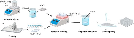
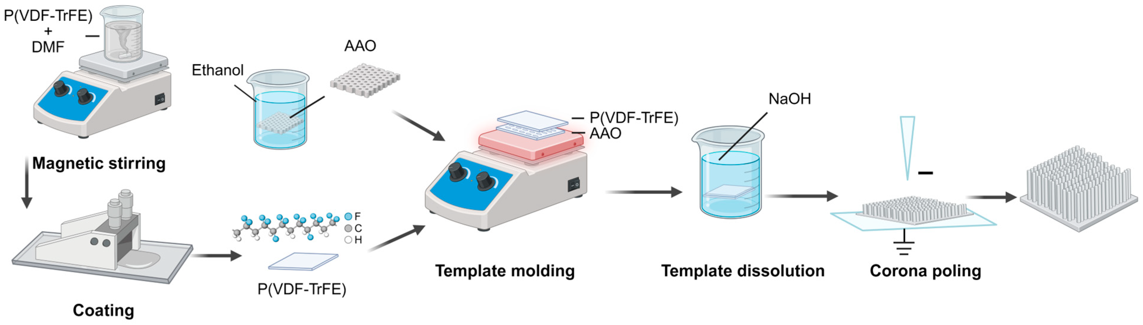
Corona poling was performed using a needle-to-plate configuration. The P(VDF-TrFE) nanofiber-clustered film was placed on a grounded metal electrode with the nanofiber-clustered side facing the corona needle. A negative DC high voltage was applied to the needle electrode, and the needle-to-sample distance was maintained at 1 cm. For clarity, the planar P(VDF-TrFE) film, unpoled P(VDF-TrFE) nanofiber-clustered film, and corona-poled P(VDF-TrFE) nanofiber-clustered film were denoted as PF, PN-UP, and PN-P, respectively. In this study, the PN-P samples used for subsequent KPFM characterization and cellular experiments were poled at −8.2 kV for 40 min. Unless otherwise specified, sample preparation and characterization were conducted under ambient laboratory conditions at approximately 25 °C and 50% relative humidity. The fabrication and corona poling processes are schematically illustrated in Figure 1.
Figure 1.
Schematic illustration of the fabrication and corona poling processes of P(VDF-TrFE) nanofiber-clustered films. Planar P(VDF-TrFE) films were first prepared by coating a P(VDF-TrFE)/DMF solution onto a glass substrate. Nanofiber-clustered surfaces were then constructed by AAO template-assisted molding. After dissolution and removal of the AAO template using NaOH, the resulting nanofiber-clustered films were subjected to corona poling. In the corona-poling schematic, the upper needle electrode is connected to a negative high-voltage source (−), while the lower electrode is grounded.
2.5. Characterization of P(VDF-TrFE) Nanofiber-Clustered Films
The surface and cross-section morphologies of the films were observed using a scanning electron microscope (GeminiSEM 300, Zeiss, Jena, Germany) at an accelerating voltage of 3 kV. Before observation, the samples were gold sputter-coated to enhance surface conductivity and minimize charging during SEM imaging. The diameter and height distributions of the nanofibers were statistically analyzed from SEM images using ImageJ (version 1.53k, National Institutes of Health, Bethesda, MD, USA). The crystal phase structure was examined using an X-ray diffractometer (X-Pert Powder, PANalytical, Almelo, The Netherlands) with Cu Kα radiation (λ = 1.5406 Å) at a scanning rate of 2°/min over a 2θ range of 10–80°. Fourier transform infrared (FTIR) spectra were collected using an attenuated total reflectance FTIR spectrometer (Nicolet 5700, Thermo Fisher Scientific, Madison, WI, USA). The relative β-phase fraction in P(VDF-TrFE) was calculated from the characteristic absorption bands of the α and β phases according to the following equation [28]:
where F(β) is the relative β-phase fraction; Aα and Aβ are the absorbance values at 766 and 840 cm−1; and Kα and Kβ are the corresponding absorption coefficients, with values of 6.1 × 10⁴ and 7.7 × 104 cm2 mol−1, as reported by Gregorio and Cestari [28]. Static water contact angles were determined at room temperature using an optical contact angle meter (OCA 20, DataPhysics Instruments GmbH, Filderstadt, Germany). Deionized water droplets with a volume of 1 μL were used for the measurements. The piezoelectric performance of the films was evaluated by measuring the quasi-static piezoelectric coefficient d33 with a d33 m (ZJ-3A, Institute of Acoustics, Chinese Academy of Sciences, Beijing, China). Three independent samples (n = 3) were tested, with five measurements per sample. The surface potential distribution of the samples was characterized by Kelvin probe force microscopy (NTEGRA Spectra, NT-MDT, Russia) with an SCM-PIT platinum-iridium-coated silicon probe.
Protein adsorption was evaluated using bovine serum albumin (BSA; Aladdin, Shanghai, China). The samples were incubated with 500 μL of 10 mg/mL BSA solution in phosphate-buffered saline (PBS; Cellmax, Beijing, China) at 37 °C for 24 h. After incubation, the samples were rinsed with PBS for 10 s and then immersed in 300 μL of 1 mg/mL sodium dodecyl sulfate (SDS; Aladdin, Shanghai, China) solution at room temperature for 8 h to desorb the adsorbed protein. The protein content in the solution was measured using a BCA Protein Assay Kit (Thermo Fisher Scientific, Waltham, MA, USA), with three independent samples tested for each group (n = 3).
2.6. Cell Culture and Biological Evaluation
All animal experiments were approved by the Laboratory Animal Welfare and Ethics Committee of Zhejiang University (Ethics Code: ZJU20220279). Bone marrow mesenchymal stem cells (BMSCs) were isolated from the bone marrow of 3-week-old male Sprague-Dawley rats (Hangzhou Medical College) and cultured in low-glucose Dulbecco’s modified Eagle’s medium (DMEM; Cellmax, Beijing, China). The culture medium was supplemented with 10% fetal bovine serum (FBS; Cellmax, SA211.02, Beijing, China) and 1% penicillin-streptomycin solution (Gibco, Grand Island, NY, USA), corresponding to final concentrations of 100 U/mL penicillin and 100 μg/mL streptomycin. The cells were cultured at 37 °C under humidified conditions with 5% CO2. Before cell seeding, the P(VDF-TrFE) film samples were sterilized by ultraviolet irradiation. The cells were then detached with 0.25% trypsin and seeded on the samples in 24-well plates at a density of 5 × 10⁴ cells per sample.
After 1 day of culture, BMSC cytoskeletal organization and morphology on the different samples were examined. For fluorescence staining, the cells were fixed with 4% paraformaldehyde (PFA; Solarbio, Beijing, China) and permeabilized with 0.4% Triton X-100 (Sigma-Aldrich, St. Louis, MO, USA). The samples were then blocked with PBS containing 2% BSA and 2% bovine serum for 1 h. F-actin was stained with phalloidin (AAT Bioquest, Pleasanton, CA, USA), and the cell nuclei were counterstained with DAPI (Beyotime, Shanghai, China). The stained samples were observed using a confocal laser scanning microscope. For SEM observation, the cell samples were fixed with 4% PFA and dehydrated through a graded ethanol series. Cell adhesion and spreading morphology on the different film surfaces were then observed using SEM.
BMSCs adhesion and proliferation on different samples were evaluated using a Cell Counting Kit-8 assay (CCK-8; Dojindo Laboratories, Kumamoto, Japan) after 1 and 3 days of culture, respectively. For the assay, the samples were transferred to new 24-well plates to avoid interference from cells attached to the original well bottoms. Subsequently, 500 μL of low-glucose DMEM and 50 μL of CCK-8 reagent were added to each well, followed by incubation at 37 °C for 2 h in the dark. After incubation, the reaction solution was transferred to a 96-well plate, and the optical density (OD) was measured at 450 nm with a microplate reader (Multiskan MK3, Thermo Fisher Scientific, Shanghai, China). Three independent biological replicates were performed for the CCK-8 assay (n = 3).
The early osteogenesis-related activity of BMSCs was evaluated based on alkaline phosphatase (ALP) activity. From day 3 onward, the cells were cultured in osteogenic induction medium containing 1 μM dexamethasone (Sigma, St. Louis, MO, USA), 12 mM β-glycerophosphate (Sigma, St. Louis, MO, USA) and 50 μM ascorbic acid (Sigma, St. Louis, MO, USA). After 7 days of culture on the samples, the culture medium was removed, and the cells were washed two to three times with phosphate-buffered saline (PBS; Cellmax, Beijing, China). Then, 60 μL of cell lysis buffer (Boster, Wuhan, China) was added to each sample, followed by lysis on ice for 30 min. The lysates were collected into centrifuge tubes and centrifuged at 12,000 rpm for 15 min at 4 °C. The supernatants were collected, and the total protein content and ALP activity were determined using a BCA Protein Assay Kit (Thermo Fisher Scientific, Waltham, MA, USA) and a LabAssay ALP kit (Wako Pure Chemical Industries, Osaka, Japan), respectively. ALP activity was normalized to the total protein content to compare the early osteogenesis-related activity of BMSCs cultured on different samples. Five independent biological replicates were performed for the ALP assay (n = 5).
2.7. Statistical Analysis
All quantitative data are presented as the mean ± standard deviation. Statistical analyses were performed using OriginPro 2026 (OriginLab Corporation, Northampton, MA, USA). Differences among multiple groups were analyzed using one-way analysis of variance followed by Tukey’s post hoc test. A value of p < 0.05 was considered statistically significant.
3. Results and Discussion
3.1. Fabrication and Morphology of P(VDF-TrFE) Nanofiber-Clustered Films
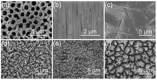
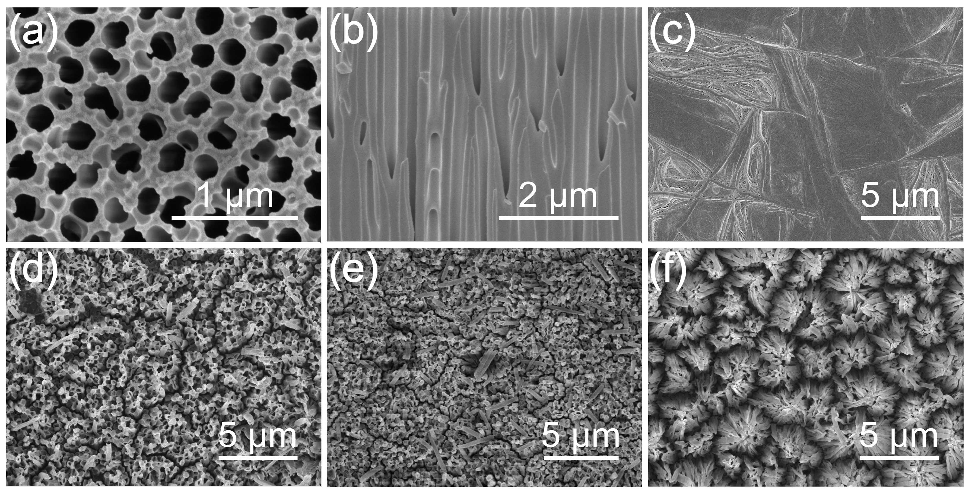
The P(VDF-TrFE) planar films and nanofiber-clustered films were prepared through solution casting and AAO template-assisted molding. The AAO template exhibited a nanoporous surface with an average pore diameter of approximately 200 nm, and vertically aligned channels in the cross-sectional view, providing a negative mold for the formation of surface nanostructures (Figure 2a,b). In the molding process, the AAO membrane allowed thermally softened P(VDF-TrFE) to infiltrate the nanopores. After removal of the AAO template, the replicated polymer structures remained on the film surface, forming nanofiber clusters. As shown in Figure 2c, the solution-cast P(VDF-TrFE) planar film, denoted as PF, exhibited a wrinkled surface morphology, which can be associated with crystallization-induced surface roughening in semicrystalline polymer films. Then the molding temperature was fixed at 160 °C, and the samples were heated for 5, 10, and 15 min, respectively, as shown in Figure 2d–f. After heating for 5 min, protruding nanofibers were formed on the film surface and assembled into relatively uniform clustered structures (Figure 2d). After heating for 10 min, the nanofibers exhibited increased bending and overlap, resulting in a less uniform clustered morphology (Figure 2e). When the heating time was further extended to 15 min, neighboring nanofibers tended to coalesce, forming more evident but still organized nanofibrous clusters (Figure 2f). These results indicate that heating time strongly affected nanofibrous morphology. Based on the surface morphology, the film prepared at 160 °C for 5 min was selected for subsequent characterization and biological evaluation because it showed a relatively uniform morphology. This clustered nanofibrous film was denoted as PN-UP before corona poling and PN-P after corona poling.
Figure 2.
Morphological characterization of the AAO template and P(VDF-TrFE) films. (a) Top-view and (b) cross-sectional SEM images of the AAO template. (c) Surface morphology of the solution-cast P(VDF-TrFE) planar film. Surface SEM images of P(VDF-TrFE) films treated at 160 °C for (d) 5 min, (e) 10 min, and (f) 15 min.
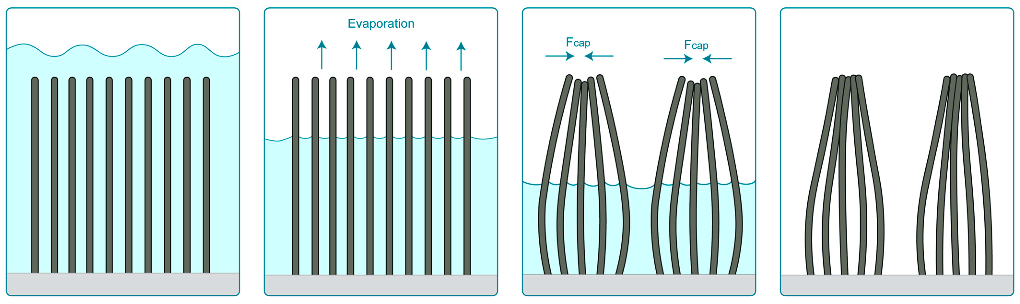
To further illustrate the possible origin of the clustered morphology, a schematic representation of the proposed formation process is presented in Figure 3. Prolonged heating may have promoted deeper polymer infiltration into the AAO nanopores, potentially resulting in replicated nanofibers with a higher aspect ratio. The formation of clustered nanofibrous structures may be associated with the high aspect ratio of the replicated fibers and the template removal process. After removal of the AAO template, the high-aspect-ratio fibers were no longer laterally constrained by the pore walls. During alkaline etching, washing, and drying, the capillary force and surface tension pull the high-aspect-ratio nanofibers to bend and come into contact with each other, thereby adhering together and forming a clustered structure [29,30].
Figure 3.
Schematic illustration of the proposed formation mechanism of the P(VDF-TrFE) nanofiber-clustered morphology. Fcap represents the capillary force. The light blue area represents the liquid, the dark grey structures represent the P(VDF-TrFE) nanofibers, and the light grey area at the bottom represents the underlying film.
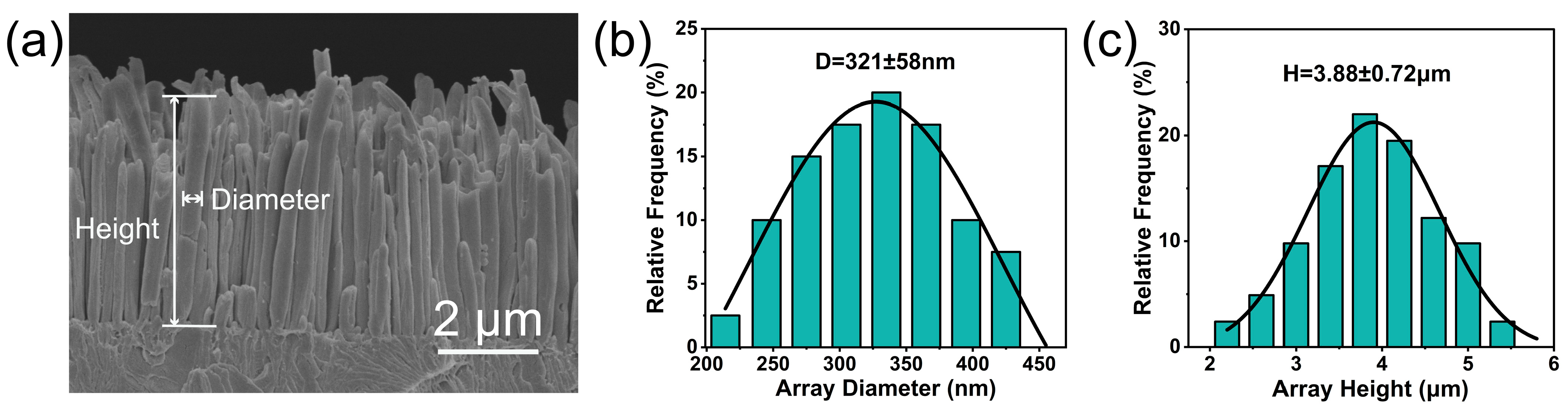
Cross-sectional SEM imaging was further performed to verify their protruding morphology and structural continuity with the underlying film. As shown in Figure 4a, a distinct nanofibrous surface layer extended from the P(VDF-TrFE) film and was directly connected to the base film, with no obvious interfacial delamination. This morphology is consistent with AAO-assisted molding, in which thermally softened P(VDF-TrFE) infiltrated the AAO nanopores and retained replicated high-aspect-ratio features after template removal. The dimensions of the nanofibers were analyzed from SEM images. As shown in Figure 4b, the average fiber diameter was 321 ± 58 nm. This diameter was larger than the pore size of the AAO template, suggesting that the replicated structures did not strictly preserve the original pore geometry. This mismatch may be related to dimensional relaxation or slight expansion after template removal, washing, and drying [31]. Similar deviations between template pore size and replicated polymer nanostructure dimensions have been reported in AAO-assisted polymer molding systems [32]. The average nanofiber height was 3.88 ± 0.72 μm, with an approximately normal distribution (Figure 4c), suggesting relatively uniform polymer infiltration under the selected condition. Slight height variations may result from local differences in polymer flow, pore filling, and template removal. Overall, the cross-sectional morphology and dimensional statistics demonstrate the successful construction of a clustered nanofibrous P(VDF-TrFE) surface with relatively consistent dimensions.
Figure 4.
(a) Cross-sectional SEM image of the P(VDF-TrFE) nanofiber-clustered film. Statistical distributions of the nanofiber (b) diameters and (c) heights. The solid lines represent Gaussian fits to the distributions.
3.2. Physicochemical and Electrical Characterization of P(VDF-TrFE) Films
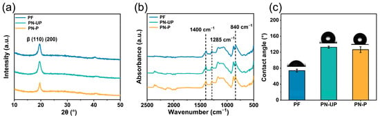
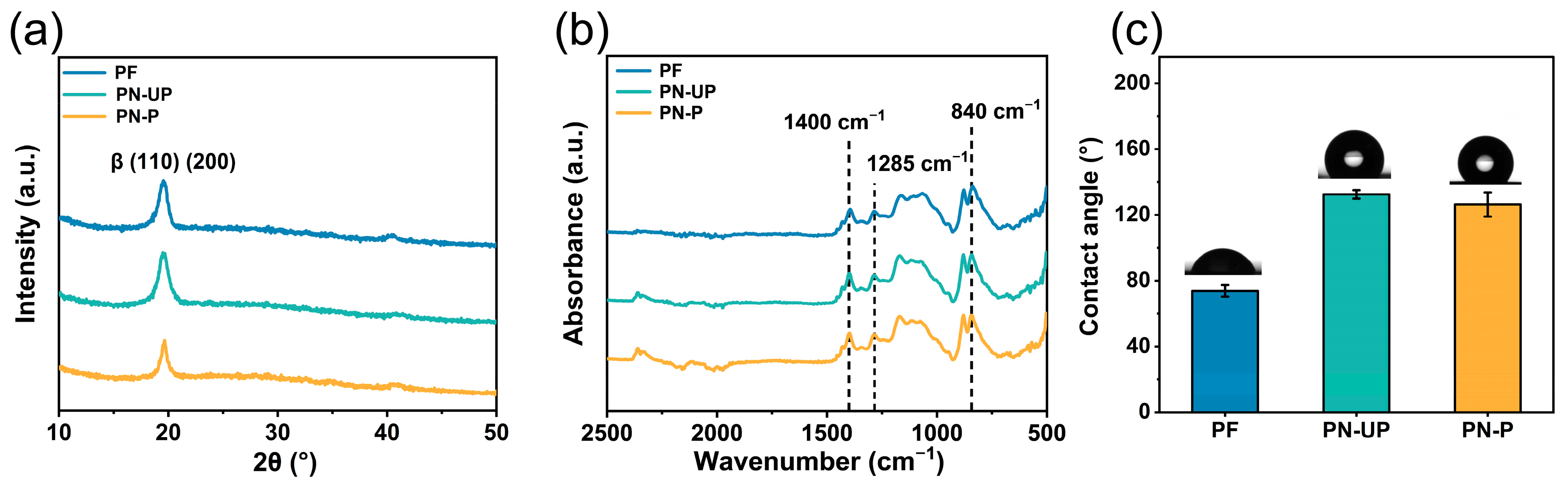
Figure 5a shows the XRD characterization results. PF, PN-UP, and PN-P all exhibited a prominent diffraction peak at 2θ ≈ 19.9°, corresponding to the overlapping (110)/(200) reflections of the polar β phase [33]. The distinct diffraction peak suggests a relatively ordered crystalline structure in the films. This polar crystalline phase is closely associated with the ferroelectric and piezoelectric properties of P(VDF-TrFE). The nearly unchanged peak positions indicate that the β-phase structure was preserved after AAO-assisted molding and corona poling. The FTIR spectra of PF, PN-UP, and PN-P are presented in Figure 5b. All three samples exhibited characteristic β-phase absorption bands at approximately 840, 1285, and 1400 cm−1. No obvious shifts in the major peak positions were observed after molding and poling, indicating the preservation of the electroactive β-phase structure [34,35,36]. Among the three spectra, the α-phase-related absorption band near 766 cm−1 gradually decreased from PF to PN-UP and PN-P, whereas the β-phase-related band near 840 cm−1 remained relatively stable. The 1285 and 1400 cm−1 bands showed higher intensities in PN-UP and PN-P than in PF. Consistent with these spectral changes, the relative β-phase fractions calculated using Equation (1) were approximately 75.49%, 82.44%, and 87.82% for PF, PN-UP, and PN-P, respectively. The gradual increase in the β-phase fraction suggests that the nanofiber-cluster fabrication process and corona poling may have promoted the formation or retention of β-phase chain conformations. This result is consistent with the XRD analysis.
Figure 5.
(a) XRD patterns, (b) FTIR spectra, and (c) water contact angles of the planar P(VDF-TrFE) film and nanofiber-clustered films before and after corona poling. The inset images in (c) show representative water droplets for contact-angle measurements.
As shown in Figure 5c, the WCA values of PF, PN-UP, and PN-P were 74.8°, 132.5°, and 126.3°, respectively. Compared with the planar PF surface, the WCA of PN-UP increased markedly, which may be related to its nanofiber-clustered surface morphology. According to the Wenzel model, surface roughness can amplify intrinsic wettability; however, the pronounced increase in WCA of PN-UP suggests that a Cassie–Baxter-like wetting state may be more relevant [37,38,39]. In this case, the rough surface structure may trap air within the gaps between nanofibers, thereby reducing the effective solid–liquid contact area and resulting in a higher apparent WCA. After corona poling, the slight decrease in WCA may be associated with changes in the surface electrical state of P(VDF-TrFE). Poling can promote dipole alignment and induce the accumulation and trapping of surface or near-surface charges, which may enhance the interaction between polar water molecules and the film surface and thereby increase wettability [40]. Minor surface chemical changes during corona treatment may also contribute to this decrease. Nevertheless, the PN-P surface remained highly hydrophobic.
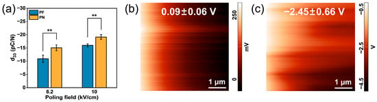
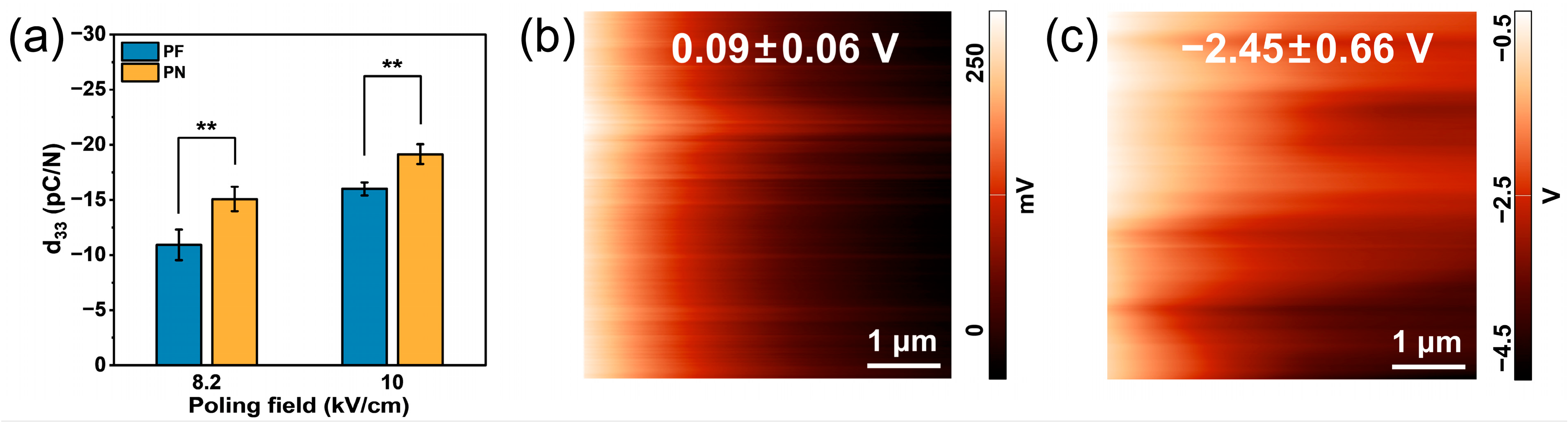
Figure 6a compares the quasi-static d33 coefficients of the planar (PF) and nanofiber-clustered (PN) films under different corona poling voltages. At poling voltages of −8.2 and −10 kV, the planar films showed d33 values of −11 and −16 pC/N, whereas the nanofiber-clustered films showed values of −15 and −19 pC/N, respectively. Previous studies have reported absolute d33 values reaching approximately 30–40 pC/N for conventionally processed and poled P(VDF-TrFE) films [41,42]. The d33 values measured in this study were within this reported range, although they remain moderate compared with some highly polarized films. The differences may be related to coating methods, film thickness, crystalline state, and poling conditions [43,44]. For both film types, the absolute d33 value increased with the magnitude of the poling voltage. At both tested poling voltages, the nanofiber-clustered films exhibited higher absolute d33 values than the planar films. This difference may be related to the altered local stress distribution and structural compliance of the nanofiber-clustered layer.
Figure 6.
(a) Piezoelectric coefficients of the films under different corona poling voltages (** p < 0.01). KPFM surface potential maps of (b) PF and (c) PN-UP.
The surface potentials of the P(VDF-TrFE) films were measured with KPFM (Figure 6b,c). The images showed nonuniform potential maps, suggesting spatial heterogeneity in the surface electrical state. Quantitative analysis showed that PF had an average surface potential of 0.09 ± 0.06 V, whereas PN-UP showed a substantially higher surface-potential magnitude, with an average value of −2.45 ± 0.66 V. This result indicates that nanofiber-clustered construction altered the surface electrical state of P(VDF-TrFE). The increased absolute surface potential may be related to nanofiber-induced surface curvature, localized charge trapping, and spatial variations in dipole distribution. After corona poling, the KPFM signal of PN-P reached the upper measurement limit under the selected conditions (approximately 12 V) and became saturated. Therefore, the surface potential of PN-P could not be reliably quantified, although the saturated KPFM response qualitatively indicated that the surface electrical state was altered after poling. This change may arise from electric-field-induced dipole orientation and surface charge deposition during corona poling. KPFM analysis confirmed that both nanofibrous construction and corona poling effectively modulated the surface potential of P(VDF-TrFE) films.
Taken together, the d33 and KPFM results demonstrate that the nanofibrous structure and corona poling treatment jointly altered the electrical characteristics of the P(VDF-TrFE) films, which may contribute to the differences in subsequent cell-material interactions.
3.3. Cellular Responses to P(VDF-TrFE) Films
To further evaluate the effects of the nanofiber-clustered structure and surface electrical properties on cell-material interactions, protein adsorption and early BMSC responses on the P(VDF-TrFE) films were investigated.
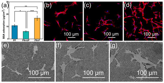
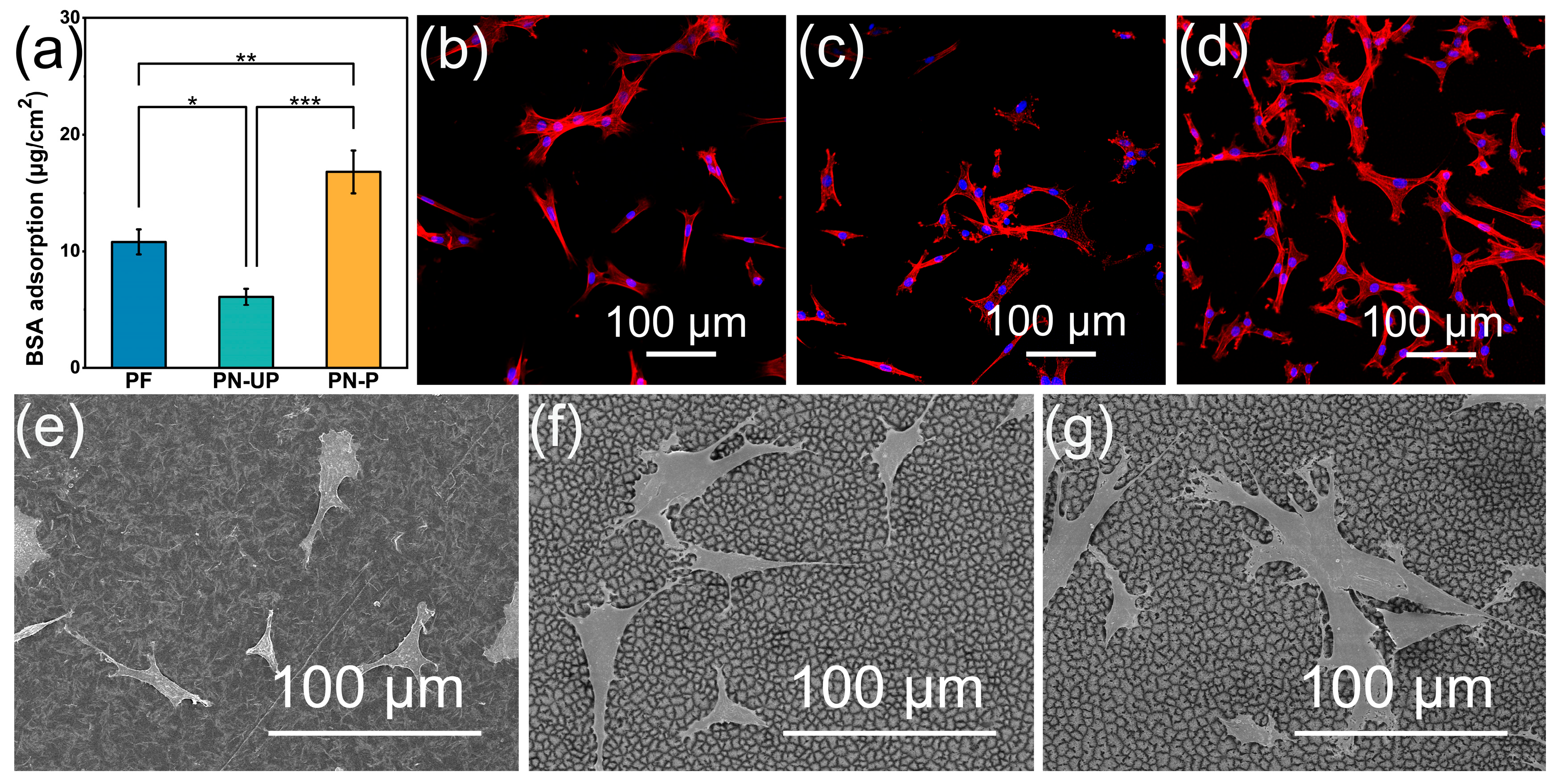
As shown in Figure 7a, the protein adsorption results showed that the PN-UP group, in which only the surface morphology was modified, exhibited reduced protein adsorption, whereas the adsorption capacity increased after polarization in the PN-P group. Protein adsorption on material surfaces is generally influenced by multiple factors, including surface morphology, wettability, and surface electrical properties [45]. Although the nanofiber-clustered structure increased the hydrophobicity of the material surface, polarization-induced changes in surface electrical properties may further regulate electrostatic and interfacial interactions between proteins and the material. Therefore, the relatively higher protein adsorption on the PN-P surface may provide a more favorable interfacial environment for subsequent cell adhesion.
Figure 7.
(a) Protein adsorption on different films after 24 h (* p < 0.05, ** p < 0.01, *** p < 0.001). (b–d) Fluorescence images of BMSCs on PF, PN-UP, and PN-P after 1 day of culture. F-actin and cell nuclei are shown in red and blue, respectively. (e–g) SEM images of BMSCs on PF, PN-UP, and PN-P after 1 day of culture.
Fluorescence images (Figure 7b–d) revealed differences in cell distribution and cytoskeletal organization among the three groups. Cells on the PN-P surface exhibited more evident spreading and a more developed F-actin cytoskeleton, suggesting that both the nanotopographical structure and polarization-induced electrical changes may contribute to cell–material interfacial interactions.
SEM images (Figure 7e–g) showed that cells on the PF surface exhibited a relatively contracted morphology, whereas cells on the PN-UP surface adhered and spread more extensively. Cells on the PN-P surface showed more pronounced spreading, with distinct filopodia-like extensions observed at the cell periphery. These observations suggest that the nanotopographical structure may contribute to improved early cell morphology, while polarization may further enhance cell spreading and cytoskeletal organization. This trend was generally consistent with the fluorescence results.
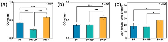
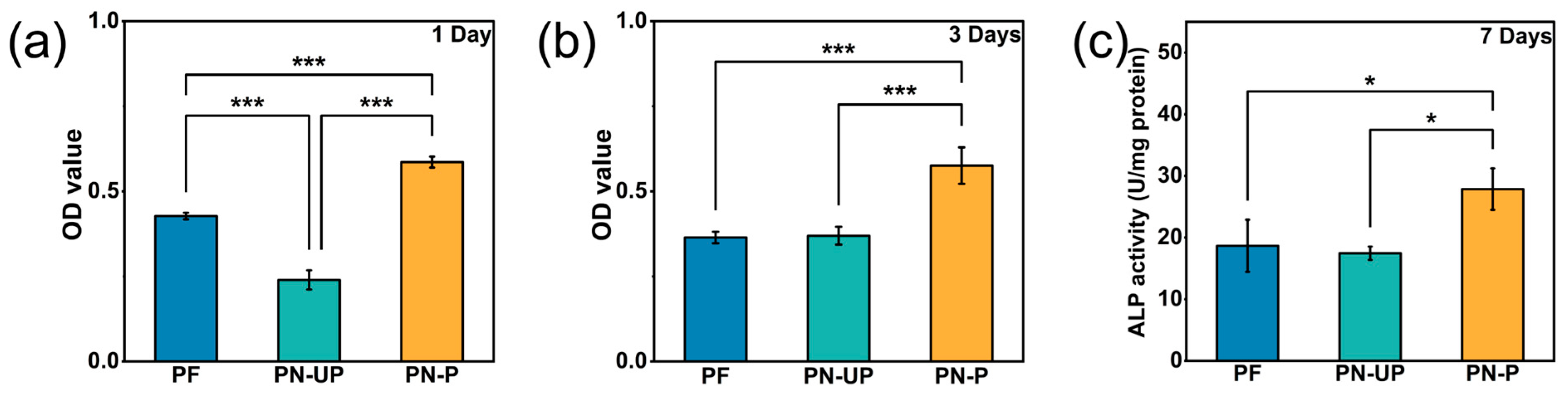
The CCK-8 results after 1 day of culture are shown in Figure 8a. Differences in OD values were observed among PF, PN-UP, and PN-P. Compared with PF, PN-UP showed a lower OD value, suggesting that the nanofiber-clustered surface alone did not improve early cellular metabolic activity or the number of viable attached cells. This result may be related to the markedly increased hydrophobicity caused by the nanofibrous structure [46]. In contrast, PN-P exhibited a higher OD value after 1 day, indicating that corona poling improved the early cellular response on the nanofiber-clustered film. This improvement may be associated with polarization-induced electrical properties, which could partly offset the unfavorable influence of increased hydrophobicity. After 3 days of culture, PN-P still showed the highest OD value among the three groups (Figure 8b), suggesting that the poled nanofibrous surface was more favorable for maintaining BMSC viability and early proliferation. However, the increase in OD values from day 1 to day 3 was limited for all groups, supporting that overall cell proliferation was not pronounced under the present conditions.
Figure 8.
(a) The adhesion (1 d) and (b) proliferation (3 d) behaviors of BMSCs on different films. (c) ALP activity of BMSCs on different films after 7 days of culture (* p < 0.05, *** p < 0.001).
ALP activity was used to evaluate the early osteogenesis-related activity of BMSCs (Figure 8c). Compared with PF, PN-UP did not markedly increase ALP activity. In contrast, PN-P showed higher ALP activity than PF and PN-UP, indicating that corona poling improved the early ALP-related activity of BMSCs. This may be associated with polarization-induced electrical properties, including altered surface potential and piezoelectric response.
Although statistically significant differences were observed among the groups, the absolute CCK-8 OD values and ALP activities remained relatively low under the present experimental conditions, suggesting that the overall magnitude of the cell responses was modest.
Collectively, the protein adsorption, cell morphology, CCK-8 and ALP results indicate that nanofiber clusters alone did not consistently improve BMSC responses, whereas corona-poled nanofiber-clustered films showed greater protein adsorption, more favorable cell spreading and cytoskeletal organization, higher cell viability and ALP activity. These results suggest that the cellular response to P(VDF-TrFE) films was not governed by surface topography alone, but by the combined effects of surface morphology, wettability, and polarization-induced electrical properties.
The nanofiber-clustered structure increased surface roughness and provided micro-/nanoscale topographical cues for cells, which may affect the contact mode between cells and the material surface, thereby influencing protein adsorption, adhesion-related structure formation, and cytoskeletal organization [6,47]. SEM observations showed that BMSCs on PN-UP were able to attach and spread, indicating that the nanotopography itself could provide morphological cues for cell spreading. However, the total protein adsorption on PN-UP was lower than that on PF, which may be related to the markedly increased hydrophobicity. Excessive hydrophobicity can affect the amount and interfacial behavior of the initial protein adsorption layer, thereby influencing subsequent cell-material interactions [48,49]. Notably, although individual cells on PN-UP showed spreading in SEM images, this was not accompanied by an increased CCK-8 signal. Therefore, the response on PN-UP may reflect a balance between favorable topographical cues and unfavorable wettability.
Corona poling appeared to shift this balance and was accompanied by increased absolute d33 values and the qualitative change in the KPFM response of PN-P. It should be noted that the cells in this study were cultured under static conditions. Since dynamic piezoelectric signals generally require mechanical deformation, the direct contribution of changes in d33 to the observed cellular responses may be limited under the present conditions. In contrast, poling-induced dipole orientation, surface charge state, and surface potential can persist without continuous mechanical stimulation, and may therefore be more relevant to the observed cellular responses under static culture conditions.
For P(VDF-TrFE), the polar β phase provides the structural basis for electroactivity, while corona poling may promote dipole alignment and surface charge deposition or trapping. In addition, the surface curvature and increased interfacial area of the nanofiber clusters may contribute to spatially heterogeneous charge distribution [44,50]. These electrical features may influence protein adsorption, cell membrane-related electrical interactions, and early osteogenesis-related signaling [27]. Consistent with this, PN-P showed higher protein adsorption than PN-UP, while fluorescence staining and SEM observations showed more extensive cell spreading and f-actin organization on PN-P. Thus, the higher CCK-8 signal and ALP activity on PN-P may be associated with the combined effects of nanofiber-clustered topography and polarization-induced electrical properties, rather than nanofibrous morphology alone [27].
This study shows that surface structuring combined with corona poling can regulate the morphology, wettability, piezoelectric response, and surface electrical state of P(VDF-TrFE) films without introducing inorganic fillers. However, several limitations remain. A poled planar control (PF-P) was not included in the present study. Therefore, the separate effects of surface morphology and corona poling could not be fully distinguished. ALP activity only reflects early osteogenesis-related activity; therefore, osteogenic gene expression, mineralization staining, and longer-term culture are required to further verify osteogenic differentiation. In addition, the protein adsorption assay only quantified the total amount of adsorbed protein, while the SEM and fluorescence observations were qualitative. The causal relationships among surface potential, protein adsorption, and cellular behavior should be further clarified through surface potential decay measurements and more systematic cellular mechanism studies.
4. Conclusions
This study presents a simple approach for preparing nanofiber-clustered structures on the surface of P(VDF-TrFE) films. The resulting nanofibrous structures showed a relatively organized surface distribution and good dimensional uniformity, with an average fiber diameter of 321 ± 58 nm and an average height of 3.9 ± 0.7 μm. Surface nanostructuring altered the wettability and interfacial characteristics of the films while maintaining the characteristic polar β phase of P(VDF-TrFE). Corona poling further increased the absolute d33 values and altered the surface electrical state. However, nanofiber clustering alone did not consistently improve the measured BMSC responses, whereas subsequent poling was accompanied by enhanced protein adsorption, more extensive cell spreading, higher CCK-8 signals and increased ALP activity. These cellular responses therefore appear to be associated with the sequential interfacial changes following nanostructuring and poling rather than with nanofibrous morphology alone. A strength of this study is the combination of a relatively simple fabrication strategy with physicochemical, electrical, protein adsorption, and cellular evaluations. However, the biological evidence in the present study is limited to early cellular responses and ALP activity, and the control-group design could be further strengthened. Overall, this strategy offers a practical route for tailoring both the surface structure and electrical state of P(VDF-TrFE) films. With further biological validation, it may support the development of electroactive polymer surfaces for bone repair and regeneration.
Author Contributions
Conceptualization, X.Y. and K.C.; methodology, Y.B.; formal analysis, Y.B., K.L., S.F., S.Z., X.H., W.W., Y.H., C.W. and K.C.; investigation, Y.B. and K.L.; data curation, Y.B. and K.L.; writing—original draft preparation, Y.B.; writing—review and editing, K.L., S.F., S.Z., X.H., W.W., X.Y., Y.H., C.W. and K.C.; supervision, X.H.; project administration, X.H.; funding acquisition, W.W., X.Y., Y.H., Y.H., C.W. and K.C. All authors have read and agreed to the published version of the manuscript.
Funding
This work was financially supported by the National Natural Science Foundation of China (Grant No. 52271252, 32271373 and U24A20762), Zhejiang Provincial Natural Science Foundation Exploration Project (LMS26E030002), the Wenzhou Municipal Applied Basic Research Program (Grant No. GK20250184), the Science and Technology Program of Zhejiang Province (2025ZY01095 and 2025E10011) and the Science and Technology Program of State Market Regulation (2024MK050 and 2025MK054).
Institutional Review Board Statement
The animal study protocol was approved by Laboratory Animal Welfare and Ethics Review Committee of Zhejiang University (protocol code ZJU20220279 and date of approval 12 July 2022).
Informed Consent Statement
Not applicable.
Data Availability Statement
The original contributions presented in this study are included in the article. Further inquiries can be directed to the corresponding authors.
Conflicts of Interest
The authors declare no conflict of interest.
References
- Sands, I.; Demarco, R.; Thurber, L.; Esteban-Linares, A.; Song, D.; Meng, E.; Chen, Y. Interface-mediated neurogenic signaling: The impact of surface geometry and chemistry on neural cell behavior for regenerative and brain-machine interfacing applications. Adv. Mater. 2024, 36, 2401750. [Google Scholar] [CrossRef] [Scilit]
- Long, Y.; Cheng, X.; Jansen, J.A.; Leeuwenburgh, S.G.C.; Mao, J.; Yang, F.; Chen, L. The molecular conformation of silk fibroin regulates osteogenic cell behavior by modulating the stability of the adsorbed protein-material interface. Bone Res. 2021, 9, 13. [Google Scholar] [CrossRef] [Scilit]
- Milos, F.; del Campo, A. Polyacrylamide hydrogels as versatile biomimetic platforms to study cell-materials interactions. Adv. Mater. Interfaces 2024, 11, 2400404. [Google Scholar] [CrossRef] [Scilit]
- Lee, K.K.; Celt, N.; Ardoña, H.A.M. Looking both ways: Electroactive biomaterials with bidirectional implications for dynamic cell–material crosstalk. Biophys. Rev. 2024, 5, 021303. [Google Scholar] [CrossRef] [Scilit]
- Cui, L.; Yao, Y.; Yim, E.K.F. The effects of surface topography modification on hydrogel properties. APL Bioeng. 2021, 5, 031509. [Google Scholar] [CrossRef] [Scilit]
- Luo, J.; Walker, M.; Xiao, Y.; Donnelly, H.; Dalby, M.J.; Salmeron-Sanchez, M. The influence of nanotopography on cell behaviour through interactions with the extracellular matrix—A review. Bioact. Mater. 2022, 15, 145–159. [Google Scholar] [CrossRef] [Scilit]
- Alvarez-Perez, M.A.; Cirillo, V.; Carbone, M.G.P.; Pannico, M.; Musto, P.; Guarino, V. In vitro cell interactions on PVDF films: Effects of surface morphology and polar phase transition. Materials 2021, 14, 5232. [Google Scholar] [CrossRef] [Scilit]
- Zhang, J.; He, X.; Zhou, Z.; Chen, X.; Shao, J.; Huang, D.; Dong, L.; Lin, J.; Wang, H.; Weng, W.; et al. The osteogenic response to chirality-patterned surface potential distribution of CFO/P(VDF-TrFE) membranes. Biomater. Sci. 2022, 10, 4576–4587. [Google Scholar] [CrossRef] [Scilit]
- Xue, G.; Zhang, Y.; Xie, T.; Zhang, Z.; Liu, Q.; Li, X.; Gou, X. Cell adhesion-mediated piezoelectric self-stimulation on polydopamine-modified poly(vinylidene fluoride) membranes. ACS Appl. Mater. Interfaces 2021, 13, 17361–17371. [Google Scholar] [CrossRef] [Scilit]
- Liu, J.; Cheng, Y.; Wang, H.; Yang, D.; Liu, C.; Dou, W.; Jiang, X.; Deng, H.; Yang, R. Regulation of TiO2 @PVDF piezoelectric nanofiber membranes on osteogenic differentiation of mesenchymal stem cells. Nano Energy 2023, 115, 108742. [Google Scholar] [CrossRef] [Scilit]
- Jun, I.; Li, N.; Shin, J.; Park, J.; Kim, Y.J.; Jeon, H.; Choi, H.; Cho, J.-G.; Choi, B.C.; Han, H.-S.; et al. Synergistic stimulation of surface topography and biphasic electric current promotes muscle regeneration. Bioact. Mater. 2022, 11, 118–129. [Google Scholar] [CrossRef] [Scilit]
- Yuan, Z.; Tong, H.; Fei, Z.; Li, C.; Liu, Y. Scalable solution-processed ferroelectric polymers exhibiting markedly enhanced piezoelectricity. Nat. Commun. 2025, 16, 8758. [Google Scholar] [CrossRef] [Scilit]
- Hu, Y.-D.; Tang, C.-Y.; Li, S.-J.; Wu, Y.-J.; Ke, K.; Chen, R.; Luo, C.; Dong, Q.-X.; Li, H.-X.; Bai, L.; et al. Flash annealing boosts piezoelectricity of PVDF-TrFE. Nat. Commun. 2025, 16, 11147. [Google Scholar] [CrossRef] [Scilit]
- Li, B.; Cai, C.; Liu, Y.; Wang, F.; Yang, B.; Li, Q.; Zhang, P.; Deng, B.; Hou, P.; Liu, W. Ultrasensitive mechanical/thermal response of a P(VDF-TrFE) sensor with a tailored network interconnection interface. Nat. Commun. 2023, 14, 4000. [Google Scholar] [CrossRef] [Scilit]
- Frick, A.; van Vliet, W.A.; Žukauskaitė, A.; Zywitzki, O.; Modes, T.; Kostenko, A.; von Hauff, E. Volumetric 3D-printed piezoelectric polymer films. Adv. Mater. Technol. 2024, 9, 2301469. [Google Scholar] [CrossRef] [Scilit]
- Liu, Y.; Wang, Q. Ferroelectric polymers exhibiting negative longitudinal piezoelectric coefficient: Progress and prospects. Adv. Sci. 2020, 7, 1902468. [Google Scholar] [CrossRef] [Scilit]
- Qu, Z.; Chen, Q.; Li, X.; Liu, J.; Lang, C.; Xu, Y.; Tang, R.; Zhang, J.; Wang, H.; Du, Y.; et al. Thermomechanical pressing and immediate quenching: Enhanced piezoelectricity and transparency in piezoelectric nanofibers. Nat. Commun. 2026, 17, 816. [Google Scholar] [CrossRef] [Scilit]
- Lozano Montero, K.; Calvo Guzman, R.; Tewari, A.; Zou, H.; Wang, Z.L.; Mäntysalo, M.; Laurila, M.-M. Printed and stretchable triboelectric energy harvester based on P(VDF-TrFE) porous aerogel. Adv. Funct. Mater. 2024, 34, 2312881. [Google Scholar] [CrossRef] [Scilit]
- Lam, T.-N.; Ma, C.-Y.; Hsiao, P.-H.; Ko, W.-C.; Huang, Y.-J.; Lee, S.-Y.; Jain, J.; Huang, E.-W. Tunable mechanical and electrical properties of coaxial electrospun composite nanofibers of P(VDF-TrFE) and P(VDF-TrFE-CTFE). Int. J. Mol. Sci. 2021, 22, 4639. [Google Scholar] [CrossRef] [Scilit]
- Lv, F.; Yu, C.; Liu, B.; Lin, J.; Hong, Z.; Wu, Y.; Ren, Z.; Zhang, Q.; Dong, S.; Luo, J.; et al. Micropatterned piezoelectric films induced by modulated corona field for weak signal detection. Chem. Eng. J. 2025, 517, 164234. [Google Scholar] [CrossRef] [Scilit]
- Liang, L.; Sun, C.; Zhang, R.; Han, S.; Wang, J.; Ren, N.; Liu, H. Piezotronic effect determined neuron-like differentiation of adult stem cells driven by ultrasound. Nano Energy 2021, 90, 106634. [Google Scholar] [CrossRef] [Scilit]
- Costa, C.M.; Cardoso, V.F.; Martins, P.; Correia, D.M.; Goncalves, R.; Costa, P.; Correia, V.; Ribeiro, C.; Fernandes, M.M.; Martins, P.M.; et al. Smart and multifunctional materials based on electroactive poly(vinylidene fluoride): Recent advances and opportunities in sensors, actuators, energy, environmental, and biomedical applications. Chem. Rev. 2023, 123, 11392–11487. [Google Scholar] [CrossRef] [Scilit]
- Arzt, E.; Quan, H.; McMeeking, R.M.; Hensel, R. Functional surface microstructures inspired by nature—From adhesion and wetting principles to sustainable new devices. Prog. Mater. Sci. 2021, 120, 100823. [Google Scholar] [CrossRef] [Scilit]
- Azimi, B.; Labardi, M.; Sorayani Bafqi, M.S.; Macchi, T.; Ricci, C.; Carnicelli, V.; Scarpelli, L.; Hussain, I.; Matino, F.; Scaglione, M.; et al. Remnant polarization and structural arrangement in P(VDF-TrFE) electrospun fiber meshes affect osteogenic differentiation of human mesenchymal stromal cells. Mater. Des. 2024, 241, 112973. [Google Scholar] [CrossRef] [Scilit]
- Ribeiro, L.M.S.; Berniak, K.; Sukumaran, S.; Gimenes, R.; Stachewicz, U. Hybrid near and far field electrospinning of PVDF-TrFE/BaTiO3 scaffolds: Morphology and osteoblast-like cell responses. Biomed. Mater. 2025, 20, 015034. [Google Scholar] [CrossRef] [Scilit]
- Tai, Y.; Tonmoy, T.I.; Win, S.; Brinkley, N.T.; Park, B.H.; Nam, J. Enhanced peripheral nerve regeneration by mechano-electrical stimulation. npj Regen. Med. 2023, 8, 57. [Google Scholar] [CrossRef] [Scilit]
- Zhang, F.; Yan, X.; Wu, M.; Chen, Y.; Zhao, H.; Zhang, C.; Dang, P.; Wei, L.; Zhu, F.; Chen, Y.; et al. Modulating lineage specification in stem cell differentiation via bioelectrical stimulation intensity matching. Adv. Mater. Interfaces 2024, 11, 2300609. [Google Scholar] [CrossRef] [Scilit]
- Gregorio, R., Jr.; Cestari, M. Effect of crystallization temperature on the crystalline phase content and morphology of poly(vinylidene fluoride). J. Polym. Sci. Part B Polym. Phys. 1994, 32, 859–870. [Google Scholar] [CrossRef] [Scilit]
- Jeong, Y.; Shin, S.; Choi, H.; Kim, S.; Kim, J.; Kwon, S.; Kim, K.-Y.; Lee, S.-H.; Jung, Y.-G.; Cho, Y.T. Fabrication of nano-micro hybrid structures by replication and surface treatment of nanowires. Crystals 2017, 7, 215. [Google Scholar] [CrossRef] [Scilit]
- Ghosh, T.; Fritz, E.-C.; Balakrishnan, D.; Zhang, Z.; Vrancken, N.; Anand, U.; Zhang, H.; Loh, N.D.; Xu, X.; Holsteyns, F.; et al. Preventing the capillary-induced collapse of vertical nanostructures. ACS Appl. Mater. Interfaces 2022, 14, 5537–5544. [Google Scholar] [CrossRef] [Scilit]
- Hwang, T.; Seo, J.; Ko, E.; Yang, C.; Lee, H.S. Synergistic control of engineered nanostructures toward sensitivity and reliability of a flexible piezoresistive pressure sensor. Mater. Adv. 2023, 4, 1326–1334. [Google Scholar] [CrossRef] [Scilit]
- Schulte, A.; Alhusaini, Q.; Awan, A.; Schönherr, H. Anodic aluminum oxide membrane assisted extrusion of polymer solutions for hydrogel formation. Macromol. Symp. 2023, 409, 2200208. [Google Scholar] [CrossRef] [Scilit]
- Mishra, H.K.; Gupta, V.; Roy, K.; Babu, A.; Kumar, A.; Mandal, D. Revisiting of δ-PVDF nanoparticles via phase separation with giant piezoelectric response for the realization of self-powered biomedical sensors. Nano Energy 2022, 95, 107052. [Google Scholar] [CrossRef] [Scilit]
- Cai, X.; Lei, T.; Sun, D.; Lin, L. A critical analysis of the α, β and γ phases in poly(vinylidene fluoride) using FTIR. RSC Adv. 2017, 7, 15382–15389. [Google Scholar] [CrossRef] [Scilit]
- Zhai, W.; Lai, Q.; Chen, L.; Zhu, L.; Wang, Z.L. Flexible piezoelectric nanogenerators based on P(VDF-TrFE)/GeSe nanocomposite films. ACS Appl. Electron. Mater. 2020, 2, 2369–2374. [Google Scholar] [CrossRef] [Scilit]
- Mireja, S.; Khakhar, D.V.V. Methods to characterize the crystal polymorphs of polyvinylidene fluoride using fourier transform infrared spectroscopy. Polym. Eng. Sci. 2023, 63, 2857–2870. [Google Scholar] [CrossRef] [Scilit]
- Cassie, A.B.D.; Baxter, S. Wettability of porous surfaces. Trans. Faraday Soc. 1944, 40, 546. [Google Scholar] [CrossRef] [Scilit]
- Lee, S.M.; Jung, I.D.; Ko, J.S. The effect of the surface wettability of nanoprotrusions formed on network-type microstructures. J. Micromechanics Microengineering 2008, 18, 125007. [Google Scholar] [CrossRef] [Scilit]
- Wenzel, R.N. Resistance of solid surfaces to wetting by water. Ind. Eng. Chem. 1936, 28, 988–994. [Google Scholar] [CrossRef] [Scilit]
- Shimada, H.; Abe, T.; Kawaguchi, D.; Tanaka, K. Poling-induced wettability transition of a uniaxially oriented poly(vinylidene fluoride) film. Macromolecules 2024, 57, 9157–9166. [Google Scholar] [CrossRef] [Scilit]
- Mahapatra, S.D.; Mohapatra, P.C.; Aria, A.I.; Christie, G.; Mishra, Y.K.; Hofmann, S.; Thakur, V.K. Piezoelectric materials for energy harvesting and sensing applications: Roadmap for future smart materials. Adv. Sci. 2021, 8, 2100864. [Google Scholar] [CrossRef] [Scilit]
- Yang, J.; Chen, Q.; Xu, F.; Jiang, H.; Liu, W.; Zhang, X.; Jiang, Z.; Zhu, G. Epitaxy enhancement of piezoelectric properties in P(VDF-TrFE) copolymer films and applications in sensing and energy harvesting. Adv. Electron. Mater. 2020, 6, 2000578. [Google Scholar] [CrossRef] [Scilit]
- Sico, G.; Montanino, M.; Loffredo, F.; Borriello, C.; Miscioscia, R. Corona poling enabling gravure printing of electroactive flexible PVDF-TrFE devices. Materials 2025, 18, 22. [Google Scholar] [CrossRef] [Scilit]
- Gasperini, L.; Selleri, G.; Pegoraro, D.; Mariani, D.; Rumi, A.; Seri, P.; Fabiani, D. Corona triode poling of P(VDF-TrFE) nanofibers: Mechanisms and optimization strategies. J. Sci. Adv. Mater. Devices 2025, 10, 100870. [Google Scholar] [CrossRef] [Scilit]
- Rabe, M.; Verdes, D.; Seeger, S. Understanding protein adsorption phenomena at solid surfaces. Adv. Colloid Interface Sci. 2011, 162, 87–106. [Google Scholar] [CrossRef] [Scilit]
- Guo, S.; Zhu, X.; Li, M.; Shi, L.; Ong, J.L.T.; Janczewski, D.; Neoh, K.G. Parallel control over surface charge and wettability using polyelectrolyte architecture: Effect on protein adsorption and cell adhesion. ACS Appl. Mater. Interfaces 2016, 8, 30552–30563. [Google Scholar] [CrossRef] [Scilit]
- Nishida, K.; Baba, K.; Murakami, D.; Tanaka, M. Nanoscopic analyses of cell-adhesive protein adsorption on poly(2-methoxyethyl acrylate) surfaces. Biomater. Sci. 2022, 10, 2953–2963. [Google Scholar] [CrossRef] [Scilit]
- Donnelly, H.; Sprott, M.R.; Poudel, A.; Campsie, P.; Childs, P.; Reid, S.; Salmeron-Sanchez, M.; Biggs, M.; Dalby, M.J. Surface-modified piezoelectric copolymer poly(vinylidene fluoride-trifluoroethylene) supporting physiological extracellular matrixes to enhance mesenchymal stem cell adhesion for nanoscale mechanical stimulation. ACS Appl. Mater. Interfaces 2023, 15, 50652–50662. [Google Scholar] [CrossRef] [Scilit]
- Hao, L.; Li, T.; Wang, L.; Shi, X.; Fan, Y.; Du, C.; Wang, Y. Mechanistic insights into the adsorption and bioactivity of fibronectin on surfaces with varying chemistries by a combination of experimental strategies and molecular simulations. Bioact. Mater. 2021, 6, 3125–3135. [Google Scholar] [CrossRef] [Scilit]
- Mallick, Z.; Sarkar, R.; Kundu, T.K.; Mandal, D. Molecular dipole regulated surface potential and ferroelectric characteristics in nanoconfined P(VDF-TrFE) architectures. Appl. Surf. Sci. 2024, 646, 158925. [Google Scholar] [CrossRef] [Scilit]
Disclaimer/Publisher’s Note: The statements, opinions and data contained in all publications are solely those of the individual author(s) and contributor(s) and not of MDPI and/or the editor(s). MDPI and/or the editor(s) disclaim responsibility for any injury to people or property resulting from any ideas, methods, instructions or products referred to in the content. |
© 2026 by the authors. Licensee MDPI, Basel, Switzerland. This article is an open access article distributed under the terms and conditions of the Creative Commons Attribution (CC BY) license.