


Abstract
Implant-associated biofilms are a major barrier to successful treatment of periprosthetic joint infection (PJI) because surface-adherent microbial communities embedded in extracellular polymeric substance exhibit marked tolerance to antimicrobial therapy and host defenses. Electrical and electrochemical approaches offer a mechanistically distinct strategy for directly disrupting these communities at conductive implant surfaces. This review critically evaluates mechanistic, in vitro, animal, translational, and early clinical evidence for electrical and electrochemical control of implant-associated biofilms, with particular emphasis on orthopedic applications. Low-intensity direct current, cathodic voltage-controlled electrical stimulation, electrolytic cleaning, alternating and pulsed fields, and radiofrequency approaches can promote biofilm detachment, bacterial killing, or antimicrobial sensitization through polarity- and dose-dependent effects involving local pH shifts, electrolysis, reactive species generation, gas evolution, matrix disruption, and altered transport across the biofilm. Experimental studies consistently demonstrate antibiofilm activity on metallic implant surfaces, and animal models provide additional support, particularly for combinations with antibiotics. However, interpretation is constrained by heterogeneous electrical dosing, simplified implant geometries, young or single-species biofilms, variable endpoints, and limited assessment of corrosion and tissue safety. Human evidence remains largely limited to dental peri-implantitis, and no completed clinical trial has established efficacy in hip or knee PJI. Electrical and electrochemical decontamination should therefore be considered promising but investigational adjuncts to surgical source control and antimicrobial therapy in orthopedic periimplantitis. Translation will require standardized dose reporting, clinically realistic biofilm and implant models, rigorous electrochemical and tissue safety assessment, and controlled trials evaluating infection-free implant retention and patient benefit.
1. Introduction
Implant-associated infection remains one of the most challenging complications of orthopedic surgery because microorganisms colonizing biomaterial surfaces can form persistent biofilms that are difficult to eradicate without removing the implanted device. Orthopedic practice relies on a wide range of permanent and temporary implants, including fracture-fixation wires, pins, plates, screws, intramedullary nails, spinal implants, megaprostheses, and total joint arthroplasties. With the increasing number of arthroplasty and fracture-fixation procedures, even relatively low infection rates translate into a substantial clinical and economic burden.
Registry-based and health-economic studies identify periprosthetic joint infection (PJI) as a major cause of revision arthroplasty and a substantial clinical and economic burden on patients and healthcare systems. The 2025 American Joint Replacement Registry (AJRR) Annual Report includes more than 4.6 million primary and revision hip and knee arthroplasty procedures recorded between 2012 and 2024 [1]. During this period, 172,817 revision hip arthroplasties and 229,826 revision knee arthroplasties were reported. Infection and inflammatory reactions were the leading recorded indications for revision, accounting for 20.8% of hip revisions and 41.2% of knee revisions across the 2012–2024 reporting period. In the 2024 reporting year, infection or inflammatory reaction accounted for approximately 26% of revision total hip arthroplasty (THA) procedures and 51.8% of revision total knee arthroplasty (TKA) procedures, compared with 45.0% of knee revisions in 2023 [1]. These differences reflect distinct reporting periods and denominators and should not be interpreted as contradictory estimates.
Importantly, the proportion of revision procedures attributed to infection must be distinguished from the incidence of PJI or the cumulative probability of infection-related revision after primary arthroplasty. Registry-wide procedure counts include primary and revision operations and cannot serve directly as denominators for calculating infection risk after primary implantation. Furthermore, diagnosed PJI, infection-related revision, and the distribution of revision indications represent different clinical endpoints. Population-based studies provide more appropriate estimates of infection-related revision risk. A Nordic Arthroplasty Register Association study of 569,463 primary THAs performed between 2004 and 2018 reported 5653 first revisions due to infection (1.0%) during a median follow-up of 5.4 years [2]. The adjusted hazard of revision for infection increased over successive periods, reaching 1.9 (95% CI: 1.7–2.0) for 2014–2018 compared with 2004–2008 [2]. Similarly, an international registry-based analysis of 2,168,924 cemented primary TKAs performed between 2010 and 2020 reported infection-related revision proportions of approximately 0.8% for implants fixed with antibiotic-loaded bone cement and 1.0% for those fixed with plain cement, without evidence of a lower one-year revision risk associated with routine antibiotic-loaded cement use [3].
The burden of arthroplasty is also substantial in Poland. National Health Fund (NFZ)-derived procedural data for 2025 indicate approximately 67,000 hip arthroplasty procedures and 45,000 knee arthroplasty procedures, including both primary and revision operations [4]. More detailed procedural records distinguish approximately 60,918 primary hip arthroplasties and 6094 hip revisions, together with 41,775 primary knee arthroplasties and 3260 knee revisions. The Polish Arthroplasty Registry provides additional national information on revision indications, including mechanical complications and implant-related infection [4].
European data further illustrate the scale of the problem. Eurostat-based analyses reported approximately 754,622 primary knee arthroplasties in Europe in 2019 [5]. A broader health-economic analysis incorporating 30 European countries estimated that 2,048,778 primary hip and knee arthroplasties were performed in 2019, comprising 1,147,316 THAs and 901,462 TKAs. Based on published infection-risk assumptions, the authors estimated 20,416 PJI cases, including 11,131 hip and 9285 knee infections, with a corresponding total healthcare reimbursement burden of €346,262,026. Hip-related PJI accounted for approximately €197.2 million, whereas knee-related PJI accounted for €149.0 million. Two-stage revision surgery represented the largest component of the estimated reimbursement burden [6].
More recent European analyses based on 2023 procedural data estimated 2,287,889 primary hip and knee arthroplasties across 30 European countries, including 1,284,887 THAs and 1,003,002 TKAs. Using published infection-rate assumptions, these procedures were associated with an estimated 22,794 PJI cases. An additional health-economic model estimated approximately 8629 septic re-revisions and a reimbursement burden of €123.6 million for these subsequent procedures, with two-stage revision strategies accounting for 82.8% of the modeled expenditure [7].
Beyond their economic impact, infection-related revisions are associated with additional surgical procedures, prolonged antimicrobial treatment, increased perioperative risk, bone and soft-tissue loss, impaired mobility, and substantial patient burden [8,9,10]. Revision arthroplasty generally produces less favorable functional outcomes than uncomplicated primary joint replacement, while PJI may be associated with increased mortality, particularly among older or medically vulnerable patients. The concept of biomaterial-centered infection further explains why even a relatively small bacterial inoculum may result in clinically significant infection once microorganisms colonize an implanted foreign surface and establish a persistent biofilm.
The central biological problem is biofilm formation. Implant-associated biofilm is not simply planktonic bacterial growth attached to metal, but a structured microbial community embedded within an extracellular polymeric substance (EPS) matrix and characterized by spatial gradients of nutrients, oxygen, and pH, heterogeneous metabolic states, and markedly reduced susceptibility to both antimicrobial therapy and host immune defenses [11,12,13,14]. Biofilm development is dynamic and involves initial attachment to the host-conditioned implant surface, microcolony formation, matrix maturation, phenotypic diversification, and eventual dispersal. Mature biofilms contain extracellular polysaccharides and proteins, extracellular DNA, water channels, metabolically inactive or slow-growing cells, and persister-like subpopulations. These features alter antimicrobial penetration and activity, generate protected microenvironments, and reduce susceptibility to phagocytosis and complement-mediated killing [15,16,17,18,19]. Biofilm age and surface-associated physicochemical properties, including surface charge, can further influence antimicrobial susceptibility, underscoring the limitations of extrapolating results from young laboratory biofilms to chronic implant-associated infection [20].
These biological characteristics are particularly important for electrical and electrochemical approaches because their therapeutic targets are not isolated planktonic bacteria but adherent, spatially heterogeneous microbial communities interacting with both the implant surface and the surrounding host environment. Electrical treatment may influence several components of this system simultaneously, including bacterial viability, EPS integrity, adhesion to the implant, antimicrobial transport, and the local electrochemical microenvironment. Consequently, reduction in surface-associated bacterial burden, biofilm detachment, and bacterial killing should not be regarded as equivalent outcomes. Detached microorganisms may remain viable and can potentially recolonize adjacent tissues or implant surfaces unless they are removed by lavage or eliminated by local or systemic antimicrobial treatment.
Orthopedic implants further increase the complexity of this interface. Arthroplasty components, fracture-fixation devices, intramedullary nails, and external-fixation pins differ in alloy composition, surface roughness and porosity, coating, geometry, and degree of integration with surrounding tissue [21,22,23]. Modular junctions, porous surfaces, cement interfaces, polyethylene components, necrotic bone, inflamed synovium, and devitalized soft tissues may all provide protected bacterial reservoirs that are not equally accessible to a surface-directed intervention [24,25,26,27]. Therefore, eradication of implant-associated infection generally requires both reduction in the adherent biofilm burden and adequate surgical source control. Electrical or electrochemical methods should consequently be considered potential adjunctive biofilm-targeting technologies rather than substitutes for established surgical and antimicrobial treatment.
Current treatment of PJI includes debridement, antibiotics, and implant retention (DAIR); one-stage or two-stage revision; usage of antibiotic-releasing spacers; resection arthroplasty; arthrodesis; and, in selected end-stage cases, amputation. DAIR is primarily considered when the infection is acute, symptoms have been present for a limited period, components remain stable, soft-tissue conditions are acceptable, and extensive mature biofilm is unlikely to have developed. Revision strategies may provide more definitive source control but are substantially more invasive and may be associated with bone loss, soft-tissue compromise, repeated anesthesia, prolonged disability, and poorer functional outcomes than uncomplicated primary arthroplasty [28,29,30]. Treatment success is strongly influenced by the presentation of infection, the causative organism, the index joint, host characteristics, biofilm maturity, and the feasibility of implant retention [31]. Culture-negative infection and diagnostic uncertainty introduce additional complexity [32,33,34,35,36]. These factors are important when evaluating novel antibiofilm technologies because a method capable of reducing surface biofilm experimentally may still fail clinically if used in an inappropriate infection phenotype or without adequate control of extra-implant bacterial reservoirs [31,37].
The limitations of existing treatments have stimulated interest in adjunctive strategies that act directly on implant-associated biofilms while preserving a mechanically stable prosthesis when implant retention is otherwise justified. Investigated physical and physicochemical approaches include ultrasonic or sonication-based strategies, photodynamic and light-based treatment, cold atmospheric plasma, laser treatment, reactive gases, microwave or inductive heating, antimicrobial coatings, local drug-delivery systems, and electrical or electrochemical interventions [38,39,40,41]. Electrical approaches are particularly attractive for metallic implants because the implant itself may serve as an electrically active interface or electrode, or may be exposed to an externally generated electric field; non-conductive polyethylene and ceramic components cannot themselves serve as metallic working electrodes, although accessible modular bearings may be exchanged during DAIR. This enables direct targeting of the biofilm at the biomaterial–microorganism interface without permanently introducing an additional antimicrobial material. Figure 1 illustrates the rationale for integrating electrical or electrochemical biofilm decontamination into conventional surgical management.
Figure 1.
Conceptual sequence illustrating the potential integration of electrical or electrochemical biofilm decontamination into orthopedic implant-retention management. The schematic shows an implant (1), attachment and maturation of biofilm (2; A–C), surgical access with adjunctive electrical/electrochemical treatment and antibiotics or antiseptics during debridement, antibiotics, and implant retention (DAIR; 3; D–F), and a retained implant after local decontamination and reassembly (4). The upper prosthetic component is omitted in stages 2 and 3 to show surgical access; this does not imply that biofilm develops only after component removal. The diagram is conceptual, not a validated clinical treatment pathway. Created in Biorender: M. Plonska-Brzezinska (2026), https://app.biorender.com/illustrations/6a4a36870ddcbcbab1c1cc85, corrected using OpenAI ChatGPT Images 2.0 (GPT-Image-2, version 2026-04-21) and subsequently scientifically verified, edited, and refined by the authors.
Recent reviews illustrate both the expanding technological landscape of implant-associated biofilm control and the increasing interest in electrically mediated approaches [42,43]. Sharma et al. provided a broad overview of emerging antibiofilm technologies for medical implants, including antimicrobial and antifouling coatings, antimicrobial peptides, nanotechnology-based platforms, photodynamic and photothermal therapies, smart coatings, and bioelectric strategies [42].
In contrast to the broad inventory of implant antibiofilm technologies summarized by Sharma et al. [42] and the focused account of cathodic hydrogen-evolution-based electrolytic cleaning by Font-Vizcarra and Sobrino-Díaz [43], the present review compares distinct electrical modalities within an orthopedic PJI decision framework. Its added value is a modality-specific separation of detachment, viable-cell killing, and antimicrobial sensitization; an explicit hierarchy spanning mechanistic, in vitro, animal, human dental, and human orthopedic evidence; and an assessment of electrical dose, implant-material compatibility, and the safety information required before a protocol could be added to DAIR. Accordingly, the review does not claim demonstrated efficacy in human hip or knee PJI, but identifies experimentally testable requirements for such translation.
More specifically, Font-Vizcarra and Sobrino-Díaz recently focused on electrolytic cleaning as a surface-directed approach in which cathodic H2 evolution and bubble formation can promote mechanical detachment of adherent biofilm from metallic implants [43]. These contributions highlight the growing translational interest in electrical technologies but also emphasize the need to distinguish between different electrical modalities, mechanisms, and therapeutic objectives. The present review therefore considers electrical and electrochemical biofilm control as a broader therapeutic framework, integrating low-intensity direct current, bioelectric and electricidal effects, cathodic voltage-controlled electrical stimulation, electrolytic cleaning, alternating and pulsed electrical exposure, and radiofrequency approaches, while critically examining their mechanistic basis, electrical dose, preclinical evidence, safety, and specific relevance to orthopedic implant retention.
Substantial mechanistic and translational constraints accompany the potential advantages of electrical biofilm control. Antibiofilm activity depends not simply on applied voltage or current but on polarity, waveform, current density, total charge, electrode geometry, implant surface area, electrolyte composition, exposure time, and the electrical properties of surrounding tissues and fluids [22,23,40]. The electrical field must reach clinically relevant implant regions while avoiding excessive local pH shifts, electrochemical corrosion, gas accumulation, thermal effects, metal-ion release, and injury to bone or soft tissue [25,44,45]. Intraoperative use introduces additional requirements: treatment must be sterile, rapid, reproducible, compatible with blood and irrigation fluids, and adaptable to cemented, cementless, modular, porous, and mixed-material reconstructions of different geometries and anatomical locations. Importantly, efficacy must be evaluated against clinically relevant comparators such as debridement, pulse lavage, antiseptic irrigation, modular component exchange, and antimicrobial therapy rather than against untreated biofilm alone [36,46,47,48,49,50,51,52].
Although the present review focuses on orthopedic implant-associated biofilms, the underlying problem belongs to the broader field of biomaterial-associated infection. Metallic devices used in orthopedics, dentistry, craniofacial reconstruction, osseointegrated limb-restoration prostheses, and other medical applications share several relevant features, including conductive surfaces, host-conditioned interfaces, complex topography, and susceptibility to microbial colonization. Figure 2 situates orthopedic implants within this broader biomaterial context and illustrates the clinical diversity of metallic implant surfaces on which biofilm may form. Evidence from non-orthopedic implant systems can therefore provide important mechanistic or feasibility information, although its direct applicability to hip and knee PJI must be evaluated with caution.
Figure 2.
Examples of metallic medical implants. The conceptual illustration shows selected clinical applications of metallic implants, including orthopedic fixation and joint-replacement devices; it does not depict stages of biofilm development. Ti, titanium; Ti6Al4V, titanium–aluminum–vanadium alloy; Co–Cr, cobalt–chromium alloy; NiTi, nickel–titanium (nitinol); Mg, magnesium. Adapted as a conceptual illustration with BioRender from Benčina et al. [53]. The figure does not contain patient data or original experimental results.
The key translational question is therefore not whether electrical exposure can affect biofilm bacteria in principle, but whether a controlled electrical or electrochemical intervention can reproducibly disrupt or kill clinically relevant implant-associated biofilms while preserving implant integrity, fixation, osseointegration, surrounding tissues, and established antimicrobial treatment pathways. Accordingly, this review critically examines the mechanistic basis of electrical and electrochemical biofilm control, the influence of waveform, polarity, and electrical dose, and the evidence obtained from in vitro, animal, and human studies. Particular attention is given to the distinction between biofilm detachment, bacterial killing, and antimicrobial sensitization; to the relevance of implant geometry and biofilm model; and to electrochemical and tissue safety.
Dental peri-implantitis studies are considered as the principal source of human feasibility evidence for implant-based electrolytic cleaning, but are explicitly distinguished from the evidence required to support application in hip and knee PJI. By integrating mechanistic evidence with translational limitations, the review aims to identify which questions must be resolved before electrical or electrochemical biofilm control can progress from experimental proof of concept to a clinically testable adjunct for orthopedic implant infection.
2. Literature Search and Evidence Appraisal
2.1. Literature Search and Study Selection
This review was designed as a narrative, clinically oriented synthesis with a structured appraisal of evidence, rather than a systematic review or meta-analysis. The objective was not only to summarize the available literature, with emphasis on relevant in vivo evidence, but also to assess the extent to which mechanistic, experimental, preclinical, and clinical evidence supports the translation of electrical and electrochemical biofilm-control strategies for orthopedic implant-associated infection.
The literature search was conducted using Scopus, PubMed/MEDLINE, and Google Scholar between February and June 2026, with a final update performed on 10 October 2026. The search strategy combined terms related to implant-associated biofilms and electrical or electrochemical interventions. Representative search queries included (biofilm OR “implant-associated infection” OR “prosthetic joint infection”) AND (“electric current” OR “electricidal” OR “bioelectric effect” OR “electrolysis” OR “cathodic voltage-controlled electrical stimulation” OR “electrochemical cleaning”), with appropriate adaptations to the syntax and search capabilities of each database.
The initial database queries yielded 9139 results before refinement; this count should not be interpreted as the number of deduplicated unique records. Following refinement of the search and screening of titles and abstracts for relevance to electrical or electrochemical biofilm control, 37 publications were identified as directly relevant to the scope of this review. An additional 15 publications were identified through examination of reference lists and related research by authors active in this field. Thus, 52 publications were identified through the primary and supplementary literature searches.
Primary experimental and clinical publications were prioritized when they addressed electrical or electrochemical effects on bacterial biofilms, implant-associated infections, antimicrobial susceptibility, or the feasibility and safety of these interventions. Experimental studies, including in vitro and in vivo investigations, as well as clinically relevant human studies, were considered. Additional publications concerning conventional management of periprosthetic joint infection, implant materials, and translational challenges were used to provide clinical and methodological context. Because this study was designed as a narrative review with a structured appraisal of available evidence rather than a systematic review, the literature was synthesized qualitatively, without formal meta-analysis or a PRISMA-based selection process.
The evidence base included mechanistic studies, in vitro and ex vivo experiments, animal models, and human studies. Because direct clinical evidence for electrical or electrochemical treatment of orthopedic PJI remains limited, studies of dental peri-implantitis were also considered when they involved metallic implants, implant-based electrodes, electrolytic cleaning, or outcomes relevant to biofilm removal, bacterial reduction, surface preservation, or re-osseointegration. These studies were treated as translational evidence rather than as direct evidence of efficacy in hip or knee PJI. Consensus documents, registry reports, and manufacturer or device-development information were used only when necessary to provide clinical context or to describe the current translational landscape. They were not considered equivalent to peer-reviewed evidence of efficacy.
Because the literature is highly heterogeneous in terminology, models used, electrical properties (electrical waveform, polarity, dose, electrode configuration), implant material and geometry, electrolyte composition, bacterial species, biofilm age, exposure time, and outcome measures, quantitative pooling was not considered appropriate. Reporting of the electrical properties of the current used was incomplete in many studies, with only one or two parameters reported.
Evidence was therefore organized according to experimental and translational context: (1) foundational studies of bioelectric and electricidal effects; (2) mechanistic and in vitro studies of electrical or electrochemical biofilm control; (3) studies using clinically relevant implant materials or implant-like geometries; (4) in vivo models of implant-associated infection; (5) human evidence, predominantly from dental peri-implantitis; and (6) translational studies addressing implementation, safety, and clinical readiness. The comparison below is therefore descriptive rather than a pooled estimate of efficacy. Cross-study inference must distinguish electrode potential (relative to a specified reference electrode) from two-terminal cell voltage; report current density only when the relevant exposed area is known; and refrain from converting noncomparable nominal voltages to a common dose. Likewise, any unreported comparator, replicate number, statistical result, biofilm maturity, or safety endpoint is classified as not reported rather than inferred.
A targeted, supplementary assessment of recent orthopedic literature was conducted to contextualize the electrical and electrochemical evidence within current PJI management. Relevant topics included DAIR, implant retention, irrigation and local decontamination, biofilm-active antimicrobial therapy, fracture- and implant-related infections, diagnostic uncertainty, culture-negative infections, sonication, and contemporary infection models. These publications were used to define the clinical comparator and translational framework rather than as evidence for the efficacy of electrical decontamination itself. An additional distinction was applied between direct mechanistic evidence and mechanistic plausibility. Mechanisms were regarded as experimentally supported only when appropriate measurements or interventions were reported, such as ROS detection with scavenger controls, near-electrode pH measurements, gas/product analysis, electrochemical monitoring or an electrolyte- and electrode-matched sham. When these controls were absent, the explanation was classified as proposed rather than proven.
2.2. Evidence Appraisal Framework
The evidence was critically appraised across six complementary domains. First, mechanistic plausibility was considered, including whether the observed antibiofilm effect could reasonably be related to electrochemical reactions, local pH changes, reactive species generation, gas evolution, changes in bacterial membrane function, altered transport through the extracellular polymeric matrix, or other electrically induced processes. Second, implant relevance was assessed according to material, surface characteristics, geometry, electrode configuration, and similarity to clinically used orthopedic devices. Third, the quality of electrical-dose reporting was evaluated, including reporting of voltage or electrode potential, current, current density, polarity, waveform, exposure duration, electrode area and spacing, total charge where available, and electrolyte composition. Fourth, the model’s microbiological relevance was considered, including bacterial species and strains, mono- versus polymicrobial conditions, biofilm age and maturity, surface conditioning, and the extent to which it reproduced the characteristics of established implant-associated biofilms. Fifth, studies were evaluated according to the clinical relevance of the comparator, with particular attention to comparisons with lavage, antiseptic irrigation, brushing, or mechanical debridement, sonication, antimicrobial therapy, and other procedures relevant to implant-retention strategies. Finally, safety and implant compatibility were assessed where data were available, including local tissue effects, pH and temperature changes, gas formation, corrosion, metal-ion release, alteration of surface coatings or passivation layers, fixation strength, and effects on osseointegration. Because of substantial methodological heterogeneity and the absence of a sufficiently uniform evidence base for quantitative synthesis, unreported bio-film age, sample size, mechanistic controls, or safety outcomes were not interpreted as negative findings. Instead, the evidence was interpreted qualitatively according to mechanistic strength, experimental relevance, reproducibility, safety assessment, and proximity to clinical orthopedic practice. An additional distinction was applied between direct mechanistic evidence and mechanistic plausibility. Mechanisms were regarded as experimentally supported only when appropriate measurements or interventions were reported, such as ROS detection with scavenger controls, near-electrode pH measurements, gas/product analysis, electrochemical monitoring or an electrolyte- and electrode-matched sham. When these controls were absent, the explanation was classified as proposed rather than proven.
3. Terminology and Mechanistic Basis
Electrical and electrochemical control of biofilm encompasses several related but mechanistically distinct phenomena. Their terminology is not used uniformly across the literature. Similar reductions in biofilm burden may arise from different combinations of physical detachment, electrochemical killing, altered antimicrobial susceptibility, and changes in the local biofilm microenvironment. Distinguishing these outcomes is essential when comparing experimental studies and assessing their relevance to implant-associated infection.
3.1. Bioelectric and Electricidal Effects
The terms bioelectric effect and electricidal effect describe overlapping but conceptually different responses of biofilms to electrical exposure. In this review, the bioelectric effect refers to the enhancement of antimicrobial activity or increased susceptibility of biofilm-associated bacteria in the presence of an electric field or current. The electricidal effect refers more specifically to a direct reduction in bacterial viability caused by electrical current or electrochemical processes generated during electrical exposure [28,29,30,54,55]. Depending on the experimental configuration, these effects may occur when electrodes are positioned adjacent to the biofilm, when an electrically conductive material or coating is energized, or when the metallic implant itself forms part of the electrical circuit. The distinction is useful but should not be interpreted as mechanistically absolute. A single electrical treatment can simultaneously alter the biofilm matrix, modify the physicochemical environment at the implant surface, change bacterial physiology, enhance antimicrobial penetration or activity, and directly reduce bacterial viability. Consequently, the experimentally observed reduction in biofilm burden may reflect several processes acting concurrently.
Three principal antibiofilm outcomes can be distinguished. First, electrical or electrochemical treatment may induce biofilm detachment or dispersal from the implant surface, an effect particularly relevant to cathodically polarized metallic substrates [54,55,56,57]. Second, treatment may produce direct bacterial killing or loss of viability, resulting from electrochemical reactions, oxidative stress, changes in the local microenvironment, or other electrically induced cellular damage [57]. Third, electrical exposure may produce antimicrobial sensitization, thereby increasing the activity of antibiotics or antiseptics against biofilm-associated bacteria [58]. Combined electrical and antimicrobial interventions have produced enhanced anti-biofilm effects in selected experimental systems, including cathodic voltage-controlled stimulation with vancomycin and with povidone–iodine [57,59,60,61]. Such findings should be interpreted as regimen- and model-specific, rather than as evidence that every antimicrobial agent exhibits electrical synergy. For consistent interpretation throughout this review, three non-interchangeable endpoint classes are used: (i) removal of attached biomass or viable cells from the treated surface; (ii) reduced viability quantified by culturable counts or validated viability assays, including both surface-retained and detached populations; and (iii) demonstrated absence of recoverable organisms at a stated assay detection limit. The third endpoint must not be described as sterilization without validated recovery and detection procedures.
These outcomes are not clinically equivalent. In particular, biofilm detachment should not be equated with bacterial eradication. Organisms released from the extracellular matrix may remain viable and retain the capacity to colonize adjacent tissue or previously treated implant surfaces. For orthopedic application, detachment is therefore therapeutically meaningful only when displaced microorganisms are simultaneously removed by lavage or debridement, killed by local or systemic antimicrobial treatment, or otherwise prevented from reseeding residual tissue and implant-associated niches.
3.2. Proposed Mechanisms
The biological response of an implant-associated biofilm to electrical treatment is multifactorial and depends on the electrical and electrochemical conditions at the biofilm–implant interface. Relevant variables include applied potential or voltage, current and current density, polarity, waveform, electrode material and geometry, electrolyte composition, exposure duration, bacterial species, biofilm maturity, and the physicochemical characteristics of the implant surface. No single mechanism adequately explains the diverse effects reported across experimental systems.
Proposed mechanisms include local changes in pH, electrolysis of aqueous and chloride-containing media, formation of ROS, hydrogen and oxygen evolution at electrode surfaces, electrophoretic and electroosmotic transport, altered permeability of the EPS, changes in bacterial membrane potential, and electrically induced metabolic or oxidative stress [54,62,63,64]. Studies using conductive polymers and wireless electrostimulation further indicate that antibiofilm activity can arise from the combined effects of electrical energy delivery, interfacial surface chemistry, and reactive species generation even when the implant itself is not the sole active electrode [65,66].
At cathodically polarized surfaces, reduction reactions can create a locally alkaline environment and promote hydrogen evolution. These processes may weaken the biofilm’s attachment to the substrate, alter the physicochemical properties of the EPS, and cause mechanical disruption through gas formation at the implant–biofilm interface. Cathodic treatment can therefore promote substantial biofilm detachment, although a reduction in bacterial viability may occur simultaneously. In contrast, anodic polarization may favor oxidative processes and the generation of bactericidal electrochemical products, resulting in more pronounced direct killing under some experimental conditions. These polarity-dependent tendencies should not, however, be considered universal because the products and biological consequences of electrolysis depend strongly on electrode material, electrolyte composition, current density, and exposure time [67,68].
The composition of the surrounding electrolyte is therefore a critical determinant of both mechanism and efficacy from a purely scientific point of view. However, in clinical practice, its composition is likely beyond control. Laboratory experiments are commonly performed in well-defined electrolytes. In contrast, implant-associated infection occurs within a chemically complex environment containing synovial fluid, inflammatory exudate, proteins, and, during revision surgery, blood, saline or other irrigation solutions, antimicrobial agents, products of electrocauterization, and potentially metallic, polyethylene or tissue debris. These components can alter conductivity, electrode reactions, local pH, diffusion of electrochemical products, and the spatial distribution of current. An electrical protocol that produces effective biofilm disruption in a simplified in vitro electrolyte may consequently behave differently under intraoperative or in vivo conditions.
Chloride-containing media merit particular attention. Under sufficiently anodic conditions, chloride oxidation can produce chlorine-containing oxidants, while cathodic polarization may favor reduction reactions, hydrogen evolution, and local alkalinization. Which products actually occur depends on electrode potential, material, current density, pH, chloride activity, proteins and other constituents; their formation must not be asserted without direct chemical analysis. Saline, phosphate-buffered media, blood, synovial fluid and antiseptic-containing irrigation solutions are therefore not electrochemically interchangeable. Intraoperative variability strengthens, rather than removes, the rationale for controlled electrolyte comparison and product monitoring.
The literature on the bioelectric effect further indicates that electrical enhancement of antibiofilm activity cannot be attributed to a single pathway. Early studies demonstrated that low-amperage direct current could influence medical-device-associated biofilms and increase the efficacy of antimicrobial agents [28,44]. Subsequent work proposed several non-exclusive explanations, including enhanced antimicrobial transport through the biofilm matrix, electrolysis-associated changes in local oxygen availability, modification of bacterial membrane potential, electrophoretic movement of charged molecules, local pH shifts, formation of bactericidal electrochemical products, and oxidative stress [46,69,70,71,72]. Reactive oxygen species (ROS) provide one mechanistically plausible connection between electrochemical reactions and bacterial killing. Brinkman et al. demonstrated that electrical current-induced biofilm cell death was mediated, at least in part, by ROS [63]. Oxidative stress alone, however, cannot account for the full range of reported effects, particularly the rapid detachment observed during cathodic polarization, where hydrogen evolution, local alkalinization, and physical disruption of the biofilm matrix may be dominant contributors. More recent studies of Pseudomonas and Staphylococcus biofilms further demonstrate that the biological outcome depends on electrical intensity, exposure duration, electrode material, treatment timing, and the biofilm’s physiological state. Under different conditions, electrical exposure may therefore be predominantly bacteriostatic, bactericidal, biofilm-detaching, or antimicrobial-sensitizing [20,73,74]. Electrical and electrochemical biofilm control should consequently be regarded as a family of related interventions rather than a single therapeutic mechanism.
Figure 3 summarizes the principal processes proposed to occur during cathodic electrochemical treatment of an implant-associated biofilm. In this conceptual model, the retained metallic implant acts as the cathode, while a separate counter-electrode serves as the anode. Cathodic reactions at the implant surface may generate hydrogen bubbles and a locally alkaline microenvironment, thereby promoting disruption of the EPS and detachment of adherent biofilm. In parallel, electrochemical reactions may generate reactive species that alter bacterial membrane function and local transport processes, thereby contributing to bacterial killing and to enhanced penetration or activity of antibiotics and antiseptics at the implant surface. The relative contribution of these mechanisms depends on polarity, applied electrical dose, electrode material and geometry, electrolyte composition, exposure time, and biofilm characteristics.
Figure 3.
Conceptual mechanisms of electrochemical disruption of biofilms on an exposed orthopedic implant. The retained conductive implant acts as the cathode and a separate counter-electrode as the anode in an irrigated, surgically accessible field. Cathodic water reduction may cause hydrogen evolution and local alkalinization, potentially weakening biofilm attachment. In chloride-containing electrolytes, sufficiently anodic potentials may generate reactive chlorine species near the anode; their formation and transport depend on electrode potential, electrolyte composition, and local conditions. The illustration distinguishes these chemically plausible processes from experimentally verified clinical outcomes and does not imply proven treatment efficacy. The initial schematic was generated using OpenAI ChatGPT Images 2.0 (GPT-Image-2, version 2026-04-21) and subsequently scientifically verified, edited, and refined by the authors.
4. Electrical Waveforms, Polarity, and Dose
Direct current (DC) is the most extensively studied mode of electrical exposure for implant-associated biofilm control. Its principal advantage is that a conductive metallic implant can be incorporated directly into the electrical circuit, thereby allowing control of the implant surface polarity and the delivered current. This makes DC particularly suitable for studying polarity-dependent effects and implant–biofilm interfacial processes. However, sustained direct current also promotes electrochemical reactions at electrode surfaces. It may therefore be associated with local pH changes, gas evolution, corrosion, metal-ion release, and tissue injury when current density or exposure duration is excessive [75,76]. Accordingly, the biological response to DC cannot be interpreted from nominal voltage or current alone. Polarity is a particularly important experimental variable. When the implant itself serves as an electrode, cathodic and anodic polarizations produce distinct interfacial electrochemical conditions and may therefore favor different antibiofilm outcomes. As discussed above, cathodic polarization is often associated with biofilm detachment and local alkalinization, whereas anodic conditions may favor more oxidative bactericidal processes. These tendencies are not universal, and their magnitude depends on electrode material, electrolyte composition, current density, exposure duration, and implant geometry. Consequently, studies using different polarities should not be treated as directly interchangeable even when similar nominal voltages or currents are applied.
Alternative electrical modalities include alternating current, electric block current, pulsed electrical stimulation, low-intensity sinusoidal electric fields, radiofrequency exposure, and wireless electrostimulation [38,39,63,66,74]. These approaches may reduce or redistribute some of the interfacial electrochemical effects associated with continuous DC and may be advantageous when repeated or prolonged stimulation is considered. However, the available evidence is considerably more heterogeneous, and direct comparisons between modalities remain difficult because experimental studies differ in waveform, frequency, electrode configuration, current density, exposure time, bacterial strain, biofilm maturity, and surface material. Different electrical modalities may therefore be better suited to different therapeutic objectives, including biofilm detachment, direct bacterial killing, antimicrobial sensitization, prophylaxis, or repeated low-intensity stimulation.
A central problem in the current literature is the absence of a uniform definition of electrical dose. Nominal voltage alone is insufficient, as the resulting current depends on electrode geometry, electrode spacing, electrolyte conductivity, tissue or medium impedance, and exposed surface area. Likewise, current alone does not fully characterize the electrochemical conditions at the implant–electrolyte interface, as the interfacial potential determines which electrochemical reactions can occur on titanium, cobalt–chromium, stainless steel, or other electrode materials. The most common approach used by researchers is to maintain a pre-specified voltage or current by adjusting the other parameter. Current density is more informative than total current for comparing surfaces of different areas, but it also requires a clearly defined electrode or implant surface area. Heterogeneity of specimens and electrodes precludes direct comparison. For example, the frequently cited CVCES condition of −1.8 V on titanium in 2015 was an electrode potential measured versus Ag/AgCl [59], not an unspecified applied voltage across two electrodes. Reports that omit a reference electrode or current/area cannot be used to infer an equivalent polarization state or delivered charge.
For implant-associated biofilm studies, electrical exposure should therefore be characterized using a multidimensional set of parameters rather than a single voltage or current value. At a minimum, studies should report waveform, polarity, applied voltage or electrode potential (where applicable), current, current density, exposure duration, electrode area, electrode spacing, implant material, and electrolyte composition. Reporting the total charge is also valuable for comparing treatments of different durations, while temperature and local pH measurements can help distinguish electrical exposure from secondary electrochemical or thermal effects [44,58,73,77]. Where a reference electrode is used, the potential of the implant or working electrode should be reported relative to that reference rather than only as the total voltage applied across the system.
Dose–response relationships are generally expected to depend on both electrical intensity and exposure duration, but increasing either parameter is likely to narrow the safety margin. Higher or longer electrical exposure may enhance biofilm reduction while increasing the likelihood of local pH excursions, gas accumulation, reactive electrochemical products, corrosion, or tissue injury, via both metal diffusion and direct electrical exposure. Consequently, the objective of dose optimization should not be maximal bacterial reduction alone, but identification of a reproducible treatment window that achieves a meaningful antibiofilm effect without compromising the implant surface or surrounding tissues.
Low-amperage direct-current protocols (often reported in microamperes) are conceptually distinct from potentiostatically controlled cathodic stimulation, for which an electrode potential relative to a reference electrode is specified, and from electrolytic cleaning designed to drive interfacial hydrogen evolution [58,78,79]. In all cases, the biological response depends on treatment duration, current distribution, exposed electrode area, electrolyte chemistry, and implant geometry [58,80,81]. A reported microampere value cannot be ranked against a reported cathodic potential without the current–time trace and electrode configuration [78,82]. Likewise, total charge, Q = ∫I(t)dt, and charge density, Q/A, describe exposure but cannot alone establish equivalence of electrochemical products [43]. We therefore compare the modalities by controllable variables, verified mechanisms, bacterial endpoints, and measured implant/tissue safety, rather than asserting a universal threshold current for biofilm elimination.
This issue becomes particularly important when translating protocols from simplified experimental systems to clinically sized orthopedic implants. A voltage or current that produces an effective treatment on a small coupon cannot be assumed to generate the same current density or interfacial conditions on a femoral component, tibial tray, acetabular shell, spinal implant, or fracture-fixation device. Differences in exposed surface area, complex three-dimensional geometry, porous coatings, adjacent non-conductive materials, and surrounding biological fluids may substantially alter current distribution. Future studies should therefore treat electrical dose with the same methodological rigor applied to pharmacological dose and report sufficient electrical and electrochemical parameters to enable reproducibility, cross-study comparison, and eventual scale-up to clinically relevant implant geometries.
5. Evidence from In Vitro Studies
In vitro studies provide the most extensive experimental evidence that electrical and electrochemical interventions can modify implant-associated biofilms. Across stainless steel, titanium, cobalt–chromium, and dental implant surfaces, as well as conductive polymeric substrates, electrical exposure has been associated with biofilm detachment, reduced bacterial viability, or increased susceptibility to antimicrobial treatment. However, the magnitude and biological meaning of these effects depend strongly on electrical dose, polarity, implant material, biofilm characteristics, and experimental geometry. Foundational studies by van der Borden et al. demonstrated that a 100 μA electrical current and 2.1 V applied for 360 min could detach 78 ± 5% of Staphylococcus epidermidis biofilm from surgical stainless steel and reduce bacterial viability [38,39]. Del Pozo et al. subsequently showed that prolonged exposure to low-intensity (up to 2000 μA for up to 7 days) electrical current reduced Staphylococcus and Pseudomonas biofilms and enhanced antimicrobial activity in an organism- and drug-dependent manner [54,55,56]; the Pseudomonas biofilms were less electrically prone than Staphylococci. These studies established two concepts that remain central to the field: that electrical exposure can directly modify surface-associated biofilm, and that its biological consequences may extend beyond physical detachment to include altered bacterial viability and antimicrobial susceptibility. At the same time, these simplified experimental systems do not define the conditions required for effective decontamination of a retained orthopedic implant.
Cathodic voltage-controlled electrical stimulation (CVCES) provides a more direct connection to clinically relevant metallic biomaterials. In the presence of vancomycin (500 μg/mL), 75 for 24 h reduces both planktonic and biofilm CFUs by 99.98% and 99.97%, respectively (3.38 × 10³ vs. 2.14 × 10⁷ CFU/mL, p < 0.00; and 4.04 × 10⁴ vs. 1.26 × 10⁸ CFU/mL, p < 0.001, respectively). The −1.8 V treatment with Betadine on an S. aureus, E. coli and P. aeruginosa knee-implant, as compared to CVCES alone, further reduces the amount of CFUs, increasing the efficacy of CVCES, especially in cemented implants.
Cathodic polarization at approximately −1.8 V vs. a reference electrode (Ag/AgCl) has been shown to prevent or reduce biofilm formation by methicillin-resistant Staphylococcus aureus and Acinetobacter baumannii on titanium surfaces [82]. Complementary electrochemical studies demonstrated that cathodic stimulation modifies the local pH microenvironment, supporting a mechanistic relationship between interfacial electrochemistry, biofilm disruption, and bacterial killing, while also identifying a potential safety constraint [75]. More recent work combining CVCES with povidone–iodine against nosocomial pathogens on implant surfaces further supports the idea that electrical treatment may be most useful as part of a multimodal decontamination strategy rather than as an isolated intervention [61].
Dental implant models provide an important intermediate level of experimental complexity because they incorporate threaded titanium surfaces and recessed geometries that more closely resemble clinically colonized biomaterials than flat coupons. Electrochemical and electrolytic cleaning have removed mature biofilms from dental implants in liquid- and gelatin-based models. Systems employing sprayed electrolyte have achieved decontamination of complex surfaces beyond that obtained with mechanical irrigation alone [68,80,83,84]; moreover, they have demonstrated that electrical exposure of an implant in intraoperatively feasible time can achieve a significant reduction in biofilm. Schneider et al. investigated electrochemical biofilm removal from titanium implant surfaces under electrolyte-dependent conditions [79]; in the study, elimination of 14-day-old, wild-type biofilm could be achieved within 30 s, with a voltage of 7.0 V and a current of 300 mA, with the implant used as the cathode in a solution containing lactic acid, which increased the efficiency of the method. Ratka et al. demonstrated that electrolytic cleaning could outperform some powder-cleaning [85]. Koch et al. evaluated electrochemical disinfection using boron-doped diamond electrodes in a peri-implantitis model [86], while Virto et al. and Jordan et al. examined electrochemical or electrolytic treatment of titanium implants carrying polymicrobial or clinically relevant biofilms [64,86].
These studies are particularly informative for understanding how surface topography, electrolyte access, and biofilm localization influence electrochemical decontamination. Nevertheless, their direct applicability to orthopedic infection remains limited. Dental and orthopedic implants share several relevant features, including roughened titanium surfaces, limited access to colonized regions, the need to preserve implant surface integrity, and the requirement for subsequent tissue reintegration. However, oral microbiota, local tissue composition, implant dimensions and coating, mechanical loading, and therapeutic objectives differ substantially from those encountered in hip and knee PJI. Dental in vitro studies should therefore be interpreted as mechanistic and translational support rather than as direct evidence of orthopedic efficacy.
Orthopedic in vitro models have begun to move beyond simplified coupons toward clinically shaped components. Tamimi et al. cultivated bacterial biofilms directly on knee arthroplasty implants [87]. They reported that 10 min exposure to electric current (sinusoidal waveform with a frequency of 10.000 Hz, an amplitude of 4.5 V, and an offset of 750 mV, from −1.5 V to +3.0 V) could decrease the number of CFUs by 96.5% ± 4.3 (p = 0.004) compared to control (208.2 ± 240.4 and 6041.6 ± 2010.7, respectively) on clinically shaped surfaces [87]. Bernaus et al. compared pulse lavage with electrical-field treatment on cobalt–chromium surfaces, a particularly relevant material for arthroplasty [88], and found that the most effective approach was a combination of pulse lavage and electrical exposure [89]. These studies represent an important methodological progression because they begin to address surface complexity, treatment access, and clinically relevant implant geometry. This progression is summarized in Figure 4, which illustrates the increasing translational fidelity of in vitro biofilm models. Simplified coupon models are valuable for defining mechanisms, polarity-dependent effects, and electrical dose. Still, progressively more clinically relevant systems should incorporate implant-specific materials and surface topography, complex three-dimensional geometries, mature or polymicrobial biofilms, biologically relevant fluids, and clinically used decontamination comparators.
Figure 4.
Increasing translational fidelity of in vitro models used to evaluate electrical and electrochemical control of implant-associated biofilms. The scheme illustrates progression from reductionist coupon models through implant-material and clinically shaped implant models to translationally realistic in vitro systems. Ti, titanium; Co–Cr, cobalt–chromium; V/I, applied voltage/current. The initial schematic was generated using OpenAI ChatGPT Images 2.0 (GPT-Image-2, version 2026-04-21) and subsequently scientifically verified, edited, and refined by the authors.
Dental and orthopedic implant models provide an intermediate step between reductionist surface experiments and translationally realistic systems that reproduce the constraints of retained implants. Increasing model complexity improves the relevance of antibiofilm efficacy data but does not substitute for in vivo validation. Electrical activity demonstrated in vitro should therefore be interpreted as mechanistic or translational evidence rather than proof of clinical effectiveness. The biological characteristics of the biofilm are as important as the implant geometry. Many studies use young, single-species biofilms under static conditions, whereas established implant-associated infections may contain mature, spatially heterogeneous, metabolically diverse, and potentially polymicrobial communities. Biofilm age, extracellular matrix composition, physiological state, surface conditioning, and microbial interactions can all influence susceptibility to electrical or electrochemical intervention. Consequently, activity demonstrated against a young laboratory biofilm should not be assumed to predict efficacy against chronic implant-associated biofilm.
The experimental environment introduces additional limitations. Simplified and controllable electrolytes, short electrode distances, and fully accessible surfaces facilitate uniform current distribution and electrochemical mass transport. In retained arthroplasty, by contrast, biofilm may occupy porous coatings, screw holes, recessed surfaces, modular junctions, cement interfaces, or regions partly shielded by bone and soft tissue. Blood, proteins, synovial fluid, inflammatory exudate, tissue, metal and polyethylene debris, and irrigation solutions may further modify conductivity, electrode reactions, and local current distribution. The most informative future in vitro models should therefore reproduce not only implant material but also clinically relevant surface geometry, biofilm maturity, biological fluid composition, and treatment access.
Comparator selection is equally important. Demonstrating superiority to an untreated control confirms biological activity but does not establish clinical usefulness. Electrical and electrochemical interventions intended for intraoperative application should be compared with procedures already used for implant decontamination, including pulse lavage, brushing or mechanical debridement, povidone–iodine or other antiseptic irrigation, antimicrobial solutions, and combined mechanical–antimicrobial protocols [22,23,41,45,47,48]. Such comparisons are necessary to determine whether electrical treatment provides additional benefit beyond current practice.
A treatment that reduces Staphylococcus aureus biofilm on a polished titanium coupon cannot automatically be generalized to coagulase-negative staphylococci, Cutibacterium acnes, enterococci, streptococci or Gram-negative organisms. Biofilms differ in extracellular polysaccharides, proteins, extracellular DNA, physiological heterogeneity and oxygen gradients, and the relative contributions of these components depend on strain, medium and maturation conditions. Because electrochemical products may act on cells, the EPS network, and the underlying adhesion interface to varying extents, comparative studies should record species and strain, biofilm age, EPS measurements, pre- and post-treatment attached CFU, viable cells recovered from the wash fluid, and microscopy-based biomass. Apparent detachment without killing is particularly important when examining possible downstream recolonization.
The in vitro literature provides robust proof of principle that electrical and electrochemical exposure can alter implant-associated biofilms and offers an essential platform for defining mechanisms, dose-dependent effects, polarity-dependent effects, and combination strategies. Future models should progressively integrate clinically relevant implant geometry, mature and polymicrobial biofilms, biologically realistic fluids, and appropriate comparators. Changes to the implant itself require further study, as alterations to the surface may lead to biocompatibility issues, increased wear, easier recolonization, metal diffusion, and metallosis. In vitro efficacy should therefore be regarded as a necessary foundation for translation, but not as evidence of clinical effectiveness.
6. Evidence from Animal Models
Animal models provide the strongest preclinical evidence that electrical and electrochemical antibiofilm effects observed in vitro can persist in the considerably more complex environment of living tissue. In vivo studies introduce variables absent from simplified experimental systems, including tissue–implant interactions, host responses, biological fluids, diffusion barriers, and bacterial reservoirs outside the exposed implant surface. However, they also differ substantially in their experimental objectives: some evaluate prevention of implant-associated infection, whereas others investigate treatment of established biofilm-associated infection. This distinction is important when assessing their relevance to orthopedic PJI.
Early in vivo evidence came from a goat external-fixator model in which a low direct current of 100 μA was delivered through stainless-steel fixation pins during a 21-day challenge with Staphylococcus epidermidis [90]. Electrical stimulation reduced clinical signs of pin-tract inflammation or infection, demonstrating that implant-based current could exert an antimicrobial effect in living tissue. The model is relevant to external fixation and transcutaneous or osseointegrated implants, where persistent microbial colonization represents a clinical problem. This field, although promising, requires further studies. Nevertheless, it primarily addressed the prevention of infection at a percutaneous implant rather than the treatment of an established mature biofilm in a deep joint environment.
Models of foreign-body osteomyelitis provided more direct evidence that low-amperage current can affect established implant-associated infection. In a rabbit model, Del Pozo et al. applied 200 μA of direct current for 21 days after the establishment of S. epidermidis foreign-body osteomyelitis. They observed a reduction in bacterial burden compared with untreated controls, with efficacy at least as high as that of doxycycline in that experimental setting [54,56]. Schmidt-Malan et al. subsequently demonstrated electrical activity against Cutibacterium acnes and examined combinations with ceftriaxone in related experimental models [58]. These studies are important because they extend the electricidal concept from surface biofilms to infection involving both an implanted foreign body and surrounding bone. At the same time, simplified implant geometry, controlled inoculation, and defined microbial populations limit direct extrapolation to chronic human PJI. A particularly coherent body of preclinical evidence has been generated using CVCES in titanium implant-associated infection. Ehrensberger et al. developed a rat model of methicillin-resistant Staphylococcus aureus infection in which the titanium implant served as the cathode [59]. A 1 h CVCES treatment reduced recoverable bacteria from the implant and surrounding tissues by 97% (1.89 × 10⁶ vs. 6.45 × 10⁴, p < 0.001) [59]. Importantly, combining CVCES with vancomycin produced greater reductions in bacterial burden than either intervention alone [57], supporting the concept that electrical treatment may act most effectively as an adjunct to antimicrobial therapy rather than as an isolated modality.
Subsequent studies strengthened this observation. Repeated or prolonged CVCES further reduced bacterial recovery without obvious histological evidence of bone necrosis in the reported small-animal experiments [60]. More recent work using implantable circuitry showed that prolonged CVCES combined with vancomycin could reduce bacterial recovery from implants and bone to undetectable levels in five out of six and four out of six treated animals, respectively (Table 1) [91]. The internal consistency of this experimental sequence is a major strength of the preclinical evidence: related models repeatedly demonstrate activity against implant-associated infection and indicate that electrical stimulation can enhance antibiotic-mediated control of biofilm-associated bacteria. At the same time, these findings should not be interpreted as evidence that an orthopedic implant infection can already be treated effectively using the same electrical conditions, as implants used differ substantially from orthopedic fixation devices or endoprostheses.
Table 1.
In vivo and preclinical animal studies on electrical or electrolytic implant decontamination.
Table 1 summarizes the available in vivo and preclinical animal evidence, including models of pin-tract infection, foreign-body osteomyelitis, titanium implant-associated infection, CVCES combined with antimicrobial therapy, electrolytic cleaning in peri-implantitis models, and low-intensity direct-current approaches. The strongest evidence concerns reduction in recoverable bacterial burden and enhancement of antimicrobial therapy; evidence for long-term eradication, possibility of implant retention, and prevention of recurrence remains substantially less developed.
Several features of the available models limit their translational fidelity. Most studies use defined inocula, predominantly single bacterial species, relatively young infections or biofilms, simplified uncoated implants, and comparatively short follow-up periods. In contrast, human PJI may involve mature biofilms, polymicrobial infection, or culture-negative presentations, prior antimicrobial exposure, host comorbidity, and bacterial reservoirs in the synovium, necrotic bone, intramedullary canals, cement interfaces, and modular junctions. These differences are particularly important for electrical treatment because organisms located outside the effective electrical field may persist even when exposed implant surfaces are successfully decontaminated. Methodological heterogeneity further complicates comparison between studies. Experimental systems differ in whether the implant is used as the anode or cathode, whether stimulation is controlled by current or potential, whether a reference electrode is employed, the duration and timing of treatment, and whether electrical exposure is combined with systemic antibiotics. Consequently, the animal literature does not yet define a single optimal electrical regimen that can be transferred directly to orthopedic practice.
Animal studies provide biological credibility rather than procedural readiness. They establish that implant-based electrical treatment can reduce bacterial recovery in living tissue and provide particularly encouraging evidence for combination with antibiotics. However, they do not yet reproduce the scale and complexity of human peri-implantitis. The absence of major histological injury in available small-animal studies is encouraging. Still, the safety of use of orthopedic devices cannot be established, as the behavior of implant-to-bone, cement-to-bone, and cement-to-implant junctions, hydroxyapatite-coated surfaces, taper connections, metal diffusion into surrounding tissues, and changes in implant geometry that influence implant-in-bone press-fit and liner stability remain unexplored. Progress toward clinical evaluation will therefore require larger, more clinically representative orthopedic models that assess not only bacterial burden but also the aforementioned factors, as well as longer, relevant follow-up periods.
7. Clinical and Near-Clinical Evidence
Human evidence for electrical and electrochemical treatment of implant-associated biofilms remains limited and is derived predominantly from dental peri-implantitis. This literature is nevertheless important because it demonstrates that implant-based electrochemical cleaning can be delivered in a clinical setting and provides information on surface decontamination, tissue healing, and potential re-osseointegration after treatment. Its relevance to orthopedic infection, however, is indirect and should not be interpreted as evidence of efficacy in PJI. Electrolytic cleaning systems used in peri-implantitis incorporate the metallic implant into an electrical circuit, with an irrigating electrolyte providing ionic conductivity around the contaminated surface. Clinical studies have reported improvements in local peri-implant parameters and radiographic bone fill after electrolytic cleaning used as part of regenerative or surgical treatment [86,94]. These findings provide proof of clinical feasibility, but the available studies are relatively small and comparisons with alternative implant-decontamination strategies remain limited.
An important translational question is whether an electrochemically treated implant surface remains compatible with subsequent tissue reintegration. Human histological evidence has indicated re-osseointegration on previously contaminated implant surfaces following electrolytic cleaning and regenerative peri-implantitis therapy [95,96]. At the same time, supportive animal histology from Schlee et al. suggested that reintegration can occur after electrolytic decontamination of previously infected implants [93]. These observations are particularly relevant because successful surface cleaning is clinically meaningful only if it does not irreversibly compromise the implant surface or prevent subsequent host-tissue integration.
Recent scoping and narrative reviews of electrolytic peri-implantitis treatment similarly conclude that the available clinical evidence is promising but still limited by heterogeneity in patient selection, defect morphology, decontamination protocols, regenerative procedures, comparator interventions, and outcome definitions [97]. The major lesson for orthopedic translation is therefore not that dental results can be transferred directly to arthroplasty, but that electrical decontamination must ultimately be evaluated alongside tissue healing, surface preservation, and reintegration, rather than by microbiological reduction alone.
Table 2 summarizes the available human and near-clinical evidence, including randomized or comparative peri-implantitis studies, follow-up data, histological observations of re-osseointegration, case-series evidence, and other bioelectric or electrolytic applications. These studies demonstrate that electrical or electrolytic treatment can be delivered to metallic implants in humans. They do not, however, establish infection-eradication endpoints comparable to those required for orthopedic PJI.
Table 2.
Summary of clinical and human studies involving electrical or electrolytic biofilm-control approaches.
Within the clinical literature assessed for this review (last search update: 10 October 2026), no completed interventional study demonstrated clinical efficacy of electrical or electrochemical implant decontamination in hip or knee PJI. In the peer-reviewed literature evaluated for this review, we did not identify a completed clinical trial establishing the efficacy of electrical or electrochemical implant decontamination in patients with hip or knee periprosthetic joint infection; this literature-based observation does not exclude registered or ongoing trials. Current clinical management of PJI continues to rely on established surgical and antimicrobial strategies, including DAIR, revision procedures, lavage, modular component exchange where appropriate, and systemic or local antimicrobial therapy [32,33,48,50]. Electrical adjuncts must therefore demonstrate additional clinical benefit over current standard-of-care treatment rather than antibiofilm efficacy in isolation.
Device development has nevertheless progressed toward orthopedic translation. Garwood Medical Devices describes BioPrax as a technology under development for the treatment of orthopedic implant-associated infection. The University at Buffalo also reports that the technology obtained FDA Breakthrough Device designation (University at Buffalo, “Garwood”, https://www.buffalo.edu/partnerships/collaborate/case-studies/garwood.html, accessed on 20 July 2026). Breakthrough Device designation is not FDA marketing authorization or evidence of clinical efficacy. Because peer-reviewed clinical outcome data are not yet available, this information should be regarded as evidence of translational and regulatory development rather than evidence of clinical efficacy.
The distinction between dental peri-implantitis and orthopedic PJI is critical. Hip and knee arthroplasty components have substantially larger surface areas and more complex geometries; additionally, they may be cemented, and access to the implant is a much more complex medical procedure. The microbial and tissue environments also differ. Consequently, a protocol that successfully decontaminates an accessible dental implant surface cannot be assumed to generate an adequate electrical field, electrochemical environment, or biofilm response throughout a retained arthroplasty component.
Apart from safety concerns present in dentistry, the size and placement of orthopedic implants introduce new ones. The sheer size, and thus the surface area, of the implant is likely to require higher current density, which may lead to excessive local heating, tissue injury, alterations of finished, weight-bearing surfaces, peeling or degradation of porous coatings, metallosis caused by excessive ion release, and loosening of implant–cement or implant–bone interfaces. Incomplete treatment of geometrically shielded surfaces is equally important, as residual biofilm can serve as a reservoir for recolonization even after substantial decontamination of accessible implant regions.
Clinical workflow represents a separate translational constraint. DAIR is a time-limited procedure that already incorporates debridement, synovectomy, lavage, replacement of removable components, and antimicrobial decision-making. An electrical adjunct would therefore need to be sterile, rapid, reproducible, and technically straightforward. It must accommodate different implant geometries and material combinations, function in a fluid environment containing blood and irrigation solutions, and integrate into the existing surgical sequence without delaying essential source-control procedures.
The most appropriate interpretation of the current literature is therefore clinical feasibility rather than orthopedic efficacy. Dental peri-implantitis studies demonstrate that electrolytic cleaning can be delivered to patients and provide preliminary evidence that decontaminated implant surfaces may remain compatible with local tissue healing and re-osseointegration.
8. Evidence Appraisal and Translational Gaps
The literature supports a coherent mechanistic and biological rationale for electrical and electrochemical control of implant-associated biofilms. Still, the strength of evidence decreases markedly along the translational pathway. The most robust evidence concerns the ability of electrical or electrochemical exposure to alter biofilm structure, promote detachment, reduce bacterial viability, and enhance antimicrobial activity on conductive or electrically exposed surfaces. In vitro studies establish reproducible antibiofilm effects and enable exploration of mechanistic and dose-dependent relationships, whereas animal models provide evidence that these effects can persist in living tissue. Human evidence, however, remains largely limited to dental peri-implantitis and therefore establishes feasibility rather than efficacy in orthopedic PJI.
This hierarchy of evidence is summarized in Table 3. Mechanistic and in vitro studies provide high biological plausibility but limited clinical specificity. Animal models offer the strongest preclinical support, particularly for implant-based electrical treatment combined with antimicrobial therapy, but remain constrained by simplified implants, controlled inocula, limited biofilm maturity, limited mechanical studies, and short follow-up. Dental peri-implantitis studies demonstrate that electrolytic cleaning can be performed in humans and may be compatible with subsequent tissue reintegration. However, the anatomical, microbiological, and biomechanical contexts differ substantially from those of deep arthroplasty infection. At present (June 2026), no orthopedic implant trial has been identified.
Table 3.
Evidence map for electrical and electrochemical decontamination of orthopedic implant biofilm.
Consequently, benefits for infection-free retention, revision avoidance, functional outcomes, and long-term safety in hip or knee PJI remain unestablished. The principal translational gap is therefore not a lack of antibiofilm activity, but the absence of evidence that this activity can be reproduced safely and effectively across the complex surfaces and biological environments of retained orthopedic implants. Several recurrent limitations contribute to this gap. First, the electrical dose is insufficiently standardized across studies. Voltage or current alone cannot be generalized between experimental systems because current distribution and interfacial electrochemistry depend on implant surface area, geometry, electrode configuration, electrolyte conductivity, tissue impedance, and exposure duration. Incomplete reporting of current density, electrode potential, total charge, waveform, polarity, and electrode geometry makes comparison and scale-up difficult. Second, microbiological endpoints are often disconnected from clinically meaningful outcomes. A reduction in colony-forming units, biofilm biomass, or surface-associated fluorescence indicates biological activity. Still, it does not establish eradication of infection, implant retention, preservation of fixation, or a reduced need for revision surgery. For translation, microbiological efficacy must therefore be linked to outcomes that reflect both infection control and implant function. Third, current experimental models do not fully capture the spatial complexity of PJI. Many studies evaluate exposed implant surfaces in isolation, whereas orthopedic infection may involve multiple interconnected reservoirs, including synovium, necrotic bone, intramedullary canals, cement interfaces, modular junctions, and adjacent soft tissues. Effective surface decontamination may therefore leave viable microorganisms outside the electrically treated region, allowing persistence or subsequent recolonization. Fourth, safety assessment remains less developed than efficacy testing. Potential consequences of electrical or electrochemical treatment include local pH excursions, gas evolution, thermal effects, corrosion, alteration of surface passivation, metal-ion release, damage to porous or functional coatings, and impaired osseointegration. These effects are particularly important for load-bearing implants, in which preservation of surface integrity and fixation is as clinically important as reduction in bacterial burden. Fifth, the experimental environment often differs substantially from the clinical setting. Few studies systematically evaluate electrical treatment in the presence of blood, proteins, synovial fluid, bone debris, inflammatory material, or clinically used irrigation and antiseptic solutions. These components can alter conductivity, current distribution, electrode reactions, and the identity and concentration of electrochemically generated products. Treatment conditions optimized in simplified electrolytes may therefore not translate directly to the operative environment.
A further limitation is the considerable heterogeneity in biofilm models. The available literature is dominated by single-species systems and, more frequently, by relatively young biofilms, whereas clinical implant-associated infections may involve mature, metabolically heterogeneous, and polymicrobial communities. The susceptibility of such biofilms to electrical treatment may differ substantially because biofilm age, extracellular matrix composition, microbial interactions, and physiological state influence both electrical response and antimicrobial sensitivity. These limitations define an important boundary for interpretation. Current evidence supports the statement that electrical and electrochemical interventions can reduce or disrupt implant-associated biofilms under experimental conditions and may enhance the efficacy of antimicrobial treatment. It does not yet support the stronger claims that these approaches can sterilize mature PJI, reliably salvage infected arthroplasties, or serve as a substitute or addition to revision surgery.
The appropriate translational question is therefore narrower: when implant retention is already clinically justified, can an electrical or electrochemical adjunct provide additional reduction in residual implant-associated biofilm without introducing unacceptable tissue, implant, or systemic risk? Answering this question will require progressive validation across clinically realistic biofilm models, relevant implant geometries, biological fluids, appropriate surgical comparators, and ultimately controlled orthopedic studies with clinically meaningful endpoints.
9. Orthopedic Translation, Clinical Implementation, and Safety
9.1. Clinical Use Case and Patient Selection
The most plausible initial role for electrical or electrochemical biofilm control in orthopedic practice is as an adjunctive intraoperative decontamination strategy when implant retention is already clinically justified. DAIR offers the clearest potential use case because major metal implant components remain in situ, and the complete mechanical removal of adherent biofilm from retained surfaces is inherently difficult; at the same time, non-conductive elements of the endoprosthesis (polyethylene liners of the acetabulum (hip), ceramic heads (hip), polyethylene liner of tibia (knee), and polyethylene patella button) are replaced. Therefore, electrochemical treatment could complement debridement, lavage, and antimicrobial therapy by targeting residual biofilm on surfaces that are not easily accessible by surgical methods. The most defensible initial clinical population would comprise selected acute postoperative or acute hematogenous infections with stable implants, adequate soft-tissue conditions, accessible metallic surfaces, and planned exchange of removable modular components [21,27,31,102]. In contrast, chronic PJI associated with a sinus tract, component loosening, extensive bone loss, mature polymicrobial biofilm, or multiple inaccessible bacterial reservoirs is unlikely to be adequately addressed by surface-directed electrical decontamination alone. Periprosthetic joint infection is not exclusively an implant-surface disease. Viable microorganisms may persist in synovium, necrotic bone, abscess cavities, intramedullary canals, cement interfaces, modular junctions, intracellular reservoirs, and other protected niches. Regarding the current state of knowledge, electrical treatment of accessible metallic surfaces should therefore be regarded as an adjunct to surgical source control rather than as a replacement for debridement, modular component exchange, lavage, or local and systemic antimicrobial therapy [22,23].
Consequently, a multi-hit strategy is the most biologically coherent model for clinical translation (Figure 5). The schematic illustrates the spatially heterogeneous nature of periprosthetic joint infection, with bacterial reservoirs located not only on accessible implant surfaces. Electrical or electrochemical treatment is therefore positioned as one component of a future, proposed combined intraoperative strategy that also includes debridement and synovectomy, exchange of removable modular components, lavage to remove disrupted or detached biofilm, and local or systemic antimicrobial therapy. These complementary interventions target different bacterial reservoirs and together aim to reduce the total residual infectious burden and improve the probability of infection-free implant retention. Surface decontamination alone should not be equated with the eradication of periprosthetic joint infection [12,37,103]. This concept is consistent with experimental evidence indicating that electrical stimulation may enhance antimicrobial activity and may therefore be particularly valuable as a combination treatment.
Figure 5.
Multi-hit strategy for adjunctive electrical or electrochemical decontamination of orthopedic implant-associated biofilm. The conceptual strategy combines debridement/synovectomy, modular component exchange, electrical or electrochemical treatment, lavage, and antimicrobial therapy to reduce residual infectious burden and support infection-free implant retention. Surface decontamination should not be equated with infection eradication. The scheme does not represent a validated clinical treatment protocol. The initial schematic was generated using OpenAI ChatGPT Images 2.0 (GPT-Image-2, version 2026-04-21) and subsequently scientifically verified, edited, and refined by the authors.
The fate of detached biofilm is an important consideration. Electrical treatment that weakens adhesion without eliminating bacterial viability may release microorganisms or biofilm fragments into the operative field. Any decontamination strategy that promotes detachment should therefore be coupled with effective lavage, physical removal, and antimicrobial coverage to minimize redistribution or recolonization.
9.2. Implant Architecture and Operative Integration
The feasibility of implant-based electrical treatment depends strongly on orthopedic implant architecture. Total joint replacements and other orthopedic devices contain both conductive and non-conductive regions, including titanium or cobalt–chromium alloys, porous metallic coatings, screws, and modular junctions, as well as polyethylene, ceramics, and bone cement. Current distribution across these complex structures will not be uniform, and electrically insulating components may shield colonized regions from implant-based stimulation. In most cases, these are removable and are replaced during revision surgery.
Backside surfaces, screw holes, taper junctions, recessed geometries, cement interfaces, porous coatings, and regions partly covered by bone or soft tissue may be particularly difficult to reach. Translation from a small experimental electrode or coupon to a full-sized arthroplasty component, therefore, requires demonstrating that a reproducible electrical or electrochemical environment can be generated across clinically relevant target surfaces.
An important step toward addressing this challenge was reported by Gupta et al. [61], who investigated cathodic voltage-controlled electrical stimulation (CVCES) using commercially available cobalt–chromium (CoCr) patellofemoral and femoral components and Ti-alloy tibial implants, including components with three-dimensional porous ingrowth surfaces. Both cemented and cementless configurations were evaluated to determine whether implant geometry, surface characteristics, and the presence of polymethylmethacrylate (PMMA) bone cement influenced antibiofilm efficacy. The experiments employed a three-electrode configuration in custom-designed agar-based chambers, with the orthopedic implant serving as the working electrode, a platinum mesh as the counter-electrode, and an Ag/AgCl reference electrode. Electrical contact with the implant was established using a monopolar needle electrode. The implant was maintained at −1.9 V vs. Ag/AgCl for 24 h, either alone or following a 3 min treatment with 10% povidone–iodine (Betadine). The study demonstrated substantial reductions in viable bacterial counts on clinically relevant implant geometries, although treatment efficacy depended on the bacterial species, the presence of bone cement, and the use of combined chemical and electrochemical decontamination. Notably, cement-containing configurations were more difficult to decontaminate under some experimental conditions, highlighting the importance of electrically inaccessible or shielded surfaces. Furthermore, although the authors recorded current, cumulative charge transfer, and changes in the surrounding medium’s pH, they did not directly map the spatial distribution of current density or electrode potential across the complete implant surface. Thus, the findings support the feasibility of applying CVCES to full-sized orthopedic components but do not establish uniform electrochemical exposure of all clinically relevant surfaces.
Electrode configuration therefore remains a critical determinant of treatment reproducibility and safety. If the retained implant serves as the active electrode, the position, size, and geometry of the counter-electrode must ensure adequate current distribution while minimizing focal electrical exposure of surrounding tissue; the electrode arrangement investigated by Gupta et al. provides a useful experimental starting point but requires further validation under clinically realistic anatomical and intraoperative conditions [61]. Electrical contact must be established reproducibly without contaminating the operative field or substantially prolonging the procedure.
Ceramic heads and liners, conventional polyethylene bearings, and most polymeric components are electrical insulators under the relevant conditions [104,105]. They cannot simply be substituted for a metallic working electrode in cathodic polarization or electrolytic cleaning protocols [59]. An externally imposed electric field or an electrode placed adjacent to an insulating component could, in principle, affect microorganisms in surrounding conductive fluid [106,107]. However, this would constitute a different intervention requiring direct assessment of electric-field distribution, antimicrobial efficacy, and tissue safety. Electrically conductive functional coatings or conductive composite architectures based on otherwise insulating polymeric substrates may offer alternative engineering strategies [108,109]. Nevertheless, their electrical continuity, coating adhesion, resistance to delamination under physiological and mechanical loading conditions, and biocompatibility would require independent validation before electrochemical biofilm-control protocols could be applied. Accordingly, evidence obtained by electrically polarizing titanium or cobalt–chromium implant components must not be interpreted as direct evidence of electrochemical cleaning of polyethylene or ceramic surfaces [43,59,82].
The clinical value of electrical decontamination will depend not only on microbiological efficacy but also on the technology’s reliability in applying to clinically relevant implant surfaces within the practical constraints of orthopedic surgery.
9.3. Safety and Implant Compatibility
Safety assessment is inseparable from clinical translation because the electrochemical processes responsible for antibiofilm activity may also affect the implant surface and surrounding tissues. Bacterial reduction alone is therefore not an adequate indicator of treatment suitability. Titanium, cobalt–chromium, stainless steel, and other implant alloys respond differently to electrochemical polarization and cannot be considered interchangeable experimental substrates. Relevant safety endpoints include local tissue histology, temperature, pH changes, gas evolution, electrochemical corrosion, alteration of passivation and in-growth layers, metal-ion release, surface roughness, coating integrity, fatigue behavior, hydrogen-related effects where applicable, and preservation of osseointegration.
The composition of the surrounding fluid is a major determinant of both efficacy and risk. Normal saline, phosphate-buffered saline, synovial fluid, blood, antiseptic solutions, antibiotic-containing fluids, and other electrolytes may produce substantially different electrode reactions and local pH profiles. In chloride-containing media, electrochemical reactions may generate reactive chlorine species with both antimicrobial and cytotoxic potential. Intraoperative bleeding and irrigation further alter conductivity and local current distribution over time. For this reason, safety testing should increasingly reproduce the clinical chemical environment rather than rely exclusively on static laboratory electrolytes. Antimicrobial efficacy, tissue compatibility, and implant integrity should be assessed together, particularly when electrical treatment is combined with povidone–iodine, chlorhexidine, antibiotics, or other local decontamination solutions [45,48,49,59].
Surface compatibility is particularly important for orthopedic implants because electrochemical treatment may alter oxide layers, roughness, hydrophilicity, passivation, protein adsorption, or metal-ion release, all of which can affect both bacterial reattachment and host-cell response. Dental studies provide evidence of changes in titanium microtopography and the potential for re-osseointegration after electrolytic cleaning [64,80,85,86,110]. Still, orthopedic implants introduce additional concerns. The preservation of mechanical fixation must therefore be incorporated into the safety evaluation. Press-fit implants depend on stable osseointegration, cemented implants depend on implant–cement and cement–bone interfaces, and fracture-fixation devices require mechanical stability throughout bone healing [41]. Relevant preclinical studies should assess fixation strength, fatigue behavior, coating integrity, surface morphology, and histological bone response after electrical treatment to establish whether antimicrobial efficacy can be achieved without compromising implant stability or osseointegration [59,80,81]. The broader experience with antimicrobial implant coatings illustrates the same translational principle: reducing bacterial burden is clinically valuable only when mechanical durability, coating adhesion, biocompatibility, and tissue integration are preserved [111,112,113].
Electrical safety should also be reported using clinically interpretable parameters. Tissue injury depends not only on total current but on current density at the tissue–electrode interface, exposure duration, local temperature, electrode geometry, impedance, electrolyte conductivity, and changes occurring during irrigation. Studies should therefore report adverse histology, macroscopic tissue injury, temperature rise, pH change, gas generation, corrosion products, and post-treatment mechanical testing where relevant.
Conventional implant-retention management relies on source control and local processing steps, including debridement, irrigation and lavage, exchange of accessible modular components, and adjunctive antimicrobial treatment [27,114]. Experimental alternatives or adjuncts include mechanical brushing, sonication-assisted cleaning, and antiseptic irrigation [81,89]. Their relevance as comparators depends on whether a study assesses attached biomass, viable bacteria, the risk of residual contamination and subsequent recolonization, or implant surface integrity [89].
Material-specific electrochemical safety is essential: anodic polarization and aggressive electrolyte chemistry can compromise passive-film stability or promote metal dissolution, depending on the alloy and exposure conditions, including in titanium, cobalt–chromium alloys, and stainless steels [115,116,117]. Cathodic treatment may also alter the interfacial chemical environment and introduce material-specific risks associated with hydrogen evolution and uptake, although material degradation is not inevitable under the investigated conditions [80,118].
Metal ion and particle release, and wear-associated metallic debris can contribute to local inflammatory reactions and, under certain circumstances, adverse local tissue responses associated with metallic debris accumulation [119,120]. However, metallosis cannot be inferred from electrical exposure alone; its assessment requires material-specific evidence of metallic debris or ion release, together with appropriate clinical and tissue evaluation where relevant [120,121]. Studies lacking appropriate measurements of metal release (e.g., ICP-MS or ICP-OES), surface microscopy, and relevant histological or cytotoxicity outcomes are therefore considered to have safety-evidence gaps rather than to demonstrate preservation of implant integrity.
The appropriate translational objective is therefore not maximal biofilm reduction but identification of a therapeutic window in which electrical or electrochemical exposure produces additional antibiofilm benefit without unacceptable damage to tissues and the implant itself. This balance between efficacy and safety should define progression from experimental models to large-animal studies and ultimately to controlled clinical evaluation.
Orthopedic translation should initially address a narrow clinical question: In patients for whom implant retention is already appropriate, can adjunctive electrical or electrochemical treatment improve residual implant-surface decontamination and infection-free retention beyond established surgical and antimicrobial management without compromising tissue or implant safety?
10. Future Research Directions and Conclusions
The next phase of research should move beyond demonstration of antibiofilm activity toward clinically realistic validation of efficacy, safety, and added therapeutic value. Priority studies should compare electrical or electrochemical decontamination directly with current intraoperative standards. The most informative in vitro models should reproduce conditions relevant to DAIR, including mature biofilms on clinically relevant implants, realistic lavage, antimicrobial exposure, and simultaneous assessment of both implant-associated and extra-implant bacterial reservoirs. Immediate reductions in colony-forming units should no longer be considered sufficient as the only endpoint.
Progression toward human evaluation should follow a staged translational pathway. Standardized in vitro studies should first establish reproducible dose–response relationships and demonstrate benefit over clinically relevant comparators on appropriate implants. These studies should then be followed by large-animal orthopedic models incorporating cemented and cementless fixation, and clinically relevant surgical access. Large-animal experiments should evaluate not only bacterial reduction but also electrochemical and thermal safety, systemic metal-ion exposure, implant-surface integrity, fixation strength, osseointegration, and recurrence over meaningful follow-up periods. Only after these requirements are met should first-in-human orthopedic studies be considered.
The microbiological scope of the evidence base also requires expansion. Current studies are dominated by Staphylococcus aureus and Staphylococcus epidermidis, whereas clinically relevant implant-associated infections may involve Pseudomonas aeruginosa, Cutibacterium acnes, Enterococcus spp., Gram-negative bacilli, anaerobic organisms, and polymicrobial communities. Species-specific differences in extracellular matrix composition, metabolic state, antimicrobial susceptibility, and response to electrochemical products may substantially influence treatment efficacy. Future studies should therefore evaluate whether electrical regimens optimized for staphylococcal biofilms remain effective in broader microbiological settings and under conditions containing host proteins, synovial fluid, inflammatory debris, and clinically used irrigation solutions.
More broadly, the field would benefit from convergence on standardized electrical-dose reporting and common experimental endpoints. Applied potential or voltage, current, current density, polarity, waveform, exposure duration, electrode geometry, implant surface area, electrolyte composition, total charge, temperature, local pH, and relevant safety variables should be reported consistently. Such standardization is necessary to distinguish true biological dose–response relationships from effects specific to a particular electrode geometry or laboratory setup that may disappear when scaled to a full-sized implant.
The available evidence supports a scientifically coherent and increasingly well-substantiated rationale for electrical and electrochemical control of implant-associated biofilms. In vitro studies repeatedly demonstrate biofilm disruption, bacterial killing, and antimicrobial sensitization; animal models show that implant-based electrical treatment can reduce bacterial burden in living tissue, enhance antibiotic efficacy and present an optimistic perspective regarding the safety profile of the therapy. The major limitation is no longer mechanistic plausibility, but the absence of direct evidence that these effects can be reproduced safely and effectively in retained orthopedic components.
The principal evidence gaps are: limited directly comparable electrical-dose data; incomplete measurement of electrochemical products and causal mechanisms; inadequate material-specific corrosion, ion-release, passivation and coating-integrity data; dominance of young single-species staphylococcal biofilms; and a lack of controlled human orthopedic efficacy studies. Implant-material comparisons should distinguish titanium and porous titanium, stainless-steel fixation devices, cobalt–chromium bearing surfaces, platinum electrodes, hydroxyapatite coatings, polymer/ceramic components, and electrically inaccessible modular junctions. Dental clinical feasibility must not be treated as evidence of efficacy in hip or knee PJI.
Electrical and electrochemical decontamination should therefore be regarded as a promising but investigational adjunct for orthopedic implant-associated infection. Its most defensible potential role is not to replace debridement, modular component exchange, lavage, antimicrobial therapy, or revision surgery, but to improve decontamination of retained conductive implant surfaces when implant retention is otherwise clinically justified. The critical question for the field is now whether this additional surface-directed effect can be converted into improved infection-free implant retention and patient benefit without compromising implant integrity, fixation, osseointegration, or tissue safety. Until such evidence is generated in clinically realistic preclinical models and controlled orthopedic trials, routine clinical implementation is not justified.
Author Contributions
J.P.A.: Conceptualization, Methodology, Writing—original draft, Writing—review and editing; J.K.: Conceptualization, Methodology, Project administration, Writing—review and editing. M.E.P.-B.: Conceptualization, Methodology, Supervision, Project administration, Visualization, Writing—original draft, and Writing—review and editing. All authors have read and agreed to the published version of the manuscript.
Funding
This research received no external funding.
Institutional Review Board Statement
Not applicable.
Informed Consent Statement
Not applicable.
Data Availability Statement
No new data were created or analyzed in this study. Data sharing is not applicable to this article.
Acknowledgments
During the preparation of this work, the authors used ChatGPT (OpenAI) to support English language correction and the development and refinement of conceptual explanatory schematics. OpenAI ChatGPT Images 2.0 (GPT-Image-2, version 2026-04-21) was used to assist in preparing the conceptual illustrations presented in Figure 1, Figure 3, Figure 4 and Figure 5. These figures are explanatory schematics only and do not represent patient data, original experimental images, or quantitative research results. After using these tools, the authors critically reviewed, verified, and edited all AI-assisted content and graphical elements as needed and take full responsibility for the accuracy, integrity, originality, and content of the published article.
Conflicts of Interest
The authors declare no conflicts of interest relevant to this manuscript.
References
- Novikov, D.; Cochrane, N.; Huddleston, J.I.; Levine, B.R.; Cohen-Rosenblum, A. Highlights of the 2025 American Joint Replacement Registry Annual Report. Arthroplast. Today 2026, 41, 102121. [Google Scholar] [CrossRef] [Scilit] [PubMed]
- Dale, H.; Fenstad, A.M.; Hallan, G.; Overgaard, S.; Pedersen, A.B.; Hailer, N.P.; Kärrholm, J.; Rolfson, O.; Eskelinen, A.; Mäkelä, K.T.; et al. Increasing Risk of Revision Due to Infection after Primary Total Hip Arthroplasty: Results from the Nordic Arthroplasty Register Association. Acta Orthop. 2023, 94, 307–315. [Google Scholar] [CrossRef] [Scilit] [PubMed]
- Leta, T.H.; Lie, S.A.; Fenstad, A.M.; Lygre, S.H.L.; Lindberg-Larsen, M.; Pedersen, A.B.; W-Dahl, A.; Rolfson, O.; Bülow, E.; Van Steenbergen, L.N.; et al. Periprosthetic Joint Infection After Total Knee Arthroplasty with or Without Antibiotic Bone Cement. JAMA Netw. Open 2024, 7, e2412898. [Google Scholar] [CrossRef] [Scilit] [PubMed]
- Czubak, J. Rejestr Endoprotezoplastyk—Raport 2024; Annual Report; Samodzielny Publiczny Szpital Kliniczny im. prof. Adama Grucy CMKP: Otwock, Poland, 2025. [Google Scholar]
- Le Stum, M.; Le Goff-Pronost, M.; Stindel, E.; Dardenne, G. Incidence Rate of Total Knee Arthroplasties in Eleven European Countries: Do They Reach a Plateau? PLoS ONE 2025, 20, e0312701. [Google Scholar] [CrossRef] [Scilit] [PubMed]
- Alt, V.; Szymski, D.; Rupp, M.; Fontalis, A.; Vaznaisiene, D.; Marais, L.C.; Wagner, C.; Walter, N. The Health-Economic Burden of Hip and Knee Periprosthetic Joint Infections in Europe: A Comprehensive Analysis Following Primary Arthroplasty. Bone Jt. Open 2025, 6, 298–311. [Google Scholar] [CrossRef] [Scilit] [PubMed]
- Szymski, D.; Baertl, S.; Walter, N.; Rupp, M.; Hierl, K.; Alt, V. Socioeconomic Burden of Septic Re-Revision Surgeries for Periprosthetic Joint Infections in Europe—A Comprehensive Analysis of Re-Revision Costs. J. Bone Jt. Infect. 2026, 11, 523–533. [Google Scholar] [CrossRef] [Scilit] [PubMed]
- American Academy of Orthopaedic Surgeons (AAOS). American Joint Replacement Registry (AJRR) 2025 Annual Report; American Academy of Orthopaedic Surgeons (AAOS): Rosemont, IL, USA, 2025. [Google Scholar]
- Wik, T.S.; Klaksvik, J.; Husby, O.S.; Rasch, A.; Winther, S.B. Patient-Reported Outcome after Primary and Aseptic Revision Hip Arthroplasty: 1-Year Follow-up of 3559 Primary and 406 Revision THAs in an Institutional Registry. Acta Orthop. 2022, 93, 132–137. [Google Scholar] [CrossRef] [Scilit] [PubMed]
- Gristina, A.G. Biomaterial-Centered Infection: Microbial Adhesion Versus Tissue Integration. Science 1987, 237, 1588–1595. [Google Scholar] [CrossRef] [Scilit] [PubMed]
- Tande, A.J.; Patel, R. Prosthetic Joint Infection. Clin. Microbiol. Rev. 2014, 27, 302–345. [Google Scholar] [CrossRef] [Scilit] [PubMed]
- Zimmerli, W.; Sendi, P. Orthopaedic Biofilm Infections. APMIS 2017, 125, 353–364. [Google Scholar] [CrossRef] [Scilit] [PubMed]
- Gbejuade, H.O.; Lovering, A.M.; Webb, J.C. The Role of Microbial Biofilms in Prosthetic Joint Infections: A Review. Acta Orthop. 2015, 86, 147–158. [Google Scholar] [CrossRef] [Scilit] [PubMed]
- Staats, A.; Li, D.; Sullivan, A.C.; Stoodley, P. Biofilm Formation in Periprosthetic Joint Infections. Ann. Jt. 2021, 6, 43. [Google Scholar] [CrossRef] [Scilit] [PubMed]
- Donlan, R.M. Biofilms: Microbial Life on Surfaces. Emerg. Infect. Dis. 2002, 8, 881–890. [Google Scholar] [CrossRef] [Scilit] [PubMed]
- Hall-Stoodley, L.; Costerton, J.W.; Stoodley, P. Bacterial Biofilms: From the Natural Environment to Infectious Diseases. Nat. Rev. Microbiol. 2004, 2, 95–108. [Google Scholar] [CrossRef] [Scilit] [PubMed]
- Flemming, H.-C.; Wingender, J. The Biofilm Matrix. Nat. Rev. Microbiol. 2010, 8, 623–633. [Google Scholar] [CrossRef] [Scilit] [PubMed]
- Flemming, H.-C.; Wingender, J.; Szewzyk, U.; Steinberg, P.; Rice, S.A.; Kjelleberg, S. Biofilms: An Emergent Form of Bacterial Life. Nat. Rev. Microbiol. 2016, 14, 563–575. [Google Scholar] [CrossRef] [Scilit] [PubMed]
- Stewart, P.S.; William Costerton, J. Antibiotic Resistance of Bacteria in Biofilms. Lancet 2001, 358, 135–138. [Google Scholar] [CrossRef] [Scilit] [PubMed]
- Lavoie, T.; Daffinee, K.E.; Vicent, M.L.; LaPlante, K.L. Staphylococcus Biofilm Dynamics and Antibiotic Resistance: Insights into Biofilm Stages, Zeta Potential Dynamics, and Antibiotic Susceptibility. Microbiol. Spectr. 2025, 13, e02915-24. [Google Scholar] [CrossRef] [Scilit] [PubMed]
- Zimmerli, W.; Trampuz, A.; Ochsner, P.E. Prosthetic-Joint Infections. N. Engl. J. Med. 2004, 351, 1645–1654. [Google Scholar] [CrossRef] [Scilit] [PubMed]
- Trampuz, A.; Piper, K.E.; Jacobson, M.J.; Hanssen, A.D.; Unni, K.K.; Osmon, D.R.; Mandrekar, J.N.; Cockerill, F.R.; Steckelberg, J.M.; Greenleaf, J.F.; et al. Sonication of Removed Hip and Knee Prostheses for Diagnosis of Infection. N. Engl. J. Med. 2007, 357, 654–663. [Google Scholar] [CrossRef] [Scilit] [PubMed]
- Bellova, P.; Knop-Hammad, V.; Königshausen, M.; Mempel, E.; Frieler, S.; Gessmann, J.; Schildhauer, T.A.; Baecker, H. Sonication of Retrieved Implants Improves Sensitivity in the Diagnosis of Periprosthetic Joint Infection. BMC Musculoskelet. Disord. 2019, 20, 623. [Google Scholar] [CrossRef] [Scilit] [PubMed]
- Arciola, C.R.; Campoccia, D.; Montanaro, L. Implant Infections: Adhesion, Biofilm Formation and Immune Evasion. Nat. Rev. Microbiol. 2018, 16, 397–409. [Google Scholar] [CrossRef] [Scilit] [PubMed]
- McConoughey, S.J.; Howlin, R.; Granger, J.F.; Manring, M.M.; Calhoun, J.H.; Shirtliff, M.; Kathju, S.; Stoodley, P. Biofilms in Periprosthetic Orthopedic Infections. Future Microbiol. 2014, 9, 987–1007. [Google Scholar] [CrossRef] [Scilit] [PubMed]
- Trampuz, A.; Zimmerli, W. Diagnosis and Treatment of Infections Associated with Fracture-Fixation Devices. Injury 2006, 37, S59–S66. [Google Scholar] [CrossRef] [Scilit] [PubMed]
- Osmon, D.R.; Berbari, E.F.; Berendt, A.R.; Lew, D.; Zimmerli, W.; Steckelberg, J.M.; Rao, N.; Hanssen, A.; Wilson, W.R. Diagnosis and Management of Prosthetic Joint Infection: Clinical Practice Guidelines by the Infectious Diseases Society of America. Clin. Infect. Dis. 2013, 56, e1–e25. [Google Scholar] [CrossRef] [Scilit] [PubMed]
- Costerton, J.W.; Ellis, B.; Lam, K.; Johnson, F.; Khoury, A.E. Mechanism of Electrical Enhancement of Efficacy of Antibiotics in Killing Biofilm Bacteria. Antimicrob. Agents Chemother. 1994, 38, 2803–2809. [Google Scholar] [CrossRef] [Scilit] [PubMed]
- Wellman, N.; Fortun, S.M.; McLeod, B.R. Bacterial Biofilms and the Bioelectric Effect. Antimicrob. Agents Chemother. 1996, 40, 2012–2014. [Google Scholar] [CrossRef] [Scilit] [PubMed]
- Del Pozo, J.L.; Rouse, M.S.; Patel, R. Bioelectric Effect and Bacterial Biofilms. A Systematic Review. Int. J. Artif. Organs 2008, 31, 786–795. [Google Scholar] [CrossRef] [Scilit] [PubMed]
- Davis, J.S.; Metcalf, S.; Clark, B.; Robinson, J.O.; Huggan, P.; Luey, C.; McBride, S.; Aboltins, C.; Nelson, R.; Campbell, D.; et al. Predictors of Treatment Success After Periprosthetic Joint Infection: 24-Month Follow up from a Multicenter Prospective Observational Cohort Study of 653 Patients. Open Forum Infect. Dis. 2022, 9, ofac048. [Google Scholar] [CrossRef] [Scilit] [PubMed]
- Zhang, C.; He, L.; Fang, X.; Huang, Z.; Bai, G.; Li, W.; Zhang, W. Debridement, Antibiotics, and Implant Retention for Acute Periprosthetic Joint Infection. Orthop. Surg. 2020, 12, 463–470. [Google Scholar] [CrossRef] [Scilit] [PubMed]
- Wang, H.; Zhang, R.; Luo, Z.; Wang, D.; Pei, F.; Tang, X.; Zhou, Z. One-Stage Arthroplasty or Revision for Seronegative Infections in Hip and Knee. Orthop. Surg. 2020, 12, 38–49. [Google Scholar] [CrossRef] [Scilit] [PubMed]
- Xu, Z.; Huang, C.; Lin, Y.; Chen, Y.; Fang, X.; Huang, Z.; Zhang, C.; Zhang, Z.; Zhang, W. Clinical Outcomes of Culture-Negative and Culture-Positive Periprosthetic Joint Infection: Similar Success Rate, Different Incidence of Complications. Orthop. Surg. 2022, 14, 1420–1427. [Google Scholar] [CrossRef] [Scilit] [PubMed]
- Tang, Y.; Zhao, D.; Wang, S.; Yi, Q.; Xia, Y.; Geng, B. Diagnostic Value of Next-Generation Sequencing in Periprosthetic Joint Infection: A Systematic Review. Orthop. Surg. 2022, 14, 190–198. [Google Scholar] [CrossRef] [Scilit] [PubMed]
- Tang, H.; Xu, J.; Yuan, W.; Wang, Y.; Yue, B.; Qu, X. Reliable Diagnostic Tests and Thresholds for Preoperative Diagnosis of Non-Inflammatory Arthritis Periprosthetic Joint Infection: A Meta-analysis and Systematic Review. Orthop. Surg. 2022, 14, 2822–2836. [Google Scholar] [CrossRef] [Scilit] [PubMed]
- Scheper, H.; Gerritsen, L.M.; Pijls, B.G.; Van Asten, S.A.; Visser, L.G.; De Boer, M.G.J. Outcome of Debridement, Antibiotics, and Implant Retention for Staphylococcal Hip and Knee Prosthetic Joint Infections, Focused on Rifampicin Use: A Systematic Review and Meta-Analysis. Open Forum Infect. Dis. 2021, 8, ofab298. [Google Scholar] [CrossRef] [Scilit] [PubMed]
- Van Der Borden, A.J.; Van Der Werf, H.; Van Der Mei, H.C.; Busscher, H.J. Electric Current-Induced Detachment of Staphylococcus epidermidis Biofilms from Surgical Stainless Steel. Appl. Environ. Microbiol. 2004, 70, 6871–6874. [Google Scholar] [CrossRef] [Scilit] [PubMed]
- Van Der Borden, A.J.; Van Der Mei, H.C.; Busscher, H.J. Electric Block Current Induced Detachment from Surgical Stainless Steel and Decreased Viability of Staphylococcus epidermidis. Biomaterials 2005, 26, 6731–6735. [Google Scholar] [CrossRef] [Scilit] [PubMed]
- Ciarolla, A.A.; Lapin, N.; Williams, D.; Chopra, R.; Greenberg, D.E. Physical Approaches to Prevent and Treat Bacterial Biofilm. Antibiotics 2023, 12, 54. [Google Scholar] [CrossRef] [Scilit] [PubMed]
- Xi, W.; Hegde, V.; Zoller, S.D.; Park, H.Y.; Hart, C.M.; Kondo, T.; Hamad, C.D.; Hu, Y.; Loftin, A.H.; Johansen, D.O.; et al. Point-of-Care Antimicrobial Coating Protects Orthopaedic Implants from Bacterial Challenge. Nat. Commun. 2021, 12, 5473. [Google Scholar] [CrossRef] [Scilit] [PubMed]
- Sharma, A.; Kaur, B.; Goyal, A.; Purvi, P.; Minhas, A.P. Advances in Antibiofilm Strategies for Medical Implant-Associated Infections: Emerging Technologies and Translational Challenges. Front. Microbiol. 2026, 17, 1880016. [Google Scholar] [CrossRef] [Scilit] [PubMed]
- Font-Vizcarra, L.; Sobrino-Díaz, B. Electrolytic Cleaning as a Biofilm Control Strategy on Medical Implants: Mechanisms, Evidence and Translational Perspectives. Front. Microbiol. 2026, 17, 1867507. [Google Scholar] [CrossRef] [Scilit] [PubMed]
- Khoury, A.E.; Lam, K.; Ellis, B.; Costerton, J.W. Prevention and Control of Bacterial Infections Associated with Medical Devices. ASAIO J. 1992, 38, M174–M178. [Google Scholar] [CrossRef] [Scilit] [PubMed]
- Wang, D.; Huang, X.; Lv, W.; Zhou, J. The Toxicity and Antibacterial Effects of Povidone-Iodine Irrigation in Fracture Surgery. Orthop. Surg. 2022, 14, 2286–2297. [Google Scholar] [CrossRef] [Scilit] [PubMed]
- Jass, J.; Lappin-Scott, H.M. The Efficacy of Antibiotics Enhanced by Electrical Currents against Pseudomonas aeruginosa Biofilms. J. Antimicrob. Chemother. 1996, 38, 987–1000. [Google Scholar] [CrossRef] [Scilit] [PubMed]
- Wang, Y.; Che, M.; Zheng, Z.; Liu, J.; Ji, X.; Sun, Y.; Xin, J.; Gong, W.; Na, S.; Jin, Y.; et al. Animal Models for Postoperative Implant-Related Spinal Infection. Orthop. Surg. 2022, 14, 1049–1058. [Google Scholar] [CrossRef] [Scilit] [PubMed]
- Wu, X.; Chen, W.; Rong, R.; Pan, B.; Hu, X.; Zheng, L.; Alimu, A.; Chu, C.; Tu, Y.; Zhang, Z.; et al. Application of the New Irrigation Protocol to Reduce Recurrence Rate in the Management of Periprosthetic Joint Infection. Orthop. Surg. 2024, 16, 577–584. [Google Scholar] [CrossRef] [Scilit] [PubMed]
- Williams, M.; Harris, R.M. Efficacy of a Novel Intraoperative Surgical Irrigant in Preventing Periprosthetic Joint Infections in Primary Knee, Hip, and Shoulder Arthroplasties: A Retrospective Analysis. Orthop. Surg. 2024, 16, 1277–1283. [Google Scholar] [CrossRef] [Scilit] [PubMed]
- Zeng, Y.; Deng, S.; Zhu, X.; Sun, X.; Feng, W.; Zeng, J.; Zhang, H.; Zeng, Y. Diagnostic Accuracy of the Synovial Fluid α-Defensin Lateral Flow Test in Periprosthetic Joint Infection: A Meta-analysis. Orthop. Surg. 2021, 13, 708–718. [Google Scholar] [CrossRef] [Scilit] [PubMed]
- Xu, H.; Zhou, J.; Huang, Q.; Huang, Z.; Xie, J.; Zhou, Z. Unreliability of Serum- or Plasma-based Assays of D-dimer or Fibrin (Fibrinogen) Degradation Product for Diagnosing Periprosthetic Joint Infection: A Prospective Parallel Study. Orthop. Surg. 2024, 16, 29–37. [Google Scholar] [CrossRef] [Scilit] [PubMed]
- Li, Z.; Li, Z.; Xu, C.; Fu, J.; Maimaiti, Z.; Hao, L.; Zhang, Q.; Chen, J. Hypoalbuminemia Is Highly Prevalent in Patients with Periprosthetic Joint Infection and Strongly Associated with Treatment Failure. Orthop. Surg. 2024, 16, 2419–2427. [Google Scholar] [CrossRef] [Scilit] [PubMed]
- Benčina, M.; Resnik, M.; Starič, P.; Junkar, I. Use of Plasma Technologies for Antibacterial Surface Properties of Metals. Molecules 2021, 26, 1418. [Google Scholar] [CrossRef] [Scilit] [PubMed]
- Del Pozo, J.L.; Rouse, M.S.; Mandrekar, J.N.; Steckelberg, J.M.; Patel, R. The Electricidal Effect: Reduction of Staphylococcus and Pseudomonas Biofilms by Prolonged Exposure to Low-Intensity Electrical Current. Antimicrob. Agents Chemother. 2009, 53, 41–45. [Google Scholar] [CrossRef] [Scilit] [PubMed]
- Del Pozo, J.L.; Rouse, M.S.; Mandrekar, J.N.; Sampedro, M.F.; Steckelberg, J.M.; Patel, R. Effect of Electrical Current on the Activities of Antimicrobial Agents against Pseudomonas aeruginosa, Staphylococcus aureus, and Staphylococcus epidermidis Biofilms. Antimicrob. Agents Chemother. 2009, 53, 35–40. [Google Scholar] [CrossRef] [Scilit] [PubMed]
- Del Pozo, J.L.; Rouse, M.S.; Euba, G.; Kang, C.-I.; Mandrekar, J.N.; Steckelberg, J.M.; Patel, R. The Electricidal Effect Is Active in an Experimental Model of Staphylococcus epidermidis Chronic Foreign Body Osteomyelitis. Antimicrob. Agents Chemother. 2009, 53, 4064–4068. [Google Scholar] [CrossRef] [Scilit] [PubMed]
- Nodzo, S.; Tobias, M.; Hansen, L.; Luke-Marshall, N.R.; Cole, R.; Wild, L.; Campagnari, A.A.; Ehrensberger, M.T. Cathodic Electrical Stimulation Combined with Vancomycin Enhances Treatment of Methicillin-Resistant Staphylococcus aureus Implant-Associated Infections. Clin. Orthop. Relat. Res. 2015, 473, 2856–2864. [Google Scholar] [CrossRef] [Scilit] [PubMed]
- Schmidt-Malan, S.M.; Karau, M.J.; Cede, J.; Greenwood-Quaintance, K.E.; Brinkman, C.L.; Mandrekar, J.N.; Patel, R. Antibiofilm Activity of Low-Amperage Continuous and Intermittent Direct Electrical Current. Antimicrob. Agents Chemother. 2015, 59, 4610–4615. [Google Scholar] [CrossRef] [Scilit] [PubMed]
- Ehrensberger, M.T.; Tobias, M.E.; Nodzo, S.R.; Hansen, L.A.; Luke-Marshall, N.R.; Cole, R.F.; Wild, L.M.; Campagnari, A.A. Cathodic Voltage-Controlled Electrical Stimulation of Titanium Implants as Treatment for Methicillin-Resistant Staphylococcus aureus Periprosthetic Infections. Biomaterials 2015, 41, 97–105. [Google Scholar] [CrossRef] [Scilit] [PubMed]
- Nodzo, S.R.; Tobias, M.; Ahn, R.; Hansen, L.; Luke-Marshall, N.R.; Howard, C.; Wild, L.; Campagnari, A.A.; Ehrensberger, M.T. Cathodic Voltage-Controlled Electrical Stimulation Plus Prolonged Vancomycin Reduce Bacterial Burden of a Titanium Implant-Associated Infection in a Rodent Model. Clin. Orthop. Relat. Res. 2016, 474, 1668–1675. [Google Scholar] [CrossRef] [Scilit] [PubMed]
- Gupta, T.T.; Zumpano, B.; Opalinski, J.; Ritchey, R.; Winter, N.; Nodzo, S.R.; Canty, M. Cathodic Voltage-Controlled Electrical Stimulation and Betadine Decontaminate Nosocomial Pathogens from Implant Surfaces. mSphere 2024, 9, e00583-23. [Google Scholar] [CrossRef] [Scilit] [PubMed]
- Clark, C.M.; Vishnoi, P.; Swihart, M.T.; Ehrensberger, M.T. The Effect of Cathodic Voltage-Controlled Electrical Stimulation of Titanium on the Surrounding Microenvironment pH: An Experimental and Computational Study. Electrochim. Acta 2021, 393, 138853. [Google Scholar] [CrossRef] [Scilit]
- Brinkman, C.L.; Schmidt-Malan, S.M.; Karau, M.J.; Greenwood-Quaintance, K.; Hassett, D.J.; Mandrekar, J.N.; Patel, R. Exposure of Bacterial Biofilms to Electrical Current Leads to Cell Death Mediated in Part by Reactive Oxygen Species. PLoS ONE 2016, 11, e0168595. [Google Scholar] [CrossRef] [Scilit] [PubMed]
- Jordan, A.; Smojver, I.; Budimir, A.; Gabrić, D.; Vuletić, M. Evaluation of Different Procedures for Titanium Dental Implant Surface Decontamination—In Vitro Study. Bioengineering 2024, 11, 326. [Google Scholar] [CrossRef] [Scilit] [PubMed]
- Zhang, J.; Neoh, K.G.; Hu, X.; Kang, E.-T. Mechanistic Insights into Response of Staphylococcus aureus to Bioelectric Effect on Polypyrrole/Chitosan Film. Biomaterials 2014, 35, 7690–7698. [Google Scholar] [CrossRef] [Scilit] [PubMed]
- Wang, H.; Tampio, A.J.F.; Xu, Y.; Nicholas, B.D.; Ren, D. Noninvasive Control of Bacterial Biofilms by Wireless Electrostimulation. ACS Biomater. Sci. Eng. 2020, 6, 727–738. [Google Scholar] [CrossRef] [Scilit] [PubMed]
- Caubet, R.; Pedarros-Caubet, F.; Chu, M.; Freye, E.; De Belém Rodrigues, M.; Moreau, J.M.; Ellison, W.J. A Radio Frequency Electric Current Enhances Antibiotic Efficacy against Bacterial Biofilms. Antimicrob. Agents Chemother. 2004, 48, 4662–4664. [Google Scholar] [CrossRef] [Scilit] [PubMed]
- Kyaw, T.T.; Hanawa, T.; Kasugai, S. Investigation of Different Electrochemical Cleaning Methods on Contaminated Healing Abutments in Vitro: An Approach for Metal Surface Decontamination. Int. J. Implant Dent. 2020, 6, 64. [Google Scholar] [CrossRef] [Scilit] [PubMed]
- Stewart, P.S.; Wattanakaroon, W.; Goodrum, L.; Fortun, S.M.; McLeod, B.R. Electrolytic Generation of Oxygen Partially Explains Electrical Enhancement of Tobramycin Efficacy against Pseudomonas aeruginosa Biofilm. Antimicrob. Agents Chemother. 1999, 43, 292–296. [Google Scholar] [CrossRef] [Scilit] [PubMed]
- Shirtliff, M.E.; Bargmeyer, A.; Camper, A.K. Assessment of the Ability of the Bioelectric Effect to Eliminate Mixed-Species Biofilms. Appl. Environ. Microbiol. 2005, 71, 6379–6382. [Google Scholar] [CrossRef] [Scilit] [PubMed]
- Sandvik, E.L.; McLeod, B.R.; Parker, A.E.; Stewart, P.S. Direct Electric Current Treatment under Physiologic Saline Conditions Kills Staphylococcus epidermidis Biofilms via Electrolytic Generation of Hypochlorous Acid. PLoS ONE 2013, 8, e55118. [Google Scholar] [CrossRef] [Scilit] [PubMed]
- Voegele, P.; Badiola, J.; Schmidt-Malan, S.M.; Karau, M.J.; Greenwood-Quaintance, K.E.; Mandrekar, J.N.; Patel, R. Antibiofilm Activity of Electrical Current in a Catheter Model. Antimicrob. Agents Chemother. 2016, 60, 1476–1480. [Google Scholar] [CrossRef] [Scilit] [PubMed]
- Chavez-Manini, C.A.; Reza-López, S.A.; Arzate-Quintana, C.; Quiñonez-Flores, C.M.; Favila-Pérez, M.A.; Camarillo-Cisneros, J.; Castillo-González, A.R. Effect of Electric Current in Viability, Biofilm Formation and Antibiotic Resistance of Pseudomonas aeruginosa: A Systematic Review. Indian J. Med. Microbiol. 2024, 52, 100735. [Google Scholar] [CrossRef] [Scilit] [PubMed]
- Balato, M.; Roscetto, E.; Petrarca, C.; Vitelli, M.; Aversa, M.; Galdiero, U.; Catania, M.R.; Costanzo, L.; Soto, S.M.; Mariconda, M.; et al. Electric Fields Unlock a Transient Vulnerability Window for Vancomycin-Based Treatment of Staphylococcal Biofilms. Sci. Rep. 2025, 15, 41991. [Google Scholar] [CrossRef] [Scilit] [PubMed]
- Ovchinnikov, E.; Silanteva, T.; Stogov, M.; Diuriagina, O.; Godovykh, N.; Kubrak, N. Suppression of Staphylococcus aureus Biofilm Formation under a Short-Term Impact of Low-Intensity Direct Current in Vitro and in a Rat Model of Implant-Associated Osteomyelitis. Iran. J. Basic Med. Sci. 2023, 27, 247–255. [Google Scholar] [CrossRef] [Scilit] [PubMed]
- Liu, W.K.; Brown, M.R.; Elliott, T.S. Mechanisms of the Bactericidal Activity of Low Amperage Electric Current (DC). J. Antimicrob. Chemother. 1997, 39, 687–695. [Google Scholar] [CrossRef] [Scilit] [PubMed]
- Kim, Y.W.; Subramanian, S.; Gerasopoulos, K.; Ben-Yoav, H.; Wu, H.-C.; Quan, D.; Carter, K.; Meyer, M.T.; Bentley, W.E.; Ghodssi, R. Effect of Electrical Energy on the Efficacy of Biofilm Treatment Using the Bioelectric Effect. npj Biofilms Microbiomes 2015, 1, 15016. [Google Scholar] [CrossRef] [Scilit] [PubMed]
- Canty, M.; Luke-Marshall, N.; Campagnari, A.; Ehrensberger, M. Cathodic Voltage-Controlled Electrical Stimulation of Titanium for Prevention of Methicillin-Resistant Staphylococcus aureus and Acinetobacter baumannii Biofilm Infections. Acta Biomater. 2017, 48, 451–460. [Google Scholar] [CrossRef] [Scilit] [PubMed]
- Schneider, S.; Rudolph, M.; Bause, V.; Terfort, A. Electrochemical Removal of Biofilms from Titanium Dental Implant Surfaces. Bioelectrochemistry 2018, 121, 84–94. [Google Scholar] [CrossRef] [Scilit] [PubMed]
- Al-Hashedi, A.A.; Laurenti, M.; Abdallah, M.-N.; Albuquerque, R.F.; Tamimi, F. Electrochemical Treatment of Contaminated Titanium Surfaces in Vitro: An Approach for Implant Surface Decontamination. ACS Biomater. Sci. Eng. 2016, 2, 1504–1518. [Google Scholar] [CrossRef] [Scilit] [PubMed]
- Zhu, Y.; Xu, Y.; Ling, Z.; Zhao, C.; Xu, A.; He, F. The Biofilm Removal Effect and Osteogenic Potential on the Titanium Surface by Electrolytic Cleaning: An in Vitro Comparison of Electrolytic Parameters and Five Techniques. Clin. Oral Implants Res. 2024, 35, 454–466. [Google Scholar] [CrossRef] [Scilit] [PubMed]
- Weeks, K.; Clark, C.; McDermott, E.; Mohanraj, G.; Tobias, M.; Titus, A.; Duquin, T.; Ehrensberger, M.T. In Vitro and in Vivo Assessment of Extended Duration Cathodic Voltage-controlled Electrical Stimulation for Treatment of Orthopedic Implant-associated Infections. J. Orthop. Res. 2023, 41, 2756–2764. [Google Scholar] [CrossRef] [Scilit] [PubMed]
- Mohn, D.; Zehnder, M.; Stark, W.J.; Imfeld, T. Electrochemical Disinfection of Dental Implants—A Proof of Concept. PLoS ONE 2011, 6, e16157. [Google Scholar] [CrossRef] [Scilit] [PubMed]
- Sahrmann, P.; Zehnder, M.; Mohn, D.; Meier, A.; Imfeld, T.; Thurnheer, T. Effect of Low Direct Current on Anaerobic Multispecies Biofilm Adhering to a Titanium Implant Surface. Clin. Implant Dent. Relat. Res. 2014, 16, 552–556. [Google Scholar] [CrossRef] [Scilit] [PubMed]
- Ratka, C.; Weigl, P.; Henrich, D.; Koch, F.; Schlee, M.; Zipprich, H. The Effect of In Vitro Electrolytic Cleaning on Biofilm-Contaminated Implant Surfaces. J. Clin. Med. 2019, 8, 1397, Correction in J. Clin. Med. 2022, 11, 882. https://doi.org/10.3390/jcm11030882. [Google Scholar] [CrossRef] [Scilit] [PubMed]
- Koch, M.; Göltz, M.; Xiangjun, M.; Karl, M.; Rosiwal, S.; Burkovski, A. Electrochemical Disinfection of Dental Implants Experimentally Contaminated with Microorganisms as a Model for Periimplantitis. J. Clin. Med. 2020, 9, 475. [Google Scholar] [CrossRef] [Scilit] [PubMed]
- Tamimi, I.; Gasca, M.; Halbardier, A.; Martin, S.; Martin Caballero, G.; Lucena Serrano, C.; Martin, E.; Tamimi, F.; González-Quevedo, D.; García De Quevedo, D.; et al. The Treatment of Bacterial Biofilms Cultivated on Knee Arthroplasty Implants Using the Bioelectric Effect. Front. Bioeng. Biotechnol. 2024, 12, 1426388. [Google Scholar] [CrossRef] [Scilit] [PubMed]
- Bernaus, M.; López, Y.; Soto, S.M.; Bermúdez-Castel, A.; Calero, J.A.; Torres, D.; Veloso, M.; Font-Vizcarra, L. Pulse Lavage and Electrical Fields to Remove Biofilms from Cobalt-Chrome Surfaces: An In Vitro Study. Bioelectricity 2025, 7, 71–75. [Google Scholar] [CrossRef] [Scilit] [PubMed]
- McManus, T.G.; Fort, M.W.; Barrack, K.E.; Ray, G.S.; Sparks, M.B.; McGuire, K.J.; O’Toole, G.A.O. Combating Orthopedic Implant Biofilms—SABER (Study on Agitation for Biofilm Eradication and Reduction) Evaluates Mechanical, Sonication, and Radiofrequency Approaches: A Preclinical in Vitro Study. Acta Orthop. 2026, 97, 209–216. [Google Scholar] [CrossRef] [Scilit] [PubMed]
- Van Der Borden, A.J.; Maathuis, P.G.M.; Engels, E.; Rakhorst, G.; Van Der Mei, H.C.; Busscher, H.J.; Sharma, P.K. Prevention of Pin Tract Infection in External Stainless Steel Fixator Frames Using Electric Current in a Goat Model. Biomaterials 2007, 28, 2122–2126. [Google Scholar] [CrossRef] [Scilit] [PubMed]
- Schmidt-Malan, S.M.; Brinkman, C.L.; Greenwood-Quaintance, K.E.; Karau, M.J.; Mandrekar, J.N.; Patel, R. Activity of Electrical Current in Experimental Propionibacterium acnes Foreign-Body Osteomyelitis. Antimicrob. Agents Chemother. 2017, 61, e01863-16. [Google Scholar] [CrossRef] [Scilit] [PubMed]
- Schmidt-Malan, S.M.; Brinkman, C.L.; Greenwood-Quaintance, K.E.; Karau, M.J.; Mandrekar, J.N.; Patel, R. Activity of Fixed Direct Electrical Current in Experimental Staphylococcus aureus Foreign-Body Osteomyelitis. Diagn. Microbiol. Infect. Dis. 2019, 93, 92–95. [Google Scholar] [CrossRef] [Scilit] [PubMed]
- Schlee, M.; Naili, L.; Rathe, F.; Brodbeck, U.; Zipprich, H. Is Complete Re-Osseointegration of an Infected Dental Implant Possible? Histologic Results of a Dog Study: A Short Communication. J. Clin. Med. 2020, 9, 235. [Google Scholar] [CrossRef] [Scilit] [PubMed]
- Schlee, M.; Rathe, F.; Brodbeck, U.; Ratka, C.; Weigl, P.; Zipprich, H. Treatment of Peri-Implantitis—Electrolytic Cleaning Versus Mechanical and Electrolytic Cleaning—A Randomized Controlled Clinical Trial—Six-Month Results. J. Clin. Med. 2019, 8, 1909. [Google Scholar] [CrossRef] [Scilit] [PubMed]
- Schlee, M.; Wang, H.-L.; Stumpf, T.; Brodbeck, U.; Bosshardt, D.; Rathe, F. Treatment of Periimplantitis with Electrolytic Cleaning versus Mechanical and Electrolytic Cleaning: 18-Month Results from a Randomized Controlled Clinical Trial. J. Clin. Med. 2021, 10, 3475. [Google Scholar] [CrossRef] [Scilit] [PubMed]
- Bosshardt, D.D.; Brodbeck, U.R.; Rathe, F.; Stumpf, T.; Imber, J.-C.; Weigl, P.; Schlee, M. Evidence of Re-Osseointegration after Electrolytic Cleaning and Regenerative Therapy of Peri-Implantitis in Humans: A Case Report with Four Implants. Clin. Oral Investig. 2022, 26, 3735–3746. [Google Scholar] [CrossRef] [Scilit] [PubMed]
- Rodrigues, F.; Rodrigues Da Silva, M.; Silva, F.S.; Madeira, S.; Carvalho, Ó. Electric Current Application on Dental Implant Biofilms: Review. J. Funct. Biomater. 2024, 15, 197. [Google Scholar] [CrossRef] [Scilit] [PubMed]
- Gianfreda, F.; Punzo, A.; Pistilli, V.; Bollero, P.; Cervino, G.; D’Amico, C.; Cairo, F.; Cicciù, M. Electrolytic Cleaning and Regenerative Therapy of Peri-Implantitis in the Esthetic Area: A Case Report. Eur. J. Dent. 2022, 16, 950–956. [Google Scholar] [CrossRef] [Scilit] [PubMed]
- Kim, Y.W.; Lee, J.; Lee, T.-H.; Lim, S. Bioelectric Effect Utilized a Healthcare Device for Effective Management of Dental Biofilms and Gingivitis. Med. Eng. Phys. 2022, 104, 103804. [Google Scholar] [CrossRef] [Scilit] [PubMed]
- Hakkers, J.; Meijer, H.J.A.; De Waal, Y.C.M.; Raghoebar, G.M. Electrolytic Cleaning as Part of Surgical Reconstructive Peri-Implantitis Treatment: A Case Series. Dent. J. 2025, 13, 237. [Google Scholar] [CrossRef] [Scilit] [PubMed]
- Puisys, A.; Akhondi, S.; Vindasiute-Narbute, E.; Zvirblis, T.; Gallucci, G.O.; Pedrinaci, I. Peri-Implant Reconstruction with Autogenous Bone and Electrolytic Therapy: A Randomized Controlled Clinical Trial. Clin. Implant Dent. Relat. Res. 2026, 28, e70123. [Google Scholar] [CrossRef] [Scilit] [PubMed]
- Grant, A.R.; Nin, D.Z.; Chen, Y.-W.; Niu, R.; Esantsi, M.; Talmo, C.T.; Hollenbeck, B.L.; Chang, D.C.; Mattingly, D.A.; Smith, E.L. The Fate of the DAIR, Outcomes after 1 Year: A Large Database Study. J. Knee Surg. 2025, 38, 282–289. [Google Scholar] [CrossRef] [Scilit] [PubMed]
- Karlsen, Ø.E.; Borgen, P.; Bragnes, B.; Figved, W.; Grøgaard, B.; Rydinge, J.; Sandberg, L.; Snorrason, F.; Wangen, H.; Witsøe, E.; et al. Rifampin Combination Therapy in Staphylococcal Prosthetic Joint Infections: A Randomized Controlled Trial. J. Orthop. Surg. Res. 2020, 15, 365. [Google Scholar] [CrossRef] [Scilit] [PubMed]
- Bal, B.S.; Garino, J.; Ries, M.; Rahaman, M.N. A Review of Ceramic Bearing Materials in Total Joint Arthroplasty. HIP Int. 2007, 17, 21–30. [Google Scholar] [CrossRef] [Scilit] [PubMed]
- Zagra, L.; Gallazzi, E. Bearing Surfaces in Primary Total Hip Arthroplasty. EFORT Open Rev. 2018, 3, 217–224. [Google Scholar] [CrossRef] [Scilit] [PubMed]
- Giladi, M.; Porat, Y.; Blatt, A.; Wasserman, Y.; Kirson, E.D.; Dekel, E.; Palti, Y. Microbial Growth Inhibition by Alternating Electric Fields. Antimicrob. Agents Chemother. 2008, 52, 3517–3522. [Google Scholar] [CrossRef] [Scilit] [PubMed]
- Chong, P.; Erable, B.; Bergel, A. How Bacteria Use Electric Fields to Reach Surfaces. Biofilm 2021, 3, 100048. [Google Scholar] [CrossRef] [Scilit] [PubMed]
- He, M.; Zhu, C.; Xu, H.; Sun, D.; Chen, C.; Feng, G.; Liu, L.; Li, Y.; Zhang, L. Conducting Polyetheretherketone Nanocomposites with an Electrophoretically Deposited Bioactive Coating for Bone Tissue Regeneration and Multimodal Therapeutic Applications. ACS Appl. Mater. Interfaces 2020, 12, 56924–56934. [Google Scholar] [CrossRef] [Scilit] [PubMed]
- Liu, X.; Zhang, H.; Tan, J.; Lu, Z.; Peng, X.; Ouyang, L.; Liu, X. “Janus” PEEK Implant with Sandwich Mg-Containing Coating for Infected Tissue Repair. Compos. Part B Eng. 2025, 289, 111938. [Google Scholar] [CrossRef] [Scilit]
- Klein, A.; Rasperini, G.; Gruber, R.; Andrukhov, O.; Rausch-Fan, X. Electrolytic Cleaning of Dental Implants: A Scoping Review of Clinical Studies. Dent. J. 2025, 13, 172. [Google Scholar] [CrossRef] [Scilit] [PubMed]
- Bohara, S.; Suthakorn, J. Surface Coating of Orthopedic Implant to Enhance the Osseointegration and Reduction of Bacterial Colonization: A Review. Biomater. Res. 2022, 26, 26. [Google Scholar] [CrossRef] [Scilit] [PubMed]
- Kaspiris, A.; Vasiliadis, E.; Pantazaka, E.; Lianou, I.; Melissaridou, D.; Savvidis, M.; Panagopoulos, F.; Tsalimas, G.; Vavourakis, M.; Kolovos, I.; et al. Current Progress and Future Perspectives in Contact and Releasing-Type Antimicrobial Coatings of Orthopaedic Implants: A Systematic Review Analysis Emanated from In Vitro and In Vivo Models. Infect. Dis. Rep. 2024, 16, 298–316. [Google Scholar] [CrossRef] [Scilit] [PubMed]
- Mao, Z.; Dong, C.; Jia, Y.; Li, B.; Zhijian, W.; Hu, M. Antibacterial and Immunomodulatory Coatings for Orthopedic Metal Implants: Biological Rationale, Design Strategies, and Translational Challenges. Adv. Healthc. Mater. 2026, 15, e00008. [Google Scholar] [CrossRef] [Scilit] [PubMed]
- Sigmund, I.K.; Ferry, T.; Sousa, R.; Soriano, A.; Metsemakers, W.-J.; Clauss, M.; Trebse, R.; Wouthuyzen-Bakker, M. Debridement, Antimicrobial Therapy, and Implant Retention (DAIR) as Curative Strategy for Acute Periprosthetic Hip and Knee Infections: A Position Paper of the European Bone & Joint Infection Society (EBJIS). J. Bone Jt. Infect. 2025, 10, 101–138. [Google Scholar] [CrossRef] [Scilit] [PubMed]
- Watkins, K.G.; Younis, S.B.; Davies, D.E.; Williams, K. A Preliminary Investigation of the Electrochemical Properties of a Nitrided Stainless Steel for Dental Applications. Biomaterials 1986, 7, 147–151. [Google Scholar] [CrossRef] [Scilit] [PubMed]
- Yu, X.; Zhu, W.; Chen, W.; Chen, W.; Zhang, S.; Qiu, J. Osteoclast-Mediated Biocorrosion of Pure Titanium in an Inflammatory Microenvironment. Mater. Sci. Eng. C 2021, 119, 111610. [Google Scholar] [CrossRef] [Scilit] [PubMed]
- Luo, H.; Dong, C.; Xiao, K.; Li, X. Passive Film Properties and Electrochemical Behavior of Co-Cr-Mo Stainless Steel in Chloride Solution. J. Mater. Eng. Perform. 2017, 26, 2237–2243. [Google Scholar] [CrossRef] [Scilit]
- Vishnoi, P.; Tobias, M.E.; Swihart, M.T.; Ehrensberger, M.T. In Vitro and in Silico Evaluation of Hydrogen Embrittlement of Cathodically Stimulated Titanium. Mater. Today Commun. 2024, 40, 109636. [Google Scholar] [CrossRef] [Scilit]
- Natu, S.; Sidaginamale, R.P.; Gandhi, J.; Langton, D.J.; Nargol, A.V.F. Adverse Reactions to Metal Debris: Histopathological Features of Periprosthetic Soft Tissue Reactions Seen in Association with Failed Metal on Metal Hip Arthroplasties. J. Clin. Pathol. 2012, 65, 409–418. [Google Scholar] [CrossRef] [Scilit] [PubMed]
- Perino, G.; De Martino, I.; Zhang, L.; Xia, Z.; Gallo, J.; Natu, S.; Langton, D.; Huber, M.; Rakow, A.; Schoon, J.; et al. The Contribution of the Histopathological Examination to the Diagnosis of Adverse Local Tissue Reactions in Arthroplasty. EFORT Open Rev. 2021, 6, 399–419. [Google Scholar] [CrossRef] [Scilit] [PubMed]
- Chalmers, B.P.; Perry, K.I.; Taunton, M.J.; Mabry, T.M.; Abdel, M.P. Diagnosis of Adverse Local Tissue Reactions Following Metal-on-Metal Hip Arthroplasty. Curr. Rev. Musculoskelet. Med. 2016, 9, 67–74. [Google Scholar] [CrossRef] [Scilit] [PubMed]
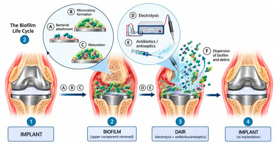
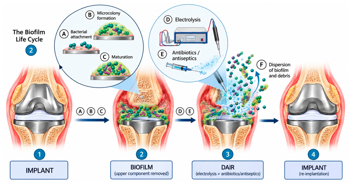
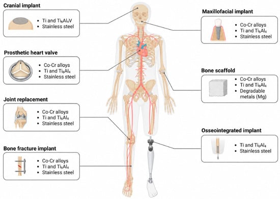
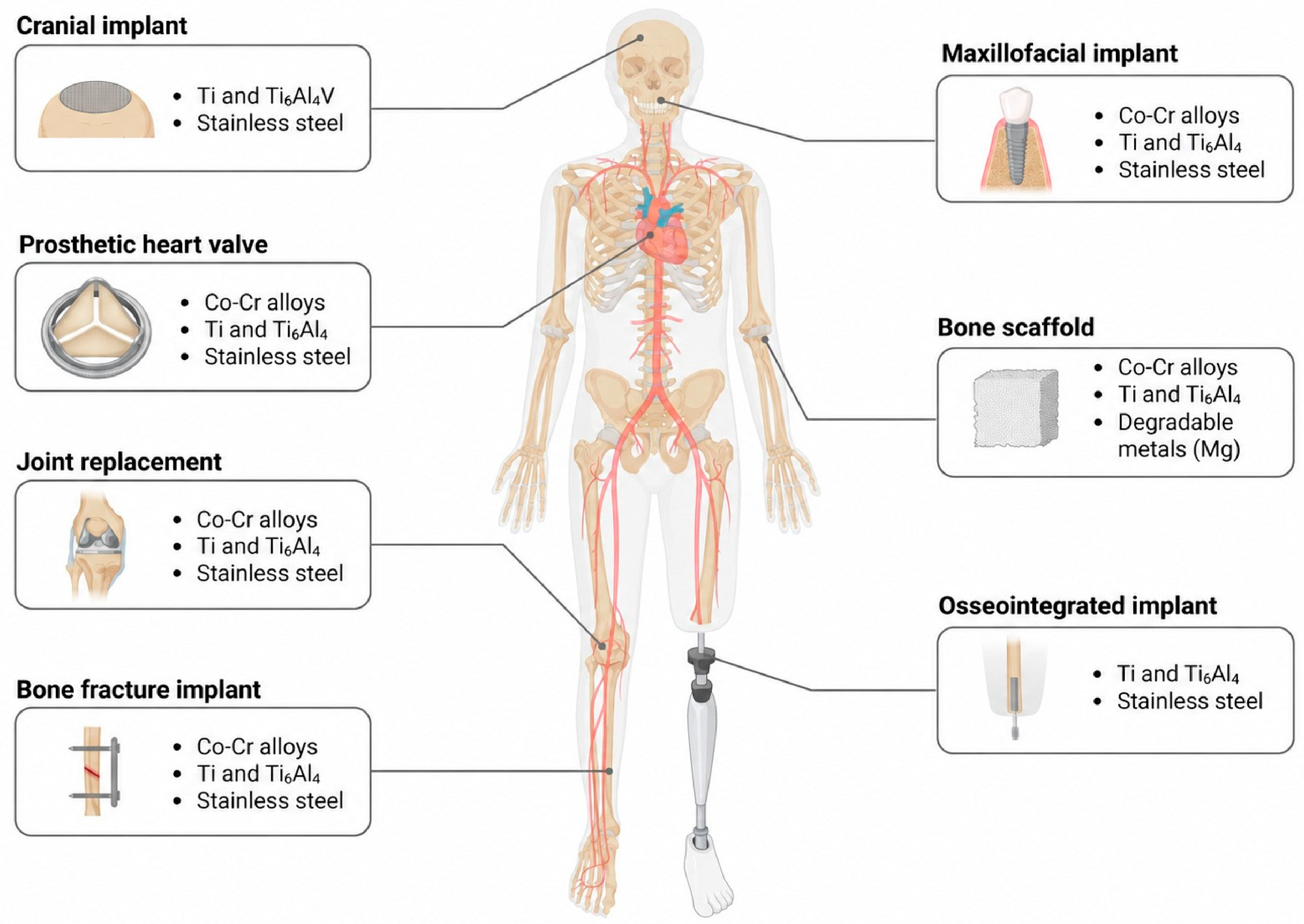
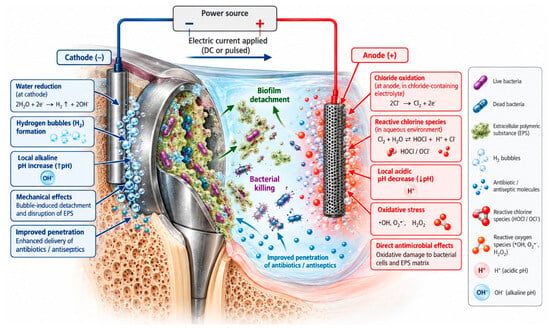
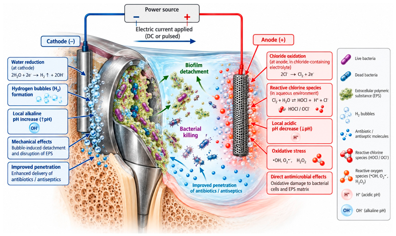
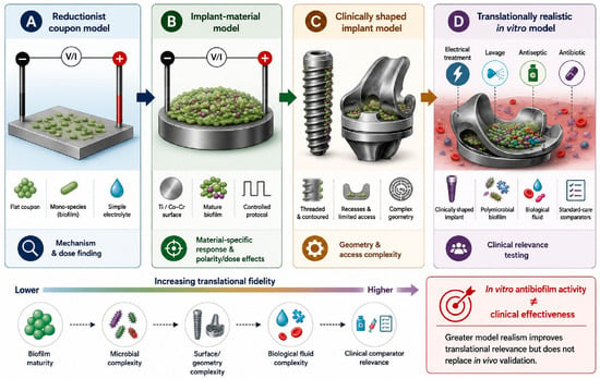
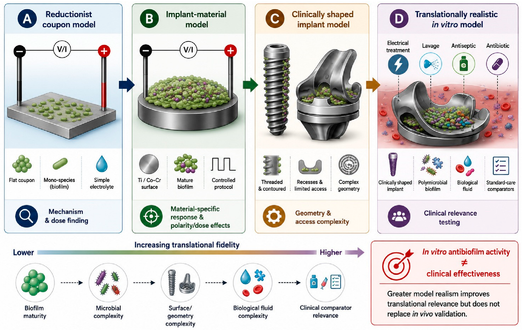
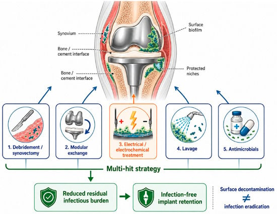
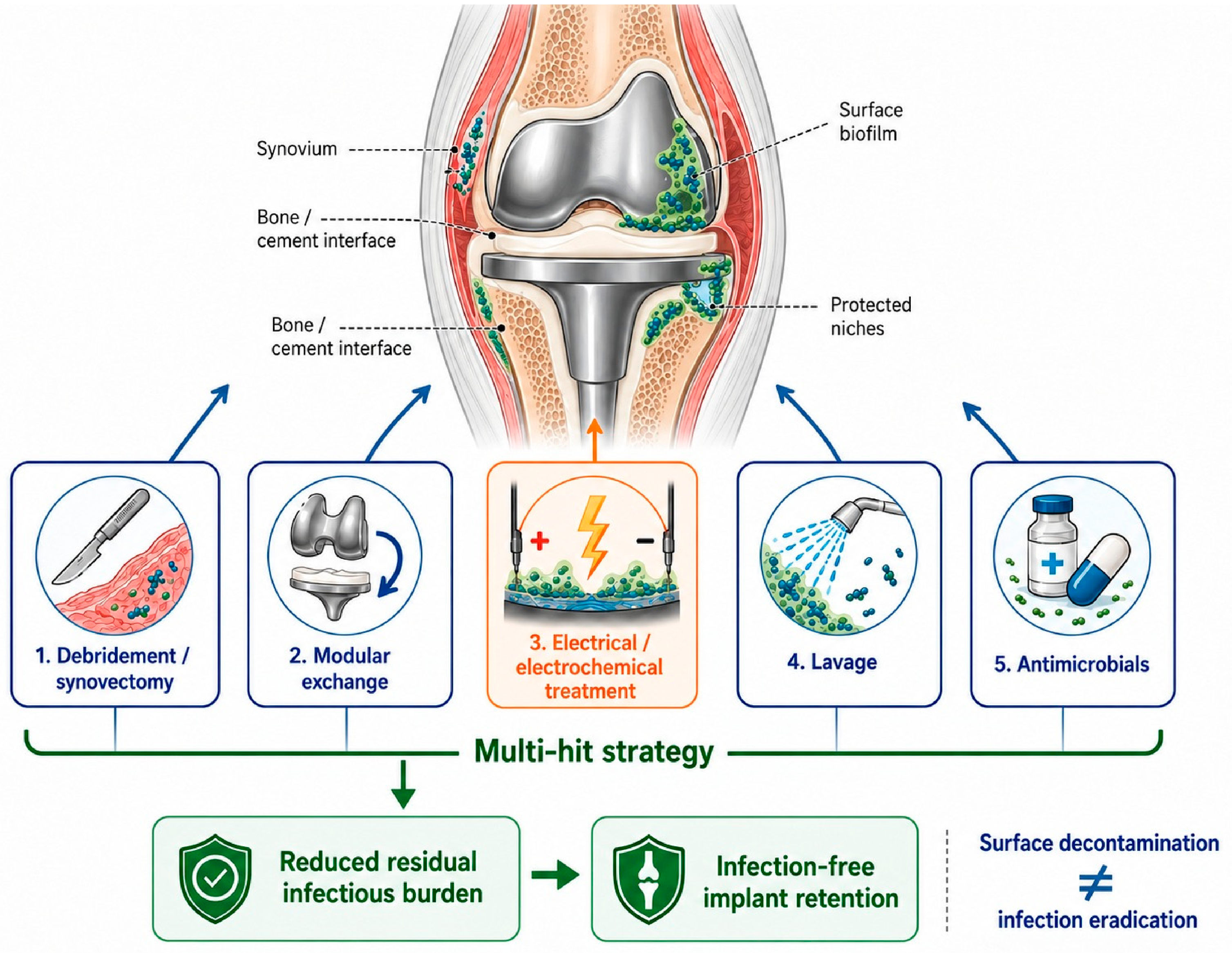

Disclaimer/Publisher’s Note: The statements, opinions and data contained in all publications are solely those of the individual author(s) and contributor(s) and not of MDPI and/or the editor(s). MDPI and/or the editor(s) disclaim responsibility for any injury to people or property resulting from any ideas, methods, instructions or products referred to in the content. |
© 2026 by the authors. Licensee MDPI, Basel, Switzerland. This article is an open access article distributed under the terms and conditions of the Creative Commons Attribution (CC BY) license.