


Abstract
Background: Urinary tract infections (UTIs) are a major global health burden that drives substantial antimicrobial use. Microbiological culture, the reference standard, can miss uropathogens and delay appropriate treatment, promoting interest in genomic approaches. Here, we systematically review the diagnostic accuracy of genomic methods and discuss their positivity criteria, turnaround time (TAT) and clinical impact. Methods: The Scopus and PubMed databases were searched for studies published between January 2020 and January 2026 that evaluated genomic methods for UTI diagnosis against microbiological culture and reported accuracy. Sensitivity and specificity were pooled separately using binomial-normal generalised linear mixed models (GLMM). Quality assessment was conducted using QUADAS-2. TAT and clinical impact were synthesised narratively. The review protocol was registered with PROSPERO (CRD420261294559). Results: Thirteen studies were included. Genomic methods showed high pooled sensitivity with moderate heterogeneity (0.94; 95% CI 0.87–0.98, τ2 = 1.43). However, pooled specificity was moderate and highly variable (0.61; 95% CI 0.23–0.89; τ2 = 5.92). The criteria used to define a positive genomic result varied widely, the shortest TAT was 4–6 h, and evidence on clinical outcomes was limited to two studies. Conclusions: Genomic methods are sensitive, potentially rapid, detect organisms missed by culture and characterise microbial diversity. However, their moderate, variable specificity likely reflects both limitations of culture as a reference standard and inconsistent genomic positivity criteria. As culture-negative, genomic-positive results may have explanations other than infection, they should be interpreted alongside symptoms and pre-test probability. Standardised positivity criteria and evidence of clinical benefit are needed before routine implementation.
1. Introduction
Urinary tract infections (UTIs) remain a major health concern around the globe, with an estimated 449 million incident cases reported in 2021 [1]. Clinical manifestations vary widely, ranging from minor self-limiting symptoms and acute uncomplicated cystitis to recurrent or complicated infections. UTIs can also be a source of more serious infections. For example, UTIs are the most common source (42.2%) of bloodstream infections [2]. Accordingly, treatment also varies from no treatment for self-limited infection through single short antimicrobial course use to prolonged or combination therapy [3,4,5]. Across this spectrum, accurate diagnosis and identification of the causative pathogen are pivotal to effective management of the infection, appropriate antimicrobial use and optimal use of healthcare resources [6,7].
In practice, however, UTIs are frequently treated empirically, before or without microbiological confirmation. In a recent analysis of electronic health data records, 64% of the patients received antibiotics despite only 41% having a positive urine culture [6]. Urine culture has long been the reference standard for diagnosing UTIs [8,9] because it establishes organism viability and identifies antimicrobial susceptibility [9]. However, culture typically takes 24–72 h to yield clinically useful results, during which time antimicrobial treatment is usually initiated empirically [10]. Moreover, culture can only reveal infections caused by culturable microorganisms, leaving infections caused by viruses, fastidious or slow-growing bacteria, and polymicrobial infections outside its diagnostic scope [11]. These factors, together with the variable diagnostic thresholds applied to the quantity of detected microorganisms [9], contribute to misdiagnosis and to delayed or inappropriate antimicrobial use, which is a major driver of antimicrobial resistance.
Identification of microorganisms using genomic methods offers an attractive diagnostic alternative to culture as it has the potential to expedite diagnosis, shorten turnaround time (TAT) and identify clinically relevant unculturable microorganisms, for example symptomatic patients with negative cultures and infections by fastidious uropathogens [12,13,14]. Several studies have investigated the identification of previously known and unknown uropathogens using untargeted sequencing strategies such as whole-genome sequencing (WGS) and metagenomic next-generation sequencing (mNGS) as well as targeted sequencing strategies such as 16S rRNA amplification-based sequencing and a high-plex targeted sequencing panel [13,14,15,16,17]. Beyond pathogen identification, untargeted sequencing approaches can also identify functional genes and therefore have the potential to characterise antimicrobial resistance genes and predict antimicrobial susceptibility genotypically [13,14,15]. However, the detection of a resistance gene does not always correlate with phenotypic resistance [16].
The diagnostic value of genomic approaches has been assessed in two recent systematic reviews [18,19]. Both reviews found that mNGS detected more pathogens than urine culture, although this difference reached statistical significance in only He et al., 2024 [18]. He et al. pooled the accuracy of mNGS against culture, reporting high sensitivity (0.89) and lower, more variable specificity (0.75). Jeff et al. [19], on the other hand, presented sensitivity and specificity for individual studies but did not pool them, given the high heterogeneity between studies. Across the studies, sensitivity was consistently high, whereas specificity varied widely.
However, these reviews focused on mNGS alone and did not evaluate the broader range of sequence-based methods used in UTI diagnosis. Moreover, the diagnostic accuracy of parameters beyond pathogen detection, such as microbial diversity, was not considered. Therefore, we build on these previous meta-analyses by evaluating the diagnostic accuracy of genomic methods for UTI across sequencing technologies, including mNGS, 16S rRNA sequencing, and high-plex targeted sequencing panels. Moreover, we include research diagnosing UTI based on microbial community diversity, such as the Shannon diversity index. We also describe the positivity parameters used in each study and discuss their turnaround time and clinical impact.
2. Results
2.1. Characteristics of Included Studies
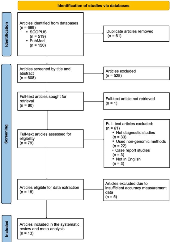
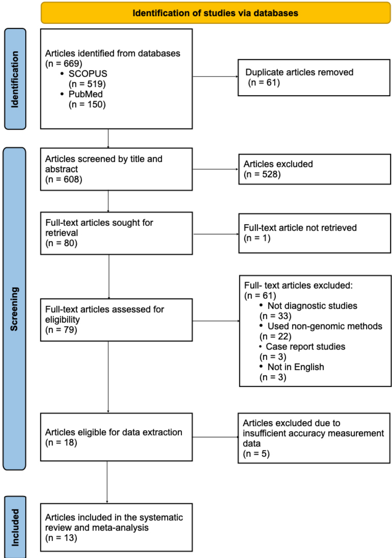
A total of 669 records were retrieved from both databases. Sixty-one duplicates were removed, leaving 608 records for title and abstract screening. Of these, 528 were excluded and 80 records underwent full text assessment for eligibility. One full text was not retrievable. Of the remaining 79 articles, 61 were excluded, 33 as non-diagnostic studies, 22 for using non-genomic diagnostic methods, three for not being written in English, and three for being case reports. Of the remaining 18 studies, five lacked sufficient accuracy data. The remaining 13 studies met the eligibility criteria and were included in the meta-analysis (Figure 1).
Figure 1.
Flow chart of study selection.
Of the 13 observational studies, six were conducted in China [13,14,15,20,21,22], two in the United States of America [17,23], and one each in Japan [12], South Korea [24], Germany [25], the Netherlands [16], and Thailand [26]. Most of the studies were conducted in hospital settings (n = 9), though a few reported other mixed settings (n = 4). UTI was classified as complicated (n = 2), uncomplicated (n = 2), mixed (n = 2), or recurrent (n = 1), and was unspecified in six studies. Patients ranged from infants to older adults (up to 89 years), and study sample sizes ranged from 10 to 488 (Table 1).
Table 1.
Study characteristics of the 13 included studies. All studies were observational, used culture as the reference standard, and examined urine samples from humans.
The genomic methods reported (index test) were mNGS, 16S rRNA sequencing, and high-plex targeted sequencing panels. The standard (reference) method was urine culture in every included study (Table 1).
2.2. Quality Assessment
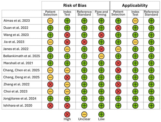
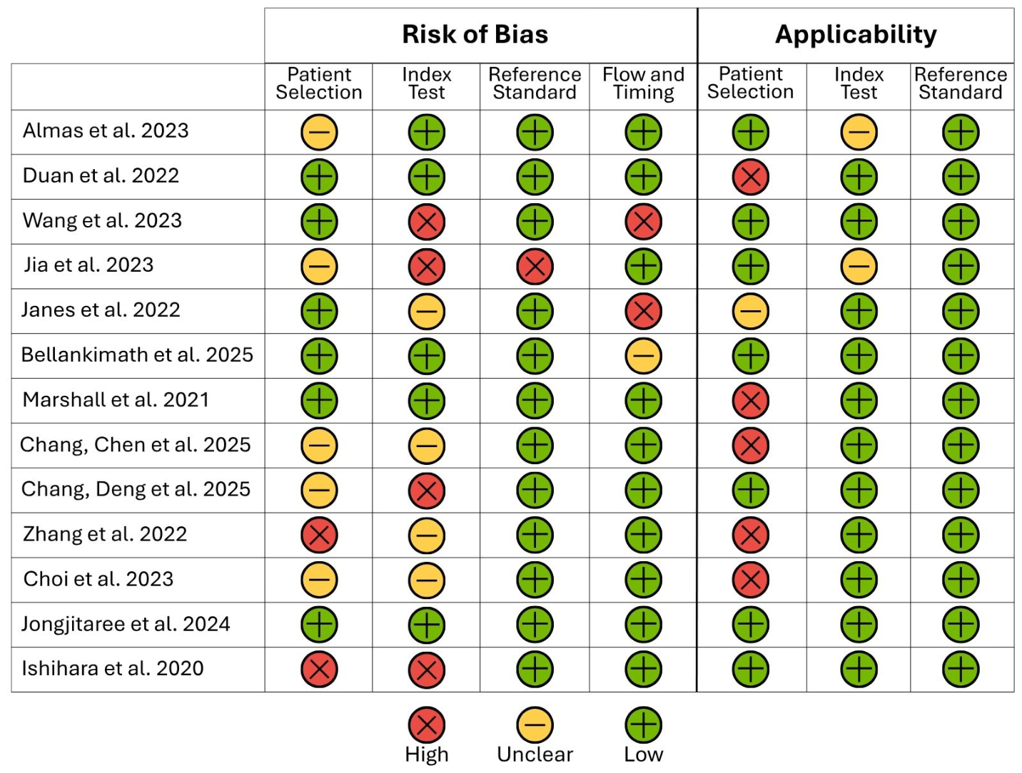
The risk of bias was assessed for the 13 included articles using QUADAS-2 (Figure 2). Applicability concerns were low for the reference standard domain, as all studies defined UTI in a comparable way. Similarly, the index test domain showed low concern for applicability as the genomic methods used could realistically be applied in clinical settings. The risk of bias in the reference standard domain was also low, with 12 of 13 studies rated as low risk, since they had defined detection thresholds. The most concerning domains were the index test risk of bias (where 4 studies were rated as high) and patient selection applicability (where 5 studies were of high concern). Overall, only 3 of the 13 studies were at low risk of bias across all four domains.
Figure 2.
Risk of bias and applicability assessment (QUADAS-2). Methodological quality of the 13 included studies across four risks-of-bias domains and three applicability domains rated as low, unclear, or high. Only three studies were at low risk of bias across all domains. Two studies with the same first author were distinguished by the second author.
2.3. Diagnostic Accuracy
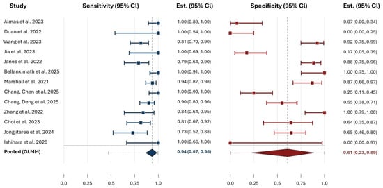
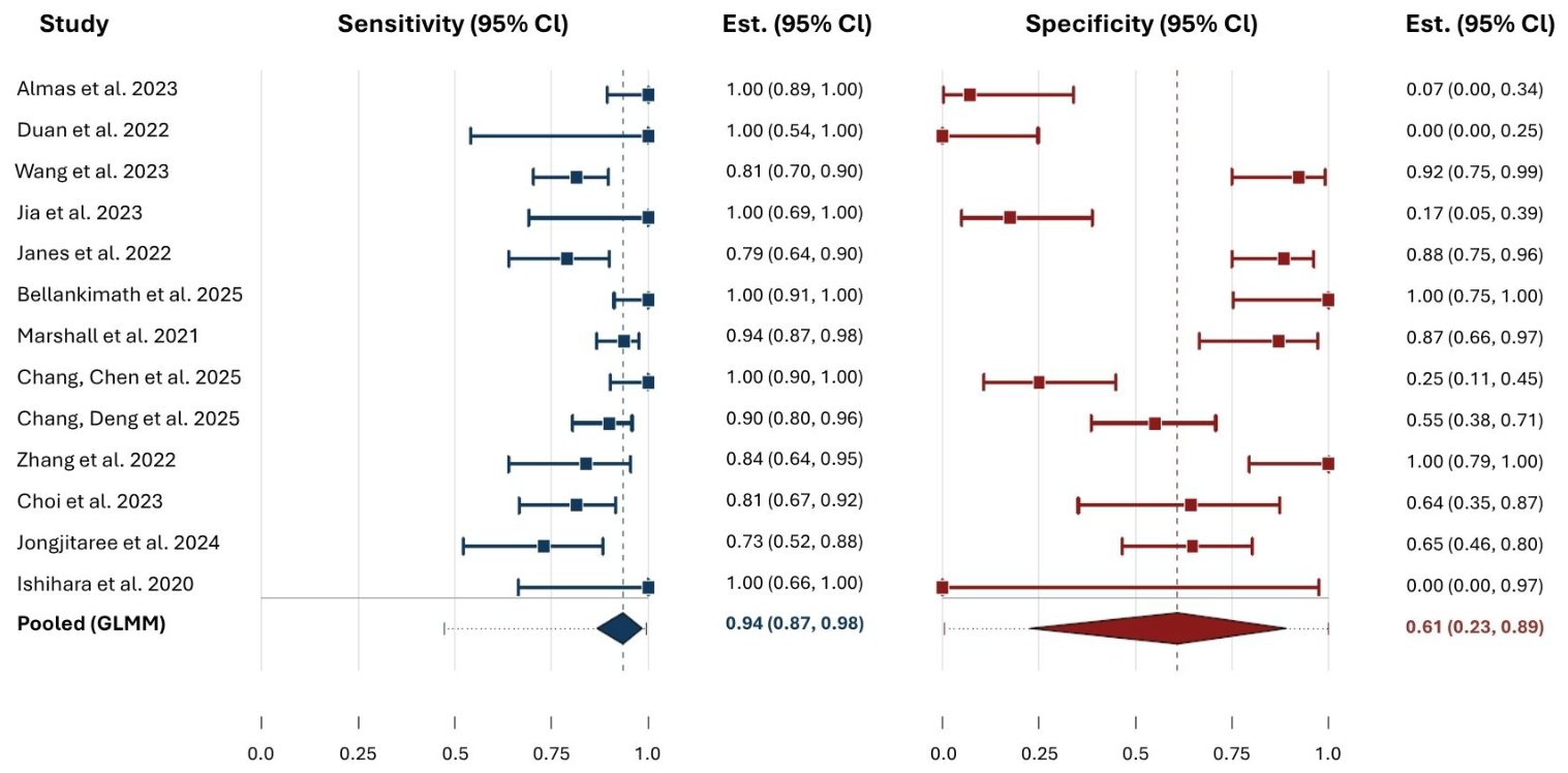
The extracted 2 × 2 values used to calculate the accuracy measurements are provided in Table 1. Across individual studies, estimated sensitivity ranged from 0.73 (95% CI 0.52–0.88) to 1.00 (0.54, 1.00) and the pooled estimate was 0.94 (95% CI 0.87–0.98), with moderate between-study variance (τ2 = 1.43), Figure 3. However, specificity showed a very wide range, with a pooled estimate of 0.61 (95% CI 0.23–0.89) and a larger heterogeneity (τ2 = 5.92) (Figure 3). Two studies, Duan et al. [20] and Ishihara et al. [12], reported a specificity of 0.00 because no true negatives were recorded.
Figure 3.
Pooled sensitivity and specificity. Per-study and pooled sensitivity and specificity of genomic methods compared with urine culture for the diagnosis of urinary tract infection. Pooled sensitivity and specificity were 0.94 (95% CI 0.87–0.98) and 0.61 (95% CI 0.23–0.89), respectively, and represented by the dashed vertical line in each panel. Squares and diamonds denote per-study and pooled point estimates, respectively, with 95% confidence intervals. The horizontal dotted line through each diamond denotes the 95% prediction interval. Between-study heterogeneity was moderate for sensitivity (τ2 = 1.43) but substantial for specificity (τ2 = 5.92). Duan et al. [20] and Ishihara et al. [12] show a specificity of 0.00 because neither study recorded a true negative, as sequencing returned a positive result for every culture-negative sample.
2.4. Uropathogens Detected by Genomic Methods
Genomic methods consistently detected more pathogens than culture, including polymicrobial infections and fastidious organisms that culture would otherwise have missed. The fastidious anaerobe, Actinotignum schaalii, for example, was identified by the genomic method but missed by culture in three studies [12,16,25]. Almas et al. [23] and Jia et al. [22] reported fungal and viral species detected by metagenomic sequencing methods but not by culture. Moreover, several studies reported substantial numbers of additional organisms. Wang et al. [21] and Duan et al. [20] identified 47 and 27 further pathogens, respectively, that culture failed to detect. Genomic methods also detected culturable pathogens (Enterococcus, Escherichia, and Proteus) that had been missed by culture due to low organism concentrations [17].
2.5. UTI Definition in Standard and Genomic Methods
Culture positivity thresholds varied across the included studies, ranging from 102 to 105 CFU/mL (Table 2). One study applied a threshold of ≥102 [21], two used ≥103 [16,25], five used ≥104 or ≥5 × 10⁴ [14,15,20,23,24], and one used ≥105 [17], while one study applied a range of 102–105 [13]. The culture threshold was not reported in the remaining three studies [12,22,26]. Genomic parameters for a positive UTI result varied significantly (Table 2). However, seven studies used the number of reads and relative abundance of pathogens as indicators, with only one study identifying a numeric threshold [21]. Interestingly, Marshall et al. [17] used low alpha diversity as a parameter to assign a UTI-positive result with a threshold of Shannon index ≤1 (Table 2).
Table 2.
How UTI was identified by culture and genomic method in each of the 13 studies.
2.6. Turnaround Time
TAT was reported for the genomic method in 10 of the 13 included studies and not reported by Jia et al. [22], Choi et al. [24], and Marshall et al. [17]. Genomic methods consistently had a shorter turnaround time than culture. The rapid metagenomic and nanopore workflows were the fastest methods, reporting TATs between 4–6 h [13,16,21,25] versus at least 24 h for culture. However, two mNGS studies reported longer TATs. Almas et al. [23] and Duan et al. [20] reported at least 24 h and 48 h, respectively. High-plex targeted sequencing panels reported also longer TATs at 13 h [14,15] and 48–72 h [26].
2.7. Impact of Genomic Diagnosis on Clinical Outcome
Direct evidence of an effect on clinical management was limited, with only two studies reporting treatment outcomes. Duan et al. [20], whose study included 19 patients with recurrent UTIs, reported that antimicrobial treatment was adjusted based on mNGS results in 76.9% of culture-negative cases and 33.3% of culture-positive cases. However, this did not lead to a statistically significant reduction in recurrent UTI episodes after 6-month follow-up. On the other hand, Jia et al. [22], whose study included 33 UTI patients, reported treatment adjustment in 92% of the study cohort with statistically significant symptom relief following treatment guided by mNGS.
3. Discussion
Our study found that genomic methods, including both targeted and untargeted approaches, demonstrated a high pooled sensitivity (0.94; 95% CI 0.87–0.98) but moderate and highly variable specificity (0.61; 95% CI 0.23–0.89) against urine culture. Included studies reported that genomic methods frequently identified organisms not recovered by culture, and turnaround times were generally shorter than for culture. The definition of a positive genomic result varied widely across studies with no standardised threshold, with one study using a microbial diversity metric, the Shannon diversity index, as a diagnostic marker. Direct evidence that genomic testing improves clinical outcomes was limited to two studies.
The high sensitivity shows that genomic methods identified most culture-positive infections. The moderate specificity, however, reflects the large proportion of samples that were genomic-positive but culture-negative. A culture-negative, genomic-positive result may represent a true infection that culture failed to detect, for example, a fastidious, slow-growing, non-bacterial organism or even shifts in microbial diversity [14,27,28]. However, it may equally reflect colonisation, asymptomatic bacteriuria, contamination, or the presence of non-viable microbial DNA [29,30]. Therefore, the pooled estimates should be read as measures of agreement with culture, an imperfect reference method with inherent limitations in organism detection, rather than as definitive measures of the true diagnostic accuracy of genomic testing.
Moreover, the wide variation in individual study specificity may be attributed to the variable thresholds used to define a positive culture (102 to 105 CFU/mL) and to the differing parameters used to define a positive result by genomic methods. These parameters were based on uropathogen identification [12,20,23,24,25,26], microbial diversity [17], the number of sequence reads [13,14,15,16,21,22,25], relative abundance of organisms [12,16,17,22], or a combination of qualitative and quantitative rules [14,15,16,21,22] (Table 2). This variation was also reflected in the QUADAS-2 assessment, in which risk of bias was greatest for the index test, largely because few studies prespecified the threshold used to define a positive genomic result. This has direct consequences for the pooled estimates, as the absence of such criteria or post-hoc selection of cut-offs can alter apparent accuracy within a study and make results difficult to compare across studies. The moderate and highly variable pooled specificity should therefore be interpreted in this light, as it reflects not only genuine differences between tests but also inconsistencies in how a positive result was defined. Moreover, other factors that could have influenced which organisms are detected and how a positive result is classified would contribute to the heterogeneity in specificity, for example, different patient populations, differences in the sequencing platforms and bioinformatic pipelines used, and variation in specimen collection and processing.
This uncertainty has direct implications for antimicrobial stewardship. Treating every genomic-positive result as infection would risk unnecessary antibiotic exposure, particularly given the now well-recognised existence of a resident urinary microbiome [31]. In practice, a genomic-positive result should therefore be interpreted in the context of the patient’s symptoms, pre-test probability, and other laboratory findings, rather than in isolation. For example, a febrile infant has a high pre-test probability of UTI, whereas in an older adult the same result may instead represent asymptomatic bacteriuria. Several included studies applied this principle explicitly: Jia et al. [22] and Wang et al. [21] incorporated clinical judgement or a composite reference standard, and Jia et al. used negative controls and a confidence score combining relative abundance, read number, and known uropathogenicity to distinguish likely pathogens from incidental detections. Distinguishing true infection from colonisation or clinically irrelevant detection remains one of the central challenges to the clinical adoption of genomic UTI diagnostics.
The included studies spanned clinically heterogeneous populations, in whom the significance of a positive result differs, so the pooled accuracy measures should be read with this diversity in mind. However, subgroup analysis by age or UTI complexity was not feasible, given the small number of studies (k = 13). Similarly, subgroup analysis by positivity criterion could not be performed, as the criteria were distributed across several categories, two of which (diversity-based and composite rules) were each represented by only a single study. The absence of standardised and clinically validated positivity thresholds therefore represents one of the principal barriers to routine implementation, and the standardisation of these criteria remains a priority for future research.
Beyond pathogen detection, genomic methods can also assess microbial community structure, offering a diagnostic dimension that culture cannot access [17]. For example, Marshall et al. [17] used the Shannon diversity index as a diagnostic marker, where low diversity (indicating a dominance by a single organism) was classified as likely UTI. Such approaches may support the future diagnosis of UTI based on disturbances in the urinary microbiome, an association increasingly supported by previous research [32,33,34,35]. However, like the other genomic criteria discussed above, this approach currently lacks a clinically validated threshold, which limits its routine application.
Genomic methods were generally faster than culture, although TAT varied considerably between platforms and laboratory workflows. Some studies reported a TAT as short as 4 to 6 h [13,16,21,25], which can be of clinical value, allowing appropriate treatment without an initial course of empirical antimicrobials in selected clinical settings. This potential clinical benefit should, however, be considered alongside the broader operational and economic implications of genomic testing. Although genomic methods are often assumed to be more costly than culture, any cost-effectiveness assessment should also account for potential reductions in laboratory workload, expert staffing demands, and the capacity for high-throughput automated processing [25,36].
Direct evidence of an effect on infection management was limited, with only two studies reporting it [20,22]. Duan et al. [20] identified a considerable proportion of newly detected organisms in culture-negative samples. However, the associated results did not reach statistical significance when clinical impact was evaluated, probably due to the small sample size. In contrast, Jia et al. [22], with a slightly larger sample size, reported a statistically significant improvement in symptoms following the application of genomic methods. Case reports [37,38,39] outside our included studies reinforce this. For example, Li et al. [37] described how mNGS identified Enterococcus faecalis within 24 h in a patient with persistent UTI symptoms and repeatedly negative cultures. The management of this case was changed, the patient’s symptoms improved, and mNGS confirmed a fall in the E. faecalis read counts over time. Although these findings suggest potential clinical utility, a wider implementation of genomic methods in routine practice will require unified standards for the parameters and thresholds used to define a positive genomic UTI result.
In this systematic review, several limitations should be acknowledged. The number of eligible studies was relatively small (k = 13), and there was substantial heterogeneity in specificity between the included studies, which may have affected the pooled estimate. The number of included studies also prevented a subgroup analysis by sequencing platform. The heterogeneity was also influenced by several studies with zero-cell specificity data, which we addressed by using GLMM models, a method well suited to meta-analyses of sparse diagnostic data that avoid the continuity correction required by conventional approaches [40]. The included studies were observational studies rather than randomised controlled trials, and some enrolled small or highly selected patient populations, which may have affected the precision of the accuracy estimates and may limit generalisability. Moreover, we could not pool TAT quantitatively, as it was reported inconsistently across the included studies. The hospital-based nature of the majority of the included studies may also have limited the generalisability of findings to community settings, where genomic methods could potentially have application in the detection of bacteriuria, particularly among asymptomatic older adults, in whom its prevalence increases substantially after the age of 65 years [41].
4. Materials and Methods
This systematic review and meta-analysis was conducted following the Preferred Reporting Items for Systematic Reviews and Meta-Analyses (PRISMA) guidelines [42]. The study protocol was registered with PROSPERO before data extraction under registration number CRD420261294559. More details of the protocol can be found via https://www.crd.york.ac.uk/PROSPERO/view/CRD420261294559 (accessed on 23 September 2026).
4.1. Search Strategy
The literature search was conducted using the Scopus and PubMed databases, with the last search done on 9 January 2026. The search was performed independently by two authors (FD and MJ) according to a prespecified search strategy. The search terms combined the following keywords and their synonyms using the Boolean operators (AND/OR): (urinary tract infection OR UTI OR uropathogens OR bacteriuria OR viruria OR urinary infection) AND (genomic OR genomics OR whole genome sequencing OR WGS OR metagenomics OR next-generation sequencing OR NGS OR 16S ribosomal RNA OR 16S rRNA OR 16S sequencing OR shotgun sequencing OR genome sequencing) AND (diagnosis OR diagnostic OR diagnose OR diagnostic test OR molecular diagnosis OR molecular assay OR detect OR detection). We restricted the search to studies published between 1 January 2020 and 9 January 2026 to capture the period reflecting current sequencing technologies.
4.2. Eligibility Criteria
Studies were eligible if they met the following: (1) Peer-reviewed primary research; (2) case-control, cohort, or clinical trial studies; (3) conducted in human samples using genomic methods to diagnose UTIs; (4) reported diagnostic accuracy data or 2 × 2 confusion matrix from which accuracy measurements could be calculated; (5) published in English between 1 January 2020 and 9 January 2026.
Studies were excluded if they were: (1) animal studies; (2) epidemiological studies or studies primarily focused on the treatment or prevention of UTI; (3) studies that used genetic methods such as PCR or qPCR to detect only one or a few organisms; (4) case report studies, conference abstracts, book chapters that duplicate original research findings, narrative and systematic review articles, or editorials; (5) studies published in a language other than English.
4.3. Selection Process
All studies retrieved from the database searches were imported into Rayyan [43] for screening and duplicate records were removed. Titles and abstracts were screened against the eligibility criteria by five members of the research team (MJ, FD, FO, ZA, and MT). Each record was screened independently by two authors. Any screening and verification inconsistencies were resolved through discussion among the team and the supervisor (AG).
4.4. Data Extraction
Data were extracted independently by two reviewers, with five reviewers (MJ, FD, FO, ZA, and MT) contributing to the process. Disagreements were resolved through discussion until a consensus was reached. Data were extracted using a standardised data extraction form in Microsoft Excel. Extracted study characteristics included publication details (author, year, journal, and DOI), study design, healthcare settings, sample size, and UTI type. Extracted data also included:
Reference standard: Microbiological culture and its positive threshold where reported. Where a study reported accuracy measurements against more than one reference standard, culture-referenced data were used to maintain a consistent comparator.
Index test: Genomic methods and their parameters for positive genomic results (organism quantity as sequencing reads, relative abundance, organism identity, combination of organism quantity and identity, or a microbial diversity metric).
Accuracy measurements: Sensitivity and specificity. When these measures were not directly reported, they were calculated from the reported true positive (TP), true negative (TN), false negative (FN), and false positive (FP) values.
Turnaround time: Due to variations in both the definitions and the reporting format of TAT across studies, TAT was not pooled and was instead summarised narratively by comparing the range of reported values with and across studies, following the Synthesis Without Meta-analysis (SWiM) guideline [44].
4.5. Study Outcome
The primary outcome was the diagnostic accuracy (sensitivity and specificity) of genomic methods for the detection of uropathogens compared to standard microbiological culture. The secondary outcomes were TAT and the clinical impact of genomic detection in the management of UTI.
4.6. Risk of Bias
The Quality Assessment of Diagnostic Accuracy Studies (QUADAS-2) tool was applied to assess the risk of bias and study quality [45]. Five authors (MJ, FD, FO, ZA, and MT) contributed to the quality assessment with each study independently assessed by two authors. Disagreements were resolved through discussions. Risk of bias was used to interpret the pooled accuracy estimates.
4.7. Statistical Analysis
For each study, TP, FP, TN, and FN were extracted. Sensitivity and specificity were calculated with exact (Clopper–Pearson) 95% confidence intervals (CI) [46], as not all studies reported 95% CIs. Sensitivity and specificity were pooled separately using binomial-normal generalised linear mixed models (GLMM) with a random study effect. This model was chosen as it works directly from reported counts rather than from transformed proportions, which allows it to handle studies in which a cell was zero (6 cells for sensitivity and 4 cells for specificity, Table 1) without the arbitrary corrections that approximation methods would require in such cases [40]. The model was fitted using adaptive Gauss–Hermite quadrature [47], and the estimates were stable when checked across different quadrature node counts. CIs for the pooled estimates were obtained by profile likelihood which is suitable for a relatively small number of studies. Between-study heterogeneity was summarised using tau-squared (τ2), and 95% prediction intervals were calculated to show the range in which a future study’s result would be expected to fall [48]. Analyses were performed in R version 4.3.3.
5. Conclusions
Currently, microbiological urine culture remains the standard for the diagnosis of UTI. However, its limitations, including prolonged TAT and dependence on successful microbial growth, can restrict pathogen detection. Genomic methods may address these limitations and show considerable potential to improve UTI diagnostics, with high sensitivity, broader organism detection, and substantially shorter TAT. However, these findings are based on a small and heterogeneous evidence base (13 studies) with variable definitions of a positive genomic result, and the high pooled sensitivity should therefore be interpreted with caution rather than as evidence that genomic methods are clinically superior to culture. Importantly, improved pathogen detection has not yet been shown to translate consistently into improved clinical outcomes: only two of the included studies evaluated the effect of genomic testing on patient management. However, potential benefits of genomic diagnosis may be particularly relevant for patients with sterile pyuria, recurrent UTIs, persistent symptoms despite antibiotic treatment, and those with repeated mixed-growth urine cultures involving organisms such as Escherichia coli and Enterococcus. These populations represent promising groups in which the diagnostic value of genomic methods should be evaluated in future studies. Further studies are also needed to strengthen the evidence of the diagnostic value and clinical utility of genomic methods, to establish standardised positivity thresholds and to include samples from community settings and uncomplicated UTI.
Author Contributions
Conceptualisation, A.A.H.G., O.A.; methodology, A.A.H.G., M.J., F.W.D., F.G.O., Z.A.H., M.T.; formal analysis, A.A.H.G., M.J., F.W.D., F.G.O., Z.A.H., M.T.; investigation, A.A.H.G., M.J., F.W.D., F.G.O., Z.A.H., M.T.; data curation, A.A.H.G., M.J., F.W.D., F.G.O., Z.A.H.; writing—original draft preparation, A.A.H.G., O.A., M.J., F.W.D., F.G.O., Z.A.H., M.T.; writing—review and editing, A.A.H.G., O.A., M.J., F.W.D., F.G.O., Z.A.H., M.T.; visualisation, A.A.H.G., F.W.D.; supervision, A.A.H.G. All authors have read and agreed to the published version of the manuscript.
Funding
This research received no external funding.
Institutional Review Board Statement
Not applicable.
Informed Consent Statement
Not applicable.
Data Availability Statement
No new data were created or analysed in this study. Data sharing is not applicable to this article.
Acknowledgments
The authors thank all researchers whose work contributed to this systematic review. Generative AI (Opus 5.5, Claude, Anthropic) was used to assist with language editing (grammar, spelling, punctuation, and phrasing). All AI-edited text was subsequently reviewed and verified by the authors.
Conflicts of Interest
The authors declare no conflicts of interest.
References
- He, Y.; Zhao, J.; Wang, L.; Han, C.; Yan, R.; Zhu, P.; Qian, T.; Yu, S.; Zhu, X.; He, W. Epidemiological Trends and Predictions of Urinary Tract Infections in the Global Burden of Disease Study 2021. Sci. Rep. 2025, 15, 4702. [Google Scholar] [CrossRef] [Scilit]
- The BALANCE Investigators Antibiotic Treatment for 7 versus 14 Days in Patients with Bloodstream Infections. N. Engl. J. Med. 2025, 392, 1065–1078. [Google Scholar] [CrossRef] [Scilit]
- Sabih, A.; Leslie, S.W. Complicated Urinary Tract Infections. In StatPearls; StatPearls Publishing: Treasure Island, FL, USA, 2026. [Google Scholar]
- Trautner, B.W.; Cortés-Penfield, N.W.; Gupta, K.; Hirsch, E.B.; Horstman, M.; Moran, G.J.; Colgan, R.; O’Horo, J.C.; Ashraf, M.S.; Connolly, S.; et al. Clinical Practice Guidelines by Infectious Diseases Society of America (IDSA): 2025 Guidelines on Management and Treatment of Complicated Urinary Tract Infections—Duration of Antibiotics for Complicated UTI. Clin. Infect. Dis. 2025, 82, i79–i88. [Google Scholar] [CrossRef] [Scilit]
- Valentine-King, M.; Trautner, B.; Zoorob, R.; Germanos, G.; Salemi, J.; Gupta, K.; Grigoryan, L. Analysis of Recurrent Urinary Tract Infection Management in Outpatient Settings Reveals Opportunities for Antibiotic Stewards. Antimicrob. Steward. Healthc. Epidemiol. 2021, 1, s34. [Google Scholar] [CrossRef] [Scilit]
- Murray, K.; Shimabukuro, J.; Khalfay, N.; Chiang, J.N.; Lenore Ackerman, A. Antibiotic Overprescription for “Urinary Tract Infections” Is Associated with Poor Diagnostic Stewardship and Low Adherence to Guidelines. Neurourol. Urodyn. 2025, 44, 382–389. [Google Scholar] [CrossRef] [Scilit]
- Shafrin, J.; Marijam, A.; Joshi, A.V.; Mitrani-Gold, F.S.; Everson, K.; Tuly, R.; Rosenquist, P.; Gillam, M.; Ruiz, M.E. Impact of Suboptimal or Inappropriate Treatment on Healthcare Resource Use and Cost among Patients with Uncomplicated Urinary Tract Infection: An Analysis of Integrated Delivery Network Electronic Health Records. Antimicrob. Resist. Infect. Control 2022, 11, 133. [Google Scholar] [CrossRef] [Scilit]
- Kass, E.H. Asymptomatic Infections of the Urinary Tract. 1956. J. Urol. 2002, 167, 1016–1019; discussion 1019–1021. [Google Scholar] [CrossRef] [Scilit]
- Schmiemann, G.; Kniehl, E.; Gebhardt, K.; Matejczyk, M.M.; Hummers-Pradier, E. The Diagnosis of Urinary Tract Infection. Dtsch. Ärztebl. Int. 2010, 107, 361–367. [Google Scholar] [CrossRef] [Scilit]
- Davenport, M.; Mach, K.E.; Shortliffe, L.M.D.; Banaei, N.; Wang, T.-H.; Liao, J.C. New and Developing Diagnostic Technologies for Urinary Tract Infections. Nat. Rev. Urol. 2017, 14, 296–310. [Google Scholar] [CrossRef] [Scilit]
- Price, T.K.; Dune, T.; Hilt, E.E.; Thomas-White, K.J.; Kliethermes, S.; Brincat, C.; Brubaker, L.; Wolfe, A.J.; Mueller, E.R.; Schreckenberger, P.C. The Clinical Urine Culture: Enhanced Techniques Improve Detection of Clinically Relevant Microorganisms. J. Clin. Microbiol. 2016, 54, 1216–1222. [Google Scholar] [CrossRef] [Scilit]
- Ishihara, T.; Watanabe, N.; Inoue, S.; Aoki, H.; Tsuji, T.; Yamamoto, B.; Yanagi, H.; Oki, M.; Kryukov, K.; Nakagawa, S.; et al. Usefulness of Next-Generation DNA Sequencing for the Diagnosis of Urinary Tract Infection. Drug Discov. Ther. 2020, 14, 42–49. [Google Scholar] [CrossRef] [Scilit]
- Zhang, L.; Huang, W.; Zhang, S.; Li, Q.; Wang, Y.; Chen, T.; Jiang, H.; Kong, D.; Lv, Q.; Zheng, Y.; et al. Rapid Detection of Bacterial Pathogens and Antimicrobial Resistance Genes in Clinical Urine Samples with Urinary Tract Infection by Metagenomic Nanopore Sequencing. Front. Microbiol. 2022, 13, 858777. [Google Scholar] [CrossRef] [Scilit]
- Chang, Z.; Deng, J.; Zhang, J.; Wu, H.; Wu, Y.; Bin, L.; Li, D.; Liu, J.; Yu, R.; Lin, H.; et al. Rapid and Accurate Diagnosis of Urinary Tract Infections Using Targeted Next-Generation Sequencing: A Multicenter Comparative Study with Metagenomic Sequencing and Traditional Culture Methods. J. Infect. 2025, 90, 106459. [Google Scholar] [CrossRef] [Scilit]
- Chang, Z.; Chen, J.; Chen, J.; Zhu, Y.; Zhang, Y.; Liu, J.; Deng, J.; Wu, H.; Bin, L.; Lin, H.; et al. Precision Diagnosis of Preoperative Infection in Urolithiasis: Integrating Targeted next-Generation Sequencing for Enhanced Accuracy—A Multicenter Cohort Study. BMC Infect. Dis. 2025, 25, 1767. [Google Scholar] [CrossRef] [Scilit]
- Janes, V.A.; Matamoros, S.; Munk, P.; Clausen, P.T.L.C.; Koekkoek, S.M.; Koster, L.A.M.; Jakobs, M.E.; De Wever, B.; Visser, C.E.; Aarestrup, F.M.; et al. Metagenomic DNA Sequencing for Semi-Quantitative Pathogen Detection from Urine: A Prospective, Laboratory-Based, Proof-of-Concept Study. Lancet Microbe 2022, 3, e588–e597. [Google Scholar] [CrossRef] [Scilit]
- Marshall, C.W.; Kurs-Lasky, M.; McElheny, C.L.; Bridwell, S.; Liu, H.; Shaikh, N. Performance of Conventional Urine Culture Compared to 16S rRNA Gene Amplicon Sequencing in Children with Suspected Urinary Tract Infection. Microbiol. Spectr. 2021, 9, e01861-21. [Google Scholar] [CrossRef] [Scilit]
- He, S.; Liu, H.; Hu, X.; Zhao, J.; Liang, J.; Zhang, X.; Chen, J.; Zeng, H.; Sun, G. Exploring the Clinical and Diagnostic Value of Metagenomic Next-Generation Sequencing for Urinary Tract Infection: A Systematic Review and Meta-Analysis. BMC Infect. Dis. 2024, 24, 1000. [Google Scholar] [CrossRef] [Scilit]
- Jeff, C.; Quarton, S.; Hatton, C.; Parekh, D.; Thickett, D.; McNally, A.; Sapey, E. Metagenomics in the Diagnosis and Treatment of Urinary Tract Infections: A Systematic Review and Meta-Analysis. Diagn. Microbiol. Infect. Dis. 2025, 113, 116995. [Google Scholar] [CrossRef] [Scilit]
- Duan, W.; Yang, Y.; Zhao, J.; Yan, T.; Tian, X. Application of Metagenomic Next-Generation Sequencing in the Diagnosis and Treatment of Recurrent Urinary Tract Infection in Kidney Transplant Recipients. Front. Public Health 2022, 10, 901549. [Google Scholar] [CrossRef] [Scilit]
- Wang, Y.; Chen, T.; Zhang, S.; Zhang, L.; Li, Q.; Lv, Q.; Kong, D.; Jiang, H.; Ren, Y.; Jiang, Y.; et al. Clinical Evaluation of Metagenomic Next-Generation Sequencing in Unbiased Pathogen Diagnosis of Urinary Tract Infection. J. Transl. Med. 2023, 21, 762. [Google Scholar] [CrossRef] [Scilit]
- Jia, K.; Huang, S.; Shen, C.; Li, H.; Zhang, Z.; Wang, L.; Zhao, G.; Wu, Z.; Lin, Y.; Xia, H.; et al. Enhancing Urinary Tract Infection Diagnosis for Negative Culture Patients with Metagenomic Next-Generation Sequencing (mNGS). Front. Cell. Infect. Microbiol. 2023, 13, 1119020. [Google Scholar] [CrossRef] [Scilit]
- Almas, S.; Carpenter, R.E.; Rowan, C.; Tamrakar, V.K.; Bishop, J.; Sharma, R. Advantage of Precision Metagenomics for Urinary Tract Infection Diagnostics. Front. Cell. Infect. Microbiol. 2023, 13, 1221289. [Google Scholar] [CrossRef] [Scilit]
- Choi, J.H.; Yoon, Y.M.; Kim, Y.-J.; Han, K.H. Role of 16S Ribosomal RNA Analysis in Identification of Microorganisms in Febrile Urinary Tract Infection of Infants. Indian J. Pediatr. 2023, 90, 660–664. [Google Scholar] [CrossRef] [Scilit]
- Bellankimath, A.B.; Branders, S.; Kegel, I.; Ali, J.; Asadi, F.; Johansen, T.E.B.; Imirzalioglu, C.; Hain, T.; Wagenlehner, F.; Ahmad, R. Metagenomic Sequencing Enables Accurate Pathogen and Antimicrobial Susceptibility Profiling in Complicated UTIs in Approximately Four Hours. Nat. Commun. 2025, 17, 187. [Google Scholar] [CrossRef] [Scilit]
- Jongjitaree, K.; Sheetz, T.; Finegan, J.; Bechis, S.K.; Sur, R.L.; Monga, M. The Application of Next-Generation Sequencing in Preoperative Evaluation for Urologic Stone Surgery. J. Endourol. 2024, 38, 908–915. [Google Scholar] [CrossRef] [Scilit]
- Kline, K.A.; Lewis, A.L. Gram-Positive Uropathogens, Polymicrobial Urinary Tract Infection, and the Emerging Microbiota of the Urinary Tract. In Urinary Tract Infections; Wiley: Hoboken, NJ, USA, 2016; Volume 4. [Google Scholar] [CrossRef] [Scilit]
- Sinawe, H.; Casadesus, D. Urine Culture. In StatPearls; StatPearls Publishing: Treasure Island, FL, USA, 2026. [Google Scholar]
- Nicolle, L.E.; Gupta, K.; Bradley, S.F.; Colgan, R.; DeMuri, G.P.; Drekonja, D.; Eckert, L.O.; Geerlings, S.E.; Köves, B.; Hooton, T.M.; et al. Clinical Practice Guideline for the Management of Asymptomatic Bacteriuria: 2019 Update by the Infectious Diseases Society of America. Clin. Infect. Dis. 2019, 68, e83–e110. [Google Scholar] [CrossRef] [Scilit]
- Musleh, L.; Montilli, M.; Ammendolia, M.G.; Sciarra, A.; Riccioli, A.; Maurizi, L.; Longhi, C. More than an Infection: The Ecological Puzzle of Recurrent Urinary Tract Infections. Front. Cell. Infect. Microbiol. 2026, 16, 1927507. [Google Scholar] [CrossRef] [Scilit]
- Wolfe, A.J.; Toh, E.; Shibata, N.; Rong, R.; Kenton, K.; FitzGerald, M.; Mueller, E.R.; Schreckenberger, P.; Dong, Q.; Nelson, D.E.; et al. Evidence of Uncultivated Bacteria in the Adult Female Bladder. J. Clin. Microbiol. 2012, 50, 1376–1383. [Google Scholar] [CrossRef] [Scilit]
- Huang, L.; Li, X.; Zheng, B.; Li, P.; Wei, D.; Huang, C.; Sun, L.; Li, H. Differential Urinary Microbiota Composition Between Women with and Without Recurrent Urinary Tract Infection. Front. Microbiol. 2022, 13, 888681. [Google Scholar] [CrossRef] [Scilit]
- Moustafa, A.; Li, W.; Singh, H.; Moncera, K.J.; Torralba, M.G.; Yu, Y.; Manuel, O.; Biggs, W.; Venter, J.C.; Nelson, K.E.; et al. Microbial Metagenome of Urinary Tract Infection. Sci. Rep. 2018, 8, 4333. [Google Scholar] [CrossRef] [Scilit]
- Neugent, M.L.; Hulyalkar, N.V.; Nguyen, V.H.; Zimmern, P.E.; De Nisco, N.J. Advances in Understanding the Human Urinary Microbiome and Its Potential Role in Urinary Tract Infection. mBio 2020, 11, e00218-20. [Google Scholar] [CrossRef] [Scilit]
- Yoo, J.-J.; Shin, H.; Song, J.; Kim, M.; Yun, J.; Kim, Z.; Lee, Y.; Lee, S.; Lee, K.; Kim, W.; et al. Urinary Microbiome Characteristics in Female Patients with Acute Uncomplicated Cystitis and Recurrent Cystitis. J. Clin. Med. 2021, 10, 1097. [Google Scholar] [CrossRef] [Scilit]
- Irvine, A.; Watt, J.; Kurth, M.J.; Lamont, J.; Fitzgerald, P.; Ruddock, M. The Importance of Diagnostics in the Treatment of Urinary Tract Infections in the United Kingdom. Res. Rep. Urol. 2024, 16, 327–335. [Google Scholar] [CrossRef] [Scilit]
- Li, M.; Yang, F.; Lu, Y.; Huang, W. Identification of Enterococcus Faecalis in a Patient with Urinary-Tract Infection Based on Metagenomic next-Generation Sequencing: A Case Report. BMC Infect. Dis. 2020, 20, 467. [Google Scholar] [CrossRef] [Scilit]
- Zhang, M.; Wang, W.; Li, X.; Zhang, X.; Yang, D. Fast and Precise Pathogen Detection and Identification of Overlapping Infection in Patients with CUTI Based on Metagenomic Next-Generation Sequencing: A Case Report. Medicine 2021, 100, e27902. [Google Scholar] [CrossRef] [Scilit]
- Chen, L.; Weng, W.; Li, D.; Xie, W.; Lu, L.; Li, S. Case Report: A Clinically Relevant Isolation of Gardnerella Leopoldii Guided by Morphological and Molecular Evidence from a Urinary Tract Infection Case. Front. Med. 2025, 12, 1548067. [Google Scholar] [CrossRef] [Scilit]
- Hamza, T.H.; Van Houwelingen, H.C.; Stijnen, T. The Binomial Distribution of Meta-Analysis Was Preferred to Model within-Study Variability. J. Clin. Epidemiol. 2008, 61, 41–51. [Google Scholar] [CrossRef] [Scilit]
- Kim, K.H.; Yang, H.J. Asymptomatic Bacteriuria in Older Adults—Diagnosis, Management, and Future Directions: A Narrative Review. Urogenit. Tract. Infect. 2025, 20, 58–66. [Google Scholar] [CrossRef] [Scilit]
- Page, M.J.; McKenzie, J.E.; Bossuyt, P.M.; Boutron, I.; Hoffmann, T.C.; Mulrow, C.D.; Shamseer, L.; Tetzlaff, J.M.; Akl, E.A.; Brennan, S.E.; et al. The PRISMA 2020 Statement: An Updated Guideline for Reporting Systematic Reviews. BMJ 2021, 372, n71. [Google Scholar] [CrossRef] [Scilit]
- Ouzzani, M.; Hammady, H.; Fedorowicz, Z.; Elmagarmid, A. Rayyan—A Web and Mobile App for Systematic Reviews. Syst. Rev. 2016, 5, 210. [Google Scholar] [CrossRef] [Scilit]
- Campbell, M.; McKenzie, J.E.; Sowden, A.; Katikireddi, S.V.; Brennan, S.E.; Ellis, S.; Hartmann-Boyce, J.; Ryan, R.; Shepperd, S.; Thomas, J.; et al. Synthesis without Meta-Analysis (SWiM) in Systematic Reviews: Reporting Guideline. BMJ 2020, 368, l6890. [Google Scholar] [CrossRef] [Scilit]
- Whiting, P.F.; Rutjes, A.W.S.; Westwood, M.E.; Mallett, S.; Deeks, J.J.; Reitsma, J.B.; Leeflang, M.M.G.; Sterne, J.A.C.; Bossuyt, P.M.M.; QUADAS-2 Group. QUADAS-2: A Revised Tool for the Quality Assessment of Diagnostic Accuracy Studies. Ann. Intern. Med. 2011, 155, 529–536. [Google Scholar] [CrossRef] [Scilit]
- Brown, L.D.; Cai, T.T.; DasGupta, A. Interval Estimation for a Binomial Proportion. Stat. Sci. 2001, 16, 101–117. [Google Scholar] [CrossRef] [Scilit]
- Pinheiro, J.C.; Bates, D.M. Approximations to the Log-Likelihood Function in the Nonlinear Mixed-Effects Model. J. Comput. Graph. Stat. 1995, 4, 12–35. [Google Scholar] [CrossRef] [Scilit]
- Higgins, J.P.T.; Thompson, S.G.; Spiegelhalter, D.J. A Re-Evaluation of Random-Effects Meta-Analysis. J. R. Stat. Soc. Ser. A Stat. Soc. 2009, 172, 137–159. [Google Scholar] [CrossRef] [Scilit]

Disclaimer/Publisher’s Note: The statements, opinions and data contained in all publications are solely those of the individual author(s) and contributor(s) and not of MDPI and/or the editor(s). MDPI and/or the editor(s) disclaim responsibility for any injury to people or property resulting from any ideas, methods, instructions or products referred to in the content. |
© 2026 by the authors. Licensee MDPI, Basel, Switzerland. This article is an open access article distributed under the terms and conditions of the Creative Commons Attribution (CC BY) license.