


Abstract
Antimicrobial susceptibility testing (AST) is evolving from conventional culture-based methods toward rapid phenotypic, molecular, sequencing, and artificial intelligence-assisted approaches. This narrative review examines current and emerging strategies for Gram-negative bacteria and proposes a conceptual round-trip framework for integrating genotype, phenotype, and clinical outcome evaluation. Phenotypic AST remains the functional reference because it measures growth inhibition under standardized conditions and provides drug-specific minimum inhibitory concentrations or categorical results. Rapid phenotypic platforms can shorten time to actionable susceptibility information, whereas multiplex molecular panels provide earlier pathogen identification and detection of selected resistance mechanisms. Whole-genome, metagenomic, and long-read sequencing extend this information to resistance architecture, plasmid context, transmission, and surveillance, while validated machine-learning approaches may support drug-specific susceptibility prediction. However, genotype–phenotype discordance, incomplete resistance-gene coverage, workflow constraints, and limited evidence for consistent improvement in patient outcomes remain important limitations. The proposed model links early molecular information with phenotypic testing, discordance review, expert clinical interpretation, and governed investigational learning. Reference phenotypic AST supplies susceptibility labels; clinical outcomes are evaluated separately. External validation and prospective assessment are required before the proposed learning extension can support clinical care.
1. Introduction
Antimicrobial resistance (AMR) threatens effective treatment across community and hospital settings [1]. Gram-negative bacteria present particular challenges because of their outer-membrane barrier, genomic diversity, and ability to acquire mobile resistance determinants. Clinically important pathogens, including Klebsiella pneumoniae, Enterobacter cloacae complex, Pseudomonas aeruginosa, and Acinetobacter baumannii, can exhibit multidrug-resistant (MDR), extensively drug-resistant (XDR), or pandrug-resistant (PDR) phenotypes [2,3,4]. Resistance may arise from β-lactamase production, target modification, reduced permeability, porin loss, increased efflux, or combinations of these mechanisms, complicating empirical antimicrobial selection [5].
Phenotypic antimicrobial susceptibility testing (AST) measures bacterial growth inhibition during exposure to an antimicrobial under standardized in vitro conditions [6]. Disk diffusion, broth microdilution, agar dilution, gradient diffusion, and automated growth-based systems provide categorical interpretations, quantitative minimum inhibitory concentrations (MICs), or both [7,8]. Their principal strength is direct measurement of the expressed susceptibility phenotype, including the combined effects of resistance mechanisms present in the tested isolate [9]. Phenotypic AST therefore remains the basis for drug-specific susceptibility assessment, although its turnaround time and limited mechanistic information can constrain early treatment decisions [7]. In vitro susceptibility must still be interpreted in relation to drug exposure, infection site, and patient factors.
Molecular resistance detection and genotypic susceptibility prediction provide different types of information. Targeted PCR, multiplex molecular panels, and microarrays detect predefined resistance genes or mutations; they identify selected mechanisms rather than directly measuring bacterial susceptibility [6,8,10]. Sequencing expands the range of detectable determinants, while rule-based or machine-learning models may use genomic features to predict an MIC or susceptibility category for a specified organism–drug combination. Such predictions require validation against an appropriate phenotypic reference. Detection of a resistance determinant does not establish a complete susceptibility profile, and absence of a target on a limited panel cannot exclude resistance caused by other mechanisms. We therefore use molecular resistance detection for targeted assays and genotypic susceptibility prediction for models that generate drug-specific predictions.
Rapid phenotypic testing and molecular resistance detection can inform different stages of antimicrobial decision-making [6,10]. Phenotypic methods measure drug-specific susceptibility under standardized in vitro conditions, whereas molecular assays can provide earlier mechanistic information and support infection prevention, outbreak investigation, and provisional treatment decisions. This narrative review examines how these approaches can be integrated for Gram-negative infections. The proposed round-trip framework makes genotype–phenotype discordance review explicit and separates established diagnostic practice from a conceptual learning component based on linked laboratory and clinical data. Herein, an isolated susceptibility profile refers to results for an individual organism; a cumulative antibiogram refers to an aggregate summary of susceptibility in a defined population and period. The round-trip term describes the proposed workflow and does not denote a new validated AST method or a replacement for a conventional cumulative antibiogram.
Review Scope and Literature Selection
This article is a narrative review rather than a systematic review or meta-analysis. The relevant literature was identified through PubMed/MEDLINE using combinations of terms addressing “Gram-negative bacteria,” “antimicrobial susceptibility testing,” “antimicrobial resistance,” “rapid AST,” “phenotypic AST,” “genotypic testing,” “genotype–phenotype discordance,” “carbapenemases,” “whole-genome sequencing,” “metagenomic sequencing,” “long-read sequencing,” “machine learning,” and “artificial intelligence.” Τhe literature search covered publications from database inception through August 2026. The final PubMed/MEDLINE search was conducted on 28 August 2026.
The analysis considered each diagnostic approach according to its intended use, specimen requirements, organism and antimicrobial coverage, identification requirements, output, and reported testing interval. For phenotypic assays, analytical performance was assessed using essential and categorical agreement and very major, major, and minor error rates, where reported. Molecular assays were assessed according to their target coverage and ability to attribute resistance determinants to a specific organism. Sequencing and computational approaches were evaluated in relation to reference phenotypic labels, performance for specific organism–drug combinations, external validation, calibration, and clinical utility. The literature search also included studies that reported diagnostic turnaround times, changes in antimicrobial treatment, and patient outcomes.
Analytical performance, workflow effects, effects on antimicrobial use, and patient outcomes were assessed separately. Findings were interpreted within the context of each study, taking into account differences in populations, specimens, comparators, assay panels, and interpretation criteria. Numerical estimates were not pooled or used to rank platforms indirectly. Review articles provided biological and technological background, while professional guidance, testing standards, regulatory records, and manufacturer or corporate documents informed breakpoint interpretation and the authorized uses of assays.
2. Phenotypic Antimicrobial Susceptibility Testing
Phenotypic AST remains central to clinical microbiology because it measures bacterial growth or another validated functional response during antimicrobial exposure [6]. It captures the combined effects of resistance mechanisms expressed under the test conditions, including β-lactamase production, reduced permeability, efflux, and target modification [5,6]. However, a standardized in vitro result does not reproduce every condition at the infection site or necessarily detect small resistant subpopulations. Results are reported as MICs, inhibition-zone diameters, or susceptibility categories and interpreted using organism- and drug-specific breakpoints [11,12].
Clinical breakpoints translate MICs or inhibition-zone diameters into categories that reflect the likelihood of therapeutic success at specified antimicrobial exposures. EUCAST defines susceptible, standard dosing regimen (S); susceptible, increased exposure (I); and resistant (R) [11]. Both S and I are susceptible categories; EUCAST I must not be equated with CLSI intermediate or grouped with R as “non-susceptible.” CLSI uses susceptible (S), intermediate (I), resistant (R), and, for specified organism–drug combinations, susceptible-dose dependent (SDD) [12]. At the regulatory and guidance update in October 2026, the current documents were EUCAST clinical breakpoint tables version 16.1 and CLSI M100, 36th edition, including its published corrections [11,12]. The edition used in each validation study must nevertheless be retained when reporting its results. Different thresholds, category definitions, or software implementations can change categorical agreement and error rates even when the measured MICs are identical; essential agreement, which compares MICs, addresses a different question.
2.1. Conventional Phenotypic AST
Conventional phenotypic AST includes disk diffusion, broth and agar dilution, gradient diffusion, and automated growth-based systems [13]. Broth microdilution is the reference MIC method for many aerobic organism–drug combinations; the appropriate reference method must be selected for the antimicrobial being tested [7,8,13]. Disk diffusion measures inhibition-zone diameters around antimicrobial-impregnated disks on standardized inoculated agar. It is inexpensive and adaptable, provided that medium, inoculum, incubation, and interpretation follow the relevant standard [7,13]. Gradient diffusion strips estimate MICs from a continuous concentration gradient, whereas automated systems use miniaturized testing formats and growth-detection algorithms [7,8]. Most workflows require a pure culture before AST. Conventional incubation commonly takes 18–24 h after inoculation; the total interval from specimen collection to a final report may be 48–72 h, depending on culture growth, identification, laboratory opening hours, and reporting arrangements [7,8].
EUCAST rapid AST (RAST) uses disk diffusion directly from positive blood culture bottles, with dedicated reading instructions and organism–drug breakpoints at 4, 6, and/or 8 h; 16–20 h readings are available when earlier readings are impractical [14,15,16]. The current RAST breakpoint tables are version 9.0 (2026) [16]. Supported Gram-negative organisms include E. coli, the K. pneumoniae complex, Salmonella enterica, P. aeruginosa, and A. baumannii; four Gram-positive species are also supported [16]. Identification, quality control, and strict adherence to the specified reading time are essential. Standard overnight disk-diffusion breakpoints must not be substituted for RAST criteria. Polymicrobial cultures, insufficient growth, and zones within the area of technical uncertainty can prevent early interpretation [16,17]. In a retrospective study of 187 eligible Gram-negative isolates, RAST showed categorical agreement of 99.4% at 6 h and 100% at 16–20 h against BD Phoenix M50 (Becton, Dickinson and Company, Franklin Lakes, NJ, USA); only 70% of results were interpretable at 6 h [18]. The estimated opportunity to modify treatment in 32.1% of patients was a retrospective assessment, rather than an observed intervention effect [18]. That study used RAST v7.0 and 11 drugs; the comparator breakpoint edition was not reported. At 6 h, published very major (VME), major (ME) and minor (mE) error rates were 0.4/0.3/0.2%, respectively; no category errors were reported at 16–20 h. Categorical agreement (CA) excluded results within the area of technical uncertainty, and carbapenem-resistant isolates accounted for only 1.1%. These data do not validate every combination in current RAST v9.0 [18].
2.2. Bacterial Identification Within the Phenotypic AST Workflow
Accurate identification determines which antimicrobial panel, expected resistance patterns, and organism-specific breakpoints should be used [7,11,12]. Culture and Gram staining provide an initial assessment of morphology and Gram reaction. Selective media, lactose fermentation, colony morphology, and biochemical tests help distinguish Enterobacterales from non-fermenters such as P. aeruginosa and A. baumannii [19]. Automated biochemical systems, including VITEK 2 (bioMérieux, Marcy-l’Étoile, France), BD Phoenix (Becton, Dickinson and Company, Franklin Lakes, NJ, USA), and DxM MicroScan WalkAway (Beckman Coulter, Brea, CA, USA), can integrate identification and AST [7,19]. Their performance depends on culture purity, database coverage, and the organism–drug combination [20]. Unexpected or clinically consequential results may require confirmation with an appropriate reference method. For colistin, broth microdilution is particularly important because several commercial MIC methods have shown inadequate accuracy [21].
Matrix-assisted laser desorption ionization–time of flight mass spectrometry (MALDI-TOF MS) identifies bacteria by their proteomic profiles, usually from cultured colonies and, in validated workflows, directly from positive blood cultures or after short incubation [22,23,24,25]. It therefore complements culture-based AST but should not be described as a growth-based susceptibility test. Earlier species identification permits appropriate panel selection and interpretation of expected resistance patterns and species-specific breakpoints [25]. Identification alone does not establish acquired antimicrobial resistance. Selected MALDI-TOF MS applications detect antimicrobial hydrolysis or resistance-associated spectral features, but their scope and validation are assay-specific [23,26]. These applications supplement standardized AST and do not provide a complete susceptibility profile.
2.3. Phenotypic Identification of β-Lactamases
β-Lactamase detection can clarify a resistance mechanism, support surveillance and infection prevention, and help resolve unexpected AST results [5,8]. It must be distinguished from susceptibility categorization. With current CLSI/EUCAST breakpoints, routine ESBL confirmation is not required before reporting susceptibility; EUCAST recommends reporting cephalosporin and aztreonam results as tested rather than automatically changing the category because an ESBL is present [11,12]. If mechanism characterization is indicated, clavulanate-based combination-disk or double-disk synergy tests can support ESBL detection. For validated gradient-strip assays, an eightfold or greater MIC reduction with clavulanate is one assay-specific criterion [7,27]. Boronic acid- or cloxacillin-based assays can support AmpC detection, but performance depends on the organism and coexisting mechanisms, and these results do not reliably distinguish acquired AmpC from chromosomal overexpression [28,29]. No CLSI-endorsed clinical method for detecting AmpC enzymes or ampC genes is available [30]. Clinical treatment decisions may still incorporate species-associated inducible AmpC risk, infection site, and mechanism-specific evidence; this is separate from automatically editing the reported AST category.
Carbapenemase characterization uses functional, proteomic, immunochromatographic, or molecular methods [7,8]. Functional assays such as Carba NP and the modified carbapenem inactivation method (mCIM) detect enzyme activity without identifying every enzyme family. Carba NP detects hydrolysis-associated acidification; mCIM assesses carbapenem-disk inactivation [31]. The Modified Hodge Test is a historical method with specificity limitations and should not be presented as a preferred contemporary confirmatory test. Chromogenic media are screening tools rather than proof of carbapenemase activity. Inhibitor-based methods can suggest metallo-β-lactamase or serine-carbapenemase activity, although mixed mechanisms and assay-specific limitations complicate interpretation [8,31]. Lateral-flow immunoassays detect carbapenemase proteins, such as KPC, NDM, VIM, IMP, and OXA-48-like enzymes, typically within approximately 15 min; they do not detect the corresponding bla genes [8,32]. Their target coverage varies by product, and a negative result does not exclude a carbapenemase outside the validated panel.
2.4. Advanced Phenotypic AST
Rapid phenotypic AST platforms aim to shorten the interval between blood culture positivity and susceptibility reporting while measuring a functional response to antimicrobial exposure [10]. Their readouts include growth imaging, metabolic activity, mass measurement, light scattering, and flow cytometry. Most systems discussed here test positive blood culture material and produce MICs or categorical results within hours [10,33,34]. Assay run time excludes the preceding blood culture incubation and may also exclude identification, sample preparation, queueing, and clinical reporting; these intervals must be stated separately.
2.4.1. Main Platforms and Mechanisms
Rapid phenotypic AST platforms differ in specimen requirements, organism coverage, identification requirements, antimicrobial menus, readouts, throughput, and authorized indications [35]. Most require a separate identification result before final interpretation. Analytical estimates are also specific to the studied panel and software version. Table 1 provides a concise platform comparison with representative study-specific results. Interpretation depends on the breakpoint version, error denominators, discrepancy-resolution procedures and reporting limitations [36,37,38]. These data support assessment of local suitability; they do not constitute an indirect ranking of platforms.
Accelerate PhenoTest BC (Accelerate Diagnostics, Tucson, AZ, USA) was an early integrated identification/AST platform for positive blood cultures. It combines fluorescence in situ hybridization with morphokinetic growth analysis, with reported identification and AST intervals of approximately 2 and 7 h, respectively [39]. Performance must be linked to the tested kit and software version. For example, Patel et al. used CLSI disk diffusion as the comparator, so essential agreement could not be assessed; their older-version evaluation showed substantial drug-specific categorical variability [40]. The supplier filed for Chapter 11 in May 2025, completed a selected-asset sale in August, and had a liquidation plan confirmed in August 2025 [41,42]. Current global supply and support for PhenoTest were not established from these corporate disclosures. A regulatory clearance does not, by itself, establish current commercial availability. The cited validation included 284 Gram-negative cultures (263 Enterobacterales and 21 P. aeruginosa), using kit 1.4.3.1/software 1.4.1.28 and CLSI M100, 28th edition (2018). Aggregate CA was not reported. Enterobacterales/amikacin VME was 60%; for P. aeruginosa/cefepime, CA was 79%, VME 50% (one error), ME 9% (one error), and mE 7% (1/14). Small resistant denominators and incomplete VME verification limit interpretation [40].
ASTar (Q-linea, Uppsala, Sweden) separates bacteria from positive blood cultures, standardizes the inoculum, and monitors growth at multiple antimicrobial concentrations to generate MICs in approximately 6 h [35,43]. Identification is performed separately. In a 2026 evaluation, agreement differed between prospective clinical cultures and resistant challenge isolates; the study used MicroScan WalkAway and 2021 FDA interpretive criteria, with BMD for discrepancy resolution [44]. Its retrospective predictions of treatment changes were hypothetical, rather than observed clinical outcomes. The expanded ASTar BC G− kit received FDA clearance in April 2026; results from earlier panels should not be assumed to validate that updated menu [45]. The 23-agent investigational panel was evaluated in 76 prospective clinical cultures and 32 seeded resistant challenge specimens. Published clinical VME/ME rates were 0/0.1%, compared with 1.4/3.7% in the challenge set; mE and category-specific denominators were not given in the main summary. That summary does not fully separate initial from resolved results; per-drug resolved data are supplementary. Mean positivity-to-result time was 13.5 h in 69 cultures [44].
VITEK REVEAL (bioMérieux, Marcy-l’Étoile, France) uses chemical sensor arrays to detect volatile compounds produced during bacterial metabolism in antimicrobial-containing wells, providing MICs with separate organism identification [46,47]. A prospective 2025 comparison against reference BMD reported 97.1% essential agreement and 98.3% categorical agreement for 3603 evaluable results, using EUCAST version 14.0 [36]. Aggregate agreement concealed lower performance for several organism–drug combinations. Additional clinical evaluations should be interpreted within their comparators and breakpoint editions [48,49]; the retrospective critically ill cohort assessed clinical management separately from analytical accuracy [50]. FDA clearance K260160 (July 2026) added P. aeruginosa/ceftazidime and updated or removed specified organism–drug claims following changes in interpretive criteria [51]. The validation comprised 164 Enterobacterales, 22 P. aeruginosa and 14 A. baumannii isolates, with 25 drugs across eligible species. Eleven results were excluded, including ten within the area of technical uncertainty. The published aggregate VME, ME and mE rates were 1.1% (11 errors), 0.8% (21) and 2.1% (29), respectively, using study-specific denominators. Enterobacterales/amoxicillin–clavulanate VME was 8.3% (6/72 reference-resistant results), illustrating why overall agreement is insufficient [36]. The July 2026 clearance removed P. aeruginosa/amikacin and gentamicin and Acinetobacter/tetracycline claims [51].
LifeScale AST (Affinity Biosensors, Santa Barbara, CA, USA) uses microfluidic resonant-mass measurements to monitor bacterial population changes during antimicrobial exposure and generate MICs [35,52]. It requires separate identification. In a 2026 study of 156 Gram-negative blood cultures, the mean instrument time was 4.2 h, whereas the median interval from culture positivity to reportable results was 8.6 h [53]. Using CLSI M100, 35th edition (2025), the 14-agent evaluation yielded 1360 comparisons against MicroScan. Only discrepant results underwent BMD. Initially, VME was 5.22% (13/249 resistant), ME 0.65% (7/1081 susceptible), and mE 3.82% (52/1360); after adjudication, these were 0% (0/238), 0.37% (4/1091), and 3.90% (53/1360), respectively. The predominance of E. coli (114/156 cultures) and limited non-fermenter representation restrict extrapolation [53]. This is a growth-associated functional assay; describing it as “growth-independent” would be misleading.
Selux next-generation phenotyping (Selux Diagnostics, Charlestown, MA, USA) combines fluorescent viability and surface-area measurements to determine MICs [35,54]. Direct positive-blood-culture testing uses a separator, inoculator, and analyzer, with separate identification and a reported workflow of approximately 6–8 h [55]. The multicenter evaluation included fresh clinical, seeded clinical, and resistant challenge samples; its primary results used FDA interpretive criteria current at the February 2024 clearance, with a separate CLSI analysis [55]. These distinctions explain why a single pooled agreement statement is insufficient. The direct-culture workflow has FDA clearances, including K244044 dated March 2025 [56]. The four-site study included 162 fresh clinical, 307 seeded clinical and 87 seeded challenge cultures, with blinded replicate BMD. FDA-based agreement ranges in Table 1 are organism–drug results rather than pooled rates. The primary analysis had one VME (E. coli/cefazolin, 1.9%); ME and mE were tabulated by combination, without a single aggregate rate. A separate CLSI M100, 34th edition (2024), analysis identified an amikacin/Enterobacterales VME. Some low-agreement combinations are suppressed from reporting [55].
QuickMIC (Gradientech, Uppsala, Sweden) combines microfluidic antibiotic gradients in agarose with live imaging of microcolony growth [57,58]. A multicenter validation reported a mean instrument time of 3 h 13 min and used EUCAST version 13.0 [58]. Continuous-gradient MIC output offers finer numerical resolution than twofold dilutions, but should not be described as inherently more accurate or as proven to improve dosing. Error rates, including false susceptibility among reference-resistant results, remain essential to interpretation. Subsequent workflow studies document shorter laboratory reporting times relative to local routine systems [59,60]. As of October 2026, the manufacturer identified the system and Gram-negative panel as CE-marked under IVDR and investigational for US use, without FDA 510(k) clearance [61]. The validation included 148 clinical and 411 seeded challenge cultures, 12 drugs and software v1.0.7, without discrepancy retesting. Its combined VME rate was 4.6% among reference-resistant results, ME 0.8% among reference-susceptible results, and mE 2.4% overall. The abstract reports VME 1.0% and ME 0.6% using all determinations; these denominators are not interchangeable. Clinical-only VME was 3/63 (4.8%), and mean unload-to-result time was 9.2 h [58].
QMAC-dRAST (QuantaMatrix, Seoul, Republic of Korea) monitors bacterial growth by time-lapse microscopy in agarose matrices and generates MIC-based results directly from positive blood cultures [62]. In a study of version 2.5, 153 Gram-negative isolates were compared with reference BMD using CLSI M100, 33rd edition (2023). Direct-culture essential agreement was 95.3% and categorical agreement 92.1%; major errors occurred in 3.5% of reference-susceptible results [63]. Performance varied substantially by drug, and the median instrument interval was 5.9 h. Other evaluations used different comparators and must be considered separately [64]. The manufacturer reports European IVDR certification [65]. The set included 141 clinical and 12 seeded carbapenemase-producing Enterobacterales isolates (123 Enterobacterales, 19 P. aeruginosa and 11 Acinetobacter overall). Among 1902 direct-PBC comparisons, VME was 1.8% (11/611 resistant), ME 3.5% (42/1217 susceptible), and mE 5.2% (98/1902). Colony and direct PBC results are distinct; no initial versus resolved aggregate analysis was reported [63].
FASTinov (Porto, Portugal) uses flow cytometry to identify early changes in bacterial physiology during antimicrobial exposure, with categorical results in under 2 h [66]. Its 2021 multicenter validation used EUCAST and CLSI 2019 criteria and reported agreement after discrepancy resolution; essential agreement is not applicable to its categorical output. The study included 447 cultures (256 GN/189 GP; 100 clinical/347 seeded overall), with 2790 GN determinations and disk diffusion or BMD comparators as specified by drug. Under EUCAST v9.0, GN VME/ME/mE were 0.6% (6/958 resistant), 1.3% (24/1808 susceptible) and 2.1% (59/2790); under CLSI M100, 29th edition, they were 0.4% (4/927), 0.9% (16/1805) and 2.9% (80/2790). Initial aggregate error rates were not reported [66].
Alfred 60AST (Alifax, Padova, Italy) monitors growth by light scattering and also provides categorical results [67]. The cited Alfred study evaluated a mixed Gram-negative/Gram-positive cohort against routine laboratory methods, with adjudication of discrepancies, rather than reference BMD for every result. Its overall agreement must therefore not be presented as a Gram-negative-only estimate. Current indications and local availability require product-specific assessment. Of 249 attempted cultures, 233 were analyzed (138 Enterobacterales, 13 P. aeruginosa and 82 GP), using EUCAST v9.0 (2019). Following Etest/disk-diffusion resolution, mixed-cohort VME was 7.4% (14/189 resistant), ME 3.0% (24/808 susceptible), and mE 1.8% (13/715), as published. The minor-error denominator differs from the 1008 total determinations; GN-only aggregate metrics were not reported [67].
PhAST (PhAST Corp., Boston, MA, USA) uses video microscopy and automated analysis of single-cell responses to provide categorical susceptibility results for a 15-antimicrobial panel [68]. FDA clearance K253316 was issued in June 2026. The FDA review reported mean times of 99 ± 7 min for Enterobacterales and Acinetobacter spp. and 141 ± 6 min for P. aeruginosa, measured after organism identity entry. The output is categorical and does not include MICs. Several organism–drug combinations are excluded from the indication or require alternative testing; implementation must follow those restrictions rather than extrapolate performance across the entire panel [68]. The regulatory example in Table 1 concerns gentamicin in 570 Enterobacterales, with 150 resistant and 418 susceptible reference results. FDA susceptibility test interpretive criteria (STIC) were used, treating SDD as intermediate for performance assessment; exact drug-specific criteria are provided in the decision summary. Cross-panel aggregate error rates were not reported. This example does not establish performance for every organism–drug combination [68].
Platform selection should begin with the local organism–drug spectrum, the proportion of results that can be reported, laboratory staffing and throughput, identification capacity, total time to clinical action, and cost [36,37,38]. A high overall agreement estimate cannot establish adequate performance for every drug or resistant phenotype. Direct comparisons using the same isolates, reference method, breakpoints, and reporting rules are more informative than pooled ranges from unrelated evaluations. Even head-to-head analytical comparisons do not establish comparative patient benefit.
Table 1.
Rapid phenotypic antimicrobial susceptibility testing platforms relevant to Gram-negative bloodstream infections with representative study-specific performance and status checked in October 2026.
Agreement rates should be interpreted alongside clinically relevant testing errors. Essential agreement (EA) indicates that the measured MIC differs from the reference MIC by no more than one twofold dilution, whereas categorical agreement (CA) indicates matching susceptibility categories. Very major errors (VMEs) occur when reference-resistant isolates are reported as susceptible, potentially leading to ineffective treatment; their rate is calculated among reference-resistant results. Major errors (MEs) occur when reference-susceptible isolates are reported as resistant, potentially excluding an active drug; their rate is calculated among reference-susceptible results. Minor errors involve discrepancies concerning the intermediate or susceptible-dose dependent (SDD) categories and may affect dosing decisions. Published error rates retain each study’s definitions and denominators; missing rates are not inferred from CA. Initial and post-resolution estimates are distinguished because adjudication can change both results and denominators. A zero observed error rate in a limited resistant sample does not establish zero risk. Differences in specimen populations, organism–drug coverage, breakpoint systems, panel/software versions, exclusions and reportable results yield preclude indirect ranking; workflow and patient benefit require separate evaluation [36,37,38].
2.4.2. Clinical Impact
Evidence should be assessed at five distinct levels: analytical validity (MIC agreement and errors in susceptibility classification); performance in clinical specimens (coverage, the proportion of specimens yielding interpretable results, handling of mixed cultures, and test failures in the intended population); workflow impact (time from culture positivity to reporting and from reporting to clinical action); effects on antimicrobial use (time to effective therapy, treatment escalation, narrowing of therapy, and antimicrobial exposure); and patient outcomes (survival, adverse events, and length of stay). Table 1 summarizes analytical performance and findings from testing clinical specimens. Clinical trials evaluated changes in antimicrobial treatment and patient outcomes. A retrospective study that estimates possible treatment changes does not measure the effect of implementing the test in clinical practice.
In the RAPIDS-GN trial, rapid identification plus phenotypic AST with Accelerate PhenoTest was compared with standard testing, with antimicrobial stewardship in both groups [74]. At the workflow level, mean times to identification and AST were 2.7 and 13.5 h in the rapid arm, compared with 11.7 and 44.9 h under standard care. At the antimicrobial-use level, median time to the first antibiotic modification was 8.6 versus 14.9 h. Patient outcomes, including mortality and length of stay, did not differ significantly. These results support earlier treatment modification within the studied workflow, without establishing a mortality benefit.
The FAST trial randomized 899 hospitalized adults and children at seven centers in Greece, India, Israel, and Spain; 850 were included in the primary analysis (413 rapid AST plus standard testing; 437 standard testing), with stewardship review in both groups [75]. At the antimicrobial-use level, median time to escalation or de-escalation was 22 versus 36 h, whereas time to effective therapy did not differ in the overall population. At the patient-outcome level, rapid testing did not achieve superiority for the day-30 desirability-of-outcome ranking (probability of a more favorable outcome, 48.8%; 95% CI, 45.3–52.4%); mortality and length of stay were similar. In the prespecified carbapenem-resistant subgroup, time to effective therapy was numerically shorter (9.5 versus 28 h), but the difference’s 95% CI included no effect (−18 h; 95% CI, −42 to 6). This subgroup finding supports further targeted evaluation and does not establish subgroup superiority or improved survival.
These trials demonstrate that rapid testing can improve antimicrobial-use processes while patient benefit depends on the intervention, comparator workflow, baseline therapy, and patient population [74,75]. Selecting patients at high risk of initially inactive treatment is a plausible implementation strategy, but requires prospective assessment of benefit, safety, and resource use. Analytical agreement, faster reporting, and patient outcomes should therefore be evaluated as separate endpoints.
3. Genotypic Molecular Pathogen Identification and Resistance-Determinant Detection
Targeted molecular assays provide pathogen identification and resistance-determinant detection. These tests report selected organisms, genes, or mutations; they do not directly measure bacterial growth under antimicrobial exposure. Genotype-based susceptibility prediction uses validated interpretive rules or models to translate molecular or genomic features into predictions for specific organism–drug combinations. Neither detection of a resistance gene nor its absence on a limited panel is equivalent to a complete phenotypic susceptibility profile [10,76].
3.1. Direct Molecular Identification from Whole Blood
Direct whole-blood molecular assays detect microbial nucleic acid before blood culture becomes positive and before a viable isolate is recovered. They may therefore provide earlier pathogen identification, including after antimicrobial exposure or when conventional cultures remain negative [77]. However, low microbial abundance, small sample volumes, host DNA, and off-panel organisms can limit sensitivity. Detection may also reflect DNA from nonviable organisms. These assays are adjuncts to blood culture rather than susceptibility tests: pathogen identification alone neither establishes a drug-specific susceptibility category nor provides an MIC. Blood cultures remain necessary for isolate recovery, phenotypic AST, and investigation of unexpected results [77].
InfectID-BSI (Microbio, Brisbane, QLD, Australia) is a direct whole-blood qPCR assay for the identification of sepsis-associated bacteria and yeasts without preculture [78]. The current manufacturer menu comprises 26 pathogen targets, including 20 bacterial and six yeast targets, with a stated laboratory time of less than 3 h [78]. A two-hospital evaluation tested 375 whole-blood samples from 203 patients with suspected bloodstream infection, sepsis, or septic shock. The assay detected more microorganisms than blood culture, including fastidious organisms, but these additional detections should not be interpreted as equivalent to independently confirmed infection [79]. InfectID-BSI provides pathogen identification; subsequent culture and phenotypic AST are required to determine antimicrobial susceptibility.
Sepsis-Q (Genes2Me, Gurugram, India) is a multiplex real-time PCR assay for qualitative detection of sepsis-associated pathogen DNA extracted from whole blood or positive blood culture samples [80]. The manufacturer offers version-specific menus covering Gram-negative, Gram-positive, and fungal targets; the applicable instructions for use should define the exact target coverage, compatible instrument, and intended use [81,82]. Pathogen detection should be distinguished from resistance-gene detection and drug-specific susceptibility prediction. Independently validated clinical performance and local regulatory status should be established before implementation, and culture-based AST remains necessary.
The T2Bacteria Panel (T2 Biosystems, Lexington, MA, USA) used T2 magnetic resonance technology to detect selected bloodstream pathogens directly from whole blood. It did not provide a complete susceptibility profile and required concomitant blood cultures for phenotypic AST and detection of off-panel organisms [83]. T2 Biosystems announced completion of its operational wind-down and voluntary liquidation in February 2026 [84]. The platform is therefore discussed as a historical example; its commercial discontinuation does not establish that direct-from-blood pathogen detection is biologically ineffective.
3.2. Molecular Identification and Resistance Detection from Positive Blood Cultures
Molecular testing of positive blood cultures provides rapid pathogen identification and detection of selected resistance determinants before subculture results become available. The increased microbial biomass after bottle positivity generally facilitates nucleic acid detection relative to direct whole-blood testing [10,77]. In Gram-negative bloodstream infection, identification of the organism together with a clinically relevant resistance marker can support provisional treatment modification and infection-control measures [10,85,86]. Such results provide mechanism-level information. They do not constitute an MIC or a complete susceptibility profile, and a negative resistance-marker result cannot establish susceptibility to an individual drug.
Representative syndromic blood-culture assays include BIOFIRE BCID2 (bioMérieux, Marcy-l’Étoile, France) [87,88], cobas eplex BCID-GN (Roche Diagnostics, Rotkreuz, Switzerland) [33,89,90], VERIGENE BC-GN(DiaSorin, Saluggia, Italy) [91,92], and Unyvero BCU (Curetis, Holzgerlingen, Germany) [93,94]. These panels identify selected pathogens and resistance determinants from positive blood culture broth. A recent addition is the QIAstat-Dx BCID GN Plus AMR Panel (QIAGEN, Hilden, Germany), launched in Europe with CE-IVDR certification in July 2026. It detects 13 Gram-negative pathogen targets and 18 resistance-marker groups in approximately 1 h [95]. The panel received FDA clearance in September 2026 (K262168) [96]. The earlier evaluation of QIAstat-Dx BCID GN and GPF kits [97] concerns preceding panel configurations and should not be assumed to validate all targets in the expanded Plus AMR panel. Table 2 summarizes specimen scope, marker coverage, and status as of October 2026.
Positive-blood-culture molecular panels can improve antimicrobial-use outcomes when results trigger a prompt stewardship response [85,86]. In a 2026 randomized trial of 418 patients, both groups received stewardship support, and adding BCID2 reduced the median time from randomization to the first effective antimicrobial modification from 64.5 to 23.2 h. The corresponding Gram-negative subgroup times were 77.1 and 30.5 h (p < 0.001). Persistent bloodstream infection occurred less frequently in the BCID2 group (5.7% versus 11.5%, p = 0.038), whereas 30-day mortality and hospital length of stay did not differ significantly [98]. Separately, a 2026 systematic review of rapid blood-based molecular diagnostics reported pooled reductions of 21.4 h in time to pathogen identification and 13.8 h in time to optimal, adjusted, or appropriate treatment; its 30-day mortality estimate was OR 0.97 (95% CI 0.88–1.07) [99]. These workflow, antimicrobial-use, microbiological, and patient outcomes should be distinguished. Pooled results are not platform-specific effects or a basis for indirect ranking among assays.
3.3. Resistance-Gene Detection from Cultured Isolates
Molecular testing of a pure cultured isolate provides a technically simpler setting for resistance-gene detection and more secure attribution of the detected determinant to a specific organism [10]. A principal application is rapid identification of acquired carbapenemase genes in carbapenem-non-susceptible Enterobacterales, P. aeruginosa, and A. baumannii complex [8,76]. Mechanism identification can inform provisional drug selection because the activity of newer β-lactam/β-lactamase inhibitor combinations varies by enzyme class [30,100]. It also supports infection-control and epidemiological investigations [8]. Nonetheless, enzyme class alone does not establish susceptibility to a particular agent; phenotypic AST remains necessary to account for additional resistance mechanisms and variant-specific effects.
Xpert Carba-R (Cepheid, Sunnyvale, CA, USA) is a cartridge-based real-time PCR assay for five major carbapenemase gene families, with results available in approximately 50–60 min. Its approved specimen types include specified screening swabs and cultured isolates, depending on the jurisdiction and assay labeling [8,101]. Published evaluations demonstrate strong performance for common targeted families, but detection may vary for uncommon variants [102]. A negative result means that the assay did not detect an included target under the test conditions; it does not exclude an undetected target variant, an off-panel carbapenemase, or non-carbapenemase-mediated carbapenem resistance [8,76].
Revogene Carba C (Meridian Bioscience, Cincinnati, OH, USA) detects five carbapenemase gene families from characterized carbapenem-non-susceptible cultured Gram-negative isolates in approximately 70 min [103]. It is FDA-cleared (K190275) [104,105]. The historical Acuitas AMR Gene Panel (OpGen, Rockville, MD, USA) provided broader isolate-based molecular profiling, detecting 28 resistance markers associated with nine antimicrobial classes or subclasses and interpreting findings according to the separately identified organism [10,106]. OpGen discontinued this FDA-cleared assay in October 2023 [107]. Its historical clearance should therefore be distinguished from current commercial availability.
3.4. Molecular Testing of Respiratory and Other Clinical Specimens
Syndrome-specific molecular panels extend pathogen identification and selected resistance-marker detection to respiratory, joint, implant-associated, intra-abdominal, and urinary specimens [10]. Respiratory examples include the BIOFIRE Pneumonia/Pneumonia plus Panels and Unyvero respiratory applications [8,33,108]. Their specimen types and marker menus differ by application and jurisdiction. These are molecular diagnostic panels rather than stand-alone AST systems.
The BIOFIRE Pneumonia/Pneumonia plus Panels are closed, sample-to-answer multiplex PCR assays for specified lower respiratory tract specimens, including sputum, endotracheal aspirates, bronchoalveolar lavage (BAL), and mini-BAL. Results are available in approximately 1 h [108,109,110]. The Gram-negative resistance-marker menu includes blaCTX-M and selected carbapenemase genes (Table 2) [10,109]. Semiquantitative bacterial reporting estimates target abundance, but DNA copy number is not equivalent to viable bacterial count and cannot independently distinguish pneumonia from airway colonization [108].
Unyvero respiratory applications use multiplex PCR followed by hybridization, with a sample-to-answer time of approximately 5 h [111,112]. The CE-IVD HPN and FDA-cleared LRT/LRT BAL configurations should be distinguished because their specimen claims and target menus are not identical. The HPN menu includes selected β-lactamase genes, carbapenemase families, Acinetobacter-associated OXA genes, and non-β-lactam resistance determinants (Table 2). Broader marker coverage provides additional mechanistic information, but mixed respiratory specimens may prevent reliable assignment of a detected gene to a particular organism. The presence of a marker also does not establish its expression or the final drug-specific susceptibility phenotype.
Molecular detection in respiratory specimens does not establish that the detected organism is causing pneumonia [77,108,113]. Airway colonization, prior antimicrobial exposure, aspiration, polymicrobial microbiota, and nucleic acid from nonviable organisms can produce discordance with culture [108,113,114]. Off-panel organisms and unmeasured resistance mechanisms further limit interpretation. Treatment decisions should therefore incorporate specimen quality, bacterial abundance, clinical and radiological findings, prior therapy, and local epidemiology. Resistance-marker findings may justify early mechanism-informed treatment in a compatible clinical setting, but neither detection of colonizing organisms nor a negative marker panel should automatically determine escalation or de-escalation. Culture and phenotypic AST remain necessary [77,113].
Respiratory-panel studies primarily support earlier antimicrobial optimization. A meta-analysis of five randomized trials comprising 2466 critically ill adults with pneumonia found improved adequacy of initial therapy and an approximately 28 h reduction in time to effective treatment, with substantial heterogeneity for both outcomes. In-hospital mortality was not significantly different (RR 1.04, 95% CI 0.90–1.21) [115]. Stewardship-supported implementation studies also reported earlier treatment modification [77,113,114]. A national Veterans Health Administration difference-in-differences analysis associated BIOFIRE Pneumonia Panel implementation with increased early de-escalation and, among culture-positive patients, more appropriate early therapy [116]. These findings support use within an explicit interpretation and stewardship pathway; they do not establish that the panel provides a complete susceptibility result.
For joint infection, the BIOFIRE Joint Infection Panel tests synovial fluid in approximately 1 h and detects selected resistance determinants (Table 2) [10,117]. Its menu omits important prosthetic joint infection pathogens, including Staphylococcus epidermidis and Cutibacterium acnes. A negative result therefore cannot exclude prosthetic joint infection, particularly chronic infection [117,118,119,120]. Unyvero ITI covers a broader range of implant- and tissue-associated pathogens and resistance markers, but published diagnostic sensitivity varies by specimen type and clinical setting [121,122].
The Unyvero IAI application detects selected bacterial and fungal pathogens together with resistance determinants in intra-abdominal specimens (Table 2) [112,123,124]. A prospective evaluation reported additional molecular detections and imperfect concordance with conventional microbiology; the clinical relevance of each additional organism or marker therefore requires assessment [124]. The Unyvero UTI application similarly detects selected urinary pathogens and resistance determinants directly from urine [112]. Neither application provides a complete phenotypic susceptibility profile, and its intended use and regulatory status must be checked for the specific version and jurisdiction.
Across non-blood specimens, molecular panels can accelerate access to microbiological information, but expanded target coverage does not resolve every diagnostic uncertainty [10,77]. In a mixed sample, the organism carrying a resistance determinant may remain unidentified; in a non-sterile sample, microbial detection may reflect colonization rather than infection. These limitations should be stated in laboratory reports and incorporated into stewardship algorithms. A negative limited resistance panel must not be reported as a susceptible antibiogram [76,77,113].
Table 2.
Molecular assays and syndromic panels for pathogen identification and detection of selected resistance determinants relevant to Gram-negative infections [77,85,86,87,88,89,91,92,93,94,95,97,101,102,103,106,107,108,109,111,112,113,114,115,116,117,118,119,120,121,122,123,124].
3.5. Guidance and Clinical Integration
Rapid molecular testing is most useful within a defined pathway linking specimen selection, laboratory reporting, stewardship review, and subsequent phenotypic AST. The Italian multidisciplinary guideline supports rapid pathogen and resistance-mechanism detection in hospitalized and critically ill patients, with timely communication to the treating team [129]. The 2026 IDSA guidance similarly places resistance-mechanism information alongside organism identification, infection syndrome, and in vitro susceptibility when selecting therapy for resistant Gram-negative infections [30]. A practical pathway should specify who receives results, which findings trigger provisional treatment changes or infection-control action, when phenotypic results are reviewed, and how discordant findings are resolved. Evaluation should assess reporting times, changes in antimicrobial use and patient outcomes separately.
4. Interpreting Genotype–Phenotype Discordance
Molecular panels applied to positive blood cultures detect selected resistance determinants and generally do not measure MICs or bacterial growth inhibition [10,76,130]. Their results must therefore be interpreted for the identified organism, the particular drug, and the assay’s target coverage. A detected gene may not produce resistance above the clinical breakpoint because its effect depends on the allele, expression, copy number, and other resistance mechanisms [76,130,131]. A negative result means that the targeted determinants were not detected; interpretation remains limited by target coverage, analytical sensitivity, and specimen quality.
Discordance occurs in both directions. An isolate with a detected resistance gene but a susceptible phenotype may carry a poorly expressed, truncated, or nonfunctional determinant, or a mechanism whose effect does not exceed the breakpoint. An isolate with a negative molecular panel but a resistant phenotype may have an off-panel determinant, a chromosomal mutation, altered permeability, increased efflux, or several interacting mechanisms [5,76,130]. Concordance consequently varies by organism, antimicrobial, database, and prediction method; it is particularly difficult to generalize for species with complex regulatory and permeability mechanisms, such as P. aeruginosa [131,132,133].
KPC-producing K. pneumoniae illustrates why mechanism detection cannot establish drug-specific susceptibility. Identification of KPC may support consideration of agents active against this carbapenemase, including ceftazidime–avibactam, meropenem–vaborbactam, and imipenem–relebactam, subject to current treatment guidance [30,100]. However, activity can differ among these combinations. KPC variants, increased expression or gene copy number, additional β-lactamases, and permeability changes can confer resistance to an individual agent [5,134]. Drug-specific phenotypic AST remains necessary to confirm the activity of the contemplated treatment.
Clinically important discordance should trigger a structured laboratory review. This includes checking culture purity and organism identification, repeating susceptibility testing with an appropriate reference method when indicated, confirming the applicable breakpoint system and version, and reviewing molecular target coverage and organism–gene assignment [130,131]. Persistent discrepancies may warrant expanded molecular testing or sequencing to investigate allele integrity, regulatory changes, porins, target mutations, or genomic context [131]. Final reporting should follow the applicable CLSI or EUCAST guidance and expert rules, with explanatory comments and clinical consultation when needed; neither result should be applied as an unconditional override. Table 3 summarizes common discrepancies and their investigation.
Table 3.
Common genotype–phenotype discordance scenarios in Gram-negative antimicrobial susceptibility testing and a practical approach to resolution [10,76,130,131,132,133,134].
5. Sequencing for Resistance Characterization and Susceptibility Prediction
Sequencing extends resistance investigation from predefined molecular targets to genome-wide characterization of pathogens, resistance determinants, and transmission [135,136]. Sanger sequencing remains useful for confirming selected genes or mutations but has limited throughput [131,137]. Short-read platforms, including Illumina (San Diego, CA, USA) and Ion Torrent (Thermo Fisher Scientific, Waltham, MA, USA), sequence many fragments in parallel, whereas long-read platforms from Oxford Nanopore Technologies (ONT; Oxford, UK) and Pacific Biosciences (PacBio; Menlo Park, CA, USA) can resolve larger genomic structures [135,137,138,139]. Clinical applications include isolate whole-genome sequencing (WGS), targeted sequencing, and shotgun metagenomics. These approaches differ in specimen requirements, analytical coverage, and the extent to which detected resistance determinants can be assigned to a specific organism [135,136,138]. Sequencing-based resistance characterization should be distinguished from a validated model that predicts susceptibility.
5.1. Short-Read Sequencing
Short-read sequencing is widely used in bacterial genomics because of its high base-level accuracy, established analytical pipelines, and scalable throughput [137,138]. Short-read workflows generate reads typically tens to a few hundred base pairs long, which are aligned to reference sequences or assembled computationally [135,137]. Applications include isolate WGS, targeted sequencing, and shotgun metagenomics [135,136]. Analytical performance depends on sample quality, sequencing coverage, and the reference databases and pipelines used.
5.1.1. Whole-Genome Sequencing
WGS is usually performed on a cultured isolate and provides genome-wide information about resistance genes, chromosomal mutations, multilocus sequence type, and strain relatedness [135,136,138]. Its established applications include outbreak investigation and AMR surveillance. Curated resources such as CARD, ResFinder, and AMRFinderPlus support identification of known resistance determinants [131,138]. Drug-specific prediction can agree closely with phenotypic AST for some organism–drug combinations, but performance is less reliable when resistance depends on regulation, interacting mechanisms, or determinants absent from the database [131,136]. Isolate recovery adds time before sequencing; optimized workflows may yield genomic results within approximately 24–48 h after isolate availability, but this is not a universal clinical reporting interval [138]. Short reads can also leave plasmids, repetitive regions, and mobile elements incompletely resolved [138,139]. The EUCAST 2025 update, published in 2026, reports progress in WGS-based susceptibility prediction while emphasizing reference-quality phenotypic testing, representative datasets, harmonized interpretation, and external quality assessment before broader clinical adoption [131].
5.1.2. Targeted Next-Generation Sequencing
Targeted sequencing enriches predefined loci by amplification or capture before sequencing [131,136]. Broad-range bacterial 16S rRNA sequencing can support identification, while dedicated panels that include AMR loci can characterize selected resistance genes or mutations; identification targets alone do not provide susceptibility information. Enrichment can reduce sequencing requirements and host-background data compared with shotgun metagenomics when the diagnostic targets are known [131,138]. Reported workflows differ substantially, so results obtained within approximately 8–24 h in selected studies should not be generalized to every specimen or assay [138]. Targeted sequencing can help investigate culture-negative infection or characterize defined pathogen and resistance targets, but it will miss organisms and mechanisms outside the enriched regions [131,135,136]. It is therefore an expanded targeted molecular approach rather than a complete susceptibility profile.
5.1.3. Shotgun Metagenomic Sequencing
Shotgun metagenomic sequencing analyzes nucleic acids extracted from a clinical specimen without selecting a fixed pathogen panel [135,136,140]. DNA- and RNA-based workflows have different coverage; detection of bacteria, fungi, parasites, and viruses depends on the extraction and library-preparation protocol, sequencing depth, and analytical pipeline. Broad pathogen detection can be useful in culture-negative, unusual, or polymicrobial infections. Resistance genes and strain-level features may also be recovered when microbial coverage is sufficient, although linking a gene to its host organism can be difficult in mixed specimens [131,136,140]. Host-background reads, low microbial burden, contamination, cost, and uncertainty about clinical significance limit interpretation. Detection in respiratory specimens or other non-sterile sites does not establish causation [136,138,140]. Blood culture enrichment increases microbial biomass: a pilot study correctly identified the bacterial or fungal organism in 30 of 30 positive blood culture samples [141]. This small identification study does not establish the accuracy of susceptibility prediction or patient benefit, and the workflow still depends on blood culture positivity.
5.2. Long-Read Sequencing
Long-read sequencing, principally with ONT or PacBio, improves assembly continuity and can resolve plasmids, insertion sequences, integrons, and resistance islands that are difficult to reconstruct from short reads [138,139]. ONT supports real-time data generation and portable instrumentation, while PacBio HiFi provides highly accurate long reads for genome assembly. For Gram-negative AMR, long reads can help establish whether a determinant is chromosomal or plasmid-borne and whether resistance genes are physically linked. These structural findings support investigation and surveillance; they do not by themselves establish an MIC or clinical susceptibility category.
ONT sequencing can be applied to positive blood culture material without an intervening subculture, enabling early species identification and resistance-gene detection in optimized workflows [138,142,143]. A Thai tertiary-hospital pilot examined 40 positive blood culture bottles and reported initial identification and AMR-gene results within approximately 6–8 h of blood culture positivity [142]. Sequencing continued for up to 72 h to improve genomic completeness. The study demonstrated workflow feasibility and genomic characterization rather than validated treatment benefit. Its reported comparison with a 5–7-day conventional workflow should not be taken as a general benchmark for routine identification and AST. Interpretation is also limited by the small sample, mixed-organism discrepancies, absence of dedicated extraction or library negative controls, and lack of a formal economic analysis [142]. Direct-from-whole-blood performance was not evaluated.
PacBio HiFi sequencing is useful for reference-quality genome assembly, plasmid closure, and analysis of complex resistance regions. Infrastructure, cost, and sample-preparation requirements limit its use for immediate treatment decisions. Hybrid sequencing combines the base-level accuracy of short reads with the structural resolution of long reads, although it adds processing and analytical requirements [138,139,143].
The choice of sequencing approach depends on the question being asked. Short reads are suitable for identifying known resistance genes and point mutations and for single-nucleotide-variant analysis in outbreak investigations. Long reads are particularly useful for locating determinants and resolving mobile elements, which can help investigate discordance or distinguish clonal spread from plasmid-mediated transmission [136,138,139,143]. Both require quality-controlled analysis and an appropriate clinical interpretation. Table 4 compares their principal applications and limitations.
Table 4.
Comparison of short-read and long-read sequencing approaches for antimicrobial resistance characterization in Gram-negative bacteria [131,135,136,137,138,139,140,141,142,143].
6. Machine Learning for Genomic Susceptibility Prediction
Machine learning (ML) can combine resistance genes, sequence variants and other genomic features to estimate drug-specific resistance probabilities, susceptibility categories or MICs [131,144,145]. These outputs are predictions of a phenotype, rather than direct measurements of antimicrobial activity. Their potential value is greatest when susceptibility depends on several interacting mechanisms, as frequently occurs in Gram-negative bacteria.
Common approaches include random forests, support-vector machines, gradient boosting and neural networks. Compared with rules based on gene presence alone, these models may capture combinations of features associated with a particular organism–drug phenotype. However, apparent performance depends on the quality of the reference AST labels, the breakpoint standard and version, resistance prevalence and the population represented in the training data [131,144]. Predictive associations, including those linked to bacterial lineage or plasmid carriage, should not automatically be interpreted as causal resistance mechanisms.
Wan et al. analyzed WGS data reported for 1952 E. coli isolates and developed resistance classifiers for ten antimicrobials [145]. Discrimination differed by drug and genomic feature set, with greater heterogeneity for beta-lactams. Importantly, evaluation used repeated random splits without independent external validation, and the models predicted binary resistance categories rather than MICs. The study therefore supports further development of genomic prediction, but does not establish readiness for clinical reporting.
ML also uses non-genomic laboratory data. Xu and Gao evaluated MALDI-TOF MS spectra from 484 K. pneumoniae isolates to predict susceptibility to nine antimicrobials [146]. This represents proteomic susceptibility prediction, rather than genotypic testing. Its clinical translation requires independent validation across laboratories, instruments, bacterial lineages and resistance epidemiology.
The reverse task is illustrated by MALCA (MAchine Learning CArbapenemase), a random-forest pipeline developed by Emeraud et al. to infer carbapenemase production and type from routine disk-diffusion patterns [147]. Two classifiers using panels of 22 or eight antibiotics were evaluated in an external dataset of 8514 Enterobacterales isolates; both achieved sensitivity and specificity above 96% for carbapenemase-producing Enterobacterales detection. Typing was strongest for OXA-48-like, NDM and KPC producers, whereas rare or combined carbapenemases were more difficult to classify. MALCA provides a mechanism prediction from an observed phenotype; it does not directly detect a carbapenemase gene or protein, and uncertain predictions require confirmation.
Clinical implementation requires more than overall accuracy or AUC. Performance should be reported separately for each organism–drug combination, using an explicitly identified reference method and breakpoint version. Reports should include the number of resistant and susceptible isolates, sensitivity and specificity, false-susceptible and false-resistant rates with their respective denominators and confidence intervals, and results for clinically important resistant subgroups. For MIC models, essential agreement and categorical errors should also be assessed. Independent geographic and temporal validation is necessary; data splitting must prevent leakage from repeated patient samples, replicate spectra or closely related isolates [131,148].
Probability models also require calibration: predicted resistance probabilities should agree with observed frequencies in the intended population. Evaluation should include calibration plots, calibration intercept and slope, with uncertainty; the Brier score can additionally summarize overall probability accuracy. A well-discriminating model can still provide poorly calibrated probabilities, especially when resistance prevalence or case mix changes. Recalibration must be assessed on data separate from those used to evaluate the final updated model [149].
Clinical utility should be assessed for a prespecified decision, such as avoiding an agent above a defined resistance-risk threshold. Decision-curve analysis can compare net benefit across clinically justified thresholds with current practice and suitable default strategies, making the consequences of false susceptibility and unnecessary broader therapy explicit [149,150]. It does not replace prospective assessment of prescribing, toxicity, treatment adequacy or patient outcomes. Models should specify how uncertain or unsupported predictions trigger abstention and phenotypic confirmation; monitoring and version control are needed after deployment.
General-purpose large language models have a different role from models trained and validated for susceptibility prediction. They may assist with retrieval or presentation of laboratory and therapeutic information, but fluent output does not establish microbiological accuracy. The 2026 ESGAID publication describes a protocol for developing specialty-specific consensus statements; it is not a completed consensus guideline or a validation study [151]. Any proposed clinical application therefore requires evidence of reliability, appropriate use of local data, privacy protection, accountability and clinician oversight. General-purpose language models should not independently assign susceptibility categories or select patient treatment.
7. Emerging Approaches to Near-Patient Resistance Detection and Susceptibility Testing
Emerging technologies seek to provide actionable information closer to specimen collection. Their outputs differ: targeted nucleic-acid assays detect predefined resistance determinants; miniaturized phenotypic methods measure bacterial responses to antimicrobial exposure; and spectral models predict resistance from associated features [10,33,35]. These distinctions matter when judging clinical readiness. Near-patient prescribing studies, isolate-based validation and proof-of-concept experiments provide different levels of evidence.
CRISPR-based assays often combine isothermal amplification with sequence-specific recognition by Cas12 or Cas13. Target recognition activates cleavage of a labeled reporter, producing a fluorescent or lateral-flow signal. A LAMP–CRISPR–Cas13a assay detected genes encoding OXA-48-like and GES carbapenemases in a characterized collection of 164 Gram-negative clinical isolates in less than 2 h; the investigators reported 100% sensitivity and specificity in the evaluated collection [152]. This is analytical evidence for targeted gene detection, not evidence that broad susceptibility can be determined directly from clinical specimens. Such assays do not provide MICs and cannot exclude resistance caused by off-panel determinants; broader multiplexing and prospective specimen-based validation remain necessary [152,153].
Miniaturized growth-based methods provide functional susceptibility results. In a clinical evaluation of urine from 278 women with suspected uncomplicated UTI, the Sysmex PA-100 AST System (Sysmex Astrego, Uppsala, Sweden) reported bacteriuria within approximately 15 min and phenotypic AST within 45 min for samples and organisms meeting its specifications [154]. A 2026 primary-care intervention found that access to PA-100 results changed antibiotic selection in women with urinary symptoms [155]. That study did not follow patients for infection clearance, so it demonstrates an effect on prescribing rather than improved patient outcomes. At an earlier stage of development, a reactive-oxygen-species droplet microfluidic platform produced MIC estimates within 90 min; results for 20 clinical isolates were within one twofold dilution of broth microdilution [156].
Optical and proteomic methods offer other routes to rapid susceptibility information. Raman spectroscopy and surface-enhanced Raman spectroscopy can measure antimicrobial-associated biochemical changes, but specimen background, instrument variation and linkage to standardized susceptibility interpretation remain challenges [33,35]. In a study of 262 P. aeruginosa isolates from two hospitals, MALDI-TOF MS models predicted non-susceptibility to eight agents with external-validation AUCs of 0.841–0.936 [157]. A larger deep-learning study illustrates the difficulty of transfer over time: median resistance-prediction AUC decreased from 0.94 internally to 0.55 in a temporal test dataset and improved to 0.61 after fine-tuning [158]. These findings support monitoring and controlled revalidation of updated models, rather than assuming that initial discrimination will persist.
WGS and long-read or metagenomic sequencing add mechanistic and epidemiological information, but their readiness for susceptibility reporting depends on the organism, antimicrobial and intended use [131]. The EUCAST update, published in 2026 with “2025 update” in its title, identifies substantial progress alongside unresolved requirements for standardized interpretation, representative datasets, reference phenotypes and external quality assessment [131]. MALCA and spectral ML instead infer mechanisms or resistance from existing laboratory measurements [147,157,158]. Table 5 distinguishes these approaches by readout and current evidence; none should be regarded as a specimen-independent replacement for standardized phenotypic AST.
Table 5.
Emerging approaches to resistance detection and susceptibility prediction in Gram-negative bacteria: readouts, representative evidence and limitations [33,34,35,131,144,145,146,147,151,152,153,154,155,156,157,158].
8. The Round-Trip Framework: Established Care and Proposed Learning
We propose the “round-trip” framework as a conceptual model linking established diagnostic and stewardship practice with a controlled process for developing and evaluating future susceptibility predictions. Its contribution is to make discordance investigation, data curation and model validation explicit. The “round-trip” comprises curated laboratory observations, offline model development, independent validation and controlled introduction of an evaluated version into laboratory interpretation for future cases. Figure 1 distinguishes established clinical practice (A; solid teal arrows) from the proposed learning pathway (B; dashed purple arrows). The complete framework has not been prospectively validated.
Figure 1.
Round-trip framework linking established clinical practice with proposed investigational learning in Gram-negative bacteria [6,10,30,74,75,77,130,131,148,159,160]. Established clinical pathway ((A); solid teal arrows). Molecular testing (1a) provides organism identification and detects selected resistance genes or mutations. Phenotypic AST (1b) provides MICs or susceptibility categories. Testing may proceed in parallel. Integrated laboratory review (2) considers the organism, antimicrobial, assay coverage and applicable breakpoints. Discordant findings prompt verification and, where indicated, further testing and reassessment. Clinicians and stewardship teams interpret reviewed results (3) alongside infection site, host factors and drug exposure. Follow-up records treatment response, microbiological clearance, recurrence and safety. Proposed investigational pathway ((B); dashed purple arrows). Reviewed laboratory findings enter a curated dataset of paired molecular and reference phenotypic results (4). Clinical outcomes are evaluated separately to assess clinical utility. Reference phenotypic AST supplies susceptibility labels for offline development of candidate AI/ML models (5); paired data also support descriptive summaries linking genotype and phenotype. Model updates are controlled and versioned. Before clinical use, candidate models pass a validation gate (6; amber), including independent external validation, assessment of false susceptibility and false resistance for each organism–drug combination, calibration where applicable, and evaluation of clinical utility. Following approval and controlled release, validated versions may support integrated laboratory review (2) for future cases, completing the proposed round trip. Abbreviations: AI, artificial intelligence; AST, antimicrobial susceptibility testing; ID, organism identification; MIC, minimum inhibitory concentration; ML, machine learning.
The clinical pathway combines organism identification, molecular resistance detection and phenotypic AST. Molecular and phenotypic testing may proceed in parallel, with findings reviewed as they become available. Molecular results describe selected determinants within the assay’s validated scope; absence of a panel target does not establish susceptibility [10,77]. Phenotypic AST provides measured MICs or susceptibility categories. Discordant findings require review of organism identity, culture purity, assay coverage, testing conditions and breakpoint interpretation. Investigation may include repeat or reference AST, targeted mechanism testing or sequencing when appropriate [130,131]. Clinical interpretation also considers infection site, severity, source control, antimicrobial exposure and host factors [30].
The learning pathway begins with representative paired molecular and phenotypic observations. Records should include the organism, specimen, antimicrobial agent, testing method, raw MIC where available, susceptibility standard and version, and resolution of discrepancies. Measured phenotypes, detected determinants and computational predictions should remain distinguishable. Sampling should represent the intended population rather than only resistant isolates or discordant results. Unresolved findings should retain their uncertainty rather than receive an unsupported susceptibility label.
Candidate models would be developed offline under a prespecified plan and fixed before independent evaluation. Data partitioning should address repeated samples and related isolates that could inflate performance estimates. Validation should examine relevant organisms, antimicrobial agents, specimens and settings, including failed predictions. Assessment should include agreement and clinically important errors, calibration when probabilities are produced, and independent temporal or geographic evaluation [131,148]. Prospective assessment of clinical utility should reflect the model’s intended influence on reporting and treatment decisions.
Clinical outcomes belong to a separate evaluation dataset. Treatment failure despite a susceptible result may reflect inadequate exposure, poor source control, severe illness or host factors; improvement despite a resistant result does not establish susceptibility. Evaluation should prespecify the diagnostic strategy, comparator, start of follow-up and endpoints, while recording treatment timing, dosing and relevant clinical characteristics. Randomized or cluster randomized comparisons are preferable where feasible. Observational studies may use target-trial emulation and appropriate adjustment, including methods addressing treatment and severity changes over time, but unmeasured confounding may remain [160]. Outcomes should not automatically relabel susceptibility or retrain clinical models.
The validation gate permits controlled introduction of an evaluated version for future cases, with documented scope, version and monitoring. Changes in performance should trigger investigation and appropriate reassessment before release. Local summaries linking genotype and phenotype may support surveillance, provided observed results remain separate from predictions. Conventional cumulative antibiograms remain summaries of standardized phenotypic results, with defined sampling, deduplication, reporting periods and breakpoint policies.
9. Clinical Implementation: From Technology to Patient Benefit
Implementation should begin with a defined clinical use case, local validation and a reporting pathway. Hybrid testing may be most useful when initially inactive therapy is plausible or when early narrowing has substantial value, including selected patients with septic shock, ICU bloodstream infection, hematological malignancy or previous carriage of carbapenem-resistant organisms [35,75,77]. These are candidate priorities for local evaluation, rather than a validated universal testing rule. Genomic or spectral susceptibility prediction requires organism–drug-specific validation before clinical reporting [131,145,157,158]. Integration of outcomes into predictive learning remains a research objective.
A local implementation plan should specify specimen eligibility, organism and drug coverage, operating hours, quality control, result communication, responsibility for interpretation and escalation or de-escalation actions [35,74,75,159,161]. It should also distinguish analytical endpoints from time to reporting, time to treatment modification, antimicrobial exposure and patient outcomes.
Economic assessment should compare the complete testing-and-stewardship pathway with current practice. Costs include equipment acquisition or leasing, maintenance, reagents and consumables, repeat or confirmatory tests, reference AST, laboratory and stewardship staffing, out-of-hours coverage, training, quality assurance, information-system integration, and, for sequencing or ML, bioinformatics and model maintenance [131,161,162]. Potential benefits include earlier effective therapy, reduced exposure to unnecessarily broad agents, avoidance of drug toxicity and, where demonstrated, fewer complications or shorter hospitalization. These benefits should be measured locally rather than assumed from analytical speed. A decision-analytic study of bloodstream-infection diagnostics suggests that rapid testing combined with stewardship can be cost-effective, but its modeled results do not establish cost-effectiveness for every contemporary platform or setting [162].
Risk-targeted deployment may increase the probability that a result changes treatment, but reduces test volume and may increase the equipment and staffing cost per actionable result. Universal deployment has different throughput and budget implications. Future evaluations should compare these strategies using local resistance prevalence, baseline empirical-treatment adequacy and operating hours, and report budget impact, cost per actionable result and, where appropriate, incremental cost per patient benefit or quality-adjusted life-year. Sensitivity analyses should vary test price, workload, staffing and the size of any hospitalization benefit. Cost-effectiveness of the proposed hybrid strategy remains an important unresolved implementation question [131,162].
10. Discussion
The evidence supports complementary use of phenotypic AST, molecular determinant detection and validated susceptibility prediction, with each result interpreted according to its intended use. Mechanism detection can be available early and can inform specific therapeutic questions; phenotypic AST measures inhibition under standardized conditions and supplies drug-specific susceptibility information [6,10,30,130,131]. The round-trip framework organizes these contributions and identifies where discordance requires adjudication and where further validation is needed. Its added value is conceptual clarity and a research structure; improvement in clinical outcomes from the complete framework has not been demonstrated.
Clinical effects vary with resistance epidemiology, empirical therapy and workflow. The randomized trials demonstrate earlier antimicrobial modification, while improvement in patient outcomes has been inconsistent [74,75]. When empirical therapy is usually active, the principal opportunity may be earlier narrowing; when initially inactive therapy is common, early resistance information may support escalation [74,75,77]. Observational implementation studies and meta-analyses also report antimicrobial-use and patient outcomes, but those findings should be interpreted with their design limitations and stewardship context [99,159,163]. This heterogeneity argues for evaluating the intended local clinical pathway rather than extrapolating benefit from assay performance alone.
Platform comparisons must retain their study context. Differences in organism and drug panels, resistance prevalence, reference methods, breakpoint versions and error definitions prevent reliable indirect ranking by turnaround time or agreement alone [35,36,37,38]. Practical selection also depends on throughput, staffing, reporting integration, availability of active agents and total implementation cost [159,161]. Comparative studies should evaluate complete diagnostic-and-stewardship strategies, with prespecified analytical, workflow, antimicrobial-use, patient and economic endpoints.
Long-term stewardship effects remain uncertain. Most evaluations measure prescribing changes during the index episode, short-term mortality, length of stay or hospitalization cost [75,99,159,163]. Future studies should assess cumulative broad-spectrum days of therapy, recurrent infection, new carriage or infection with resistant organisms, and resistance emerging during or after treatment. Institution-level antimicrobial consumption and resistance surveillance may identify ecological effects, but these are influenced by infection-control practices, transmission and prescribing beyond the tested patient [164]. Longitudinal multicenter studies are therefore needed before short-term prescribing improvements are attributed to durable reductions in antimicrobial resistance.
Biological complexity also limits prediction. Expression, allelic variation, copy number, permeability, regulation and mixed populations can produce genotype–phenotype discordance [130,131,132,133]. Such findings require verification and documented interpretation, rather than automatic precedence of either modality. For predictive models, representative reference phenotypes, organism–drug-specific error assessment, calibration, external validation and prospective utility evaluation are essential [131,148,150]. For the proposed learning extension, clinical outcomes must remain separate from susceptibility labels, and causal evaluation must account for treatment and patient context [160]. These limitations define a research agenda for the framework rather than establish its effectiveness.
11. Conclusions
Standardized phenotypic AST remains the reference for evaluating expressed in vitro susceptibility. Targeted molecular assays detect selected resistance determinants, while genomic and other computational models predict organism–drug phenotypes within a validated scope. In Gram-negative bacteria, these approaches can provide complementary information, particularly when mechanisms are complex or results are discordant.
The round-trip framework explicitly links established diagnostic and stewardship practice to proposed discordance-informed learning. Curated laboratory data may support future prediction, and carefully analyzed clinical outcomes may evaluate pathway effectiveness; the latter are not susceptibility ground truth. Clinical adoption of predictive extensions requires independent validation and controlled updating. The practical goal is reliable, timely information that supports an appropriate antimicrobial decision.
Author Contributions
Conceptualization, I.K., G.V., A.V., D.R.G. and P.M.; methodology, I.K., G.V., A.V., D.R.G. and P.M.; investigation, I.K., G.V., A.V., D.R.G. and P.M.; writing—original draft preparation, I.K.; writing—review and editing, I.K., G.V., A.V., D.R.G. and P.M.; visualization, I.K.; supervision, I.K. and P.M.; project administration, I.K. All authors have read and agreed to the published version of the manuscript.
Funding
This research received no external funding.
Institutional Review Board Statement
Not applicable.
Informed Consent Statement
Not applicable.
Data Availability Statement
No new data were created or analyzed in this study. Data sharing is not applicable to this article.
Acknowledgments
Authors used ChatGPT (GPT-5.6 Sol, OpenAI) to assist with English-language editing, figure preparation and table formatting. The authors retain responsibility for the accuracy and final content of the publication.
Conflicts of Interest
Outside the submitted work, I.K. has received speaker honoraria from Pfizer, Menarini, and bioMérieux. G.V. has nothing to declare. Outside the submitted work, D.R.G. reports investigator-initiated grants from bioMérieux, Pfizer, Menarini, Advanz Pharma, Shionogi, Tillotts Pharma, and Gilead Italia; travel support from Pfizer, bioMérieux, and Shionogi; and speaker/advisor fees from Menarini, MSD, Advanz Pharma, Tillotts Pharma, Pfizer, and Shionogi. Outside the submitted work, A.V. reports personal fees for speaker/advisor from Pfizer Inc., Shionogi, Tillotts Pharma, Menarini, Gilead Italia, Mundipharma, Advanz Pharma and MSD. Outside the submitted work, P.M. reports speaker and/or consultant activities for Angelini, Basilea, Gilead, Merck Sharp and Dohme (MSD), Menarini, Mundipharma, Pfizer, Roche, and Viatris, and for scientific societies and non-profit foundations (Fundación de Ciencias de la Salud, UIMP, and Future Day Foundation); and research support from FIS, CIBER de Enfermedades Respiratorias, Mutua Madrileña, and European Community Funds.
Abbreviations
AI, artificial intelligence; AMR, antimicrobial resistance; AST, antimicrobial susceptibility testing; AUC, area under the curve; BAL, bronchoalveolar lavage; BCID, blood culture identification; BL/BLI, beta-lactam/beta-lactamase inhibitor; BMD, broth microdilution; BSI, bloodstream infection; CA, categorical agreement; CARD, Comprehensive Antibiotic Resistance Database; CE, Conformité Européenne; CI, confidence interval; CLSI, Clinical and Laboratory Standards Institute; CPE, carbapenemase-producing Enterobacterales; CRISPR, clustered regularly interspaced short palindromic repeats; DNA, deoxyribonucleic acid; EA, essential agreement; ESBL, extended-spectrum beta-lactamase; EUCAST, European Committee on Antimicrobial Susceptibility Testing; FDA, United States Food and Drug Administration; FISH, fluorescence in situ hybridization; ICU, intensive care unit; ID, identification; IDSA, Infectious Diseases Society of America; ITS, internal transcribed spacer; IVD, in vitro diagnostic; IVDR, In Vitro Diagnostic Medical Devices Regulation; LAMP, loop-mediated isothermal amplification; MALCA, MAchine Learning CArbapenemase; MALDI-TOF MS, matrix-assisted laser desorption/ionization time-of-flight mass spectrometry; MDR, multidrug resistant; ME, major error; mE, minor error; MIC, minimum inhibitory concentration; ML, machine learning; mNGS, metagenomic next-generation sequencing; NGS, next-generation sequencing; ONT, Oxford Nanopore Technologies; OR, odds ratio; PBC, positive blood culture; PCR, polymerase chain reaction; PDR, pandrug resistant; PJI, prosthetic joint infection; PK/PD, pharmacokinetics/pharmacodynamics; POC, point of care; RAST, rapid antimicrobial susceptibility testing; RPA, recombinase polymerase amplification; SDD, susceptible dose dependent; SERS, surface-enhanced Raman spectroscopy; SMRT, single-molecule real-time; STIC, susceptibility test interpretive criteria; TTR, time to result; UTI, urinary tract infection; VME, very major error; WGS, whole-genome sequencing; XDR, extensively drug resistant.
References
- Oliveira, M.; Antunes, W.; Mota, S.; Madureira-Carvalho, Á.; Dinis-Oliveira, R.J.; Dias da Silva, D. An Overview of the Recent Advances in Antimicrobial Resistance. Microorganisms 2024, 12, 1920. [Google Scholar] [CrossRef] [Scilit] [PubMed]
- Grossman, J.M.; Thompson, D.K. Clinically Significant Carbapenemases in Gram-Negative Pathogens: Molecular Diversity and Advances in β-Lactamase Inhibitor Therapy. Antibiotics 2026, 15, 413. [Google Scholar] [CrossRef] [Scilit] [PubMed]
- Giamarellou, H.; Karaiskos, I. Current and Potential Therapeutic Options for Infections Caused by Difficult-to-Treat and Pandrug Resistant Gram-Negative Bacteria in Critically Ill Patients. Antibiotics 2022, 11, 1009. [Google Scholar] [CrossRef] [Scilit] [PubMed]
- Magiorakos, A.P.; Srinivasan, A.; Carey, R.B.; Carmeli, Y.; Falagas, M.E.; Giske, C.G.; Harbarth, S.; Hindler, J.F.; Kahlmeter, G.; Olsson-Liljequist, B.; et al. Multidrug-resistant, extensively drug-resistant and pandrug-resistant bacteria: An international expert proposal for interim standard definitions for acquired resistance. Clin. Microbiol. Infect. 2012, 18, 268–281. [Google Scholar] [CrossRef] [Scilit] [PubMed]
- Galani, I.; Karaiskos, I.; Giamarellou, H. Multidrug-resistant Klebsiella pneumoniae: Mechanisms of resistance including updated data for novel β-lactam-β-lactamase inhibitor combinations. Expert Rev. Anti Infect. Ther. 2021, 19, 1457–1468. [Google Scholar] [CrossRef] [Scilit] [PubMed]
- Humphries, R.M. Update on Susceptibility Testing: Genotypic and Phenotypic Methods. Clin. Lab. Med. 2020, 40, 433–446. [Google Scholar] [CrossRef] [Scilit] [PubMed]
- Gajic, I.; Kabic, J.; Kekic, D.; Jovicevic, M.; Milenkovic, M.; Mitic Culafic, D.; Trudic, A.; Ranin, L.; Opavski, N. Antimicrobial Susceptibility Testing: A Comprehensive Review of Currently Used Methods. Antibiotics 2022, 11, 427. [Google Scholar] [CrossRef] [Scilit] [PubMed]
- Simner, P.J.; Pitout, J.D.D.; Dingle, T.C. Laboratory detection of carbapenemases among Gram-negative organisms. Clin. Microbiol. Rev. 2024, 37, e0005422. [Google Scholar] [CrossRef] [Scilit] [PubMed]
- Maugeri, G.; Lychko, I.; Sobral, R.; Roque, A.C.A. Identification and Antibiotic-Susceptibility Profiling of Infectious Bacterial Agents: A Review of Current and Future Trends. Biotechnol. J. 2019, 14, e1700750. [Google Scholar] [CrossRef] [Scilit] [PubMed]
- Hattab, S.; Ma, A.H.; Tariq, Z.; Vega Prado, I.; Drobish, I.; Lee, R.; Yee, R. Rapid Phenotypic and Genotypic Antimicrobial Susceptibility Testing Approaches for Use in the Clinical Laboratory. Antibiotics 2024, 13, 786. [Google Scholar] [CrossRef] [Scilit] [PubMed]
- European Committee on Antimicrobial Susceptibility Testing (EUCAST). Breakpoint Tables for Interpretation of MICs and Zone Diameters. Version 16.1, Valid from June 2026 to December 2026. Available online: https://www.eucast.org/bacteria/clinical-breakpoints-and-interpretation/clinical-breakpoint-tables/ (accessed on 20 October 2026).
- Clinical and Laboratory Standards Institute (CLSI). Performance Standards for Antimicrobial Susceptibility Testing, 36th ed.; CLSI Supplement M100, Including July 2026 Corrections; CLSI: Wayne, PA, USA, 2026; Available online: https://clsi.org/shop/standards/m100/ (accessed on 20 October 2026).
- Salam, M.A.; Al-Amin, M.Y.; Pawar, J.S.; Akhter, N.; Lucy, I.B. Conventional methods and future trends in antimicrobial susceptibility testing. Saudi J. Biol. Sci. 2023, 30, 103582. [Google Scholar] [CrossRef] [Scilit] [PubMed]
- Ekwall-Larson, A.; Fröding, I.; Mert, B.; Åkerlund, A.; Özenci, V. Analytical Performance and Potential Clinical Utility of EUCAST Rapid Antimicrobial Susceptibility Testing in Blood Cultures after Four Hours of Incubation. Microbiol. Spectr. 2023, 11, e0500122. [Google Scholar] [CrossRef] [Scilit] [PubMed]
- Jonasson, E.; Matuschek, E.; Kahlmeter, G. Evaluation of prolonged incubation time of 16-20 h with the EUCAST rapid antimicrobial susceptibility disc diffusion testing method. J. Antimicrob. Chemother. 2023, 78, 2926–2932. [Google Scholar] [CrossRef] [Scilit] [PubMed]
- European Committee on Antimicrobial Susceptibility Testing (EUCAST). Rapid AST Directly from Blood Culture Bottles and Breakpoint Tables for Rapid AST. Version 9.0, 2026. Available online: https://www.eucast.org/bacteria/rapid-ast-in-blood-culture/rapid-ast-directly-from-blood-culture-bottles/ (accessed on 20 October 2026).
- Åkerlund, A.; Jonasson, E.; Matuschek, E.; Serrander, L.; Sundqvist, M.; Kahlmeter, G.; RAST Study Group. EUCAST rapid antimicrobial susceptibility testing (RAST) in blood cultures: Validation in 55 European laboratories. J. Antimicrob. Chemother. 2020, 75, 3230–3238. [Google Scholar] [CrossRef] [Scilit] [PubMed]
- Tian, P.P.; Yi, H.W.; Wang, T.; Yang, H.; Du, M.Y.; Zhu, L.S.; Wang, X.M.; Xie, L.C.; Wan, L.; Tian, T. Clinical evaluation of EUCAST rapid antimicrobial susceptibility testing in Gram-negative bacilli bloodstream infections: A real-world retrospective study. Front. Microbiol. 2026, 17, 1874736. [Google Scholar] [CrossRef] [Scilit] [PubMed]
- Giuliano, C.; Patel, C.R.; Kale-Pradhan, P.B. A Guide to Bacterial Culture Identification And Results Interpretation. P T. 2019, 44, 192–200. [Google Scholar]
- Benkova, M.; Soukup, O.; Marek, J. Antimicrobial susceptibility testing: Currently used methods and devices and the near future in clinical practice. J. Appl. Microbiol. 2020, 129, 806–822. [Google Scholar] [CrossRef] [Scilit] [PubMed]
- Matuschek, E.; Åhman, J.; Webster, C.; Kahlmeter, G. Antimicrobial susceptibility testing of colistin—Evaluation of seven commercial MIC products against standard broth microdilution for Escherichia coli, Klebsiella pneumoniae, Pseudomonas aeruginosa, and Acinetobacter spp. Clin. Microbiol. Infect. 2018, 24, 865–870. [Google Scholar] [CrossRef] [Scilit] [PubMed]
- Hou, T.Y.; Chiang-Ni, C.; Teng, S.H. Current status of MALDI-TOF mass spectrometry in clinical microbiology. J. Food Drug Anal. 2019, 27, 404–414. [Google Scholar] [CrossRef] [Scilit] [PubMed]
- Oviaño, M.; Rodríguez-Sánchez, B. MALDI-TOF mass spectrometry in the 21st century clinical microbiology laboratory. Enferm. Infecc. Microbiol. Clin. 2021, 39, 192–200. [Google Scholar] [CrossRef] [Scilit] [PubMed]
- Calderaro, A.; Chezzi, C. MALDI-TOF MS: A Reliable Tool in the Real Life of the Clinical Microbiology Laboratory. Microorganisms 2024, 12, 322. [Google Scholar] [CrossRef] [Scilit] [PubMed]
- Barth, P.O.; Roesch, E.W.; Lutz, L.; de Souza, Â.C.; Goldani, L.Z.; Pereira, D.C. Rapid bacterial identification by MALDI-TOF MS directly from blood cultures and rapid susceptibility testing: A simple approach to reduce the turnaround time of blood cultures. Braz. J. Infect. Dis. 2023, 27, 102721. [Google Scholar] [CrossRef] [Scilit] [PubMed]
- Vrioni, G.; Tsiamis, C.; Oikonomidis, G.; Theodoridou, K.; Kapsimali, V.; Tsakris, A. MALDI-TOF mass spectrometry technology for detecting biomarkers of antimicrobial resistance: Current achievements and future perspectives. Ann. Transl. Med. 2018, 6, 240. [Google Scholar] [CrossRef] [Scilit] [PubMed]
- Garrec, H.; Drieux-Rouzet, L.; Golmard, J.L.; Jarlier, V.; Robert, J. Comparison of nine phenotypic methods for detection of extended-spectrum beta-lactamase production by Enterobacteriaceae. J. Clin. Microbiol. 2011, 49, 1048–1057. [Google Scholar] [CrossRef] [Scilit] [PubMed]
- Peter-Getzlaff, S.; Polsfuss, S.; Poledica, M.; Hombach, M.; Giger, J.; Böttger, E.C.; Zbinden, R.; Bloemberg, G.V. Detection of AmpC beta-lactamase in Escherichia coli: Comparison of three phenotypic confirmation assays and genetic analysis. J. Clin. Microbiol. 2011, 49, 2924–2932. [Google Scholar] [CrossRef] [Scilit] [PubMed]
- Polsfuss, S.; Bloemberg, G.V.; Giger, J.; Meyer, V.; Böttger, E.C.; Hombach, M. Practical approach for reliable detection of AmpC beta-lactamase-producing Enterobacteriaceae. J. Clin. Microbiol. 2011, 49, 2798–2803. [Google Scholar] [CrossRef] [Scilit] [PubMed]
- Tamma, P.D.; Bonomo, R.A.; Heil, E.L.; Justo, J.A.; Satlin, M.J.; Mathers, A.J. Infectious Diseases Society of America 2026 Guidance on the Treatment of Antimicrobial-Resistant Gram-Negative Infections. Clin. Infect. Dis. 2026. Epub ahead of printing. [Google Scholar] [CrossRef] [Scilit] [PubMed]
- Tamma, P.D.; Simner, P.J. Phenotypic Detection of Carbapenemase-Producing Organisms from Clinical Isolates. J. Clin. Microbiol. 2018, 56, e01140-18. [Google Scholar] [CrossRef] [Scilit] [PubMed]
- Boutal, H.; Naas, T.; Devilliers, K.; Oueslati, S.; Dortet, L.; Bernabeu, S.; Simon, S.; Volland, H. Development and Validation of a Lateral Flow Immunoassay for Rapid Detection of NDM-Producing Enterobacteriaceae. J. Clin. Microbiol. 2017, 55, 2018–2029. [Google Scholar] [CrossRef] [Scilit] [PubMed]
- Nicoletti, D.; Goodacre, R.; Jindal, S.; Kell, D.B. Rapid antimicrobial susceptibility testing in bloodstream infections: Current landscape and emerging technologies. Analyst 2026, 151, 3613–3627. [Google Scholar] [CrossRef] [Scilit] [PubMed]
- Rattin, J.; Boswell, M.; Rhoads, D. Emerging technologies for rapid phenotypic antimicrobial susceptibility testing of clinical isolates of bacteria. J. Clin. Microbiol. 2025, 63, e0067425. [Google Scholar] [CrossRef] [Scilit] [PubMed]
- Glover, J.S.; Humphries, R. Novel Rapid Phenotypic Susceptibility Testing Techniques That Enhance Antimicrobial Stewardship and Clinical Decision-Making. Clin. Chem. 2026. Epub ahead of printing. [Google Scholar] [CrossRef] [Scilit] [PubMed]
- Squitieri, D.; Menchinelli, G.; Magrì, C.; D’Inzeo, T.; Fiori, B.; Cacaci, M.; Sanguinetti, M.; De Angelis, G.; Posteraro, B. Improving time-to-result: Head-to-head comparison of three rapid AST systems for Gram-negative bacteremia, including the newly developed VITEK REVEAL. J. Clin. Microbiol. 2025, 63, e0105025. [Google Scholar] [CrossRef] [Scilit] [PubMed]
- Park, J.; Chung, H.-S.; Kim, M.; Bala, Y.; Hong, G.; Lee, M.; So, M.-K. Clinical performance of the VITEK REVEAL fast antimicrobial susceptibility test within a real-world workflow for gram-negative bacteremia: Comparison with QMAC-dRAST and conventional methods. Microbiol. Spectr. 2026, 14, e0197225. [Google Scholar] [CrossRef] [Scilit] [PubMed]
- Degel-Brossmann, N.; Kimkes, T.; Reibenspies, L.; Huang, J.; Seifert, H.; Higgins, P.; Christner, M.; Aepfelbacher, M.; Johansson, C.; Malmberg, C.; et al. Comparative evaluation of EUCAST RAST and QuickMIC for rapid antimicrobial susceptibility testing of carbapenem-resistant organisms directly from positive blood cultures. Microbiol. Spectr. 2026, 14, e0395625. [Google Scholar] [CrossRef] [Scilit] [PubMed]
- Chapot, V.; Effenberg, L.; Dohmen-Ruetten, J.; Buer, J.; Kehrmann, J. Evaluation of the Accelerate Pheno System for Rapid Identification and Antimicrobial Susceptibility Testing of Positive Blood Culture Bottles Inoculated with Primary Sterile Specimens from Patients with Suspected Severe Infections. J. Clin. Microbiol. 2021, 59, e02637-20. [Google Scholar] [CrossRef] [Scilit] [PubMed]
- Patel, Y.A.; Kirn, T.J.; Weinstein, M.P.; Uprety, P. Systematic Evaluation of the Accelerate Pheno System for Susceptibility Testing of Gram-Negative Bacteria Isolated from Blood Cultures. Microbiol. Spectr. 2021, 9, e0183621. [Google Scholar] [CrossRef] [Scilit] [PubMed]
- PR Newswire by Cision. Accelerate Diagnostics Files for Chapter 11 Protection and Agrees to Terms to Sell Substantially All Assets. Available online: https://www.prnewswire.com/news-releases/accelerate-diagnostics-files-for-chapter-11-protection-and-agrees-to-terms-to-sell-substantially-all-assets-302450047.html (accessed on 20 October 2026).
- Accelerate Diagnostics, Inc. Current Report, Form 8-K, August 2025 (Events in August 2025). US Securities and Exchange Commission. Available online: https://www.sec.gov/Archives/edgar/data/727207/000110465925079855/tm2523695d1_8k.htm (accessed on 20 October 2026).
- Coulson, H.; Ivin, A.; Day, K.M.; Fenwick, D.J.C.; Marrs, E.C.L.; Mpwilu, P.; Perry, J.D. Evaluation of the ASTar Automated Antimicrobial Testing System for Gram-Negative Bacteria in Positive Blood Cultures. Antibiotics 2025, 14, 249. [Google Scholar] [CrossRef] [Scilit] [PubMed]
- Chaudhary, S.; Henry, N.; Binsol, P.D.; Sheth, M.; Ranganath, S.; Midturi, J.K.; Goldson, T.; LaDow, T.; Rao, A.; Mutnal, M. Advancing antimicrobial therapy: Evaluating the ASTar (Q-linea) System for rapid AST in Gram-negative bloodstream infections. Microbiol. Spectr. 2026, 14, e0358125. [Google Scholar] [CrossRef] [Scilit] [PubMed]
- US Food and Drug Administration. ASTar BC G− Kit. 510(k) K253573. Decision Date: April 2026. Available online: https://www.accessdata.fda.gov/scripts/cdrh/cfdocs/cfPMN/pmn.cfm?ID=K253573 (accessed on 20 October 2026).
- Bianco, G.; Boattini, M.; Comini, S.; Bondi, A.; Curtoni, A.; Piccinini, G.; Musso, T.; Broccolo, F.; Cavallo, R.; Nordmann, P.; et al. Detection of volatile organic compounds as new paradigm to accelerate antimicrobial susceptibility testing: Performance evaluation of VITEK REVEAL. J. Antimicrob. Chemother. 2024, 79, 2237–2245, Correction in J. Antimicrob. Chemother. 2024, 79, 3054. [Google Scholar] [CrossRef] [Scilit] [PubMed]
- Tibbetts, R.; George, S.; Burwell, R.; Rajeev, L.; Rhodes, P.A.; Singh, P.; Samuel, L. Performance of the Reveal Rapid Antibiotic Susceptibility Testing System on Gram-Negative Blood Cultures at a Large Urban Hospital. J. Clin. Microbiol. 2022, 60, e0009822. [Google Scholar] [CrossRef] [Scilit] [PubMed]
- Ostermann, G.; Körber-Irrgang, B.; Krüger, A.; Singh, P.; Vo, K.; Gielen, J.; Aurbach, U.; Wisplinghoff, H.; Jazmati, N. Performance evaluation of the Specific Reveal system for rapid antibiotic susceptibility testing from positive blood cultures containing Gram-negative pathogens. J. Clin. Microbiol. 2024, 62, e0069224. [Google Scholar] [CrossRef] [Scilit] [PubMed]
- Xue, Y.C.; Williams-Bouyer, N.; Ren, P. Single-center clinical laboratory evaluation of VITEK REVEAL for rapid phenotypic antimicrobial susceptibility testing of Gram-negative organisms directly from positive blood cultures. Front. Microbiol. 2026, 17, 1890096. [Google Scholar] [CrossRef] [Scilit] [PubMed]
- Rotundo, S.; Russo, A.; Morena, R.; Garofalo, E.; Morrone, H.L.; Mazza, G.; Neri, G.; Marascio, N.; Bruni, A.; Matera, G.; et al. Early targeted therapy guided by rapid phenotypic antimicrobial susceptibility testing in critically ill patients with Gram-negative bacterial bloodstream infections: A retrospective cohort study. J. Antimicrob. Chemother. 2026, 81, dkag056, Correction in J. Antimicrob. Chemother. 2026, 81, dkag256. [Google Scholar] [CrossRef] [Scilit] [PubMed]
- US Food and Drug Administration. VITEK REVEAL GN AST Assay and VITEK REVEAL AST System. 510(k) K260160. Decision Date: July 2026. Available online: https://www.accessdata.fda.gov/scripts/cdrh/cfdocs/cfPMN/pmn.cfm?ID=K260160 (accessed on 20 October 2026).
- Snyder, J.W.; Chaudhry, N.; Hoffmann, W. Performance of the LifeScale automated rapid phenotypic antimicrobial susceptibility testing on Gram-negative rods directly from positive blood cultures. J. Clin. Microbiol. 2024, 62, e0092224, Correction in J. Clin. Microbiol. 2025, 63, e0015625. [Google Scholar] [CrossRef] [Scilit] [PubMed]
- Chaudhary, S.; Henry, N.; Teckenbrock, K.; Hege, M.; Quitoriano, N.; Rao, A.; Mutnal, M. Rapid phenotypic antimicrobial susceptibility testing of Gram-negative bloodstream isolates: Clinical evaluation of the LifeScale AST system. BMC Microbiol. 2026, 26, 671. [Google Scholar] [CrossRef] [Scilit] [PubMed]
- Baker, K.R.; Flentie, K.; Spears, B.R.; Mozharov, S.; Roberts, K.; El Ganbour, A.; Somers, M.; Calkwood, J.; Liu, J.; DaPonte, K.; et al. Multicenter evaluation of the Selux Next-Generation Phenotyping antimicrobial susceptibility testing system. J. Clin. Microbiol. 2024, 62, e0054623. [Google Scholar] [CrossRef] [Scilit] [PubMed]
- Streva, V.; Gajewski, J.; Pento, J.; Chandrasekaran, A.; Green, M.; Lindley, J.; Huband, M.; El Ganbour, A.; Roberts, K.; Flentie, K.; et al. Multi-center evaluation of the Selux next-generation phenotyping system for gram-negative direct-from-positive blood culture antimicrobial susceptibility testing. J. Clin. Microbiol. 2025, 63, e0181924. [Google Scholar] [CrossRef] [Scilit] [PubMed]
- US Food and Drug Administration. PBC Separator with Selux AST System. 510(k) K244044. Decision Date: March 2025. Available online: https://www.accessdata.fda.gov/scripts/cdrh/cfdocs/cfPMN/pmn.cfm?ID=K244044 (accessed on 20 October 2026).
- Malmberg, C.; Torpner, J.; Fernberg, J.; Öhrn, H.; Ångström, J.; Johansson, C.; Tängdén, T.; Kreuger, J. Evaluation of the Speed, Accuracy and Precision of the QuickMIC Rapid Antibiotic Susceptibility Testing Assay with Gram Negative Bacteria in a Clinical Setting. Front. Cell. Infect. Microbiol. 2022, 12, 758262. [Google Scholar] [CrossRef] [Scilit] [PubMed]
- Berinson, B.; Davies, E.; Torpner, J.; Flinkfeldt, L.; Fernberg, J.; Åman, A.; Bergqvist, J.; Öhrn, H.; Ångström, J.; Johansson, C.; et al. A multicenter evaluation of a novel microfluidic rapid AST assay for Gram-negative bloodstream infections. J. Clin. Microbiol. 2024, 62, e0045824. [Google Scholar] [CrossRef] [Scilit] [PubMed]
- Åhman, A.K.; Englöf, V.; Knagge, K.; Reibenspies, L.; Avolio, M.; Cabral da Silva, P.; Cambieri, P.; Costa, C.; Coutinho, A.; Dotta, M.; et al. Rapid AST in practice—A workflow analysis of the QuickMIC® rapid AST system at multiple clinical laboratories in Europe. Front. Cell. Infect. Microbiol. 2026, 16, 1823965. [Google Scholar] [CrossRef] [Scilit] [PubMed]
- Olsson, A.; Rafeletou, A.; Åman, A.; Athanasiou, N.; García-Rivera, C.; Gerstbrein, D.; Mesich, B.; Patterson, L.; Vahturova, J.; VanZeeland, N.; et al. Multicenter evaluation of the QuickMIC® rapid AST system in clinical practice: Impact on turnaround time compared to routine AST systems. Eur. J. Clin. Microbiol. Infect. Dis. 2026, 45, 185–196. [Google Scholar] [CrossRef] [Scilit] [PubMed]
- Gradientech. QuickMIC Publications and Regulatory Information; Gradientech Receives IVDR Certification for QuickMIC, October 2025. Available online: https://gradientech.se/quickmic/publications/ (accessed on 20 October 2026).
- Morecchiato, F.; Coppi, M.; Chilleri, C.; Antonelli, A.; Giani, T.; Rossolini, G.M. Evaluation of Quantamatrix dRASTTM system for rapid antimicrobial susceptibility testing of bacterial isolates from positive blood cultures, in comparison with commercial Micronaut broth microdilution system. Diagn. Microbiol. Infect. Dis. 2024, 110, 116436. [Google Scholar] [CrossRef] [Scilit] [PubMed]
- Kim, T.Y.; Kang, M.; Shim, H.J.; Kang, O.K.; Huh, H.J.; Lee, N.Y. Evaluation of the QMAC-dRAST System Version 2.5 for Rapid Antimicrobial Susceptibility Testing of Gram-Negative Bacteria from Positive Blood Culture Broth and Subcultured Colony Isolates. J. Clin. Lab. Anal. 2024, 38, e25043. [Google Scholar] [CrossRef] [Scilit] [PubMed]
- Gallois, E.; Fihman, V.; Danjean, M.; Gomart, C.; Kimseng, H.; Le Guen, R.; Royer, G.; Woerther, P.L. QMAC-dRAST for the direct testing of antibiotic susceptibility for Enterobacterales in positive blood-culture broth: A comparison of the performances with the MicroScan system and direct disc diffusion testing methods. J. Antimicrob. Chemother. 2023, 78, 684–691, Correction in J. Antimicrob. Chemother. 2023, 78, 1136. [Google Scholar] [CrossRef] [Scilit] [PubMed]
- QuantaMatrix. DRAST Solution Receives European CE Marking IVDR. March 2023. Available online: https://www.quantamatrix.com/news/drast-solution-receives-european-ce-marking-ivdr/ (accessed on 20 October 2026).
- Silva-Dias, A.; Pérez-Viso, B.; Martins-Oliveira, I.; Gomes, R.; Rodrigues, A.G.; Cantón, R.; Pina-Vaz, C. Evaluation of FASTinov Ultrarapid Flow Cytometry Antimicrobial Susceptibility Testing Directly from Positive Blood Cultures. J. Clin. Microbiol. 2021, 59, e0054421. [Google Scholar] [CrossRef] [Scilit] [PubMed]
- Cupaiolo, R.; Cherkaoui, S.; Serrano, G.; Dauby, N.; Georgala, A.; Blumental, S.; Maillart, E.; Hites, M.; Hallin, M.; Martiny, D. Antimicrobial susceptibility testing determined by Alfred 60/AST (Alifax®) in a multi-sites lab: Performance’s evaluation and optimization of workflow. J. Microbiol. Methods 2022, 194, 106433. [Google Scholar] [CrossRef] [Scilit] [PubMed]
- US. Food and Drug Administration (FDA). PhAST Instrument and PhAST Blood Culture Gram Negative Panel. 510(k) K253316. Applicant: PhAST Corp., Boston, MA, USA. FDA. 2026. Available online: https://www.accessdata.fda.gov/scripts/cdrh/cfdocs/cfpmn/pmn.cfm?ID=K253316 (accessed on 20 October 2026).
- bioMérieux. BioMérieux Receives US FDA 510(k) Clearance for Its AST System VITEK REVEAL. June 2024. Available online: https://www.biomerieux.com/corp/en/journalists/press-releases/vitek-reveal-fda-clearance-ast-system.html (accessed on 20 October 2026).
- Q-linea. Annual Report 2024. 2025; p. 13. Available online: https://qlinea.com/0986741_wp-uploads/2025/04/Qlinea_2024_ENG.pdf (accessed on 20 October 2026).
- US Food and Drug Administration. LifeScale Gram Negative Kit (LSGN) with the LifeScale AST System. 510(k) K251875. Decision Date: October 2025. Available online: https://www.accessdata.fda.gov/scripts/cdrh/cfdocs/cfPMN/pmn.cfm?ID=K251875 (accessed on 20 October 2026).
- FASTinov. FASTinov AST: Ultra-Rapid Phenotypic Susceptibility Testing. Available online: https://www.fastinov.com/products/ast/index.html (accessed on 20 October 2026).
- Alifax. Laser Light Scattering: ALFRED 60/AST. Available online: https://www.alifax.com/products/laser-light-scattering/ (accessed on 20 October 2026).
- Banerjee, R.; Komarow, L.; Virk, A.; Rajapakse, N.; Schuetz, A.N.; Dylla, B.; Earley, M.; Lok, J.; Kohner, P.; Ihde, S.; et al. Randomized Trial Evaluating Clinical Impact of RAPid IDentification and Susceptibility Testing for Gram-negative Bacteremia: RAPIDS-GN. Clin. Infect. Dis. 2021, 73, e39–e46. [Google Scholar] [CrossRef] [Scilit] [PubMed]
- Banerjee, R.; Komarow, L.; Li, Y.; Mau, D.; Dodd, A.; Geres, H.; Greenwood-Quaintance, K.; Adler, A.; Baliga, S.; Chowers, M.; et al. Fast Antimicrobial Susceptibility Testing for Gram-Negative Bacteremia: The FAST Randomized Clinical Trial. JAMA 2026, 335, 1762–1773. [Google Scholar] [CrossRef] [Scilit] [PubMed]
- Burillo, A.; Serrano-Lobo, J.; Bouza, E.; Muñoz, P. Is it possible to identify genotypes underlying resistant phenotypes in Gram-negative pathogens? Curr. Opin. Infect. Dis. 2025, 38, 605–615. [Google Scholar] [CrossRef] [Scilit] [PubMed]
- Giacobbe, D.R.; Giani, T.; Bassetti, M.; Marchese, A.; Viscoli, C.; Rossolini, G.M. Rapid microbiological tests for bloodstream infections due to multidrug resistant Gram-negative bacteria: Therapeutic implications. Clin. Microbiol. Infect. 2020, 26, 713–722. [Google Scholar] [CrossRef] [Scilit] [PubMed]
- Microbio. InfectID-BSI Bacterial and Yeast Combined Detection Assay Kit for qPCR. Available online: https://microbio.com.au/product (accessed on 20 October 2026).
- Simms, L.A.; Davies, C.; Jayasundara, N.; Sandhu, S.; Pintara, A.; Pretorius, A.; Nimmo, G.R.; Harper, J.; Hiskens, M.; Smith, K.; et al. Performance evaluation of InfectID-BSI: A rapid quantitative PCR assay for detecting sepsis-associated organisms directly from whole blood. J. Microbiol. Methods 2023, 211, 106783. [Google Scholar] [CrossRef] [Scilit] [PubMed]
- Genes2Me (G2M). Infectious Diseases Diagnostic Kits. 2025. Available online: https://www.genes2me.com/ (accessed on 20 October 2026).
- Genes2Me. Sepsis-Q Real-Time PCR Kit: Infectious Diseases Diagnostics Solutions. Available online: https://www.genes2me.com/ivd-real-time-pcr/infectious-diseases-diagnostics-solutions/sepsis-causing-pathogens-rt-pcr-kit (accessed on 20 October 2026).
- Genes2Me. Sepsis RT-PCR Diagnostic Panel: Point-of-Care Infectious Diseases Testing Solutions. Available online: https://www.genes2me.com/poct/infectious-diseases-testing-solutions/sepsis-rt-pcr-diagnostic-panel (accessed on 20 October 2026).
- Giannella, M.; Pankey, G.A.; Pascale, R.; Miller, V.M.; Miller, L.E.; Seitz, T. Antimicrobial and resource utilization with T2 magnetic resonance for rapid diagnosis of bloodstream infections: Systematic review with meta-analysis of controlled studies. Expert Rev. Med. Devices 2021, 18, 473–482. [Google Scholar] [CrossRef] [Scilit] [PubMed]
- PR Newswire by Cision. T2 Biosystems, Inc. Announces Intent to Complete Winding down of Operations. 2026. Available online: https://www.prnewswire.com/news-releases/t2-biosystems-inc-announces-intent-to-complete-winding-down-of-operations-302677856.html (accessed on 20 October 2026).
- Chen, H.Y.; Tseng, H.Y.; Chen, C.L.; Lin, Y.C.; Liang, S.J.; Tu, C.Y.; Chen, W.C.; Hsueh, P.R. The real-world impact of the BioFire FilmArray blood culture identification 2 panel on antimicrobial stewardship among patients with bloodstream infections in intensive care units with a high burden of drug-resistant pathogens. J. Microbiol. Immunol. Infect. 2024, 57, 580–593. [Google Scholar] [CrossRef] [Scilit] [PubMed]
- Tseng, H.Y.; Chen, C.L.; Chen, W.C.; Kuo, Y.C.; Liang, S.J.; Tu, C.Y.; Lin, Y.C.; Hsueh, P.R. Reduced mortality with antimicrobial stewardship guided by BioFire FilmArray Blood Culture Identification 2 panel in critically ill patients with bloodstream infection: A retrospective propensity score-matched study. Int. J. Antimicrob. Agents 2024, 64, 107300. [Google Scholar] [CrossRef] [Scilit] [PubMed]
- Naruse, H.; Watanabe, N.; Koyama, S.; Tanaka, S.; Taji, Y.; Ebihara, Y. Clinical performance of the BioFire Blood Culture Identification 2 panel for microorganism species identification and resistance gene detection in blood culture-positive specimens. PLoS ONE 2026, 21, e0350768. [Google Scholar] [CrossRef] [Scilit] [PubMed]
- Holma, T.; Torvikoski, J.; Friberg, N.; Nevalainen, A.; Tarkka, E.; Antikainen, J.; Martelin, J.J. Rapid molecular detection of pathogenic microorganisms and antimicrobial resistance markers in blood cultures: Evaluation and utility of the next-generation FilmArray Blood Culture Identification 2 panel. Eur. J. Clin. Microbiol. Infect. Dis. 2022, 41, 363–371, Correction in Eur. J. Clin. Microbiol. Infect. Dis. 2022, 41, 373. [Google Scholar] [CrossRef] [Scilit] [PubMed]
- McCarty, T.P.; Cumagun, P.; Meeder, J.; Moates, D.; Edwards, W.S.; Hutchinson, J.; Lee, R.A.; Leal, S.M., Jr. Test Performance and Potential Clinical Utility of the GenMark Dx ePlex Blood Culture Identification Gram-Negative Panel. Microbiol. Spectr. 2023, 11, e0409222. [Google Scholar] [CrossRef] [Scilit] [PubMed]
- Roche Diagnostics. Cobas Eplex Blood Culture Identification Gram-Negative (BCID-GN) Panel. Available online: https://diagnostics.roche.com/us/en/products/lab/cobas-eplex-bcid-gn-panel-pid00000003.html (accessed on 20 October 2026).
- Kim, J.S.; Kang, G.E.; Kim, H.S.; Kim, H.S.; Song, W.; Lee, K.M. Evaluation of Verigene Blood Culture Test Systems for Rapid Identification of Positive Blood Cultures. BioMed Res. Int. 2016, 2016, 1081536. [Google Scholar] [CrossRef] [Scilit] [PubMed]
- Uno, N.; Suzuki, H.; Yamakawa, H.; Yamada, M.; Yaguchi, Y.; Notake, S.; Tamai, K.; Yanagisawa, H.; Misawa, S.; Yanagihara, K. Multicenter evaluation of the Verigene Gram-negative blood culture nucleic acid test for rapid detection of bacteria and resistance determinants in positive blood cultures. Diagn. Microbiol. Infect. Dis. 2015, 83, 344–348. [Google Scholar] [CrossRef] [Scilit] [PubMed]
- Liborio, M.P.; Harris, P.N.A.; Ravi, C.; Irwin, A.D. Getting Up to Speed: Rapid Pathogen and Antimicrobial Resistance Diagnostics in Sepsis. Microorganisms 2024, 12, 1824. [Google Scholar] [CrossRef] [Scilit] [PubMed]
- Burrack-Lange, S.C.; Personne, Y.; Huber, M.; Winkler, E.; Weile, J.; Knabbe, C.; Görig, J.; Rohde, H. Multicenter assessment of the rapid Unyvero Blood Culture molecular assay. J. Med. Microbiol. 2018, 67, 1294–1301. [Google Scholar] [CrossRef] [Scilit] [PubMed]
- QIAGEN, N.V. QIAGEN Expands QIAstat-Dx Bloodstream Infection Testing Menu in Europe. July 2026. Available online: https://corporate.qiagen.com/English/newsroom/press-releases/press-release-details/2026/QIAGEN-Expands-QIAstat-Dx-Bloodstream-Infection-Testing-Menu-in-Europe/default.aspx (accessed on 20 October 2026).
- U.S. Food and Drug Administration. QIAstat-Dx BCID GN Plus AMR Panel. 510(k) K262168; Decision Date: September 2026. Available online: https://www.accessdata.fda.gov/scripts/cdrh/cfdocs/cfpmn/pmn.cfm?ID=K262168 (accessed on 20 October 2026).
- Ng, S.; Tan, C.Y.; Yah, J.Y.; Osman, N.A.B.; Chew, K.L. Evaluation of the QIAstat-Dx BCID GN and GPF kits for direct identification and antimicrobial resistance prediction from blood culture bottles. J. Clin. Microbiol. 2024, 62, e01169-24. [Google Scholar] [CrossRef] [Scilit] [PubMed]
- Kim, K.J.; Yoon, Y.K.; Kim, J.Y.; Kim, M.J.; Sohn, J.W.; Nam, M.H. Clinical Impact of Rapid Molecular Diagnosis of Bloodstream Infections: A Randomized Controlled Trial. Clin. Infect. Dis. 2026. Epub ahead of printing. [Google Scholar] [CrossRef] [Scilit] [PubMed]
- Kyriazopoulou, E.; Patras, K.; Metallidis, S.; Gartzonika, K.; Akinosoglou, K.; Orfanidou, M.; Karaiskos, I.; Vrioni, G.; Giamarellos-Bourboulis, E.J.; Tsakris, A. Evaluating rapid diagnostics for severe infections and sepsis: A systematic review and meta-analysis on behalf of the Hellenic Microbiological Society and the Hellenic Society of Chemotherapy. Int. J. Antimicrob. Agents 2026, 67, 107937. [Google Scholar] [CrossRef] [Scilit] [PubMed]
- Karaiskos, I.; Gkoufa, A.; Kyriazopoulou, E.; Polyzou, E.; Rapti, V.; Ioannidis, K.; Chrysanthopoulou, E.; Athanassa, Z.; Akinosoglou, K.; Poulakou, G. Study Group of the Hellenic Group for the Combat of Antimicrobial Resistance in Critically Ill Patients of the Hellenic Society of Chemotherapy. An update on beta-lactam–beta-lactamase inhibitor combinations for the treatment of carbapenem-resistant Gram-negative pathogens. Expert Opin. Drug Metab. Toxicol. 2026, 22, 505–544. [Google Scholar] [CrossRef] [Scilit] [PubMed]
- Bai, Y.; Hao, Y.; Shao, C.; Wang, Y.; Jin, Y. Accuracy of Xpert Carba-R Assay for the Diagnosis of Carbapenemase-Producing Organisms from Rectal Swabs and Clinical Isolates: A Meta-Analysis of Diagnostic Studies. J. Mol. Diagn. 2021, 23, 1534–1544. [Google Scholar] [CrossRef] [Scilit] [PubMed]
- Estabrook, M.; Streit, C.; Townsend, A.; Stone, G.; Kamat, S.; Sahm, D. The in vitro activity of aztreonam-avibactam and cefiderocol against globally collected clinical metallo-β-lactamase- and/or serine-carbapenemase-positive Enterobacterales isolates and the utility of commonly used in vitro diagnostic kits. Microbiol. Spectr. 2026, 14, e0382025. [Google Scholar] [CrossRef] [Scilit] [PubMed]
- Riccobono, E.; Cervini, C.; Morecchiato, F.; Antonelli, A.; Rossolini, G.M. Evaluation of Revogene Carba C assay for the rapid detection of carbapenemase genes in gram-negative pathogens. Diagn. Microbiol. Infect. Dis. 2022, 102, 115614. [Google Scholar] [CrossRef] [Scilit] [PubMed]
- U.S. Food and Drug Administration. GenePOC Carba (Subsequently Revogene Carba C). 510(k) K190275; Decision Date: May 2019. Available online: https://www.accessdata.fda.gov/scripts/cdrh/cfdocs/cfpmn/pmn.cfm?ID=K190275 (accessed on 20 October 2026).
- Meridian Bioscience. Revogene Carba C. Instructions for Use, SN134793, Revision 02/20, Catalog 410500. Available online: https://www.meridianbioscience.com/uploads/CLSI-Revogene-Carba-.pdf (accessed on 20 October 2026).
- Simner, P.J.; Musser, K.A.; Mitchell, K.; Wise, M.G.; Lewis, S.; Yee, R.; Bergman, Y.; Good, C.E.; Abdelhamed, A.M.; Li, H.; et al. Multicenter Evaluation of the Acuitas AMR Gene Panel for Detection of an Extended Panel of Antimicrobial Resistance Genes among Bacterial Isolates. J. Clin. Microbiol. 2022, 60, e0209821. [Google Scholar] [CrossRef] [Scilit] [PubMed]
- OpGen, I. OpGen Reports Third Quarter 2023 Financial Results and Provides Business Update. November 2023. U.S. Securities and Exchange Commission, Exhibit 99.1. Available online: https://www.sec.gov/Archives/edgar/data/1293818/000107997323001602/ex99x1.htm (accessed on 20 October 2026).
- Adams, J.; Ferguson, K.; Hirschy, R.; Konopka, E.; Meckel, J.; Benanti, G.; Kuhrau, S.; Albarillo, F.; Chang, K.; Santarossa, M.; et al. Antimicrobial Stewardship Techniques for Critically Ill Patients with Pneumonia. Antibiotics 2023, 12, 295. [Google Scholar] [CrossRef] [Scilit] [PubMed]
- Murphy, C.N.; Fowler, R.; Balada-Llasat, J.M.; Carroll, A.; Stone, H.; Akerele, O.; Buchan, B.; Windham, S.; Hopp, A.; Ronen, S.; et al. Multicenter Evaluation of the BioFire FilmArray Pneumonia/Pneumonia Plus Panel for Detection and Quantification of Agents of Lower Respiratory Tract Infection. J. Clin. Microbiol. 2020, 58, e00128-20. [Google Scholar] [CrossRef] [Scilit] [PubMed]
- bioMérieux. BIOFIRE FILMARRAY Pneumonia Panels. Available online: https://www.biomerieux.com/corp/en/our-offer/clinical-products/biofire-filmarray-pneumonia-panels.html (accessed on 20 October 2026).
- Klein, M.; Bacher, J.; Barth, S.; Atrzadeh, F.; Siebenhaller, K.; Ferreira, I.; Beisken, S.; Posch, A.E.; Carroll, K.C.; Wunderink, R.G.; et al. Multicenter Evaluation of the Unyvero Platform for Testing Bronchoalveolar Lavage Fluid. J. Clin. Microbiol. 2021, 59, e02497-20. [Google Scholar] [CrossRef] [Scilit] [PubMed]
- Curetis GmbH. Unyvero Platform and Syndromic Applications. 2026. Available online: https://www.curetis.com/products (accessed on 20 October 2026).
- Cano, S.; Clari, M.Á.; Albert, E.; Liñan, J.; Juan, M.; Olea, B.; Carbonell, N.; Navarro, D. Protocolized antimicrobial stewardship following FilmArray Pneumonia Plus panel testing in mechanically ventilated patients with severe lower respiratory tract infection. Sci. Rep. 2026, 16, 8338. [Google Scholar] [CrossRef] [Scilit] [PubMed]
- Latorre Ibars, R.; Carvalho-Brugger, S.; Rodríguez Ibáñez, P.; Vallverdú Vidal, M.; Iglesias Moles, S.; Miralbés Torner, M.; Bellés Bellés, A.; Castellano, A.; Campi, D.; Caballero, J.; et al. Evaluating the Impact of Filmarray Pneumonia Plus Panel in Therapeutic Decision-Making in Critical Patients with Suspected Respiratory Infection. Antibiotics 2026, 15, 521. [Google Scholar] [CrossRef] [Scilit] [PubMed]
- de Albuquerque Pessoa Dos Santos, Y.; Tomazini, B.M.; Claro Dos Santos, M.H.; Lyra de Queiroz, E.; Pastore Júnior, L.; Costa, E.L.V.; da Silva Ramos, F.J. Impact of syndromic molecular diagnostics on antimicrobial adequacy and time to therapy in critically ill patients with pneumonia: A systematic review and meta-analysis of randomized trials. Crit. Care 2025, 29, 379. [Google Scholar] [CrossRef] [Scilit] [PubMed]
- Britt, N.S.; Khader, K.; He, T.; Prinzi, A.M.; Hommel, B.; Timbrook, T.T.; Potter, E.M.; Lodise, T.P. Clinical Impact of the BIOFIRE® FILMARRAY® Pneumonia Panel in the Veterans Affairs Healthcare System: A Difference-in-Differences Study. Clin. Infect. Dis. 2026, 83, e461–e471, Correction in Clin. Infect. Dis. 2026, 83, e688. [Google Scholar] [CrossRef] [Scilit] [PubMed]
- Esteban, J.; Salar-Vidal, L.; Schmitt, B.H.; Waggoner, A.; Laurent, F.; Abad, L.; Bauer, T.W.; Mazariegos, I.; Balada-Llasat, J.-M.; Horn, J.; et al. Multicenter evaluation of the BIOFIRE Joint Infection Panel for the detection of bacteria, yeast, and AMR genes in synovial fluid samples. J. Clin. Microbiol. 2023, 61, e0035723. [Google Scholar] [CrossRef] [Scilit] [PubMed]
- Schoenmakers, J.W.A.; de Boer, R.; Gard, L.; Kampinga, G.A.; van Oosten, M.; van Dijl, J.M.; Jutte, P.C.; Wouthuyzen-Bakker, M. First evaluation of a commercial multiplex PCR panel for rapid detection of pathogens associated with acute joint infections. J. Bone Jt. Infect. 2023, 8, 45–50. [Google Scholar] [CrossRef] [Scilit] [PubMed]
- Lin, T.-L.; Yeh, C.-W.; Tsai, C.-H.; Ho, M.-W.; Lin, H.-H.; Hsueh, P.-R. High sensitivity and enhanced antibiotic stewardship of the BioFire Joint Infection Panel in acute, but not chronic, prosthetic joint infection of the knee. Microbiol. Spectr. 2025, 13, e0028625. [Google Scholar] [CrossRef] [Scilit] [PubMed]
- Fox-Lewis, S.; Douglass, M.; Roberts, S. Performance and potential utility of the BioFire Joint Infection Panel with synovial fluid and joint tissue specimens. J. Clin. Microbiol. 2025, 63, e0059425. [Google Scholar] [CrossRef] [Scilit] [PubMed]
- Hischebeth, G.T.R.; Randau, T.M.; Buhr, J.K.; Wimmer, M.D.; Hoerauf, A.; Molitor, E.; Bekeredjian-Ding, I.; Gravius, S. Unyvero i60 implant and tissue infection (ITI) multiplex PCR system in diagnosing periprosthetic joint infection. J. Microbiol. Methods 2016, 121, 27–32. [Google Scholar] [CrossRef] [Scilit] [PubMed]
- Zannoli, S.; Sambri, A.; Morotti, M.; Tassinari, M.; Torri, A.; Bianchi, G.; De Paolis, M.; Donati, D.M.; Sambri, V. Unyvero ITI® system for the clinical resolution of discrepancies in periprosthetic joint infection diagnosis. Musculoskelet. Surg. 2021, 105, 39–42. [Google Scholar] [CrossRef] [Scilit] [PubMed]
- Ciesielczuk, H.; Wilks, M.; Castelain, S.; Choquet, M.; Morotti, M.; Pluquet, E.; Sambri, V.; Tassinari, M.; Zannoli, S.; Cavalié, L.; et al. Multicenter performance evaluation of the Unyvero IAI cartridge for detection of intra-abdominal infections. Eur. J. Clin. Microbiol. Infect. Dis. 2018, 37, 2107–2115. [Google Scholar] [CrossRef] [Scilit] [PubMed]
- Horn, J.; Höhn, P.; Strotmann, J.; Majchrzak-Stiller, B.; Buchholz, M.; Uhl, W.; Herzog, T. Next-generation microbiological testing in intraabdominal infections with PCR technology. Langenbecks Arch. Surg. 2024, 409, 108. [Google Scholar] [CrossRef] [Scilit] [PubMed]
- DiaSorin. VERIGENE Gram-Negative Blood Culture Test. Available online: https://us.diasorin.com/en/molecular-diagnostics/kits-reagents/verigene-gram-negative-blood-culture-test (accessed on 20 October 2026).
- bioMérieux. BioMérieux Receives IVDR Class D CE Marking for BIOFIRE Respiratory 2.1 Plus Panel and BIOFIRE FILMARRAY Pneumonia Plus Panel. September 2026. Available online: https://www.biomerieux.com/corp/en/journalists/press-releases/IVDR-Class-D-CE-Marking-BIOFIRE.html (accessed on 20 October 2026).
- A. Menarini Diagnostics. Unyvero Panel Overview: European HPN, BCU, ITI, IAI, and UTI Applications. Available online: https://www.menarinidiagnostics.com/content/dam/diagnostics/assets/pdf/PANEL_OVERVIEW.pdf (accessed on 20 October 2026).
- U.S. Food and Drug Administration. Unyvero System/Unyvero LRT BAL Application. 510(k) K191967, Decision Summary. Available online: https://www.accessdata.fda.gov/cdrh_docs/reviews/K191967.pdf (accessed on 20 October 2026).
- Tiseo, G.; Brigante, G.; Giacobbe, D.R.; Maraolo, A.E.; Gona, F.; Falcone, M.; Giannella, M.; Grossi, P.; Pea, F.; Rossolini, G.M.; et al. Diagnosis and management of infections caused by multidrug-resistant bacteria: Guideline endorsed by the Italian Society of Infection and Tropical Diseases (SIMIT), the Italian Society of Anti-Infective Therapy (SITA), the Italian Group for Antimicrobial Stewardship (GISA), the Italian Association of Clinical Microbiologists (AMCLI) and the Italian Society of Microbiology (SIM). Int. J. Antimicrob. Agents 2022, 60, 106611. [Google Scholar] [CrossRef] [Scilit] [PubMed]
- Yee, R.; Dien Bard, J.; Simner, P.J. The Genotype-to-Phenotype Dilemma: How Should Laboratories Approach Discordant Susceptibility Results? J. Clin. Microbiol. 2021, 59, e00138-20. [Google Scholar] [CrossRef] [Scilit] [PubMed]
- Samuelsen, Ø.; López-Causapé, C.; Aarestrup, F.M.; Bortolaia, V.; Brouwer, M.S.M.; Cantón, R.; Egli, A.; Grad, Y.H.; Hamprecht, A.; Haussler, S.; et al. The role of whole genome sequencing in antimicrobial susceptibility prediction of bacteria: 2025 update from the European Committee on Antimicrobial Susceptibility Testing Subcommittee. Clin. Microbiol. Infect. 2026, 32, S1–S34. [Google Scholar] [CrossRef] [Scilit] [PubMed]
- Eladawy, M.; Heslop, N.; Negus, D.; Thomas, J.C.; Hoyles, L. Phenotype–genotype discordance in antimicrobial resistance profiles of Gram-negative uropathogens recovered from catheter-associated urinary tract infections in Egypt. J. Antimicrob. Chemother. 2025, 80, 3123–3138. [Google Scholar] [CrossRef] [Scilit] [PubMed]
- Alshehri, A.A. Antibiotic-Specific Genotype–Phenotype Concordance and Cross-Database Interoperability in Public Escherichia coli/Shigella WGS-AMR Metadata. Int. J. Mol. Sci. 2026, 27, 7157. [Google Scholar] [CrossRef] [Scilit] [PubMed]
- Karaiskos, I.; Galani, I.; Daikos, G.L.; Giamarellou, H. Breaking Through Resistance: A Comparative Review of New Beta-Lactamase Inhibitors (Avibactam, Vaborbactam, Relebactam) Against Multidrug-Resistant Superbugs. Antibiotics 2025, 14, 528. [Google Scholar] [CrossRef] [Scilit] [PubMed]
- Hilt, E.E.; Ferrieri, P. Next Generation and Other Sequencing Technologies in Diagnostic Microbiology and Infectious Diseases. Genes 2022, 13, 1566. [Google Scholar] [CrossRef] [Scilit] [PubMed]
- Rodino, K.G.; Simner, P.J. Status check: Next-generation sequencing for infectious-disease diagnostics. J. Clin. Investig. 2024, 134, e178003. [Google Scholar] [CrossRef] [Scilit] [PubMed]
- Satam, H.; Joshi, K.; Mangrolia, U.; Waghoo, S.; Zaidi, G.; Rawool, S.; Thakare, R.P.; Banday, S.; Mishra, A.K.; Das, G.; et al. Next-Generation Sequencing Technology: Current Trends and Advancements. Biology 2023, 12, 997, Correction in Biology 2024, 13, 286. [Google Scholar] [CrossRef] [Scilit] [PubMed]
- Papamentzelopoulou, M.; Vrioni, G.; Pitiriga, V. Comparative Evaluation of Sequencing Technologies for Detecting Antimicrobial Resistance in Bloodstream Infections. Antibiotics 2025, 14, 1257. [Google Scholar] [CrossRef] [Scilit] [PubMed]
- Oehler, J.B.; Burns, K.; Warner, J.; Schmitz, U. Long-Read Sequencing for the Rapid Response to Infectious Diseases Outbreaks. Curr. Clin. Microbiol. Rep. 2025, 12, 10. [Google Scholar] [CrossRef] [Scilit] [PubMed]
- Rong, R.; Long, Y.; Li, Y.; Lin, L.; Yang, J.; Hu, Z.; Liu, D.; Chen, P. Metagenomic and Targeted Next-Generation Sequencing in Infectious Disease Diagnostics: Current Applications, Challenges, and Future Perspectives. Diagnostics 2026, 16, 991. [Google Scholar] [CrossRef] [Scilit] [PubMed]
- Kamau, E.; Yang, S. Metagenomic Sequencing of Positive Blood Culture Fluid for Accurate Bacterial and Fungal Species Identification: A Pilot Study. Microorganisms 2023, 11, 1259. [Google Scholar] [CrossRef] [Scilit] [PubMed]
- Yaikhan, T.; Wongsurawat, T.; Jenjaroenpan, P.; Thaipisuttikul, I.; Chayakulkeeree, M.; Tribhuddarat, C.; Nitayanon, P.; Peizner, M.T.; Tansirichaiya, S.; Kamolvit, W.; et al. Evaluating long-read metagenomics for bloodstream infection diagnostics: A pilot study from a Thai Tertiary Hospital. Sci. Rep. 2026, 16, 9330. [Google Scholar] [CrossRef] [Scilit] [PubMed]
- Zi, G.R.; Zhang, D.-J.; He, D.-L.; Shu, F.; Ou, Y.; Ke, C.-X. Current status and prospects of nanopore sequencing technology in the detection of pathogenic microorganisms. Front. Microbiol. 2026, 17, 1843102. [Google Scholar] [CrossRef] [Scilit] [PubMed]
- Sakagianni, A.; Koufopoulou, C.; Feretzakis, G.; Kalles, D.; Verykios, V.S.; Myrianthefs, P.; Fildisis, G. Using Machine Learning to Predict Antimicrobial Resistance—A Literature Review. Antibiotics 2023, 12, 452. [Google Scholar] [CrossRef] [Scilit] [PubMed]
- Wan, F.; Tong, W.; Wu, W.; Liu, J.; Huang, H.; Zhang, P.; Jiang, W. Utilizing whole genome sequencing data for machine learning driven prediction of antibiotic resistance in Escherichia coli. Front. Microbiol. 2026, 17, 1842717. [Google Scholar] [CrossRef] [Scilit] [PubMed]
- Xu, X.; Gao, Y. Machine learning powered profiling: Rapid identification of Klebsiella pneumoniae drug resistance from MALDI-TOF MS. J. Microbiol. Methods 2025, 238, 107291. [Google Scholar] [CrossRef] [Scilit] [PubMed]
- Emeraud, C.; Benzerara, Y.; De Swardt, H.; Aubry, A.; Veziris, N.; Jousset, A.B.; Rezzoug, I.; Latour, L.; Pages, A.; Ronsin, S.; et al. Direct carbapenemase typing from disc diffusion antibiograms with MALCA (MAchine Learning CArbapenemase). Nat. Commun. 2026, 17, 6316. [Google Scholar] [CrossRef] [Scilit] [PubMed]
- Collins, G.S.; Moons, K.G.M.; Dhiman, P.; Riley, R.D.; Beam, A.L.; Van Calster, B.; Ghassemi, M.; Liu, X.; Reitsma, J.B.; van Smeden, M.; et al. TRIPOD+AI statement: Updated guidance for reporting clinical prediction models that use regression or machine learning methods. BMJ 2024, 385, e078378, Correction in BMJ 2024, 385, q902. [Google Scholar] [CrossRef] [Scilit] [PubMed]
- Van Calster, B.; Collins, G.S.; Vickers, A.J.; Wynants, L.; Kerr, K.F.; Barreñada, L.; Varoquaux, G.; Singh, K.; Moons, K.G.M.; Hernandez-Boussard, T.; et al. Evaluation of performance measures in predictive artificial intelligence models to support medical decisions: Overview and guidance. Lancet Digit. Health 2025, 7, 100916. [Google Scholar] [CrossRef] [Scilit] [PubMed]
- Vickers, A.J.; Elkin, E.B. Decision curve analysis: A novel method for evaluating prediction models. Med. Decis. Mak. 2006, 26, 565–574. [Google Scholar] [CrossRef] [Scilit] [PubMed]
- Giacobbe, D.R.; Lucis, R.; Banerjee, S.; Carra, G.; Dexter, E.; McFadden, B.; Rizzo, A. European Society of Clinical Microbiology and Infectious Diseases Study Group for Artificial Intelligence and Digitalisation consensus statements on key aspects in the interaction of clinical microbiologists and infectious disease specialists with general-purpose large language model-based systems: Project protocol. Ther. Adv. Infect. Dis. 2026, 13, 20499361261467605. [Google Scholar] [CrossRef] [Scilit] [PubMed]
- Ortiz-Cartagena, C.; Pablo-Marcos, D.; Fernández-García, L.; Blasco, L.; Pacios, O.; Bleriot, I.; Siller, M.; López, M.; Fernández, J.; Aracil, B.; et al. CRISPR-Cas13a-Based Assay for Accurate Detection of OXA-48 and GES Carbapenemases. Microbiol. Spectr. 2023, 11, e01329-23. [Google Scholar] [CrossRef] [Scilit] [PubMed]
- Demirayak, P.S.; Akay Sazaklioglu, S. CRISPR for detection of drug resistance genes. Clin. Chim. Acta 2026, 579, 120626. [Google Scholar] [CrossRef] [Scilit] [PubMed]
- Alonso-Tarrés, C.; Benjumea Moreno, C.; Navarro, F.; Habison, A.C.; Gonzàlez-Bertran, E.; Blanco, F.; Borràs, J.; Garrigó, M.; Saker, J. Bacteriuria and phenotypic antimicrobial susceptibility testing in 45 min by point-of-care Sysmex PA-100 System: First clinical evaluation. Eur. J. Clin. Microbiol. Infect. Dis. 2024, 43, 1533–1543. [Google Scholar] [CrossRef] [Scilit] [PubMed]
- Martínez-Berganza Asensio, L.; Largo-Rojo, G.; Menéndez-Fernández, A.I.; Solano-Villarrubia, C.; Fuentes-Romero, M.; Medina-Polo, J. Impact of PA-100 AST System Rapid Antibiotic Susceptibility Test on Antibiotic Prescription for Community-Acquired Urinary Tract Infections in Spanish Primary Care Settings. Antibiotics 2026, 15, 520. [Google Scholar] [CrossRef] [Scilit] [PubMed]
- Kim, J.S.; Hwang, B.H.; Kim, J.; Heo, H.Y.; Song, H.; Hazra, A.; Kim, J.S.; Kim, W.; Lee, C.S. A ROS-based droplet microfluidic platform for rapid antimicrobial susceptibility testing and single-cell heterogeneity analysis. Sens. Actuators B Chem. 2026, 458, 139747. [Google Scholar] [CrossRef] [Scilit]
- Ren, J.; Zhang, M.; Wu, J.; Li, Y.; Liu, C.; Li, S.; Cao, W. MALDI-TOF mass spectrometry and SHAP guided machine learning for multicenter antibiotic resistance prediction in Pseudomonas aeruginosa. J. Antimicrob. Chemother. 2026, 81, dkag259. [Google Scholar] [CrossRef] [Scilit] [PubMed]
- Wang, C.H.; Tsao, S.Y.; Hsieh, Y.C.; Liao, P.C.; Pan, Y.C.; Ma, M.H.M.; Sheng, W.H.; Chen, L.Y.; Lee, C.C. A deep learning system for bacterial identification and resistance prediction from MALDI-TOF data. npj Digit. Med. 2026, 9, 708. [Google Scholar] [CrossRef] [Scilit] [PubMed]
- Halperin, A.V.; Gómez-León, S.; Escudero-Sánchez, R.; Corbacho-Loarte, M.D.; Suárez-Carantoña, C.; Saez de la Fuente, J.; Retamar-Gentil, P.; Cantón, R. Structured integration of rapid antimicrobial susceptibility testing and antimicrobial resistance detection into an antimicrobial stewardship programme for bacteraemia. JAC Antimicrob. Resist. 2026, 8, dlag067. [Google Scholar] [CrossRef] [Scilit] [PubMed]
- Hernán, M.A.; Robins, J.M. Using Big Data to Emulate a Target Trial When a Randomized Trial Is Not Available. Am. J. Epidemiol. 2016, 183, 758–764. [Google Scholar] [CrossRef] [Scilit] [PubMed]
- McDaneld, P.M.; Okeyo Owuor, T.; Keelara, S.; Lasky, C.E.; Prinzi, A.M. Real-world laboratory functionality requirements and implementation considerations for fast phenotypic antimicrobial susceptibility testing. Diagn. Microbiol. Infect. Dis. 2026, 114, 117279. [Google Scholar] [CrossRef] [Scilit] [PubMed]
- Pliakos, E.E.; Andreatos, N.; Shehadeh, F.; Ziakas, P.D.; Mylonakis, E. The Cost-Effectiveness of Rapid Diagnostic Testing for the Diagnosis of Bloodstream Infections with or without Antimicrobial Stewardship. Clin. Microbiol. Rev. 2018, 31, e00095-17. [Google Scholar] [CrossRef] [Scilit] [PubMed]
- Muqtadir, J.; Bhatti, S.; Batool, I.; Lesho, E.P. Impact of Multiplex PCR Blood-Culture Identification Panels on Clinical Outcomes, Antimicrobial Stewardship, and Economic Impact in US Hospitals: A Systematic Review and Meta-Analysis. Open Forum Infect. Dis. 2026, 13, ofag370. [Google Scholar] [CrossRef] [Scilit] [PubMed]
- Krishnamoorthy, Y.; Govindan, D.; Karunakaran, M.; Parveen, M.; Manohar, A.; Al-Tawfiq, J.A. Global impact of antimicrobial stewardship programs in healthcare: An umbrella review of effectiveness, cost-efficiency, and implementation strategies. J. Infect. Chemother. 2025, 31, 102753. [Google Scholar] [CrossRef] [Scilit] [PubMed]
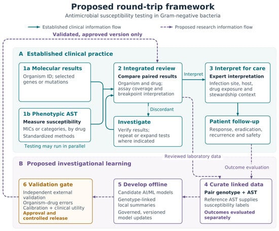
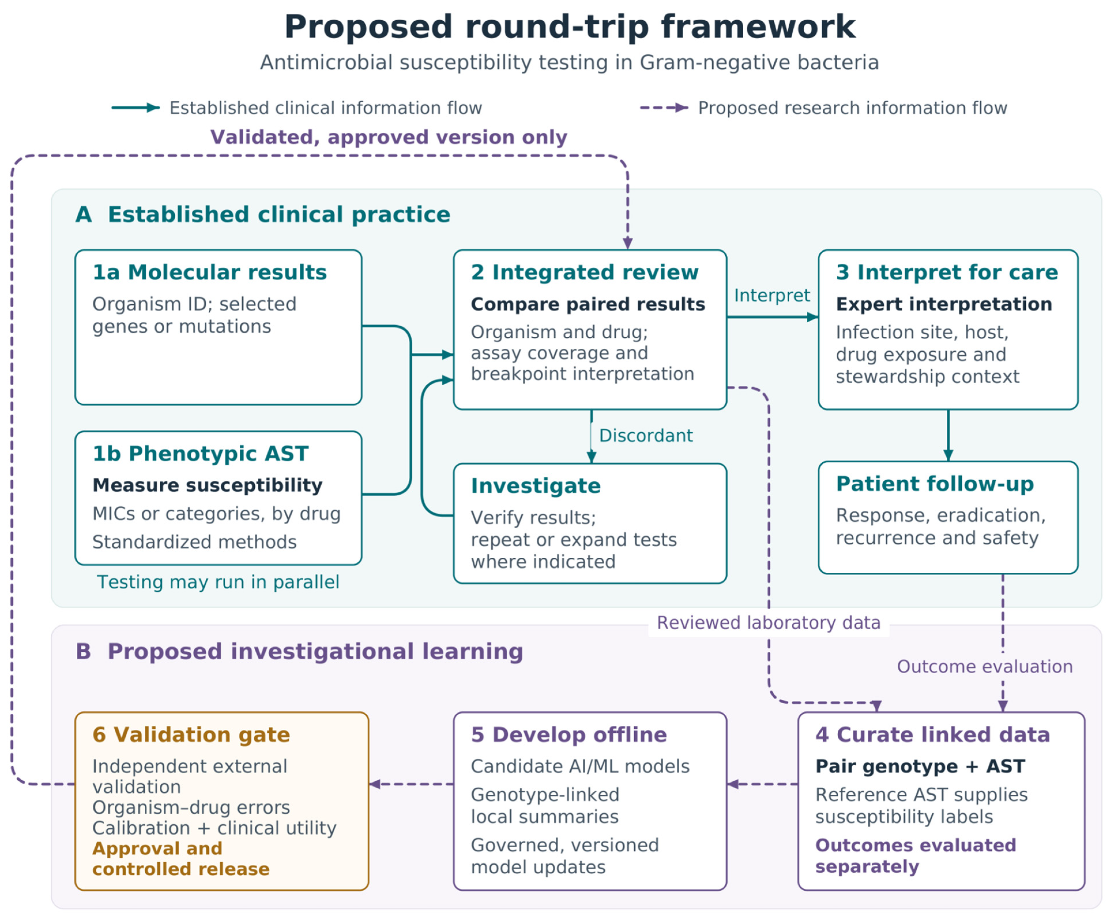

Disclaimer/Publisher’s Note: The statements, opinions and data contained in all publications are solely those of the individual author(s) and contributor(s) and not of MDPI and/or the editor(s). MDPI and/or the editor(s) disclaim responsibility for any injury to people or property resulting from any ideas, methods, instructions or products referred to in the content. |
© 2026 by the authors. Licensee MDPI, Basel, Switzerland. This article is an open access article distributed under the terms and conditions of the Creative Commons Attribution (CC BY) license.