


Abstract
Periprosthetic joint infection (PJI) is a severe and difficult-to-treat complication following artificial joint replacement, occurring in approximately 1–2% of total joint arthroplasties and normally caused by the adhesion of bacteria to the prosthesis and the formation of biofilms. Since this disease lacks a single definitive diagnostic indicator, a comprehensive judgment needs to be made by combining serum inflammatory indicators, joint synovial fluid tests, microbial cultures, imaging, and intraoperative pathology. Although microbial culture remains the gold standard, it usually requires more than 3 days. Microfluidics breaks through the limitations of traditional cultivation and conventional nucleic acid testing, which are characterized by long time consumption, large sample requirements, complex operations, and bulky equipment. Microfluidics has the irreplaceable advantage in real-time, micro-scale, and multi-indicator synchronous detection of bacterial pathogens, while the serious consequences of PJI require more advanced and rapid detection technologies to enable more effective treatment. In this review, in addition to summarizing the advances of microfluidic technology in the field of bacterial detection over the past five years, we also propose and analyze the microfluidic technology strategies that are likely to develop in the field of PJI detection in the future.
1. Introduction
Periprosthetic joint infection (PJI) is a serious complication related to total joint arthroplasty (TJA), which is one of the causes leading to implant failure and revision surgery [1]. The main cause of PJI is that bacteria invade the surrounding tissues of the prosthesis through the surgical process or hematogenous dissemination and form biofilms that are difficult to eradicate [2,3]. Despite continuous improvements in surgical asepsis and perioperative management, PJI still occurs in approximately 1–2% of primary TJA and accounts for a substantial and growing proportion of revision procedures worldwide [1]. Each episode of PJI is associated with prolonged hospitalization, repeated surgeries, and a heavy economic burden, and the mortality of patients with PJI has even been reported to be comparable to that of many malignancies [4]. These facts highlight that PJI is not merely a technical complication, but a life-changing event for patients and a major challenge for healthcare systems. Furthermore, among these pathogenic bacteria, Staphylococcus aureus (S. aureus) and Staphylococcus epidermidis (S. epidermidis) are the most important pathogens associated with PJI, because of their high virulence and recurrence characteristics [5,6]. The clinical similarities between PJI and other post-operative complications present significant challenges in developing timely and reliable PJI diagnosis.
As shown in Figure 1, diagnostic criteria of the Musculoskeletal Infection Society/International Consensus Meeting (2018) are the primary basis of PJI diagnosis, which include serological examination, joint synovial fluid analysis, pathology examination, imaging examination, and microbial cultivation. Clinically, the combination of bacterial culturing and biochemical identification is still the gold standard to detect PJI pathogens [7,8]. These methods can provide accurate results and enable antimicrobial susceptibility testing (AST) and antibiotic-related experiments, offering valuable clinical information. However, it usually takes 2 to 3 days, which is time-consuming and does not allow rapid detection [9]. This is the primary reason why empirical antibiotic treatments are frequently administered. Furthermore, biomolecular analysis technologies like mNGS (metagenomic Next-Generation Sequencing) can identify pathogenic bacteria and measure their drug resistance by amplifying specific gene sequences and drug resistance genes of the bacteria [10,11]. However, many related studies have also shown that bacterial resistance is not directly related to the presence or absence of specific genes [12]. Meanwhile, digital PCR technology can shorten the detection time to less than 1 h [13]. However, it is limited by certain requirements for the bacterial concentration in the detection sample and cannot provide information on the minimum inhibitory concentration (MIC) of antibiotics [14].
Figure 1.
Comprehensive diagnostic methods for PJI in clinical practice. (Created with assistance of Gemini Pro 3.1).
In conclusion, there is an urgent need for rapid, simple, highly sensitive, and high-throughput analysis technologies in the clinical diagnosis of PJI, including pathogen identification, concentration determination, AST, and the minimum inhibitory concentration of antibiotics. Current detection and clinical techniques for PJI are continuously advancing towards improved precision and speed. The traditional stable bacterial culture technique achieves enhanced PJI detection performance and shorter detection time by integrating advanced characterization technologies [15]. In terms of diagnosis, traditional serological markers (ESR, CRP) and synovial fluid testing systems have been continually refined. The application of novel synovial fluid biomarkers, including α-defensin and leukocyte esterase, has elevated diagnostic specificity to over 90% [16]. Also, most recently developed microfluidic qPCR devices can further reduce bacterial pathogen detection time to less than 60 min, achieving both sensitivity and specificity as high as over 90%, overcoming the long wait times which typically exceed days associated with conventional culture methods [17]. When current clinical tests are combined with an optimized microbiological approach incorporating metagenomic next-generation sequencing and bacterial pathogen culturing, the likelihood of missing low-abundance or low-virulence strains is significantly reduced, making this combination the best technological partner for PJI detection [10,18]. Overall, the currently common clinical detection techniques for PJI mainly rely on microbial culture, synovial fluid biomarker detection, and molecular testing, as shown in Table 1 [19,20,21,22,23,24,25,26,27,28].
Table 1.
Current clinical methods for detecting PJI.
However, in terms of detection time, accuracy, technical accessibility, and drug susceptibility testing, no existing clinical method achieves a satisfactory balance, and the final diagnosis still relies on waiting for the lengthy pathogen culture results. Microfluidic technology has overcome the inherent limitations of traditional bacterial detection, which relied on large laboratories and involved cumbersome operations and long processing times. It integrates the entire sample-processing and signal-reading process onto a chip, requiring only a small amount of sample to complete the detection within several minutes. This not only enables rapid bedside screening and immediate response for on-site pathogen identification, but also enhances the detection sensitivity to the level of individual bacterial pathogens, significantly reducing reagent and labor costs [29,30]. In scenarios such as emergency response for public health incidents, early diagnosis and treatment of infectious diseases, and rapid identification of pathogens in grassroots medical settings and resource-poor areas, it fills the capability gap of traditional detection methods, providing key technical support for precise bacterial infection control and rapid clinical decision-making [31,32]. Because microfluidic studies directly targeting PJI are extremely scarce, we exhaustively retrieved all available publications strongly related to both PJI and microfluidic detection, regardless of publication year. On the other hand, for the broader advances in microfluidic bacterial detection, we focused primarily on representative works published within the last five years (2020–2025). As a result, it is believed that microfluidic technology holds great potential in addressing the issue of improving the detection methods for PJI. This review work focuses on a summary of advances in microfluidic technology for pathogenic bacterial detection and its potential for use in PJI pathogen detection.
2. Advances of Microfluidic Technology in Bacterial Pathogen Detection
Taking advantage of integration [33], rapidity, high sensitivity, and low consumption [28], microfluidic technology has established several mature and cutting-edge applications in the field of bacterial detection. Normally, these technologies can be categorized based on the methods for functionality, mainly including optical detection [34,35,36], nucleic acid amplification (e.g., PCR [37,38]), electrochemistry [39,40], and protein biomarkers (as shown in Figure 2). As one of the most popular application fields, a substantial body of literature has accumulated over the past few decades, and many researchers have summarized and analyzed these works on microfluidic bacterial detection. In the following paragraphs, we summarize the novel related works over the past five years.
Figure 2.
The application of microfluidic technology for bacterial detection. Reprinted with permission from [35,36,37,38,39,40].
2.1. Optical Detection
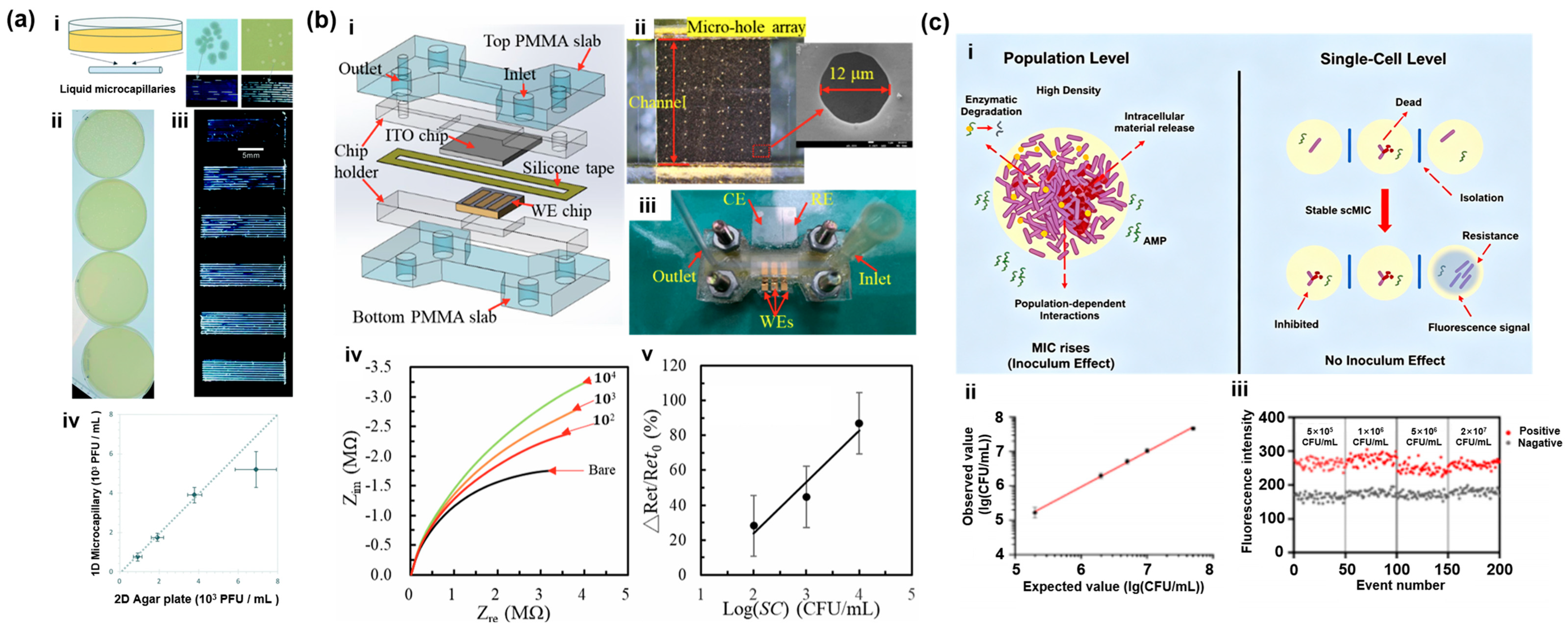
From the outset, optical technology has been serving as a perfectly compatible supporting method for microfluidics in the bacterial detection application [41]. In microfluidics, optical systems are typically integrated by embedding miniaturized optical fibers, waveguides, or microlenses directly into the chip substrate (such as PDMS or glass) during the microfabrication process. This precise on-chip alignment allows light to directly intersect the microchannel with minimal scattering or signal loss [42]. Due to its advantages of high sensitivity, fast response, and ease of integration, it has significantly enhanced the efficiency and accuracy of bacterial detection, leading to the development of multiple mature applications [43]. Laliwala et al. developed a paper-based fluorescence sensor array for rapid analysis of pathogenic bacteria [44]. The researchers pre-adsorbed environmentally sensitive fluorescent dyes onto paper microplates using lithography technology, allowing them to interact with bacterial cell membranes and generate unique fluorescence response patterns. By analyzing these response patterns using a neural network model, the sensor was able to effectively distinguish and identify 16 different bacterial species and their Gram staining status with an identification accuracy rate exceeding 90%. Dönmez et al. developed a microfluidic capillary-based label-free “dip-and-test” test strip platform for rapid and accurate one-dimensional (1D) counting of microbial colonies and phage plaques [45]. As shown in Figure 3a, 200 μm fluoropolymer microcapillaries were used as solid culture dishes. To achieve real-time monitoring of in situ growth of microfluidic microorganisms, the growing bacteria and the regions lysed by phages could be directly identified as “segments” or “gaps” in the capillary through light scattering. The test results showed that the entire detection routine was significantly shortened to 4 to 8 h, which was 6 times faster than traditional methods. Additionally, microfluidics is also commonly used for antibiotic testing related to bacteria. Tong et al. developed a laser-printed, paper-based microfluidic chip (mCD-μPAD) based on a multi-color fluorescent carbon dot biosensor for the on-site visual determination of various antibiotics in aquatic products [46].
In practice, the boundary between optical technologies and molecular biology detection techniques is often blurred. Optical sensors are one of the best methods for visualizing the results of molecular biology reactions [47,48]. Trieu et al. proposed a fully integrated, paper-based origami microfluidic device for POCT testing, capable of achieving rapid colorimetric identification of live pathogenic bacteria through nucleic acid amplification testing [49]. Furthermore, Tavakoli et al. developed a fully paper-based microfluidic analysis device (μFPAD) integrating loop-mediated isothermal amplification (LAMP) and single-stranded DNA (ssDNA)-functionalized graphene oxide (GO) nano-biosensors for rapid diagnosis of infectious diseases [50]. The test results showed that the prototype could provide flexible qualitative diagnosis within 1 h with a limit of detection (LOD) of 6 DNA copies per detection zone. Subsequently, Zhuang et al. introduced a microfluidic paper-based analysis device (RPA-Cas12a-μPAD) combining recombinase polymerase amplification (RPA), CRISPR/Cas12a technology, and surface-enhanced Raman scattering (SERS) for ultra-sensitive, immediate detection of pathogenic bacteria in food samples [51].
2.2. Electrochemistry Detection
Unlike conventional electrochemical sensors with rod or wire electrodes, microfluidic electrochemical systems integrate miniaturized electrodes directly onto the inner surfaces of the microchannels through advanced microfabrication techniques such as photolithography, screen-printing, or thin-film metal deposition [52]. Electrochemical sensors enable chemical signals of the analyte to be converted into electrical signals via electrodes, capacitating detection at the micrometer scale. This approach offers advantages such as high sensitivity, low cost, and ease of miniaturized integration [53]. As shown in Figure 3b, Police Patil et al. introduced a rapid, highly sensitive detection for Salmonella choleraesuis using positive dielectrophoresis (pDEP) enrichment and electrochemical impedance spectroscopy (EIS) sensing technologies [54]. pDEP was applied within the microelectrode pores of the microfluidic chip to rapidly collect and concentrate these bacterial complexes with immunomagnetic beads (IMBs) functionalized with anti-Salmonella antibodies. It achieved quantitative determination of bacterial concentration via EIS. Test results demonstrated excellent analytical performance of this microfluidic platform in spiked, undiluted milk samples in only 50 min. The detection range was from 10 to 104 CFU/mL, with a low LOD of 2 CFU/mL. Guo et al. proposed an electrochemical biosensor combining the CRISPR/Cas12a system with a microfluidic chip for simultaneous and rapid detection of foodborne pathogens [55]. To target Staphylococcus aureus and Salmonella, a dual-layer, vertically integrated microfluidic chip was used to load the sample and combine recombinase polymerase amplification (RPA) with the CRISPR/Cas12a target-recognition system. A three-electrode system modified with a composite material of single-walled carbon nanohorns, polypyrrole, and gold nanoparticles (SWCNHs-ppy-AuNPs) could simultaneously analyze both pathogens within 65 min.
At the same time, Jiang et al. exploited a novel gene sensor based on electrochemical cotton-based devices (ECDSs) for low-cost, high-sensitivity nucleic acid detection [56]. Using carbon ink and solid wax screen-printing technology, they fabricated ultra-low-cost sensing devices on fabric and significantly enhanced electrochemical signals by modifying the sensing interface with a CdTe quantum dots/multi-walled carbon nanotubes (CdTe QDs/MWCNTs) nanocomposite. Hou et al. also proposed an ultra-sensitive biosensing system based on 3D magnetic grid separation and urease-catalyzed signal amplification for rapid detection of Salmonella in large-volume complex samples [39]. This innovative system enables highly sensitive quantitative identification in actual food samples through exceptional enrichment and signal amplification capabilities, achieving a LOD as low as 10 CFU/mL. Jiang et al. developed a label-free electrochemical aptasensor based on a microfluidic device for rapid detection of Vibrio parahaemolyticus in seafood [57]. The study demonstrated that the sensor could complete the entire detection within just 30 min, with a dynamic detection range of 10 to 106 CFU/mL and an LOD as low as 5.74 CFU/mL.
2.3. Protein Biomarkers
Conventional protein biomarker profiling, such as traditional ELISA, relies on macroscopic well plates requiring prolonged incubation times and large sample volumes. In contrast, the integration of biomarker detection within microfluidic systems revolves around on-chip surface functionalization and compartmentalization. Antibodies or capture probes are covalently immobilized directly onto the inner walls of the microchannels or conjugated onto magnetic microbeads that are dynamically controlled by external magnets integrated into the chip [58]. In rapid bacterial pathogen detection and identification of drug-resistant bacteria, biomarkers are the key targets for achieving highly specific and sensitive detection in microfluidic bacterial assays [59]. Biomarkers can distinguish the target species from other contaminants, avoiding false positives and enabling detection in microfluidic systems with minute sample volumes [31]. By leveraging the signal amplification characteristics of the biomarkers, microfluidic methods can accurately detect rare target bacterial pathogens from samples at low concentrations [60]. Frigoli et al. introduced a thermal sensor based on molecularly imprinted polymers (MIPs) to achieve indirect and rapid detection of the pathogen Pseudomonas aeruginosa by detecting the main toxin, pyocyanin [61]. Pyocyanin could cause a change in thermal resistance, and by measuring this thermal resistance parameter, the sensor could achieve accurate quantification.
Mei et al. utilized a microfluidic device to explore the inoculum effect produced by antibacterial peptides when facing high bacterial-load infections [62]. As shown in Figure 3c, bacterial populations were sorted into single cells and distributed in independent micro-chambers, thereby eliminating the interference at the population level. The study found that the MIC of the population would significantly increase with the increase in bacterial inoculation density, while the MIC at the single-cell level remained unchanged. Additionally, Guo et al. introduced a compact smartphone microfluidic platform integrating double membrane filtration enrichment and Au@Pt nanozyme catalytic colorimetric immunoassay [63]. This innovative system can rapidly and quantitatively detect uropathogenic E. coli in real clinical urine samples within 15 min, with an accurate LOD of 105 CFU/mL. The system uses a double-layer membrane microfluidic chip to efficiently filter out large volumes of urine sediment and physically retain the target bacteria. Then, porous spherical Au@Pt nanozyme probes modified with specific antibodies cascaded catalysis of the colorless substrate (TMB) to oxidize into blue ox-TMB, generating a significant optical signal amplification interaction. The colorimetric sensing smartphone provides an anti-interference, digital, visual, real-time diagnostic tool to reduce the risk of blind empirical medication due to subjective misjudgment or diagnostic delay.
In recent years, the combination of microfluidics technology and protein biomarkers has developed rapidly. Some studies have proposed exploring the impact of the integration and fixation of protein biomarkers on microfluidic chips. For instance, Kim et al. comprehensively evaluated diverse protein immobilization techniques for microfluidic assays [64]. Their work highlighted the distinct functional advantages of direct covalent immobilization on glass and polymer substrates, the use of three-dimensional packed beds to increase surface area, and the deployment of stimulus-responsive “smart immobilization” strategies. Building on the need for advanced fluid control and assay stability, Mok et al. developed a digital microfluidic platform that physically decouples the biomarker capture chambers from the downstream detection zones [65]. This architectural separation proved highly effective, as it allows for the independent optimization of fluid flow rates and wash steps, thereby preserving the structural stability of the immobilized proteins while minimizing shear stress during pressure-driven or continuous flow operations. More recently, Sathish et al. provided a comprehensive blueprint for microfluidic sensor development, extensively analyzing how different spatial immobilization architectures directly influence fluid dynamics [66]. They elucidated how physicochemical surface alterations during protein integration dictate continuous capillary flow stability and mass-transport limits. Furthermore, their study demonstrated how specific integration methods, such as the transition from flat functionalized walls to monolithic porous structures or packed beads, significantly alter back-pressure profiles and structural stability within pressure-driven flow systems.
2.4. Polymerase Chain Reaction
Microfluidic PCR bacterial detection technology is a molecular diagnostic technique that miniaturizes and highly integrates the nucleic acid amplification process of traditional centralized laboratories onto microfluidic chips [67]. Molecular biology technologies focused on biological macromolecules such as nucleic acids and proteins have developed a comprehensive technical category covering the entire chain, from amplification and detection to sequencing, editing, and interaction analysis [68]. In this technological field, nucleic acid-related technologies, protein-related technologies, gene cloning and expression technology, as well as gene editing and regulation technologies all play important and irreplaceable roles in multiple research and industrial fields [69]. Moreover, cutting-edge new technologies, such as optogenetics [70,71], chromatin immunoprecipitation (ChIP) [72], methylation-specific PCR (MSP) [73], ATAC-seq [74], etc., are constantly being proposed. Indeed, all of the aforementioned microfluidic bacteria detection technologies cannot be completely separated from the molecular biology technologies, as their mutual combination has led to better detection functionality. It is the core technical foundation for the implementation of precision medicine, and it reshapes the medical model throughout the entire process from prevention and diagnosis to treatment [75].
If we summarize broadly, applications related to molecular biology account for at least half of all the work. Here, we listed some of the recent research works whose highlights were entirely focused on molecular biology. Yang et al. introduced a microfluidic chip using a continuous-flow polymerase chain reaction (CF-PCR) array for simultaneously amplifying the target genes of multiple periodontal pathogenic bacteria [76]. This array platform was successfully applied to the gene amplification of common periodontal pathogenic bacteria, Porphyromonas gingivalis, Treponema denticola, and Tannerella forsythia. A dual-layer droplet CF-PCR microfluidic chip was designed by Li et al. to achieve ultra-fast amplification and detection of bacteria by reducing fluid resistance [77]. The test results showed that by increasing the height of the serpentine nanoscale droplet microfluidic channel with the double-layer structure, the flow resistance could be significantly reduced, and the chip could stably generate droplets with a volume of only 0.032 nL. Then, Wan et al. proposed a super-fast PCR system based on digital microfluidics for efficient POC diagnosis of pathogens [78]. They implemented a series of ingenious hardware and sensing innovations to ensure system stability in the generation of microdroplets in extreme thermal cycling.
Additionally, Li et al. introduced an automated and portable microfluidic nucleic acid testing device, innovatively integrating a rotation-valve-assisted sample pretreatment with the RPA-T7-Cas13a reaction system in a single-chip chamber to achieve “one-step” nucleic acid testing [79]. This platform could accurately detect Group B Streptococcus (GBS) from 8 types of pathogenic bacteria and achieved a 100% accuracy rate in tests on 16 positive and 24 negative clinical samples. The entire automated detection process could be completed within 30 min. Most recently, Zheng et al. proposed a high-throughput single-cell sequencing technology called Microbe-seq, using microfluidic droplets to achieve the release of nucleic acids, whole-genome amplification, and barcode labeling of individual microorganisms in complex microbial communities [80]. Thereby, it could obtain single-microorganism amplified genomes (SAGs) with strain-level resolution without the need for pure cultivation. The researchers applied this technology to human gut microbiome research and sequenced over 20,000 microbial single cells from a single donor. Nearly 100 high-quality bacterial genomes were assembled, including multiple multi-strain species at the subspecies level. Finally, Murphy et al. developed a semi-automated NGS library construction platform based on droplet microfluidics technology [81]. It aimed to solve the problems of sample loss, cumbersome operation, and batch errors in the conventional manual library construction process for extremely low-mass samples. Computer-controlled multi-channel micropumps and PDMS chips were used to highly integrate the complex, multi-step library preparation process of DNA end repair, adapter connection, and purification.
Figure 3.
Advances in microfluidic technology in bacterial pathogen detection. (a) Microfluidic optical methods for detecting bacteria: Label-free 1D microfluidic dipstick counting of microbial colonies and bacteriophage plaques. (i) A miniaturized one-dimensional method for enumerating liquid colonies and plaques. (ii) Representative endpoint images displaying five ascending concentrations of phage K, alongside a phage-free control, plated in Petri dishes using the traditional double agar layer technique. (iii) Evaluation of identical phage K concentrations utilizing microcapillary dip-strips. (iv) Comparison of plaque-forming units determined via the microcapillaries versus the double agar layer method. Due to near-complete clearance within the capillary at the highest concentration, plaque enumeration was unfeasible; therefore, only four concentration levels are graphed. The plotted data represents the mean counts derived from 10 replicate capillaries, with error bars denoting ±1 standard deviation. The dashed line illustrates a 1:1 correlation between the two quantification methods. Reprinted with permission from [45]. (b) Microfluidic electrochemical methods for detecting bacteria: Microfluidic platform for integrating dielectrophoretic concentration and impedimetric sensing for rapid Salmonella choleraesuis detection. (i) Exploded view of the microfluidic chip. (ii) Optical photograph of a single gold working electrode (Au WE), accompanied by a SEM image detailing a microhole on the WE surface. (iii) A photographic image of the fully assembled device. (iv) Nyquist plots of IMBs@SC complexes at varying concentrations, obtained from 100 μL milk samples. (v) Calibration curves corresponding to (iv). Each data point represents the statistical mean derived from three independent WEs. Reprinted with permission from [54]. (c) Microfluidic detection methods for bacteria based on protein biomarkers: Investigation of the inoculum effect of antimicrobial peptides utilizing a microfluidic chip platform. (i) Schematic diagram of the principle. (ii) Linear correlation between positive well counts and E. coli inoculation density. (iii) Fluorescence intensity comparison of positive versus negative E. coli reaction wells. Reprinted with permission from [62].
Synovial fluid is a highly viscous and compositionally complex biological matrix containing abundant hyaluronic acid, proteins, inflammatory cells, and cellular debris. These characteristics, together with blood contamination and low bacterial concentrations, could cause microchannel clogging, unstable fluid transport, nonspecific adsorption, and interference with downstream sensing, thereby increasing the difficulty of direct on-chip analysis. As a result, conventional centrifugation of synovial fluid is still an effective approach for sample pretreatment, which could solve the above complex composition issue as well as enrich the sample for high pathogen concentration. Furthermore, the volume of synovial fluid obtained by joint aspiration varies considerably among patients, ranging from insufficient samples or even unsuccessful aspirations to several milliliters or more. In current clinical PJI diagnosis, the sample volume required for individual laboratory tests generally ranges from approximately 20 μL to 2 mL, depending on the analytical method. For example, approximately 0.5 mL is commonly required for synovial leukocyte counting or α-defensin analysis, at least 1 mL is generally recommended for microbial culture, and approximately 2 mL may be required for next-generation sequencing [82]. Depending on the intended assay, additional preprocessing steps, such as sample homogenization, viscosity reduction, centrifugation, bacterial isolation, cell lysis, and nucleic acid extraction, may be required before the samples are suitable for analysis. On the contrary, these complex and mature processing steps are not suitable for being fully integrated onto the chip, as this would reduce the reliability of the microfluidic chip. In contrast, microfluidic technologies are expected to offer precise controllability on small-volume samples as well as pathogens. Therefore, the advantages of microfluidic technology, including low sample and reagent consumption, efficient pathogen enrichment, high integration, and the potential for rapid and highly sensitive detection, are particularly relevant to the clinical analysis of limited and complex synovial fluid specimens.
3. Microfluidic Methods for PJI Detection
Compared with blood, urine, etc., less than 0.1 mL of synovial fluid can be extracted from the cavities of some specific patients [83]. As a result, microfluidics should be inherently competent for the PJI detection task. However, it is found that only a very small amount of microfluidic research is related to PJI detection technologies. To provide a comprehensive overview of the current landscape of microfluidic technologies for PJI diagnosis, Table 2 summarizes and compares various methodologies. These approaches are broadly categorized into two primary diagnostic strategies, including the analysis of synovial biomarkers and molecular diagnostics [84,85,86,87,88]. Over the past decade, Lee’s team has focused on the automated and precise diagnosis of PJI. They first developed a microfluidic platform for aptamer screening. They successfully completed the high-specificity nucleic acid aptamer screening for the crucial biomarker of PJI, human neutrophil peptide 1 (HNP-1, also known as alpha-defensin). Subsequently, they further optimized the microfluidic screening scheme for alpha-defensin and thoroughly verified its application potential in clinical synovial fluid analysis [89].
Table 2.
Microfluidic methods for PJI detection.
Afterward, they shifted their focus to the development of miniaturized detection methods and prototype devices. A miniaturized aptamer sandwich method was established by combining the selected aptamers with microfluidic technology, which achieved rapid and highly sensitive quantitative detection of HNP-1 in synovial fluid [90]. Then, to further reduce the sample volume required for clinical testing and enhance portability, a paper-based aptamer analysis platform was developed, promising precise detection with extremely low sample volumes. It was based on the innovation in material heterogeneity and leveraged the capillary-driven advantages of paper-based microfluidics [91]. Most recently, in addition to HNP-1, a dual aptamer sandwich method on an integrated platform was developed for detecting C-reactive protein (CRP) in synovial fluid, which demonstrated the versatility of this detection scheme, as shown in Figure 4a [86]. Additionally, as shown in Figure 4b, Chang et al. developed an integrated microfluidic system specifically designed for rapid isolation and diagnosis of live bacteria from human joint fluid [92]. This system innovatively combined the pre-treatment technology of azide-ethidium bromide with the bacterial primer-probe PCR amplification. Azide-ethidium bromide could specifically penetrate the cell membranes of dead bacteria and bind to their DNA to inhibit subsequent amplification, thereby avoiding false positives due to interference from dead bacterial DNA. It was shown that the microfluidic platform could complete the analysis process of clinical PJI samples within 55 min, and the LOD for typing bacteria in the joint fluid matrix reached 102–104 CFU/mL. Finally, they presented a fully automated microfluidic system to achieve simultaneous detection of CRP and HNP-1, the dual biomarkers of PJI, in the synovial fluid sample within 45 min. This achievement eliminated the risks of false positives or false negatives that might arise from traditional single indicators. It was closely aligned with the immediate testing (POCT) requirements in real medical scenarios.
Figure 4.
Microfluidic methods for PJI detection. (a) A dual-aptamer sandwich assay by detection of C-reactive protein in synovial fluid on an integrated microfluidic system for diagnosis of PJI. (i) Schematic of the on-chip magnetic bead-based dual-aptamer CRP assay. CRP in positive samples or synovial fluid is captured by St1 primary aptamer-coated magnetic beads. Following magnetic isolation and two PBS washes, the complexes are incubated with a secondary aptamer (St1 or St2). After a subsequent isolation and washing cycle, the complexes undergo horseradish peroxidase (HRP) incubation and TMB assay. (ii) Fluorescence microscopy visualization (50×) of the assay at various CRP concentrations (0, 0.1, 1, and 10 mg/L). Reprinted with permission from [86]. (b) Rapid isolation and diagnosis of live bacteria from human joint fluids by using an integrated microfluidic system. (i) Illustration of the microfluidic chip. Photographs of the (ii) bottom and (iii) upper layers of the chip. (iv) The fluorescent results from the integrated microfluidic system (* p < 0.05). (v) Slab gel electrophoresis results confirming the fluorescence readouts. Reprinted with permission from [92]. (c) Rapid detection of bacteria in PJI via microfluidic qPCR chip. (i) Illustration of microfluidic chip (ii) Photograph of the microfluidic chip. (iii) Box plots of patient bacterial loads. (1) S. aureus bacterial load for all patients. (2) S. epidermidis bacterial load for all patients. (3) S. aureus bacterial load below the positivity threshold. (4) S. epidermidis bacterial load below the positivity threshold (**** p < 0.001). Reprinted with permission from [17].
In recent years, PCR technology has remained the most mature and commonly used detection principle in microfluidic technology for the detection of PJI. As shown in Figure 4c, Feng et al. clinically validated a microfluidic qPCR chip across 310 PJI and aseptic failure patients for the rapid screening of Staphylococcus aureus and Staphylococcus epidermidis [17]. Compared with conventional joint fluid culture and standard qPCR, their microfluidic system markedly reduced the turnaround time to 59.4 ± 4.2 min while enabling the sensitive detection of sub-threshold, low-load bacterial infections. To address the limited sensitivity and standard-curve dependency of conventional real-time PCR in early-stage PJI screening, Tak et al. established a 16S rDNA-targeted droplet digital PCR platform for precise pathogen quantification [88]. Using synthesized model plasmids, the authors demonstrated that ddPCR could detect down to 400 ag of target DNA, lowering the limit of detection by over an order of magnitude compared to real-time PCR. When further challenged with bacterial genomic DNA, the ddPCR assay successfully identified target sequences at ultra-low levels of 50 fg for E. coli, 70 fg for S. epidermidis, and 90 fg for S. aureus, demonstrating superior analytical sensitivity for early PJI intervention.
Therefore, although microfluidic technology has advanced rapidly in bacterial detection, it remains in its infancy for PJI. Three interrelated reasons explain this gap. First, PJI exceeds the recognition capability of typical microfluidic assays, as PJI involves diverse and polymicrobial pathogens, and affinity ligands frequently cross-react among closely related staphylococci. On the other hand, for typical positive synovial fluid (102–103 CFU/mL), a microliter-scale aliquot contains only a few or zero target cells, so detection is influenced by Poisson statistics and diffusion-limited capture within the microchannels [93,94]. Then, PJI is a niche problem with a large interdisciplinary barrier. Few microfluidic researchers are familiar with this specialized complication; clinical validation samples are scarce. Finally, biofilms are physically incompatible with microfluidic architectures, while metabolically dormant persister cells further compromise activity- and growth-based readouts [95].
4. Perspectives
4.1. Microwell Chips
Among the numerous microfluidic device designs, it is believed that the microwell chip holds great potential in addressing the challenges faced in PJI detection. A microfluidic device with multiple individual cavities that are used as chambers for on-chip physical, biological, and chemical micro-reactions is broadly referred to as a microwell chip.
Microwell chips, due to their superior ability to precisely control small volumes of fluids, can better achieve precise detection of pathogens at the single-cell level. In contrast to numerous diseases, PJI diagnosis physically imposes a combination of constraints that is rarely encountered in other applications. The sample is extremely scarce and hard-won, and spatially heterogeneous after biofilm dispersion from the prosthesis. Most established microfluidic biosensing formats are optimized for either abundant samples, high target concentrations, or single readouts; therefore, they do not map naturally onto the PJI problem. Meanwhile, practically, PJI is a low-incidence complication with a narrow patient pool, so clinical validation samples are scarce. The strong interdisciplinary barrier between microfluidics and orthopedic infection means few researchers recognize this unmet need.
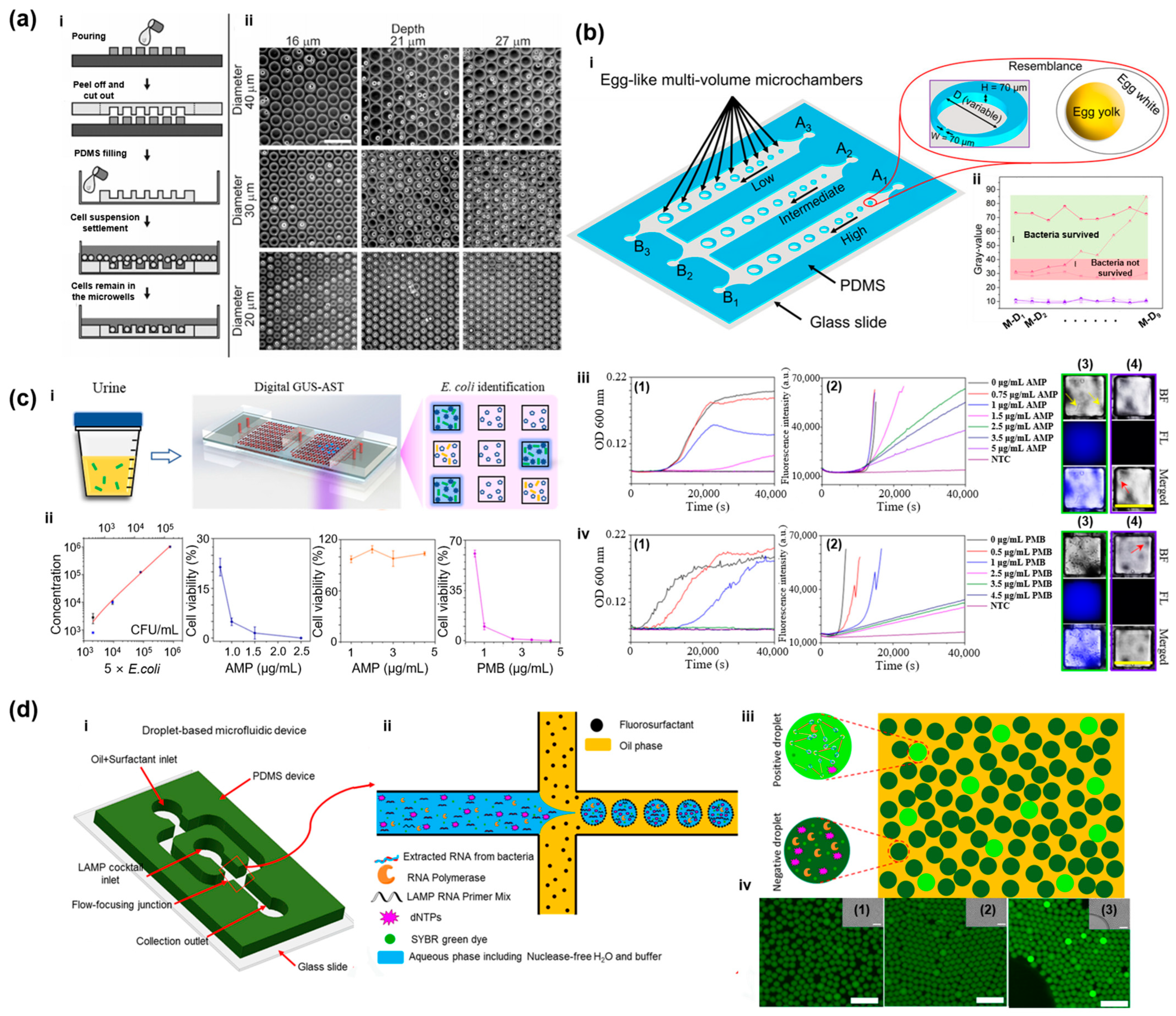
Taylor et al. introduced a chip with microwell arrays fabricated by an etching method to accommodate individual viable cells for biosensing assays [96]. This pioneering work could be the origin of the array of microwell structure inspiration and microwell chips. At almost the same time, Inoue et al. used the microwell chips to isolate, localize, and experimentalize on single cells in separated micro-sized wells [97]. Then, the Folch group pioneered the use of the PDMS microwell chip comprising tens of thousands of microwells [98]. In Figure 5a, the effects of microwell design and dimensions on single-cell trapping efficiency were systematically studied in this work. After the nascent stage, the development of microwell chips primarily focused on achieving on-chip single-cell encapsulation. These devices largely relied on gravity sedimentation or surface fluid flow to passively deposit cells into cylindrical or square cavities. These studies defined the iconic design of microwell chips and laid the physical and application foundation for microwell chip functionalities over the following decade. In addition to droplet microfluidics, microwell chips are another option for digital PCR applications. Fan et al. utilized microwell chips for high-throughput single-cell gene expression profiling, establishing the cavity array as a core technological paradigm for single-cell sequencing [99]. Subsequently, microwell-based platforms like BD Rhapsody and 10× Genomics’ iterative microfluidic technologies have transformed single-cell transcriptomic and proteomic analyses into standard scientific tools.
Microwell chips have also diversified extensive applications in bacterial detection, including AST and the determination of the MIC for various antibiotics [100,101]. Jalali et al. designed a microwell chip composed of a cavity array to confine a small number of bacteria within a restricted spatial volume, which allowed for high-precision monitoring of their growth [102]. Utilizing this chip in conjunction with monitoring equipment, they investigated the effects of different antibiotics on the growth of S. aureus. The experiments successfully determined the MICs of various antibiotics within 3 h, significantly shortening the detection time and simultaneously distinguishing the specific effects of different antibiotic classes on S. aureus’s proliferation. Similarly, as shown in Figure 5b, Azizi et al. reported an egg-like multivolume microchamber (EL-MVM2) microfluidic platform for the rapid generation of antibiotic concentration gradients [103].
In the last few years, our team has also persisted in systematic and progressive research to explore the development of microwell chips in the fields of single-particle manipulation, single-cell analysis, and rapid clinical pathogen detection. To enhance the microwell chip detection functionality, a self-driven carbon-doped high-density microwell array was designed utilizing pre-stored negative pressure in PDMS material for autonomous sample loading and incorporating carbon powder doping to significantly reduce crosstalk from stray fluorescent signals between microwells. The proposed revised microwell chip achieved dynamic monitoring of single-cell energy metabolism with a high signal-to-noise ratio. The research emphasized improving the pump-free integration of microwell devices and lowering the optical detection limit, which greatly promoted the usability of such microwell technologies and prototypes [104]. Then, for precise detection of foodborne and waterborne pathogens, a chemiluminescence digital microwell array chip was developed, combining Poisson-distributed monodisperse droplet encapsulation with the hydrolysis reaction of a fluorescent substrate by E. coli-specific β-D-glucuronidase (GUS) [105]. This method enabled absolute quantification of E. coli and rapid differentiation between live and dead bacteria in complex matrices. It addressed the long processing time and susceptibility to sample turbidity inherent in conventional culture methods. Based on the above research, as shown in Figure 5c, a complete set of E. coli clinical test methods was proposed using the refined three-dimensional microwell chip called “digital GUS-AST”. The key mechanism of this detection method leveraged the phenomenon that a specific metabolism-related enzyme, GUS, could translate the presence of E. coli into a bright fluorescence signal [35]. The digital GUS-AST could quantify the E. coli concentration and directly indicate antibiotic efficacy (AST) and determine the MIC for the analysis of separated bacterial populations in urine. It was demonstrated that the entire detection workflow can be completed within 4.5 h with a satisfactory limit of detection reaching 39 CFU/mL. To further advance the technology towards point-of-care testing (POCT), our team subsequently developed a ready-to-use 3D micropore array chip, incorporating internal capillary valve fluid distributors, preloaded lyophilized reagents, and vacuum-sealed pre-packaging features, enabling the direct, instrument-free single-cell-level AST and MIC determination directly from clinical urine samples [106].
In addition to microwell arrays, droplet microfluidics represents another prominent miniaturized system that addresses the challenges of low-volume sample analysis. In bacterial detection, microwell chips leverage spatial fixation by capturing individual cells within microscopic wells and seamlessly enable continuous, time-lapse imaging of in situ growth dynamics and morphological changes. However, droplet microfluidics detects bacteria by rapidly encapsulating single cells into picoliter-scale water-in-oil emulsion droplets, offering dynamic fluid manipulation. Compared with microwell chips, droplet microfluidics have the advantage of high throughput, enabling rapid pathogen detection and high-throughput phenotypic analysis.
Most recently, droplet microfluidics have demonstrated significant potential in microbial analysis and pathogen detection. Hsieh et al. developed a rapid single-cell pathogen diagnostic technology utilizing picoliter droplets [107]. By encapsulating individual bacteria within microdroplets and employing fluorescent peptide nucleic acid probes targeting 16S rRNA, this platform achieves highly sensitive bacterial identification without amplification. Additionally, Tiwari et al. introduced a label-free bacterial detection methodology [108]. They encapsulated bacteria or bacteria-phage mixtures into microdroplets and, by coupling Z-stack bright-field microscopy with morphology-based deep learning algorithms, successfully achieved precise differentiation of morphologically distinct bacterial species. When processing complex matrix samples, the droplet compartmentalization effect can effectively sequester background inhibitors. As shown in Figure 5d, Azizi et al. proposed a quantitative detection protocol that couples droplet microfluidics with RNA-based loop-mediated isothermal amplification (LAMP) [109]. By co-partitioning bacterial lysates, targeting primers, and enzymatic reagents into nanoliter-scale droplets, this method dramatically enhances both the specificity and sensitivity of pathogen detection, successfully enabling the rapid identification of Salmonella typhimurium in contaminated milk samples. To further expand multiplexing capabilities, Shang et al. pioneered an innovative diagnostic platform integrating droplet microfluidics with a recombinase-aided amplification (RAA)-assisted one-pot CRISPR/Cas13a assay [110]. The platform has achieved precise quantitative analysis of the endpoints and can identify up to seven different foodborne pathogens.
Transitioning from targeted identification to the comprehensive profiling of genetic heterogeneity, droplet microfluidics has equally propelled the advancement of single-cell sequencing. Lan et al. developed single-cell genomic sequencing (SiC-seq), which utilizes droplet microfluidics to isolate, fragment, and barcode the genomes of single cells, followed by pooled Illumina sequencing [111]. In a synthetic community comprising Gram-negative bacteria, Gram-positive bacteria, and fungi, this study demonstrated an ultra-high-throughput sequencing capacity of over 50,000 cells per run.
Figure 5.
Microwell chips and droplet microfluidics for bacterial detection. (a) Large-scale single-cell trapping and imaging using microwell arrays. (i) Cell seeding in a microchip. (ii) Nine representative images of RBL-1 cells in microwells across all combinations of three diameters (D = 20–40 μm) and three depths (H = 16–27 μm). Reprinted with permission from [98]. (b) Antimicrobial Susceptibility Testing in a Rapid Single Test via an Egg-like Multivolume Microchamber-Based Microfluidic Platform. (i) Perspective view of the EL-MVM2 platform (inset: an individual microchamber embedded in the main channel). The platform is named after its egg-like microchambers, which comprise an “egg white” (PDMS wall) and an “egg yolk” (bacterial suspension thermally incubated with the drug). (ii) Quantitative determination of the MIC based on resazurin fluorescence intensity changes during the AST assay. Reprinted with permission from [103]. (c) All-In-One Escherichia coli viability assay for multi-dimensional detection of uncomplicated urinary tract infections. (i) Principle of the digital GUS-AST assay for E. coli identification and quantification, alongside AST and MIC determination. (ii) Quantification of EK-19 recovered from urine. Black and blue squares represent the MPN estimate and Poisson distribution-derived values, respectively, alongside MIC determinations of E. coli strains against AMP and PMB. Error bars denote the standard deviation of three replicates. (iii) Conformity testing for AMP susceptibility between broth microdilution (1) and fluorescent GUS assays (2). Digital GUS-AST assay of strain O114 exposed to (3) 0.75 μg/mL and (4) 2.5 μg/mL AMP. (iv) Conformity testing for PMB susceptibility between broth microdilution (1) and fluorescent GUS assays (2). Digital GUS-AST assay of EK-19 exposed to (3) 1 μg/mL and (4) 3.5 μg/mL PMB. Yellow and red arrows indicate growing and static E. coli. Scale bars denote 50 μm. Reprinted with permission from [35]. (d) Pathogenic bacteria detection using RNA-based loop-mediated isothermal-amplification-assisted nucleic acid amplification via droplet microfluidics. (i) Illustration of the microfluidic flow-focusing chip designed to compartmentalize the LAMP reaction mixture into water-in-oil (W/O) droplets. (ii) Enlarged view of the flow-focusing cross-junction, from which the generated W/O droplets are exported into a PCR tube. (iii) Post-amplification fluorescence detection and binary (“Yes/No”) digital quantification of positive droplets. (iv) Representative fluorescence micrographs of droplets containing S. flexneri-specific primers paired with: (1) no RNA template (negative control), (2) non-target S. typhimurium RNA (negative), and (3) target S. flexneri RNA (positive). Reprinted with permission from [109].
The above description of the microwell chip reveals its tremendous application prospects in the clinical diagnosis of PJI. Facing the major clinical challenges in the diagnosis of PJI, the microwell chip possesses inherent advantages to provide solutions to these problems [112]. Although the limited volume of patients’ synovial fluid samples is sometimes even insufficient for necessary NGS and culture tests [113], a microwell chip with tens of thousands of cavities merely requires a few microliters of sample for effective detection [84,114]. The aliquot sample could be enriched, then injected into the microwell chip at a higher concentration. This significant advantage enables the microwell chip not only to avoid disrupting the existing detection process, but also to provide a fallback solution in extreme patient situations [115]. Furthermore, the typical 3 to 14 days’ detection time of traditional microbial culture could be significantly shortened to a few hours [116,117]. Likewise, compared with molecular technologies, microwell chips enable highly sensitive and specific single bacterial detection [105,118], alongside the capability to perform AST and MIC assays taking advantage of their single-bacteria packing and culturing [119,120]. The functionalities of microwell chips are highly compatible with the current diagnostic limitations of PJI. In the near future, once the microwell chips acquire an appropriate detection principle for PJI pathogens, it is believed that microwell chips hold the promise to be one of the candidates for PJI diagnosis and assist physicians in prescribing timely targeted therapies [112].
4.2. Microfluidic Detection Study on PJI’s Pathogen
The PJI pathogens are primarily bacteria, with a few being fungi [2]. The presence of multiple pathogens makes detection techniques based on specific reactions unsuitable [121]. Although hardly any microfluidic researchers have focused their interests on PJI detection, it was found that PJI pathogens have been frequently studied in the past decade [6]. We have summarized the research on the application of microfluidic technology in the detection of PJI-related pathogens, and have organized the characteristics of the technical methods proposed by each work into Table 3 [122,123,124,125,126,127,128,129,130].
Table 3.
Microfluidic detection methods on PJI pathogens.
4.2.1. E. coli
E. coli is not only relevant in the application of PJI detection, but it is also basically the most representative bacterial pathogen of microfluidic detection technology [30]. In recent years, PJI has been a severe and difficult-to-treat complication following artificial joint replacement, caused by the adhesion of bacteria to the prosthesis and the formation of biofilms.
Microfluidic technology for rapid detection of E. coli has been accelerating its evolution towards a highly integrated and multi-dimensional detection approach with enhanced performance in all aspects [131]. In sample pretreatment processing, emerging devices aim to break through the bottleneck of the cumbersome and time-consuming traditional nucleic acid extraction. Gong et al. developed a microfluidic system based on double-layer membranes and high-throughput droplets with an integrated pipetting automation module to achieve automatic sampling of E. coli from environmental samples, pL droplet encapsulation, and quantitative enrichment, which effectively solved the problem of cumbersome traditional operations [122]. During the nucleic acid release process, in order to overcome the technical drawbacks of traditional chemical lysis being time-consuming and requiring additional purification, Cheng et al. most recently developed an integrated microfluidic biochip and introduced an electrolytic lysis module [132]. This module could efficiently lyse E. coli in a very short time, laying a solid miniaturization foundation for downstream rapid molecular detection.
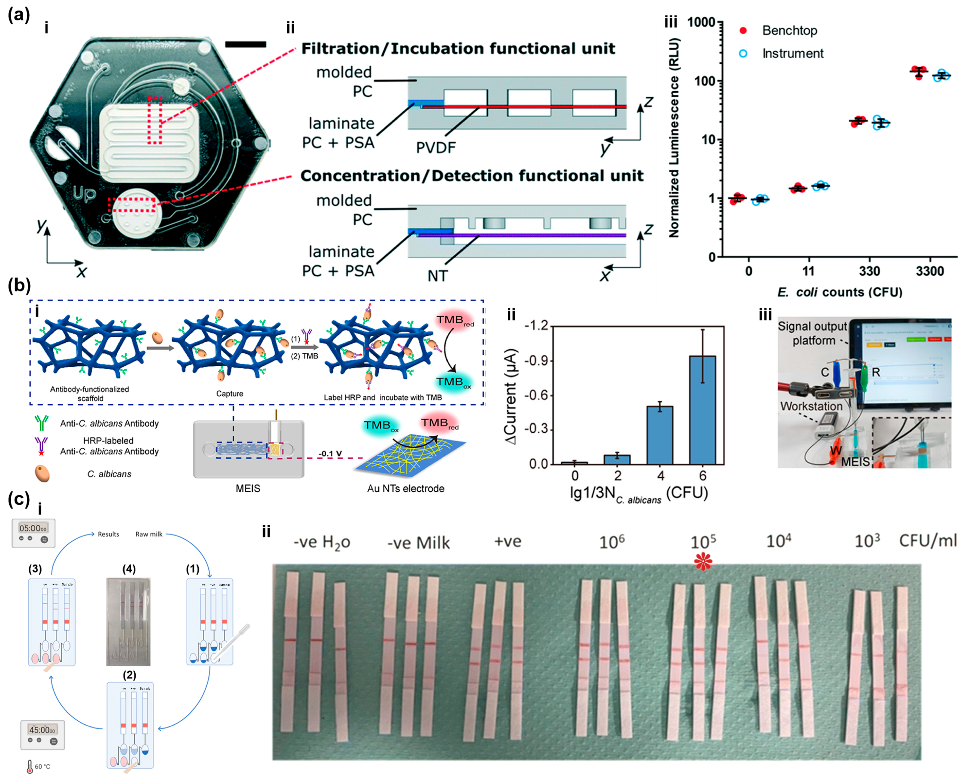
Due to the intuitive and sensitive characteristics, optical detection holds a dominant position in pathogen microfluidic analysis of E. coli [133]. The recent trend is to introduce multiple signal amplification mechanisms and approaches to challenge extremely low detection limits. As shown in Figure 6a, Alonzo et al. constructed a microfluidic filtration system combining gene-engineered phage bioluminescence to enable sensitive detection of as few as several colony-forming units of E. coli in complex water samples without the need for large instruments [134]. In another study at the same time, Sun et al. designed a microfluidic chemiluminescence sensor with a microarray [124]. This chip ingeniously combined catalytic hairpin self-assembly (CHA) and gold nanoparticles (Au NPs) catalytic reactions for multiple signal amplification, significantly improving the specificity and sensitivity in actual sample analysis. Yin et al. proposed a novel poker-shaped micro-mixer chip integrating microfluidic technology and machine learning, which is used for the sensitive detection and classification of bacteria [135]. This chip utilizes chaotic flow and the Dean vortex effect to enhance mixing efficiency, and based on the characteristic of bacteria metabolizing glucose, it constructs a dual-modal detection strategy of colorimetry and fluorescence. This method has extremely high sensitivity for Escherichia coli, with detection limits as low as 8.8 CFU/mL and 7.6 CFU/mL, respectively. Then, Jayan et al. developed a microfluidic-SERS platform specifically for the rapid detection of E. coli in food [123]. The highlight of this device was the in-situ synthesis of nanoparticles within the chip, which significantly enhanced the reproducibility of SERS signals. Moreover, to address the global antimicrobial resistance (AMR) crisis, the goal of microfluidic bacterial detection is shifting from pathogen identification to resistance gene analysis. Man et al. presented a microfluidic-based electrochemical nucleic acid biosensor [136]. This system used cyclic voltammetry (CV) and EIS for dual-mode detection, enabling direct detection of antibiotic resistance DNA sequences in lysed E. coli. Furthermore, this microfluidic device can distinguish single-base mismatches, providing a low-cost and portable solution for early screening of clinical and environmental resistant bacteria.
4.2.2. Fungus
Although fungal infections account for only about 1% of all PJI cases, fungal infections possess the unexpected characteristics of limited effective drugs, long treatment duration, and poor prognosis [137,138]. Usually, surgical treatment combined with long-term antifungal drug therapy is required, and the overall treatment failure risk is higher than that of bacterial PJI. As an example, Candida are already normal colonizers of human skin and mucous membranes, and they are often mistakenly regarded as contamination, thereby delaying the diagnosis [139]. In the applications of microfluidic technology, it is difficult to find by which fluid turbulence was significantly enhancing the capture efficiency representative significant works and achievements, but some related research can still be retrieved. Not long ago, Lin et al. presented a microfluidic detection system combining PCR amplification technology with reverse dot hybridization microfluidic chips, enabling the direct and simultaneous identification of 13 common fungal pathogens within 3–4 h [125]. It was demonstrated that this microfluidic system achieved a sensitivity of up to 96.7% and a specificity of 100%, significantly overcoming the long, time-consuming defect of traditional culture methods. It provided an efficient solution for the early and precise diagnosis of clinical bloodstream infections like fungal sepsis. In Figure 6b, Liang et al. reported a microfluidic electrochemical integrated sensor (MEIS) [126]. It was composed of a 3D chaotic flow separator and an electrochemical detector, in which fluid turbulence significantly enhanced the capture efficiency of pathogens, and enzyme-linked immunosensory electrochemistry provided precise quantitative signals. It was reported that this sensor could achieve a capture rate of over 95% for Candida albicans in saliva samples within 1 h, providing a highly sensitive and efficient solution for immediate testing of oral candidiasis and other clinical infections.
4.2.3. S. aureus and S. epidermidis
As the most important Gram-positive bacterial pathogens, S. aureus and S. epidermidis have not received much research interest from microfluidic researchers. To detect S. aureus in hospital air, Jiang et al. introduced a microfluidic chip with an interleaved trapezoidal mixer to efficiently capture liquid aerosols and seamlessly transfer them to the loop-mediated isothermal amplification (LAMP) module for in situ genetic analysis, which provided a LOD as low as 27 cells [128]. Meanwhile, Song et al. proposed a portable microfluidic system using immunomicrospheres coated with antibodies to specifically capture and rapidly read signals from pathogenic bacteria [140]. The results showed that the microfluidic device could automatically quantitatively analyze S. aureus within 1.5 h. Recently, Celik et al. presented a ready-to-use colorimetric microfluidic chip based on smartphone image analysis for the AST of methicillin-resistant S. aureus (MRSA) [141]. It provided a low-cost and easy-to-operate solution for rapid (1.5 h) and precise medication guidance in response to antibiotic-resistant infections of S. aureus. As shown in Figure 6c, Shalaby et al. reported an integrated “sample input, result output” microfluidic nucleic acid detection device [142]. As shown by the experimental results, the microfluidic device successfully achieved rapid detection of the S. aureus enterotoxin gene in dairy products from ruminants.
Figure 6.
Microfluidic technology for detecting E. coli, fungus, and S. aureus. (a) A microfluidic device and instrument prototypes for the detection of Escherichia coli in water samples using a phage-based bioluminescence assay. (i) Valveless microfluidic device for E. coli detection. (ii) Cross-sectional views of the functional dead-end filtration subunits, comprising injection-molded polycarbonate (gray), laminate polycarbonate with double-sided acrylic-based PSA (blue), PVDF membrane (red), and NT membrane (purple). (iii) Validation of instrumentation prototypes for portable and semi-automated operation, demonstrating equivalent performance of the phage-based E. coli assay between the prototype and a standard laboratory benchtop. Reprinted with permission from [134]. (b) A microfluidic electrochemical integrated sensor for efficient and sensitive detection of Candida albicans. (i) MEIS-mediated capture and detection of C. albicans. (ii) Detection of C. albicans in real saliva samples. (iii) Integration of the MEIS with a portable workstation. Reprinted with permission from [126]. (c) Sample-to-answer microfluidic device towards the point-of-need detection of Staphylococcus aureus enterotoxin genes in ruminant milk. (i) (1) Adding milk to the diluent chamber (containing PCR-grade nuclease-free water) and actuating a finger pump dilutes and mixes the sample with LAMP reagents in the amplification chamber. (2) Amplification proceeds at 60 °C for 45 min. (3) Primer-mediated incorporation of biotin and FITC labels enables amplicon capture by streptavidin-coated beads and the test line for lateral flow visualization. (4) Simple microfluidic format integrates dilution, amplification, and readout via finger-actuated liquid transfer. (ii) Limit of detection of the LAMP assay for S. aureus enterotoxin A genes, * in the panel represent the LOD. Reprinted with permission from [142].
Moreover, research on S. epidermidis detection with microfluidic technology is even rarer, as it was found that no microfluidic applications involved relevant fields in the decade. It seems that the detection method for S. epidermidis remains the commonly used approach in clinical and laboratory settings, i.e., three major categories, such as microbiological testing, biochemical identification, and molecular biological testing [143]. On the contrary, some works have presented new findings and perspectives on S. epidermidis’ underlying mechanisms [144]. For example, Weaver et al. reported a parallel microfluidic device capable of precise fluid control [145]. The device utilized tunable flow velocity distributions within microchannels to accurately simulate the microscopic physical shear forces generated by fluid flow inside medical devices such as catheters. More importantly, this microfluidic platform provided the first direct evidence that fluid shear stress alone could directly induce biofilm formation in clinically isolated S. epidermidis.
4.3. Artificial Intelligence
Moreover, AI could be the optimal matching technology for microwell chips to achieve effective PJI detection [115,146]. While the microwell chip benefits from single-bacteria packing functionality, it faces a heavy burden from monitoring tens of thousands of cavities simultaneously. AI might be the perfect solution to this issue, while the stronger generalization ability of AI can also better match the individual differences in organisms [147,148]. In the constructed PJI detection system, the microwell chip will separate and encapsulate the pathogenic bacteria, and an integrated AI module could continuously monitor the reactions of bacteria and reagents [149,150], which should be the most effective and feasible technical solution for achieving the clinical PJI detection function in the short term.
In recent years, the combination of AI and microfluidic technology has led to numerous innovative applications, such as biomanufacturing [151,152], environmental monitoring [153], pharmaceuticals [154,155], and medical sciences [156]. Moreover, it is still continuously exploring new directions. Chen et al. combined feature extraction and deep learning with photon-supported high-throughput quantitative imaging, achieving a high-precision unlabeled cell classification method [157]. The system captured quantitative optical phase and intensity images and extracted multiple biological physical features of individual cells. Nitta et al. first combined deep learning with high-speed microscopic imaging, microfluidic technology, and integrated the functionalities, including high-throughput microscopes, focusing, and sorting, onto a hybrid infrastructure [158]. Prototype technology promised real-time automated data acquisition, data processing, decision-making, and execution operations, as well as real-time intelligent cell sorting based on images.
Intelligent image processing is one of the representative functions of AI [159]. Building upon the foundation of the Vision Transformer (ViT) proposed by Dosovitskiy et al. [160], subsequent ViT-derived models experienced explosive growth in microfluidic applications. Cao et al. proposed a Swin-Unet transformer for medical image segmentation [161]. Meanwhile, Guo et al. introduced a study that integrated the YOLOv8 object detection model and path planning algorithm on a digital microfluidic platform, achieving cell sorting based on their morphology [162]. It was demonstrated to achieve extremely high purity and recovery rates in the separation experiments of HeLa cells and impurities.
Additionally, with the growing development of microfluidic technology’s functionalities and the requirements from applications in research and industrial fields, on-demand design of microfluidic devices has become crucial to overcome the bottlenecks of traditional trial-and-error methods and intensive computational fluid dynamics (CFD) simulations [163,164]. However, the design of microstructures has always been a challenge for microfluidic engineers. So far, most of the initial design relies on the accumulated experience of researchers and uncertain finite element simulations [165]. Although microfluidic experiments with small sample capacity were originally not suitable for the full utilization of AI capabilities, the researchers found some solutions for this challenge through combining finite element simulation [166]. Lashkaripour et al. proposed a machine learning-based microfluidic computing framework providing methodological support for the performance evaluation and reverse design of new microstructures [167]. By constructing an efficient surrogate model, the framework could significantly reduce the time consumed by fluid dynamics simulations while maintaining extremely high prediction accuracy. Then, they expanded the intelligent design paradigm to more complex double-emulsion droplet systems [168]. Compared with the single-phase droplet, the generation mechanism of double-phase emulsion droplets was more complex due to the dynamic coupling of multiple interfaces and the precise spatial control of multiphase flow. A more advanced predictive framework was constructed to achieve a comprehensive analysis of the generation processes of single-phase and double-phase emulsion droplets.
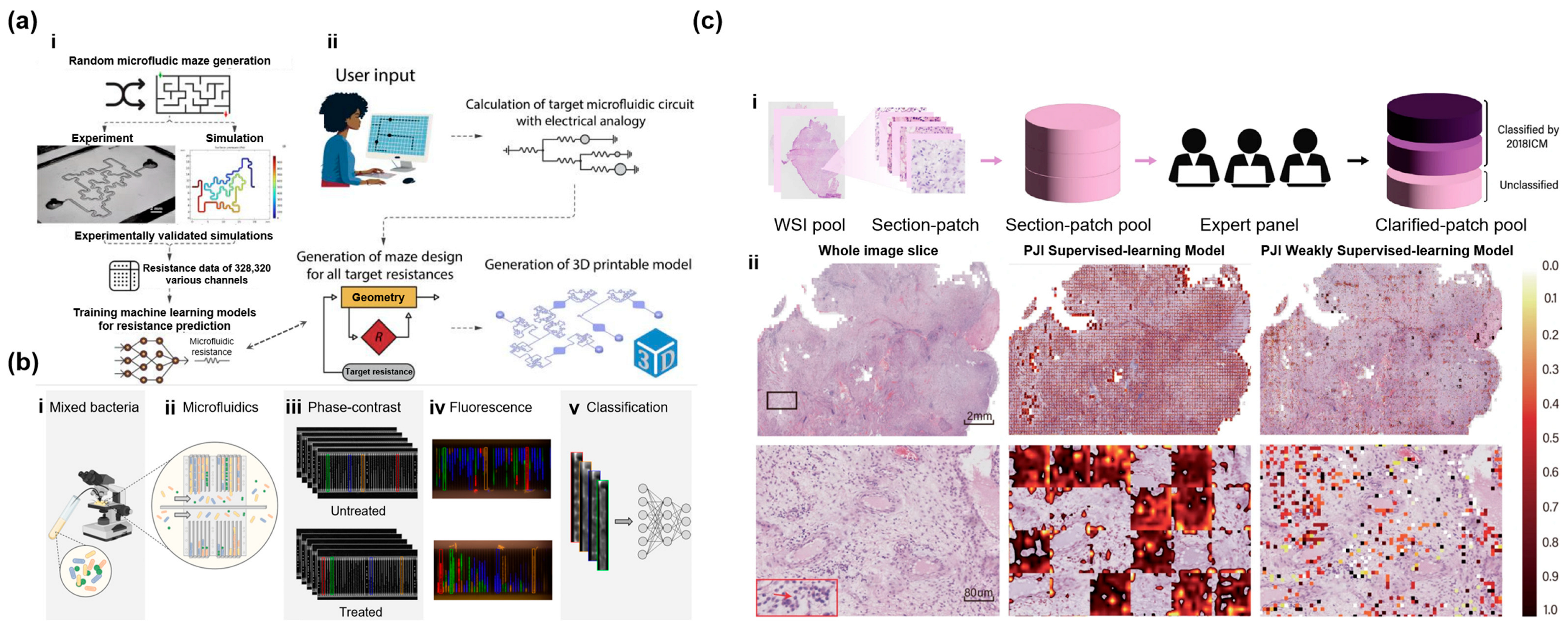
Praljak et al. have demonstrated the significant advantages of integrating AI with microfluidic technology in the analysis of complex cell morphology [169]. They utilized a microfluidic platform to simulate the flow environment within the blood vessels of patients with sickle cell anemia, and simultaneously leveraged the deep learning model to automatically classify abnormal red blood cells adhering to laminin through their biophysical properties. This approach overcomes the subjectivity and low efficiency of traditional manual assessment, providing a high-throughput and label-free evaluation tool for pathological research and precise diagnosis of blood system diseases. As shown in Figure 7a, Birtek et al. proposed an open-source platform named μFluidicGenius (μFG), which was able to automatically generate precise labyrinth-shaped microfluidic resistance network structures based on machine learning and mathematical modeling [170]. This work significantly lowered the design threshold for complex fluid circuits such as multi-organ chips.
Furthermore, AI also offers new strategies for traditional microfluidic methods in the field of bacterial detection [171]. As shown in Figure 7b, Hallström et al. demonstrated the unlabeled classification of live bacteria using ViT on time-lapse phase-contrast microscopic images in microfluidic chips [172]. The system could determine the bacterial species as the bacteria reproduced in the microfluidic device, thereby simultaneously determining the sensitivity to antibiotics, with an accuracy rate exceeding 98%. Later, they further extended the model to identify 7 common pathogens causing human infections using the ViT’s strong spatiotemporal feature extraction ability [173]. Experiments showed that the model could extract spatiotemporal features from video data of bacterial cell division, and this method could be used simultaneously with phenotypic antibiotic sensitivity detection on the microfluidic chip. McMahon et al. presented a study to develop a large-scale visual Transformer framework for the extremely time-consuming Gram staining analysis in the diagnosis of clinical blood infections [174]. Lee et al. proposed a deep learning framework based on ViT for alignment-free bacterial pathogen identification in high-resolution microscopy [175]. The study innovatively combined a two-stage image enhancement strategy including offline data synthesis and online variation, effectively overcoming the common class imbalance problem in clinical samples. This model achieved an accuracy of 98.51% in identifying six highly pathogenic bacteria.
Moreover, researchers have found that AI could also play a significant role in solving challenging PJI detection problems [176,177]. To overcome the substantial noise in CT images and the disparity in data volume between CT images and text data, Li et al. presented a hierarchical GCN (graph convolutional network)-based Transformer (HGT) method to combine features from CT images and patients’ text data [178]. Then, as shown in Figure 7c, Tao et al. innovatively employed a ViT architecture based on a self-supervised learning method [179]. Through feature pre-training on a large number of unlabeled pathological microscopic images, a highly accurate AI pathological quantitative analysis model was constructed, which could significantly reduce the false negative rate in the diagnosis of PJI.
Figure 7.
Integrative studies of microfluidics and AI. (a) ML-automated microfluidic circuit design. (i) Description of the data generation method. Following experimental validation of the simulation approach across six geometries, a dataset comprising 328,320 simulated random mazes was generated to train the ML algorithm for microfluidic resistance prediction. (ii) The μFG employs an electrical circuit analogy to calculate the resistance requirements for each channel. A generative algorithm then devises geometries meeting these values, subsequently exporting a 3D-printable .stl file for the user. Reprinted with permission from [170]. (b) Label-free deep learning-based species classification of bacteria imaged by phase-contrast microscopy. (i,ii) Illustration of a mixed bacterial sample loaded onto the microfluidic chip. (iii) Time-lapse phase-contrast images of bacterial proliferation in media with (bottom) and without (top) antibiotics. (iv) Species identification via correspondence between the fluorescence and final phase-contrast images: Enterococcus faecalis (red), Escherichia coli (green), Klebsiella pneumoniae (blue), and Pseudomonas aeruginosa (orange). (v) Single-frame or time-lapse video inputs for the neural network. Reprinted with permission from [172]. (c) Clinically applicable optimized PJI diagnosis via AI-based pathology. (i) Data processing: WSI datasets were segmented into 600 × 600-pixel patches and partitioned for DINO v2 training, testing, and additional training. (ii) Left to right: a whole-slide image (WSI), followed by visualization heatmaps of the supervised and weakly supervised PJI models. The light-to-dark color gradient indicates increasing diagnostic weight. Neutrophil cytoplasmic and nuclear morphologies vary with proximity to blood vessels (indicated by the red arrow). Reprinted with permission from [179].
A critical obstacle to deploying AI in PJI detection is the scarcity of training data. As a niche complication, PJI offers a very limited number of positive cases per institution, which is insufficient for training deep models. Few-sample learning algorithms provide practical solutions to this problem. Transfer learning, which pre-trains models on large natural-image or general microbiology datasets and fine-tunes them on small PJI-specific datasets, has become the most widely adopted strategy and has shown promising performance in bacterial classification from microscopic images [180]. Self-supervised learning (e.g., DINO, SimCLR, MAE) exploits large amounts of unlabeled microfluidic or pathological images to learn robust representations, substantially reducing the dependence on annotated samples [181]. Beyond the limited sample size, clinical PJI data are distributed across multiple medical centers with heterogeneous imaging equipment, staining protocols, bacterial strains, antibiotic resistance profiles, and baseline levels of laboratory tests. Directly pooling such data raises privacy concerns, while training models on a single center leads to poor generalization. Federated learning offers a privacy-preserving solution by aggregating model parameters rather than raw data from multiple centers, and has been increasingly applied in medical image analysis and infectious disease diagnosis [182]. Moreover, PJI diagnosis inherently relies on multi-modal information, including CT/MRI images, pathological sections, serological indicators, synovial fluid test results, and microfluidic readouts, so hierarchical or attention-based multi-modal fusion strategies will be indispensable for building a robust AI-assisted PJI diagnostic system [183]. We believe that the combination of few-sample learning and multi-center data fusion will make AI-driven microfluidic PJI detection not only technically feasible but also clinically deployable.
The breakthrough of AI successfully transformed the prediction and classification capability of machine learning into a directly manufacturable engineering physical model and interpretation tool for professional images, promoting the evolution of microfluidic design and integrated systems towards fully automated implementation [184,185]. It demonstrates high engineering application value in complex microfluidic devices. Ultimately, we believe that AI represents the final piece of the puzzle in achieving the microfluidic PJI detection method [186].
5. Conclusions
It is known that current microfluidic technology has achieved mature, multi-scenario applications in the detection of bacterial pathogens. It overcomes the limitations of traditional culture methods, such as long processing times and complex procedures, by integrating sample preparation, pathogen enrichment, identification, and phenotypic antimicrobial susceptibility testing onto a compact chip. This enables precise bacterial identification within 1 h and simultaneous susceptibility results within 5 h, with sensitivity significantly higher than conventional methods. However, microfluidic applications specifically targeting PJI detection remain scarce. In this review, it is analyzed that this might be caused by the complex characteristics of PJI infection pathogens, combined with the fact that it is a relatively highly specialized disease. On the contrary, it is believed that, in the future, microfluidic technology will play a significant role in the development of PJI detection, and PJI detection technology will evolve in the future along four key directions: speed, multifunctionality, precision, and intelligent analysis.
However, several obstacles remain to be addressed. For AI, data limitations and weaknesses in the reliability of clinical decision-making are nonnegligible. In cases of multidrug-resistant bacteria and multispecies mixed infections, the diagnostic and treatment recommendations provided by AI still align with those of experienced multidisciplinary clinical teams in less than 30% of cases, and the large regional variations in pathogen resistance profiles and clinical test baselines make general AI models difficult to adapt to localized needs. Nevertheless, as prototype technology, AI holds tremendous potential for full-process intelligent auxiliary diagnosis, personalized risk assessment and treatment support, and dynamic management, which may be unlocked by few-sample learning algorithms and multi-center heterogeneous data fusion strategies. From a hardware perspective, we consider microwell chips a promising platform to upgrade existing culturing technology. Their closed microwell array effectively reduces the risk of sample cross-contamination, and their multi-well design enables quantitative parallel screening of pathogens and simultaneous multiple sets of antibiotic susceptibility tests. However, the difficulty is the mismatch between scarce samples and the detection limits of chip-based assays. At typical synovial fluid levels of 102–103 CFU/mL, a microliter-scale chip might contain few or even zero target cells, so that Poisson sampling statistics, rather than nominal assay sensitivity, become the hard boundary. This difficulty could be solved with microfluidic pretreatment technology. Once equipped with appropriate detection principles for PJI pathogens, such platforms could substantially assist physicians in prescribing timely, targeted therapies.
Author Contributions
D.W., S.Y. and Q.Z. conceived the manuscript; Y.B., Q.Z. and D.W. wrote and revised the manuscript; L.M. and L.W. collected and organized the literature and relevant materials; G.C. provided guidance on parts of the manuscript. All authors have read and agreed to the published version of the manuscript.
Funding
This work was supported by the Science and Technology Research Project of Higher Education Institutions in Hebei Province (BJK2024150), Natural Science Foundation of Hebei Province (H2026202013, C2026202004), National Natural Science Foundation of China (52271245), Natural Science Foundation of Tianjin (24JCYBJC01100).
Institutional Review Board Statement
Not applicable.
Informed Consent Statement
Not applicable.
Data Availability Statement
No primary research results, new data, software, or code have been included as part of this review.
Acknowledgments
Some elements of the drawing in Figure 1 were created with assistance from AI tools (Gemini Pro 3.1). The authors have reviewed and edited the output and take full responsibility for the content of this publication.
Conflicts of Interest
The authors declare no competing interests.
References
- Kapadia, B.H.; Berg, R.A.; Daley, J.A.; Fritz, J.; Bhave, A.; Mont, M.A. Periprosthetic joint infection. Lancet 2016, 387, 386–394. [Google Scholar] [CrossRef] [Scilit] [PubMed]
- Zimmerli, W.; Trampuz, A.; Ochsner Peter, E. Prosthetic-Joint Infections. N. Engl. J. Med. 2004, 351, 1645–1654. [Google Scholar] [CrossRef] [Scilit] [PubMed]
- Arciola, C.R.; Campoccia, D.; Montanaro, L. Implant infections: Adhesion, biofilm formation and immune evasion. Nat. Rev. Microbiol. 2018, 16, 397–409. [Google Scholar] [CrossRef] [Scilit] [PubMed]
- Lum, Z.C.; Natsuhara, K.M.; Shelton, T.J.; Giordani, M.; Pereira, G.C.; Meehan, J.P. Mortality During Total Knee Periprosthetic Joint Infection. J. Arthroplast. 2018, 33, 3783–3788. [Google Scholar] [CrossRef] [Scilit] [PubMed]
- Tong, S.Y.; Davis, J.S.; Eichenberger, E.; Holland, T.L.; Fowler, V.G., Jr. Staphylococcus aureus infections: Epidemiology, pathophysiology, clinical manifestations, and management. Clin. Microbiol. Rev. 2015, 28, 603–661. [Google Scholar] [CrossRef] [Scilit] [PubMed]
- Tande Aaron, J.; Patel, R. Prosthetic Joint Infection. Clin. Microbiol. Rev. 2014, 27, 302–345. [Google Scholar] [CrossRef] [Scilit] [PubMed]
- Osmon, D.R.; Berbari, E.F.; Berendt, A.R.; Lew, D.; Zimmerli, W.; Steckelberg, J.M.; Rao, N.; Hanssen, A.; Wilson, W.R. Diagnosis and Management of Prosthetic Joint Infection: Clinical Practice Guidelines by the Infectious Diseases Society of America. Clin. Infect. Dis. 2013, 56, e1–e25. [Google Scholar] [CrossRef] [Scilit] [PubMed]
- Parvizi, J.; Tan, T.L.; Goswami, K.; Higuera, C.; Della Valle, C.; Chen, A.F.; Shohat, N. The 2018 Definition of Periprosthetic Hip and Knee Infection: An Evidence-Based and Validated Criteria. J. Arthroplast. 2018, 33, 1309–1314. [Google Scholar] [CrossRef] [Scilit] [PubMed]
- Burnham, C.-A.D.; Leeds, J.; Nordmann, P.; O’Grady, J.; Patel, J. Diagnosing antimicrobial resistance. Nat. Rev. Microbiol. 2017, 15, 697–703. [Google Scholar] [CrossRef] [Scilit] [PubMed]
- Thoendel, M.J.; Jeraldo, P.R.; Greenwood-Quaintance, K.E.; Yao, J.Z.; Chia, N.; Hanssen, A.D.; Abdel, M.P.; Patel, R. Identification of Prosthetic Joint Infection Pathogens Using a Shotgun Metagenomics Approach. Clin. Infect. Dis. 2018, 67, 1333–1338. [Google Scholar] [CrossRef] [Scilit] [PubMed]
- Tarabichi, M.; Shohat, N.; Goswami, K.; Alvand, A.; Silibovsky, R.; Belden, K.; Parvizi, J. Diagnosis of Periprosthetic Joint Infection: The Potential of Next-Generation Sequencing. J. Bone Jt. Surg. 2018, 100, 147–154. [Google Scholar] [CrossRef] [Scilit] [PubMed]
- Ellington, M.J.; Ekelund, O.; Aarestrup, F.M.; Canton, R.; Doumith, M.; Giske, C.; Grundman, H.; Hasman, H.; Holden, M.T.G.; Hopkins, K.L.; et al. The role of whole genome sequencing in antimicrobial susceptibility testing of bacteria: Report from the EUCAST Subcommittee. Clin. Microbiol. Infect. 2017, 23, 2–22. [Google Scholar] [CrossRef] [Scilit] [PubMed]
- Zhang, K.; Qin, S.; Wu, S.; Liang, Y.; Li, J. Microfluidic systems for rapid antibiotic susceptibility tests (ASTs) at the single-cell level. Chem. Sci. 2020, 11, 6352–6361. [Google Scholar] [CrossRef] [Scilit] [PubMed]
- Aldea, A.C.; Diguṭă, F.C.; Presacan, O.; Voaideṣ, C.; Toma, R.C.; Matei, F. Detecting antibiotic resistance: Classical, molecular, advanced bioengineering, and AI-enhanced approaches. Front. Microbiol. 2025, 16, 1673343. [Google Scholar] [CrossRef] [Scilit] [PubMed]
- Assaf, E.; Amerschläger, C.F.; Nessler, V.B.; Ali, K.; Ossendorff, R.; Jaenisch, M.; Strauss, A.C.; Burger, C.; Hischebeth, G.T.; Walmsley, P.J.; et al. A Novel Approach for Tissue Analysis in Joint Infections Using the Scattered Light Integrating Collector (SLIC). Biosensors 2025, 15, 795. [Google Scholar] [CrossRef] [Scilit] [PubMed]
- Wyatt, M.C.; Beswick, A.D.; Kunutsor, S.K.; Wilson, M.J.; Whitehouse, M.R.; Blom, A.W. The Alpha-Defensin Immunoassay and Leukocyte Esterase Colorimetric Strip Test for the Diagnosis of Periprosthetic Infection: A Systematic Review and Meta-Analysis. J. Bone Jt. Surg. Am. 2016, 98, 992–1000. [Google Scholar] [CrossRef] [Scilit] [PubMed]
- Feng, Z.; Ji, Q.; Zheng, Q.; Zhao, R.; Zhang, Y.; Shen, X.; Li, P.; Zhang, X.; Hao, L.; Wang, Y.; et al. Rapid detection of bacteria in periprosthetic joint infection via microfluidic qPCR chip. Int. J. Infect. Dis. 2026, 164, 108334. [Google Scholar] [CrossRef] [Scilit] [PubMed]
- Chiu, C.Y.; Miller, S.A. Clinical metagenomics. Nat. Rev. Genet. 2019, 20, 341–355. [Google Scholar] [CrossRef] [Scilit] [PubMed]
- Trampuz, A.; Piper Kerryl, E.; Jacobson Melissa, J.; Hanssen Arlen, D.; Unni Krishnan, K.; Osmon Douglas, R.; Mandrekar Jayawant, N.; Cockerill Franklin, R.; Steckelberg James, M.; Greenleaf James, F.; et al. Sonication of Removed Hip and Knee Prostheses for Diagnosis of Infection. N. Engl. J. Med. 2007, 357, 654–663. [Google Scholar] [CrossRef] [Scilit] [PubMed]
- Tarabichi, S.; Goh, G.S.; Zanna, L.; Qadiri, Q.S.; Baker, C.M.; Gehrke, T.; Citak, M.; Parvizi, J. Time to Positivity of Cultures Obtained for Periprosthetic Joint Infection. J. Bone Jt. Surg. 2023, 105, 107–112. [Google Scholar] [CrossRef] [Scilit] [PubMed]
- Chen, Y.; Kang, X.; Tao, J.; Zhang, Y.; Ying, C.; Lin, W. Reliability of synovial fluid alpha-defensin and leukocyte esterase in diagnosing periprosthetic joint infection (PJI): A systematic review and meta-analysis. J. Orthop. Surg. Res. 2019, 14, 453. [Google Scholar] [CrossRef] [Scilit] [PubMed]
- Trampuz, A.; Hanssen, A.D.; Osmon, D.R.; Mandrekar, J.; Steckelberg, J.M.; Patel, R. Synovial fluid leukocyte count and differential for the diagnosis of prosthetic knee infection. Am. J. Med. 2004, 117, 556–562. [Google Scholar] [CrossRef] [Scilit] [PubMed]
- Olearo, F.; Zein, S.E.; Portillo, M.E.; Zapf, A.; Rohde, H.; Berbari, E.F.; Wouthuyzen-Bakker, M. Diagnostic accuracy of 16S rDNA PCR, multiplex PCR and metagenomic next-generation sequencing in periprosthetic joint infections: A systematic review and meta-analysis. Clin. Microbiol. Infect. 2025, 31, 1115–1125. [Google Scholar] [CrossRef] [Scilit] [PubMed]
- Jun, Y.; Jianghua, L. Diagnosis of Periprosthetic Joint Infection Using Polymerase Chain Reaction: An Updated Systematic Review and Meta-Analysis. Surg. Infect. 2018, 19, 555–565. [Google Scholar] [CrossRef] [Scilit] [PubMed]
- Sigmund, I.K.; Windhager, R.; Sevelda, F.; Staats, K.; Puchner, S.E.; Stenicka, S.; Thalhammer, F.; Holinka, J. Multiplex PCR Unyvero i60 ITI application improves detection of low-virulent microorganisms in periprosthetic joint infections. Int. Orthop. 2019, 43, 1891–1898. [Google Scholar] [CrossRef] [Scilit] [PubMed]
- Hantouly, A.T.; Alzobi, O.; Toubasi, A.A.; Zikria, B.; Al Dosari, M.A.A.; Ahmed, G. Higher sensitivity and accuracy of synovial next-generation sequencing in comparison to culture in diagnosing periprosthetic joint infection: A systematic review and meta-analysis. Knee Surg. Sports Traumatol. Arthrosc. 2023, 31, 3672–3683. [Google Scholar] [CrossRef] [Scilit] [PubMed]
- Wang, L.; Feng, S.; Zhao, Z.; Zhao, Y.; Dong, S.; Cao, L.; Song, K. Comparative diagnostic accuracy of next-generation sequencing in different specimen types for periprosthetic joint infection: A systematic review and meta-analysis. Knee Surg. Sports Traumatol. Arthrosc. 2026, 34, 1725–1739. [Google Scholar] [CrossRef] [Scilit] [PubMed]
- Li, H.; Niu, E.; Fu, J.; Huang, Y.; Gao, Y.; Chai, W.; Chen, J. The Clinical Impact of Metagenomic Next-Generation Sequencing for the Diagnosis of Periprosthetic Joint Infection. Infect. Drug Resist. 2023, 16, 6521–6533. [Google Scholar] [CrossRef] [Scilit] [PubMed]
- Chin, C.D.; Laksanasopin, T.; Cheung, Y.K.; Steinmiller, D.; Linder, V.; Parsa, H.; Wang, J.; Moore, H.; Rouse, R.; Umviligihozo, G.; et al. Microfluidics-based diagnostics of infectious diseases in the developing world. Nat. Med. 2011, 17, 1015–1019. [Google Scholar] [CrossRef] [Scilit] [PubMed]
- Mi, F.; Hu, C.; Wang, Y.; Wang, L.; Peng, F.; Geng, P.; Guan, M. Recent advancements in microfluidic chip biosensor detection of foodborne pathogenic bacteria: A review. Anal. Bioanal. Chem. 2022, 414, 2883–2902. [Google Scholar] [CrossRef] [Scilit] [PubMed]
- Nasseri, B.; Soleimani, N.; Rabiee, N.; Kalbasi, A.; Karimi, M.; Hamblin, M.R. Point-of-care microfluidic devices for pathogen detection. Biosens. Bioelectron. 2018, 117, 112–128. [Google Scholar] [CrossRef] [Scilit] [PubMed]
- Whitesides, G.M. The origins and the future of microfluidics. Nature 2006, 442, 368–373. [Google Scholar] [CrossRef] [Scilit] [PubMed]
- Foudeh, A.M.; Fatanat Didar, T.; Veres, T.; Tabrizian, M. Microfluidic designs and techniques using lab-on-a-chip devices for pathogen detection for point-of-care diagnostics. Lab Chip 2012, 12, 3249–3266. [Google Scholar] [CrossRef] [Scilit] [PubMed]
- He, S.; Qin, X.; Zhu, X.; Mu, G.; Yang, G.; Ma, J.; Zhang, Z. Auto-Fluorescence Cellulose Paper Based on High Affinity of Sodium Lignosulfonate for Microfluidic Detection Platform. J. Anal. Test. 2025, 9, 662–667. [Google Scholar] [CrossRef] [Scilit]
- Wu, W.; Zhao, Q.; Cai, G.; Zhang, B.; Suo, Y.; Liu, Y.; Jin, W.; Mu, Y. All-In-One Escherichia coli Viability Assay for Multi-dimensional Detection of Uncomplicated Urinary Tract Infections. Anal. Chem. 2022, 94, 17853–17860. [Google Scholar] [CrossRef] [Scilit] [PubMed]
- Yedire, S.G.; Khan, H.; AbdelFatah, T.; Siavash Moakhar, R.; Mahshid, S. Microfluidic-based colorimetric nucleic acid detection of pathogens. Sens. Diagn. 2023, 2, 763–780. [Google Scholar] [CrossRef] [Scilit]
- Yang, B.; Wang, P.; Li, Z.; You, Q.; Sekine, S.; Ma, J.; Zhuang, S.; Zhang, D.; Yamaguchi, Y. Simultaneous amplification of DNA in a multiplex circular array shaped continuous flow PCR microfluidic chip for on-site detection of bacterial. Lab Chip 2023, 23, 2633–2639. [Google Scholar] [CrossRef] [Scilit] [PubMed]
- Xia, Y.; Feng, Y.; Zhao, H.; Du, B.; Pu, Y.; Sun, S.; Feng, J.; Yan, C.; Cui, J.; Gan, L.; et al. A one-pot, visual, multiplex microfluidic digital PCR chip for simultaneous point-of-care detection, genotyping, and macrolide resistance assessment of Mycoplasma pneumoniae. Microchim. Acta 2025, 192, 839. [Google Scholar] [CrossRef] [Scilit] [PubMed]
- Fande, S.; Banerjee, A.; Goel, S. Bacteria-on-chip: A multiplexed point-of-care electrochemical platform for rapid detection of Escherichia coli and antimicrobial susceptibility testing. Sens. Diagn. 2026, 5, 1002–1014. [Google Scholar] [CrossRef] [Scilit]
- Liu, Z.; Song, H.; Lin, G.; Zhong, W.; Zhang, Y.; Yang, A.; Liu, Y.; Duan, J.; Zhou, Y.; Jiao, K.; et al. Wireless Intelligent Patch for Closed-loop In Situ Wound Management. Adv. Sci. 2024, 11, 2400451. [Google Scholar] [CrossRef] [Scilit] [PubMed]
- Kuswandi, B.; Nuriman; Huskens, J.; Verboom, W. Optical sensing systems for microfluidic devices: A review. Anal. Chim. Acta 2007, 601, 141–155. [Google Scholar] [CrossRef] [Scilit] [PubMed]
- Zhou, P.; He, H.; Ma, H.; Wang, S.; Hu, S. A Review of Optical Imaging Technologies for Microfluidics. Micromachines 2022, 13, 274. [Google Scholar] [CrossRef] [Scilit] [PubMed]
- Guliy, O.I.; Karavaeva, O.A.; Smirnov, A.V.; Eremin, S.A.; Bunin, V.D. Optical Sensors for Bacterial Detection. Sensors 2023, 23, 9391. [Google Scholar] [CrossRef] [Scilit] [PubMed]
- Laliwala, A.; Svechkarev, D.; Sadykov, M.R.; Endres, J.; Bayles, K.W.; Mohs, A.M. Simpler Procedure and Improved Performance for Pathogenic Bacteria Analysis with a Paper-Based Ratiometric Fluorescent Sensor Array. Anal. Chem. 2022, 94, 2615–2624. [Google Scholar] [CrossRef] [Scilit] [PubMed]
- Dönmez, S.; Needs, S.H.; Osborn, H.M.I.; Reis, N.M.; Edwards, A.D. Label-free 1D microfluidic dipstick counting of microbial colonies and bacteriophage plaques. Lab Chip 2022, 22, 2820–2831. [Google Scholar] [CrossRef] [Scilit] [PubMed]
- Tong, X.; Lin, X.; Duan, N.; Wang, Z.; Wu, S. Laser-Printed Paper-Based Microfluidic Chip Based on a Multicolor Fluorescence Carbon Dot Biosensor for Visual Determination of Multiantibiotics in Aquatic Products. ACS Sens. 2022, 7, 3947–3955. [Google Scholar] [CrossRef] [Scilit] [PubMed]
- Yoo, S.M.; Lee, S.Y. Optical Biosensors for the Detection of Pathogenic Microorganisms. Trends Biotechnol. 2016, 34, 7–25. [Google Scholar] [CrossRef] [Scilit] [PubMed]
- Dey, S.; Dolci, M.; Zijlstra, P. Single-Molecule Optical Biosensing: Recent Advances and Future Challenges. ACS Phys. Chem. AU 2023, 3, 143–156. [Google Scholar] [CrossRef] [Scilit] [PubMed]
- Trieu, P.T.; Lee, N.Y. Paper-Based All-in-One Origami Microdevice for Nucleic Acid Amplification Testing for Rapid Colorimetric Identification of Live Cells for Point-of-Care Testing. Anal. Chem. 2019, 91, 11013–11022. [Google Scholar] [CrossRef] [Scilit] [PubMed]
- Tavakoli, H.; Hirth, E.; Luo, M.; Sharma Timilsina, S.; Dou, M.; Dominguez, D.C.; Li, X. A microfluidic fully paper-based analytical device integrated with loop-mediated isothermal amplification and nano-biosensors for rapid, sensitive, and specific quantitative detection of infectious diseases. Lab Chip 2022, 22, 4693–4704. [Google Scholar] [CrossRef] [Scilit] [PubMed]
- Zhuang, J.; Zhao, Z.; Lian, K.; Yin, L.; Wang, J.; Man, S.; Liu, G.; Ma, L. SERS-based CRISPR/Cas assay on microfluidic paper analytical devices for supersensitive detection of pathogenic bacteria in foods. Biosens. Bioelectron. 2022, 207, 114167. [Google Scholar] [CrossRef] [Scilit] [PubMed]
- Wang, J. Electrochemical detection for microscale analytical systems: A review. Talanta 2002, 56, 223–231. [Google Scholar] [CrossRef] [Scilit] [PubMed]
- Rackus, D.G.; Shamsi, M.H.; Wheeler, A.R. Electrochemistry, biosensors and microfluidics: A convergence of fields. Chem. Soc. Rev. 2015, 44, 5320–5340. [Google Scholar] [CrossRef] [Scilit] [PubMed]
- Police Patil, A.V.; Chuang, Y.S.; Lin, C.W.; Yang, C.Y.; Yasukawa, T.; Wu, C.C. A Microfluidic Platform Integrating Dielectrophoretic Concentration and Impedimetric Sensing for Rapid Salmonella choleraesuis Detection. ACS Sens. 2025, 10, 6512–6520. [Google Scholar] [CrossRef] [Scilit] [PubMed]
- Guo, Y.; Guo, W.; Wu, Z.; Xu, H.; Zhang, X.; Zou, X.; Sun, Z. A Microfluidic Chip-Based Electrochemical Biosensor Coupled with CRISPR/Cas12a for Simultaneous Detection of Foodborne Pathogens. Anal. Chem. 2025, 97, 27266–27275. [Google Scholar] [CrossRef] [Scilit] [PubMed]
- Jiang, J.; Wu, H.; Su, Y.; Liang, Y.; Shu, B.; Zhang, C. Electrochemical Cloth-Based DNA Sensors (ECDSs): A New Class of Electrochemical Gene Sensors. Anal. Chem. 2020, 92, 7708–7716. [Google Scholar] [CrossRef] [Scilit] [PubMed]
- Jiang, H.; Sun, Z.; Guo, Q.; Weng, X. Microfluidic thread-based electrochemical aptasensor for rapid detection of Vibrio parahaemolyticus. Biosens. Bioelectron. 2021, 182, 113191. [Google Scholar] [CrossRef] [Scilit] [PubMed]
- Vafadar, A.; Takallu, S.; Alashti, S.K.; Rashidi, S.; Bahrani, S.; Tajbakhsh, A.; Mirzaei, E.; Savardashtaki, A. Advancements in microfluidicplatforms for rapid biomarker diagnostics of infectious diseases. Microchem. J. 2025, 208, 112296. [Google Scholar] [CrossRef] [Scilit]
- Shin, D.J.; Andini, N.; Hsieh, K.; Yang, S.; Wang, T.H. Emerging Analytical Techniques for Rapid Pathogen Identification and Susceptibility Testing. Annu. Rev. Anal. Chem. 2019, 12, 41–67. [Google Scholar] [CrossRef] [Scilit] [PubMed]
- Xing, G.; Shang, Y.; Ai, J.; Lin, H.; Wu, Z.; Zhang, Q.; Lin, J.M.; Pu, Q.; Lin, L. Nanozyme-Mediated Catalytic Signal Amplification for Microfluidic Biosensing of Foodborne Bacteria. Anal. Chem. 2023, 95, 13391–13399. [Google Scholar] [CrossRef] [Scilit] [PubMed]
- Frigoli, M.; Lowdon, J.W.; Caldara, M.; Arreguin-Campos, R.; Sewall, J.; Cleij, T.J.; Diliën, H.; Eersels, K.; van Grinsven, B. Thermal Pyocyanin Sensor Based on Molecularly Imprinted Polymers for the Indirect Detection of Pseudomonas aeruginosa. ACS Sens. 2023, 8, 353–362. [Google Scholar] [CrossRef] [Scilit] [PubMed]
- Mei, H.; Yin, W.; Zou, Z.; Wang, Y.; Chen, J.; Liao, Z.; Liu, J.; Mu, Y. Investigation of the Inoculum Effect of Antimicrobial Peptides Utilizing a Microfluidic Chip Platform. Anal. Chem. 2026, 98, 13460–13468. [Google Scholar] [CrossRef] [Scilit] [PubMed]
- Guo, Y.; Wang, T.; Tu, Z.; Chen, H.; Wang, S.; Sun, G.; Rong, Z. Microfluidic filtration enrichment cascade nanozyme-catalyzed colorimetric immunoassay for rapid and ultrasensitive detection of Escherichia coli in urine with smartphone readout. Anal. Methods 2026, 18, 5093–5104. [Google Scholar] [CrossRef] [Scilit] [PubMed]
- Kim, D.; Herr, A.E. Protein immobilization techniques for microfluidic assays. Biomicrofluidics 2013, 7, 041501. [Google Scholar] [CrossRef] [Scilit] [PubMed]
- Mok, J.; Mindrinos, M.N.; Davis, R.W.; Javanmard, M. Digital microfluidic assay for protein detection. Proc. Natl. Acad. Sci. USA 2014, 111, 2110–2115. [Google Scholar] [CrossRef] [Scilit] [PubMed]
- Sathish, S.; Shen, A.Q. Toward the Development of Rapid, Specific, and Sensitive Microfluidic Sensors: A Comprehensive Device Blueprint. JACS Au 2021, 1, 1815–1833. [Google Scholar] [CrossRef] [Scilit] [PubMed]
- Ahrberg, C.D.; Manz, A.; Chung, B.G. Polymerase chain reaction in microfluidic devices. Lab Chip 2016, 16, 3866–3884. [Google Scholar] [CrossRef] [Scilit] [PubMed]
- Shendure, J.; Balasubramanian, S.; Church, G.M.; Gilbert, W.; Rogers, J.; Schloss, J.A.; Waterston, R.H. DNA sequencing at 40: Past, present and future. Nature 2017, 550, 345–353. [Google Scholar] [CrossRef] [Scilit] [PubMed]
- Doudna, J.A.; Charpentier, E. Genome editing. The new frontier of genome engineering with CRISPR-Cas9. Science 2014, 346, 1258096. [Google Scholar] [CrossRef] [Scilit] [PubMed]
- Deisseroth, K. Optogenetics. Nat. Methods 2011, 8, 26–29. [Google Scholar] [CrossRef] [Scilit] [PubMed]
- Barros, B.J.; Cunha, J.P.S. Neurophotonics: A comprehensive review, current challenges and future trends. Front. Neurosci. 2024, 18, 1382341. [Google Scholar] [CrossRef] [Scilit] [PubMed]
- Nakato, R.; Sakata, T. Methods for ChIP-seq analysis: A practical workflow and advanced applications. Methods 2021, 187, 44–53. [Google Scholar] [CrossRef] [Scilit] [PubMed]
- Ku, J.L.; Jeon, Y.K.; Park, J.G. Methylation-specific PCR. Methods Mol. Biol. 2011, 791, 23–32. [Google Scholar] [CrossRef] [Scilit] [PubMed]
- Buenrostro, J.D.; Wu, B.; Litzenburger, U.M.; Ruff, D.; Gonzales, M.L.; Snyder, M.P.; Chang, H.Y.; Greenleaf, W.J. Single-cell chromatin accessibility reveals principles of regulatory variation. Nature 2015, 523, 486–490. [Google Scholar] [CrossRef] [Scilit] [PubMed]
- Oushyani Roudsari, Z.; Esmaeili, Z.; Nasirzadeh, N.; Heidari Keshel, S.; Sefat, F.; Bakhtyari, H.; Nadri, S. Microfluidics as a promising technology for personalized medicine. Bioimpacts 2025, 15, 29944. [Google Scholar] [CrossRef] [Scilit] [PubMed]
- Yang, B.; Wang, P.; Li, Z.; Tao, C.; You, Q.; Sekine, S.; Zhuang, S.; Zhang, D.; Yamaguchi, Y. A continuous flow PCR array microfluidic chip applied for simultaneous amplification of target genes of periodontal pathogens. Lab Chip 2022, 22, 733–737. [Google Scholar] [CrossRef] [Scilit] [PubMed]
- Li, Z.; Wang, Y.; Gao, Z.; Sekine, S.; You, Q.; Zhuang, S.; Zhang, D.; Feng, S.; Yamaguchi, Y. Lower fluidic resistance of double-layer droplet continuous flow PCR microfluidic chip for rapid detection of bacteria. Anal. Chim. Acta 2023, 1251, 340995. [Google Scholar] [CrossRef] [Scilit] [PubMed]
- Wan, L.; Li, M.; Law, M.K.; Mak, P.I.; Martins, R.P.; Jia, Y. Sub-5-Minute Ultrafast PCR using Digital Microfluidics. Biosens. Bioelectron. 2023, 242, 115711. [Google Scholar] [CrossRef] [Scilit] [PubMed]
- Li, Z.; Hua, L.; Xie, L.; Wang, D.; Jiang, X. Automated Microfluidic Nucleic Acid Detection Platform-Integrated RPA-T7-Cas13a for Pathogen Diagnosis. Anal. Chem. 2023, 95, 6940–6947. [Google Scholar] [CrossRef] [Scilit] [PubMed]
- Zheng, W.; Zhao, S.; Yin, Y.; Zhang, H.; Needham, D.M.; Evans, E.D.; Dai, C.L.; Lu, P.J.; Alm, E.J.; Weitz, D.A. High-throughput, single-microbe genomics with strain resolution, applied to a human gut microbiome. Science 2022, 376, 1483. [Google Scholar] [CrossRef] [Scilit] [PubMed]
- Murphy, T.W.; Hsieh, Y.P.; Zhu, B.; Naler, L.B.; Lu, C. Microfluidic Platform for Next-Generation Sequencing Library Preparation with Low-Input Samples. Anal. Chem. 2020, 92, 2519–2526. [Google Scholar] [CrossRef] [Scilit] [PubMed]
- Quinlan, N.D.; Jennings, J.M. Joint aspiration for diagnosis of chronic periprosthetic joint infection: When, how, and what tests? Arthroplasty 2023, 5, 43. [Google Scholar] [CrossRef] [Scilit] [PubMed]
- Li, R.; Lu, Q.; Chai, W.; Hao, L.B.; Lu, S.B.; Chen, J.Y. Saline Solution Lavage and Reaspiration for Culture with a Blood Culture System Is a Feasible Method for Diagnosing Periprosthetic Joint Infection in Patients with Insufficient Synovial Fluid. J. Bone Jt. Surg. Am. 2019, 101, 1004–1009. [Google Scholar] [CrossRef] [Scilit] [PubMed]
- Gandotra, R.; Wu, H.-B.; Kuo, F.-C.; Lee, M.S.; Lee, G.-B. Advancing the Diagnosis of Periprosthetic Joint Infections: Integrated Microfluidic Platform for Alpha-Defensins-Specific Aptamer Selection and Its Analytical Applications. ACS Sens. 2024, 9, 1775–1784. [Google Scholar] [CrossRef] [Scilit] [PubMed]
- Maritati, M.; Vogl, M.; Sonnleitner, M.; Trampuz, A. Rapid multiplex micro-ELISA assay for simultaneous measurement of synovial biomarkers: A potential aid in diagnosing periprosthetic joint infection? Bone Jt. Res. 2025, 14, 176–184. [Google Scholar] [CrossRef] [Scilit] [PubMed]
- Chen, T.-W.; Wang, C.-H.; Kuo, F.-C.; Lee, M.S.; Lee, G.-B. A dual-aptamer sandwich assay by detection of C-reactive protein in synovial fluid on an integrated microfluidic system for diagnosis of periprosthetic joint infection. Sens. Actuators B Chem. 2025, 424, 136932. [Google Scholar] [CrossRef] [Scilit]
- Chang, W.H.; Wang, C.H.; Lin, C.L.; Wu, J.J.; Lee, M.S.; Lee, G.B. Rapid detection and typing of live bacteria from human joint fluid samples by utilizing an integrated microfluidic system. Biosens. Bioelectron. 2015, 66, 148–154. [Google Scholar] [CrossRef] [Scilit] [PubMed]
- Tak, L.-J.; Shin, M.-K.; Yoo, J.-I.; Cho, M.-C.; Kim, W. Development of droplet digital PCR-based detection of bacterial pathogens in prosthetic joint infection: A preliminary study using a synthesized model plasmid. Front. Cell. Infect. Microbiol. 2023, 13, 1301446. [Google Scholar] [CrossRef] [Scilit] [PubMed]
- Gandotra, R.; Wu, H.B.; Gopinathan, P.; Tsai, Y.C.; Kuo, F.C.; Lee, M.S.; Lee, G.B. Aptamer selection against alpha-defensin human neutrophil peptide 1 on an integrated microfluidic system for diagnosis of periprosthetic joint infections. Lab Chip 2022, 22, 250–261. [Google Scholar] [CrossRef] [Scilit] [PubMed]
- Gandotra, R.; Chen, T.W.; Kuo, F.C.; Lee, M.S.; Lee, G.B. An aptamer-based sandwich assay for detection of alpha-defensin human neutrophil protein 1 on a microfluidic platform. Biosens. Bioelectron. 2023, 229, 115120. [Google Scholar] [CrossRef] [Scilit] [PubMed]
- Gandotra, R.; Kuo, F.C.; Lee, M.S.; Lee, G.B. A paper-based aptamer-sandwich assay for detection of HNP 1 as a biomarker for periprosthetic joint infections on an integrated microfluidic platform. Anal. Chim. Acta 2023, 1281, 341879. [Google Scholar] [CrossRef] [Scilit] [PubMed]
- Chang, W.H.; Wang, C.H.; Yang, S.Y.; Lin, Y.C.; Wu, J.J.; Lee, M.S.; Lee, G.B. Rapid isolation and diagnosis of live bacteria from human joint fluids by using an integrated microfluidic system. Lab Chip 2014, 14, 3376–3384. [Google Scholar] [CrossRef] [Scilit] [PubMed]
- Morgenstern, C.; Renz, N.; Cabric, S.; Maiolo, E.; Perka, C.; Trampuz, A. Thermogenic diagnosis of periprosthetic joint infection by microcalorimetry of synovial fluid. BMC Musculoskelet. Disord. 2020, 21, 345. [Google Scholar] [CrossRef] [Scilit] [PubMed]
- Hindson, B.J.; Ness, K.D.; Masquelier, D.A.; Belgrader, P.; Heredia, N.J.; Makarewicz, A.J.; Bright, I.J.; Lucero, M.Y.; Hiddessen, A.L.; Legler, T.C.; et al. High-throughput droplet digital PCR system for absolute quantitation of DNA copy number. Anal. Chem. 2011, 83, 8604–8610. [Google Scholar] [CrossRef] [Scilit] [PubMed]
- Mikziński, P.; Kraus, K.; Widelski, J.; Paluch, E. Modern Microbiological Methods to Detect Biofilm Formation in Orthopedy and Suggestions for Antibiotic Therapy, with Particular Emphasis on Prosthetic Joint Infection (PJI). Microorganisms 2024, 12, 1198. [Google Scholar] [CrossRef] [Scilit] [PubMed]
- Taylor, L.C.; Walt, D.R. Application of high-density optical microwell arrays in a live-cell biosensing system. Anal. Biochem. 2000, 278, 132–142. [Google Scholar] [CrossRef] [Scilit] [PubMed]
- Inoue, I.; Wakamoto, Y.; Moriguchi, H.; Okano, K.; Yasuda, K. On-chip culture system for observation of isolated individual cells. Lab Chip 2001, 1, 50–55. [Google Scholar] [CrossRef] [Scilit] [PubMed]
- Rettig, J.R.; Folch, A. Large-scale single-cell trapping and imaging using microwell arrays. Anal. Chem. 2005, 77, 5628–5634. [Google Scholar] [CrossRef] [Scilit] [PubMed]
- Fan, H.C.; Fu, G.K.; Fodor, S.P. Expression profiling. Combinatorial labeling of single cells for gene expression cytometry. Science 2015, 347, 1258367. [Google Scholar] [CrossRef] [Scilit] [PubMed]
- Chen, F.E.; Kaushik, A.; Hsieh, K.; Chang, E.; Chen, L.; Zhang, P.; Wang, T.H. Toward Decentralizing Antibiotic Susceptibility Testing via Ready-to-Use Microwell Array and Resazurin-Aided Colorimetric Readout. Anal. Chem. 2021, 93, 1260–1265. [Google Scholar] [CrossRef] [Scilit] [PubMed]
- Yoon, J.; Kim, Y.; Suh, J.W.; Jin, Y.Y.; Jung, Y.G.; Park, W. Bacterial Isolation Microwell-Plug (μWELLplug) for Rapid Antibiotic Susceptibility Testing Using Morphology Analysis. ACS Appl. Bio Mater. 2020, 3, 4798–4808. [Google Scholar] [CrossRef] [Scilit] [PubMed]
- Jalali, F.; Ellett, F.; Irimia, D. Rapid antibiotic sensitivity testing in microwell arrays. Technology (Singap. World Sci.) 2017, 5, 107–114. [Google Scholar] [CrossRef] [Scilit] [PubMed]
- Azizi, M.; Nguyen, A.V.; Dogan, B.; Zhang, S.; Simpson, K.W.; Abbaspourrad, A. Antimicrobial Susceptibility Testing in a Rapid Single Test via an Egg-like Multivolume Microchamber-Based Microfluidic Platform. ACS Appl. Mater. Interfaces 2021, 13, 19581–19592. [Google Scholar] [CrossRef] [Scilit] [PubMed]
- Wu, W.; Nguyen, B.T.T.; Liu, P.Y.; Cai, G.; Feng, S.; Shi, Y.; Zhang, B.; Hong, Y.; Yu, R.; Zhou, X.; et al. A self-driven carbon-doped high-density microwell array for single cell analysis. Sens. Actuators B Chem. 2022, 368, 132198. [Google Scholar] [CrossRef] [Scilit]
- Wu, W.; Nguyen, B.T.T.; Liu, P.Y.; Cai, G.; Feng, S.; Shi, Y.; Zhang, B.; Hong, Y.; Yu, R.; Zhou, X.; et al. Single Escherichia coli bacteria detection using a chemiluminescence digital microwell array chip. Biosens. Bioelectron. 2022, 215, 114594. [Google Scholar] [CrossRef] [Scilit] [PubMed]
- Wu, W.; Cai, G.; Liu, Y.; Suo, Y.; Zhang, B.; Jin, W.; Yu, Y.; Mu, Y. Direct single-cell antimicrobial susceptibility testing of Escherichia coli in urine using a ready-to-use 3D microwell array chip. Lab Chip 2023, 23, 2399–2410. [Google Scholar] [CrossRef] [Scilit] [PubMed]
- Hsieh, K.; Mach, K.E.; Zhang, P.; Liao, J.C.; Wang, T.-H. Combating Antimicrobial Resistance via Single-Cell Diagnostic Technologies Powered by Droplet Microfluidics. Acc. Chem. Res. 2022, 55, 123–133. [Google Scholar] [CrossRef] [Scilit] [PubMed]
- Tiwari, A.; Daniels, A.M.; Chait, R.; Manley, R.; Gielen, F. Harnessing droplet microfluidics and morphology-based deep learning for the label-free study of polymicrobial-phage interactions. Commun. Biol. 2025, 8, 1556. [Google Scholar] [CrossRef] [Scilit] [PubMed]
- Azizi, M.; Zaferani, M.; Cheong, S.H.; Abbaspourrad, A. Pathogenic Bacteria Detection Using RNA-Based Loop-Mediated Isothermal-Amplification-Assisted Nucleic Acid Amplification via Droplet Microfluidics. ACS Sens. 2019, 4, 841–848. [Google Scholar] [CrossRef] [Scilit] [PubMed]
- Shang, Y.; Xing, G.; Lin, J.; Li, Y.; Lin, Y.; Chen, S.; Lin, J.M. Multiplex bacteria detection using one-pot CRISPR/Cas13a-based droplet microfluidics. Biosens. Bioelectron. 2024, 243, 115771. [Google Scholar] [CrossRef] [Scilit] [PubMed]
- Lan, F.; Demaree, B.; Ahmed, N.; Abate, A.R. Single-cell genome sequencing at ultra-high-throughput with microfluidic droplet barcoding. Nat. Biotechnol. 2017, 35, 640–646. [Google Scholar] [CrossRef] [Scilit] [PubMed]
- Pellegrino, L.; Bulgarelli, A.; Belgiovine, C.; Loppini, M.; Rusconi, R. Microfluidic and lab-on-a-chip devices for detection and diagnosis of periprosthetic joint infections. Biomed. Microdevices 2025, 27, 38. [Google Scholar] [CrossRef] [Scilit] [PubMed]
- Fang, X.; Cai, Y.; Shi, T.; Huang, Z.; Zhang, C.; Li, W.; Zhang, C.; Yang, B.; Zhang, W.; Guan, Z. Detecting the presence of bacteria in low-volume preoperative aspirated synovial fluid by metagenomic next-generation sequencing. Int. J. Infect. Dis. 2020, 99, 108–116. [Google Scholar] [CrossRef] [Scilit] [PubMed]
- Belanger, C.R.; Locher, K.; Velapatiño, B.; Charles, M.K. Detection of bacterial pathogens directly from synovial fluids using digital PCR: A proof of concept study. Diagn. Microbiol. Infect. Dis. 2025, 112, 116749. [Google Scholar] [CrossRef] [Scilit] [PubMed]
- Maritati, M.; De Rito, G.; Zanoli, G.A.; Ning, Y.; Guarino, M.; De Giorgio, R.; Contini, C.; Trampuz, A. Beyond Cultures: The Evolving Role of Molecular Diagnostics, Synovial Biomarkers and Artificial Intelligence in the Diagnosis of Prosthetic Joint Infections. J. Clin. Med. 2025, 14, 6886. [Google Scholar] [CrossRef] [Scilit] [PubMed]
- Azad, M.A.; Patel, R. Practical Guidance for Clinical Microbiology Laboratories: Microbiologic diagnosis of implant-associated infections. Clin. Microbiol. Rev. 2024, 37, e0010423. [Google Scholar] [CrossRef] [Scilit] [PubMed]
- Peel, T.N.; Dylla, B.L.; Hughes, J.G.; Lynch, D.T.; Greenwood-Quaintance, K.E.; Cheng, A.C.; Mandrekar, J.N.; Patel, R. Improved Diagnosis of Prosthetic Joint Infection by Culturing Periprosthetic Tissue Specimens in Blood Culture Bottles. mBio 2016, 7, e01776-15. [Google Scholar] [CrossRef] [Scilit] [PubMed]
- Zhang, J.; Xue, J.; Luo, N.; Chen, F.; Chen, B.; Zhao, Y. Microwell array chip-based single-cell analysis. Lab Chip 2023, 23, 1066–1079. [Google Scholar] [CrossRef] [Scilit] [PubMed]
- Li, H.; Hsieh, K.; Wong, P.K.; Mach, K.E.; Liao, J.C.; Wang, T.H. Single-cell pathogen diagnostics for combating antibiotic resistance. Nat. Rev. Methods Primers 2023, 3, 6. [Google Scholar] [CrossRef] [Scilit] [PubMed]
- Wan, C.; Yuan, H.; Dai, C.; Chen, D.; Zhao, X.; Ma, X.; Liu, X.; Shu, Y.; Li, S.; Miao, Z.; et al. Culture-Free Microfluidics for Ultra-Rapid Antimicrobial Susceptibility Testing with AI in Resource-Limited Settings. Anal. Chem. 2026, 98, 8349–8366. [Google Scholar] [CrossRef] [Scilit] [PubMed]
- Marculescu, C.E.; Cantey, J.R. Polymicrobial prosthetic joint infections: Risk factors and outcome. Clin. Orthop. Relat. Res. 2008, 466, 1397–1404. [Google Scholar] [CrossRef] [Scilit] [PubMed]
- Gong, P.; Gao, Z.; Sun, R.; He, B.; Yuan, F.; Chen, C.; Zhao, J.; Su, H.; Wang, L.; Liu, B.; et al. A microfluidic system for rapid enrichment and sensitive detection of E. coli based on bilayer membrane and high flux droplets. Talanta 2025, 293, 128005. [Google Scholar] [CrossRef] [Scilit] [PubMed]
- Jayan, H.; Zhou, R.; Zheng, Y.; Xue, S.; Yin, L.; El-Seedi, H.R.; Zou, X.; Guo, Z. Microfluidic-SERS platform with in-situ nanoparticle synthesis for rapid E. coli detection in food. Food Chem. 2025, 471, 142800. [Google Scholar] [CrossRef] [Scilit] [PubMed]
- Sun, D.; Fan, T.; Liu, F.; Wang, F.; Gao, D.; Lin, J.-M. A microfluidic chemiluminescence biosensor based on multiple signal amplification for rapid and sensitive detection of E. coli O157:H7. Biosens. Bioelectron. 2022, 212, 114390. [Google Scholar] [CrossRef] [Scilit] [PubMed]
- Lin, C.; Tang, H.; Hu, X.; Li, G.; Jiang, T.; Yang, W.; Xia, Z.; Zhu, Y.; Xu, H.; Zhou, J.; et al. A PCR-Reverse Dot Blot Hybridization Based Microfluidics Detection System for the Rapid Identification of 13 Fungal Pathogens Directly After Blood Cultures Over a Period of Time. Infect. Drug Resist. 2023, 16, 5347–5357. [Google Scholar] [CrossRef] [Scilit] [PubMed]
- Liang, Y.-X.; Wang, Y.-K.; Meng, W.-J.; Wang, Q.; Li, J.-X.; Huang, W.-H.; Xie, M. Microfluidic Electrochemical Integrated Sensor for Efficient and Sensitive Detection of Candida albicans. Anal. Chem. 2024, 96, 10013–10020. [Google Scholar] [CrossRef] [Scilit] [PubMed]
- Asghar, W.; Sher, M.; Khan, N.S.; Vyas, J.M.; Demirci, U. Microfluidic Chip for Detection of Fungal Infections. ACS Omega 2019, 4, 7474–7481. [Google Scholar] [CrossRef] [Scilit] [PubMed]
- Jiang, X.; Liu, Y.; Liu, Q.; Jing, W.; Qin, K.; Sui, G. Rapid Capture and Analysis of Airborne Staphylococcus aureus in the Hospital Using a Microfluidic Chip. Micromachines 2016, 7, 169. [Google Scholar] [CrossRef] [Scilit] [PubMed]
- Chen, Y.; Wu, L.; Ding, W.; Dong, Z.; Zhang, Y. A manually driven centrifugal microfluidic LAMP platform for rapid visual detection of waterborne pathogens in aquatic sports. Sci. Rep. 2025, 16, 2293. [Google Scholar] [CrossRef] [Scilit] [PubMed]
- Liu, T.-H.; Cheng, S.-S.; You, H.-L.; Lee, M.S.; Lee, G.-B. Bacterial detection and identification from human synovial fluids on an integrated microfluidic system. Analyst 2019, 144, 1210–1222. [Google Scholar] [CrossRef] [Scilit] [PubMed]
- Olanrewaju, A.O.; Ng, A.; DeCorwin-Martin, P.; Robillard, A.; Juncker, D. Microfluidic Capillaric Circuit for Rapid and Facile Bacteria Detection. Anal. Chem. 2017, 89, 6846–6853. [Google Scholar] [CrossRef] [Scilit] [PubMed]
- Cheng, J.; Pan, Z.; Sun, R.; Jia, C.; Wu, S.; Cai, G.; Zhang, E.; Zhao, J.; Liu, B.; Feng, S. Integrated microfluidic biochip with electrolytic lysis module for rapid detection of Escherichia coli. Talanta 2026, 304, 129508. [Google Scholar] [CrossRef] [Scilit] [PubMed]
- Guo, Y.; Zhao, J.; Ma, X.; Cai, M.; Liu, S.; Sun, C.; Chi, Y.; Xu, K. A colorimetric biosensor with infrared sterilization based on CuSe nanoparticles for the detection of E. coli O157:H7 in food samples. Microbiol. Spectr. 2024, 12, e0397823. [Google Scholar] [CrossRef] [Scilit] [PubMed]
- Alonzo, L.F.; Hinkley, T.C.; Miller, A.; Calderon, R.; Garing, S.; Williford, J.; Clute-Reinig, N.; Spencer, E.; Friend, M.; Madan, D.; et al. A microfluidic device and instrument prototypes for the detection of Escherichia coli in water samples using a phage-based bioluminescence assay. Lab Chip 2022, 22, 2155–2164. [Google Scholar] [CrossRef] [Scilit] [PubMed]
- Yin, B.; Wang, J.; Zhu, H.; Yue, W.; Muhammad, R.; Liu, J. Glucose metabolism-inspired poker-pattern microfluidic chip for bacterial detection and identification. Sens. Actuators B Chem. 2026, 447, 138822. [Google Scholar] [CrossRef] [Scilit]
- Man, H.M.; Omar, C.; Freisa, M.; Bouville, D.; Baptiste, T.; Haghiri-Gosnet, A.-M.; Jacquier, H.; Le Potier, I.; Gamby, J. Microfluidics-Based Electrochemical Detection of Antimicrobial-Resistant DNA Sequence in Lysed Escherichia coli Medium. ACS Electrochem. 2025, 1, 886–896. [Google Scholar] [CrossRef] [Scilit]
- Sznajder, W.; Jankowska-Polańska, B.; Tański, W. A Narrative Review of Fungal Periprosthetic Joint Infections of the Hip and Knee: Risk Factors, Microbiological Profiles, and Treatment Challenges. J. Clin. Med. 2025, 14, 206. [Google Scholar] [CrossRef] [Scilit] [PubMed]
- Chisari, E.; Lin, F.; Fei, J.; Parvizi, J. Fungal periprosthetic joint infection: Rare but challenging problem. Chin. J. Traumatol. 2022, 25, 63–66. [Google Scholar] [CrossRef] [Scilit] [PubMed]
- Sambri, A.; Zunarelli, R.; Fiore, M.; Bortoli, M.; Paolucci, A.; Filippini, M.; Zamparini, E.; Tedeschi, S.; Viale, P.; De Paolis, M. Epidemiology of Fungal Periprosthetic Joint Infection: A Systematic Review of the Literature. Microorganisms 2022, 11, 84. [Google Scholar] [CrossRef] [Scilit] [PubMed]
- Song, B.; Wang, J.; Yan, Z.; Liu, Z.; Pan, X.; Zhang, Y.; Zhang, X. Microfluidics for the rapid detection of Staphylococcus aureus using antibody-coated microspheres. Bioengineered 2020, 11, 1137–1145. [Google Scholar] [CrossRef] [Scilit] [PubMed]
- Celik Yoldas, C.; Ildiz, N.; Demir, N.Y.; Duman, M.; Yoldas, E.; Kucukgunay, S.; Erol, K.; Ocsoy, I. Colorimetric Ready-to-Use Microfluidic Chips for Rapid Antibiotic Susceptibility Testing of Methicillin-Resistant Staphylococcus aureus by a Smartphone-Based Analysis. ACS Omega 2026, 11, 18144–18154. [Google Scholar] [CrossRef] [Scilit] [PubMed]
- Shalaby, M.; Busin, V.; Yan, X.; Cengiz, S.; Adiguzel, M.C.; Cooper, J.M.; Forde, T.; Reboud, J. Sample-to-answer microfluidic device towards the point-of-need detection of Staphylococcus aureus enterotoxin genes in ruminant milk. Lab Chip 2025, 25, 524–535. [Google Scholar] [CrossRef] [Scilit] [PubMed]
- von Eiff, C.; Proctor, R.A.; Peters, G. Coagulase-negative staphylococci. Pathogens have major role in nosocomial infections. Postgrad. Med. 2001, 110, 63–64, 69–70, 73–76. [Google Scholar]
- Fisher, C.R.; Masters, T.L.; Johnson, S.; Greenwood-Quaintance, K.E.; Chia, N.; Abdel, M.P.; Patel, R. Comparative transcriptomic analysis of Staphylococcus epidermidis associated with periprosthetic joint infection under in vivo and in vitro conditions. Int. J. Med. Microbiol. 2024, 315, 151620. [Google Scholar] [CrossRef] [Scilit] [PubMed]
- Weaver, W.M.; Milisavljevic, V.; Miller, J.F.; Di Carlo, D. Fluid flow induces biofilm formation in Staphylococcus epidermidis polysaccharide intracellular adhesin-positive clinical isolates. Appl. Environ. Microbiol. 2012, 78, 5890–5896. [Google Scholar] [CrossRef] [Scilit] [PubMed]
- Zhang, D.; Lv, X.; Jiang, H.; Fan, Y.; Liu, K.; Wang, H.; Deng, Y. AI-Enabled Microfluidics for Respiratory Pathogen Detection. Sensors 2025, 25, 5791. [Google Scholar] [CrossRef] [Scilit] [PubMed]
- Riordon, J.; Sovilj, D.; Sanner, S.; Sinton, D.; Young, E.W.K. Deep Learning with Microfluidics for Biotechnology. Trends Biotechnol. 2019, 37, 310–324. [Google Scholar] [CrossRef] [Scilit] [PubMed]
- Mairi, A.; Hamza, L.; Touati, A. Artificial intelligence and its application in clinical microbiology. Expert Rev. Anti Infect. Ther. 2025, 23, 469–490. [Google Scholar] [CrossRef] [Scilit] [PubMed]
- Zagajewski, A.; Turner, P.; Feehily, C.; El Sayyed, H.; Andersson, M.; Barrett, L.; Oakley, S.; Stracy, M.; Crook, D.; Nellåker, C.; et al. Deep learning and single-cell phenotyping for rapid antimicrobial susceptibility detection in Escherichia coli. Commun. Biol. 2023, 6, 1164. [Google Scholar] [CrossRef] [Scilit] [PubMed]
- Raza, R.S.; Fatah, S.A.; Ameen, S.S.M.; Omer, K.M. Machine learning-empowered smartphone platforms for sustainable point-of-care bacterial detection. Talanta 2026, 307, 129901. [Google Scholar] [CrossRef] [Scilit] [PubMed]
- Ibrahim, M.S.; Kim, M. Artificial Intelligence-Aided Microfluidic Cell Culture Systems. Biosensors 2025, 16, 16. [Google Scholar] [CrossRef] [Scilit] [PubMed]
- Khalid, Z.; Chen, Y.; Liu, X.; Noureen, B.; Chen, Y.; Wang, M.; Ma, Y.; Du, L.; Wu, C. Recent Advances and Unaddressed Challenges in Biomimetic Olfactory- and Taste-Based Biosensors: Moving Towards Integrated, AI-Powered, and Market-Ready Sensing Systems. Sensors 2025, 25, 7000. [Google Scholar] [CrossRef] [Scilit] [PubMed]
- Zhang, Y.; Li, J.; Zhou, Y.; Zhang, X.; Liu, X. Artificial Intelligence-Based Microfluidic Platform for Detecting Contaminants in Water: A Review. Sensors 2024, 24, 4350. [Google Scholar] [CrossRef] [Scilit] [PubMed]
- Yin, H.; Li, Z.; Shen, Z.; Wang, S.; Du, N.; Cheng, S.; Zhou, J.; Li, Y.; Jia, Y.; Li, Y. AI-integrated microfluidics for drug screening: From single cell to organ-on-a-chip. Acta Pharm. Sin. B 2026, 16, 1175–1200. [Google Scholar] [CrossRef] [Scilit] [PubMed]
- Chen, C.; Du, X.; Yang, J.; Zeng, Y.; Xie, H.; Wang, D.; Qi, P.; Zeng, Y. From microfluidics to nanodelivery: Artificial intelligence reshapes neuropharmacology research strategies. Front. Pharmacol. 2026, 17, 1796070. [Google Scholar] [CrossRef] [Scilit] [PubMed]
- Zare Harofte, S.; Soltani, M.; Siavashy, S.; Raahemifar, K. Recent Advances of Utilizing Artificial Intelligence in Lab on a Chip for Diagnosis and Treatment. Small 2022, 18, e2203169. [Google Scholar] [CrossRef] [Scilit] [PubMed]
- Chen, C.L.; Mahjoubfar, A.; Tai, L.C.; Blaby, I.K.; Huang, A.; Niazi, K.R.; Jalali, B. Deep Learning in Label-free Cell Classification. Sci. Rep. 2016, 6, 21471. [Google Scholar] [CrossRef] [Scilit] [PubMed]
- Nitta, N.; Sugimura, T.; Isozaki, A.; Mikami, H.; Hiraki, K.; Sakuma, S.; Iino, T.; Arai, F.; Endo, T.; Fujiwaki, Y.; et al. Intelligent Image-Activated Cell Sorting. Cell 2018, 175, 266–276.e213. [Google Scholar] [CrossRef] [Scilit] [PubMed]
- Litjens, G.; Kooi, T.; Bejnordi, B.E.; Setio, A.A.A.; Ciompi, F.; Ghafoorian, M.; van der Laak, J.; van Ginneken, B.; Sánchez, C.I. A survey on deep learning in medical image analysis. Med. Image Anal. 2017, 42, 60–88. [Google Scholar] [CrossRef] [Scilit] [PubMed]
- Dosovitskiy, A.; Beyer, L.; Kolesnikov, A.; Weissenborn, D.; Zhai, X.; Unterthiner, T.; Dehghani, M.; Minderer, M.; Heigold, G.; Gelly, S.; et al. An Image is Worth 16x16 Words: Transformers for Image Recognition at Scale. arXiv 2021. [Google Scholar] [CrossRef] [Scilit]
- Cao, H.; Wang, Y.; Chen, J.; Jiang, D.; Zhang, X.; Tian, Q.; Wang, M. Swin-Unet: Unet-Like Pure Transformer for Medical Image Segmentation. In Proceedings of the Computer Vision—ECCV 2022 Workshops; Springer: Cham, Switzerland, 2023; pp. 205–218. [Google Scholar]
- Guo, Z.; Li, F.; Li, H.; Zhao, M.; Liu, H.; Wang, H.; Hu, H.; Fu, R.; Lu, Y.; Hu, S.; et al. Deep Learning-Assisted Label-Free Parallel Cell Sorting with Digital Microfluidics. Adv. Sci. 2025, 12, e2408353. [Google Scholar] [CrossRef] [Scilit] [PubMed]
- Kundacina, I.; Kundacina, O.; Miskovic, D.; Radonic, V. Advancing microfluidic design with machine learning: A Bayesian optimization approach. Lab Chip 2025, 25, 657–672. [Google Scholar] [CrossRef] [Scilit] [PubMed]
- Emmerich, M.; Costamoling, F.; Wille, R. Modular and extendable 1D-simulation for microfluidic devices. Sci. Rep. 2024, 14, 26311. [Google Scholar] [CrossRef] [Scilit] [PubMed]
- Talebjedi, B.; Heydari, M.; Taatizadeh, E.; Tasnim, N.; Li, I.T.S.; Hoorfar, M. Neural Network-Based Optimization of an Acousto Microfluidic System for Submicron Bioparticle Separation. Front. Bioeng. Biotechnol. 2022, 10, 878398. [Google Scholar] [CrossRef] [Scilit] [PubMed]
- Zheng, J.; Cole, T.; Zhang, Y.; Kim, J.; Tang, S.Y. Exploiting machine learning for bestowing intelligence to microfluidics. Biosens. Bioelectron. 2021, 194, 113666. [Google Scholar] [CrossRef] [Scilit] [PubMed]
- Lashkaripour, A.; Rodriguez, C.; Mehdipour, N.; Mardian, R.; McIntyre, D.; Ortiz, L.; Campbell, J.; Densmore, D. Machine learning enables design automation of microfluidic flow-focusing droplet generation. Nat. Commun. 2021, 12, 25. [Google Scholar] [CrossRef] [Scilit] [PubMed]
- Lashkaripour, A.; McIntyre, D.P.; Calhoun, S.G.K.; Krauth, K.; Densmore, D.M.; Fordyce, P.M. Design automation of microfluidic single and double emulsion droplets with machine learning. Nat. Commun. 2024, 15, 83. [Google Scholar] [CrossRef] [Scilit] [PubMed]
- Praljak, N.; Iram, S.; Goreke, U.; Singh, G.; Hill, A.; Gurkan, U.A.; Hinczewski, M. Integrating deep learning with microfluidics for biophysical classification of sickle red blood cells adhered to laminin. PLoS Comput. Biol. 2021, 17, e1008946. [Google Scholar] [CrossRef] [Scilit] [PubMed]
- Birtek, M.T.; Aktas, V.; Aktas, B.; Abdullah, A.C.; Ozcan, A.; Tasoglu, S. ML-automated microfluidic circuit design. Sci. Adv. 2026, 12, eaea7598. [Google Scholar] [CrossRef] [Scilit] [PubMed]
- Zhang, X.; Zhu, W.; Mei, L.; Zhang, S.; Liu, J.; Wang, F. Machine Learning-Enhanced Bacteria Detection Using a Fluorescent Sensor Array with Functionalized Graphene Quantum Dots. ACS Appl. Mater. Interfaces 2025, 17, 3084–3096. [Google Scholar] [CrossRef] [Scilit] [PubMed]
- Hallström, E.; Kandavalli, V.; Ranefall, P.; Elf, J.; Wählby, C. Label-free deep learning-based species classification of bacteria imaged by phase-contrast microscopy. PLoS Comput. Biol. 2023, 19, e1011181. [Google Scholar] [CrossRef] [Scilit] [PubMed]
- Hallström, E.; Kandavalli, V.; Wählby, C.; Hast, A. Rapid label-free identification of seven bacterial species using microfluidics, single-cell time-lapse phase-contrast microscopy, and deep learning-based image and video classification. PLoS ONE 2025, 20, e0330265. [Google Scholar] [CrossRef] [Scilit] [PubMed]
- McMahon, J.; Tomita, N.; Tatishev, E.S.; Workman, A.A.; Costales, C.R.; Banaei, N.; Martin, I.W.; Hassanpour, S. A novel framework for the automated characterization of Gram-stained blood culture slides using a large-scale vision transformer. J. Clin. Microbiol. 2025, 63, e0151424. [Google Scholar] [CrossRef] [Scilit] [PubMed]
- Lee, M.-H.; Tsai, Y.-T.; Huang, T.-Y.; Liew, D.-J.; Candia, I.; Chang, Y.-C. Alignment-free bacterial pathogen identification using vision transformer and image augmentation techniques in high-resolution microscopy. Biomed. Signal Process. Control 2026, 112, 108401. [Google Scholar] [CrossRef] [Scilit]
- Parr, J.; Thai-Paquette, V.; Worden, A.; Baker, J.; Edwards, P.; Toler, K.O. Machine Learning-Driven Probability Scoring Enhances Diagnostic Certainty and Reduces Costs in Suspected Periprosthetic Joint Infection. Diagnostics 2026, 16, 626. [Google Scholar] [CrossRef] [Scilit] [PubMed]
- Chen, W.; Hu, X.; Gu, C.; Zhang, Z.; Zheng, L.; Pan, B.; Wu, X.; Sun, W.; Sheng, P. A machine learning-based model for “In-time” prediction of periprosthetic joint infection. Digit. Health 2024, 10, 20552076241253531. [Google Scholar] [CrossRef] [Scilit] [PubMed]
- Li, R.; Yang, F.; Liu, X.; Shi, H. HGT: A Hierarchical GCN-Based Transformer for Multimodal Periprosthetic Joint Infection Diagnosis Using Computed Tomography Images and Text. Sensors 2023, 23, 5795. [Google Scholar] [CrossRef] [Scilit] [PubMed]
- Tao, Y.; Luo, Y.; Hu, H.; Wang, W.; Zhao, Y.; Wang, S.; Zheng, Q.; Zhang, T.; Zhang, G.; Li, J.; et al. Clinically applicable optimized periprosthetic joint infection diagnosis via AI based pathology. npj Digit. Med. 2024, 7, 303. [Google Scholar] [CrossRef] [Scilit] [PubMed]
- Huang, Y.; Cao, L.; Xue, H.; Zhang, M.; Liu, Y.; Huang, X.; Liu, A. Application of few-shot learning and transfer learning based on YOLOv6 in the recognition of bacteria in sputum M-ROSE. Front. Cell. Infect. Microbiol. 2026, 16, 1791986. [Google Scholar] [CrossRef] [Scilit] [PubMed]
- Chen, R.J.; Ding, T.; Lu, M.Y.; Williamson, D.F.K.; Jaume, G.; Song, A.H.; Chen, B.; Zhang, A.; Shao, D.; Shaban, M.; et al. Towards a general-purpose foundation model for computational pathology. Nat. Med. 2024, 30, 850–862. [Google Scholar] [CrossRef] [Scilit] [PubMed]
- Hu, J.; Yang, Z.; Wang, P.; Zhao, G.; Huang, H.; Zong, Z.; Wu, D.O. Federated Learning for Medical Image Analysis: Privacy-Preserving Paradigms and Clinical Challenges. Trans. Artif. Intell. 2025, 1, 153–169. [Google Scholar] [CrossRef] [Scilit]
- Dragosloveanu, S.; Vulpe, D.E.; Andrei, C.A.; Nedelea, D.-G.; Garofil, N.D.; Anghel, C.; Dragosloveanu, C.D.M.; Cergan, R.; Scheau, C. Predicting periprosthetic joint Infection: Evaluating supervised machine learning models for clinical application. J. Orthop. Transl. 2025, 54, 51–64. [Google Scholar] [CrossRef] [Scilit] [PubMed]
- Nguyen, D.-N.; Tong, R.K.-Y.; Dinh, N.-D. DropMicroFluidAgents (DMFAs): Autonomous droplet microfluidic research framework through large language model agents. Digit. Discov. 2025, 4, 2827–2851. [Google Scholar] [CrossRef] [Scilit]
- McIntyre, D.; Lashkaripour, A.; Fordyce, P.; Densmore, D. Machine learning for microfluidic design and control. Lab Chip 2022, 22, 2925–2937. [Google Scholar] [CrossRef] [Scilit] [PubMed]
- Han, G.R.; Goncharov, A.; Eryilmaz, M.; Ye, S.; Palanisamy, B.; Ghosh, R.; Lisi, F.; Rogers, E.; Guzman, D.; Yigci, D.; et al. Machine learning in point-of-care testing: Innovations, challenges, and opportunities. Nat. Commun. 2025, 16, 3165. [Google Scholar] [CrossRef] [Scilit] [PubMed]
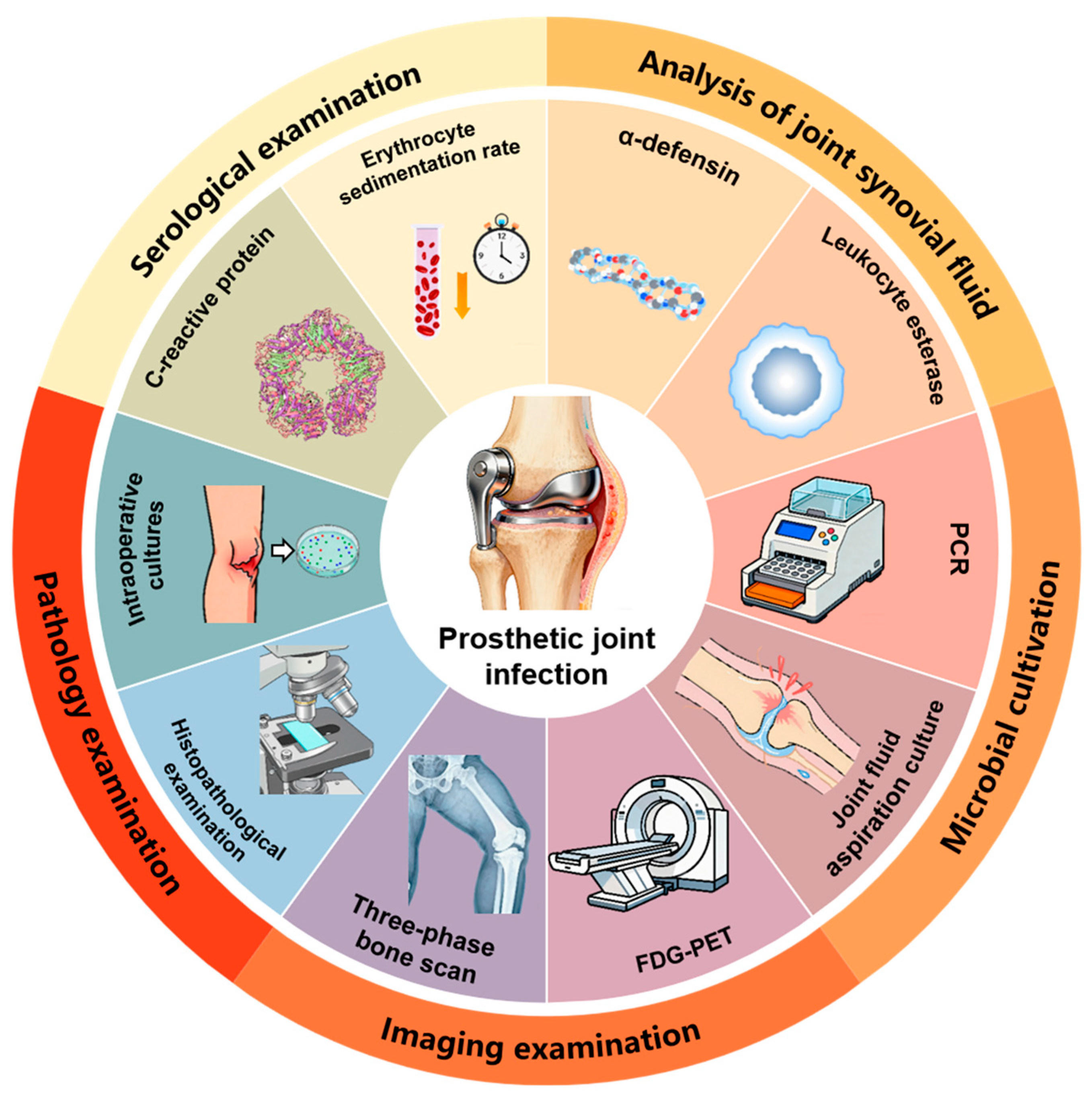
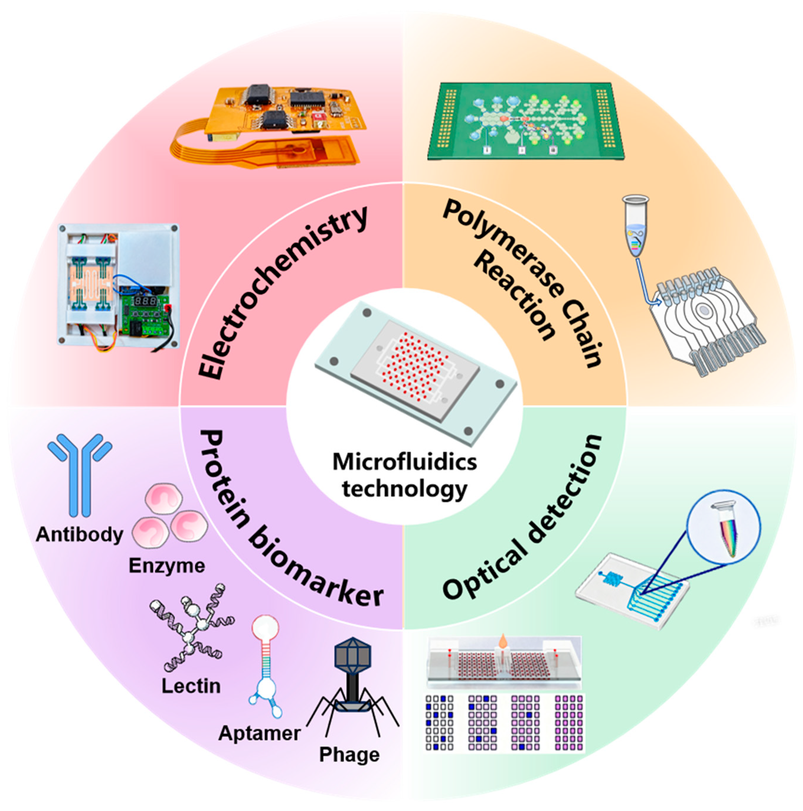
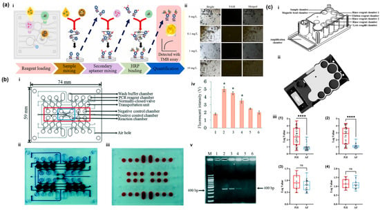
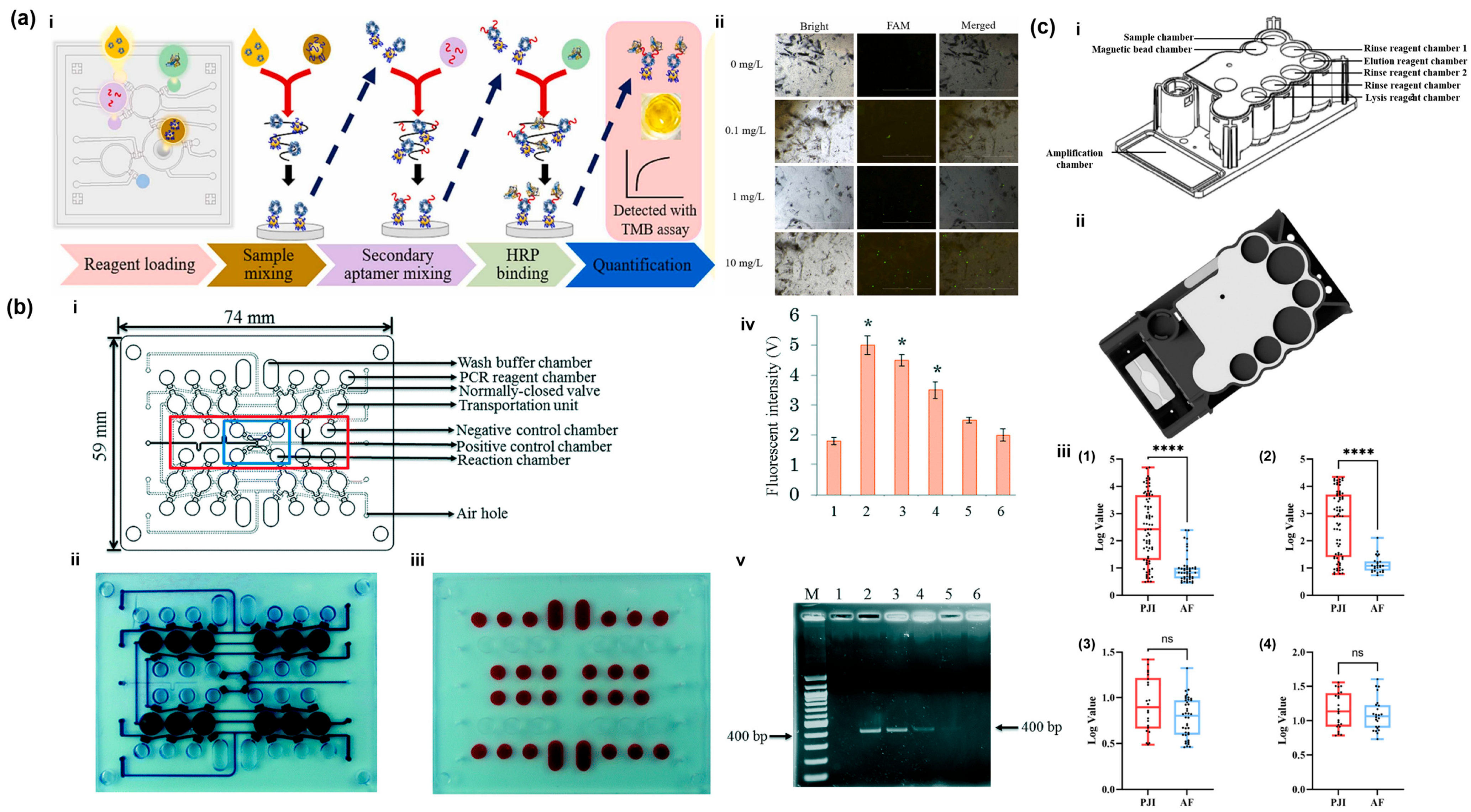

Disclaimer/Publisher’s Note: The statements, opinions and data contained in all publications are solely those of the individual author(s) and contributor(s) and not of MDPI and/or the editor(s). MDPI and/or the editor(s) disclaim responsibility for any injury to people or property resulting from any ideas, methods, instructions or products referred to in the content. |
© 2026 by the authors. Licensee MDPI, Basel, Switzerland. This article is an open access article distributed under the terms and conditions of the Creative Commons Attribution (CC BY) license.