


Abstract
Microneedle (MN)-based optical biosensors are emerging as minimally invasive platforms for point-of-care testing (POCT) and wearable glucose monitoring by enabling direct access to interstitial fluid (ISF), which closely reflects plasma glucose dynamics. This review summarizes recent advances in MN-based glucose sensing with particular emphasis on optical biosensors. Blood-to-ISF glucose transport, physiological time lag, MN architectures, fabrication strategies, and ISF sampling mechanisms are discussed. Glucose recognition mechanisms are outlined together with optical transduction approaches, including colorimetric, fluorescence, surface-enhanced Raman scattering (SERS), photonic, and plasmonic sensing. Many colorimetric MN platforms currently function as single-use or endpoint POCT systems, whereas reversible fluorescence- and photonic-based sensors provide greater potential for repeated measurements and continuous glucose monitoring. Advanced functional materials, smartphone-assisted readout, and emerging artificial intelligence/machine-learning approaches for signal processing, calibration, and glucose prediction are also examined. Key analytical, mechanical, biocompatibility, and ex vivo/in vivo performance parameters are critically evaluated, together with patents, clinical investigations, and commercialization status. Compared with previous reviews that mainly address ISF sampling, general MN biosensing, glucose monitoring across multiple sensing modalities, or wearable optical sensors separately. This review provides an integrated glucose-specific framework linking ISF physiology, MN-mediated sampling, point-of-care and emerging continuous optical sensing, advanced materials, digital/AI analysis, performance validation, and clinical translation. Finally, current limitations involving biofouling, signal drift, long-term stability, manufacturing scalability, and clinical validation are discussed to highlight future directions toward advanced point-of-care platforms and emerging intelligent, wearable, and continuous glucose-monitoring systems.
1. Introduction
Diabetes mellitus (DM) describes a group of chronic and metabolic diseases characterized by the dysregulation of glucose homeostasis, resulting in high levels of blood glucose (hyperglycemia). It is also a remarkably prevalent and challenging health problem worldwide. As reported by the International Diabetes Federation (IDF), the global population of adults aged 20–60 with diabetes was approximately 537 million [1], accounting for 10.5% of the global adult population. This condition causes approximately 1.5 million people to die yearly [2]. DM has become a main cause of several health issues such as cardiovascular disease, kidney failure, blindness, stroke, and limb amputation in many countries. In recent years, point-of-care testing (POCT) has become a transformative tool and has gained significant attention in advancing proactive healthcare by offering simplicity, affordability, and accessibility for clinical applications [3]. These devices enable rapid disease monitoring and screening. Monitoring blood-glucose levels is a core component of managing diabetes and diagnosing disorders associated with glucose metabolism [4]. Glucose monitoring techniques include invasive finger-prick testing, minimally invasive continuous glucose monitoring (CGM) systems, and emerging non-invasive optical or biochemical approaches [5]. These methods aim to improve diabetes management by enhancing patient comfort, reducing the burden of frequent blood sampling, and providing accurate, real-time glucose data for better glycemic control.
Blood tests have long been considered the common standard for clinical diagnosis. Traditional blood-glucose level testing typically requires invasive blood collection through a fingerstick, which frequently results in pain, potential a risk of cross-contamination, low patient adherence, and inconvenience [6,7]. These have become issues that often contribute to trypanophobia among diabetic patients. Furthermore, it fails to satisfy the need for real-time blood-glucose monitoring due to the requirement of multiple daily punctures, and the absence of critical physiological signals can lead to delays in medical action. Currently, novel or alternative approaches for glucose monitoring have been developed to overcome the limitations of traditional blood-glucose monitoring. These approaches shift from invasive methods to minimally invasive and non-invasive techniques, including non-invasive optical methods [8,9] and sampling from sites other than the fingertip [10,11,12]. For minimally invasive applications, microneedles (MNs) biosensors offer a transformative and patient-friendly diagnostic platform. They integrate the minimally painful penetration of MNs with the immediate feedback, enabling simple and rapid detection. By accessing interstitial fluid (ISF) beneath the skin surface, these devices enable real-time, in situ analysis and provide a practical alternative to conventional blood-based testing methods [13].
Glucose biosensors generally consist of a biorecognition element coupled with a transducer that converts glucose-dependent biochemical interactions into measurable signals. Glucose recognition is commonly achieved through enzymatic systems, particularly glucose oxidase (GOx) and glucose dehydrogenase (GDH), or through non-enzymatic strategies using catalytic materials and synthetic receptors. The resulting biochemical response can be converted through electrochemical or optical transduction mechanisms. Electrochemical glucose biosensors remain the most established approach because of their high sensitivity, rapid response, and compatibility with miniaturized electronics, whereas optical biosensors provide complementary advantages, including resistance to electromagnetic interference, remote signal acquisition, multiplexing capability, and compatibility with fluorescence, colorimetric, Raman, photonic, and plasmonic readouts. These sensing principles provide the fundamental basis for the development of minimally invasive glucose-monitoring platforms [12].
Wearable biosensors extend these principles by integrating sensing components with flexible substrates, miniaturized electronics, wireless communication, and digital interfaces to enable continuous or near-continuous monitoring under real-life conditions. For glucose monitoring, successful wearable systems require not only reliable molecular recognition but also efficient access to biofluids, stable signal acquisition, low-power operation, and robust calibration during prolonged use [14]. Microfluidic technologies are particularly important in this context because they can regulate the collection, transport, metering, and delivery of small-volume biofluids to sensing regions while minimizing sample loss and contamination [15]. When integrated with MNs, microfluidic channels and capillary-driven structures can facilitate ISF transport from the skin toward optical or electrochemical sensing elements. While hollow, porous, and hydrogel-forming MNs can further enhance fluid access through pressure-driven flow, capillary action, or swelling-assisted extraction [16,17]. Therefore, the convergence of glucose biosensing, wearable electronics, microfluidics, and MNs-mediated ISF access provides a technological foundation for next-generation minimally invasive glucose-monitoring systems.
An important distinction should be made between continuous glucose monitoring and discrete MN-based glucose measurements. Continuous glucose monitoring refers to repeated or automated glucose measurements acquired over an extended period while the sensing device remains in place, thereby providing temporal glucose profiles, trends, and rates of change. In contrast, intermittent monitoring involves repeated but discrete sampling or measurements at selected time intervals, often requiring separate MNs application or ISF collection for each analysis. Single-point measurement represents a single glucose determination obtained from one sampling event and does not provide information on temporal glucose dynamics. Accordingly, MNs platforms that only extract ISF followed by off-device analysis or provide a single colorimetric, fluorescence, or SERS measurement should not be classified as continuous glucose-monitoring systems unless repeated longitudinal measurements have been experimentally demonstrated.
This narrative review provides an integrated overview of recent advances in MNs-based biosensors for glucose monitoring in ISF. It focuses on the fundamental relationship between blood and ISF glucose, MNs design and fabrication strategies, ISF sampling mechanisms, and the integration of MNs with electrochemical and optical sensing platforms. Particular emphasis is placed on emerging sensing approaches, including fluorescence, colorimetric, surface-enhanced Raman scattering (SERS), nanomaterial-assisted detection, and wearable biosensor systems. The review also evaluates key analytical performance parameters, such as sensitivity, selectivity, response time, stability, reproducibility, and resistance to biological interference. In addition, recent progress in artificial intelligence-assisted signal processing, calibration, glucose trend prediction, and personalized monitoring is discussed. Clinical translation, patents, commercialization, and regulatory considerations are further summarized to highlight the gap between laboratory-scale prototypes and practical glucose-monitoring devices. Finally, current challenges related to ISF extraction efficiency, signal drift, biofouling, long-term stability, manufacturing scalability, and clinical validation are critically examined, together with future perspectives toward intelligent, minimally invasive, and continuous glucose-monitoring systems.
Although several reviews have addressed MNs-mediated ISF sampling, MNs-based glucose monitoring, general MN biosensors, and wearable optical MN sensors, the available literature remains fragmented across these individual domains. The present review provides a distinct synthesis by specifically linking blood-to-ISF glucose physiology and physiological time lag with MNs architecture, ISF sampling mechanisms, glucose-recognition chemistry, and optical signal transduction. Particular emphasis is placed on colorimetric, fluorescence, SERS, photonic, and plasmonic MNs platforms and the advanced functional materials that enable these sensing mechanisms. In addition, this review integrates smartphone-assisted readout and emerging AI/ML approaches for optical image processing, spectral analysis, multivariable calibration, interference correction, and glucose prediction. Importantly, analytical and mechanical performance, ex vivo/in vivo validation, biocompatibility, patents, clinical investigations, and commercialization are evaluated within the same glucose-specific framework. Thus, rather than providing another general overview of MNs biosensors, this review establishes a translational pathway from ISF glucose physiology and MN-mediated sensing to intelligent optical glucose-monitoring systems. Comparison of the present review with previous reviews on MNs-based biosensing and glucose monitoring was represented in Table 1.
Table 1.
Comparison of the present review with previous reviews on MNs-based biosensing and glucose monitoring.
This article was conducted as a narrative review to provide an integrated overview of microneedle-based optical biosensors for glucose monitoring in ISF. Relevant literature was identified through searches of PubMed, Scopus, Web of Science, and Google Scholar, focusing primarily on publications from 2015 to 2026, while earlier seminal studies were included when necessary to describe fundamental concepts and technological development. Search terms included combinations of “microneedle”, “glucose biosensor”, “interstitial fluid”, “optical biosensor”, “colorimetric”, “fluorescence”, “SERS”, “photonic”, “plasmonic”, “continuous glucose monitoring”, “wearable biosensor”, and “artificial intelligence”. Priority was given to original research articles describing MNs-mediated ISF sampling, glucose-sensing mechanisms, optical transduction, analytical performance, in vitro/ex vivo/in vivo validation, and translational development. Relevant review articles, patents, clinical-trial records, and commercial-device information were additionally considered to provide technological and clinical context.
2. Interstitial Fluid (ISF) for Glucose Monitoring
Alternative biological samples for blood-glucose (BG) monitoring include sweat [25], saliva [26], tears [26], urine [26], and ISF [27]. Among these biofluids, ISF has emerged as a particularly suitable source for biomarker analysis because of its close compositional similarity to plasma, the presence of distinct biomarkers that may not be readily detectable in plasma, and its abundance within the extracellular compartment. ISF accounts for approximately 75% of extracellular fluid [28] and 15–25% of body weight [29]. It occupies the interstitial spaces between cells and is primarily derived from plasma that extravasates through capillary walls [30]. ISF contains water, electrolytes, nutrients, dissolved gases, metabolites, and other essential molecules required for cellular function. Accordingly, it serves as an immediate medium for nutrient delivery, waste removal, maintenance of cellular hydration, and intercellular communication [31,32]. Importantly, glucose concentrations in ISF are strongly correlated with those in plasma, supporting its use as an alternative fluid for glucose monitoring. For non-diabetic individuals, ISF glucose concentrations have been proposed to fall within approximately 70–140 mg/dL (3.9–7.8 mmol/L). In patients with type 1 diabetes (T1D) or type 2 diabetes (T2D), clinical recommendations for ISF-based continuous glucose monitoring (CGM) generally target a glucose range of 70–180 mg/dL for at least 70% of the monitored period [33].
Glucose is transported from the vascular compartment into the interstitial space across the capillary endothelium, after which it diffuses through the extracellular matrix before being taken up by surrounding cells. Therefore, ISF glucose concentration is determined by a dynamic balance between capillary glucose delivery, endothelial transport, interstitial diffusion, and local cellular-glucose consumption. Because equilibration between blood and ISF is not instantaneous, changes in ISF glucose generally follow changes in blood glucose with a physiological time lag of approximately 5–10 min, although the magnitude of this delay is variable rather than fixed. The lag may become more pronounced during rapid glucose excursions, such as after meals, insulin administration, exercise, or hypoglycemia, because the rate of glucose appearance or disappearance in blood may temporarily exceed the rate of equilibration with the interstitial compartment [34]. Local tissue perfusion and capillary permeability also influence glucose delivery to ISF, whereas cellular uptake and metabolic activity affect glucose removal from the interstitial space. Consequently, factors such as blood flow, exercise, temperature, hydration status, anatomical site, tissue composition, and the direction and rate of glucose change can alter the relationship between blood and ISF glucose concentrations [35]. For MN-based biosensors, additional local factors, including insertion depth, tissue compression, inflammation, ISF availability, and biofouling, may further influence glucose transport and sensing performance. Importantly, this physiological blood-to-ISF lag should be distinguished from sensor-related delay, which may arise from glucose diffusion through membranes or hydrogels, enzymatic reaction kinetics, optical or electrochemical signal generation, data acquisition, calibration, and algorithmic filtering. Thus, the total delay observed by an MN glucose sensor may represent the combined effects of physiological transport and device-specific response kinetics [36]. Glucose transport from the blood circulation into ISF, the physiological blood–ISF glucose time lag, and MNs-mediated access to dermal ISF for glucose sensing are illustrated in Figure 1.
Figure 1.
Schematic illustration of glucose transport from the blood circulation into ISF, the physiological blood–ISF glucose time lag, and MNs-mediated access to dermal ISF for glucose sensing.
Despite these advantages, the broader utilization of ISF as a diagnostic fluid remains limited by the lack of rapid, minimally invasive techniques capable of obtaining sufficient sample volumes, typically approximately 1–10 µL [30,37]. Conventional ISF sampling approaches include suction blistering, sonophoresis, reverse iontophoresis, microdialysis, and thermal ablation [38]. However, some of these methods can disrupt the stratum corneum, induce local tissue damage, require prolonged sampling periods, or depend on specialized equipment, calibration, and trained personnel. These limitations restrict their practicality for routine point-of-care and self-monitoring applications. Therefore, the development of rapid, reliable, and minimally invasive ISF sampling and sensing technologies remains essential. MNs provide a promising strategy because they can selectively access the epidermal or dermal interstitial space while minimizing pain and tissue damage. Integration of MNs with biosensing platforms may therefore enable efficient ISF access and real-time glucose detection, providing a promising foundation for next-generation minimally invasive glucose monitoring systems [19].
3. Fundamentals of MNs Technology
At present, MNs are gaining increasing interest as a promising alternative to conventional blood-based diagnostics due to their micron-scale dimensions, minimal invasiveness, and reduced pain compared with standard hypodermic needles. In contrast to hypodermic needles, MNs penetration is typically limited to the superficial skin layers and does not reach dermal nerves or blood vessels, enabling painless and blood-free ISF collection, with sufficient ISF volumes for analysis obtainable within ∼1–10 min [39,40,41]. The interstitial fluid of the skin is found under the stratum corneum and is plentiful within the dermal layer, which has a thickness ranging approximately 1500–3000 µm [42]. MNs typically range from 25 to 2500 µm in heights that can penetrate the epidermis. They have a thickness of 50–200 µm for the extraction of ISF from the dermis and a tip diameter between 1 and 100 µm [43,44]. Importantly, their length should be appropriately designed to penetrate the stratum corneum (10–20 µm) [45] and reach the epidermal (50–150 µm) or dermal layers (500–2000 µm) [46]. Insertion pain can be minimized by optimizing MNs design parameters, including diameter, length, width, tip size, tip angle, and needle density. MNs with heights of approximately 500–900 µm are commonly used to reach the dermis for interstitial fluid extraction, providing safe and effective skin penetration depending on factors such as skin type, ethnicity, and age [47]. In addition, MNs length has the greatest influence on perceived pain, with longer MNs (>1450 µm) producing several-fold higher pain scores compared with shorter MNs (<700 µm) [47]. Currently, MNs are commonly fabricated from materials such as silicon, polymers, ceramics, and metals [48]. Recent advancements in micro/nano-fabrication, materials science, digital innovations, biosensing platforms, and flexible electronics have accelerated the development of integrated, miniaturized, and functional micro-sensing systems, particularly for MNs-based transdermal sensors used in point-of-care testing.
4. Types of MNs Structures and Mediated Interstitial Fluid Sampling
There are various types of MNs addressed in this review. MNs can be classified according to their design, fabrication strategies, and function, including applications in drug administration and biofluid extraction. They are commonly divided into six categories: solid, coated, hollow, porous, dissolving, and hydrogel-forming MNs (Figure 2). Materials used in MNs fabrication usually include silicon, stainless steel, biomaterials, and polymers [49,50]. The selection of an appropriate fabrication method depends on the chosen material, critical geometric parameters, and the intended functionality of the MNs. Another important consideration is the skin structure. Human skin is composed of three main layers: the epidermis, dermis, and hypodermis. Among these, the dermis contains a higher concentration of ISF, making it an optimal target for MNs-based biosensors for glucose monitoring. The comparative performance of representative MN-based optical glucose biosensors is reported in Table 2.
Figure 2.
Schematic representation of solid, coated, hollow, porous, dissolving, hydrogel-forming MNs platforms for ISF access and integration with optical glucose biosensing systems.
4.1. Solid MNs
The solid MNs system was the first developed and is primarily composed of metals (including nickel, palladium, stainless steel, gold, and titanium), silicon, and various polymers [51]. They are easy to make and generally have great mechanical strength. Solid MNs pierce the skin to form microincisions, allowing body fluids to be collected. They are mechanically stable and easy to modify [52]. Solid MNs cannot penetrate deep enough to trigger pain receptors. As a result, its use is usually painless and may increase patient compliance [53]. To date, colorimetric or fluorescent solid MNs for glucose detection in ISF have not been reported, while existing approaches predominantly rely on electrochemical sensor integration. Previously, solid MNs fabricated from silicon and integrated with an electrochemical biosensor for glucose detection have been reported, demonstrating a detection range of 2–13.5 mM [54]. However, solid MNs have limitations in drug administration and ISF extraction due to their low drug-loading capacity and low fluid-collection effectiveness. To overcome these issues, other type of MNs such as hollow, porous, coated, dissolving, and hydrogel MNs were developed.
4.2. Coated MNs
Coated MNs are generally fabricated by depositing functional sensing materials onto mechanically robust solid MNs substrates, commonly composed of stainless steel, titanium, silicon, or polymers. The MNs surface can be modified with metals, conductive polymers, enzymes, hydrogels, or nanomaterials using dip-coating, spray-coating, electrodeposition, or layer-by-layer techniques [14]. This configuration combines the high mechanical strength of solid MNs with a functional coating that directly contacts ISF, allowing rapid analyte diffusion and signal generation. For glucose monitoring, coated MNs have predominantly been integrated with electrochemical sensing systems. Previously, gold-coated MNs functionalized with glucose oxidase (GOx) through a self-assembled monolayer demonstrated glucose detection over 30–400 mg/dL (approximately 1.7–22.2 mM) [55]. Non-enzymatic glucose sensing has also been demonstrated using Nafion and platinum-black-coated MNs electrodes, providing a linear detection range of 1–40 mM [56]. More recently, stainless-steel MNs coated with a GOx–chitosan layer and an alginate–polyvinyl alcohol hydrogel achieved glucose detection over 1–40 mM in artificial ISF [57]. However, coated MNs remain limited by non-uniform coating, coating delamination during skin insertion, restricted loading capacity, enzyme instability, and biofouling during prolonged use. These limitations have encouraged the development of alternative MNs platforms, including porous, hollow, dissolving, and hydrogel-forming MNs.
4.3. Hollow MNs
Hollow MNs can be fabricated from diverse materials, including metals (such as stainless steel, nickel, and titanium), polymers (e.g., polydimethylsiloxane and poly(methyl methacrylate)), silicon, glass, and ceramic materials (including silicon carbide and alumina) [58]. These hollow MNs feature microfluidic channels similar to those seen in ordinary hypodermic needles, allowing for the delivery of higher amounts of fluid. Previously, a hollow MNs array with integrated fluidic microchannels was fabricated on a bulk silicon wafer for in vivo ISF sampling and in situ colorimetric glucose detection. Mukerjee et al. designed hollow MNs with a 10 μm hole diameter and a shank height of 250–350 μm, which were successfully demonstrated for glucose detection by extracting ISF from the human earlobe via capillary action, with a sampling time of 15–20 min [16]. In another study, a vacuum-pressure-assisted blood-extraction system incorporating nickel hollow MNs was developed for in vivo glucose and cholesterol diagnosis. The device integrated blood collection, serum separation, and colorimetric paper-based sensing into a single platform, with all processes sequentially automated through one-touch activation [59]. Similarly, a hollow MNs integrated with a paper-based sensor was developed for colorimetric glucose detection. The MNs featured a sharp beveled edge and a length of 400 µm, enabling rapid fluid uptake via capillary action within 5 s. The device allowed simple and rapid glucose detection within 30 s and enabled discrimination between normal glycaemia (4–7 mM) and hyperglycaemia (>7 mM) [60].
4.4. Porous MNs
Porous MNs are designed with micro- or nanoscale pores either on their surface or throughout the entire needle body. Following skin insertion, these micro- or nanoscale pores can extract ISF through capillary action. The collected ISF can then be analyzed directly at the site of collection using surface functionalization or integrated sensing platforms [61,62]. Porous MNs are fabricated from biocompatible metals, ceramics, or biodegradable polymers such as polyethylene glycol (PEG) and poly(lactic-co-glycolic acid) (PLGA). Consequently, porous MNs are less prone to breakage than solid MNs. Several studies have reported porous colorimetric MNs integrated with optical sensing elements for glucose detection in ISF. For example, Zeng et al. developed porous PGMA-based colorimetric MNs functionalized with GOx and horseradish peroxidase (HRP), along with a 3,3′,5,5′-tetramethylbenzidine (TMB)-containing backing layer, allowing in situ visual glucose detection in rat skin with quantitative analysis over a 50–400 mg/dL range using RGB image analysis [63]. Interestingly, the porous MNs integrated with SERS probes and GOx was fabricated using PEG as a porogen and a monomer matrix, enabling in vivo glucose detection over a concentration range of 0–15 mM [64]. Furthermore, Zhu et al. extended this platform by developing a porous MNs integrated with SERS probes for multiplex detection of glucose, uric acid, and cholesterol, demonstrating detection ranges of 0–15 mM for glucose, 0–0.9 mM for uric acid, and 0–9 mM for cholesterol in ISF [65].
4.5. Dissolving MNs
Dissolving MNs are commonly referred to as “poke-and-release” systems. Biodegradable, bioresorbable, or water-soluble polymers such as polyvinyl alcohol (PVA), polyvinylpyrrolidone (PVP), cellulose derivatives, and sugars are often employed in the fabrication process, which is normally done via micro-molding technologies [66]. After MNs implantation into the skin, drug release begins through polymer dissolution, followed by gradual degradation of the biodegradable matrix [51]. The insertion of dissolving MNs forms microchannels in the skin that allow ISF to be extracted using a vacuum pump or captured by an absorbent membrane. Ito et al. developed dissolving MNs made from sodium chondroitin sulfate and dextran to collect ISF from rat skin for glucose monitoring using a moist nonwoven cloth, whereby increasing the cloth-skin contact time from 0.1 to 5.0 min significantly increased ISF glucose extraction from 1.99 to 5.04 µg, and normal skin physiology recovered within 1 h after MNs removal. This study also reported a strong correlation between the measured ISF glucose levels and blood-glucose concentrations [67]. Dissolving MNs help reduce biohazardous sharp waste and improve dosing accuracy but may exhibit inconsistent skin penetration and an increased risk of breakage during use [68,69].
4.6. Hydrogel-Forming MNs
Hydrogel-forming MNs are generally known as swelling MNs. They are fabricated by crosslinking two or more polymers, including chitosan, PVA, PLGA, hyaluronic acid (HA), methacrylated hyaluronic acid (MeHA), polymethyl vinyl ether-alt-maleic anhydride (PMVE/MA), and gelatin methacryloyl (GelMA), which are commonly preferred in fabrication methods [70]. These polymers can be cross-linked to form hydrogel MNs with more effective swelling, depending on the applied chemical, physical, or photo-crosslinking method. The polymer swells after skin application due to uptake of ISF [71]. The development of hydrogel MNs is based on the use of swelling or super-swelling polymers, which are often crosslinked to improve their ability to absorb fluids. There are several reports where the hydrogel-based MNs for glucose detection utilized the chosen different polymer material. For example, hydrogel MNs fabricated from MeHA and subsequently crosslinked via UV irradiation were shown to rapidly collect interstitial fluid. These MNs were able to collect 1.4 μL of ISF within 1 min and 2.3 μL within 10 min from mouse skin [39]. It has been reported that GelMA-based MNs showed swelling ratios of 293–423% and a compressive modulus of 3.34–7.23 MPa demonstrated the capability to extract glucose and vancomycin from ISF [40]. In addition, Bao et al. also utilized GelMA mixed with PVA at a 1:1 ratio to fabricate hydrogel MNs exhibiting a swelling rate exceeding 300% within 60 min. These hydrogel MNs enabled colorimetric detection of four biomarkers including pH, glucose, uric acid, and insulin and detected glucose over a concentration range of 0 to 24 mM in vitro. In vivo studies also showed that the MNs absorbed 45 mg of ISF within 10 min after application, resulting in visible color changes corresponding to the four biomarkers. Similarly, He et al. reported the development of a hydrogel MNs patch composed of PVA and chitosan for extracting multiple biomarkers, including glucose, chloride, lactate, and bovine serum albumin (BSA). The PVA and chitosan formulation at a 1:1 ratio exhibited the highest swelling capacity and extraction efficiency. For glucose detection, the MNs extracted 1.25 mg within 10 min in rabbit skin, and the micropores created in the skin disappeared rapidly within 3 min [72]. However, one limitation associated with hydrogel MNs is the difficulty in preserving the geometric integrity of the fabricated structures [73]. Furthermore, certain polymer systems require cross linking to enhance mechanical strength, regulate swelling behavior, and improve the overall stability of the MNs.
Table 2.
Comparative performance of representative MN-based optical glucose biosensors.
In addition to passive capillary and swelling mechanisms, external forces have been introduced to accelerate MNs-mediated ISF extraction. These approaches include vacuum pressure, osmotic pressure, electroosmosis, mechanical vibration, and integrated microfluidic pumps. Such strategies can increase sampling volume and shorten extraction time, which is particularly important because only microliter-scale volumes of ISF are typically available. Nevertheless, increasing extraction force may alter local tissue perfusion or introduce blood contamination, while prolonged extraction can affect the representativeness of the collected sample. Therefore, an optimal MNs sampling platform must balance extraction efficiency with tissue integrity, analyte recovery, and sampling reproducibility [14,95].
For glucose monitoring, the integration of ISF sampling and sensing within a single MNs platform is particularly attractive because it minimizes sample handling and may enable near-real-time or continuous measurements. Hollow and porous MNs can directly transport glucose-containing ISF toward electrochemical or optical sensing regions, whereas hydrogel-forming MNs can absorb glucose into a hydrated sensing matrix. Current challenges include obtaining sufficient and reproducible ISF volumes, maintaining unobstructed mass transport, preventing blood contamination and biofouling, minimizing sampling delay, and achieving stable calibration during prolonged skin contact. Addressing these limitations will be essential for translating MNs-mediated ISF sampling from laboratory prototypes into reliable wearable glucose-monitoring systems [14,18].
5. Fabrication Technologies for MNs Biosensors
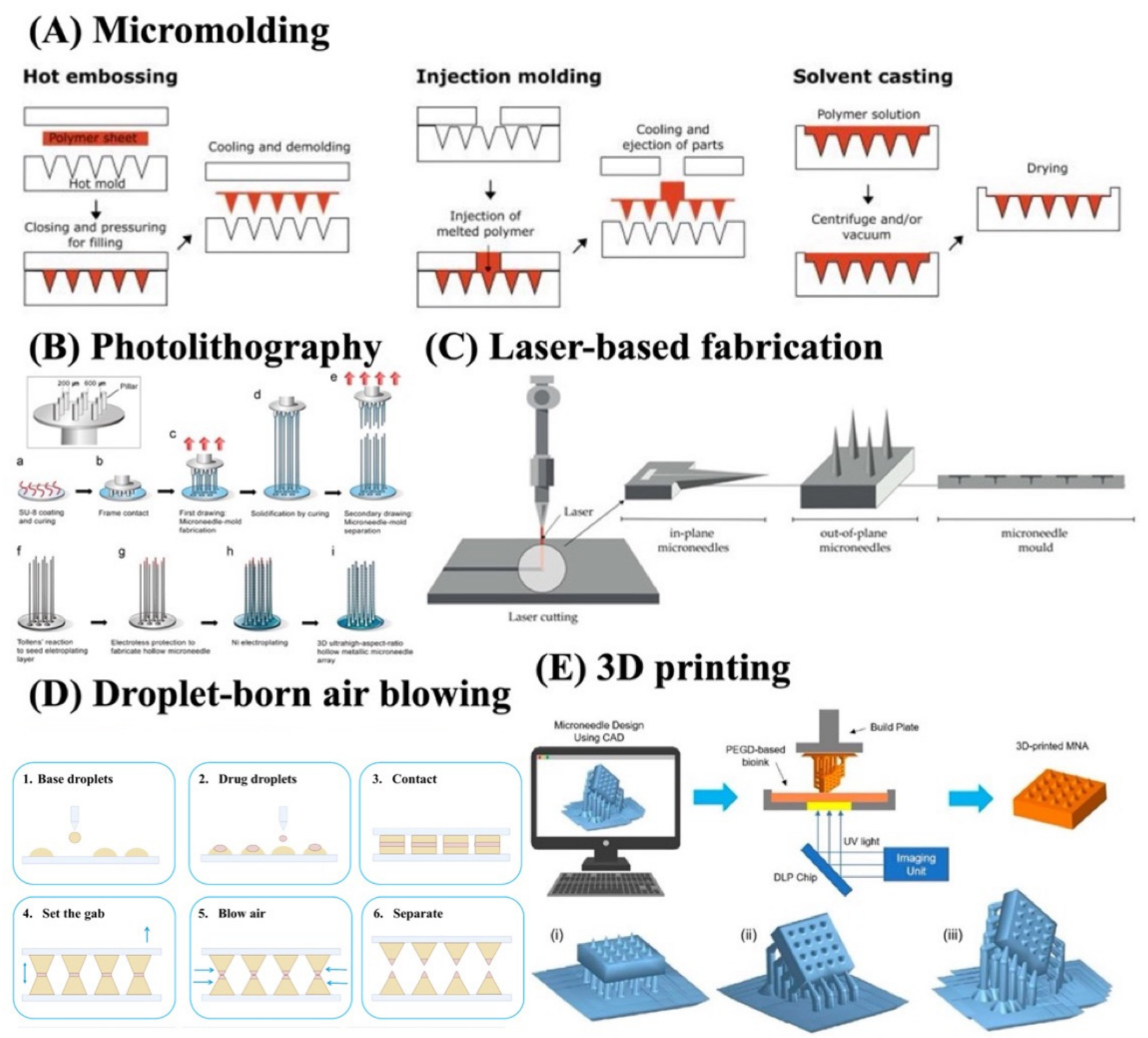
Fabrication technology is a critical determinant of the geometry, mechanical strength, reproducibility, surface properties, and sensing performance of MNs-based optical biosensors. The selection of an appropriate manufacturing method depends on the MNs type, constituent material, target insertion depth, sensing mechanism, and required integration with electrodes, enzymes, nanomaterials, or optical reporters. Conventional fabrication approaches include micromolding, photolithography, etching, micromachining, and laser-based processing, whereas additive manufacturing and advanced surface-modification techniques have recently expanded the design flexibility of MNs biosensors [50]. The fabrication technologies for MNs biosensors were illustrated in Figure 3.
5.1. Micromolding
Micromolding is one of the most widely used methods for fabricating dissolving, and hydrogel-forming MNs. In this approach, polymer solutions or prepolymer mixtures are filled into negative molds, commonly prepared from polydimethylsiloxane (PDMS), followed by drying, polymerization, or crosslinking. Vacuum, centrifugation, or pressure-assisted filling may be employed to ensure complete filling of the needle cavities. Micromolding offers relatively low cost, simple processing, and good scalability; however, the final MN geometry is dependent on mold quality, and incorporation of sensing materials may require additional coating or immobilization steps [50].
5.2. Photolithography and Etching Techniques
Photolithography and etching techniques are frequently employed for the fabrication of silicon and metallic MNs. Photolithography enables precise definition of MNs dimensions and array patterns, while wet or dry etching can generate sharp and highly reproducible structures. Deep reactive ion etching is particularly suitable for producing high-aspect-ratio silicon MNs with controlled geometry. These methods provide excellent dimensional accuracy and are compatible with microelectronic fabrication, making them attractive for electrochemical biosensors. Nevertheless, they generally require cleanroom facilities, specialized equipment, multiple processing steps, and relatively high manufacturing costs [50].
5.3. Laser-Based Fabrication and Micromachining
Laser-based fabrication and micromachining provide alternative routes for manufacturing metallic and polymeric MNs. Laser cutting, laser ablation, and laser micromachining can rapidly generate MNs structures from stainless steel, titanium, polymers, or composite materials without the need for complex masks. These approaches offer flexibility in modifying needle length, width, spacing, and tip geometry. However, thermal damage, surface roughness, and limited resolution may occur depending on the laser parameters and material properties [50].
5.4. Droplet-Born Air Blowing
Droplet-born air blowing (DAB) is a mold-free fabrication technique developed for dissolving MNs. In this approach, controlled droplets of a water-soluble polymer are sequentially dispensed onto a substrate, brought into contact with an opposing surface, and elongated into MNs structures. Air blowing is subsequently applied to dry and solidify the elongated droplets while maintaining the needle geometry. The droplet volume can be precisely controlled by adjusting the dispensing pressure and time, allowing reproducible control of MNs dimensions and incorporated material content. Importantly, DAB operates under mild conditions of approximately 4–25 °C and enables rapid MNs fabrication within 10 min, without requiring heating or ultraviolet irradiation. These characteristics are advantageous for incorporating temperature- or light-sensitive biomolecules, including enzymes and other biological sensing components. However, precise control of droplet rheology, polymer viscosity, drying conditions, and needle geometry is required to obtain reproducible MNs arrays [96].
5.5. Three-Dimensional (3D) Printing
3D printing has emerged as a promising technology for the rapid and customizable fabrication of MNs biosensors. Techniques such as stereolithography, digital light processing, two-photon polymerization, and fused deposition modeling enable the production of complex MNs geometries directly from computer-aided designs (CADs). In particular, high-resolution photopolymerization techniques allow precise control over needle height, base diameter, tip radius, and array configuration. 3D printing also facilitates rapid prototyping and patient-specific design. However, limitations remain regarding printing resolution, biocompatibility of photopolymerizable resins, post-processing requirements, and the electrical conductivity of printed materials [97].
For biosensing applications, MNs fabrication is commonly followed by surface functionalization or coating to introduce sensing components. Conductive metals, carbon-based materials, conducting polymers, enzymes, antibodies, aptamers, fluorophores, and nanomaterials can be deposited using dip coating, spray coating, electrodeposition, sputtering, inkjet printing, or layer-by-layer assembly. For glucose sensing, GOx or glucose dehydrogenase (GDH) is frequently immobilized on conductive MNs electrodes, while optical platforms may incorporate fluorescent probes, plasmonic nanoparticles, or colorimetric reagents. The uniformity and stability of these functional layers are essential because coating defects or delamination during skin insertion can substantially reduce analytical sensitivity and reproducibility [14,36]. Future manufacturing strategies are expected to combine high-resolution additive manufacturing with automated surface functionalization, conductive materials, and scalable roll-to-roll or batch-processing techniques. Such integrated approaches will be important for achieving reproducible, low-cost, and clinically translatable MNs biosensors for continuous glucose monitoring in interstitial fluid [14].
Figure 3.
Schematic illustration of MNs fabrication techniques: (A) Micromolding [98], (B) photolithography and etching [99], (C) laser-based fabrication and micromachining [100], (D) droplet-born air blowing [96], and (E) three-dimensional (3D) printing [101]. Adapted from Refs. [96,98,99,100,101] under the Creative Commons Attribution (CC BY 4.0) license.
6. Principles of Glucose Biosensing
Glucose biosensors convert glucose concentration into a measurable analytical signal through the combination of a recognition element and a transducer. The recognition component selectively interacts with glucose, whereas the transducer converts this interaction into an electrical, optical, or other physicochemical signal. In MNs-based systems, glucose sensing is predominantly achieved through electrochemical or optical mechanisms following direct contact with ISF. According to the glucose-recognition mechanism, sensing platforms can be broadly classified as enzymatic and non-enzymatic systems [14,102]. The principles of glucose biosensing were illustrated in Figure 4.
Figure 4.
Schematic illustration of the principles of glucose biosensing, including enzymatic, non-enzymatic, and glucose-selective recognition mechanisms.
6.1. Enzymatic Glucose Sensing
Enzymatic glucose biosensors use glucose-specific oxidoreductases to catalyze glucose conversion into products that can subsequently be quantified. GOx and GDH are the most widely employed enzymes because of their high catalytic activity and glucose-recognition capability. In electrochemical systems, glucose oxidation generates electron transfer that can be measured as current, whereas optical systems may detect changes in oxygen, hydrogen peroxide, pH, fluorescence, or color produced during the enzymatic reaction. Enzyme-based sensors generally provide high sensitivity and selectivity but are susceptible to enzyme denaturation, environmental pH and temperature, immobilization conditions, and long-term stability limitations [102,103]. Enzymatic electrochemical glucose biosensors are commonly described in three generations according to the electron-transfer mechanism. First-generation sensors use molecular oxygen as the natural electron acceptor and quantify oxygen consumption or hydrogen peroxide formation. Second-generation systems employ artificial redox mediators, such as ferrocene derivatives, ferricyanide, or osmium complexes, to facilitate electron transfer between the enzyme and electrode and reduce oxygen dependence. Third-generation sensors attempt direct electron transfer between the enzyme redox center and electrode surface without a diffusible mediator, potentially simplifying sensor architecture and lowering operating potential [14,103].
6.1.1. Glucose Oxidase-Based Sensing
GOx is the most extensively employed enzyme in glucose biosensors because of its high glucose selectivity, catalytic activity, relative stability, and established immobilization chemistry. GOx contains flavin adenine dinucleotide (FAD) as its redox cofactor and catalyzes the oxidation of β-D-glucose to glucono-δ-lactone while FAD is reduced to FADH2. Molecular oxygen subsequently reoxidizes FADH2, generating hydrogen peroxide, which can be electrochemically or optically quantified as an indirect measure of glucose concentration [14,102]. GOx is particularly attractive for MNs biosensors because it can be immobilized onto metallic electrodes, conductive polymers, nanomaterials, or hydrogel matrices positioned directly within the ISF-accessing region. However, first-generation GOx systems depend on local oxygen availability, which may vary within skin tissue and introduce measurement error. Hydrogen peroxide detection may also require relatively high electrode potentials, increasing interference from electroactive compounds such as uric acid and ascorbic acid. Mediators, nanostructured electrodes, and selective membranes are therefore frequently incorporated to improve electron transfer, decrease operating potential, and enhance glucose selectivity [14,95].
6.1.2. Glucose Dehydrogenase-Based Sensing
GDH provides an alternative enzymatic approach that is largely independent of dissolved oxygen. Different GDH families use cofactors including pyrroloquinoline quinone (PQQ), nicotinamide adenine dinucleotide (NAD), and flavin adenine dinucleotide (FAD). During glucose oxidation, these cofactors accept electrons that are subsequently transferred to an electrode through mediators or direct electron-transfer pathways. The oxygen independence of GDH can be advantageous for ISF sensing, where local oxygen concentrations may fluctuate [95,104]. Nevertheless, each GDH system presents specific limitations. PQQ-GDH exhibits rapid electron-transfer kinetics but may respond to other sugars, including maltose, galactose, and xylose, depending on the enzyme variant. NAD-dependent GDH requires an additional diffusible cofactor and efficient electrochemical regeneration of NADH, complicating sensor design. FAD-GDH provides improved substrate specificity and oxygen-independent activity and has therefore attracted increasing interest for glucose biosensors. Appropriate enzyme selection is particularly important for MNs-based continuous monitoring, where long-term selectivity, catalytic stability, and resistance to physiological interference are essential [105,106].
6.2. Non-Enzymatic Glucose Sensing
Non-enzymatic glucose sensing eliminates biological enzymes and typically relies on direct catalytic oxidation of glucose at an electrode surface. Noble and transition metals, including Pt, Au, Ni, and Cu, as well as their oxides, alloys, sulfides, carbon composites, metal-organic frameworks, and other nanostructured materials have been investigated. Their high surface area and catalytic activity can improve electron transfer and increase glucose sensitivity. Compared with enzymatic sensors, non-enzymatic systems generally offer greater thermal and chemical stability, simpler storage, and reduced susceptibility to enzyme degradation [103,104]. However, direct glucose oxidation frequently requires specific electrode potentials or alkaline conditions and may suffer from interference by electroactive compounds and other carbohydrates. Chloride adsorption and surface fouling may additionally reduce catalytic activity in physiological fluids. These issues are particularly important for MNs biosensors operating directly in ISF. Consequently, nanostructuring, alloy formation, protective membranes, and surface functionalization are increasingly employed to enhance catalytic activity and selectivity under physiologically relevant conditions [103].
6.3. Recognition Elements and Glucose-Selective Materials
Beyond enzyme-mediated catalysis, glucose recognition can be achieved using synthetic receptors and functional materials. Phenylboronic acid (PBA) and diboronic acid derivatives are particularly important for optical glucose sensing because boronic acids reversibly bind cis-diol groups of saccharides to form boronate esters. Binding can induce changes in fluorescence intensity, wavelength, color, swelling, or other physicochemical properties that are translated into glucose-dependent signals. Diboronic acid architectures have been developed to improve glucose selectivity by providing two appropriately positioned boronic acid recognition sites [107]. Other recognition strategies include molecularly imprinted polymers (MIPs), which contain synthetic binding cavities complementary to glucose, and catalytic nanomaterials that combine recognition and transduction functions. For optical MNs biosensors, glucose-responsive hydrogels, fluorophores, plasmonic nanoparticles, and Raman-active substrates may be incorporated directly into the MNs structure or sensing layer. Selection of the recognition material should consider glucose affinity within the physiological ISF range, reversibility, resistance to interference, biocompatibility, and stability during repeated or continuous monitoring [103,107].
7. Microneedle-Based Electrochemical Glucose Biosensors
MNs-based electrochemical biosensors combine minimally invasive access to ISF with electrochemical transduction, allowing biochemical reactions occurring at the MNs–ISF interface to be converted into measurable electrical signals. Compared with conventional blood-based electrochemical glucose testing, MNs platforms can provide direct access to dermal ISF while minimizing tissue injury and enabling repeated or continuous measurements. Electrochemical MNs sensors are particularly attractive because of their high sensitivity, rapid response, low power consumption, ease of miniaturization, and compatibility with wearable electronics. Depending on the transduction principle, these systems can operate through amperometric, potentiometric, or impedimetric detection, although amperometric sensing currently dominates MNs-based glucose monitoring [108]. The MNs-based electrochemical glucose biosensors were illustrated in Figure 5.
Figure 5.
Schematic classification of MNs-based electrochemical glucose biosensors, including (A) amperometric, (B) potentiometric, and (C) impedimetric sensing modes for interstitial-fluid glucose detection.
7.1. Amperometric Sensing
Amperometric sensing is the most extensively investigated electrochemical approach for MNs-based glucose monitoring. In this method, a constant potential is applied between the working and reference electrodes, and the resulting current generated by an oxidation or reduction reaction is measured. For GOx-based sensors, glucose is enzymatically oxidized to gluconolactone, accompanied by the generation of hydrogen peroxide (H2O2) or reduction of an electron-transfer mediator. The electrochemical oxidation or reduction of these products generates a current proportional to glucose concentration. Because amperometric measurements provide high sensitivity, rapid response, and straightforward miniaturization, they are highly suitable for continuous ISF glucose monitoring [109].
7.2. Potentiometric Sensing
Potentiometric sensing measures the potential difference between an indicator electrode and a stable reference electrode under conditions of negligible current flow. The resulting potential is generally governed by the activity of a target ionic species according to the Nernst relationship. Potentiometric MNs sensors are therefore well established for monitoring electrolytes such as Na+, K+, and H+ in ISF. However, direct potentiometric glucose detection is less common because glucose is electrically neutral and does not itself generate an ion-selective potential. Consequently, potentiometric glucose biosensors typically require an indirect recognition mechanism in which enzymatic glucose conversion produces changes in proton concentration, ionic composition, or interfacial potential that can be measured potentiometrically [108,110].
7.3. Impedimetric Sensing
Impedimetric sensing measures changes in the electrical impedance of the electrode–solution interface over a defined frequency range, most commonly using electrochemical impedance spectroscopy (EIS). Unlike conventional amperometry, EIS can monitor changes in interfacial resistance, capacitance, dielectric properties, or charge-transfer processes using relatively small excitation signals. Glucose recognition may therefore be translated into concentration-dependent changes in impedance through enzyme immobilization, glucose-responsive polymers, molecularly imprinted layers, or changes occurring at the electrode interface [108].
8. Microneedle-Based Optical Glucose Biosensors
Recently, MNs-based sensors have been developed by integrating biosensing elements onto MNs surfaces or within their structures for real-time detection of biomarkers in ISF or blood. MNs and biosensor technologies have emerged as innovative platforms offering minimal pain, precise delivery, improved bioavailability, and increased user adherence. It can also detect a wide range of biomarkers, including electrolytes, nucleic acids, polysaccharides, proteins, and drugs, as well as metabolites including glucose in ISF [20,111].
8.1. Fundamentals of Optical Glucose Sensing
MNs-based sensing platforms integrate two fundamental functions: ISF extraction or access and biochemical glucose detection through an integrated sensing mechanism. For direct ISF sampling using MNs, the extraction process generally involves two primary stages: (1) successful insertion of the MNs through the stratum corneum into the viable epidermis or superficial dermis, and (2) subsequent accumulation or collection of ISF. ISF harvesting can be achieved through multiple strategies, including coupling the MNs array with an external absorbent reservoir (e.g., paper-based collectors) to wick fluid from the skin, or employing driving forces such as suction-assisted extraction, pressure-driven convection, passive diffusion, or osmotic gradients [112]. Glucose detection in MNs-based systems relies on specific molecular-recognition elements integrated with an appropriate signal-transduction mechanism. The detection principle varies according to the sensing modality and can be broadly categorized into enzymatic detection, non-enzymatic detection, and aptamer-based recognition strategies.
Optical glucose sensing relies on conversion of glucose recognition into a measurable change in an optical property, including fluorescence intensity or lifetime, visible color, Raman spectral intensity, reflectance wavelength, or refractive index (Figure 6). Glucose recognition can occur enzymatically, most commonly through GOx, or non-enzymatically using synthetic receptors such as PBA. In GOx-based systems, glucose oxidation produces gluconolactone and H2O2, and the resulting products can trigger fluorogenic, chromogenic, or Raman-active reactions. Alternatively, reversible binding between boronic acid groups and glucose cis-diols can alter hydrogel swelling, fluorophore behavior, or photonic structure, thereby generating an optical response without enzymatic catalysis [23]. In aptamer-integrated MNs platforms, single-stranded DNA or RNA aptamers are engineered to selectively recognize glucose. Nevertheless, as aptamers serve as molecular recognition elements and lack intrinsic signal-generating capability, they require integration with a suitable signal transducer to translate target binding into a quantifiable response [15].
Figure 6.
Schematic illustration of sensor-integrated MNs platforms for optical glucose detection in interstitial fluid: (A) colorimetric MNs, (B) fluorometric MNs, and (C) surface-enhanced Raman scattering (SERS) MNs.
Developments of MNs sensing platforms are primarily integrated with electrochemical and optical detection methods [113,114]. However, electrochemical MNs sensors exhibit several limitations, including signal drift, electrode degradation, susceptibility to biofouling and electrochemical interference, frequent calibration requirements, and limited multiplexing capability when compared with optical MNs sensors [20,115,116]. Apart from the electrochemical MNs sensor, the optical MNs sensor exhibits strong resistance to electromagnetic interference and does not rely on a reference electrode. It prevents potential drift and provides a stable signal for long-term monitoring, as well as more detailed chemical insights. Additionally, optical analysis minimizes disturbance to the biological environment while avoiding stability issues correlated with electrolytic reactions or electrode passivation.
8.2. Optical MNs Sensor Platforms for Glucose Detection
Optical MNs-based sensors represent a promising approach for minimally invasive glucose monitoring in ISF because of their high sensitivity, versatility, and compatibility with non-electrical signal readout. According to the optical signal-transduction mechanism, these sensors are generally classified into three main categories, such as fluorescent, colorimetric, and surface-enhanced Raman scattering (SERS) sensors (Figure 7). In general, optical biosensors operate through chemical or biological interactions that induce measurable changes in optical properties, such as light absorbance or emission. MNs-based optical-sensors fabrication processes are comprised of four features: the target analyte, the probe-sensing element, signal acquisition, and data readout (Figure 7). Target analytes are selectively recognized by probes and converted into measurable signals through a transducer, which are then displayed on readable devices [22]. Optical biosensors have gained widespread attention due to their versatility, high sensitivity and specificity, affordability, and portability, as well as their compatibility with MNs platforms. Importantly, optical transducers offer the potential for label-free and real-time detection.
Figure 7.
Schematic representation of the fabrication process of MN-based sensors, showing target analytes, probes, sensor types, and data collection and analysis [22].
The operating mechanisms of glucose biosensors are typically based on either oxygen consumption or hydrogen peroxide production resulting from glucose oxidation during the enzyme-catalyzed reaction, as shown in Equation (1) [117].
GOx serves as the enzymatic recognition element in MNs-based sensors. However, when applied in MNs sensors, the inherent instability of GOx is a significant challenge, as its enzymatic activity can be degraded during the coating steps on the MNs surface and upon exposure to biological fluids [118]. In contrast, non-enzymatic electrocatalysts, such as metal-based nanomaterials, have also been applied in MNs sensors [119]. The following sections provide a summary of glucose optical-sensing approaches used in MNs systems ISF, including colorimetric, fluorescence, and SERS methods (Table 3).
Table 3.
Comparison of MNs-based optical sensing strategies for glucose monitoring in ISF.
8.3. Colorimetric MNs Sensors for Glucose Detection
Colorimetric signals can be analyzed by the naked eye or captured using a smartphone and quantitatively analyzed via RGB value. The integration of colorimetric strategies with MNs patches facilitates efficient sampling and detection of biomarkers from ISF. Color generation originates from analyte-induced changes in the chemical structure, density, morphology, size of chromogenic agents, or orientation. According to the underlying color-development mechanisms, chromogenic systems can be broadly categorized into redox-responsive agents and nanostructure-based platforms, which commonly rely on redox reactions, chromogenic substrates, or structural color changes [127]. Most colorimetric MNs patches are designed as redox-responsive systems that rely on enzymatic reactions and incorporate nanostructures such as gold nanoparticles or nanozymes to enhance catalytic efficiency and amplify colorimetric signals.
According to the color change induced by redox-responsive agents, colorimetric sensing relies on enzyme-mediated reactions that generate visually observable signals. Glucose is initially oxidized by glucose oxidase, producing gluconic acid and H2O2. The generated H2O2 subsequently reacts with H2O2-responsive chromogenic systems, such as iodide [128] or HRP coupled with TMB, resulting in color changes visible to the naked eye [129]. Meanwhile, gluconic acid directly causes a decrease in the local solution pH, which can be monitored using a pH-responsive biosensor. For example, Li et al. reported the first development of MNs patch based on AuNP nanozymes with GOx-like activity for in situ glucose detection in ISF [87]. The MNs consisted of two layers: a tip layer embedded with AuNPs and GOx, which generated H2O2 as a byproduct of glucose oxidation, and a backing layer encapsulating HRP, which catalyzed the oxidation of TMB by H2O2 to form oxTMB, resulting in a visible color change. This MNs patch can collect 60.2 mg of ISF in vitro within 10 min, while in vivo the MNs exhibited a blue-green color in the ISF of hyperglycemic rats and the glucose level by analyzing the RGB value (Figure 8A).
Figure 8.
MNs arrays integrated with a colorimetric sensor platform for transdermal glucose monitoring. (A) Swellable colorimetric MNs for glucose detection based on GOx-like gold nanoparticles [87]. (B) Glucose detection: in situ colorimetric analysis with double-layer hydrogel MNs patch based on polyvinyl alcohol and carboxymethyl chitosan [89]. (C) Wearable MNs patch for colorimetric detection of multiple signature biomarkers in vivo toward diabetic diagnosis [88]. (D) Colloidal crystal MNs patch for glucose monitoring [76]. (E) Colorimetric dermal-tattoo biosensor fabricated by MNs patch for multiplexed detection of health-related biomarkers [82]. (F) Controllable-pore porous MNs for high-speed extraction and biomarker detection of ISF [81]. Adapted from Refs. [81,82] under the Creative Commons Attribution (CC BY 4.0) license.
In 2024, the same research group further reported a double-layer hydrogel MNs patch for in situ colorimetric detection of hyperglycemia [89]. The tip layer (PVA/CMCS) contained GOx, while the backing layer consisted of an immobilized poly(vinyl alcohol)–chitosan–horseradish peroxidase (PVA-CS-HRP) hydrogel loaded with TMB. The MNs tip array exhibited good swelling properties, enabling the extraction of 10 mg of ISF within 10 min. The MNs patch identified hyperglycemia in rats within 5 min after insertion, and the rat skin fully recovered within 10 min (Figure 8B).
For multibiomarker detection, Bao et al. demonstrated a colorimetric MNs patch based on a double-crosslinked hydrogel (GelMA mixed with PVA) for the detection of four type 2 diabetes (T2D)-related biomarkers, including glucose, uric acid, insulin, and pH [88]. The MNs absorbed approximately 45 mg of ISF within 10 min in vivo. After MNs application to diabetic rats, the glucose concentration was estimated to be 18.63 mM based on color changes, which was close to the glucose meter reading of 21.9 mM (Figure 8C). Additionally, Gu et al. developed rigid-resin MNs decorated with colloidal crystal structures for glucose visualization, in which periodic SiO2 nanoparticle nanostructures were embedded within a glucose-responsive fluorophenylboronic acid (FPBA) matrix [76]. When glucose diffused into the FPBA-SiO2 colloidal crystals, swelling of the FPBA matrix increased the spacing between the periodically arranged SiO2 nanoparticles, resulting in a redshift of the MNs from blue (200 mg dL−1 glucose) to green (400 mg dL−1 glucose). No significant inflammation was observed after GCC–MNs insertion, demonstrating the good biosafety of the GCC–MNs patch (Figure 8D).
Furthermore, He et al. developed a multiplex MNs-based skin-tattoo sensor on rabbit skin by encapsulating colorimetric reagents within hyaluronic-acid MNs [82]. This platform enabled the simultaneous detection of multiple health-related biomarkers, including pH, glucose, uric acid, and body temperature. For glucose sensing, the biosensor exhibited a distinct colorimetric response, with the glucose-responsive region changing from yellow at 6 mM to red at 8 mM, while the color signals corresponding to the other biomarkers remained unchanged. However, this colorimetric MNs sensor achieved only semiquantitative detection because of its limited sensitivity and accuracy. In addition, the safety of dermal tattoo-based sensing remains controversial, as the potential leakage of dye molecules and H2O2 into the skin raises biocompatibility concerns (Figure 8E).
Interestingly, Qin et al. reported the fabrication of porous MNs composed of PLA microspheres that exhibited sufficient mechanical strength to penetrate human skin and achieved an in vitro ISF extraction rate of 0.95 μL min−1 per needle, representing the highest value reported to date as of 2025 [81]. Furthermore, glucose detection was demonstrated using a porous MNs-integrated paper sensor with a diffusion layer applied to a skin model containing different glucose concentrations (1–20 mM), achieving an LOD of 0.41 mM. The colorimetric response of the system originated from the immobilization of GOx and HRP-TMB on filter paper. However, the flexibility of the porous MNs, particularly the substrate component, should be further improved to enhance conformal contact with skin and overall system performance (Figure 8F).
8.4. Fluorescent MNs Sensors for Glucose Detection
The integration of MNs with fluorescence biosensors represents a promising advancement in biomedical applications. Fluorescent biosensors are widely employed in biological research for chemical detection due to their rapid response, high sensitivity, and strong specificity. These sensors can incorporate biological fluorophores, synthetic dyes, or nanoparticles as fluorescent probes. Fluorescence-based MNs sensors offer enhanced sensitivity and precise target recognition, enabling accurate quantitative analysis while reducing background interference via the use of specifically designed probes. Various types of MNs have been combined with fluorescence-based sensors to improve detection sensitivity and selectivity. In 2022, Zheng et al. developed a methacrylated hyaluronic acid (MeHA) hydrogel microneedles (HMNs) biosensor incorporating fluorescently labeled aptamer probes as recognition elements for the on-needle, and reagent-free detection of biomarkers, including glucose, adenosine triphosphate (ATP), L-tyrosinamide, and thrombin [85]. The team employed a strand-displacement strategy in which fluorophore-conjugated aptamers were hybridized with complementary DNA competitor strands labeled with a quencher to enable reagentless sensing. These MNs sensor patches demonstrated rapid in vitro glucose detection within 2 min and maintained stability for up to 14 days when stored at room temperature. Furthermore, the system enabled accurate and sensitive monitoring of both rising and falling glucose levels in diabetic animal models, particularly in the severe hypoglycemic range (Figure 9A).
Figure 9.
MNs arrays using fluorescence methods for transdermal glucose monitoring. (A) HMN-assisted assay integrating aptamer probes and fluorescence detection for reagentless biomarker quantification [85]. (B) Fluorescent-based biodegradable MNs-sensor array for tether-free continuous glucose monitoring with smartphone application [83]. Adapted from Ref. [83] under the Creative Commons Attribution (CC BY 4.0) license.
In 2023, Sang et al. presented a fluorescence-based biodegradable MNs sensor for glucose detection, fabricated from silk fibroin embedded with a glucose-responsive fluorescent monomer (GF-monomer) and supported by a PVA backing layer, without the use of enzymes or additional reagents [83]. The MNs were composed of two layers: a silk fibroin needle layer containing the GF-monomer, and a PVA backing layer for structural support. The GF-monomer consisted of a diboronic-acid moiety, which served as the glucose recognition site, and an anthracene moiety responsible for fluorescence emission. Upon application to the skin and excitation at 405 nm, the MNs sensor emitted fluorescence at 490 nm. In vitro studies showed that the MNs exhibited a greater fluorescence response in the hypoglycemic range than in the hyperglycemic range (50–450 mg/dL). The sensor was further evaluated across glucose concentrations ranging from 40 mg/dL (hypoglycemia) to 280 mg/dL (hyperglycemia), demonstrating high sensitivity and suitability for long-term in vivo glucose monitoring. The MNs sensor system supports convenient at-home glucose monitoring through a wireless portable device and a smartphone app with image-processing capabilities, and it maintains reliable performance even when applied at different skin angles (Figure 9B).
Previously, Zhang et al. incorporated fluorescent nanodiamonds into a fluorophenylboronic acid (FPBA)-functionalized hydrogel capable of generating fluorescence signals in response to changes in environmental glucose levels [80]. Notably, the team fabricated a fluorescent nanodiamond boronic hydrogel system in porous MNs using porogen leaching technology. The porous stature enhanced ISF extraction via capillary action, thereby reducing response lag time and improving in vivo glucose monitoring. The device exhibited good long-term photostability and signal stability in both small and large animal models.
8.5. SERS MNs Sensors for Glucose Detection
A SERS sensor allows for highly sensitive and quantitative detection of biomolecules by amplifying the Raman signals of molecules adsorbed onto plasmonic materials, such as gold (Au) or silver (Ag) nanoparticles or metal surfaces [130]. SERS enhancement originates from two principal mechanisms: the electromagnetic (EM) mechanism and the chemical mechanism (CM). The EM mechanism is driven by localized surface plasmon resonances (LSPRs), which amplify the local electric field around metallic nanostructures, while the CM mechanism involves direct charge transfer between the adsorbed molecule and the metal surface. Upon excitation, LSPRs induce collective oscillations of conduction electrons in metal nanostructures, generating highly intensified electromagnetic fields known as “hot spots” [131] The size, shape, and crystal structure of metal nanoparticles strongly influence the LSPR properties and, consequently, the magnitude of signal enhancement [132].
In composite SERS substrates, functional materials commonly include noble metals such as gold and silver, along with copper, metal oxides, metal-organic frameworks (MOFs), conjugated polymers, two-dimensional materials, and their corresponding composite structures [21]. Among these, Au and Ag are the most commonly used materials for bioanalytical SERS applications, including SERS-based MNs sensors. Silver nanomaterials exhibit excellent SERS enhancement performance and are widely used for ultra-sensitive in vitro analysis, although their potential toxicity remains a concern [133,134]. In addition to silver, Au nanomaterials demonstrate excellent biocompatibility and chemical stability, making them more suitable for in vivo and intracellular applications. There are various shapes of plasmonic materials and multicomponent nanoparticles, such as Au nanorods (Au NRs) [135], Au nanoparticles (Au NPs) [136], Au nano-islands (Au ILs) [86], and Ag nanoparticles (Ag NPs) [137], that have been incorporated into MNs-based SERS systems. The combination of MNs and SERS has been developed for the in situ monitoring of various biomarkers, such as glucose, offering a painless, effective, and minimally invasive strategy. The detected signals are further enhanced via the incorporation of metal nanoparticles.
Yuen and Liu were the first to fabricate an Ag-coated MNs-based SERS probe for the detection of Rhodamine 6G and glucose molecules embedded at depths exceeding 700 µm within a two-layer tissue phantom designed to mimic the optical absorption and scattering properties of human skin [126]. They demonstrated that glucose concentrations ranging from 5 to 150 mM within tissue phantoms could be quantified with a root mean square error (RMSE) of 3.3 mM. This work highlighted the potential of MNs for SERS-based measurements, enabling deeper tissue penetration and enhanced signal intensity. Nonetheless, the use of stainless-steel MNs induced a significant immune response in the skin.
In 2015, this research team further advanced a hollow agarose MNs coated with Ag for SERS detection of crystal violet and glucose molecules embedded at depths greater than 700 μm beneath the surface of a skin phantom [78]. The Ag-coated agarose MNs were able to measure glucose concentrations ranging from 5 to 150 mM within the skin phantoms, achieving a root-mean-square error of 5.1 mM in under 10 s. Moreover, Liu et al. reported a SERS sensor for in situ intradermal glucose detection based on a low-cost poly(methyl methacrylate) microneedles (PMMA MNs) array [75]. After functionalizing the silver-coated array surface with 1-decanethiol (1-DT), the sensor was calibrated over a glucose concentration range of 0–20 mM in skin phantoms. The functionalized PMMA MNs array was able to directly measure glucose levels in ISF of diabetic mice within a few minutes. Additionally, the MNs array demonstrated good recovery within 10 min after application, with no noticeable adverse reactions (Figure 10A).
Figure 10.
MNs arrays employing SERS for transdermal glucose monitoring. (A) SERS based biosensor with a MNs array for minimally invasive in vivo glucose measurements [75]. (B) A porous SERS MNs for multiplex detection of glucose, uric acid, and cholesterol in interstitial fluid [65]. Adapted from Ref. [75] under the Creative Commons Attribution (CC BY 4.0) license.
In 2025, SERS probes and specific enzymes were integrated into porous MNs to enable the multiplexed detection of glucose, uric acid, and cholesterol [65]. Paraffin wax was used as a hydrophobic barrier to divide each porous MNs into three independent compartments. In these compartments, gold nanoshells (serving as SERS substrates), 4-mercaptophenylboronic acid (4-MPBA), and a specific enzyme were incorporated into each compartment. In this system, enzymatic reactions generate H2O2, which subsequently induces the conversion of substrate-immobilized 4-MPBA to 4-mercaptophenol (4-MPhOH), resulting in changes in the SERS signal. For glucose detection, the MNs platform demonstrated an in vitro detection range of 0–15 mM. The performance of the porous SERS MNs was further validated in rat models, confirming their capability for multiplexed in vivo detection (Figure 10B).
8.6. Photonic and Plasmonic Glucose Sensing
Photonic crystal MNs sensors rely on periodic nanostructures that selectively reflect specific wavelengths of visible light. When incorporated into glucose-responsive hydrogels, glucose binding can alter hydrogel swelling and consequently change the periodic spacing of the photonic structure. This shifts the reflected wavelength and produces a visible structural color change. Unlike conventional dye-based colorimetric systems, the resulting optical response originates from structural changes rather than consumption of a chromogenic reagent, potentially improving reversibility and enabling repeated measurements [23,138].
Zhang et al. developed a wearable MNs platform containing a PBA-functionalized photonic crystal hydrogel. Glucose binding altered hydrogel volume and shifted the reflected wavelength, allowing glucose concentration to be visualized by naked-eye color changes and quantitatively analyzed using smartphone images. The system demonstrated enzyme-free glucose sensing following ISF transport through the MNs platform and was evaluated using porcine skin as a human-skin model (Figure 11A) [138]. More recently, core–shell photonic crystal MNs have incorporated mechanically robust outer shells with PBA-responsive photonic hydrogel cores to improve skin insertion while preserving glucose-dependent structural-color sensing (Figure 11B) [139].
Figure 11.
Representative photonic crystal MNs-based optical glucose biosensors. (A) Core–shell photonic crystal hydrogel MNs using phenylboronic acid–glucose recognition to induce hydrogel swelling, structural-color changes, and a glucose-dependent red shift in the reflection spectrum [139]. (B) Wearable transdermal MNs platform integrating a phenylboronic acid-functionalized photonic crystal hydrogel with smartphone-assisted color analysis for quantitative glucose detection [138]. Adapted from Refs. [138,139] under the Creative Commons Attribution (CC BY 4.0) license.
Plasmonic MNs biosensors instead exploit localized electromagnetic enhancement generated by metallic nanostructures such as gold nanorods or silver nanoparticles. In glucose sensing, the most established plasmonic MNs application is currently SERS, in which localized surface plasmon resonance enhances Raman scattering. Plasmonic paper integrated with MNs has previously demonstrated on-patch SERS analysis of molecules collected from ISF, establishing a general platform that subsequently supported glucose-specific SERS MNs development [140].
8.7. Smartphone-Assisted Optical Detection
Smartphones provide an attractive interface for optical MNs biosensors because their integrated cameras, processors, wireless connectivity, and user interfaces can convert visually observable signals into quantitative glucose measurements. In colorimetric systems, RGB values, hue, saturation, or luminosity can be extracted from images and correlated with glucose concentration. For photonic sensors, smartphone imaging can quantify structural-color changes or reflected wavelength shifts, whereas fluorescence-based systems can employ portable excitation modules together with camera or photodetector-based acquisition [138].
Sang et al. integrated their fluorescent biodegradable MNs sensor with a mobile application and portable optical accessories for tether-free CGM (Figure 12A) [83], while Zeng et al. used smartphone imaging to quantify colorimetric responses over 50–400 mg/dL [141]. Similarly, the photonic crystal MNs system reported by Zhang et al. used mobile-phone imaging to convert structural color into quantitative glucose information (Figure 12B) [138]. More recently, transparent chitosan-based MNs optical biosensors have also been coupled with Bluetooth-enabled readout electronics for real-time glucose measurements, highlighting increasing convergence between optical sensing, smartphones, and digital health technologies [142]. For reliable clinical use, however, smartphone-based optical quantification requires normalization for ambient illumination, camera model, viewing angle, skin pigmentation, sensor position, and optical background. Standardized illumination chambers, internal reference regions, ratiometric analysis, and computational image correction will therefore be important for reducing inter-device and inter-user variability.
Figure 12.
Smartphone-assisted optical MNs platforms for glucose monitoring. (A) Biodegradable fluorescence-based MNs sensor integrated with a mobile application for image processing and continuous glucose monitoring [83]. (B) Photonic crystal-hydrogel MNs sensor coupled with smartphone-based color analysis for quantitative glucose determination [138]. Adapted from Refs. [83,138] under the Creative Commons Attribution (CC BY 4.0) license.
9. Advanced Materials for MNs-Based Optical Glucose Biosensors
Advanced functional materials play a central role in determining the sensitivity, selectivity, optical stability, response time, and biocompatibility of MNs-based optical glucose biosensors. Unlike conventional MNs that primarily provide access to ISF, optical MNs systems require materials capable of simultaneously recognizing glucose and converting this interaction into measurable fluorescence, colorimetric, Raman, photonic, or plasmonic signals. Recent developments therefore increasingly combine nanomaterials, glucose-responsive polymers, hydrogels, and multifunctional composites within a single MNs platform. Metallic nanoparticles, metal–organic frameworks (MOFs), fluorescent nanomaterials, photonic crystals, and stimuli-responsive hydrogels have been particularly important in advancing minimally invasive optical glucose sensing [14].
9.1. Metallic and Semiconductor Nanomaterials
Metallic and semiconductor nanomaterials provide unique optical properties derived from their nanoscale dimensions, including localized surface plasmon resonance, enhanced electromagnetic fields, catalytic activity, fluorescence, and wavelength-dependent light absorption or scattering. These characteristics can amplify weak optical responses and improve the detection of glucose within the small volumes of ISF accessible through MNs. Metallic nanostructures are particularly important in SERS and colorimetric sensing, whereas semiconductor nanomaterials can serve as fluorescent reporters, photocatalysts, or components of photonic structures [23].
Nanoparticles can also contribute structurally to optical MNs sensors. For example, ordered colloidal particles embedded in glucose-responsive hydrogels can create photonic band gaps whose reflected wavelength changes when the hydrogel swells. Liu et al. developed core–shell photonic crystal MNs consisting of a PBA-functionalized polyacrylamide hydrogel containing ordered SiO2 nanoparticles and a mechanically robust hyaluronic acid shell. Glucose–PBA binding increased hydrogel swelling and lattice spacing, resulting in a concentration-dependent red shift of the structural color and reflection spectrum [139].
Although semiconductor materials such as ZnO, TiO2, and semiconductor quantum dots are widely investigated in optical glucose biosensors, their direct integration into optical MNs glucose sensors remains considerably less developed than metallic, hydrogel, and photonic-crystal systems. Their high refractive index, fluorescence, catalytic properties, and ease of surface functionalization nevertheless make them attractive candidates for future MNs platforms, particularly when combined with PBA receptors or enzyme-mediated glucose recognition.
9.2. Gold and Silver Nanoparticles
Gold nanoparticles (AuNPs) and silver nanoparticles (AgNPs) are among the most important metallic nanomaterials for optical glucose sensing because of their strong plasmonic properties, large surface area, facile surface functionalization, and catalytic activity. Their localized surface plasmon resonance can enhance optical absorption and electromagnetic fields, while nanoscale Au and Ag surfaces generate intense SERS enhancement. These properties permit amplification of weak Raman signals and facilitate sensitive glucose detection at or near the MNs–ISF interface [14].
Ag has been particularly important for MNs-based SERS glucose sensing. Ag-coated polymeric MNs have been used as plasmonic substrates for direct detection of glucose in dermal ISF, demonstrating that MNs insertion can position SERS-active surfaces directly within the sensing compartment and overcome some limitations associated with transcutaneous Raman measurements. More recent porous SERS MNs architectures have integrated ISF transport, glucose recognition, and plasmonic signal enhancement within the same structure, further improving the potential for minimally invasive optical monitoring [14].
AuNPs can additionally function as nanozymes rather than solely as plasmonic materials. Li et al. developed swellable colorimetric MNs containing AuNPs with glucose oxidase-like activity. The AuNPs catalyzed glucose oxidation and H2O2 generation, while HRP subsequently mediated TMB oxidation to produce a visible color signal. The system enabled differentiation of normal and hyperglycemic states and allowed further quantification through digital image analysis [143]. This approach demonstrates how metallic nanoparticles can simultaneously contribute to glucose recognition, catalytic amplification, and optical signal generation.
9.3. Carbon Nanotubes and Graphene-Based Materials
Carbon nanotubes (CNTs), graphene, graphene oxide (GO), and related carbon nanomaterials possess high surface areas, tunable surface chemistry, excellent mechanical properties, and distinctive optical and electronic characteristics. In glucose biosensing, CNTs and graphene are predominantly used to enhance electrochemical rather than optical MNs sensing. For example, PBA-containing glucose-responsive polymers combined with CNTs have been coated onto MNs, where glucose-induced polymer swelling alters CNT electrical percolation and produces an electrical response [83].
Nevertheless, carbon nanomaterials also offer considerable potential for optical MNs biosensors. Graphene and GO are efficient fluorescence quenchers and can therefore function as acceptors in fluorescence-resonance energy transfer or fluorescence recovery systems. PBA-functionalized reduced graphene oxide has previously been demonstrated as an enzyme-free fluorescence glucose sensor, in which competitive glucose binding modifies fluorophore–graphene interactions and restores fluorescence [144]. Similarly, single-walled CNTs possess intrinsic near-infrared fluorescence that can be modulated by molecular recognition and has been investigated for optical glucose sensing [145].
However, direct integration of CNT- or graphene-based fluorescence glucose sensors into MNs platforms remains limited. Thus, this material class should presently be regarded as an emerging opportunity rather than an established optical MNs glucose-sensing strategy. Combining PBA-functionalized graphene, fluorescent CNTs, or graphene quantum dots with hydrogel-forming MNs may offer future routes toward highly sensitive, photostable, and reversible optical sensing.
9.4. Metal–Organic Frameworks
Metal–organic frameworks are porous crystalline materials composed of metal ions or clusters connected by organic ligands. Their exceptionally high surface area, tunable pore structure, catalytic activity, and ability to encapsulate enzymes or chromogenic reagents make MOFs particularly attractive for optical glucose biosensing. In MNs platforms, MOFs can function as enzyme carriers, catalytic nanozymes, or signal-amplification materials while maintaining analyte diffusion through their porous structures.
A direct example was reported by Wang et al., who developed Zr-MOF@Pt nanocomposites with peroxidase-like activity for visual glucose detection. GOx-mediated glucose oxidation generated H2O2, which was subsequently decomposed by Zr-MOF@Pt to oxidize TMB and generate a blue color. Incorporation of the Zr-MOF@Pt/TMB/GOx sensing system into a hydrogel MNs array produced a portable visual glucose assay, demonstrating a linear sensing range of 0.5–800 μM in the underlying colorimetric platform [91]. MOFs can also protect natural enzymes from environmental degradation. More recently, Li et al. incorporated both GOx and HRP within hydrophilic MAF-7 and embedded the dual-enzyme MOF system into hydrogel colorimetric MNs. The resulting platform combined ISF extraction with glucose detection across 1–25 mM, illustrating the potential of enzyme-loaded MOFs to improve catalytic integration within MNs-based optical systems [94]. However, MOF biocompatibility, metal-ion release, batch reproducibility, enzyme accessibility, and long-term stability require further investigation before prolonged in vivo use.
9.5. Quantum Dots and Fluorescent Nanomaterials
Quantum dots (QDs) and other fluorescent nanomaterials offer high brightness, narrow and tunable emission spectra, broad excitation ranges, and greater photostability than many conventional organic dyes. These properties are particularly attractive for MNs systems because low ISF volumes and limited optical path lengths require strong and stable fluorescence signals. Surface functionalization with glucose-recognition elements such as PBA or glucose-responsive enzymes can convert QDs into glucose-responsive optical probes. Graphene quantum dots and carbon dots are increasingly attractive because they may reduce concerns associated with heavy-metal-containing semiconductor QDs [146].
Among the most promising fluorescent nanomaterials for MNs glucose sensing are fluorescent nanodiamonds (FNDs). Their nitrogen-vacancy centers provide exceptional resistance to photobleaching and photochemical degradation. Zhang et al. incorporated FNDs into a PBA-functionalized hydrogel and coupled the sensing matrix with porous MNs. Glucose binding altered hydrogel swelling and consequently modulated FND fluorescence transmission, enabling reversible and prolonged glucose monitoring. The photostability of FNDs is particularly advantageous for continuous monitoring, where conventional fluorophores may progressively lose signal intensity [80].
Fluorescent glucose sensing can also be achieved without inorganic QDs. Hu et al. subsequently developed a wearable MNs patch incorporating reversible FRET-based hydrogel sensors, where glucose-specific PBA groups and polymerized FRET pairs generated concentration-dependent fluorescence changes. The FRET hydrogel achieved an LOD of 0.193 mM and a response time of 7.7 min in human serum [90].
10. Artificial Intelligence and Data-Driven MNs-Based Optical Glucose Biosensors
Artificial intelligence (AI) and data-driven analysis are emerging as promising approaches for improving the analytical performance of MNs-based optical glucose biosensors. These platforms generate diverse optical outputs, including fluorescence intensity, RGB color values, reflection-wavelength shifts, and Raman spectra, which can be affected by illumination, optical alignment, sensor-to-sensor variability, skin characteristics, temperature, and ISF volume. Digital preprocessing, such as background subtraction, normalization, region-of-interest selection, and feature extraction, is therefore important before glucose quantification. Current wearable optical sensing literature increasingly identifies AI and machine learning (ML) as useful tools for multidimensional signal analysis, calibration, and prediction, providing a foundation for intelligent MNs-based glucose monitoring [138,147].
AI and ML can contribute to MNs-based optical glucose monitoring at several distinct stages of the sensing pipeline. First, in signal processing, AI can support background subtraction, denoising, image segmentation, region-of-interest selection, spectral preprocessing, and automated extraction of optical features from fluorescence, colorimetric, photonic, or SERS signals. Second, multivariable calibration models can relate these features to glucose concentration while simultaneously accounting for confounding variables such as pH, temperature, illumination, skin characteristics, ISF volume, and sensor-to-sensor variability. Third, data-driven models can be used for drift correction by learning gradual changes associated with photobleaching, biofouling, enzyme degradation, substrate aging, or changes in optical alignment. Fourth, time-series models can analyze sequential ISF glucose measurements to estimate rates of change and predict future glucose concentrations or impending hypo- and hyperglycemic events. Finally, AI may assist physiological-lag compensation by modeling the temporal relationship between blood and ISF glucose together with device-specific response delays, thereby estimating a more clinically relevant glucose trajectory. These functions should be considered complementary components of the analytical pipeline rather than a single generic application of AI [148,149].
Smartphone-assisted optical analysis represents an important intermediate step between conventional optical sensing and fully AI-enabled glucose monitoring. Zhang et al. developed a PBA-functionalized photonic crystal hydrogel MNs platform in which smartphone-derived RGB values were converted into CIE chromaticity coordinates and subsequently related to glucose-dependent reflection wavelengths [138]. Sang et al. similarly integrated biodegradable fluorescent MNs with a mobile application for tether-free glucose monitoring. These systems primarily rely on conventional image processing rather than AI; however, recent smartphone glucose assays outside the MNs field demonstrate that convolutional neural networks (CNNs) can directly classify glucose-dependent colorimetric images while accounting for variations in cameras and illumination [83]. Kanchan et al., for example, reported a smartphone-based colorimetric glucose assay analyzed by a two-dimensional CNN, illustrating a computational strategy that could potentially be transferred to optical MNs platforms [150].
ML may further improve optical glucose quantification by modeling nonlinear relationships among multiple sensing and environmental variables. Potential model inputs include fluorescence intensity or ratios, RGB/HSV parameters, reflection wavelength, Raman spectral features, temperature, pH, sampling time, and sensor-specific characteristics. Algorithms such as partial least-squares regression (PLSR), support vector regression (SVR), random forest (RF), gradient boosting, artificial neural networks (ANNs), and CNNs could therefore support multivariable calibration and interference correction. This capability may be particularly important for PBA-functionalized photonic MNs because their optical response depends not only on glucose binding but also on hydrogel swelling, pH, ionic strength, and polymer characteristics. Liu et al., for example, showed that core–shell PBA-functionalized photonic crystal MNs exhibited both glucose- and pH-dependent reflection changes, highlighting the need for multivariable calibration rather than reliance on a single optical parameter [151,152].
AI may provide particularly substantial benefits for SERS-based MNs biosensors, because SERS measurements produce high-dimensional spectra with overlapping bands, baseline variations, background fluorescence, noise, and substrate-dependent signal variability. Conventional glucose analysis generally relies on selected characteristic peaks or peak-intensity ratios, whereas ML can simultaneously exploit information across an entire spectrum. PCA, PLSR, LDA, SVM, RF, ANN, and CNN approaches have been applied to Raman and SERS datasets for feature extraction, classification, and quantitative analysis. Deep-learning studies have further demonstrated that CNNs can distinguish structurally similar molecules from complex SERS signals, emphasizing the potential of this approach for biomolecular sensing [153,154]. However, these AI-SERS studies are not specific to glucose-sensing MNs; thus, their application to MNs-SERS glucose monitoring should currently be considered a promising future direction rather than an established technology.
Continuous optical MNs biosensors may additionally generate longitudinal datasets suitable for AI-based time-series analysis. Such models could use present and historical ISF glucose signals to identify glucose rates of change, compensate for sensor drift, distinguish transient artifacts from physiological variation, and forecast impending hypo- or hyperglycemia. Recurrent neural networks, long short-term memory networks, temporal convolutional networks, and related models have already been widely investigated using conventional CGM datasets. A systematic review and network meta-analysis of 46 studies demonstrated extensive application of neural networks, SVMs, RF, recurrent networks, and related models for glucose and hypoglycemia prediction, while AI-focused reviews of CGM identify sensor calibration, glucose prediction, and closed-loop control as major areas of application [148,149]. These established CGM frameworks could ultimately be adapted to compensate for physiological blood-to-ISF lag and optical sensor-response delays in continuous MNs systems.
Despite these opportunities, direct integration of AI with MNs-based optical glucose biosensors remains at an early stage, and most published optical MNs systems still employ conventional calibration equations, image processing, or spectral analysis rather than independently trained AI models. Major challenges include limited datasets, batch-to-batch differences in optical materials and MNs fabrication, variations in skin and ISF characteristics, ambient illumination, camera dependence, biofouling, sensor drift, and inter-individual physiological variability. Rigorous model development should therefore use subject- or sample-level separation between training and test datasets, independent external validation, standardized acquisition protocols, and prospective human evaluation. These considerations are especially important for SERS, where technical-replicate leakage and substrate batch effects can substantially inflate apparent ML performance [154]. Ultimately, integration of optical signals with physiological and temporal variables may enable AI-assisted self-calibration, drift and interference correction, physiological-lag compensation, and personalized glucose prediction, thereby transforming optical MN biosensors from passive detectors into intelligent glucose-monitoring systems.
11. Performance Evaluation of MNs-Based Optical Glucose Biosensors
11.1. Analytical-Performance Parameters
The performance of MNs-based glucose biosensors should be evaluated using standardized analytical parameters. Sensitivity represents the change in sensor response per unit change in glucose concentration and is generally obtained from the slope of the calibration curve. High sensitivity is particularly important for ISF analysis because only small sample volumes are available. Selectivity describes the ability to discriminate glucose from potentially interfering substances, including lactate, uric acid, ascorbic acid, acetaminophen, and other saccharides. Limit of detection (LOD) represents the lowest glucose concentration that can be reliably distinguished from background noise, whereas the Linear range defines the concentration interval over which sensor response remains proportional to glucose concentration. For glucose monitoring in ISF, the useful analytical range should ideally encompass both hypoglycemic and hyperglycemic concentrations [14,102].
Response time describes how rapidly the sensor reaches a defined fraction of its steady-state response after a change in glucose concentration and is particularly important for continuous glucose monitoring because sensor-related delay adds to the physiological blood-to-ISF lag. Stability reflects preservation of sensor response during storage and repeated or prolonged operation, whereas Reproducibility indicates the consistency of measurements among independently fabricated sensors or repeated analyses. For wearable MNs biosensors, these parameters should ideally be assessed not only in buffer but also in artificial ISF, ex vivo tissue, and ultimately in vivo conditions because biofouling, enzyme degradation, tissue response, temperature, and mechanical deformation can substantially alter performance [14,102].
11.2. Mechanical Characterization of Microneedle
Mechanical testing of MNs is essential to characterize their properties and to ensure their safety, efficacy, and overall quality [155]. Key evaluations include measuring the compression force of the MNs, the base-plate break strength and flexibility of the base plate, and the transverse-failure force of MNs arrays. Such assessments are also typically required during the regulatory review process. Therefore, these tests must be reliable, standardized, and reproducible. In addition, accurate evaluation of their physical dimensions and mechanical characteristics is critical, as these parameters play a vital role in optimizing device design and performance [156].
The dimensional characteristics of MNs must be carefully evaluated to ensure their safety and performance. Key parameters include needle length, height, inner and outer diameters (for hollow MNs), radius, and overall geometry. These dimensions can be accurately examined using scanning electron microscopy (SEM). Maintaining appropriate needle length and height is particularly critical, as excessive dimensions may penetrate too deeply and potentially damage the underlying layers of the skin [157]. Therefore, strict control of these parameters is essential during design and manufacturing. Additionally, the sterility of MNs arrays is a critical performance parameter, particularly for clinical applications. Ensuring the absence of bacteria, fungi, and other microorganisms is essential to prevent infection and to safeguard patient safety during use. This evaluation focuses on assessing the mechanical strength and structural integrity of the MNs. Important parameters include tip sharpness, tip geometry, and the ability of the MNs to withstand applied mechanical forces or pressure. MNs must possess sufficient mechanical strength to resist breakage during insertion into the skin. Ensuring adequate mechanical performance is essential to prevent device failure and to ensure safe and effective application [158].
11.3. Ex Vivo and In Vivo Performance
Ex vivo and in vivo evaluation is essential for determining whether MNs-based optical glucose biosensors can maintain reliable sensing performance under tissue-relevant and physiological conditions. Ex vivo studies commonly use porcine or rodent skin to assess MNs insertion, ISF transport, glucose recovery, and optical response. For example, Zhang et al. demonstrated glucose-dependent structural-color changes in a photonic crystal hydrogel MNs platform applied to glucose-equilibrated porcine skin, with results consistent with a commercial glucose meter, while Zeng et al. reported a strong linear response of porous colorimetric MNs in isolated rat skin over 25–400 mg/dL (R2 = 0.9910) [138]. In vivo studies further assess dynamic glucose tracking, physiological lag, signal stability, skin recovery, and agreement with reference blood-glucose measurements. SERS-based MNs demonstrated direct glucose measurement in diabetic mice, with 93% of values located within zones A and B of the Clarke error grid, whereas biodegradable fluorescent MNs successfully tracked glucose changes after glucose and insulin administration in mice [83]. Fluorescent nanodiamond–boronic hydrogel MNs further enabled continuous glucose monitoring in mice and pigs, showing physiological lag times of approximately 3.8 ± 1.3 min during increasing glucose and 6.3 ± 3.3 min during decreasing glucose, together with prolonged in vivo sensing capability [80]. Overall, future validation should include standardized metrics such as correlation analysis, Bland–Altman analysis, Clarke or consensus error grids, lag-time assessment, mean absolute relative difference (MARD), prolonged wear, repeated-use safety, and ultimately prospective human studies to establish clinical reliability.
11.4. Biocompatibility and Safety
Biocompatibility and safety are critical considerations for MNs-based optical glucose biosensors because these devices directly contact viable skin and may remain in place for prolonged or repeated monitoring. Evaluation should include cytotoxicity, skin irritation, inflammatory response, tissue recovery, biodegradation, and possible accumulation or leaching of sensing components such as fluorescent probes, metallic nanoparticles, photonic materials, and glucose-responsive polymers. Similarly, fluorescent nanodiamond–boronic hydrogel MNs caused only mild and localized erythema after insertion, with significantly lower macrophage and T-cell infiltration than full-thickness wounds. In pigs, three-day continuous monitoring produced no significant skin inflammation, and the skin completely recovered within approximately seven days after device removal [80]. For photonic crystal MNs, Liu et al. reported cell viabilities of approximately 94–98% following exposure to the constituent materials, indicating favorable preliminary cytocompatibility. Nevertheless, long-term safety should also consider repeated insertion, chronic inflammation, foreign-body responses, phototoxicity, nanoparticle retention, degradation products, and systemic exposure to sensing materials [139]. Therefore, comprehensive in vitro cytotoxicity, histological evaluation, inflammatory-marker analysis, biodistribution, repeated-use studies, and long-term in vivo testing are required before optical MNs glucose biosensors can be translated into routine clinical monitoring.
12. Clinical Translation, Patents, and Commercialization
Wearable minimally invasive continuous glucose-monitoring (CGM) systems have substantially transformed diabetes management by providing real-time glucose trends and alerts that support therapeutic decision-making, particularly in individuals requiring insulin therapy. Several commercially available systems, including Dexcom G7®, FreeStyle Libre®, and Guardian Connect®, measure glucose in interstitial fluid using subcutaneously inserted sensing elements and predominantly rely on electrochemical detection. For example, Dexcom G7 and Guardian systems use sensor filaments positioned within the subcutaneous ISF compartment. Optical glucose monitoring has also reached commercialization through Eversense 365, which employs a fully implantable fluorescence-based glucose sensor together with an external transmitter and mobile application; however, it is not a MNs-based device. In parallel, non-invasive Raman-based platforms such as MOGLU are under clinical development, illustrating growing interest in optical alternatives to electrochemical CGM, although such systems have not yet achieved the same commercial maturity as established CGMs. Importantly, MNs-based optical glucose biosensors have not yet been commercially launched, and most reported systems remain at the laboratory, preclinical, or early translational stage. Thus, currently marketed CGMs provide an important performance benchmark for future MNs optical technologies in terms of analytical accuracy, response time, operational lifetime, user comfort, wireless connectivity, and long-term reliability.
Nevertheless, increasing patent activity and early clinical investigations indicate substantial translational interest in MNs-based glucose monitoring. As summarized in Table 4, patented technologies encompass hydrogel MNs for electrochemical sensing and ISF extraction, aptamer-functionalized MNs biosensors, and optical platforms incorporating fluorescent nanodiamonds and glucose-responsive polymers. Human clinical investigations have also evaluated MNs or microprobe arrays for ISF glucose monitoring, indicating that the basic concept of minimally invasive MNs-mediated glucose sensing is clinically feasible. However, successful commercialization of optical MNs glucose biosensors will require several additional advances, including reproducible MNs fabrication and skin penetration, reliable ISF access, stable optical calibration, resistance to biofouling and environmental interference, long-term biocompatibility, sterilization compatibility, shelf stability, and scalable low-cost manufacturing. Optical systems must also demonstrate clinical accuracy against established CGMs or reference blood-glucose methods using standardized metrics such as MARD, Clarke, or consensus error-grid analysis, and lag-time assessment in sufficiently powered human studies. Therefore, although the growing patent portfolio, clinical investigations, and commercialization of non-MNs optical CGMs demonstrate a favorable translational pathway, a clear gap remains between current optical MNs prototypes and market-ready continuous glucose monitoring devices.
Table 4.
Summary of commercialization status and patents related to MNs-based sensors for glucose detection in ISF.
13. Current Challenges and Future Perspectives
The advancement of MNs sensing platforms has significantly transformed traditional invasive, blood-based diagnostics by enabling direct and continuous access to ISF, particularly for glucose monitoring. Throughout this review, we have demonstrated that integrating MNs with various optical sensing strategies provides minimally invasive and continuous glucose detection in ISF. Although several commercial continuous glucose monitoring systems are currently available, their high-cost limits widespread accessibility within the broader diabetic population. Meanwhile, researchers continue to develop innovative technologies for continuous glucose monitoring, aiming to enhance sensitivity, shorten assay time, reduce required sample volume, and improve overall analytical performance. However, several critical challenges must be addressed to achieve successful and widely adoptable continuous glucose detection in ISF. These challenges involve efficient functionalization with biorecognition elements (e.g., enzymes, antibodies, nucleic acids, and aptamers), efficient ISF extraction and sampling, high sensitivity and selectivity for low-abundance biomarkers, long-term biocompatibility and mechanical stability, robust sensor integration without structural compromise, reliable clinically relevant signal generation, and optimized optical performance with good skin compatibility [15].
At present, Raman methods, colorimetry, and fluorescence-sensing mechanisms have been integrated with MNs systems to enhance the development of MNs sensors and overcome the limitation of traditional blood-glucose monitoring. In future research, MNs-based optical sensing devices are expected to integrate wireless communication and connectivity capabilities, enabling continuous data transmission to smartphones or cloud-based platforms. This advancement will facilitate remote, real-time monitoring and feedback, thereby empowering users to make informed decisions regarding their health management. Furthermore, the integration of MNs-based optical sensors with artificial intelligence (AI) and machine-learning (ML) algorithms is anticipated to enable advanced data analysis and interpretation, supporting personalized detection and data-driven health recommendations. Machine learning can not only process complex spectral data to improve detection accuracy and resistance to interference, but also facilitate early disease warning and personalized health management through long-term continuous monitoring, pattern recognition, and trend prediction.
For clinical translation, future MNs-based optical glucose biosensors should be evaluated against quantitative performance benchmarks rather than analytical feasibility alone. Clinical accuracy should ideally approach that of established CGM technologies, with a MARD of approximately ≤10%, a high proportion of measurements within clinically acceptable Clarke or consensus error-grid zones, and a total blood-to-sensor lag preferably below 10 min. For wearable continuous monitoring, operational stability should extend from several days toward at least 7–14 days while maintaining calibration accuracy, optical signal stability, mechanical integrity, and biocompatibility [166]. Manufacturing translation will additionally require reproducible MNs geometry, sensing performance, and surface functionalization across production batches, together with scalable fabrication, sterilization compatibility, and acceptable shelf stability. Wireless systems should provide reliable real-time or near-real-time transmission to smartphones or external receivers throughout the intended wear period with minimal data loss and appropriate data-security safeguards. Finally, regulatory translation will require standardized analytical and clinical validation, biocompatibility testing, human-factor assessment, manufacturing quality control, and prospective human studies demonstrating safety and performance relative to established glucose-monitoring technologies. These targets should be regarded as practical translational benchmarks rather than universal regulatory thresholds [167,168].
In addition, further clinical investigations of MNs-based optical sensing systems for glucose detection are required to establish detailed correlations and validate their clinical reliability. Ensuring the biosafety of MNs sensors remains essential for long-term application. Moreover, the development of scalable and cost-effective manufacturing technologies is urgently needed to accelerate commercialization and broaden the practical deployment of MNs sensor arrays. Comprehensive evaluation of material and fabrication costs, as well as feasibility for industrial-scale production, is also necessary. From a SWOT (strengths, weaknesses, opportunities, and threats) perspective, MNs-based optical glucose biosensors possess several strengths, including minimally invasive access to ISF, the potential integration of sampling and sensing within a single wearable platform, compatibility with diverse optical modalities, and suitability for smartphone-assisted and data-driven analysis. Nevertheless, important weaknesses remain, particularly the limited and variable volume of accessible ISF, physiological blood-to-ISF lag, sensor-response delay, biofouling, signal drift, photobleaching, environmental interference, and insufficient long-term operational stability. Emerging opportunities include the integration of artificial intelligence and machine learning for multivariable calibration, interference and drift correction, glucose prediction, and physiological-lag compensation, together with wireless communication, cloud-based monitoring, advanced manufacturing technologies, and multiplex detection of additional ISF biomarkers. Conversely, major threats to clinical translation include competition from well-established commercial CGM technologies, stringent regulatory and clinical-performance requirements, batch-to-batch variability in MN fabrication and sensing materials, high manufacturing and validation costs, and unresolved issues related to sterilization, shelf stability, repeated-use safety, long-term biocompatibility, and user adherence. Collectively, this analysis indicates that future development should focus not only on improving analytical sensitivity, but also on achieving reproducible manufacturing, prolonged sensing stability, clinically acceptable accuracy, and robust validation under real-world conditions.
Author Contributions
S.S.: investigation, data curation, and writing—original draft preparation. B.P.: visualization, proofreading, and editing. T.N.: resources, and funding acquisition. P.O.: resources, and funding acquisition. P.S.: conceptualization, formal analysis, supervision, resources, funding acquisition, writing—original draft preparation, and corresponding author. All authors have read and agreed to the published version of the manuscript.
Funding
This research received no external funding.
Institutional Review Board Statement
Not applicable.
Data Availability Statement
No new data were created or analyzed in this study. Data sharing is not applicable to this article.
Acknowledgments
This research was supported by the Postdoctoral Fellowship Program, Silpakorn University, Thailand, fiscal year 2025. During the preparation of this manuscript, the authors used the generative AI tool ChatGPT (GPT-5.6 Sol; OpenAI) and Quillbot version 4.120.1 for the purposes of enhancing readability, improving the language, and assisting in the preparation of graphical illustrations. The authors have reviewed and edited the output and take full responsibility for the content of this publication.
Conflicts of Interest
The authors report no conflicts of interest. This research received no specific grant from funding agencies in the public, commercial, or not-for-profit sectors.
References
- Mohd, S.; Kumar, L.L.; Harish, V.; Kumar, R.; Chaudhary, A.; Sharma, V. Diabetes mellitus: Complications, emerging therapeutic targets, and evolving treatment approaches. Obes. Med. 2025, 58, 100652. [Google Scholar] [CrossRef] [Scilit]
- Milani, R.V.; Lavie, C.J. Health care 2020: Reengineering health care delivery to combat chronic disease. Am. J. Med. 2015, 128, 337–343. [Google Scholar] [CrossRef] [Scilit] [PubMed]
- Yan, H.; Ma, H.; Jiang, X.; Zheng, J.; Chen, M.; Awawdeh, K.; Jiang, X.; Zhou, R.; Yu, J.; Luo, H.; et al. 3D-Printed microneedle assays for point-of-care applications. Biomed. Anal. 2025, 2, 67–78. [Google Scholar] [CrossRef] [Scilit]
- Saeedi, P.; Petersohn, I.; Salpea, P.; Malanda, B.; Karuranga, S.; Unwin, N.; Colagiuri, S.; Guariguata, L.; Motala, A.A.; Ogurtsova, K.; et al. Global and regional diabetes prevalence estimates for 2019 and projections for 2030 and 2045: Results from the International Diabetes Federation Diabetes Atlas, 9th edition. Diabetes Res. Clin. Pract. 2019, 157, 107843. [Google Scholar] [CrossRef] [Scilit] [PubMed]
- Bader, H.D.; Jarjees, M.S.; Ahmed, B.T. Invasive and Non-Invasive Glucose Monitoring Systems: A Review and Comparative Study. Przegląd Elektrotechniczny 2023, 2023, 114–120. [Google Scholar]
- Pavithran, A.A.; Ramamoorthy, L.; Murugesan, R.; MJ, K. Comparison of fingertip vs palm site sampling on pain perception, and variation in capillary blood glucose level among patients with diabetes mellitus. J. Caring Sci. 2020, 9, 182. [Google Scholar]
- Thompson, N.D.; Perz, J.F. Eliminating the blood: Ongoing outbreaks of hepatitis B virus infection and the need for innovative glucose monitoring technologies. J. Diabetes Sci. Technol. 2009, 3, 283–288. [Google Scholar] [CrossRef] [Scilit] [PubMed]
- Elsherif, M.; Hassan, M.U.; Yetisen, A.K.; Butt, H. Hydrogel optical fibers for continuous glucose monitoring. Biosens. Bioelectron. 2019, 137, 25–32. [Google Scholar] [CrossRef] [Scilit] [PubMed]
- Singh, K.; Agarwal, T.; Kumar, U.; Pal, S.; Runthala, A.; Pan, T.-M.; Wu, C.C. Optical biosensors for diabetes management: Advancing into stimuli-responsive sensing mechanisms. Smart Mater. Med. 2023, 4, 91–101. [Google Scholar] [CrossRef] [Scilit]
- Chen, Y.; Lu, S.; Zhang, S.; Li, Y.; Qu, Z.; Chen, Y.; Lu, B.; Wang, X.; Feng, X. Skin-like biosensor system via electrochemical channels for noninvasive blood glucose monitoring. Sci. Adv. 2017, 3, e1701629. [Google Scholar] [CrossRef] [Scilit] [PubMed]
- Bandodkar, A.J.; Gutruf, P.; Choi, J.; Lee, K.; Sekine, Y.; Reeder, J.T.; Jeang, W.J.; Aranyosi, A.J.; Lee, S.P.; Model, J.B.; et al. Battery-free, skin-interfaced microfluidic/electronic systems for simultaneous electrochemical, colorimetric, and volumetric analysis of sweat. Sci. Adv. 2019, 5, eaav3294. [Google Scholar] [CrossRef] [Scilit] [PubMed]
- Teymourian, H.; Barfidokht, A.; Wang, J. Electrochemical glucose sensors in diabetes management: An updated review (2010–2020). Chem. Soc. Rev. 2020, 49, 7671–7709. [Google Scholar] [CrossRef] [Scilit] [PubMed]
- Xu, N.; Xu, W.; Zhang, M.; Yu, J.; Ling, G.; Zhang, P. Microneedle-based technology: Toward minimally invasive disease diagnostics. Adv. Mater. Technol. 2022, 7, 2101595. [Google Scholar] [CrossRef] [Scilit]
- Wang, Y.; Wu, Y.; Lei, Y. Microneedle-based glucose monitoring: A review from sampling methods to wearable biosensors. Biomater. Sci. 2023, 11, 5727–5757. [Google Scholar] [CrossRef] [Scilit] [PubMed]
- Zhao, P.; Zhou, Z.; Wolter, T.; Womelsdorf, J.; Somers, A.; Feng, Y.; Nuutila, K.; Tian, Z.; Chen, J.; Tamayol, A. Engineering microneedles for biosensing and drug delivery. Bioact. Mater. 2025, 52, 36–59. [Google Scholar] [CrossRef] [Scilit] [PubMed]
- Mukerjee, E.V.; Collins, S.D.; Isseroff, R.R.; Smith, R.L. Microneedle array for transdermal biological fluid extraction and in situ analysis. Sens. Actuators A Phys. 2004, 114, 267–275. [Google Scholar] [CrossRef] [Scilit]
- Kulkarni, M.B.; Goel, S. Microthermofluidic systems: From conceptualization to implementation for point of care diagnostic applications. Sens. Actuators A Phys. 2025, 391, 116689. [Google Scholar] [CrossRef] [Scilit]
- Saifullah, K.M.; Faraji Rad, Z. Sampling Dermal Interstitial Fluid Using Microneedles: A Review of Recent Developments in Sampling Methods and Microneedle-Based Biosensors. Adv. Mater. Interfaces 2023, 10, 2201763. [Google Scholar] [CrossRef] [Scilit]
- Kim, G.; Ahn, H.; Chaj Ulloa, J.; Gao, W. Microneedle sensors for dermal interstitial fluid analysis. Med-X 2024, 2, 15. [Google Scholar] [CrossRef] [Scilit] [PubMed]
- Hu, Y.; Chatzilakou, E.; Pan, Z.; Traverso, G.; Yetisen, A.K. Microneedle Sensors for Point-of-Care Diagnostics. Adv. Sci. 2024, 11, e2306560. [Google Scholar] [CrossRef] [Scilit] [PubMed]
- Yang, W.; Chen, Y.; Cheng, X.; Liu, S.; Zhu, H.; Hu, Y. Recent Progress on the Application of Microneedles for In Situ Sampling in Surface-Enhanced Raman Scattering Detection. Biosensors 2025, 15, 350. [Google Scholar] [CrossRef] [Scilit] [PubMed]
- Xiao, J.; Zhou, Z.; Zou, F.; Xu, T. Advanced microneedle-based wearable optical sensors for healthcare monitoring. Chem. Eng. J. 2025, 526, 171057. [Google Scholar] [CrossRef] [Scilit]
- Sun, Y.; Qi, F.; Ding, C. Optical microneedle patches: From design strategies to point-of-care analysis. TrAC Trends Anal. Chem. 2026, 201, 118861. [Google Scholar] [CrossRef] [Scilit]
- Cho, J.; Son, H.; Kim, J.; Song, H.S.; Lee, W. Wearable microneedle sensors for continuous interstitial fluid monitoring. Biosens. Bioelectron. 2026, 297, 118385. [Google Scholar] [CrossRef] [Scilit] [PubMed]
- Heikenfeld, J. Non-invasive analyte access and sensing through eccrine sweat: Challenges and outlook circa 2016. Electroanalysis 2016, 28, 1242–1249. [Google Scholar] [CrossRef] [Scilit]
- Lankelma, J.; Nie, Z.; Carrilho, E.; Whitesides, G.M. Paper-based analytical device for electrochemical flow-injection analysis of glucose in urine. Anal. Chem. 2012, 84, 4147–4152. [Google Scholar] [CrossRef] [Scilit] [PubMed]
- Brown, S.; Zambrana, P.N.; Ge, X.; Bagdure, D.; Stinchcomb, A.L.; Rao, G.; Tolosa, L. Minimally invasive technique for measuring transdermal glucose with a fluorescent biosensor. Anal. Bioanal. Chem. 2018, 410, 7249–7260. [Google Scholar] [CrossRef] [Scilit] [PubMed]
- Lipani, L.; Dupont, B.G.R.; Doungmene, F.; Marken, F.; Tyrrell, R.M.; Guy, R.H.; Ilie, A. Non-invasive, transdermal, path-selective and specific glucose monitoring via a graphene-based platform. Nat. Nanotechnol. 2018, 13, 504–511. [Google Scholar] [CrossRef] [Scilit] [PubMed]
- Kool, J.; Reubsaet, L.; Wesseldijk, F.; Maravilha, R.T.; Pinkse, M.W.; D’Santos, C.S.; van Hilten, J.J.; Zijlstra, F.J.; Heck, A.J. Suction blister fluid as potential body fluid for biomarker proteins. Proteomics 2007, 7, 3638–3650. [Google Scholar] [CrossRef] [Scilit] [PubMed]
- Samant, P.P.; Niedzwiecki, M.M.; Raviele, N.; Tran, V.; Mena-Lapaix, J.; Walker, D.I.; Felner, E.I.; Jones, D.P.; Miller, G.W.; Prausnitz, M.R. Sampling interstitial fluid from human skin using a microneedle patch. Sci. Transl. Med. 2020, 12, eaaw0285. [Google Scholar] [CrossRef] [Scilit] [PubMed]
- Tran, B.Q.; Miller, P.R.; Taylor, R.M.; Boyd, G.; Mach, P.M.; Rosenzweig, C.N.; Baca, J.T.; Polsky, R.; Glaros, T. Proteomic Characterization of Dermal Interstitial Fluid Extracted Using a Novel Microneedle-Assisted Technique. J. Proteome Res. 2018, 17, 479–485. [Google Scholar] [CrossRef] [Scilit] [PubMed]
- Fogh-Andersen, N.; Altura, B.M.; Altura, B.T.; Siggaard-Andersen, O. Composition of interstitial fluid. Clin. Chem. 1995, 41, 1522–1525. [Google Scholar] [CrossRef] [Scilit]
- Battelino, T.; Danne, T.; Bergenstal, R.M.; Amiel, S.A.; Beck, R.; Biester, T.; Bosi, E.; Buckingham, B.A.; Cefalu, W.T.; Close, K.L.; et al. Clinical Targets for Continuous Glucose Monitoring Data Interpretation: Recommendations From the International Consensus on Time in Range. Diabetes Care 2019, 42, 1593–1603. [Google Scholar] [CrossRef] [Scilit] [PubMed]
- Basu, A.; Dube, S.; Slama, M.; Errazuriz, I.; Amezcua, J.C.; Kudva, Y.C.; Peyser, T.; Carter, R.E.; Cobelli, C.; Basu, R. Time lag of glucose from intravascular to interstitial compartment in humans. Diabetes 2013, 62, 4083–4087. [Google Scholar] [CrossRef] [Scilit] [PubMed]
- Moser, O.; Yardley, J.E.; Bracken, R.M. Interstitial Glucose and Physical Exercise in Type 1 Diabetes: Integrative Physiology, Technology, and the Gap In-Between. Nutrients 2018, 10, 93. [Google Scholar] [CrossRef] [Scilit] [PubMed]
- Haider, K.; Dalton, C. Recent Developments in Microneedle Biosensors for Biomedical and Agricultural Applications. Micromachines 2025, 16, 929. [Google Scholar] [CrossRef] [Scilit] [PubMed]
- Friedel, M.; Thompson, I.A.P.; Kasting, G.; Polsky, R.; Cunningham, D.; Soh, H.T.; Heikenfeld, J. Opportunities and challenges in the diagnostic utility of dermal interstitial fluid. Nat. Biomed. Eng. 2023, 7, 1541–1555. [Google Scholar] [CrossRef] [Scilit] [PubMed]
- Kiistala, U. Suction blister device for separation of viable epidermis from dermis. J. Investig. Dermatol. 1968, 50, 129–137. [Google Scholar] [CrossRef] [Scilit] [PubMed]
- Chang, H.; Zheng, M.; Yu, X.; Than, A.; Seeni, R.Z.; Kang, R.; Tian, J.; Khanh, D.P.; Liu, L.; Chen, P.; et al. A Swellable Microneedle Patch to Rapidly Extract Skin Interstitial Fluid for Timely Metabolic Analysis. Adv. Mater. 2017, 29, 1702243. [Google Scholar] [CrossRef] [Scilit] [PubMed]
- Zhu, J.; Zhou, X.; Kim, H.J.; Qu, M.; Jiang, X.; Lee, K.; Ren, L.; Wu, Q.; Wang, C.; Zhu, X.; et al. Gelatin Methacryloyl Microneedle Patches for Minimally Invasive Extraction of Skin Interstitial Fluid. Small 2020, 16, e1905910. [Google Scholar] [CrossRef] [Scilit] [PubMed]
- Zheng, M.; Wang, Z.; Chang, H.; Wang, L.; Chew, S.W.; Lio, D.C.S.; Cui, M.; Liu, L.; Tee, B.C.; Xu, C. Osmosis-powered hydrogel microneedles for microliters of skin interstitial fluid extraction within minutes. Adv. Healthc. Mater. 2020, 9, 1901683. [Google Scholar] [CrossRef] [Scilit] [PubMed]
- Mandal, A. An Alternative Diagnostic Method Using Microneedles for Sampling the Immune System In Situ. Doctoral Dissertation, Massachusetts Institute of Technology, Cambridge, MA, USA, 2017. [Google Scholar]
- Gattu, K.; Godugu, D.; Jain, H.; Jadhav, K.; Cho, H.; Rojekar, S. Microneedle Technologies for Drug Delivery: Innovations, Applications, and Commercial Challenges. Micromachines 2026, 17, 102. [Google Scholar] [CrossRef] [Scilit] [PubMed]
- Prajapat, M.; Gholap, A.D.; Shinde, S.; Padhiyar, D.; Butani, S.; Shah, S.; Rajput, A. 3D printing technology in microneedles: An emerging era in transdermal drug delivery. Hybrid Adv. 2025, 10, 100447. [Google Scholar] [CrossRef] [Scilit]
- Planz, V.; Lehr, C.-M.; Windbergs, M. In vitro models for evaluating safety and efficacy of novel technologies for skin drug delivery. J. Control. Release 2016, 242, 89–104. [Google Scholar] [CrossRef] [Scilit] [PubMed]
- Ventrelli, L.; Marsilio Strambini, L.; Barillaro, G. Microneedles for transdermal biosensing: Current picture and future direction. Adv. Healthc. Mater. 2015, 4, 2606–2640. [Google Scholar] [CrossRef] [Scilit] [PubMed]
- Gill, H.S.; Denson, D.D.; Burris, B.A.; Prausnitz, M.R. Effect of microneedle design on pain in human volunteers. Clin. J. Pain 2008, 24, 585–594. [Google Scholar] [CrossRef] [Scilit] [PubMed]
- Yuan, Z.; Zhang, H.; Hu, W.; Yu, X.; Qin, S.; Wang, C.; Zhang, F. State-of-the-art Review of Metallic Microneedles: Structure, Fabrication, and Application. Chin. J. Mech. Eng. 2025, 38, 21. [Google Scholar] [CrossRef] [Scilit]
- Lee, H.; Choi, T.K.; Lee, Y.B.; Cho, H.R.; Ghaffari, R.; Wang, L.; Choi, H.J.; Chung, T.D.; Lu, N.; Hyeon, T.; et al. A graphene-based electrochemical device with thermoresponsive microneedles for diabetes monitoring and therapy. Nat. Nanotechnol. 2016, 11, 566–572. [Google Scholar] [CrossRef] [Scilit] [PubMed]
- Luo, X.; Yang, L.; Cui, Y. Microneedles: Materials, fabrication, and biomedical applications. Biomed. Microdevices 2023, 25, 20. [Google Scholar] [CrossRef] [Scilit] [PubMed]
- Chen, J.; Huang, W.; Huang, Z.; Liu, S.; Ye, Y.; Li, Q.; Huang, M. Fabrication of tip-dissolving microneedles for transdermal drug delivery of meloxicam. AAPS PharmSciTech 2018, 19, 1141–1151. [Google Scholar] [CrossRef] [Scilit] [PubMed]
- Choi, S.O.; Kim, Y.C.; Lee, J.W.; Park, J.H.; Prausnitz, M.R.; Allen, M.G. Intracellular protein delivery and gene transfection by electroporation using a microneedle electrode array. Small 2012, 8, 1081–1091. [Google Scholar] [CrossRef] [Scilit] [PubMed]
- Chen, B.Z.; Liu, J.L.; Li, Q.Y.; Wang, Z.N.; Zhang, X.P.; Shen, C.B.; Cui, Y.; Guo, X.D. Safety evaluation of solid polymer microneedles in human volunteers at different application sites. ACS Appl. Bio Mater. 2019, 2, 5616–5625. [Google Scholar] [CrossRef] [Scilit] [PubMed]
- Barrett, C.; Dawson, K.; O’Mahony, C.; O’Riordan, A. Development of low cost rapid fabrication of sharp polymer microneedles for in vivo glucose biosensing applications. ECS J. Solid State Sci. Technol. 2015, 4, S3053. [Google Scholar] [CrossRef] [Scilit]
- Hwa, K.-Y.; Subramani, B.; Chang, P.-W.; Chien, M.; Huang, J.-T. Transdermal Microneedle Array-based Sensor for Real Time Continuous Glucose Monitoring. Int. J. Electrochem. Sci. 2015, 10, 2455–2466. [Google Scholar] [CrossRef] [Scilit]
- Chinnadayyala, S.R.; Park, I.; Cho, S. Nonenzymatic determination of glucose at near neutral pH values based on the use of nafion and platinum black coated microneedle electrode array. Microchim. Acta 2018, 185, 250. [Google Scholar] [CrossRef] [Scilit] [PubMed]
- Mansouri Majd, S. Hydrogel-coated microneedle biosensor for selective and low-potential glucose detection in skin-mimicking interstitial fluid phantoms. Talanta 2026, 297, 128555. [Google Scholar] [CrossRef] [Scilit] [PubMed]
- Dharadhar, S.; Majumdar, A.; Dhoble, S.; Patravale, V. Microneedles for transdermal drug delivery: A systematic review. Drug Dev. Ind. Pharm. 2019, 45, 188–201. [Google Scholar] [CrossRef] [Scilit] [PubMed]
- Li, C.G.; Joung, H.-A.; Noh, H.; Song, M.-B.; Kim, M.-G.; Jung, H. One-touch-activated blood multidiagnostic system using a minimally invasive hollow microneedle integrated with a paper-based sensor. Lab Chip 2015, 15, 3286–3292. [Google Scholar] [CrossRef] [Scilit] [PubMed]
- Nicholas, D.; Logan, K.A.; Sheng, Y.; Gao, J.; Farrell, S.; Dixon, D.; Callan, B.; McHale, A.P.; Callan, J.F. Rapid paper based colorimetric detection of glucose using a hollow microneedle device. Int. J. Pharm. 2018, 547, 244–249. [Google Scholar] [CrossRef] [Scilit] [PubMed]
- Bollella, P.; Sharma, S.; Cass, A.E.; Tasca, F.; Antiochia, R. Minimally invasive glucose monitoring using a highly porous gold microneedles-based biosensor: Characterization and application in artificial interstitial fluid. Catalysts 2019, 9, 580. [Google Scholar] [CrossRef] [Scilit]
- Lee, H.; Bonfante, G.; Sasaki, Y.; Takama, N.; Minami, T.; Kim, B. Porous microneedles on a paper for screening test of prediabetes. Med. Devices Sens. 2020, 3, e10109. [Google Scholar] [CrossRef] [Scilit]
- Zeng, Q.; Xu, M.; Hu, W.; Cao, W.; Zhan, Y.; Zhang, Y.; Wang, Q.; Ma, T. Porous Colorimetric Microneedles for Minimally Invasive Rapid Glucose Sampling and Sensing in Skin Interstitial Fluid. Biosensors 2023, 13, 537. [Google Scholar] [CrossRef] [Scilit] [PubMed]
- Zhu, G.; Liu, C.; Lu, Y.; Mo, X.; Dong, J.; Xu, K.; Huang, Y.; Chen, C.; Lv, X.; Yang, X. Porous SERS Microneedles Integrating Sampling and Sensing for Interstitial Glucose Detection. ACS Appl. Nano Mater. 2025, 8, 7867–7875. [Google Scholar] [CrossRef] [Scilit]
- Zhu, G.; Wang, Y.; Yang, X.; Chen, C.; Lv, X.; Huang, Y.; Zhao, X. A porous SERS microneedle for multiplex detection of glucose, uric acid, and cholesterol in interstitial fluid. Anal. Chim. Acta 2025, 1380, 344788. [Google Scholar] [CrossRef] [Scilit] [PubMed]
- Yu, X.; Zhao, J.; Fan, D. The progress in the application of dissolving microneedles in biomedicine. Polymers 2023, 15, 4059. [Google Scholar] [CrossRef] [Scilit] [PubMed]
- Ito, Y.; Taniguchi, M.; Hayashi, A.; Anai, M.; Morita, S.; Ko, E.; Yoshimoto, N.; Yoshii, Y.; Kobuchi, S.; Sakaeda, T. Application of dissolving microneedles to glucose monitoring through dermal interstitial fluid. Biol. Pharm. Bull. 2014, 37, 1776–1781. [Google Scholar] [CrossRef] [Scilit] [PubMed]
- Wang, P.M.; Cornwell, M.; Prausnitz, M.R. Minimally invasive extraction of dermal interstitial fluid for glucose monitoring using microneedles. Diabetes Technol. Ther. 2005, 7, 131–141. [Google Scholar] [CrossRef] [Scilit] [PubMed]
- Amarnani, R.; Shende, P. Microneedles in diagnostic, treatment and theranostics: An advancement in minimally-invasive delivery system. Biomed. Microdevices 2022, 24, 4. [Google Scholar] [CrossRef] [Scilit] [PubMed]
- Piao, H.; Choi, Y.-H.; Kim, J.; Park, D.; Lee, J.; Khang, D.-Y.; Choi, H.-J. Impedance-based polymer microneedle patch sensor for continuous interstitial fluid glucose monitoring. Biosens. Bioelectron. 2024, 247, 115932. [Google Scholar] [CrossRef] [Scilit] [PubMed]
- Yang, Y.; Sheng, C.; Dong, F.; Liu, S. An integrated wearable differential microneedle array for continuous glucose monitoring in interstitial fluids. Biosens. Bioelectron. 2024, 256, 116280. [Google Scholar] [CrossRef] [Scilit] [PubMed]
- Kim, J.H.; Hwang, C.; Kim, D.Y. Structurally Engineered All-Polymeric Microneedle Sensor for Highly Sensitive and Real-Time Monitoring of Glucose in Interstitial Fluids. ACS Sens. 2025, 10, 7025–7034. [Google Scholar] [CrossRef] [Scilit] [PubMed]
- Holman, J.B.; Mushiana, T.; Yang, C.; Jin, Y.; Shi, Z.; Qiu, B.; Li, C.; Ding, W. A wearable paper-based SGR/MCC microneedle array sensor for continuous glucose monitoring. Microsyst. Nanoeng. 2026, 12, 214. [Google Scholar] [CrossRef] [Scilit] [PubMed]
- Cheng, Y.; Gong, X.; Yang, J.; Zheng, G.; Zheng, Y.; Li, Y.; Xu, Y.; Nie, G.; Xie, X.; Chen, M.; et al. A touch-actuated glucose sensor fully integrated with microneedle array and reverse iontophoresis for diabetes monitoring. Biosens. Bioelectron. 2022, 203, 114026. [Google Scholar] [CrossRef] [Scilit] [PubMed]
- Ju, J.; Hsieh, C.M.; Tian, Y.; Kang, J.; Chia, R.; Chang, H.; Bai, Y.; Xu, C.; Wang, X.; Liu, Q. Surface Enhanced Raman Spectroscopy Based Biosensor with a Microneedle Array for Minimally Invasive In Vivo Glucose Measurements. ACS Sens. 2020, 5, 1777–1785. [Google Scholar] [CrossRef] [Scilit] [PubMed]
- Zeng, Y.; Wang, J.; Wang, Z.; Chen, G.; Yu, J.; Li, S.; Li, Q.; Li, H.; Wen, D.; Gu, Z.; et al. Colloidal crystal microneedle patch for glucose monitoring. Nano Today 2020, 35, 100984. [Google Scholar] [CrossRef] [Scilit]
- Kang, J.; Kim, K.Y.; Kim, S.; Hong, H.; Bae, B.-S.; Kang, S.-K.; Lee, W. A conformable microneedle sensor with photopatternable skin adhesive and gel electrolyte for continuous glucose monitoring. Device 2023, 1, 100112. [Google Scholar] [CrossRef] [Scilit]
- Yuen, C.; Liu, Q. Hollow agarose microneedle with silver coating for intradermal surface-enhanced Raman measurements: A skin-mimicking phantom study. J. Biomed. Opt. 2015, 20, 61102. [Google Scholar] [CrossRef] [Scilit] [PubMed]
- Chen, C.; Fu, Y.; Lin, Y.; Liu, Y.; Du, D.; Qiu, K. 3D-printed hollow microneedle-based electrochemical sensor for wireless glucose monitoring. Analyst 2026, 151, 1182–1194. [Google Scholar] [CrossRef] [Scilit] [PubMed]
- Zhang, J.; Zheng, Y.; Lee, J.; Hoover, A.; King, S.A.; Chen, L.; Zhao, J.; Lin, Q.; Yu, C.; Zhu, L.; et al. Continuous Glucose Monitoring Enabled by Fluorescent Nanodiamond Boronic Hydrogel. Adv. Sci. 2023, 10, e2203943. [Google Scholar] [CrossRef] [Scilit] [PubMed]
- Qin, B.; Park, J.; Kim, S.J.; Kim, B. Controllable-pore porous microneedles for high-speed extraction and biomarker detection of interstitial fluid. Microsyst. Nanoeng. 2025, 11, 240. [Google Scholar] [CrossRef] [Scilit] [PubMed]
- He, R.; Liu, H.; Fang, T.; Niu, Y.; Zhang, H.; Han, F.; Gao, B.; Li, F.; Xu, F. A Colorimetric Dermal Tattoo Biosensor Fabricated by Microneedle Patch for Multiplexed Detection of Health-Related Biomarkers. Adv. Sci. 2021, 8, e2103030. [Google Scholar] [CrossRef] [Scilit] [PubMed]
- Sang, M.; Cho, M.; Lim, S.; Min, I.S.; Han, Y.; Lee, C.; Shin, J.; Yoon, K.; Yeo, W.-H.; Lee, T. Fluorescent-based biodegradable microneedle sensor array for tether-free continuous glucose monitoring with smartphone application. Sci. Adv. 2023, 9, eadh1765. [Google Scholar] [CrossRef] [Scilit] [PubMed]
- He, R.; Niu, Y.; Li, Z.; Li, A.; Yang, H.; Xu, F.; Li, F. A Hydrogel Microneedle Patch for Point-of-Care Testing Based on Skin Interstitial Fluid. Adv. Healthc. Mater. 2020, 9, e1901201. [Google Scholar] [CrossRef] [Scilit] [PubMed]
- Zheng, H.; GhavamiNejad, A.; GhavamiNejad, P.; Samarikhalaj, M.; Giacca, A.; Poudineh, M. Hydrogel Microneedle-Assisted Assay Integrating Aptamer Probes and Fluorescence Detection for Reagentless Biomarker Quantification. ACS Sens. 2022, 7, 2387–2399. [Google Scholar] [CrossRef] [Scilit] [PubMed]
- Mei, R.; Wang, Y.; Shi, S.; Zhao, X.; Zhang, Z.; Wang, X.; Shen, D.; Kang, Q.; Chen, L. Highly sensitive and reliable internal-standard surface-enhanced Raman scattering microneedles for determination of bacterial metabolites as infection biomarkers in skin interstitial fluid. Anal. Chem. 2022, 94, 16069–16078. [Google Scholar] [CrossRef] [Scilit] [PubMed]
- Li, X.; Lv, J.; Zhao, J.; Ling, G.; Zhang, P. Swellable colorimetric microneedles for glucose detection based on glucose oxidase-like gold nanoparticles. Anal. Chim. Acta 2024, 1288, 342152. [Google Scholar] [CrossRef] [Scilit] [PubMed]
- Bao, Z.; Lu, S.; Zhang, D.; Wang, G.; Cui, X.; Liu, G. Wearable Microneedle Patch for Colorimetric Detection of Multiple Signature Biomarkers in vivo Toward Diabetic Diagnosis. Adv. Healthc. Mater. 2024, 13, e2303511. [Google Scholar] [CrossRef] [Scilit] [PubMed]
- Yin, Y.; Li, X.; Wang, M.; Ling, G.; Zhang, P. Glucose detection: In-situ colorimetric analysis with double-layer hydrogel microneedle patch based on polyvinyl alcohol and carboxymethyl chitosan. Int. J. Biol. Macromol. 2024, 277, 134408. [Google Scholar] [CrossRef] [Scilit] [PubMed]
- Hu, Y.; Pan, Z.; De Bock, M.; Tan, T.X.; Wang, Y.; Shi, Y.; Yan, N.; Yetisen, A.K. A wearable microneedle patch incorporating reversible FRET-based hydrogel sensors for continuous glucose monitoring. Biosens. Bioelectron. 2024, 262, 116542. [Google Scholar] [CrossRef] [Scilit] [PubMed]
- Wang, N.; Li, Z.; Wu, X.; Zhao, Y.; Su, X. A novel and sensitive microneedle array platform for visual detection of glucose based on Zr-MOF@Pt nanozyme with peroxidase-like activity. Microchem. J. 2024, 203, 110954. [Google Scholar] [CrossRef] [Scilit]
- Saifullah, K.M.; Azarikhah, P.; Faraji Rad, Z. Synthesis-free swellable hydrogel microneedles for rapid interstitial fluid extraction and on-site glucose detection via an electrochemical biosensor system. Mater. Today Chem. 2025, 45, 102661. [Google Scholar] [CrossRef] [Scilit]
- Saifullah, K.M.; Faraji Rad, Z. Single-use microneedle biosensor for detecting clinically relevant glucose in interstitial fluid. Biosens. Bioelectron. 2026, 297, 118387. [Google Scholar] [CrossRef] [Scilit] [PubMed]
- Li, H.; Wei, W.; Wang, X.; Xiong, R.; Ling, G.; Zhang, P. Hydrogel colorimetric microneedle patches embedded with dual-enzyme active metal-organic frameworks for glucose detection. Mater. Today Chem. 2026, 53, 103603. [Google Scholar] [CrossRef] [Scilit]
- Zhang, X.; Zhang, W.; Wu, W.; Chen, J. Recent advances in the preparation of microneedle patches for interstitial fluid extraction and analysis. Microchem. J. 2023, 195, 109477. [Google Scholar] [CrossRef] [Scilit]
- Kim, J.D.; Kim, M.; Yang, H.; Lee, K.; Jung, H. Droplet-born air blowing: Novel dissolving microneedle fabrication. J. Control. Release 2013, 170, 430–436. [Google Scholar] [CrossRef] [Scilit] [PubMed]
- Loh, J.M.; Lim, Y.J.L.; Tay, J.T.; Cheng, H.M.; Tey, H.L.; Liang, K. Design and fabrication of customizable microneedles enabled by 3D printing for biomedical applications. Bioact. Mater. 2024, 32, 222–241. [Google Scholar] [CrossRef] [Scilit] [PubMed]
- Jamaledin, R.; Di Natale, C.; Onesto, V.; Taraghdari, Z.B.; Zare, E.N.; Makvandi, P.; Vecchione, R.; Netti, P.A. Progress in Microneedle-Mediated Protein Delivery. J. Clin. Med. 2020, 9, 542. [Google Scholar] [CrossRef] [Scilit] [PubMed]
- Lee, K.; Lee, H.C.; Lee, D.-S.; Jung, H. Drawing Lithography: Three-Dimensional Fabrication of an Ultrahigh-Aspect-Ratio Microneedle. Adv. Mater. 2010, 22, 483–486. [Google Scholar] [CrossRef] [Scilit] [PubMed]
- Tucak, A.; Sirbubalo, M.; Hindija, L.; Rahić, O.; Hadžiabdić, J.; Muhamedagić, K.; Čekić, A.; Vranić, E. Microneedles: Characteristics, Materials, Production Methods and Commercial Development. Micromachines 2020, 11, 961. [Google Scholar] [CrossRef] [Scilit] [PubMed]
- Razzaghi, M.; Akbari, M. The Effect of 3D Printing Tilt Angle on the Penetration of 3D-Printed Microneedle Arrays. Micromachines 2023, 14, 1157. [Google Scholar] [CrossRef] [Scilit] [PubMed]
- Pullano, S.A.; Greco, M.; Bianco, M.G.; Foti, D.; Brunetti, A.; Fiorillo, A.S. Glucose biosensors in clinical practice: Principles, limits and perspectives of currently used devices. Theranostics 2022, 12, 493–511. [Google Scholar] [CrossRef] [Scilit] [PubMed]
- Hassan, M.H.; Vyas, C.; Grieve, B.; Bartolo, P. Recent Advances in Enzymatic and Non-Enzymatic Electrochemical Glucose Sensing. Sensors 2021, 21, 4672. [Google Scholar] [CrossRef] [Scilit] [PubMed]
- Waly, Y.; Hussain, A.; Al-Majmuei, A.; Alatoom, M.; Alaraibi, A.J.; Alaysereen, A.; Deen, G.R. Evolution of Biosensors and Current State-of-the-Art Applications in Diabetes Control. Biosensors 2026, 16, 39. [Google Scholar] [CrossRef] [Scilit] [PubMed]
- Ferri, S.; Kojima, K.; Sode, K. Review of glucose oxidases and glucose dehydrogenases: A bird’s eye view of glucose sensing enzymes. J. Diabetes Sci. Technol. 2011, 5, 1068–1076. [Google Scholar] [CrossRef] [Scilit] [PubMed]
- Olansky, L.; Kennedy, L. Finger-stick glucose monitoring: Issues of accuracy and specificity. Diabetes Care 2010, 33, 948–949. [Google Scholar] [CrossRef] [Scilit] [PubMed]
- Nan, K.; Jiang, Y.N.; Li, M.; Wang, B. Recent Progress in Diboronic-Acid-Based Glucose Sensors. Biosensors 2023, 13, 618. [Google Scholar] [CrossRef] [Scilit] [PubMed]
- Abdullah, H.; Phairatana, T.; Jeerapan, I. Tackling the challenges of developing microneedle-based electrochemical sensors. Mikrochim. Acta 2022, 189, 440. [Google Scholar] [CrossRef] [Scilit] [PubMed]
- Zhao, L.; Wen, Z.; Jiang, F.; Zheng, Z.; Lu, S. Silk/polyols/GOD microneedle based electrochemical biosensor for continuous glucose monitoring. RSC Adv. 2020, 10, 6163–6171. [Google Scholar] [CrossRef] [Scilit] [PubMed]
- Ju, J.; Li, L.; Regmi, S.; Zhang, X.; Tang, S. Microneedle-Based Glucose Sensor Platform: From Vitro to Wearable Point-of-Care Testing Systems. Biosensors 2022, 12, 606. [Google Scholar] [CrossRef] [Scilit] [PubMed]
- Reza, M.S.; Song, H.; Lee, Y.; Asaduzzaman, M.; Cha, C.; Park, J.Y. An electrochemical microneedle biosensor with wide linear range for continuous glucose monitoring. In Proceedings of the 2023 IEEE SENSORS; IEEE: New York, NY, USA, 2023; pp. 1–4. [Google Scholar]
- Samant, P.P.; Prausnitz, M.R. Mechanisms of sampling interstitial fluid from skin using a microneedle patch. Proc. Natl. Acad. Sci. USA 2018, 115, 4583–4588. [Google Scholar] [CrossRef] [Scilit] [PubMed]
- Zhu, D.D.; Zheng, L.W.; Duong, P.K.; Cheah, R.H.; Liu, X.Y.; Wong, J.R.; Wang, W.J.; Tien Guan, S.T.; Zheng, X.T.; Chen, P. Colorimetric microneedle patches for multiplexed transdermal detection of metabolites. Biosens. Bioelectron. 2022, 212, 114412. [Google Scholar] [CrossRef] [Scilit] [PubMed]
- Bakhshandeh, F.; Zheng, H.; Barra, N.G.; Sadeghzadeh, S.; Ausri, I.; Sen, P.; Keyvani, F.; Rahman, F.; Quadrilatero, J.; Liu, J.; et al. Wearable Aptalyzer Integrates Microneedle and Electrochemical Sensing for In Vivo Monitoring of Glucose and Lactate in Live Animals. Adv. Mater. 2024, 36, e2313743. [Google Scholar] [CrossRef] [Scilit] [PubMed]
- Ai, D.; Tang, X.; Meng, Z.; Yu, Y.; Guo, X.; Wang, Y. Porous microneedle-based electrochemical sensor for potassium and glucose in skin interstitial fluid. Anal. Methods 2025, 17, 7541–7550. [Google Scholar] [CrossRef] [Scilit] [PubMed]
- Tang, L.; Li, D.; Liu, W.; Tang, Y.; Zhang, R.; Tian, Y.; Tan, R.; Yang, X.; Sun, L. Microneedle electrochemical sensor based on disposable stainless-steel wire for real-time analysis of indole-3-acetic acid and salicylic acid in tomato leaves infected by Pst DC3000 in situ. Anal. Chim. Acta 2024, 1316, 342875. [Google Scholar] [CrossRef] [Scilit] [PubMed]
- Chen, J.; Ma, Q.; Li, M.; Chao, D.; Huang, L.; Wu, W.; Fang, Y.; Dong, S. Glucose-oxidase like catalytic mechanism of noble metal nanozymes. Nat. Commun. 2021, 12, 3375. [Google Scholar] [CrossRef] [Scilit] [PubMed]
- Lipińska, W.; Grochowska, K.; Siuzdak, K. Enzyme Immobilization on Gold Nanoparticles for Electrochemical Glucose Biosensors. Nanomaterials 2021, 11, 1156. [Google Scholar] [CrossRef] [Scilit] [PubMed]
- Lee, S.J.; Yoon, H.S.; Xuan, X.; Park, J.Y.; Paik, S.-J.; Allen, M.G. A patch type non-enzymatic biosensor based on 3D SUS micro-needle electrode array for minimally invasive continuous glucose monitoring. Sens. Actuators B Chem. 2016, 222, 1144–1151. [Google Scholar] [CrossRef] [Scilit]
- Cheng, J.; Huang, J.; Xiang, Q.; Dong, H. Hollow microneedle microfluidic paper-based chip for biomolecules rapid sampling and detection in interstitial fluid. Anal. Chim. Acta 2023, 1255, 341101. [Google Scholar] [CrossRef] [Scilit] [PubMed]
- Lin, S.; Ouyang, Y.; Lin, W.; Zhou, X.; Miao, M.; Cheng, E.; Jiang, Y.; Meng, Z.; Jin, M.; Zhang, S.; et al. Microenvironment-optimized GelMA microneedles for interstitial fluid extraction and real-time glucose detection. Surf. Interfaces 2024, 45, 103847. [Google Scholar] [CrossRef] [Scilit]
- Wang, Z.; Li, H.; Wang, J.; Chen, Z.; Chen, G.; Wen, D.; Chan, A.; Gu, Z. Transdermal colorimetric patch for hyperglycemia sensing in diabetic mice. Biomaterials 2020, 237, 119782. [Google Scholar] [CrossRef] [Scilit] [PubMed]
- Hsu, W.-L.; Huang, C.-Y.; Hsu, Y.-P.; Hwang, T.-L.; Chang, S.-H.; Wang, H.-Y.J.; Feng, L.-Y.; Tzou, S.-J.; Wei, K.-C.; Yang, H.-W. On-skin glucose-biosensing and on-demand insulin-zinc hexamers delivery using microneedles for syringe-free diabetes management. Chem. Eng. J. 2020, 398, 125536. [Google Scholar] [CrossRef] [Scilit]
- Razzaghi, M.; Seyfoori, A.; Pagan, E.; Askari, E.; Hassani Najafabadi, A.; Akbari, M. 3D Printed Hydrogel Microneedle Arrays for Interstitial Fluid Biomarker Extraction and Colorimetric Detection. Polymers 2023, 15, 1389. [Google Scholar] [CrossRef] [Scilit] [PubMed]
- Lu, M.; Zhang, X.; Xu, D.; Li, N.; Zhao, Y. Encoded Structural Color Microneedle Patches for Multiple Screening of Wound Small Molecules. Adv. Mater. 2023, 35, e2211330. [Google Scholar] [CrossRef] [Scilit] [PubMed]
- Yuen, C.; Liu, Q. Towards in vivo intradermal surface enhanced Raman scattering (SERS) measurements: Silver coated microneedle based SERS probe. J. Biophotonics 2014, 7, 683–689. [Google Scholar] [CrossRef] [Scilit] [PubMed]
- Cui, Y.; Zhao, J.; Li, H. Chromogenic Mechanisms of Colorimetric Sensors Based on Gold Nanoparticles. Biosensors 2023, 13, 801. [Google Scholar] [CrossRef] [Scilit] [PubMed]
- Liu, Y.; Lei, L.; Zhang, Z. An ultrasensitive colorimetric immunoassay based on glucose oxidase catalyzed cascade formation of blue–black iodine–starch complex. Sens. Actuators B Chem. 2017, 248, 195–200. [Google Scholar] [CrossRef] [Scilit]
- Chance, B.; Maehly, A. [136] Assay of catalases and peroxidases. Methods Enzymol. 1955, 2, 764–775. [Google Scholar]
- Kong, S.M.; Shin, D.; Oh, J.-W.; Park, H.; Lee, J.S.; Won, N.-I.; Na, Y.H. One-pot platform for the collection and detection of nanoparticles: Flexible surface-enhanced Raman scattering (SERS) substrates with nano-pore structure. Chem. Eng. J. 2023, 471, 144753. [Google Scholar] [CrossRef] [Scilit]
- Yang, B.; Jin, S.; Guo, S.; Park, Y.; Chen, L.; Zhao, B.; Jung, Y.M. Recent development of SERS technology: Semiconductor-based study. Acs Omega 2019, 4, 20101–20108. [Google Scholar] [CrossRef] [Scilit] [PubMed]
- Alzoubi, F.; Ahmad, A.A.; Aljarrah, I.A.; Migdadi, A.; Al-Bataineh, Q.M. Localize surface plasmon resonance of silver nanoparticles using Mie theory. J. Mater. Sci. Mater. Electron. 2023, 34, 2128. [Google Scholar] [CrossRef] [Scilit]
- Wang, Y.; Zhao, C.; Wang, J.; Luo, X.; Xie, L.; Zhan, S.; Kim, J.; Wang, X.; Liu, X.; Ying, Y. Wearable plasmonic-metasurface sensor for noninvasive and universal molecular fingerprint detection on biointerfaces. Sci. Adv. 2021, 7, eabe4553. [Google Scholar] [CrossRef] [Scilit] [PubMed]
- He, X.; Fan, C.; Luo, Y.; Xu, T.; Zhang, X. Flexible microfluidic nanoplasmonic sensors for refreshable and portable recognition of sweat biochemical fingerprint. npj Flex. Electron. 2022, 6, 60. [Google Scholar] [CrossRef] [Scilit]
- Park, J.E.; Yonet-Tanyeri, N.; Vander Ende, E.; Henry, A.-I.; Perez White, B.E.; Mrksich, M.; Van Duyne, R.P. Plasmonic microneedle arrays for in situ sensing with surface-enhanced Raman spectroscopy (SERS). Nano Lett. 2019, 19, 6862–6868. [Google Scholar] [CrossRef] [Scilit] [PubMed]
- Yi, X.; Yuan, Z.; Yu, X.; Zheng, L.; Wang, C. Novel microneedle patch-based surface-enhanced Raman spectroscopy sensor for the detection of pesticide residues. ACS Appl. Mater. Interfaces 2023, 15, 4873–4882. [Google Scholar] [CrossRef] [Scilit] [PubMed]
- Huang, X.; Chen, L.; Sha, T.; Lin, Y.; Zeng, R.; Xu, J.; Chen, S.; Cai, H.-H.; Zhang, J.; Zhou, H. In situ tyrosinase monitoring by wearable microneedle patch toward clinical melanoma screening. ACS Nano 2023, 17, 20073–20086. [Google Scholar] [CrossRef] [Scilit] [PubMed]
- Zhang, P.-P.; Zhu, J.-C.; Zhao, B.-J.; Xu, S.-H.; Wang, L.; Luo, X.-L. Wearable transdermal microneedle patch based on photonic crystal hydrogel for glucose monitoring. Chin. J. Anal. Chem. 2022, 50, 100054. [Google Scholar] [CrossRef] [Scilit]
- Liu, W.; Qiu, Y.; Zhao, W.; Zhu, A.; Zhang, G.; Wu, Z. Core/shell structure photonic crystal microneedle patch for painless, in situ and visualization in glucose monitoring. Colloids Surf. A Physicochem. Eng. Asp. 2024, 687, 133487. [Google Scholar] [CrossRef] [Scilit]
- Kolluru, C.; Gupta, R.; Jiang, Q.; Williams, M.; Gholami Derami, H.; Cao, S.; Noel, R.K.; Singamaneni, S.; Prausnitz, M.R. Plasmonic Paper Microneedle Patch for On-Patch Detection of Molecules in Dermal Interstitial Fluid. ACS Sens. 2019, 4, 1569–1576. [Google Scholar] [CrossRef] [Scilit] [PubMed]
- Aronson, J.K. (Ed.) Artificial sweeteners. In Meyler’s Side Effects of Drug, 16th ed.; Elsevier: Oxford, UK, 2016; pp. 713–716. [Google Scholar] [CrossRef] [Scilit]
- Kwon, N.; Hong, Y.; Yoo, D.; Eo, Y.; Jung, S.; Yang, H.; Lee, J.; Lee, S.; Jang, W.; Kim, H.-J.; et al. Fabrication of Transparent Microneedle Patch for Minimally Invasive Glucose Monitoring Optical Biosensor. Small Struct. 2026, 7, e70437. [Google Scholar] [CrossRef] [Scilit]
- Li, L.; Zhou, Y.; Sun, C.; Zhou, Z.; Zhang, J.; Xu, Y.; Xiao, X.; Deng, H.; Zhong, Y.; Li, G.; et al. Fully integrated wearable microneedle biosensing platform for wide-range and real-time continuous glucose monitoring. Acta Biomater. 2024, 175, 199–213. [Google Scholar] [CrossRef] [Scilit] [PubMed]
- Basiruddin, S.K.; Swain, S.K. Phenylboronic acid functionalized reduced graphene oxide based fluorescence nano sensor for glucose sensing. Mater. Sci. Eng. C 2016, 58, 103–109. [Google Scholar] [CrossRef] [Scilit] [PubMed]
- Acharya, R.; Patil, T.V.; Dutta, S.D.; Lee, J.; Ganguly, K.; Kim, H.; Randhawa, A.; Lim, K.-T. Single-Walled Carbon Nanotube-Based Optical Nano/Biosensors for Biomedical Applications: Role in Bioimaging, Disease Diagnosis, and Biomarkers Detection. Adv. Mater. Technol. 2024, 9, 2400279. [Google Scholar] [CrossRef] [Scilit]
- Duarah, R.; Torné-Morató, H.; Zhang, G.; Amin, Y.; Pabast, M.; Sharma, N.; He, S.; Das, M.R.; Pompa, P.P. Next-Generation Wearable Optical Sensors for Personalized Health and Point-of-Care Diagnostics—A Systematic Review. Adv. Healthc. Mater. 2026, 15, e04419. [Google Scholar] [CrossRef] [Scilit] [PubMed]
- Wang, J.; Luo, Y.; Zhou, Z.; Xiao, J.; Xu, T.; Zhang, X. Epidermal wearable optical sensors for sweat monitoring. Commun. Mater. 2024, 5, 77. [Google Scholar] [CrossRef] [Scilit]
- Jin, X.; Cai, A.; Xu, T.; Zhang, X. Artificial intelligence biosensors for continuous glucose monitoring. Interdiscip. Mater. 2023, 2, 290–307. [Google Scholar] [CrossRef] [Scilit]
- Liu, K.; Li, L.; Ma, Y.; Jiang, J.; Liu, Z.; Ye, Z.; Liu, S.; Pu, C.; Chen, C.; Wan, Y. Machine Learning Models for Blood Glucose Level Prediction in Patients With Diabetes Mellitus: Systematic Review and Network Meta-Analysis. JMIR Med. Inform. 2023, 11, e47833. [Google Scholar] [CrossRef] [Scilit] [PubMed]
- Kanchan, M.; Arun, P.A.H.; Chutke, S.R. Pilot study: A low-cost, point-of-care microfluidic chip for smartphone-integrated colorimetric detection using convolutional neural networks. Discov. Appl. Sci. 2025, 7, 829. [Google Scholar] [CrossRef] [Scilit]
- Qi, Y.; Hu, D.; Jiang, Y.; Wu, Z.; Zheng, M.; Chen, E.X.; Liang, Y.; Sadi, M.A.; Zhang, K.; Chen, Y.P. Recent Progresses in Machine Learning Assisted Raman Spectroscopy. Adv. Opt. Mater. 2023, 11, 2203104. [Google Scholar] [CrossRef] [Scilit]
- Mekonnen, B.K.; Yang, W.; Hsieh, T.-H.; Liaw, S.-K.; Yang, F.-L. Accurate prediction of glucose concentration and identification of major contributing features from hardly distinguishable near-infrared spectroscopy. Biomed. Signal Process. Control 2020, 59, 101923. [Google Scholar] [CrossRef] [Scilit]
- Lebrun, A.; Fortin, H.; Fontaine, N.; Fillion, D.; Barbier, O.; Boudreau, D. Pushing the Limits of Surface-Enhanced Raman Spectroscopy (SERS) with Deep Learning: Identification of Multiple Species with Closely Related Molecular Structures. Appl. Spectrosc. 2022, 76, 609–619. [Google Scholar] [CrossRef] [Scilit] [PubMed]
- Singh, N.; Trinh, B.T.; Nguyen, S.K.; Nguyen, K.V.; Kim, C.; Kim, B.; Sohn, Y.; Yoon, I. Integration and Implementation of Machine Learning & Artificial Intelligence in Surface-Enhanced Raman Spectroscopy. Adv. Sens. Res. 2026, 5, e70183. [Google Scholar] [CrossRef] [Scilit]
- Donnelly, R.F.; Majithiya, R.; Singh, T.R.; Morrow, D.I.; Garland, M.J.; Demir, Y.K.; Migalska, K.; Ryan, E.; Gillen, D.; Scott, C.J.; et al. Design, optimization and characterisation of polymeric microneedle arrays prepared by a novel laser-based micromoulding technique. Pharm. Res. 2011, 28, 41–57. [Google Scholar] [CrossRef] [Scilit] [PubMed]
- He, J.; Zhang, Z.; Zheng, X.; Li, L.; Qi, J.; Wu, W.; Lu, Y. Design and evaluation of dissolving microneedles for enhanced dermal delivery of propranolol hydrochloride. Pharmaceutics 2021, 13, 579. [Google Scholar] [CrossRef] [Scilit] [PubMed]
- Yang, J.; Gong, X.; Zheng, Y.; Duan, H.; Chen, S.; Wu, T.; Yi, C.; Jiang, L.; Haick, H. Microneedle-based integrated pharmacokinetic and pharmacodynamic evaluation platform for personalized medicine. Nat. Commun. 2025, 16, 6260. [Google Scholar] [CrossRef] [Scilit] [PubMed]
- Choi, H.; Seo, H.; Yoon, H. Fabrication Methods of Microneedles with Polymeric Materials. Korean J. Chem. Eng. 2025, 42, 1–11. [Google Scholar] [CrossRef] [Scilit]
- Zisser, H.C.; Bailey, T.S.; Schwartz, S.; Ratner, R.E.; Wise, J. Accuracy of the SEVEN continuous glucose monitoring system: Comparison with frequently sampled venous glucose measurements. J. Diabetes Sci. Technol. 2009, 3, 1146–1154. [Google Scholar] [CrossRef] [Scilit] [PubMed]
- Blum, A. Freestyle Libre Glucose Monitoring System. Clin. Diabetes 2018, 36, 203–204. [Google Scholar] [CrossRef] [Scilit] [PubMed]
- Kumar Das, S.; Nayak, K.; Krishnaswamy, P.; Kumar, V.; Bhat, N. Review—Electrochemistry and other emerging technologies for continuous glucose monitoring devices. ECS Sens. Plus 2022, 1, 031601. [Google Scholar] [CrossRef] [Scilit]
- Kropff, J.; Choudhary, P.; Neupane, S.; Barnard, K.; Bain, S.C.; Kapitza, C.; Forst, T.; Link, M.; Dehennis, A.; DeVries, J.H. Accuracy and longevity of an implantable continuous glucose sensor in the PRECISE study: A 180-day, prospective, multicenter, pivotal trial. Diabetes Care 2017, 40, 63–68. [Google Scholar] [CrossRef] [Scilit] [PubMed]
- Poudineh, M.; Ghavami Nejad, A.; GhavamiNejad, P.; Odinotski, S. Microneedle Electrodes for Biosensing; United States Patent and Trademark Office: Alexandria, VA, USA, 2023. [Google Scholar]
- Khademhosseini, A.; Sun, W. Gelatin-Based Microneedle Patch for Minimally-Invasive Extraction of Bodily Fluids. US12544547B2, 23 October 2023. [Google Scholar]
- Wu, X.; Zhang, J. Continuous Monitoring with Nano-Diamond Hydrogel in Microneedles. US20240374169A1, 14 November 2024. [Google Scholar]
- Tauschmann, M.; Forlenza, G.; Hood, K.; Cardona-Hernandez, R.; Giani, E.; Hendrieckx, C.; DeSalvo, D.J.; Laffel, L.M.; Saboo, B.; Wheeler, B.J.; et al. ISPAD Clinical Practice Consensus Guidelines 2022: Diabetes technologies: Glucose monitoring. Pediatr. Diabetes 2022, 23, 1390–1405. [Google Scholar] [CrossRef] [Scilit] [PubMed]
- Garg, S.K.; Kipnes, M.; Castorino, K.; Bailey, T.S.; Akturk, H.K.; Welsh, J.B.; Christiansen, M.P.; Balo, A.K.; Brown, S.A.; Reid, J.L.; et al. Accuracy and Safety of Dexcom G7 Continuous Glucose Monitoring in Adults with Diabetes. Diabetes Technol. Ther. 2022, 24, 373–380. [Google Scholar] [CrossRef] [Scilit] [PubMed]
- Laffel, L.M.; Bailey, T.S.; Christiansen, M.P.; Reid, J.L.; Beck, S.E. Accuracy of a Seventh-Generation Continuous Glucose Monitoring System in Children and Adolescents With Type 1 Diabetes. J. Diabetes Sci. Technol. 2023, 17, 962–967. [Google Scholar] [CrossRef] [Scilit] [PubMed]
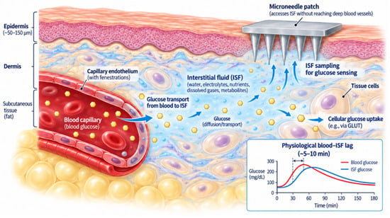
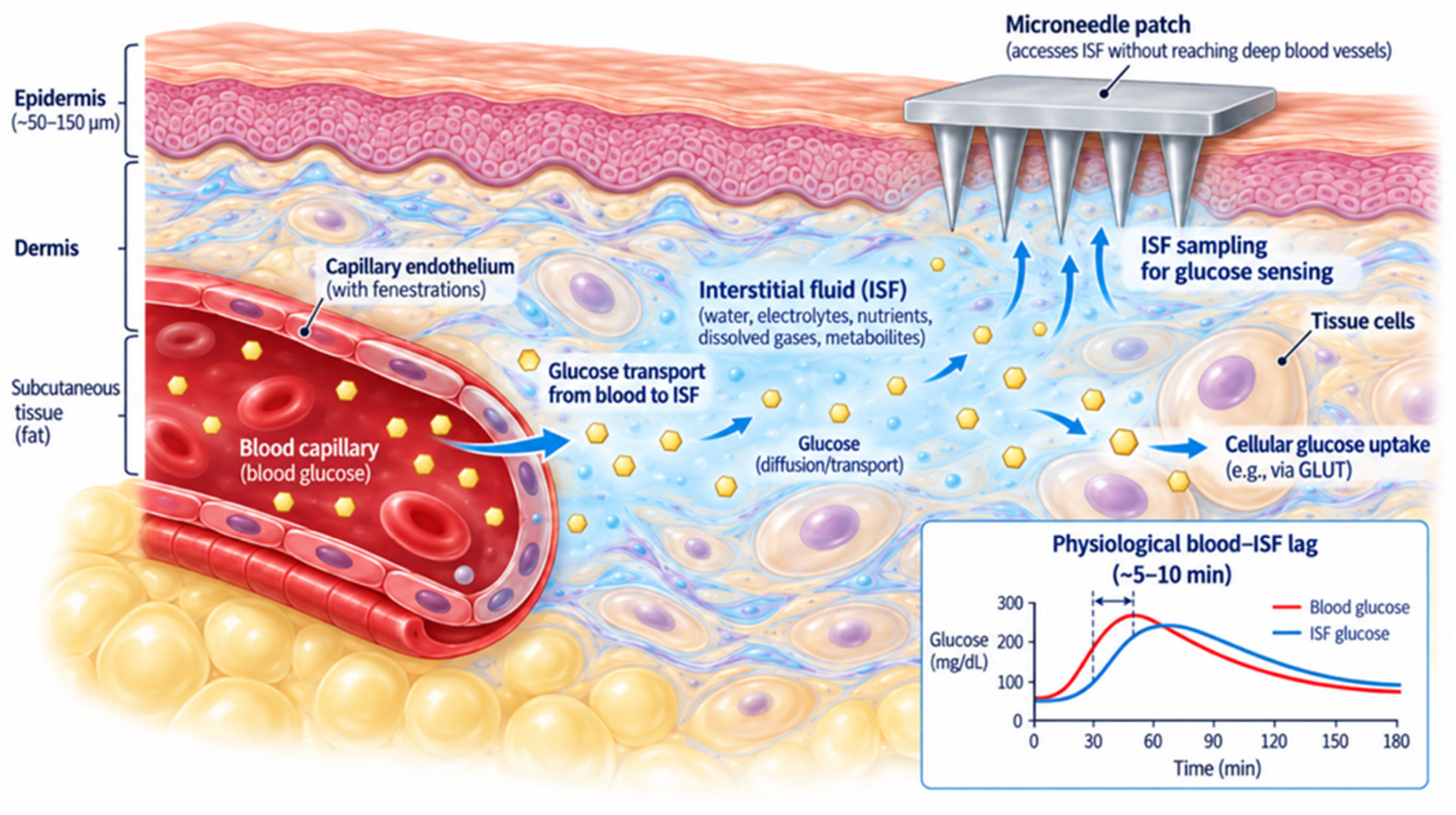
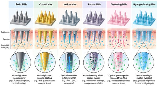
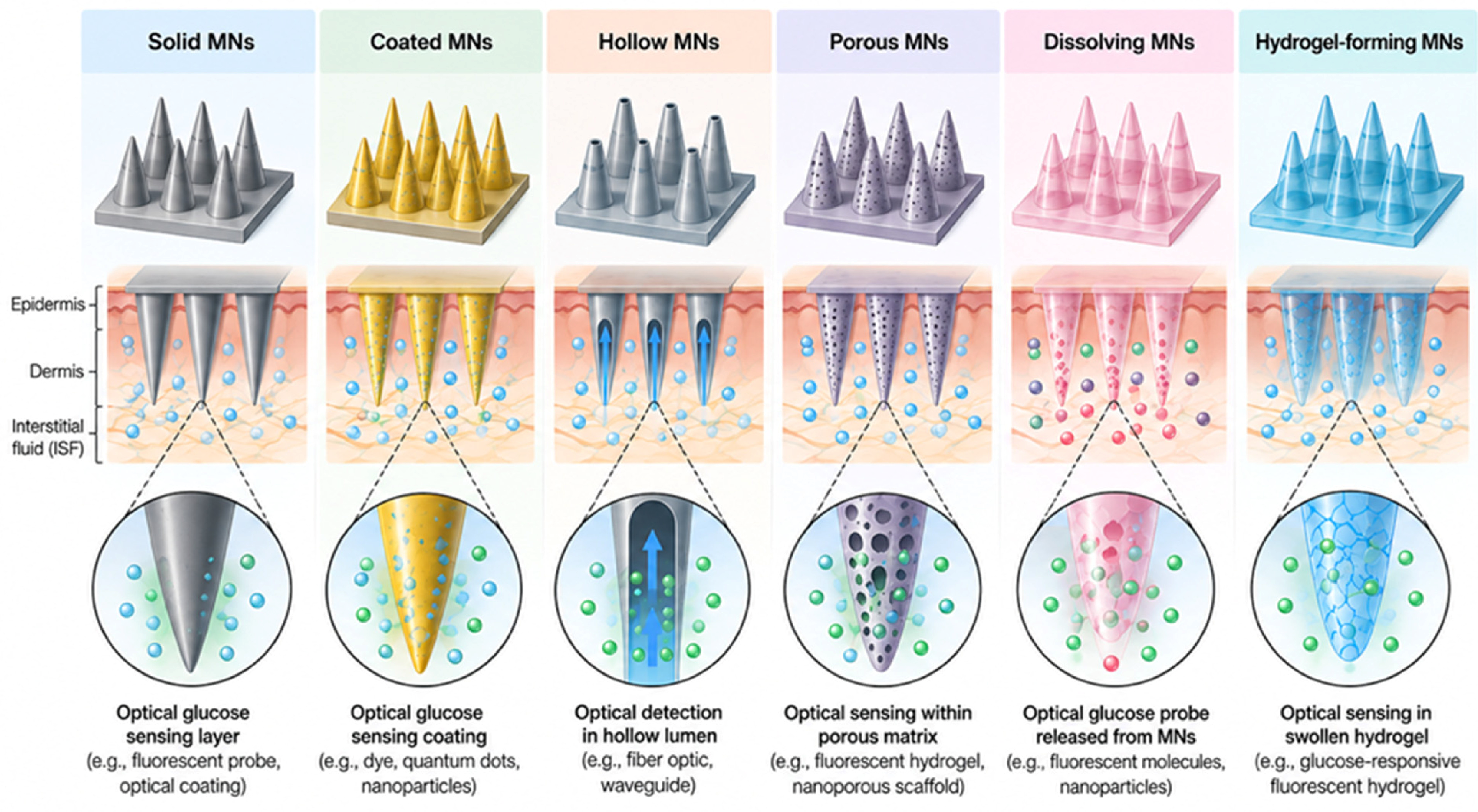
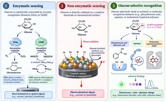
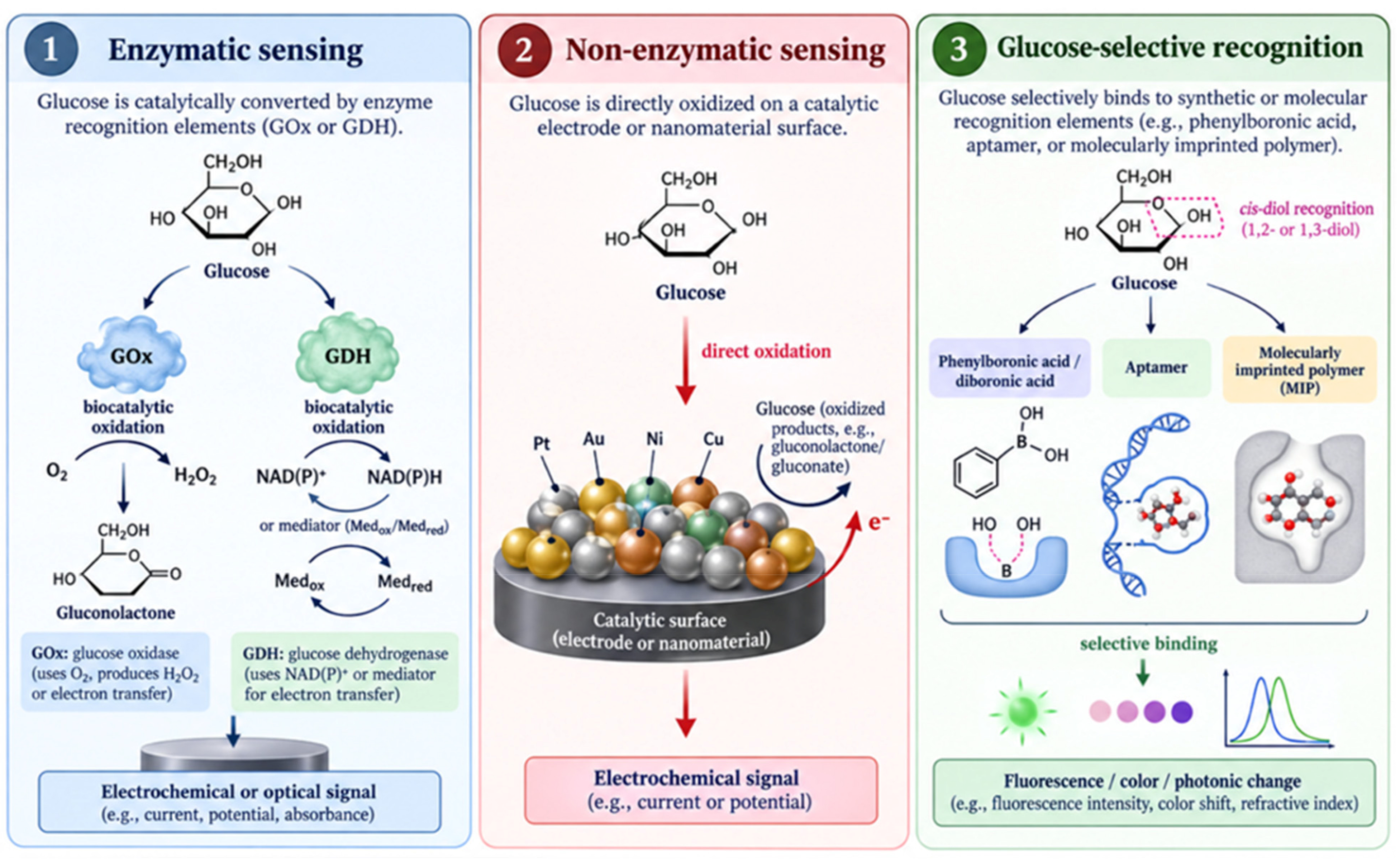
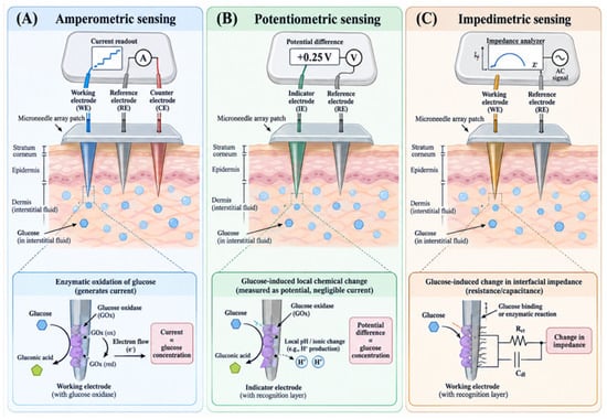
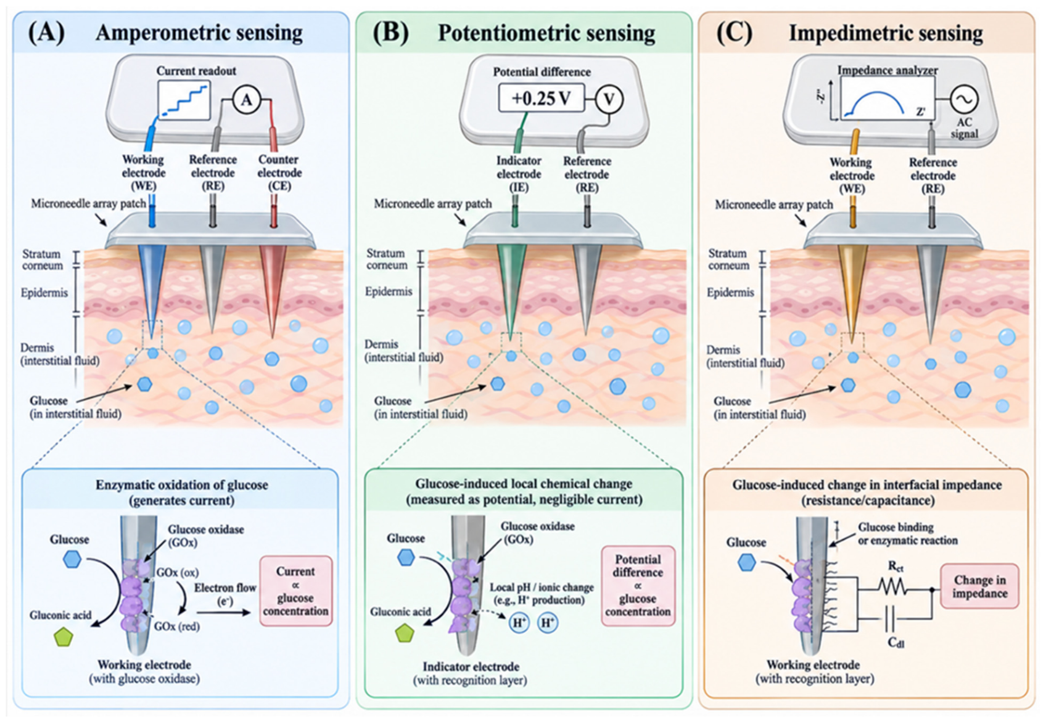
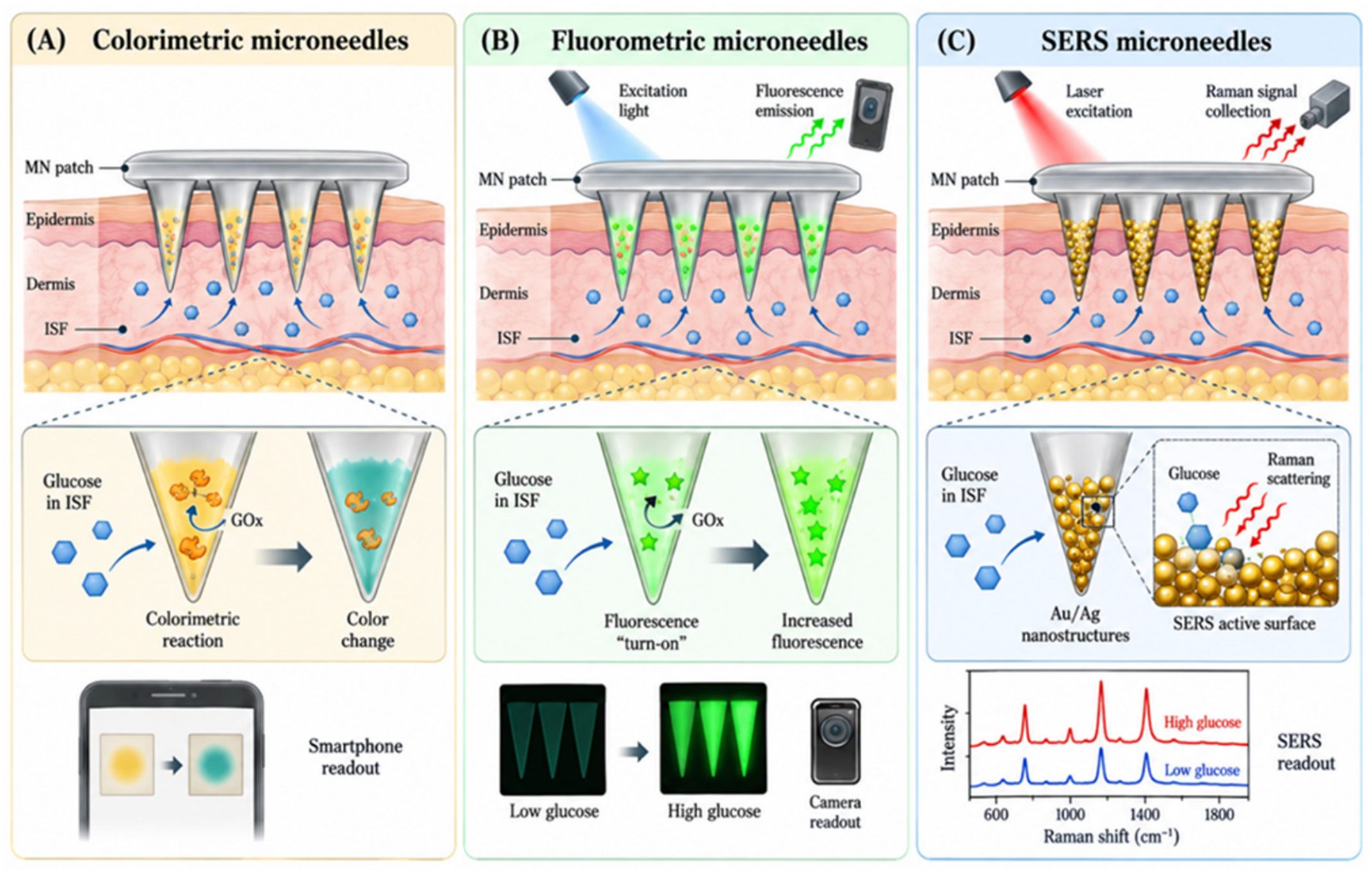
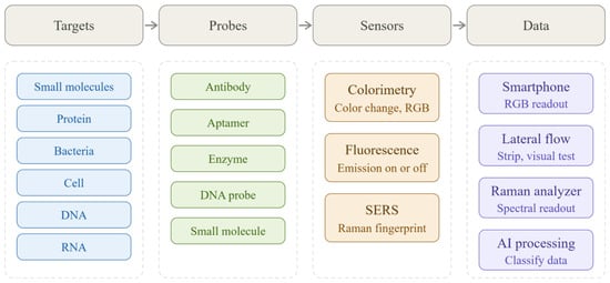
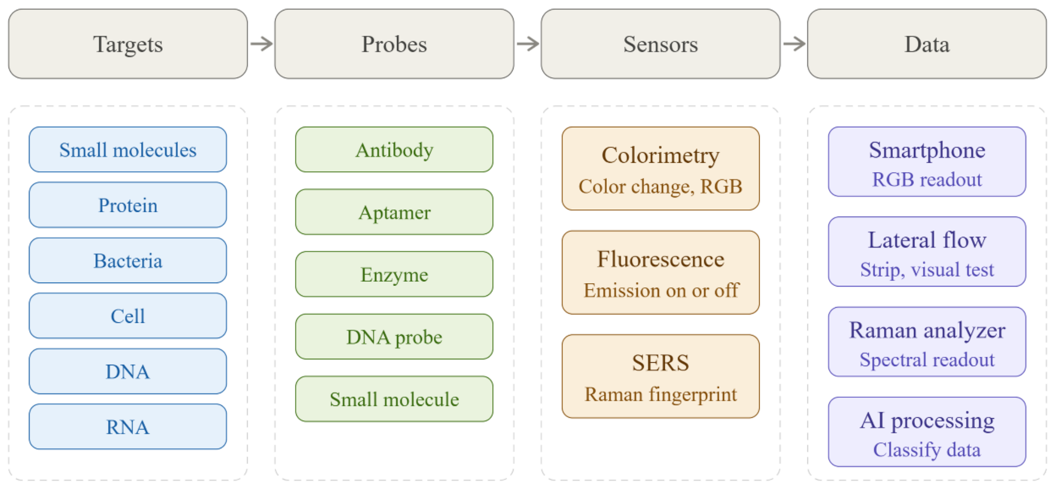
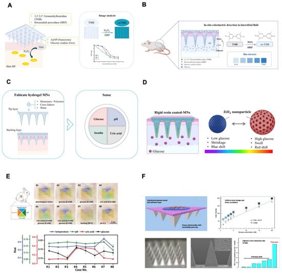
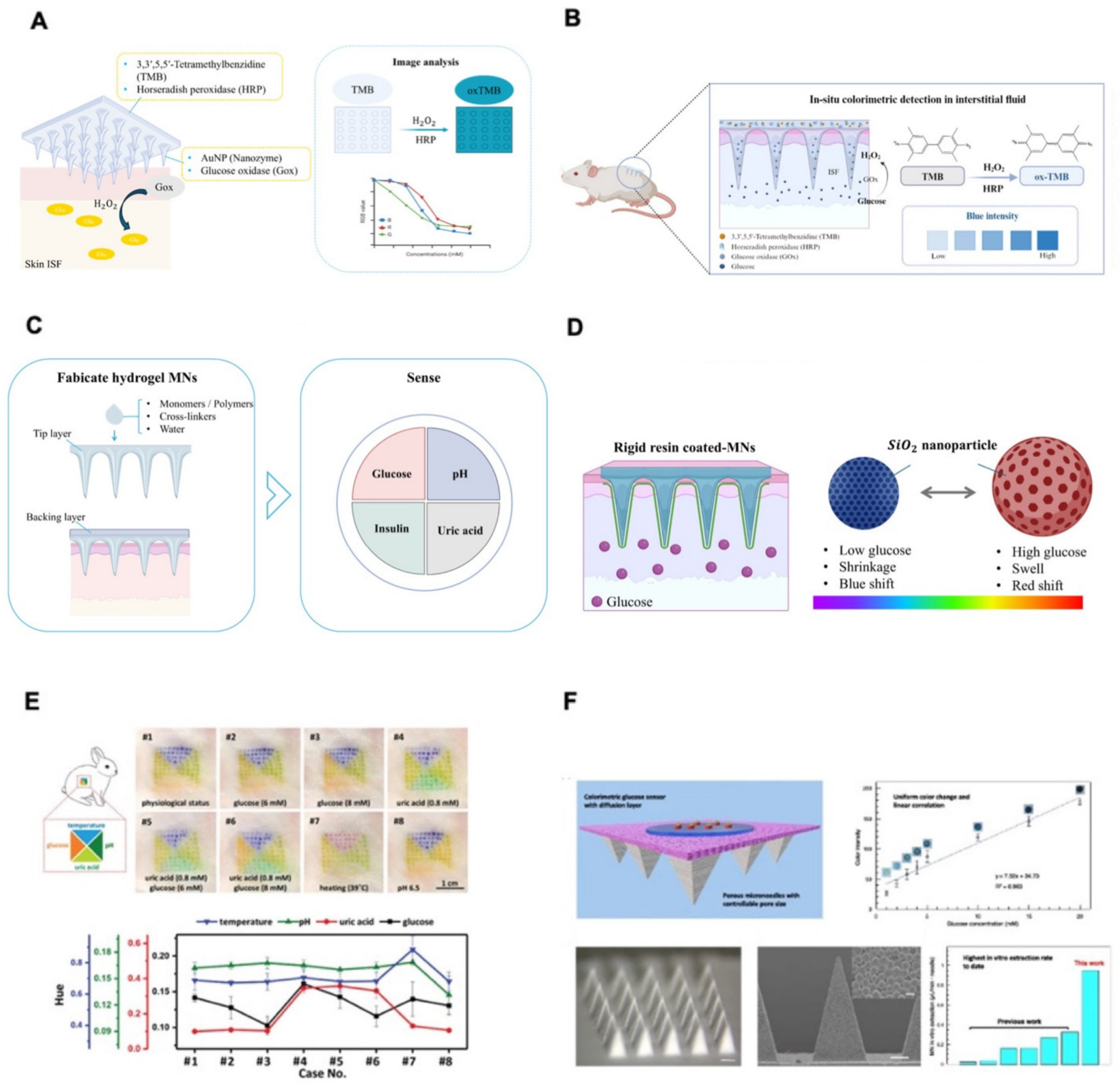
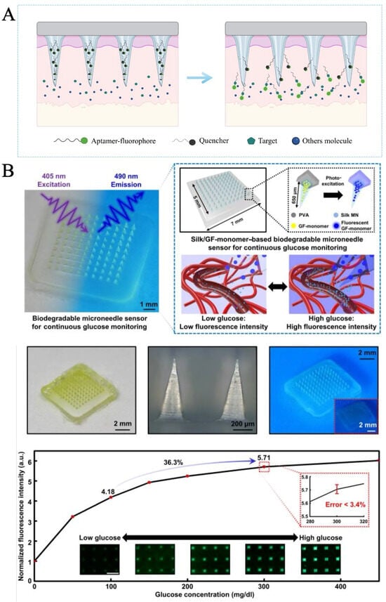
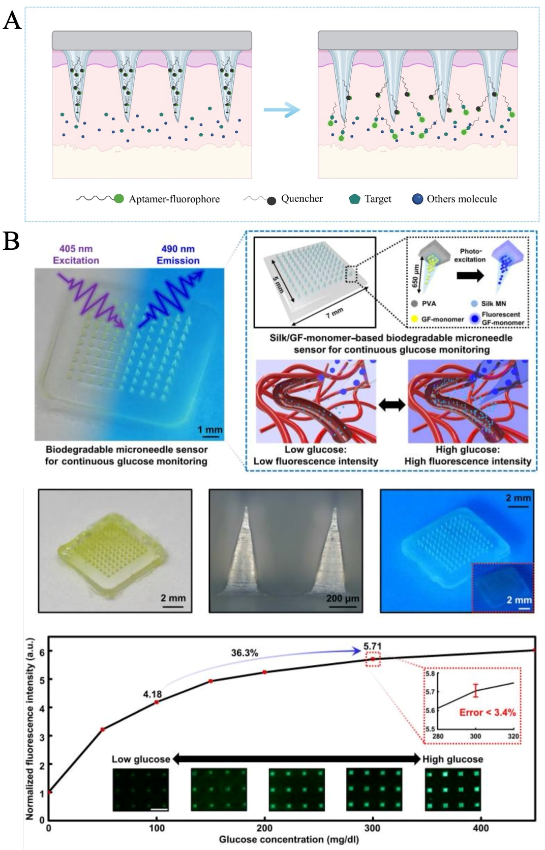
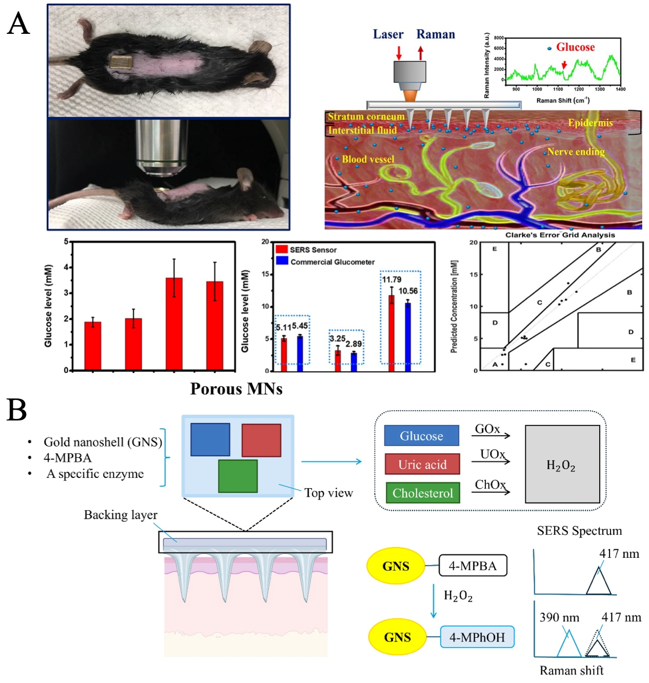
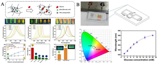
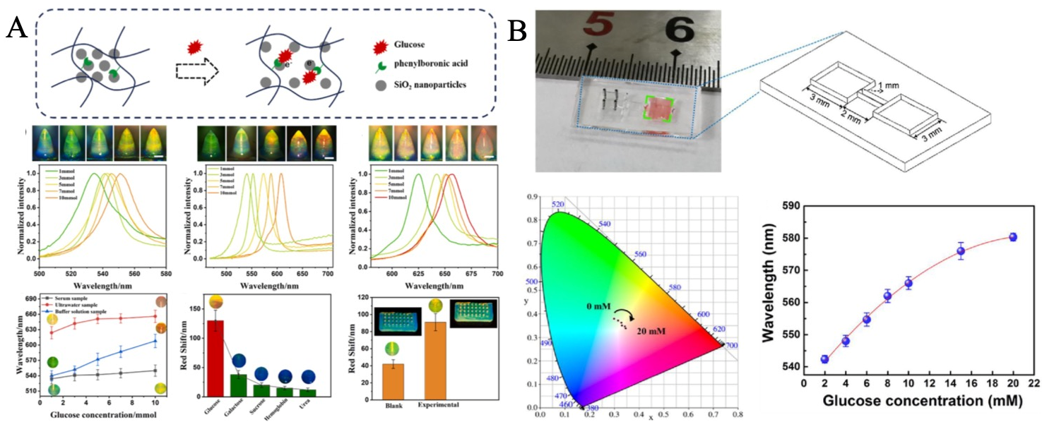
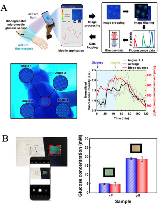
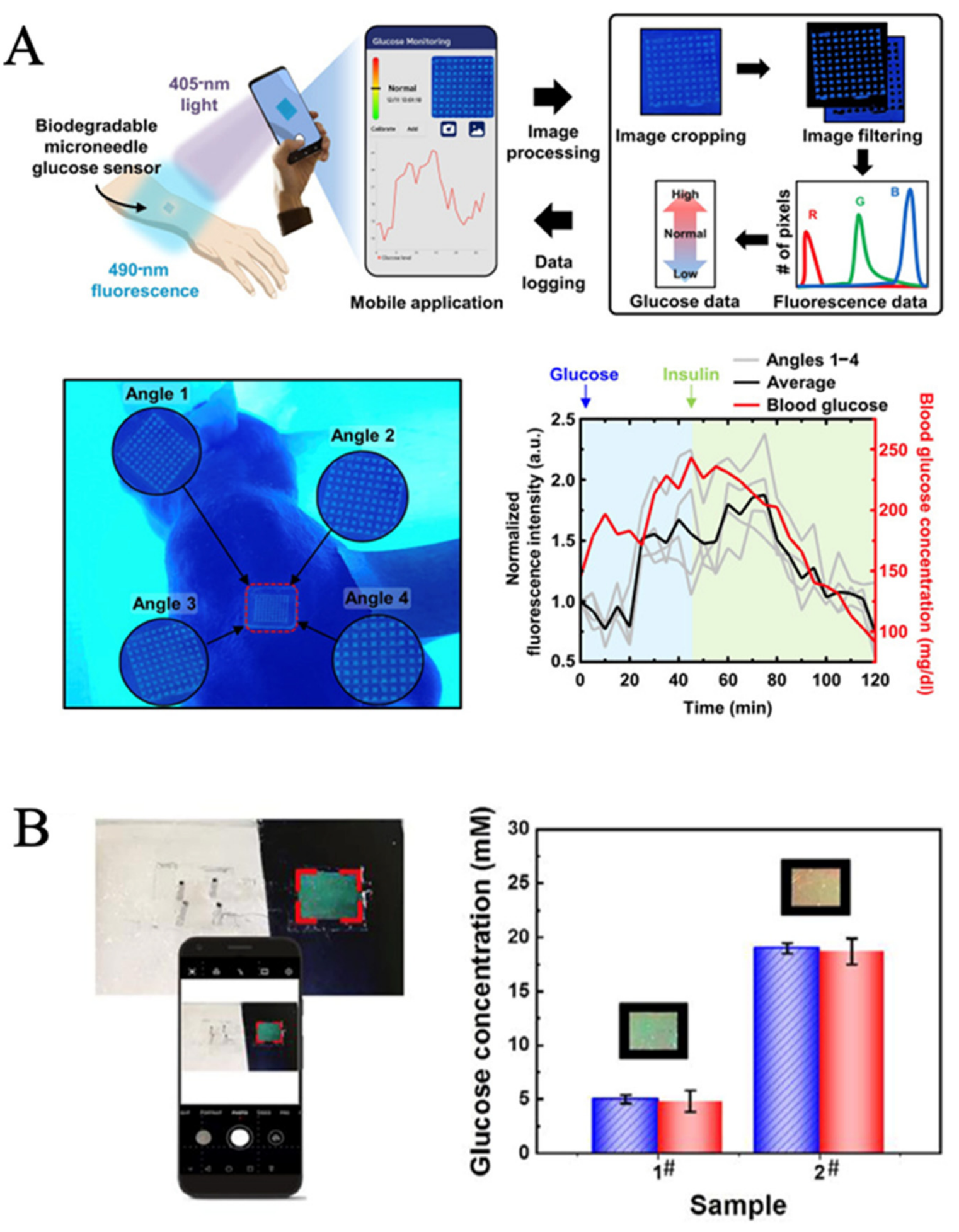

Disclaimer/Publisher’s Note: The statements, opinions and data contained in all publications are solely those of the individual author(s) and contributor(s) and not of MDPI and/or the editor(s). MDPI and/or the editor(s) disclaim responsibility for any injury to people or property resulting from any ideas, methods, instructions or products referred to in the content. |
© 2026 by the authors. Licensee MDPI, Basel, Switzerland. This article is an open access article distributed under the terms and conditions of the Creative Commons Attribution (CC BY) license.