


Abstract
Snakebite envenoming remains an important global public health problem, and Brazil ranks among the countries with the highest notification rates, especially in the Northern region. Therapeutic intervention is decisive in snakebite cases, as early administration of antivenom reduces the severity of systemic and local manifestations. However, in the absence of a diagnostic test, identification of the snake responsible for envenoming remains restricted to clinical and epidemiological assessment. In this context, this study demonstrates the potential for developing a diagnostic assay for snakebite envenoming using nanobioconjugates composed of antivenom VHHs against rattlesnakes and jararacas, medically important snakes in the country. In dot blot assays, the anti-Bothrops nanobioconjugates showed visible reactivity against Bothrops jararacussu venom down to 10 ng/µL and more consistent reactivity at concentrations ≥ 50 ng/µL. The anti-Crotalus nanobioconjugates detected Crotalus durissus terrificus venom at concentrations as low as 400 ng/µL and also showed reactivity toward three additional Crotalus subspecies venoms. In control assays, no cross-reactivity was observed with the heterologous venoms evaluated. These results provide initial evidence for the potential use of VHH-based nanobioconjugates for venom detection, while indicating that further optimization of analytical sensitivity is required before clinical application.
1. Introduction
Considered by the World Health Organization (WHO) a priority neglected tropical disease since 2017, snakebite affects about 2.7 million people annually, resulting in 81,000–138,000 deaths [1]. The severe clinical picture that envenoming can cause, the complexity for the production of antivenoms, the lack of rapid diagnosis methods, under-reporting and the high number of cases in underdeveloped countries are among the aggravating factors that make snakebite envenoming a priority neglected tropical disease [1,2].
In this context, snakebite envenoming represents an important global public health problem, highlighting the need for faster and more specific diagnostic strategies. In Brazil, since 2010, this disease has been part of the National Compulsory Notification List, and the records indicate the highest incidence rates in the North (51.84 per 100,000 inhabitants) and Midwest (19.26 per 100,000 inhabitants) regions. Of the four genera of medical importance in the country (Bothrops, Crotalus, Micrurus and Lachesis), those of the Bothrops genus, responsible for approximately 67% of notifications, and those of the Crotalus genus, which cause the highest lethality and represent 8% of cases, receive the most attention [3].
The therapeutic approach for snakebite cases in Brazil is based on the administration of anti-Bothrops (pentavalent), anti-Crotalus, anti-Micrurus (bivalent), and anti-Lachesis antivenom sera, in addition to the combined anti-Bothrops–anti-Crotalus and anti-Bothrops–anti-Lachesis formulations [4]. The determination of antivenom therapy is a central step in the management of snakebite envenoming, and in the absence of specific diagnostic tests, the definition depends mainly on the experience of the medical team to evaluate the symptomatic pattern of the victims, especially in regions where different genera of snakes can coexist. However, even when there is expertise, the similarity of clinical manifestations, including accidents caused by snakes of different genera, can impair the speed of an accurate diagnosis [5,6].
Previous studies have reported the use of different diagnostic approaches for snake venom detection. Double-antibody sandwich ELISA assays developed for snake venom identification have achieved detection limits as low as 6.25 ng/mL [7]. A commercially available example of a diagnostic test for snakebite envenoming is the Snake Venom Detection Kit (SVDK, bioCSL Pty Ltd. Parkville, Australia), developed for medically important Australian snakes, with results available within 15–25 min. More recently, multiplex immunofluorescence lateral flow assays have demonstrated detection limits ranging from 0.6 to 1.2 ng/mL, with no cross-reactivity among four medically important snake venoms and analysis times of approximately 5 min [8]. In Brazil, a multiplex lateral flow assay capable of differentiating Bothrops and Lachesis venoms provided results within 15 min and showed detection limits of 10–50 ng/mL in plasma and urine and 50–500 ng/mL in serum [9]. To date, the use of VHHs as capture and detection antibodies in lateral flow assays for snakebite diagnosis remains largely unexplored.
In this context, the availability of a rapid and accurate diagnosis method, such as lateral flow, would impact therapeutic success, reducing the recovery time of victims and the costs that may arise from nonspecific treatments [5,10]. From this perspective, the present work sought to develop nanobioconjugates consisting of VHH (variable domain of heavy chain-only antibodies) conjugated to gold nanoparticles for the detection of venoms from two medically important snake genera in Brazil, Bothrops and Crotalus.
For the constitution of nanobioconjugates, the recognized attributes of VHH antibodies were considered, such as high stability, specificity, affinity, ability to interact with epitopes often inaccessible to conventional antibodies and biocompatibility with nanomaterials [11,12]. From previously selected and characterized VHHs [13,14], the potential of antivenom VHH-based nanobioconjugates for the detection of envenomings caused by the genera Bothrops and Crotalus was explored.
2. Materials and Methods
2.1. Obtaining the Antivenom VHHs
The anti-Crotalus VHHs KF498604 [13] and PZ837026, as well as the anti-Bothrops VHHs KF498607 and KC329718 [14], were selected based on their previously characterized recognition profiles. VHHs KC329718 and KF498607 were selected based on their complementary binding profiles against BthTX-I and BthTX-II, exhibiting KD values of 53.9 nM and 770.2 nM for KC329718 and 628.7 nM and 28.0 nM for KF498607, respectively. Likewise, VHH KF498604 was selected based on its recognition of crotoxin and its binding affinity for this toxin (KD = 81.3 nM). Although affinity measurements were not determined for PZ837026, its selection was supported by its selective recognition profile toward crotalic venoms and by molecular docking analyses predicting interactions with functionally relevant regions of crotoxin (unpublished data), and its recognition profile was further evaluated in the present study.
Clones KF498607 and PZ837026, cloned into pET-22b(+), were expressed in E. coli BL21(DE3) induced with 0.3 mM IPTG and incubation at 25 °C for VHH KF498607 and at 30 °C for VHH PZ837026. Clone KC329718, in pET-28a(+), was expressed in E. coli SHuffle® T7 Express (New England Biolabs, Ipswich, MA, USA) induced with 0.3 mM IPTG and incubation at 30 °C. VHH KF498604, also in pET-28a(+), was expressed with 0.5 mM IPTG at 25 °C.
Purification of the VHHs was performed by immobilized metal affinity chromatography (IMAC) using the HiTrap TALON® crude 1 mL column containing cobalt (Sigma-Aldrich, St. Louis, MO, USA). Protein quantification was done by the BCA method (Pierce BCA Protein Assay Kit, Thermo Fisher Scientific, Waltham, MA, USA), and the electrophoretic profile was evaluated by 15% SDS-PAGE [15]. ELISA assays were performed in a direct binding format, as described by Luiz et al. [13] and Prado et al. [14]. Venoms and toxins were adsorbed onto 96-well plates at a concentration of 1 µg/well. Purified VHHs were used at 10 µg/mL. Bound VHHs were detected using an HRP-conjugated anti-VHH antibody (MonoRab™ Rabbit Anti-Camelid VHH Antibody [HRP], mAb, GenScript, Piscataway, NJ, USA) diluted 1:10,000. The cut-off value was defined as 2 × SD + 2 × mean absorbance of the negative control.
2.2. Synthesis of Colloidal Gold Nanoparticles
The Turkevich method [16] was used as the basis for the process of making colloidal gold nanoparticles. The initial characterization of AuNPs was performed by spectrophotometry in the region of the visible light spectrum, 400 to 750 nm filter. The parameters used to calculate the size and concentration of AuNPs were based on the studies by Haiss et al. [17]. By means of the ratio between the absorbance at the wavelength of maximum light absorption (λmax) and the absorbance at the wavelength at 450 nm (λ450), the diameter of the nanoparticles was calculated. The extinction coefficient (Ɛ450) was defined from the diameter provided. The concentration of the gold solution was calculated by the ratio between the absorbance at 450 nm and the Ɛ450. Additionally, transmission electron microscopy (TEM) was performed on a Jeol JEM-1400PLUS microscope, in the Confocal and Electron Microscopy subunit of the Carlos Chagas Institute, ICC/PR (https://plataformas.fiocruz.br/, (accessed on 1 March 2023)).
2.3. Bioconjugation of VHHs with Gold Nanoparticles
The optimization data presented in this study correspond to PZ837026 (anti-Crotalus nanobioconjugates) and KC329718 (anti-Bothrops nanobioconjugates), selected from the comparative screening of all four VHHs under different bioconjugation conditions, including stabilizer concentration, pH range, and antibody concentration, as these VHHs exhibited the most favorable and reproducible bioconjugation profiles. The KF498604 and KF498607 showed less suitable bioconjugation characteristics under the evaluated conditions and were consequently employed as capture antibodies. For this reason, only the optimization data of these VHHs are shown.
The optimal condition for bioconjugation KC329718 and anti-Crotalus PZ837026 to the AuNPs was defined by the assays for determining the optimal concentration of the acrylic acid stabilizing agent N-hydroxysuccinimide-ester (acrylic acid–NHS-ester) (Sigma-Aldrich, St. Louis, MO, USA) (step 1), the best pH range (step 2) and the VHH concentration required for bioconjugation (step 3). Step 1 was performed in a pH range close to the theoretical isoelectric point (pI) of each VHH, a parameter provided by the analysis of amino acid sequences with the ExPASy—ProtParam tool. After that, the AuNPs were centrifuged at 12,000 rpm for 20 min at room temperature to concentrate the solution to an optical density of 10.
The ratio of VHH and AuNPs for bioconjugation in all steps was 1:10 (v/v) [18]. During the bioconjugation optimization assays, reactions were initially evaluated in the absence of NaCl. Subsequently, NaCl was added at a final concentration of 1 M as an ionic perturbation agent to assess the stability of the bioconjugates. In the assays performed without NaCl, the reaction mixture contained only the components required for bioconjugation, and pH adjustment was performed using the minimum volume of 100 mM sodium borate buffer necessary to achieve the optimal pH for each VHH (pH 7.5 for PZ837026 and pH 8.5 for KC329718).
After bioconjugation and removal of unconjugated VHHs by centrifugation (12,000 rpm, 20 min, room temperature), the nanobioconjugates were resuspended in 50 mM KH2PO4 buffer supplemented with 1% PEG 8000 and 10% BSA. The pH of the storage buffer was adjusted according to the optimal bioconjugation conditions previously established for each VHH (pH 7.5 for PZ837026 and pH 8.5 for KC329718). After resuspension, the bioconjugates were stored at 4 °C and used in the subsequent experiments within 24 h of preparation. Long-term stability of the bioconjugates was not evaluated in this study. The VHH:AuNP stoichiometric ratios were estimated from the molar concentration of each VHH and the final concentration of AuNPs. VHH molar concentrations were calculated from their molecular weights and the final amounts used in the bioconjugation reactions. Data obtained by spectrophotometry were tabulated in the Origin 8.0 graphics program (OriginLab Corporation, Northampton, MA, USA).
2.4. Dot Blot Assay to Evaluate the Reactivity of Bioconjugates
To evaluate the reactivity of the bioconjugates, dot blot assays were performed using different experimental formats. For anti-Bothrops VHHs, sandwich assays and direct detection of the membrane-adsorbed target were conducted. In the assay with anti-Crotalus VHHs, the reactivity of PZ837026 nanobioconjugates against membrane-adsorbed KF498604 was initially evaluated. Subsequently, these nanobioconjugates were evaluated exclusively by dot blot in direct detection format, using venoms adsorbed on a nitrocellulose membrane (Table S1). All snake venoms used in these assays were obtained from the Serpentário de Proteínas Bioativas, Batatais, São Paulo, Brazil.
2.5. Sandwich Type Test with VHH KC329718 Nanobioconjugates
A nitrocellulose membrane (MFS Millipore; 0.45 μm) with previously demarcated regions received, in triplicate, 1 µL of VHH KF498607 (1 µg/µL). The membranes were then incubated at 37 °C for 2 h to allow passive adsorption of the VHHs onto the membrane. They were then subjected to 5 cycles of washing (5 min each cycle) with 0.1 × PBS plus 0.01% Tween 20. Regions without adsorbed VHH were blocked with a solution of 0.1 × PBS with 5% bovine serum albumin (BSA) (Sigma-Aldrich, St. Louis, MO, USA) for 1 h. At the end of blocking with BSA, the membranes were washed for an additional 5 cycles of 5 min each (Adapted from [19]).
Then, the membranes were incubated for 1 h with different concentrations of Bothrops jararacussu venom. Concentrations of 200; 100; 75; 50; 25; 15 and 10 ng/µL of venom were evaluated.
The last step corresponded to the incubation of the membranes with the nanobioconjugates, for 15 min and under constant agitation. After 15 min, excess bioconjugate was withdrawn with 1 cycle of washing for 2 min. At the end, the photographic record was made.
2.6. Dot Blot Assay in Direct Detection Format
In this format, the nanobioconjugates of VHHs KC329718 and PZ837026 were evaluated against the different venoms adsorbed on the nitrocellulose membrane. Then, the membranes were incubated for 1 h with homologous and heterologous venoms, as well as negative controls, as detailed in the figure legends. In addition to the whole venom of the snakes Bothrops jararacussu and Crotalus durissus terrificus, the ability of nanobioconjugates to recognize whole venom of other species was also evaluated. The detection capacity of VHH PZ837026 nanobioconjugates was evaluated against venoms from different species of the genus Crotalus. Similarly, venoms from different Bothrops species were evaluated in the assay with VHH nanobioconjugates KC329718. As an additional control, membranes containing the different venoms were stained with Ponceau S (Sigma). All dot blot assays were performed as qualitative proof-of-concept experiments. Each condition was applied in triplicate spots on the same membrane, and the interpretation was based on the presence or absence of visible reactivity. The list of venoms evaluated for each type of nanobioconjugate is detailed in Table S1.
3. Results and Discussion
3.1. Heterologous Expression and Purification of VHHs
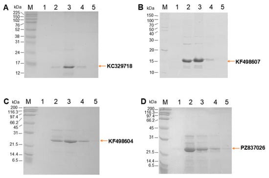
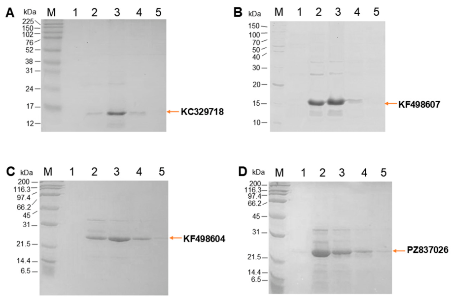
Expression of VHHs KF498607, KC329718, PZ837026 and KF498604 was confirmed by electrophoretic analysis on SDS-PAGE (Figure 1A–D), with detection of bands corresponding to the fractions eluted in 150 mM imidazole. The purified yields obtained for VHH KC329718 and PZ837026 were approximately 4.5 mg/L of bacterial culture for each VHH. After elution, the samples were subjected to diafiltration to remove residual imidazole. The observed electrophoretic profile was consistent with the expected pattern for these VHHs, in concordance with data previously described in the literature [13,14].
Figure 1.
SDS-PAGE analysis of the affinity chromatography purification of recombinant VHHs. The images (A–D) correspond to 15% SDS-PAGE gels containing, respectively, the anti-Bothrops VHHs KC329718 (A) and KF498607 (B), and the anti-Crotalus VHHs KF498604 (C) and PZ837026 (D). The arrows indicate the position of the bands corresponding to the respective VHHs. M indicates the molecular weight marker, and wells 1 to 5 correspond to the eluted fractions of the IMAC column (1st to 5th mL). Molecular weight markers used Broad Protein Molecular Weight Markers—Promega ((A,B) gels) and Broad Range—Bio-Rad Laboratories ((C,D) gels). Gels were stained with Coomassie Brilliant Blue R-250.
3.2. Assessment of VHH Reactivity by Enzymatic Immunoassay
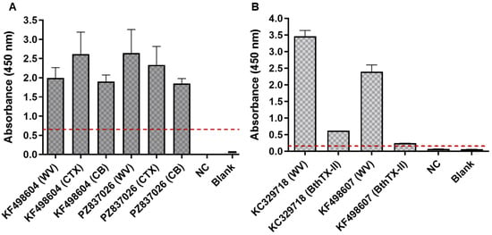
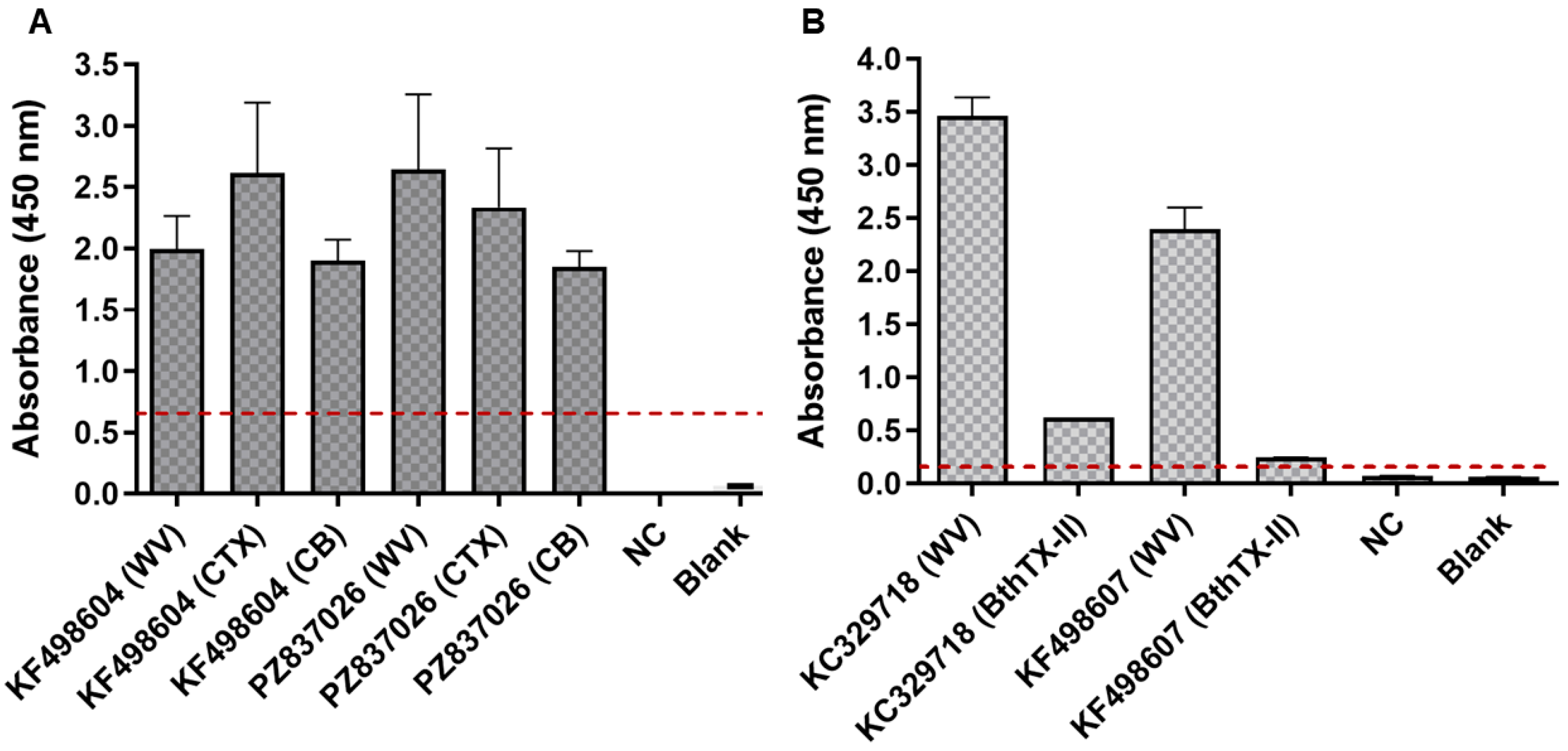
The absorbances of VHH KF498604 against crotoxin (CTX), CB subunit and whole venom (WV) of C. durissus terrificus demonstrate its reactivity against these targets, as previously described [13]. Similarly, PZ837026 also showed reactivity under the same conditions (Figure 2A). VHH KC329718 was reactive against the whole venom of B. jararacussu and bothropstoxin-II, while KF498607 showed reactivity only for the whole venom (Figure 2B), according to previous studies [14].
Figure 2.
ELISA of anti-Crotalus and anti-Bothrops VHHs. (A) Reactivity of anti-Crotalus VHHs (KF498604 and PZ837026) against Crotalus durissus terrificus whole venom (WV), crotoxin complex (CTX), and the CB subunit. (B) Reactivity of anti-Bothrops VHHs (KC329718 and KF498607) against the whole venom of Bothrops jararacussu and bothropstoxin-II (BthTX-II). NC represents the negative control using an unrelated VHH. The blank corresponds to the reaction without VHHs. The dashed red line indicates the cut-off. The results represent the mean ± standard deviation of duplicate experiments.
3.3. Characterization of Gold Nanoparticles
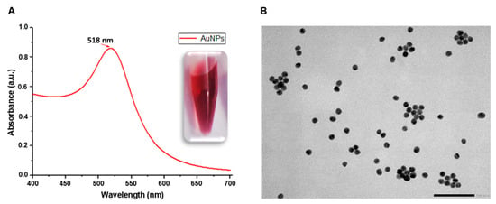
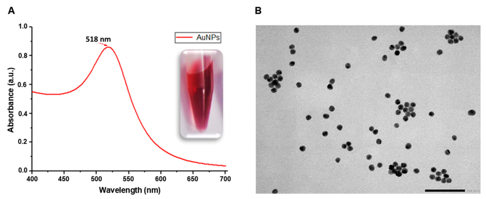
The initial characterization of AuNPs, by spectrophotometry in the visible spectrum of light (Figure 3A), showed a well-defined absorption peak, which is consistent with the formation of spherical AuNPs with a relatively homogeneous optical profile. This spectral profile is commonly associated with the formation of stable AuNP suspensions [20]. In the spectrum of Figure 3A, the wavelength of maximum absorbance (518 nm) is highlighted. In the same image, the AuNPs are arranged in a microtube where their macroscopic appearance is presented. The ratio between the absorbance of this wavelength (λmax) and the absorbance at the wavelength at 450 nm (λ450) (Table S2) resulted in the value of 1.555, which, according to Haiss et al. [17], corresponds to the approximate diameter of 12 nm.
Figure 3.
(A) Absorbance spectrum of gold nanoparticles (AuNPs). The graph shows absorbance as a function of wavelength. The red arrow indicates the wavelength at which the maximum absorbance was observed. (B) Transmission electron microscopy of AuNPs.
The size at the nanoscale is one of the factors for the colloidal gold solution to present red coloration, which is related to the phenomenon of Surface Plasmon Resonance [21]. Studies, such as that of Fatima et al. [22], demonstrate the importance of the red color of the gold solution for the easy interpretation of the test, since the positivity of the reaction is indicated by the presence of a reddish tone in the membrane. The extinction coefficient (Ɛ450) for the diameter of 12 nm is 1.09 × 10⁸ M−1 cm−1. When calculating the ratio between the absorbance at 450 nm and the Ɛ450, the concentration obtained was 10.4 nM. Along with these results, the spherical morphology of AuNPs was also confirmed by transmission electron microscopy (Figure 3B).
3.4. Optimization of VHH Bioconjugation with Gold Nanoparticles
3.4.1. Step 1: Definition of the Optimal Concentration of the Acrylic Acid N-Hydroxysuccinimide-Ester (NHS-Ester)
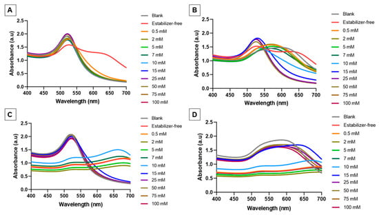
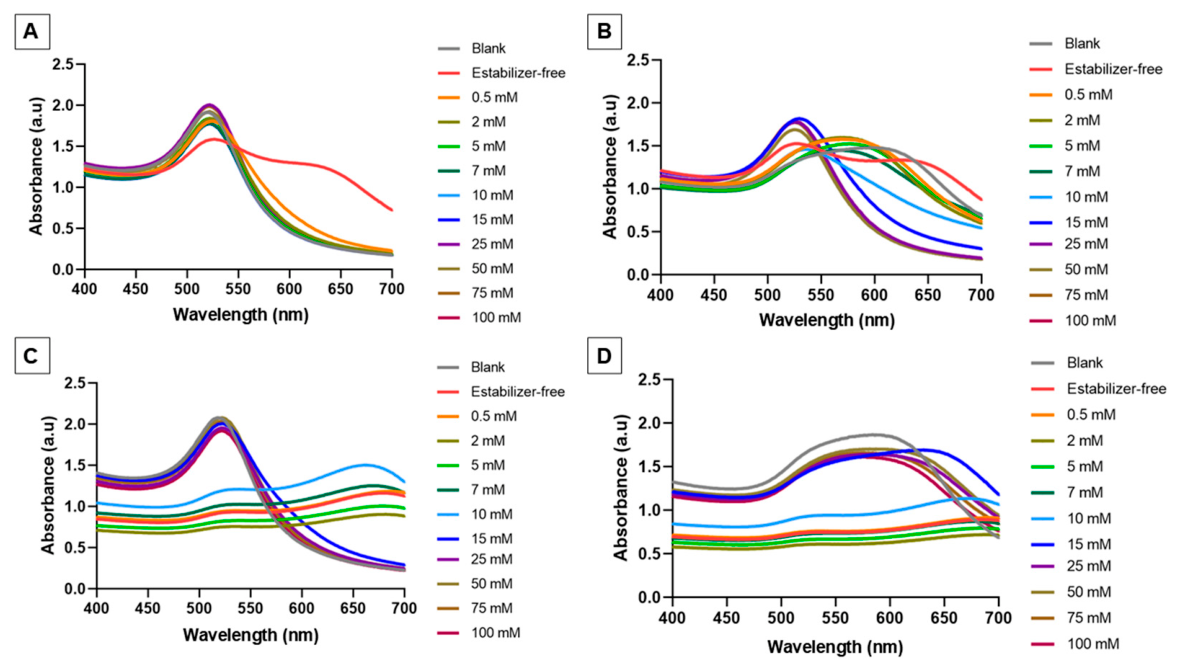
The theoretical isoelectric point (pI) of the VHHs, calculated using the ExPASy—ProtParam tool, was 7.05 for PZ837026 and 8.59 for KC329718. Visual inspection and spectrophotometric analysis of PZ837026 bioconjugation showed that 100 mM acrylic acid–NHS-ester provided the greatest reaction stability, whereas lower concentrations led to progressive darkening. In Figure S1A, it is possible to see that the concentrations of 15 to 100 mM of acrylic acid–NHS-ester were those that minimized the aggregation of AuNPs against the addition of NaCl. The evaluation of the graphs in Figure 4A,B reinforces what is observed in Figure S1, where concentrations of 15 to 100 mM acrylic acid–NHS-ester resulted in absorbance spectra close to that of the reaction blank. In this first step, significant aggregation of AuNPs in the absence of acrylic acid–NHS-ester stabilizer was demonstrated. The permanence of the red color of AuNPs in bioconjugation assays, in the presence of NaCl, is a benchmark for demonstrating stability in the fusion of biomolecules to AuNPs [23,24].
Figure 4.
Absorbance spectra, expressed in absorbance units (a.u.), obtained from the bioconjugation reactions of VHH PZ837026 (A,B) and VHH KC329718 (C,D) with different concentrations of acrylic acid–NHS-ester, in the absence (A,C) and presence (B,D) of NaCl. The blank corresponds to the reaction containing water, 100 mM acrylic acid–NHS-ester, and AuNPs at pH 7.5 for VHH PZ837026 and at pH 8.5 for VHH KC329718.
Confirmation of fusion of PZ837026, as well as the other VHHs, was performed with the spectrophotometric reading data. In the case of PZ837026, all reactions generated absorbance peaks with a distance from the assay control (Table S3). However, while the distance of the higher absorbance peak from what is observed for pure AuNPs confirms the coupling of the biomolecule to AuNPs, a high displacement of the higher absorbance peak may also indicate the instability of the bioconjugate [25,26]. Among the different stabilizer concentrations evaluated for PZ837026 without NaCl, the one of 100 mM was the one that most reduced the wave shift in greater absorbance in relation to the blank (1 nm), this being the concentration established for the tests with PZ837026 (Table S3).
Bioconjugation assays of acrylic acid–NHS-ester modified KC329718 at concentrations above 15 mM resulted in increased reaction stability (Figure S2 and Figure 4C). However, at no concentration of stabilizing agent was it possible to guarantee the stability of the reaction in the presence of NaCl (Figure 4D). The concentration of 100 mM stabilizing agent was the one that resulted in the lowest wavelength shifts in highest absorbance, with and without NaCl. After the addition of NaCl, there was a 50 nm shift relative to the bioconjugate without NaCl, and when compared to the blank without NaCl, there was a 3 nm shift (Table S4).
Although, in the presence of NaCl, the smallest displacements of the maximum absorbance peaks belong to the reactions with concentrations less than 10 mM of stabilizer (Table S4), it was under these conditions that the greatest distances from the absorbance peaks occurred before adding NaCl.
3.4.2. Step 2: Defining the pH Range for Bioconjugation
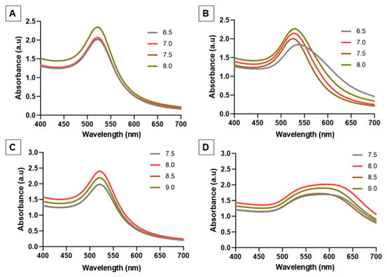
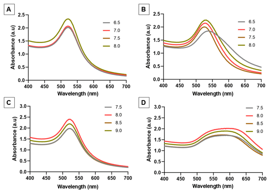
Bioconjugation of PZ837026 at different pH ranges (6.5 to 8.0) indicated that the reaction at pH 7.5 promoted greater stability after addition of NaCl. In the visual inspection (Figure S3), the color change in the AuNPs was accentuated in the 6.5 and 8.0 ranges, different from the reaction at pH 7.5 that showed the lowest color change. The absorbance spectra in the absence of NaCl (Figure 5A) are similar; however, in Figure 5B, a shift in the higher absorbance peaks is evident, particularly at pH 6.5. When calculating the wavelength shift in greater absorbance, bioconjugation at pH 7.5 was the one that remained with greater stability compared to the other pHs, since the displacement of 4 nm, in the presence of NaCl, was the lowest among all evaluated (Table S5).
Figure 5.
Absorbance spectra, expressed in absorbance units (a.u.), obtained from the bioconjugation reactions of VHH PZ837026 (A,B) and VHH KC329718 (C,D) at different pH values, in the absence (A,C) and presence (B,D) of NaCl. The pH values evaluated ranged from 6.5 to 8.0 for VHH PZ837026 and from 7.5 to 9.0 for VHH KC329718.
The bioconjugations of KC329718 at pHs of 7.5 to 9.0, with 100 mM stabilizer, remained stable in the absence of NaCl (Figure 5C and Figure S4), but after adding it, the AuNPs became unstable with consequent displacement of the higher absorbance peaks (Figure 5D). At pH 8.5, the displacement was 55 nm after the action of NaCl, being the lowest among the other pHs. In addition to the greater resistance to aggregation at pH 8.5 with NaCl, there was 3 nm from the peak of maximum absorbance in relation to the blank, a comparison carried out between the bioconjugates not subjected to ionic disturbance (Table S6). The macroscopic visualization of the blank of each pH and its respective absorbance spectra are shown in Figure S5A–C).
3.4.3. Step 3: Determination of Optimal VHH Concentration
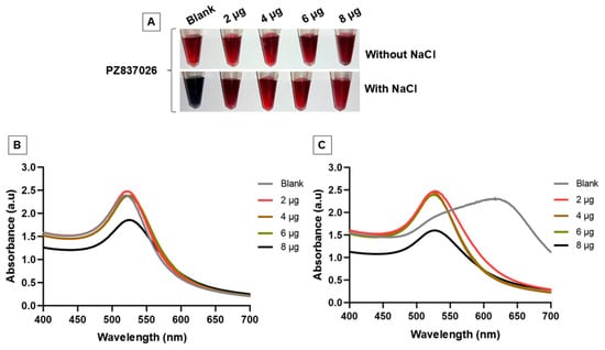
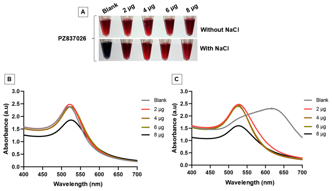
The ideal VHH PZ837026 concentration determination assays were performed at pH 7.5 and with 100 mM stabilizer, as defined in the previous steps. Macroscopic analysis did not clearly distinguish which reaction exhibited greater stability, even after NaCl addition (Figure 6A). In the analysis of the graphs of Figure 6B,C, the proximity of the peaks of greater absorbance of the four concentrations of VHH PZ837026 is evident, which explains the observation made from Figure 6A. When calculating which condition maintained the greatest stability of the bioconjugation, it was found that, with 4 µg of VHH, there was no wavelength shift. Of the other concentrations evaluated, the highest displacement was 5 nm (Table S7). Thus, the concentration of 4 µg of VHH PZ837026 was standardized for the bioconjugation assays.
Figure 6.
Photographic record of the bioconjugation reactions (A) and absorbance spectra, given in absorbance units (a.u.), of VHH PZ837026 bioconjugated with 2, 4, 6 and 8 µg without NaCl (B) and with NaCl (C). The blank is represented by the reaction consisting of water with 100 mM acrylic acid–NHS-ester and AuNPs at pH 7.5.
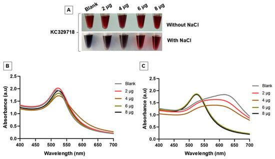
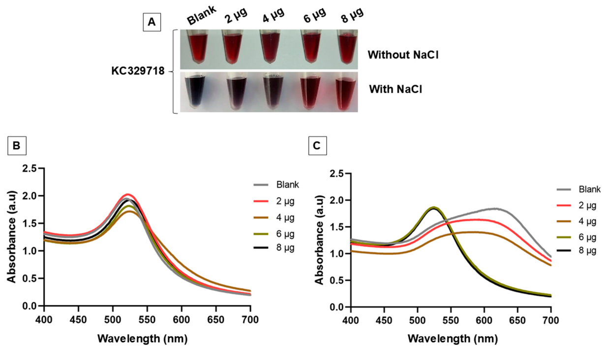
For VHH KC329718, visual inspection indicated that the two highest concentrations were more favorable for the stabilization of bioconjugates plus NaCl (Figure 7A). With 6 and 8 µg of VHH KC329718, the absorbance spectra profile remained practically unchanged in the reaction with NaCl (Figure 7C). In the two highest concentrations of VHHs, the maximum absorbance wavelength shift with NaCl did not exceed 1 nm, and with 6 µg, there was no displacement (Table S8). Regarding the blank, the peak shift in maximum absorbance was 4 nm for both concentrations (6 and 8 µg). Thus, 6 µg of VHH KC329718 was enough to prevent the aggregation of AuNPs, which was the ideal condition for the assay.
Figure 7.
Photographic record of the bioconjugation reactions (A) and absorbance spectra, given in absorbance units (a.u.), of VHH KC329718 bioconjugated with 2, 4, 6 and 8 µg without NaCl (B) and with NaCl (C). The blank is represented by the reaction consisting of water with 100 mM acrylic acid–NHS-ester and AuNPs at pH 8.5.
The data from step 1 of each bioconjugation assay demonstrated that bioconjugation without stabilizer was not feasible. Among the results that support this conclusion are, especially, those of macroscopic analysis, since, in 15 min of incubation, it was possible to evaluate the darkening of the reactions, which indicates the instability of AuNPs [23] in relation to bioconjugation with the stabilizer. One of the factors that may explain the stability achieved by the use of the acrylic acid–NHS-ester stabilizer is its property of modifying the charge of some amino acids, such as lysine. This hypothesis was carried out in the work of Filbrun et al. [27], where they discuss that, possibly, acrylic acid–NHS-ester confers neutral charge to lysine, a process that would favor a more stable bioconjugation.
The sequencing of VHHs KC329718 [14] and PZ837026 indicates the presence of lysine residues in the amino acid sequence of each VHH, which could support the discussion raised by Filbrun et al. [27] to explain the effectiveness of the stabilizer in the bioconjugation assays with the VHHs. Zhou et al. [28] demonstrate that a single lysine can bind to two distinct nanoparticles and facilitate aggregation due to approximation between AuNPs. The presence of two amine groups on the lysine is the determining factor for this process, since each of the groups is available to form stable amide-type bonds with the surface of AuNPs [29].
The best pH ranges for the bioconjugation of VHHs were close to their respective isoelectric points, such as the results presented by Goossens et al. [30]. In this condition, the balance of protein loads seems to favor a better binding to AuNP [27], a process that was observed in the results of step 2. On the other hand, the definition of the ideal VHH concentration (step 3) was the step that made it possible to achieve greater stabilization of the bioconjugates. Higher antibody concentrations (4, 6 and 8 µg) also reduced the instability of the bioconjugates, as the shifts in the maximum absorbance peaks were lower than in step 1 of the assay, where 2 µg of each VHH was applied. Possibly, the higher antibody concentrations may ensure more efficient coverage of the AuNP surface, thereby contributing to greater colloidal stability [30,31].
The concentration of AuNPs in the bioconjugation assays was 61.6 nM for PZ837026 and 60 nM for KC329718. This difference occurred due to two factors: (1) different volume of sodium borate buffer to reach the different pH ranges; and (2) definition of the OD of 10 for the bioconjugation assays. These two factors established in the methodology resulted in the small concentration variations in the AuNPs. However, these variations were taken into account for stoichiometric ratio calculations.
The calculated nominal VHH:AuNP stoichiometric ratios were approximately 32:1 for PZ837026 and 67:1 for KC329718 (Supplementary Table S9), representing the estimated number of VHH molecules that may be adsorbed onto the surface of a single AuNP. The diameter of the AuNPs and the size of the antibodies are contributing factors to the final number of antibodies that can bind to a single AuNP, as demonstrated by Sotnikov et al. [32]. By way of comparison, it is estimated that an AuNP (~15 nm) can be covered by about 28 conventional IgG, a number that may be higher as the size of AuNPs increases [32]. Although AuNPs bioconjugated to anti-Crotalus and anti-Bothrops VHHs are 12 nm, it is still demonstrated that a high number of VHHs can adsorb on the surface of AuNPs, which can be explained by the size of VHHs that is about 10 times smaller than that of an IgG [33].
3.5. Evaluation of VHH Nanobioconjugates for Venom Detection
Anti-Bothrops VHHs (KC329718 and KF498607)
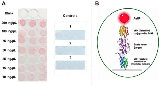
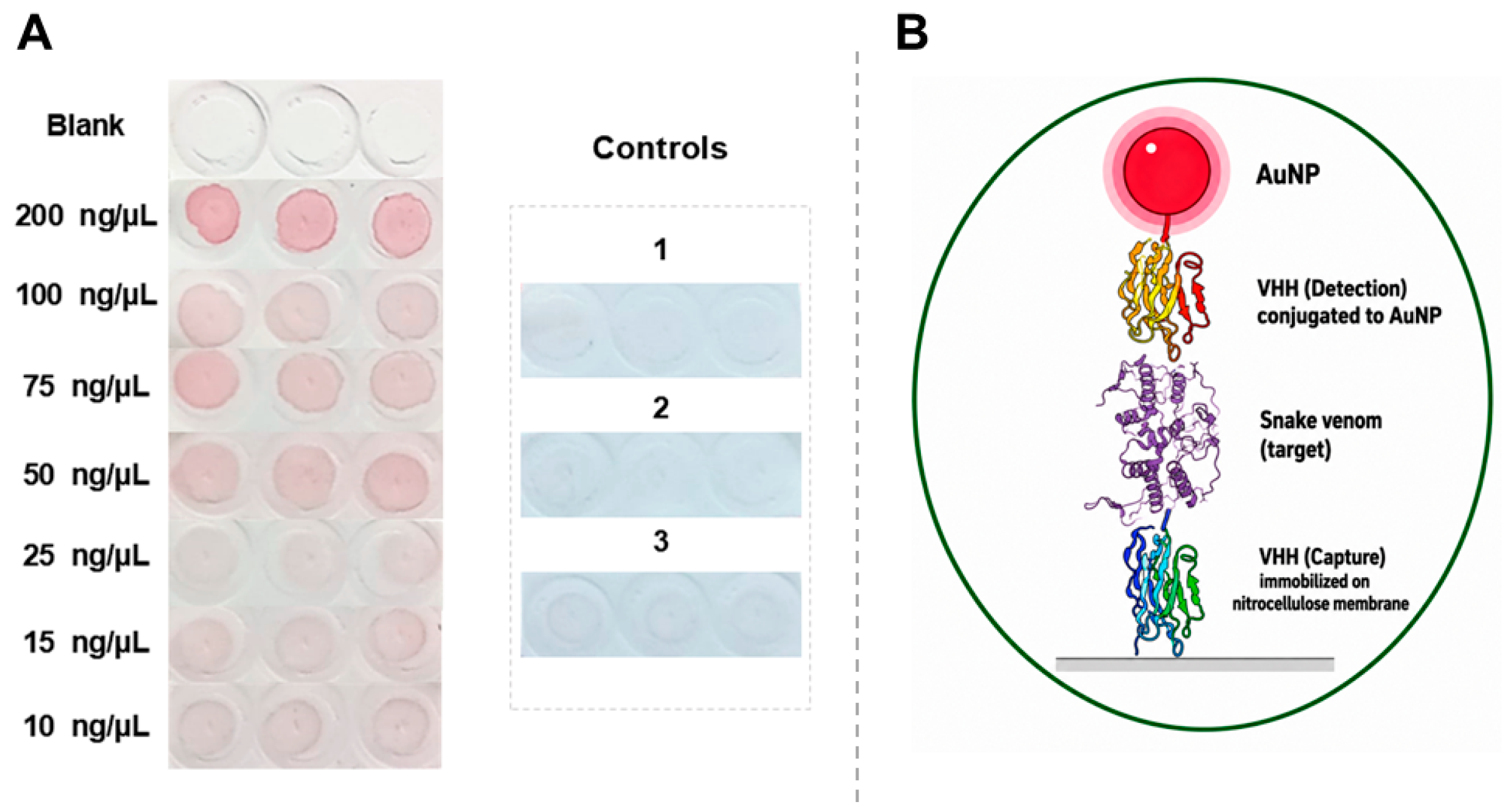
In the dot blot assay, the reactivity of the KC329718 bioconjugate was analyzed in a sandwich detection format similar to that described by Pham et al. [34]. The results presented in Figure 8A demonstrate the ability of KC329718 nanobioconjugates to detect Bothrops jararacussu whole venom, with the lowest concentration showing visible reactivity being 10 ng/µL and more consistent reactivity at concentrations ≥ 50 ng/µL, while no reactivity was observed against C. durissus terrificus and L. muta whole venoms. In some regions of Brazil, such as in the North region, snakes of the genera Bothrops and Lachesis can coexist in the same place [35] and, mainly, cause a similar symptomatological picture in the victims [36]. The distinction between envenoming by these two snake genera is of great importance for appropriate therapeutic targeting. No cross-reactivity between Bothrops and Lachesis venoms was observed under the conditions tested in this dot blot assay format.
Figure 8.
(A) Dot blot assay with VHH nanobioconjugates KC329718. Concentrations from 200 to 10 ng/µL refer to the whole venom of Bothrops jararacussu. The blank is represented by the membrane without the capture VHH (KF498607) and treated with the whole venom of Bothrops jararacussu (200 ng/µL). Controls 1, 2 and 3 correspond to membranes with VHH KF498607 and treated, respectively, with whole venom of Lachesis muta, whole venom of Crotalus durissus terrificus and distilled water. (B) Schematic representation of the molecular interactions involved in the sandwich dot blot assay shown in panel (A).
Despite the effective reactivity of the KC329718 bioconjugates in the sandwich assay, a slight reactivity of the bioconjugates against the membrane-adsorbed KF498607 capture antibody was also observed. This recognition can be observed by the red hue that appeared in the demarcated area of control 3 (Figure 8A).
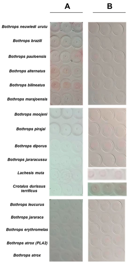
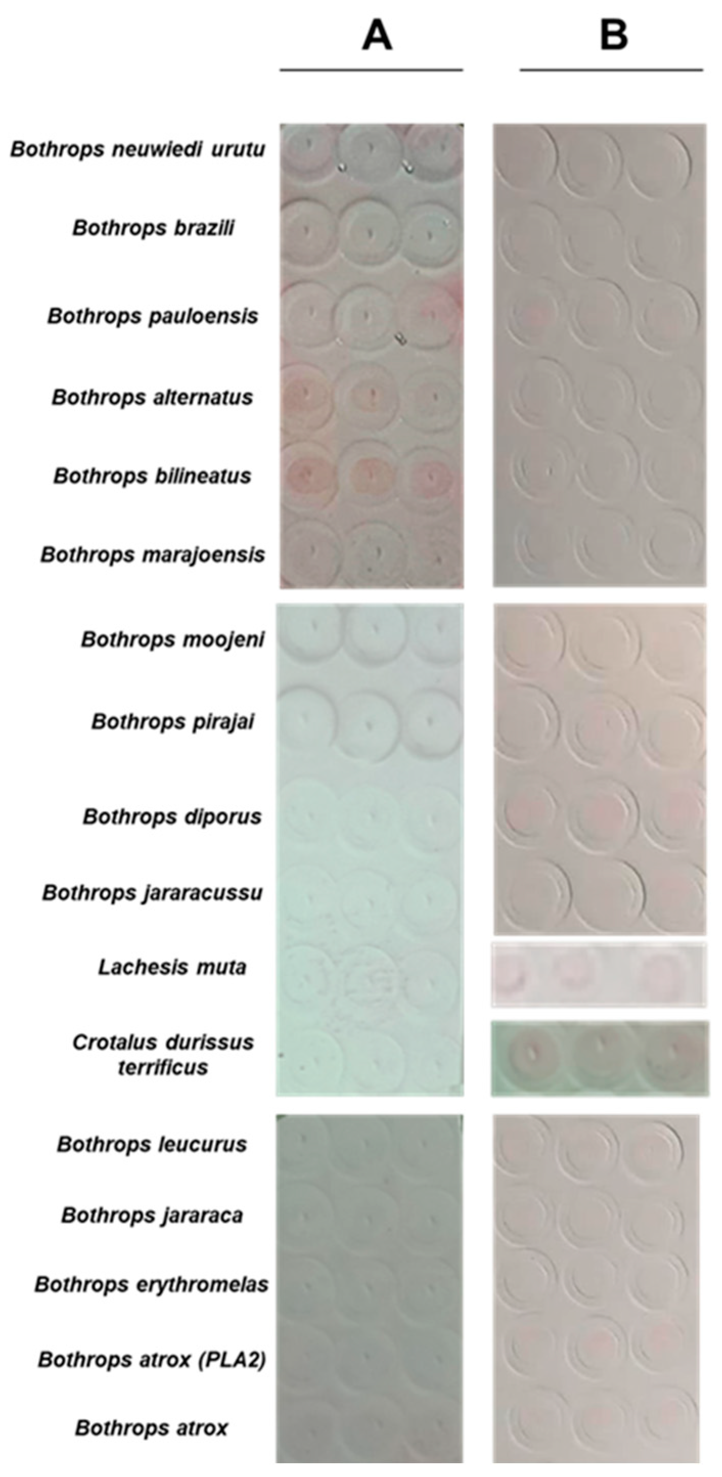
In Figure 9A, the results indicate that, in this assay, formatting the nanobioconjugates of KC329718 was unable to recognize the venom of Bothrops jararacussu and other species of the genus. Detection was visible only with Bothrops alternatus and Bothrops bilineatus venom. This result, particularly the absence of detection of B. jararacussu venom, suggests that the presence of the capture antibody adsorbed on the membrane is essential to amplify the detection signal promoted by KC329718 nanobioconjugates. For this reason, further studies involving the sandwich assay format were prioritized using B. jararacussu venom before extending this approach to other Bothrops species.
Figure 9.
(A) Dot blot assay with VHH nanobioconjugates KC329718 against venoms adsorbed at a concentration of 200 ng/µL. In (B), the corresponding membrane is stained with Ponceau S.
3.6. Anti-Crotalus VHHs (PZ837026 and KF498604)
The results demonstrated in Figure S6 indicate that PZ837026 nanobioconjugates showed considerable reactivity against membrane-adsorbed KF498604. Detection by PZ837026 nanobioconjugates was evident for concentrations up to 50 ng/µL of KF498604. The high reactivity of the PZ837026 bioconjugate against KF498604 may indicate that the blocking solution was not efficient in covering the surfaces of the nanobioconjugates. Although bovine serum albumin is one of the most used agents for blocking AuNPs, its application does not always result in effective blocking of AuNPs. Brewer et al. [37] discuss that binding of BSA molecules may not be fully effective in citrate-coated AuNPs. An alternative to be evaluated is the use of casein, another blocking agent that can be applied alternatively to BSA [38].
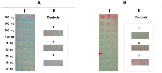
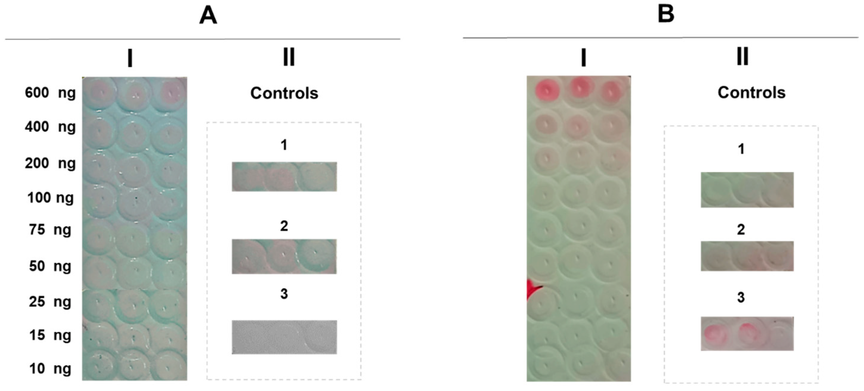
The impossibility of using VHH KF498604 as capture antibodies in the assay, as demonstrated in Figure S6, motivated the establishment of the dot blot assay in the direct format. In Figure 10A (I), PZ837026 nanobioconjugates recognized C. durissus terrificus venom at concentrations as low as 400 ng/µL. The absence of detection of the venom of L. muta (control 2) and B. jararacussu (control 3) indicates the specificity of the nanobioconjugates for Crotalus venom. In the controls, the absence of color in the triplicate containing venom of L. muta (2) and B. jararacussu (3) indicates the specificity of the nanobioconjugates for crotalic venom. However, PZ837026 nanobioconjugates showed visible reactivity with C. durissus terrificus venom, with 400 ng/µL being the lowest concentration at which reactivity was consistently observed under the conditions tested. In the corresponding membrane (B), staining with Ponceau S indicated that venom was adsorbed on the membrane at concentrations of 600, 400 and 200 ng/µL. However, it does not mean that, at concentrations below 200 ng/µL, there is no adsorbed protein, since Ponceau S staining presents a sensitivity threshold that should be considered. The literature indicates that this type of staining is capable of signaling the presence of proteins in the membrane at concentrations of approximately 200 ng/µL [39,40].
Figure 10.
(A) Dot blot (I) with PZ837026 VHH nanobioconjugates against different concentrations of C. durissus terrificus whole venom (600–10 ng). Controls (II) correspond to triplicate samples without adsorbed venom (1), with L. muta venom (2) and B. jararacussu venom (3). In (B), the corresponding membrane is stained with Ponceau S.
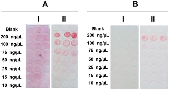
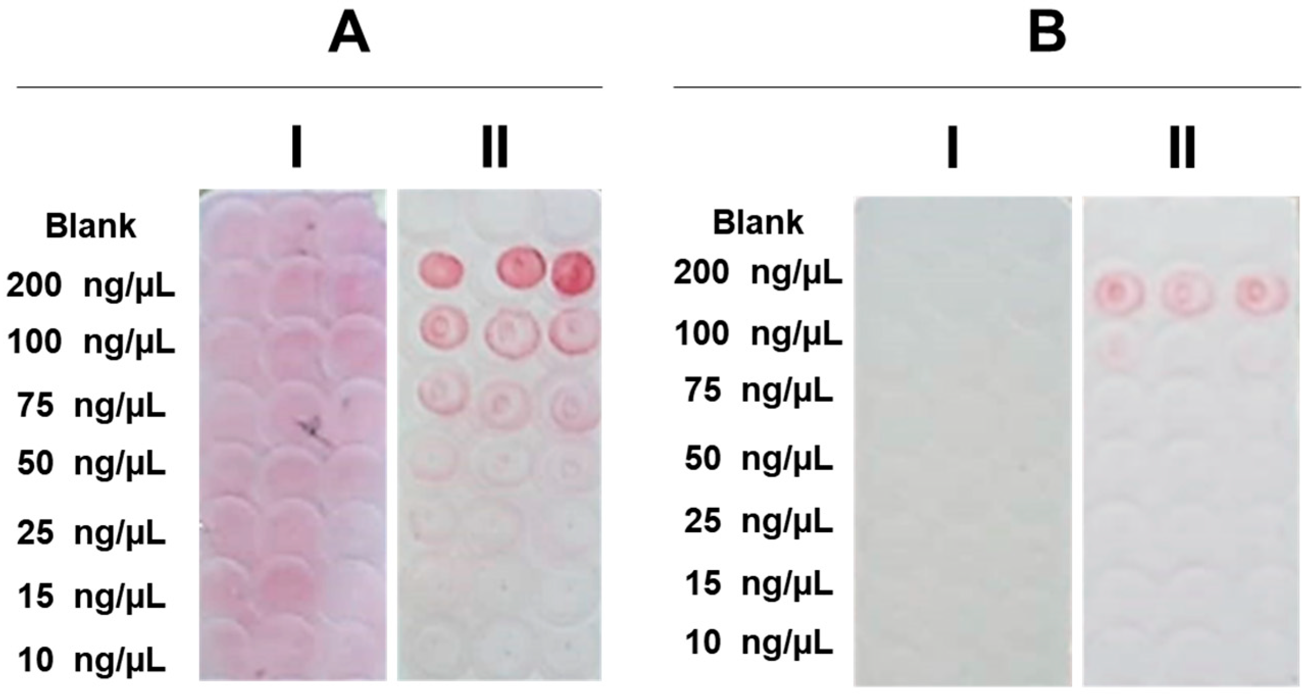
In addition to evaluating the reactivity of PZ837026 nanobioconjugates against the whole venom of C. durissus terrificus, reactivity against crotoxin (Figure 11(AI)) and the CB (PLA2) subunit (Figure 11(AII)) was also demonstrated in isolation. Unlike what was observed in the assay against whole venom, the nanobioconjugates showed no visible reactivity against crotoxin (I). However, reactivity against the CB subunit (PLA2) was evident from 200 to 75 ng/µL. When evaluating image B, Ponceau S staining was not as sensitive as nanobioconjugates to recognizing the membrane-adsorbed CB subunit at concentrations up to 75 ng/µL.
Figure 11.
(A) Dot blot assay with VHH PZ837026 nanobioconjugates against different concentrations of crotoxin (I) and its CB (PLA2) subunit (II) from C. durissus terrificus (200–10 ng). The blank is represented by the membrane without adsorbed protein. In (B), the corresponding membrane (Ponceau S staining) is shown, I and II corresponding to crotoxin and its CB (PLA2) subunit, respectively.
This apparent discrepancy could be related to differences in the way the target is presented in each assay format. Differences in immobilization may influence the orientation and accessibility of epitopes, particularly when a multicomponent protein complex is adsorbed onto a membrane. In this context, the recognition of the isolated CB subunit in the dot blot may reflect greater accessibility of the epitope recognized by PZ837026 when CB is immobilized separately, whereas its presentation in intact crotoxin may be less favorable for antibody binding. However, this interpretation remains speculative, as the present results do not provide direct evidence to establish whether differences in epitope presentation or target immobilization account for the observed discrepancy.
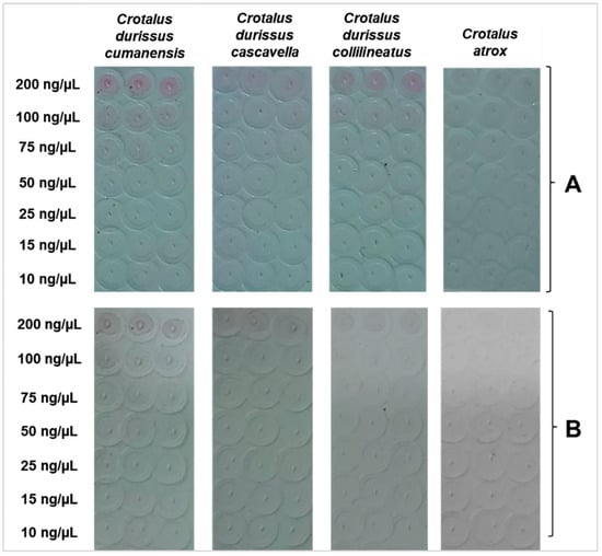
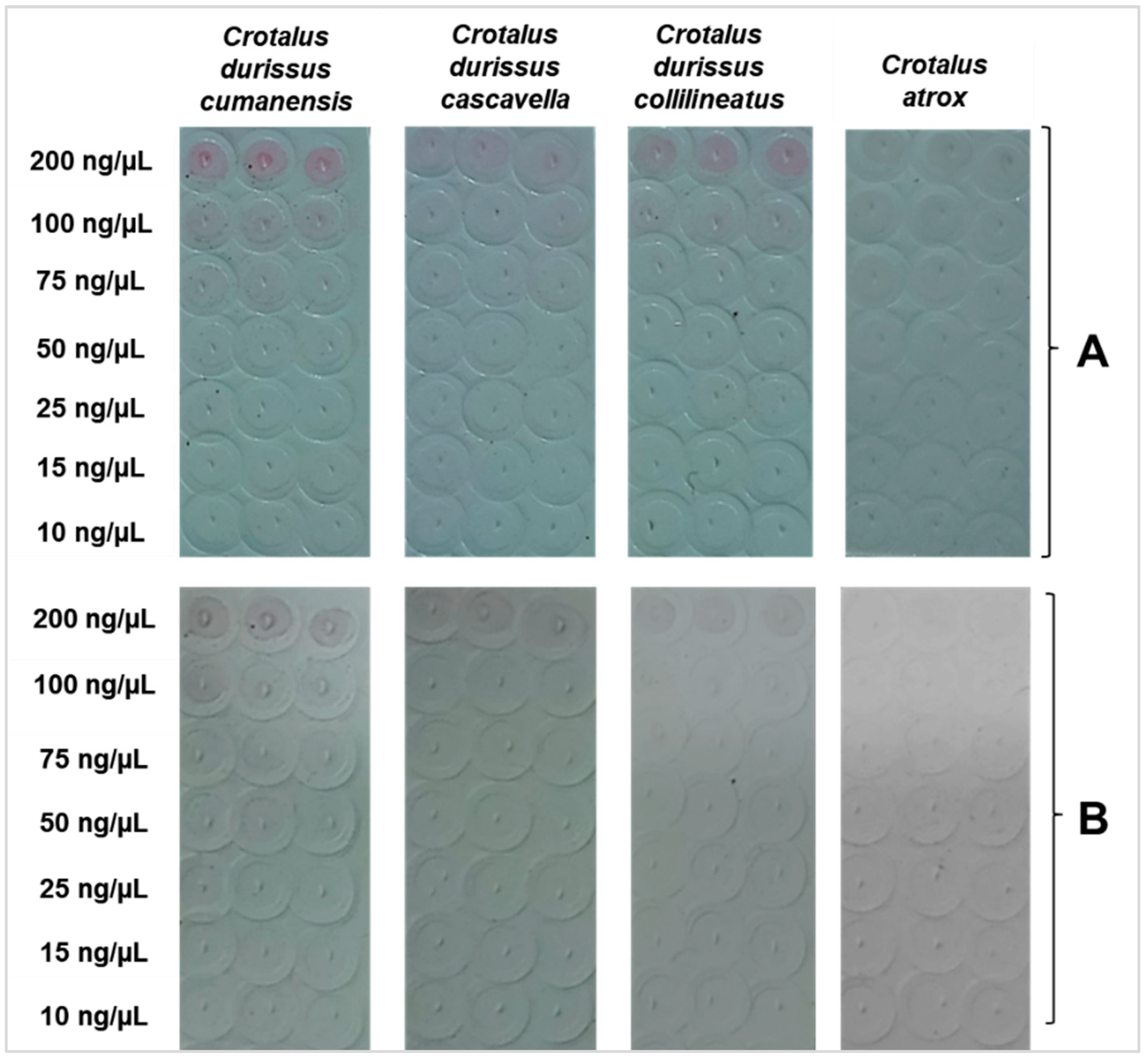
In the direct dot blot, PZ837026 nanobioconjugates were also able to signal the presence of the whole venom of the subspecies C. durissus collilineatus, C. durissus cascavella and C. durissus cumanensis (Figure 12A). For the venom of these three snakes, detection was observed at concentrations of 200 ng/µL. Although they belong to the same genus, there are differences in the protein composition of the venom of these subspecies. The crotoxin concentration, at which VHH PZ837026 shows high reactivity, is present in about 3% of C. durissus cumanensis venom [41]. In contrast, the concentration of crotoxin in the venom of C. durissus cascavella and C. durissus collilineatus corresponds to approximately 70% of the proteins that make up the venom [42], but the intensity of red color where there was the venom of C. durissus cumanensis was still higher. Thus, this result may indicate that PZ837026 nanobioconjugates are able to recognize other proteins from the venom of this subspecies, since crotoxin is in a smaller proportion in the venom of C. durissus cumanensis. The presence of color in the membrane containing C. atrox venom was not visualized, since the proteomic characterization of the venom of this snake does not indicate the presence of crotoxin [42].
Figure 12.
(A) Dot blot with VHH PZ837026 nanobioconjugates against different concentrations of venoms (200–10 ng/µL) from three subspecies of Crotalus durissus and Crotalus atrox. (B) Corresponding membrane of the reaction described in (A), stained with Ponceau S.
The lowest concentrations showing visible reactivity in the present study remain above the concentrations reported as clinically relevant for snakebite envenoming. Previous studies have shown that clinically relevant venom concentrations in snakebite patients generally occur within the range of tens to hundreds of ng/mL, depending on the snake species, severity of envenoming, and time elapsed after the bite [43,44,45,46]. Therefore, further optimization of the present nanobioconjugate will be necessary to improve analytical sensitivity and enable future evaluation in clinically relevant matrices, such as serum.
Although the present study was not designed to establish the optimal clinical matrix for venom detection, serum is currently considered the primary candidate for future evaluation of the proposed VHH-based nanobioconjugates. Previous studies using lateral flow platforms have demonstrated the feasibility of serum for snake venom detection despite the complexity of this biological matrix, particularly when appropriate sample pre-treatment strategies are employed [47,48]. Other matrices, including plasma, urine, and bite-site exudate, have also shown potential for venom detection in lateral flow assays [8,48]. However, the final selection of the most suitable clinical matrix for the VHH nanobioconjugates described here will depend on future evaluations of sensitivity, specificity, sample preparation requirements, and the final configuration of the lateral flow platform.
Beyond the optimization of analytical sensitivity and the evaluation of clinical matrices, future studies may also benefit from exploring alternative molecular designs to improve the performance of VHH-based nanobioconjugates. Further characterization of the molecular size and oligomeric state of the VHHs, particularly those showing an apparent molecular mass above their expected theoretical size in SDS-PAGE, should also be considered in future studies. Such analyses may help clarify the basis of the observed electrophoretic migration and further characterize the molecular state of these recombinant antibodies. The use of heterodimeric VHHs also represents a promising strategy for the future development of nanobioconjugates with improved specificity and sensitivity. Studies such as those by Silva et al. [49] and Calderon et al. [50], which described VHHs directed against specific targets of Bothrops venoms, may provide additional candidates for the design of heterodimeric constructs with potential diagnostic applications. Likewise, other anti-Crotalus VHH clones previously selected by Luiz et al. [13] may also represent valuable alternatives for the development of heterodimeric diagnostic platforms.
4. Conclusions
Although the anti-Bothrops and anti-Crotalus nanobioconjugates did not show cross-reactivity with the heterologous venoms evaluated in the present study, the recognition of the capture antibody, particularly in the assay for crotalic venom detection, indicates the need to evaluate alternative bioconjugation strategies, including the exploration of alternative stabilizers, optimization of bioconjugation parameters, and the use of blocking agents other than BSA. Although the sandwich dot blot assay was performed only with the anti-Bothrops nanobioconjugates, the direct format consistently demonstrated the reactivity of the anti-Crotalus nanobioconjugates. Furthermore, the analytical sensitivity achieved in the present study remains above the venom concentrations commonly reported in previous studies as clinically relevant for the diagnosis and clinical assessment of snakebite envenoming, which are generally observed in the ng/mL range. Therefore, further optimization of the proposed VHH-based nanobioconjugates will be necessary to improve analytical sensitivity before evaluation in clinically relevant matrices. Collectively, these findings provide initial evidence supporting the applicability of VHHs in the development of nanobioconjugates for the detection of botropic and crotalic venoms, while reinforcing the need for additional studies to optimize the performance of these platforms.
Supplementary Materials
The following supporting information can be downloaded at: https://www.mdpi.com/article/10.3390/bios16110609/s1, Figure S1: Photographic record of the bioconjugation reactions of VHH PZ837026 with different concentrations of acrylic acid–NHS ester, in the absence and presence of NaCl. The blank is represented by the reaction consisting of water with 100 mM acrylic acid-NHS ester and AuNPs at pH 7.5; Figure S2: Photographic record of the bioconjugation reactions of VHH KC329718 with different concentrations of acrylic acid-NHS ester, in the absence and presence of NaCl. The blank is represented by the reaction consisting of water with 100 mM acrylic acid-NHS ester and AuNPs at pH 8.5; Figure S3: Photographic record of bioconjugation of VHH PZ837026 bioconjugated at different pH ranges (6.5–8.0), in the absence and presence of NaCl; Figure S4: Photographic record of bioconjugation of VHH KC329718 bioconjugated at different pH ranges (7.5–9.0), in the absence and presence of NaCl; Figure S5: Representative images of bioconjugation reactions for visual analysis (A) and absorbance spectra, expressed in absorbance units (a.u.), of the blank at different pH values (6.0–9.0) without NaCl (B) and with NaCl (C), used as controls. The blank at each pH corresponds to a reaction containing water, 100 mM acrylic acid NHS ester, and AuNPs at the respective pH values (6.0–9.0); Figure S6: Dot blot assay with VHH PZ837026 biosensors against VHH KF498604. Concentrations from 1000 to 10 ng/µL refer to adsorbed KF498604. The blank is represented by the membrane without adsorbed VHH; Table S1: List of venoms evaluated in assays with anti-Crotalus and anti-Bothrops nanobioconjugates; Table S2: Absorbance at maximum absorption wavelength (λ) and at 450 nm; Table S3: Maximum absorbance wavelength (column 2) of VHH PZ837026 bioconjugation at different concentrations of acrylic acid NHS ester (0–100 mM). S/NaCl and C/NaCl correspond, respectively, to bioconjugates without and with NaCl. The blank corresponds to a reaction containing water, 100 mM acrylic acid NHS-ester, and AuNPs at pH 7.5; Table S4: Maximum absorbance wavelength (column 2) of VHH KC329718 bioconjugation at different concentrations of acrylic acid NHS ester (0–100 mM). S/NaCl and C/NaCl correspond, respectively, to bioconjugates without and with NaCl. The blank corresponds to a reaction containing water, 100 mM acrylic acid NHS ester, and AuNPs at pH 8.5.bsorbance wavelength (2nd column) of VHH C82 bioconjugation at different concentrations of acrylic acid NHS ester (0-100 mM). S/NaCl and C/NaCl correspond respectively to the bioconjugates without NaCl and the bioconjugates with NaCl. The blank is represented by the reaction consisting of water with 100 mM acrylic acid NHS ester and AuNPs in the pH 8.5 range; Table S5: Maximum absorbance wavelength (column 3) of VHH PZ837026 bioconjugation at different pH values (6.5–8.0). S/NaCl and C/NaCl correspond, respectively, to bioconjugates without and with NaCl. The blank for each pH corresponds to a reaction containing water, 100 mM acrylic acid NHS ester, and AuNPs at the respective pH values (6.5–8.0); Table S6: Maximum absorbance wavelength (column 3) of VHH KC329718 bioconjugation at different pH values (7.5–9.0). S/NaCl and C/NaCl correspond, respectively, to bioconjugates without and with NaCl. The blank for each pH corresponds to a reaction containing water, 100 mM acrylic acid NHS ester, and AuNPs at the respective pH values (7.5–9.0); Table S7: Maximum absorbance wavelength (column 2) of VHH PZ837026 bioconjugation with 2, 4, 6, and 8 µg. S/NaCl and C/NaCl correspond, respectively, to bioconjugates without and with NaCl. The blank corresponds to a reaction containing water, 100 mM acrylic acid NHS ester, and AuNPs at pH 7.5.bsorbance wavelength (2nd column) of bioconjugation with 2, 4, 6 and 8 µg of VHH PZ837026. S/NaCl and C/NaCl correspond respectively to the bioconjugates without NaCl and the bioconjugates with NaCl. The blank is represented by the reaction consisting of water with 100 mM acrylic acid-NHS-ester and AuNPs in the pH 7.5 range; Table S8: Maximum absorbance wavelength (column 2) of VHH KC329718 bioconjugation with 2, 4, 6, and 8 µg. S/NaCl and C/NaCl correspond, respectively, to bioconjugates without and with NaCl. The blank corresponds to a reaction containing water, 100 mM acrylic acid NHS ester, and AuNPs at pH 8.5; Table S9: Stoichiometric relationship of anti-Crotalus (PZ837026) and anti-Bothrops (KC329718) VHHs.
Author Contributions
Conceptualization, N.B.-d.-S., C.F.C.F. and S.d.S.P.; methodology, N.B.-d.-S. and L.M.-L.; formal analysis, N.B.-d.-S., S.A.R., N.D.R.P., E.L.d.S. and L.R.S.d.S.; investigation, N.B.-d.-S., S.A.R., N.D.R.P., E.L.d.S. and L.R.S.d.S.; data curation, S.A.R. and N.D.R.P.; writing—original draft preparation, N.B.-d.-S.; writing—review and editing, N.B.-d.-S., S.A.R., N.D.R.P., E.L.d.S., L.R.S.d.S. and L.M.-L.; supervision, L.M.-L. and S.d.S.P.; project administration, S.d.S.P.; funding acquisition, S.d.S.P. All authors have read and agreed to the published version of the manuscript.
Funding
This study was supported by Fundação de Amparo à Pesquisa do Estado de Rondônia (FAPERO), under Public Call PAP Pública (Notice No. 4/2025/FAPERO-DC), which will also cover the article processing charges. Additional support for the development of this research was provided by the Coordination for the Improvement of Higher Education Personnel (CAPES) and the National Council for Scientific and Technological Development (CNPq).
Institutional Review Board Statement
Not applicable.
Informed Consent Statement
Not applicable.
Data Availability Statement
Data is contained within the article and supplementary materials.
Acknowledgments
The authors acknowledge Fiocruz Rondônia for institutional support. The manuscript was reviewed for English language by a specialized scientific editing service. Generative AI was used to assist in the creation of the schematic illustration presented in Figure 8. All scientific content and the final figure were reviewed and approved by the authors. This article is a revised and expanded version of a paper entitled “Detection of rattlesnake and jararaca venoms using camelid VHH-based biosensors,” which was presented at the 10th International Symposium on Immunobiologicals (ISI 2026), Rio de Janeiro, Brazil, 6–8 May 2026 [51].
Conflicts of Interest
The authors declare no conflicts of interest.
Abbreviations
| VHH | Variable domain of heavy chain-only antibodies |
| AuNPs | Gold nanoparticles |
| IMAC | Immobilized metal affinity chromatography |
| ELISA | Enzyme-Linked Immunosorbent Assay |
| SDS-PAGE | Sodium dodecyl sulfate–polyacrylamide gel electrophoresis |
| BCA | Bicinchoninic acid assay |
| PBS | Phosphate-buffered saline |
| BSA | Bovine serum albumin |
| NHS | N-hydroxysuccinimide |
| pI | Isoelectric point |
| TEM | Transmission electron microscopy |
| CTX | Crotoxin |
| PLA2 | Phospholipase A2 |
| WV | Whole venom |
References
- WHO Snakebite Envenoming. Available online: https://www.who.int/news-room/fact-sheets/detail/snakebite-envenoming (accessed on 10 March 2026).
- Laustsen, A.H.; Johansen, K.H.; Engmark, M.; Andersen, M.R. Recombinant Snakebite Antivenoms: A Cost-Competitive Solution to a Neglected Tropical Disease? PLoS Negl. Trop. Dis. 2017, 11, e0005361. [Google Scholar] [CrossRef] [Scilit]
- BRASIL Boletim Epidemiológico—Volume 55—No 15—Ministério Da Saúde. Available online: https://www.gov.br/saude/pt-br/centrais-de-conteudo/publicacoes/boletins/epidemiologicos/edicoes/2024/boletim-epidemiologico-volume-55-no-15.pdf/view (accessed on 8 January 2025).
- BRASIL Tratamento Do Ofidismo—Ministério Da Saúde. Available online: https://www.gov.br/saude/pt-br/assuntos/saude-de-a-a-z/a/animais-peconhentos/acidentes-ofidicos/tratamento (accessed on 10 March 2026).
- Knudsen, C.; Jürgensen, J.A.; Føns, S.; Haack, A.M.; Friis, R.U.W.; Dam, S.H.; Bush, S.P.; White, J.; Laustsen, A.H. Snakebite Envenoming Diagnosis and Diagnostics. Front. Immunol. 2021, 12, 661457. [Google Scholar] [CrossRef] [Scilit]
- Garcês-Filho, A.Q.; Santos, H.H.M.; Aguiar, T.K.P.P.; Ramos, D.L.S.; Galan, L.E.B.; Dantas, D.S.M.; Cerni, F.A.; Carbonell, R.C.; Pucca, M.B. Severe Bushmaster Snakebite Envenoming: Case Report and Overview. Reports 2024, 7, 68. [Google Scholar] [CrossRef] [Scilit]
- Long, C.; Wu, F.; Lu, Q.; Xie, B.; Shen, C.; Li, J.; Deng, Y.; Liang, P.; Yu, Y.; Lai, R. A Strategy for Efficient Preparation of Genus-Specific Diagnostic Antibodies for Snakebites. Front. Immunol. 2021, 12, 775678. [Google Scholar] [CrossRef] [Scilit]
- Zeng, L.; Che, Y.; Guo, C.; Liu, L.; Wang, G.; Chen, M.; Ruan, J.; Mo, Z.; Li, Y.; Li, X.; et al. Development of an Immunofluorescence Lateral Flow Assay for Detecting Four Medically Important Snake Venoms in China. J. Trop. Med. 2026, 2026, 9614080. [Google Scholar] [CrossRef] [Scilit]
- Knudsen, C.; Jürgensen, J.A.; Knudsen, P.D.; Oganesyan, I.; Harrison, J.A.; Dam, S.H.; Haack, A.M.; Friis, R.U.W.; Vitved, L.; Belfakir, S.B.; et al. Prototyping of a Lateral Flow Assay Based on Monoclonal Antibodies for Detection of Bothrops Venoms. Anal. Chim. Acta 2023, 1272, 341306. [Google Scholar] [CrossRef] [Scilit]
- Magalhães, S.F.V.; Peixoto, H.M.; de Almeida Gonçalves Sachett, J.; Oliveira, S.S.; Alves, E.C.; dos Santos Ibiapina, H.N.; Monteiro, W.M.; de Oliveira, M.R.F. Snakebite Envenomation in the Brazilian Amazon: A Cost-of-Illness Study. Trans. R. Soc. Trop. Med. Hyg. 2020, 114, 642–649. [Google Scholar] [CrossRef] [Scilit]
- Simões, B.; Guedens, W.J.; Keene, C.; Kubiak-Ossowska, K.; Mulheran, P.; Kotowska, A.M.; Scurr, D.J.; Alexander, M.R.; Broisat, A.; Johnson, S.; et al. Direct Immobilization of Engineered Nanobodies on Gold Sensors. ACS Appl. Mater. Interfaces 2021, 13, 17353–17360. [Google Scholar] [CrossRef] [Scilit]
- Jin, B.; Odongo, S.; Radwanska, M.; Magez, S. NANOBODIES®: A Review of Diagnostic and Therapeutic Applications. Int. J. Mol. Sci. 2023, 24, 5994. [Google Scholar] [CrossRef] [Scilit]
- Luiz, M.; Pereira, S.; Prado, N.; Gonçalves, N.; Kayano, A.; Moreira-Dill, L.; Sobrinho, J.; Zanchi, F.; Fuly, A.; Fernandes, C.; et al. Camelid Single-Domain Antibodies (VHHs) against Crotoxin: A Basis for Developing Modular Building Blocks for the Enhancement of Treatment or Diagnosis of Crotalic Envenoming. Toxins 2018, 10, 142. [Google Scholar] [CrossRef] [Scilit]
- Prado, N.D.R.; Pereira, S.S.; da Silva, M.P.; Morais, M.S.S.; Kayano, A.M.; Moreira-Dill, L.S.; Luiz, M.B.; Zanchi, F.B.; Fuly, A.L.; Huacca, M.E.F.; et al. Inhibition of the Myotoxicity Induced by Bothrops Jararacussu Venom and Isolated Phospholipases A2 by Specific Camelid Single-Domain Antibody Fragments. PLoS ONE 2016, 11, e0151363. [Google Scholar] [CrossRef] [Scilit]
- Sambrook, J.; Russell, D.W. The Condensed Protocols from Molecular Cloning; Cold Spring Harbor: New York, NY, USA, 2006. [Google Scholar]
- Turkevich, J.; Stevenson, P.C.; Hillier, J. A Study of the Nucleation and Growth Processes in the Synthesis of Colloidal Gold. Discuss. Faraday Soc. 1951, 11, 55. [Google Scholar] [CrossRef] [Scilit]
- Haiss, W.; Thanh, N.T.K.; Aveyard, J.; Fernig, D.G. Determination of Size and Concentration of Gold Nanoparticles from UV−Vis Spectra. Anal. Chem. 2007, 79, 4215–4221. [Google Scholar] [CrossRef] [Scilit]
- Tanaka, R.; Yuhi, T.; Nagatani, N.; Endo, T.; Kerman, K.; Takamura, Y.; Tamiya, E. A Novel Enhancement Assay for Immunochromatographic Test Strips Using Gold Nanoparticles. Anal. Bioanal. Chem. 2006, 385, 1414–1420. [Google Scholar] [CrossRef] [Scilit]
- Bastos-Soares, E.A.; da Silva Morais, M.S.; Funes-Huacca, M.; Sousa, R.M.O.; Brilhante-Da-Silva, N.; Roberto, S.A.; Prado, N.D.R.; dos Santos, C.N.D.; Marinho, A.C.M.; Soares, A.M.; et al. Single-Domain Antibody-Gold Nanoparticle Bioconjugates as Immunosensors for the Detection of Hantaviruses. Mol. Diagn. Ther. 2024, 28, 479–494. [Google Scholar] [CrossRef] [Scilit]
- Helena, R.; Pereira, A. Ressonância de Plasmons de Superfície Localizada (LSPR) Em Dispersões Coloidais de Ouro Para Detecção de Ocratoxina A; Universidade Federal do Espírito Santo: Vitória, Brazil, 2016. [Google Scholar]
- Maier, S.A. Plasmonics: Fundamentals and Applications; Springer: New York, NY, USA, 2007; ISBN 0387331506. [Google Scholar]
- Fatima, A.; Wang, H.; Kang, K.; Xia, L.; Wang, Y.; Ye, W.; Wang, J.; Wang, X. Development of VHH Antibodies against Dengue Virus Type 2 NS1 and Comparison with Monoclonal Antibodies for Use in Immunological Diagnosis. PLoS ONE 2014, 9, e95263. [Google Scholar] [CrossRef] [Scilit]
- Zafar, M.; Shah, M.A.; Shehzad, A.; Tariq, A.; Habib, M.; Muddassar, M.; Shah, M.S.; Iqbal, M.; Hemmatzadeh, F.; Rahman, M. Characterization of the Highly Immunogenic VP2 Protrusion Domain as a Diagnostic Antigen for Members of Birnaviridae Family. Appl. Microbiol. Biotechnol. 2020, 104, 3391–3402. [Google Scholar] [CrossRef] [Scilit]
- Pinto Torres, J.E.; Goossens, J.; Ding, J.; Li, Z.; Lu, S.; Vertommen, D.; Naniima, P.; Chen, R.; Muyldermans, S.; Sterckx, Y.G.-J.; et al. Development of a Nanobody-Based Lateral Flow Assay to Detect Active Trypanosoma Congolense Infections. Sci. Rep. 2018, 8, 9019. [Google Scholar] [CrossRef] [Scilit]
- Zhang, L.; Hu, D.; Salmain, M.; Liedberg, B.; Boujday, S. Direct Quantification of Surface Coverage of Antibody in IgG-Gold Nanoparticles Conjugates. Talanta 2019, 204, 875–881. [Google Scholar] [CrossRef] [Scilit]
- Locarno, S.; Bucci, R.; Impresari, E.; Gelmi, M.L.; Pellegrino, S.; Clerici, F. Ultrashort Peptides and Gold Nanoparticles: Influence of Constrained Amino Acids on Colloidal Stability. Front. Chem. 2021, 9, 736519. [Google Scholar] [CrossRef] [Scilit]
- Filbrun, S.L.; Filbrun, A.B.; Lovato, F.L.; Oh, S.H.; Driskell, E.A.; Driskell, J.D. Chemical Modification of Antibodies Enables the Formation of Stable Antibody–Gold Nanoparticle Conjugates for Biosensing. Analyst 2017, 142, 4456–4467. [Google Scholar] [CrossRef] [Scilit]
- Nong, J.-F.; Huang, Z.; Huang, Z.-Z.; Yang, J.; Li, J.-C.; Yang, F.; Huang, D.-L.; Wang, F.; Wang, W. Development of Sandwich ELISA and Lateral Flow Assay for the Detection of Bungarus Multicinctus Venom. PLoS Negl. Trop. Dis. 2023, 17, e0011165. [Google Scholar] [CrossRef] [Scilit]
- Zakaria, H.M.; Shah, A.; Konieczny, M.; Hoffmann, J.A.; Nijdam, A.J.; Reeves, M.E. Small Molecule- and Amino Acid-Induced Aggregation of Gold Nanoparticles. Langmuir 2013, 29, 7661–7673. [Google Scholar] [CrossRef] [Scilit]
- Goossens, J.; Sein, H.; Lu, S.; Radwanska, M.; Muyldermans, S.; Sterckx, Y.G.-J.; Magez, S. Functionalization of Gold Nanoparticles with Nanobodies through Physical Adsorption. Anal. Methods 2017, 9, 3430–3440. [Google Scholar] [CrossRef] [Scilit]
- Loedsapchinda, N.; Ta-Aithuak, S.; Chueaiarrom, C.; Srikhirin, T.; Houngkamhang, N. Study of Anti-A Antibody Conjugated Gold Nanoparticles as a Biomolecular Detection Probe for a Substance in Saliva. Key Eng. Mater. 2020, 853, 87–91. [Google Scholar] [CrossRef] [Scilit]
- Sotnikov, D.V.; Byzova, N.A.; Zherdev, A.V.; Dzantiev, B.B. Retention of Activity by Antibodies Immobilized on Gold Nanoparticles of Different Sizes: Fluorometric Method of Determination and Comparative Evaluation. Nanomaterials 2021, 11, 3117. [Google Scholar] [CrossRef] [Scilit]
- Muyldermans, S. Nanobodies: Natural Single-Domain Antibodies. Annu. Rev. Biochem. 2013, 82, 775–797. [Google Scholar] [CrossRef] [Scilit]
- Pham, V.D.; Hoang, H.; Phan, T.H.; Conrad, U.; Chu, H.H. Production of Antibody Labeled Gold Nanoparticles for Influenza Virus H5N1 Diagnosis Kit Development. Adv. Nat. Sci. Nanosci. Nanotechnol. 2012, 3, 045017. [Google Scholar] [CrossRef] [Scilit]
- Schneider, M.C.; Min, K.; Hamrick, P.N.; Montebello, L.R.; Ranieri, T.M.; Mardini, L.; Camara, V.M.; Raggio Luiz, R.; Liese, B.; Vuckovic, M.; et al. Overview of Snakebite in Brazil: Possible Drivers and a Tool for Risk Mapping. PLoS Negl. Trop. Dis. 2021, 15, e0009044. [Google Scholar] [CrossRef] [Scilit]
- BRASIL Sintomas—Ministério Da Saúde. Available online: https://www.gov.br/saude/pt-br/assuntos/saude-de-a-a-z/a/animais-peconhentos/acidentes-ofidicos/sintomas (accessed on 10 March 2026).
- Brewer, S.H.; Glomm, W.R.; Johnson, M.C.; Knag, M.K.; Franzen, S. Probing BSA Binding to Citrate-Coated Gold Nanoparticles and Surfaces. Langmuir 2005, 21, 9303–9307. [Google Scholar] [CrossRef] [Scilit]
- Seele, P.P.; Dyan, B.; Skepu, A.; Maserumule, C.; Sibuyi, N.R.S. Development of Gold-Nanoparticle-Based Lateral Flow Immunoassays for Rapid Detection of TB ESAT-6 and CFP-10. Biosensors 2023, 13, 354. [Google Scholar] [CrossRef] [Scilit]
- Aebersold, R. Internal Amino Acid Sequence Analysis of Proteins after in Situ Protease Digestion on Nitrocellulose. In A Practical Guide to Protein and Peptide Purification for Microsequencing; Elsevier: Amsterdam, The Netherlands, 1993; pp. 103–124. [Google Scholar]
- Goldman, A.; Harper, S.; Speicher, D.W. Detection of Proteins on Blot Membranes. Curr. Protoc. Protein Sci. 2016, 86, 10.8.1–10.8.11. [Google Scholar] [CrossRef] [Scilit]
- Calvete, J.J.; Juárez, P.; Sanz, L. Snake Venomics. Strategy and Applications. J. Mass Spectrom. 2007, 42, 1405–1414. [Google Scholar] [CrossRef] [Scilit]
- Boldrini-França, J.; Corrêa-Netto, C.; Silva, M.M.S.; Rodrigues, R.S.; De La Torre, P.; Pérez, A.; Soares, A.M.; Zingali, R.B.; Nogueira, R.A.; Rodrigues, V.M.; et al. Snake Venomics and Antivenomics of Crotalus durissus Subspecies from Brazil: Assessment of Geographic Variation and Its Implication on Snakebite Management. J. Proteom. 2010, 73, 1758–1776. [Google Scholar] [CrossRef] [Scilit]
- Bucher, B.; Canonge, D.; Thomas, L.; Tyburn, B.; Robbe-Vincent, A.; Choumet, V.; Bon, C.; Ketterlé, J.; Lang, J. Clinical Indicators of Envenoming and Serum Levels of Venom Antigens in Patients Bitten by Bothrops lanceolatus in Martinique. Trans. R. Soc. Trop. Med. Hyg. 1997, 91, 186–190. [Google Scholar] [CrossRef] [Scilit]
- França, F.O.S.; Barbaro, K.C.; Fan, H.W.; Cardoso, J.L.C.; Sano-Martins, I.S.; Tomy, S.C.; Lopes, M.H.; Warrell, D.A.; Theakston, R.D.G. Envenoming by Bothrops jararaca in Brazil: Association between Venom Antigenaemia and Severity at Admission to Hospital. Trans. R. Soc. Trop. Med. Hyg. 2003, 97, 312–317. [Google Scholar] [CrossRef] [Scilit]
- Bucaretchi, F.; De Capitani, E.M.; Hyslop, S.; Mello, S.M.; Fernandes, C.B.; Bergo, F.; Nascimento, F.B.P. Compartment Syndrome after South American Rattlesnake (Crotalus durissus terrificus) Envenomation. Clin. Toxicol. 2014, 52, 639–641. [Google Scholar] [CrossRef] [Scilit]
- Sanhajariya, S.; Duffull, S.; Isbister, G. Pharmacokinetics of Snake Venom. Toxins 2018, 10, 73. [Google Scholar] [CrossRef] [Scilit]
- Liu, C.-C.; Yu, J.-S.; Wang, P.-J.; Hsiao, Y.-C.; Liu, C.-H.; Chen, Y.-C.; Lai, P.-F.; Hsu, C.-P.; Fann, W.-C.; Lin, C.-C. Development of Sandwich ELISA and Lateral Flow Strip Assays for Diagnosing Clinically Significant Snakebite in Taiwan. PLoS Negl. Trop. Dis. 2018, 12, e0007014. [Google Scholar] [CrossRef] [Scilit]
- Knudsen, C.; Belfakir, S.B.; Degnegaard, P.; Jürgensen, J.A.; Haack, A.M.; Friis, R.U.W.; Dam, S.H.; Laustsen, A.H.; Ross, G.M.S. Multiplex Lateral Flow Assay Development for Snake Venom Detection in Biological Matrices. Sci. Rep. 2024, 14, 2567. [Google Scholar] [CrossRef] [Scilit]
- Silva, M.C.S.; Pereira, S.S.; Gouveia, M.P.; Luiz, M.B.; Sousa, R.M.O.; Kayano, A.M.; Francisco, A.F.; Prado, N.D.R.; Dill, L.S.M.; Fontes, M.R.M.; et al. Anti-Metalloprotease P-I Single-Domain Antibodies: Tools for Next-Generation Snakebite Antivenoms. BioMed Res. Int. 2022, 2022, 2748962. [Google Scholar] [CrossRef] [Scilit]
- Bailon Calderon, H.; Yaniro Coronel, V.O.; Cáceres Rey, O.A.; Colque Alave, E.G.; Leiva Duran, W.J.; Padilla Rojas, C.; Montejo Arevalo, H.; García Neyra, D.; Galarza Pérez, M.; Bonilla, C.; et al. Development of Nanobodies Against Hemorrhagic and Myotoxic Components of Bothrops atrox Snake Venom. Front. Immunol. 2020, 11, 655. [Google Scholar] [CrossRef] [Scilit]
- Silva, N.; Roberto, S.; Prado, N.; Santos, E.; Souza, L.; Lima, L.; Fernandes, C.; Pereira, S. Detection of Rattlesnake and Jararaca Venoms Using Camelid VHH-Based Biosensors. In Proceedings of the Annals of the International Symposium on Immunobiologicals; Instituto de Tecnologia em Imunobiológicos: Rio de Janeiro, Brazil, 2026; p. 149. [Google Scholar]
Disclaimer/Publisher’s Note: The statements, opinions and data contained in all publications are solely those of the individual author(s) and contributor(s) and not of MDPI and/or the editor(s). MDPI and/or the editor(s) disclaim responsibility for any injury to people or property resulting from any ideas, methods, instructions or products referred to in the content. |
© 2026 by the authors. Licensee MDPI, Basel, Switzerland. This article is an open access article distributed under the terms and conditions of the Creative Commons Attribution (CC BY) license.