


Abstract
Protein biomarkers play an indispensable role in the early diagnosis, prognosis assessment, therapeutic monitoring, and treatment evaluation of diseases. Optical biosensors have emerged as promising alternatives for protein detection due to their label-free, real-time, and highly sensitive characteristics. Molecularly imprinted polymers (MIPs), known as “plastic antibodies,” can provide chemically stable, low-cost, and tailorable recognition elements through the formation of target-complementary cavities in polymer matrices, making them highly attractive for integration with optical sensing platforms. In this work, we systematically summarize the advances in MIP-based optical biosensors for protein detection. The fundamental principles of biosensors and recognition elements are introduced, followed by a brief overview of the key components of imprinting technology, such as imprinting materials, functional monomers, cross-linking agents, polymerization mechanisms, template fixation strategies, and template removal conditions. Then, we comprehensively evaluate the integration of MIPs with various optical transduction techniques, including fluorescence, chemiluminescence, electrochemiluminescence, surface-enhanced Raman scattering, colorimetry, and surface plasmon resonance. Finally, the major challenges are discussed, and future development directions are proposed. This review aims to provide systematic theoretical references and useful insights for the rational design, performance improvement, and clinical translation of MIP-based optical biosensors for protein detection.
1. Introduction
Proteins can serve as indicators of physiological and pathological states based on their structure and abundance changes. They play an indispensable role in the early diagnosis, prognosis assessment, therapeutic monitoring, and treatment evaluation of certain diseases [1,2]. For example, alpha-fetoprotein (AFP) is extensively employed for the early diagnosis of hepatocellular carcinoma, cardiac troponin I (cTnI) is a key biomarker for acute myocardial infarction, and prostate-specific antigen (PSA) is a commonly used biomarker for prostate diseases. The detection of protein biomarkers can not only facilitate the early identification and intervention of diseases but also provide valuable information for evaluating therapeutic efficacy, monitoring disease recurrence, and guiding personalized therapy. However, protein biomarkers are often present at extremely low concentrations in biological fluids and coexist with highly complex sample matrices, placing stringent demands on the sensitivity, specificity, and anti-interference ability of analytical methods. Traditional techniques for protein detection mainly include high-performance liquid chromatography (HPLC), liquid chromatography–tandem mass spectrometry (LC–MS/MS), and enzyme-linked immunosorbent assay (ELISA) [3]. HPLC plays an important role in protein separation and quantitative analysis owing to its high resolution, good reproducibility, and well-established separation technology. LC–MS/MS combines the separation capability of liquid chromatography with the high sensitivity and specificity of mass spectrometry, enabling simultaneous quantification of multiple proteins in complex samples. Nevertheless, these two methods are generally associated with expensive instruments, complicated procedures, specialized personnel, time-consuming sample preparation, and limited suitability for point-of-care testing. As one of the classical immunoassay methods, ELISA offers high sensitivity and specificity for protein detection, but it is still limited by antibody stability, susceptibility to cross-reactivity, and labor-intensive detection procedures.
Advancements in precision instruments and nanotechnologies have driven the widespread design and application of high-performance biosensors [4,5,6]. Based on their signal transduction mechanisms, biosensors can be mainly classified into two major categories: electrochemical and optical biosensors. Electrochemical biosensors have attracted considerable attention because of their rapid response, relatively low instrumentation cost, ease of miniaturization, and compatibility with microfluidic platforms. However, their practical application in protein detection remains challenged by several intrinsic limitations [7,8,9,10]. For example, proteins are large and structurally complex biomolecules, and their redox-active centers or specific binding sites are often buried within peptide chains or spatially shielded by post-translational modifications such as glycosylation. These structural features severely hinder electron transfer at the electrode interface, thereby limiting detection sensitivity. In complex biological samples such as serum, whole blood, and urine, interfering proteins can easily undergo non-specific adsorption onto the electrode surface, forming an insulating layer to passivate the electrode. This phenomenon substantially compromises the stability, reproducibility, and selectivity of electrochemical biosensors and increases the risk of false-positive or false-negative results. In addition, the direct immobilization of proteins on the electrode surface may induce conformational changes and loss of biological activity, further hindering the clinical translation of electrochemical biosensors.
Optical biosensors have emerged as promising alternatives due to their unique physical transduction mechanisms. Optical sensing typically relies on the interaction between light and matter to transduce molecular recognition events, and the resulting signals are largely unaffected by electroactive substances in biological samples, thereby minimizing signal suppression caused by complex sample matrices [11,12,13,14,15]. In particular, optical biosensors can enable label-free detection and real-time monitoring of the binding kinetics of targets and recognition elements in liquid-phase environments that closely approximate native physiological conditions, helping to preserve the structural integrity and biological activity of proteins. Furthermore, the surface-enhanced Raman scattering (SERS) and localized surface plasmon resonance (LSPR) effects of noble metal nanomaterials, such as gold and silver, as well as emerging two-dimensional materials, such as graphene, can generate highly enhanced local electromagnetic fields, enabling ultrasensitive detection of low-abundance protein biomarkers, potentially down to the single-molecule level [16]. Meanwhile, rapid advances in optical fibers and integrated waveguide technologies have further promoted the miniaturization and portability of optical biosensors, making them adaptable to point-of-care testing devices for rapid clinical screening. Therefore, compared with electrochemical methods, optical biosensors offer considerable potential for highly sensitive and real-time monitoring of protein biomarkers.
Biosensors can convert biometric events into quantifiable signals, typically consisting of two key components: recognition element and signal transduction. The specific interaction between recognition element with target analyte is primarily responsible for sensor selectivity, whereas transduction element converts the recognition event into electrochemical, optical, or other measurable signals [17]. At present, the most widely used recognition elements include antibodies, nucleic acids, peptides, and molecularly imprinted polymers (MIPs). Antibodies are the most extensively used biorecognition elements, and they can achieve highly specific target recognition through the antigen-binding domains located within their characteristic Y-shaped structures. Their major advantages include high affinity, excellent specificity, and commercial availability, with a wide variety of resources. However, antibodies have several notable limitations. For example, their production often involves animal immunization, making the process time-consuming, labor-intensive, and costly. Antibodies are sensitive to environmental conditions such as temperature and pH, so their stability is limited. In addition, batch-to-batch variations may compromise experimental reproducibility. Moreover, antibodies have a relatively large molecular weight (approximately 150 kDa), which may lead to steric hindrance during their immobilization on a sensor surface with high density. Nucleic acid-based biometric elements rely on base pairing to achieve specific molecular recognition. Their major advantages include low-cost and scalable chemical synthesis, excellent batch-to-batch consistency, and flexible control of recognition performance through sequence design. However, nucleic acids are negatively charged and can undergo non-specific electrostatic adsorption in complex biological matrices. They are also susceptible to degradation by nucleases, which may compromise their stability and sensing performance. Peptide-based recognition elements have attracted increasing attention. Peptides combine synthetic versatility with molecular recognition capability and offer facile chemical modification, good biocompatibility, and cost-effectiveness. However, their binding affinity to target analytes is generally relatively low, typically in the micromolar range, and their selectivity and binding strength to targets are often inferior to those of antibodies.
In order to overcome the limitations of traditional biometric elements in terms of stability, cost, and preparation time, MIPs, commonly known as “plastic antibodies”, have emerged as fully synthetic recognition elements [18,19,20,21]. They can achieve specific molecular recognition by creating three-dimensional cavities within a polymer matrix that are complementary to the target in size, shape, and functional group distribution. The synthesis of MIPs generally involves three key steps. The first is pre-polymerization, in which the template molecule, corresponding to the target analyte, and functional monomers are dissolved in a porogenic solvent. The template and monomer form a template–monomer complex through covalent or non-covalent interactions, such as hydrogen-bonding, electrostatic affinity, and hydrophobic interactions. The stability and stoichiometry of this complex directly influence the recognition performance of the resulting MIPs. The second step is polymerization, in which the mixture of polymer monomer, cross-linker, and/or initiator is added to the polymeric system to form a highly cross-linked three-dimensional polymer network, typically through oxidative or free radical polymerization. During this process, the template molecules are embedded within the polymer matrix, while their spatial configuration and functional group arrangement are effectively imprinted. The final step is template removal, in which the polymer is repeatedly washed with an appropriate solvent to extract template molecules. After template removal, the imprinted cavities complementary to the template in size, shape, and chemical functionality remain within the polymer matrix. According to the type of interaction between the template and functional monomer, imprinting methods can be broadly classified into covalent, non-covalent, and semi-covalent immobilization. The covalent imprinting strategy generally produces relatively uniform binding sites but may make template removal difficult. The non-covalent imprinting method is simpler and more versatile but often leads to heterogeneous binding sites. According to the polymerization strategy, MIPs can also be classified into bulk imprinting, surface imprinting, precipitation imprinting, and electropolymerization imprinting. These approaches differ in their emphasis on size control, accessibility of binding sites, and ease of manufacturing.
Integrating the highly selective recognition capability of MIPs with the high sensitivity of optical detection techniques has become an important research direction in the field of biosensing and imaging [22]. Optical techniques, including fluorescence, chemiluminescence (CL), electrogenerated chemiluminescence (ECL), SERS, colorimetry, and surface plasmon resonance (SPR), generally offer advantages such as label-free detection, real-time monitoring, and high sensitivity and selectivity. MIPs provide chemically stable, low-cost recognition elements that can be reasonably designed for a wide range of target analytes. Various strategies have been developed to integrate MIPs with optical sensing technologies [23]. For example, fluorescent MIPs can be constructed by incorporating fluorescent signal molecules into imprinted cavities or using fluorescent nanomaterials such as quantum dots (QDs) and carbon dots (CDs) as signal-generating components, enabling fluorescence enhancement or quenching upon target binding [24]. SERS biosensors combine the selective enrichment capability of MIPs with the electromagnetic enhancement provided by metallic nanostructures, enabling ultrasensitive detection of trace protein biomarkers [25]. SPR biosensors have been used for the label-free detection of proteins by exploiting the changes in the local refractive index induced by the binding of targets to the polymeric recognition layers [26]. For the purposes of this review, an “MIP-based optical biosensor for protein detection” is taken to mean a sensor in which (i) molecular recognition is provided by a molecularly imprinted polymer, (ii) the analyte is a protein or a protein-derived epitope, and (iii) the binding event is transduced into a measurable optical signal. These MIP-based optical biosensors have demonstrated considerable potential for the detection of disease-related protein biomarkers.
For completeness, it is necessary to benchmark MIP-based optical biosensors against the immunoassays that currently dominate protein detection. Commercial ELISA kits and automated chemiluminescence immunoassay (CLIA) platforms are well validated, highly standardized, and supported by mature quality control procedures, making them reference methods in clinical laboratories. Their primary limitations lie in the reagents and operational protocols rather than analytical sensitivity. Because the capture and detection antibodies are protein-like biorecognition elements, such kits generally require storage and transport at 2–8 °C (i.e., under cold chain conditions) and have a finite shelf life, with gradual loss of activity and appreciable batch-to-batch variation. A conventional sandwich ELISA additionally involves several sequential incubation and washing steps, often taking 2–5 h to complete. Automated CLIA platforms can shorten the hands-on time, but they rely on expensive, centralized instrumentation. In contrast, MIP-based optical biosensors are attractive precisely because they address these practical bottlenecks: the synthetic receptor can tolerate ambient storage, the material cost for each test is reduced by several orders of magnitude, and homogeneous or washing-free detection formats can shorten the assay time to a few minutes.
Significant progress has been made in the application of molecular imprinting technology to the detection of various biomarkers, including proteins, accompanied by a growing number of related reviews [27,28,29,30,31,32,33,34,35,36,37,38,39,40,41,42,43,44]. However, most of the existing reviews focus on a specific sensing technology or a broad range of analytes associated with specific diseases. For example, Yan et al. summarized molecularly imprinted fluorescent sensors for the analysis of pharmaceuticals and biomarkers [27], Yang et al. and Gupta et al. discussed various MIP-based optical fiber sensors for the detection of organic pollutants and biomolecules [28,29], and Samhitha et al. addressed the state of the art in MIP-based SERS biosensors for cancer screening and highlighted their gaps in industrialization and commercial viability compared to existing diagnostic tools [30]. In addition, although Akgönüllü et al. and Mostafa et al. discussed the synthesis and characterization of MIPs and addressed their application in different fields with a focus on their use in the analysis of protein biomarkers [32], Li’s group and Jia’s group reviewed different optical sensors for the analysis of disease biomarkers, including small molecules, proteins, extracellular vesicles, and microorganisms [11,39], Abdel-Hamied et al. critically evaluated imprinting techniques for the design of sensors and detection of different biomarkers [43], and Erdem et al. discussed the design and application of MIP-based electrochemical and optical sensors for the analysis of DNA and proteins [44], only limited sections in their studies referred to MIP-based optical detection of proteins. Thus, there is still a lack of systematic and comprehensive reviews focusing exclusively on the advancements in MIP-based optical sensing of protein biomarkers. This review systematically summarizes the developments in molecularly imprinted optical biosensors for protein detection. Studies are retrieved from the Web of Science Core Collection by combining imprinting-related terms such as “molecularly imprinting” or “molecularly imprinted” with each of the optical readouts of interest, including “fluorescence”, “chemiluminescence”, “electrochemiluminescence”, “surface-enhanced Raman scattering”, “colorimetric”, or “surface plasmon resonance”. The search covers publications from 2006 to 2026, and the records are deduplicated and then screened based on their titles and abstracts. The remaining full texts are assessed against specific criteria, and studies are included if (i) MIP serves as the recognition element, (ii) the analyte is a protein or a protein epitope, and (iii) the binding event is transduced into an optical signal for quantitative analysis. The fundamental principles of biosensors and recognition elements are discussed in Section 1. Then, the key components of imprinting technology are briefly discussed, including imprinting materials, functional monomers, polymerization mechanisms, template immobilization strategies, and template removal conditions. Based on these fundamentals, advancements in the integration of MIPs with various optical sensing techniques are comprehensively summarized and evaluated. Finally, the major challenges faced in this field are discussed, and future research directions are addressed. This review aims to provide systematic theoretical references for researchers in related fields and offer useful insights for promoting the practical application of MIP-based optical biosensors in clinical diagnosis.
2. Imprinting Materials and Methods
The incorporation of various nanomaterials into MIPs has expanded their functionality and application. Magnetic nanoparticles are the most commonly used composite materials for molecular imprinting [45]. They endow MIPs with magnetic responsiveness, enabling rapid separation and enrichment of targets under an external magnetic field, thereby simplifying sample pretreatment procedures. For signal transduction, QDs (e.g., CdTe and Mn-doped ZnS) and upconversion nanoparticles (UCNPs) have been widely employed as cores to construct imprinting optical probes because of their excellent fluorescent properties [23,46]. The binding of target analytes can induce fluorescence quenching or enhancement, enabling highly sensitive detection of proteins. In addition, metal/metal oxide nanoparticles, particularly gold and silver, are often used to enhance the fascinating features of advanced sensors due to their outstanding optical and catalytic properties, as well as excellent conductivity [47]. Metal–organic frameworks (MOFs) can improve the adsorption capacity and mass transfer efficiency of MIPs due to their extremely high specific surface areas and well-defined porous structures [48]. Carbon-based materials, such as carbon nanotubes (CNTs) and graphene, are widely used as carriers or dopants in view of their excellent electrical conductivity and mechanical strength, thereby enhancing the signal response and stability of biosensors [49]. The integration of these functional materials has also transformed MIPs from simple recognition elements into multifunctional platforms that combine target separation, sensor transduction, and signal amplification.
The recognition performance of MIPs is fundamentally governed by the selection and proportion of functional monomers and cross-linkers in the polymerization system. Functional monomers can form stable non-covalent interactions with template molecules, including hydrogen bonding, ionic interactions, and hydrophobic interactions, to generate pre-polymerization complexes [50,51]. They commonly include methacrylic acid (MAA), which offers broad applicability; acrylamide (AAm), which primarily interacts with templates through hydrogen bonding; and 4-vinylpyridine (4-VP), which can form ionic interactions with acidic templates. Additionally, ionic liquid-based functional monomers such as 1-vinyl-3-butylimidazolium chloride have also attracted increasing attention in the preparation of MIPs [52]. Their unique solvation properties and ability to participate in multiple types of interactions make them particularly suitable for imprinting biomacromolecules. Certain small molecules that are prone to polymerization under oxidative conditions can also be employed as functional components for the formation of MIPs, such as phenols. Cross-linkers facilitate the spatial arrangement of functional monomers to form a rigid three-dimensional polymer network, thereby preserving the shape and integrity of the imprinted cavities after template removal [53]. Commonly used cross-linkers include ethylene glycol dimethacrylate (EGDMA), divinylbenzene (DVB), and N,N′-methylenebisacrylamide (MBAAm). In addition, polymerization generally requires an initiator such as azobisisobutyronitrile (AIBN) to initiate the reaction under thermal or photochemical conditions. It is typically carried out in an appropriate porogenic solvent, such as acetonitrile or toluene, to generate the desired porous structure.
The physical morphology and the target recognition accessibility of MIPs are dependent on their preparation method. Although the conventional bulk imprinting method is relatively simple in operation, it suffers from significant mass transfer resistance and template entrapment issues when applied to macromolecules. For this view, surface imprinting has gradually emerged as a preferred strategy, in which the imprinted sites are confined to the substrate surface, thereby facilitating template removal and improving rebinding kinetics [54,55]. Various approaches have been developed for surface imprinting. For example, grafting polymerization, including surface-initiated atom transfer radical polymerization (SI-ATRP) and reversible addition–fragmentation chain transfer polymerization (RAFT), enables precise control over the thickness of imprinted layers by adjusting the polymerization time. By matching the polymer thickness and template size, the recognition selectivity of MIPs has been significantly improved. Photopolymerization initiated by ultraviolet irradiation can be performed at room temperature, making it particularly suitable for thermally sensitive biomacromolecules. Electropolymerization can directly oxidize monomers such as pyrrole, phenylenediamine, and phenol on the electrode surface, facilitating the formation of ultrathin imprinted films. This approach also offers precise control over film thickness without the need for external initiators or cross-linkers, and can be performed in aqueous media in a single step. It has therefore become one of the most widely used strategies for preparing MIP films for electrochemical sensing. Other methods, such as precipitation polymerization and solid-phase synthesis, can produce MIP particles with relatively uniform micro- or nanoscale dimensions. In particular, solid-phase synthesis shows considerable potential for the preparation of MIPs through affinity-based template elution to selectively enrich high-affinity particles. The choice of polymerization method should be guided by the physical and chemical properties of the template molecules, the intended application, and the required uniformity and accessibility of recognition sites.
The imprinting conditions and target immobilization methods together determine the accessibility and affinity ability of recognition sites. In conventional bulk imprinting, templates, functional monomers, and cross-linkers are copolymerized in solution to form bulk polymers, which are then crushed and sieved to obtain MIP particles. Although effective for many small-molecule templates, this approach is less suitable for the imprinting of proteins because of their slow diffusion within highly cross-linked matrices. The complete removal of protein template is also difficult, and the mechanical crushing process may damage the imprinted cavities, leading to a decrease in binding capacity and selectivity. The surface imprinting method has been developed to confine recognition sites within thin polymer layers or on the outer surface of particles, allowing the protein template to be readily accessed and removed. In addition to thin-layer polymerization on planar transducers, surface imprinting can also be achieved through electropolymerization. For example, anodic polymerization of pyrrole or phenylenediamine can directly generate ultrathin films with controllable thickness on conductive substrates without the need for an external cross-linker. Alternatively, micro- or nanoscale MIP particles can be prepared by emulsion polymerization, precipitation polymerization, or solid-phase synthesis. In the latter approach, the template is immobilized on the surface of functionalized microspheres and subsequently removed under mild conditions after polymerization, yielding highly uniform nanoscale MIPs (denoted as nanoMIPs).
Given the structural complexity of protein epitopes, epitope imprinting can further narrow the imprinting target to short, surface-exposed peptide sequences, such as N- or C-terminal peptides, or independently folded protein domains [56]. These strategies circumvent the difficulties associated with obtaining and solubilizing intact proteins, and enable recognition of native proteins using synthetically accessible peptide templates, thereby mimicking antigen–antibody recognition more closely. According to the fixation method of the template, epitope imprinting can be further classified into random, constrained, and grafted imprinting. In random imprinting, the template is mixed with the functional monomer and then polymerized without spatial self-organization. In constrained imprinting, the template is covalently immobilized on the surface of a solid support before polymerization, after which the support is removed or dissolved to expose the imprinted sites. In grafted imprinting, the template is directionally anchored to a support surface through specific interactions, such as Au–S bonds involving the thiol group, metal coordination, aptamer capture, or boronic acid–glycan interaction, followed by polymerization to from imprinting layers [57,58,59,60,61,62]. The anchoring interactions are subsequently cleaved or disrupted by electrochemical or chemical treatment to remove the template. Overall, imprinting strategies have shifted from bulk to surface imprinting, whole proteins to epitopes or protein domains, and random to oriented template immobilization, with such advances primarily driven by the need to improve template accessibility and removal efficiency. In practical applications, surface imprinting has become a major platform for protein-based MIP sensors because of its controllable film thickness, operational simplicity, and compatibility with integrated sensing and signal readout systems. Moreover, combining epitope imprinting with oriented template immobilization strategies, such as metal affinity coordination and biotin–avidin/streptavidin interactions, provides a promising route toward multiplexed protein detection and sensor regeneration.
During the preparation of MIPs, removal of the protein template is a critical step that determines the quality and accessibility of the resulting recognition sites [63,64,65]. Current template removal strategies can be broadly classified into five categories. Chaotropic treatment, using agents such as urea, sodium dodecyl sulphate (SDS), acetic acid (HAc), or oxalic acid, promotes protein denaturation and desorption by disrupting hydrogen bonding and hydrophobic interactions. Salt treatment, typically using high concentrations of sodium chloride (NaCl), facilitates template removal through charge screening and protein aggregation. Acidic cleavage, using chemical agents such as HAc or hydrochloric acid (HCl), can disrupt peptide bonds and non-covalent interactions, thereby degrading proteins into smaller peptide fragments. Alkaline treatment, using sodium hydroxide (NaOH) or carbonate solutions, as well as combined alkaline–chaotropic systems such as heated NaOH/ethanol, enables rapid template removal by inducing protein unfolding and aggregation. Proteolytic treatment, using enzymes such as trypsin or proteinase K, provides a relatively mild approach for template removal through highly specific enzymatic cleavage [66]. To date, no universally optimal template removal method has been established, and the elution conditions must be systematically optimized for each specific MIP system. Current studies have largely focused on optimizing the treatment time, whereas the mechanisms governing eluent selection and the effects of template removal on polymer stability remain insufficiently explored. Future research should therefore place greater emphasis on computationally guided optimization, the development of greener template removal strategies, and the establishment of standardized evaluation protocols.
3. MIP-Based Biosensors for Protein Detection
3.1. Fluorescence Sensing
In the development of molecularly imprinted fluorescent sensors, the selection of fluorescent materials and the design of signal transduction mechanisms are key factors governing sensing performance [67]. Based on their functional roles, fluorescent materials can be broadly divided into two categories. The first category serves solely as a signal-generating component and lacks the structural capacity to support an imprinted layer. Therefore, a suitable carrier is required to integrate these materials with MIPs. In other works, fluorescent materials are typically incorporated into or surface-functionalized on mesoporous carriers, followed by the formation of an imprinted layer on the carrier surface, including organic dyes, semiconductor QDs, CDs, metal nanoclusters, and UCNPs [68,69]. This strategy combines the intrinsic optical properties of fluorescent materials with the selective recognition capability of MIPs. A common configuration involves encapsulating dyes or QDs within the pores of mesoporous silica spheres and subsequently constructing an MIP layer on the outer surface, yielding core–shell mesoporous hybrid sensing materials. The second category of fluorescent materials serves a dual role as both signal-generating components and structural supports. These materials not only provide stable fluorescence signals but can also directly act as substrates for imprinting polymerization, eliminating the need for an additional carrier.
At the level of signal transduction, molecularly imprinted fluorescent sensors primarily rely on three classical sensing mechanisms: fluorescence resonance energy transfer (FRET), photoinduced electron transfer (PET), and inner filter effect (IFE), with plasmonic enhancement and steric modulation by the imprinted layer. FRET occurs when the emission spectrum of the donor (usually a fluorophore) overlaps with the absorption spectrum of the acceptor (such as the target analyte or quencher), and the donor–acceptor distance is in the range of 1–10 nm. Under these conditions, the excited-state donor transfers energy non-radiatively to the ground-state acceptor through dipole–dipole interactions, resulting in fluorescence quenching. Because FRET is highly sensitive to the donor–acceptor distance, it is particularly suitable for detecting targets that possess intrinsic absorption properties or can be modified with suitable chromophores. The principle of PET is based on electron transfer between the fluorophore and the target analyte or quencher. The target acting as an electron acceptor can capture the electron from the excited-state fluorophore, thereby inhibiting its radiative relaxation to the ground state and quenching the fluorescence. PET is particularly useful for detecting electron-deficient molecules, and its efficiency is governed by the electron transfer rate, usually requiring the carrier material to function simultaneously as an electron donor and a structural support for molecular imprinting. Unlike FRET and PET, IFE does not require direct interaction or energy transfer between the fluorophore and the target. On the contrary, it arises from the absorption of excitation or emission light by the target analyte. When the absorption spectrum of the target overlaps with the excitation or emission spectrum of the fluorophore, the target absorbs some of the excitation or emitted light, leading to a decrease in the fluorescence intensity. Some of the major advantages of IFE are that it does not require chemical modification of the fluorophore and that it is not limited by the donor–acceptor distance, making it a relatively simple and flexible strategy for sensing applications. In addition, FRET acts on the excited state and therefore shortens the donor lifetime, whereas IFE is a radiative process that perturbs the donor predominantly at the ground state and leaves the lifetime essentially unchanged. PET does not require spectral overlap but does require a thermodynamically favorable driving force, which should be documented by experimentally determined or computed HOMO/LUMO or band-edge energies. Plasmonic enhancement has the unique signature of enhanced intensity accompanied by a shortened lifetime, reflecting an increased radiative decay rate, and should be mapped as a function of fluorophore–metal distance against a metal-free control. Steric gating requires the use of a non-imprinted polymer as a mandatory control, together with a comparison of the Stern–Volmer quenching constants.
In the early stages, dye-labeled materials were prepared and used as fluorescence sensing probes to detect proteins (Table 1), including nanoparticles [70,71], beads [72], gels [73,74], magnetic particles [75,76], CNTs [77], and test strips or films [78,79,80,81,82]. For example, Wang et al. prepared boronic acid-functionalized molecularly imprinted nanoparticles through the supramolecular self-assembly of an amphiphilic fluorescent photo-cross-linkable copolymer in an aqueous solution with ovalbumin (OVA) as the template [70]. The copolymer was synthesized by free radical polymerization using vinylphenylboronic acid (VPBA), acrylic acid, and fluorescence N-vinylcarbazole as co-monomers. Boronate affinity drove these monomers to self-assemble around the template, followed by UV-induced cross-linking and template removal with HAc. Electron transfer from the carbazole group of the fluorophore to the rebound OVA resulted in fluorescence quenching. The biosensor exhibited a broad linear range of 0.1 fg/mL–1 μg/mL and a detection limit as low as 33.3 ag/mL. Wei et al. designed an MIP-based fluorescence probe for glycoprotein detection by incorporating fluorescent quinolinylboronic acid onto the surface of poly(glycidyl methacrylate-co-ethylene glycol dimethacrylate) (PolyPGMA-EDMA) microspheres through an amidation reaction [72]. In this work, quinolinylboronic acid served as both a functional monomer and a fluorescent reporter. A horseradish peroxidase (HRP)-imprinted layer was formed through the surface self-polymerization of aniline. Upon the formation of boronate esters between the boronic acid moieties in quinolinylboronic acid and the cis-diol groups on the glycans of HRP, the electronic structure of quinolinylboronic acid was altered, leading to a pronounced fluorescence enhancement. The sensor exhibited a linear response over 0.05–1 μM with a detection limit of 0.02 μM and was used to determine HRP in three human serum samples. Tsutsumi et al. developed molecularly imprinted nanogels using a bifunctional monomer as the key functional component through surfactant-free precipitation polymerization with human serum albumin (HSA) as the template (Figure 1A) [73]. The monomer contained a benzoic acid group for interaction with HSA and a secondary amine group for post-imprinting modification. After template removal, the fluorescent dye of ATTO 647 N was introduced into the imprinted cavities through post-imprinting modification. Upon HSA binding, changes in the microenvironment surrounding the hydrophobic fluorescent dye suppressed the PET process and induced fluorescence enhancement. The method was applied to determine HSA in a diluted clinical serum with a detection limit of 13 nM. Sunayama et al. designed and synthesized a functional monomer for the preparation of lysozyme (Lyz)-imprinted MIPs on a glass substrate (Figure 1B) [79]. The monomer contained a carboxyl group for interaction with the basic amino acid residues in the template protein and a cleavable disulfide bond for post-imprinting functionalization. Polymerization was initiated photochemically in the presence of AAm and MBAAm. After template removal, fluorescent reporters were site-specifically introduced into the imprinted cavities through a three-step post-imprinting modification process involving disulfide reduction, exchange with aminethylpyridine disulfide, and subsequent coupling with fluorescein isothiocyanate (FITC). Rebinding of Lyz altered the local electronic environment of FITC, leading to a change in the fluorescence intensity. The reversible cleavage and reformation of disulfide bonds enabled repeated removal and coupling of the fluorophore, facilitating the screening and optimization of effective fluorescent reporters.
Figure 1.
(A) Schematic illustration of the preparation of FL-MIP-NGs by molecular imprinting and PIM. Reprinted with permission from Ref. [73]. (B) Schematic illustration of a Lyz-imprinted polymer preparation combined with post-imprinting modification: (a) chemical structure of MDTA, (b) protein-imprinted polymer, (c) binding cavity, and (d) fluorophore introduction. Reprinted with permission from Ref. [79]. Copyright 2024 American Chemical Society.
Aggregation-induced emission (AIE) refers to a class of fluorophores that exhibit weak fluorescence in the dissolved or dispersed state but show markedly enhanced emission after aggregation [83]. This phenomenon is generally attributed to the restriction of intramolecular motions. When the free rotation or vibration of molecules is suppressed, the excited-state energy is preferentially released through radiation rather than dissipating in the form of heat. AIE fluorophores can be introduced into the imprinted cavities, where the binding of the target protein restricts their molecular motion, thereby “turning on” the fluorescence. Kako et al. designed and synthesized an anionic AIE-active fluorescent monomer containing the moieties of tetraphenylethene, methacrylate, and the succinic anhydride derivative of 4,4′-(1,2-diphenylethene-1,2-diyl)diphenol [84]. The monomers were copolymerized with N-isopropylacrylamide (NIPAm) in the presence of a cross-linker, polyethylene glycol diacrylate, and a template, Lyz, to yield imprinted hydrogels. The template was removed by gel sectioning, followed by washing with NaCl. The carboxyl groups in the AIE monomers could interact electrostatically with the basic amino acid residues in Lyz. Upon the binding of Lyz to the imprinted cavities in the hydrogels, the intramolecular rotation of the AIE monomers was restricted, resulting in a pronounced fluorescence enhancement. The “signal-on” mode provided a low background and high sensitivity, effectively avoiding the aggregation-caused quenching effect of conventional fluorophores. However, the design and synthesis of AIE functional monomers are relatively complex, and precise control over their distribution within the imprinted cavities is required.
In addition to organic dyes, fluorescent nanomaterials have also been integrated with MIPs for the recognition and detection of proteins (Table 1), including UCNPs [85,86,87] and QDs nanocrystals [88,89,90,91,92,93,94,95,96,97,98,99], as well as QD- and CD-loaded silica nanoparticles, MOFs, carbon nanotubes, magnetic materials, glass beads or capillaries, and test strips or films [100,101,102,103,104,105,106,107,108,109,110,111,112,113,114,115,116,117,118]. These methods combine the highly specific recognition capabilities of MIPs as “plastic antibodies” with the signal transduction properties of fluorescent nanomaterials. Through the strategies of surface imprinting, epitope imprinting, solid-phase synthesis, and post-imprinting modification, recognition sites can be constructed directly on the surface of nanomaterials, effectively overcoming the problems of slow mass transfer and incomplete template removal associated with conventional protein imprinting and dye labeling [119]. Zhang et al. developed an HRP-responsive nanosensor using CdTe nanocrystals coated with polymerizable surfactant as the cores for copolymerization of NIPAm and VPBA [90]. Free radical polymerization was performed on the surface of polymerizable micelles formed through the electrostatic self-assembly between the surfactant and CdTe. When HRP bound to the boronic acid groups in the imprinted cavities through reversible boronate affinity interaction, the fluorescence of CdTe QDs was quenched by the electron transfer effect. The sensor enabled controllable capture and release of HRP by regulating the contraction/swelling state of the imprinted cavities through the temperature- and pH-regulated formation/dissociation of boronate ester bonds. Li et al. constructed a bovine hemoglobin (BHb)-imprinted layer on the outer surface through sol–gel surface imprinting (Figure 2A) [100], in which carboxyl-functionalized CdTe QDs were modified on the surface of amino-functionalized silica nanoparticles through the formation of amide bonds. With 3-aminopropyltriethoxysilane (APTES) as the functional monomer and tetraethyl orthosilicate (TEOS) as the cross-linker for polymerization, a core–shell–shell Si-NP/CdTe/MIP hybrid structure was attained using BHb as the template. The binding of BHb to the imprinted cavities efficiently quenched the fluorescence of CdTe QDs through an electron transfer mechanism. The sensor exhibited a linear range of 0.02–2.1 μM for BHb in diluted bovine blood and urine samples, with a detection limit of 9.4 nM. Liu et al. reported a Lyz-responsive fluorescence nanosensor by covalently anchoring CDs on the surface of SiO2 nanoparticles through thiol–ene click chemistry [101]. The Lyz template was immobilized on the nanoparticle surface through the metal coordination interaction between the Cu2+–iminodiacetic acid complexes and histidine residues in Lyz. The imprinted nanolayer was formed by an aqueous precipitation polymerization system mainly comprising di(ethylene glycol) methyl ether methacrylate and MBAAm. The template and Cu2+ ions were removed by ethylenediamine tetraacetic acid and SDS/HAc. The binding of Lyz to the imprinted cavities induced an enhancement in the fluorescence. The excellent sensing performance was attributed to the metal coordination-enhanced imprinting efficiency and reduced non-specific adsorption afforded by oligo(ethylene glycol). In addition, Han et al. reported the detection of cytochrome c (Cyt c) using its C-terminal nonapeptide AYLKKTANEC as the epitope template [111]. The epitope was anchored through thiol–disulfide exchange, and MIPs were prepared via sol–gel polymerization of APTES and TEOS on the surface of fluorescent SiO2@CdTe@SiO2 nanoparticles. TCEP was used to reduce the disulfide bound for template removal. The binding of Cyt c to the oriented cavities quenched CdTe emission through the PET effect. Piloto et al. prepared imprinted CdTe QDs with AAm as the functional monomer and MBAAm as the cross-linker for myoglobin (Myo) detection (Figure 2B) [112]. The imprinted QDs were embedded in hydroxyethyl cellulose (HEC) membranes for the capture of Myo, and the fluorescence was quenched via electron or energy transfer. For assays of Myo in 1000-fold diluted human serums, the linear range was 7.39–291.3 pg/mL, and the detection limit was 3.08 pg/mL. Jin et al. developed surface-imprinted fluorescent biosensors for assays of HSA and trypsin using CD-anchored SiO2 nanoparticles (CD@SiO2) [113]. The protein templates were covalently coupled to the surface of CD@SiO2 through amide bonds and formed sandwich assemblies before radical polymerization by binding to the Cu2+–iminodiacetic acid complexes and benzamidine inhibitors. The templates were removed by reduction with dithiothreitol and washing with SDS/HAc. The binding of the target proteins to the imprinted cavities induced fluorescence enhancement. For HSA, the metal coordination interaction improved the imprinting factor from 1.6 to 4.1, while post-imprinting modification further improved it to 7.6. For trypsin, the combined recognition strategy based on the metal coordination and inhibitor yielded an imprinting factor of 13.1.
Figure 2.
(A) Schematic illustration of the preparation of Si-NP/CdTe/MIP and fluorescence quenching detection of BHb upon specific recognition. Reprinted with permission from Ref. [100]. Copyright 2013 American Chemical Society. (B) Schematic illustration of the assembly of the imprinted cellulose membranes. Reprinted with permission from Ref. [112]. Copyright 2021 American Chemical Society.
Dual-emission and ratiometric sensing modes can further improve the signal-to-noise ratio of imprinted fluorescent probes [120,121,122,123,124,125,126,127]. Wang et al. developed a ratiometric fluorescence biosensor for the visual detection of BHb by simply mixing two independent nanomaterials: green-emitting CdTe (g-CdTe) coated with a BHb-imprinted core–shell polymer (g-CdTe@MIP), and red-emitting CdTe@SiO2 (r-CdTe@SiO2) (Figure 3A) [123]. g-CdTe@MIP was prepared using a surface imprinting method with APTES as the functional monomer and TEOS as the cross-linker, in which g-CdTe QDs were encapsulated within a silica shell. r-CdTe@SiO2 was synthesized by encapsulating r-CdTe QDs within a silica matrix through sol–gel polymerization. Upon the binding of BHb to the imprinted cavities, the fluorescence of g-CdTe was quenched through the electron transfer effect, whereas that of r-CdTe remained stable because of the protective silica shell. Meanwhile, the fluorescence color gradually changed from green to red, enabling naked-eye and fluorescent dual-mode detection of BHb. Piloto et al. reported an MIP-based ratiometric biosensor for matrix metalloproteinase 7 (MMP7) detection by embedding r-CdTe QDs and blue CDs in cellulose hydrogels (Figure 3B) [125]. r-CdTe was used as the fluorescent core for the surface imprinting of MMP7, with hydroxyethyl methacrylate as the monomer and cross-linker. The capture of MMP7 by the imprinted cavities quenched the red emission of QDs, while the reference signal from the blue CDs remained unchanged. The target was determined according to the change in the red/blue ratio and the shift in the solution color from red to blue. The method was used to analyze MMP7 in 1000-fold diluted human serums, with a linear range of 14.9–1920 pg/mL and a detection limit of 4.11 pg/mL. Zhou et al. designed a single-excitation dual-signal ratiometric fluorescence sensor with Lyz and Fe3+ as dual templates [126]. Silicon-doped CDs and g-CdTe QDs were co-embedded into the polymers through sol–gel polymerization of APTES and TEOS. The resulting MIPs were coated on the inner wall of a capillary using a static gas-driven method. Fe3+ quenched the emission of CDs at 426 nm, while Lyz enhanced the emission of g-CdTe at 546 nm, producing a visible color change. The smartphone-based detection limits were 0.32 nM for the lysozyme and 0.65 nM for Fe3+.
Figure 3.
(A) Schematic illustration of the proposed synthetic and construction strategy for an MIP ratiometric sensor. Reprinted with permission from Ref. [123]. Copyright 2018 American Chemical Society. (B) Schematic illustration of the synthesis of MIP@QDs and detection of MMP7. Reprinted with permission from Ref. [125]. Copyright 2024 American Chemical Society.
Luminescent MOFs (LMOFs) are assembled from metal ions or clusters and organic ligands. Their intrinsic fluorescence can originate from the metal centers or organic ligands. The high specific surface areas and well-defined porous structures of LMOFs enable them to serve directly as supporting frameworks for the formation of MIPs. Surface imprinting on these frameworks can generate fluorescent sensing platforms with specific molecular recognition capability. Gao et al. synthesized an MOF-on-MOF heterostructure (Eu-MOF@Ce-MOF) via a two-step hydrothermal method. Its surface was coated with a molecularly imprinted SiO2 layer against the SARS-CoV-2 spike protein (S protein) [127], yielding a ratiometric fluorescent Eu-MOF@Ce-MOF@MIP sensor. In this system, Eu-MOFs were synthesized with 3,5-dicarboxyphenylboronic acid as the organic ligand, followed by the in situ growth of Ce-MOFs to form the core–shell heterostructure. Surface imprinting was then performed through a sol–gel process using APTES as the functional monomer and TEOS as the cross-linker, followed by removal of the template under acidic conditions. The cis-diol groups on the glycans of the S protein specifically interacted with the boronic acid groups in the Eu-MOFs, and the resulting electron transfer process simultaneously quenched the fluorescence emissions of Eu3+ at 616 nm and Ce3+ at 426 nm. The sensor exhibited a linear detection range of 0.1–10 μM and a detection limit of 0.012 μM, with additional validation using simulated biological matrices (serum and urine).
IFE refers to the attenuation of the observed fluorescence intensity caused by the absorption of excitation or emission light by the species present in the solution. This quenching process does not involve direct electron or energy transfer between the fluorophore and the quencher, relying solely on spectral overlap. Chen et al. reported a CD-based near-infrared (NIR) fluorescent MIP nanoprobe for trypsin detection through IFE [116]. The NIR CDs were synthesized with succulent plants as the carbon sources via a one-step solvothermal method. In this system, the NIR CDs were first embedded within the porous structure of zeolitic imidazolate framework-8 (ZIF-8), followed by surface imprinting with APTES as the functional monomer and TEOS as the cross-linker. After removing the template protein with NaCl, the UV absorption peak of trypsin at 261 nm was close to the excitation wavelength of the NIR CDs. The captured trypsin in the imprinted cavities effectively filtered the excitation light of the CDs due to its strong absorption, resulting in fluorescence quenching. The sensor exhibited a linear detection range of 0.05–9.0 ng/L, with a detection limit as low as 8.8 pg/L. It was applied to determine trypsin in serum and urine samples, with recoveries of 95.3–104.1% and 97.7–105.2%, respectively. In addition, Hu et al. constructed a dual-emission molecularly imprinted fluorescent sensor for BHb detection by employing zirconium-based MOF UiO-66 as a support for the incorporation of silicon QDs (Si QDs) and r-CdTe QDs [103]. UiO-66 was synthesized by a solvothermal method and then mixed with Si QDs and r-CdTe QDs. Surface imprinting was performed through a sol–gel process using APTES as the functional monomer and TEOS as the cross-linker, followed by removal of the template with a Triton X-100 solution. The UV absorption of BHb partially overlapped with the excitation/emission spectra of the Si QDs and r-CdTe QDs, quenching the fluorescence emissions at 438 and 658 nm through IFE.
In addition to the above-mentioned label-free strategies, proteins can also be determined based on their competitive binding with fluorescently labeled templates or targets for a limited number of MIPs [128,129]. For example, Kuwata et al. fabricated an MIP protein chip array on a glass substrate for simultaneous detection of four proteins (Cyt c, Lyz, ribonuclease A, and Myo) (Figure 4) [129]. The MIP array was prepared through a transcriptional molecular imprinting strategy by employing a nanoparticle lattice stamp as the template carrier. In this work, biotinylated target proteins were immobilized on a poly(styrene-co-methacrylic acid) nanoparticle lattice through avidin–biotin interactions to form protein stamps. A pre-polymerization solution containing AAm and MBAAm was then sandwiched between the stamp and glass substrate for photoinitiated polymerization. The target was determined based on a competitive binding mechanism accompanied by the PET effect, in which a fixed concentration of fluorescently labeled target protein competed with the free target protein for binding to the MIP cavities, resulting in a change in the fluorescence intensity. The array exhibited distinct binding patterns for different template proteins, highlighting the potential of MIP arrays for high-throughput proteomic analysis.
Figure 4.
Schematic illustration of the fabrication of transcribed MIPs (a) and MIP array (b) with patterned multiple protein-immobilized dot stamps. Reprinted with permission from Ref. [129]. Copyright 2015 American Chemical Society.
NanoMIPs can be functionalized with small molecules and fluorescent dyes, making them a powerful tool for drug delivery, biosensing, and bioimaging [130,131,132,133,134,135]. Xu et al. employed core–shell nanoMIPs as synthetic antibodies to develop sandwich fluorescence immunoassays for trypsin detection (Figure 5) [130]. The inhibitor p-aminobenzamidine (PAB) was first immobilized at the bottom of a microplate, where trypsin was captured in an oriented manner through affinity interactions, thereby preserving its native conformation. The MIPs were formed on the PAB-functionalized glass beads with NIPAm as the functional monomer and MBAAm as the cross-linker. The prepared MIP core was grafted in situ with an amine-rich shell for the labeling of FITC. The fluorescently labeled nanoMIPs could selectively recognize and bind to the captured trypsin on the microplate. The method exhibited excellent selectivity toward trypsin, with no significant cross-reactivity with kallikrein or thrombin.
Figure 5.
Schematic illustration of the solid-phase synthesis of core–shell MIPs with post functionalization of FITC on the surface of the shell. Reprinted with permission from Ref. [130]. Copyright 2017 American Chemical Society.
Mahajan et al. reported a competitive fluorescence method for displacement assays of proteins by using functionalized magnetic nanoparticles named magNPs to prepare nanoMIPs (Figure 6A) [131]. Template proteins such as trypsin and pepsin were separately immobilized on the surface of amino-functionalized, sol–gel-coated magnetic nanoparticles. Highly diluted polymerization was performed with NIPAm as the backbone monomer and MBAAm as the cross-linker. Meanwhile, a small amount of N-fluorescein acrylamide was incorporated to enable fluorescence tracking of the nanogels. The binding of the fluorescent nanoMIPs to the protein-functionalized magNPs induced a decrease in the fluorescence intensity. Free target proteins in the sample could competitively displace the nanoMIPs from the magnetic templates, resulting in an increase in the fluorescence intensity. This strategy integrated the synthesis of nanoMIPs and the binding assays within a single system, improving the yield compared with conventional glass bead-based solid-phase synthesis and thus providing a practical route toward the scalable production and facile analysis of “plastic antibodies”. Liu et al. employed epitope peptide-functionalized magNPs as templates to prepare multifunctional nanoMIPs via solid-phase synthesis for wash-free competitive fluorescence displacement detection of human angiotensin-converting enzyme 2 (ACE2) (Figure 6B) [132]. A 12-mer peptide sequence (DKFNHEAEDLFY) derived from the surface of ACE2 was selected as the epitope, in which a cysteine residue was introduced at the N-terminus to enable site-specific immobilization on the nanoparticle surface. The fluorescent monomer N-fluorescein acrylamide was incorporated during polymerization, endowing the nanoMIPs with an intrinsic fluorescence signal. The nanoMIPs initially bound to the magNPs, resulting in fluorescence quenching through aggregation-induced quenching and the PET effect. In the presence of ACE2 or free epitope peptide, competitive displacement of the nanoMIPs from the magNPs occurred, leading to their redispersion in the solution and partial recovery of fluorescence. The assay exhibited a linear range of 0.1–6 pg/mL in assays of ACE2 in human serum samples, with a detection limit as low as 81 fg/mL. The method was successfully applied to the quantification of ACE2 in human serum.
Figure 6.
(A) Schematic illustration of (a) the synthesis, affinity enrichment, and purification of FITC-labeled protein-imprinted nanogels with magnetic templates and images of a reaction vessel (P-NG) during the process and (b) the principle of the magnetic template-based displacement assay. (i) Polymerization of AAm monomers, (ii) magnetic collection and removal of unreacted monomers by rinsing, and (iii) heat-induced release of imprinted nanogel leading to enrichment of high-affinity imprinted nanogel. Reprinted with permission from Ref. [131]. (B) Schematic diagram of (a) the preparation of magNP-P by immobilizing the epitope on the magnetic nanoparticles and (b) the synthesis, collection, and purification of nanoMIPs. Reprinted with permission from Ref. [132]. Copyright 2024 American Chemical Society.
In addition, Zhang et al. reported the detection of brain natriuretic peptide (BNP) using magNPs as solid-phase substrates to covalently immobilize the C-terminal epitope peptide (KVLRRHC) of BNP (Figure 7) [134]. Through combinatorial screening and optimization of the monomer composition, CD-embedded molecularly imprinted nanoparticles (nanoMIP@CDs) were prepared by highly diluted polymerization with NIPAm as the backbone monomer and MBAAm as the cross-linker. The binding of BNP to the imprinted cavities induced fluorescence quenching. The sensor achieved a detection limit as low as 0.208 pg/mL, and the results of assays of 160 clinical serum samples showed excellent agreement with those obtained using hospital-based methods.
Figure 7.
Schematic illustration of the synthesis, affinity enrichment, and purification of fluorescent FAM-MIP nanoparticles (a), fluorescent MIP nanoparticles encapsulated with carbon dots (nanoMIP@QD) (b), and fluorescent detection of BNP in serum samples using nanoMIP5@CD (c). Reprinted with permission from Ref. [134]. Copyright 2023 Wiley-VCH GmbH.
In addition to the direct and competitive sensing strategies mentioned above, MIP-based sandwich modes have also been widely used for fluorescence assays of protein biomarkers with dual-recognition formats. MIP materials (e.g., magnetic MIPs or surface-imprinted layers) were used to selectively capture target proteins, followed by the attachment of fluorescently labeled recognition elements such as antibodies, aptamers, or boronic acid-functionalized nanoprobes to form sandwich complexes [136,137,138,139,140,141,142,143]. The recognition elements can carry a large number of fluorophores through covalent coupling or other interactions. In the presence of targets, the formed sandwich structures trigger a significant change in the fluorescence intensity. This strategy can not only improve detection selectivity but also further enhance sensitivity through multiple signal amplification. Lu et al. developed a sandwich fluorescence sensor by using carbon nanofibers loaded with Fe3O4 nanoparticles and MnO2 nanosheets as the solid-phase substrates. The imprinted polymer layers were formed through the self-polymerization of dopamine and the incorporation of MoS2 nanoflower-based fluorescent labels (Figure 8) [136]. Magnetic Fe3O4 nanoparticles were first deposited on the surface of carbon nanofibers by coprecipitation, followed by the loading of MnO2 nanosheets through a redox reaction to increase the specific surface area and protein-binding capacity. Using carcinoembryonic antigen (CEA) as the template, an imprinted layer was formed on the CF/Fe3O4/MnO2 surface through dopamine self-polymerization. After template removal, the resulting MIPs were used to selectively capture CEA. Boronic acid-functionalized MoS2–curcumin (BA-MoS2-CUR) nanolabels were then added to recognize the glycans of CEA through boronate affinity, forming sandwich “MIPs–CEA–MoS2” structures. Under alkaline conditions, curcumin was released from the MoS2 surface, generating a fluorescent signal. The high surface area of the MnO2 nanosheets promoted the capture of CEA by the MIPs, while the high loading capacity of the MoS2 nanoflowers enabled efficient enrichment of signal molecules. The sensor exhibited a linear range of 0.01 ng/mL~10 μg/mL, with a detection limit of 3.5 pg/mL for CEA in spiked human serums. Lu et al. also reported ultrasensitive “turn-on” fluorescence detection of HPR by employing boronic acid-functionalized magnetic MIPs as the capture elements and boronic acid-functionalized tetrakis(4-carboxyphenyl)porphyrin nanoparticles (BA-TCPP NPs) as the signal labels [137]. HRP was immobilized on the magnetic nanoparticle surface through boronate affinity, followed by polymerization of dopamine around the template to form the imprinted layer. After template removal, the resulting magnetic MIPs could selectively capture HRP, and BA-TCPP NPs bound to the glycans of HRP through boronate affinity. Under alkaline conditions, the TCPP NPs dissociated into numerous free porphyrin molecules, producing an amplified fluorescence signal. The sensor exhibited a linear range of 0.1 ng/mL~10 μg/mL for HRP, with a detection limit of 42 pg/mL. In addition, Lee et al. prepared magnetic peptide-imprinted pEVAL nanoparticles containing UCNPs and epitope-imprinted poly(triphenylamine rhodamine-3-acetic acid-co-3,4-ethoxylenedioxythiophene)s copolymers p(TPAR-co-EDOT) [143]. PD-L1 could approach the UCNPs and rhodamine polymer by simultaneously binding to the imprinted cavities. The variation in the FRET-modulated donor emission was dependent on the concentration of PD-L1 in A549 lung cancer cell lysate, with a linear range of 0.1–10 pg/mL.
Figure 8.
Schematic illustration of the synthesis of CF/Fe3O4/MnO2/MIPs (a) and BA-MoS2-CUR (b) and detection process based on sandwich-structured molecularly imprinted fluorescence sensors (c). Reprinted with permission from Ref. [136]. Copyright 2022 American Chemical Society.
Table 1.
Overview of MIP-based fluorescence methods for protein detection.
3.2. CL and ECL Methods
CL is a light-emitting phenomenon driven by a chemical reaction, in which the excited-state intermediates generated during the reaction release energy in the format of photons upon returning to the ground state. Unlike fluorescence or phosphorescence, CL requires excitation by an external light source and relies entirely on the chemical energy generated by the reaction itself. Typical CL systems include luminol–H2O2, oxalate–H2O2, and acridinium esters, in which the luminescent reagents are activated by oxidants, enzymes, or catalysts to generate light emission. Because no external excitation source is required, CL analysis effectively avoids background interference arising from light scattering and excitation intensity fluctuation. In addition, its simple instrumentation, high sensitivity, broad linear dynamic range, and rapid analysis make it particularly attractive for clinical diagnostics, pharmaceutical analysis, and environmental monitoring. However, in complex biological or environmental samples, coexisting matrix components can generate interfering signals due to the lack of molecular recognition capability of CL reactions, compromising the accuracy and reliability of bioassays. The introduction of MIPs provides an effective solution to the limitation of selectivity toward CL analysis. For example, MIPs can selectively recognize and efficiently capture target molecules from complex samples due to their predefined three-dimensional cavities and appropriately oriented functional groups, and the enriched targets can be introduced into the CL system for quantitative or qualitative detection, thereby reducing matrix interference and endowing CL analysis with a level of molecular specificity comparable to that of antibody–antigen recognition. Two major strategies have been developed by integrating MIPs with CL detection. In the first strategy, MIPs are directly employed as the recognition elements and coupled with CL detection [144,145]. For example, MIPs can be packed into a flow cell, where target molecules are selectively captured before the introduction of CL reagents for signal generation. In the second strategy, MIPs replace conventional antibodies as artificial antibodies and serve as recognition elements in biomimetic immunoassays [146,147,148]. This strategy not only circumvents the difficulties associated with antibody preparation, poor stability, and high cost but also broadens the applicability of CL immunoassays. MIP-based CL detection has made significant progress in food safety, particularly for the detection of antibiotics and pesticide residues, as well as in environmental monitoring of hormone-related contaminants. However, its applications in protein detection and clinical diagnostics remain relatively limited.
ECL is a special luminescence process in which a chemical reaction is initiated by applying a potential on the electrode surface. Unlike the CL method, the excitation process in ECL can be precisely controlled through electrochemical techniques. At an appropriate potential, luminophores such as tris(2,2′-bipyridyl)ruthenium(II) and luminol undergo electrochemical redox reactions to generate excited-state intermediates, releasing energy in the formation of photons upon returning to the ground state. ECL combines the advantages of CL, including the absence of an external light source and low background interference, with those of electrochemical methods, such as excellent spatiotemporal controllability and high sensitivity [149]. Although the excitation process in ECL is controlled through electrochemical techniques, the ultimately measured signal is optical; therefore, MIP-based ECL biosensors are included in this review, and they can be divided based on whether they employ signal-on or signal-off modes. In the signal-on mode, target binding promotes the enrichment of ECL reactants or facilitates electron transfer processes, resulting in an increase in the luminescence intensity with increasing target concentration. In contrast, in the signal-off mode, the target molecules captured by the imprinted cavities hinder the diffusion of ECL-active species toward the electrode surface or impede electron transfer pathways, thereby suppressing the ECL signal. These two modes have distinct advantages and can be used according to the physical and chemical properties of the target analytes and specific analytical requirements (Table 2) [150,151,152,153,154,155,156,157,158,159,160]. A representative example was reported by Li et al., who developed a strong and highly stable luminol–OH− ECL system by using hydroxide ion (OH−) as a novel co-reactant for luminol and nickel-doped, carbon-nanotube-modified tungsten oxide (Ni-WOX-CNT) as a co-reactant accelerator (Figure 9A) [150]. The system was applied to the sensing of uric acid (UA), C-reactive protein (CRP), immunoglobulin G (IgG), and glucose (Glu). Ni-WOX-CNT nanoflowers were synthesized via a one-step solvothermal method and exhibited high catalytic activity toward the conversion of OH− into reactive oxygen species (ROS). The generated ROS subsequently reacted with luminol anion radicals to produce an intense ECL emission. Based on this ECL platform, four sensing strategies were established: direct oxidation for UA detection over 0.1–10 μM, immunorecognition for IgG detection over 1–100 ng/mL, the “gating effect”-based imprinting strategy for CRP detection over 10–1000 ng/mL, and an enzyme-catalyzed strategy for Glu detection over 10–1000 μM. Wang et al. developed an ultrasensitive molecularly imprinted ECL biosensor for AFP detection using amino-functionalized Ag-coated copper nanowires (NH2-Ag@CuNWs) as quasi-three-dimensional composite luminophores that served as both ECL emitters and co-reactant accelerators [151]. NH2-Ag@CuNWs were prepared through a simple room-temperature reaction, in which AgNO3 was reduced and deposited on the surface of CuNWs using melamine as a bridging agent. Dopamine monomers were electropolymerized on the electrode surface to form PDA-MIP films. The amino groups and Ag nanoparticles in NH2-Ag@CuNWs synergistically catalyzed K2S2O8 to generate abundant SO4•− radicals. Meanwhile, the NH2-Ag layer effectively protected the CuNWs from oxidation and aggregation and shortened the reaction distance between the luminophore and co-reactant, resulting in an approximately 3-fold enhancement in the ECL intensity compared with that of pristine CuNWs. The sensor achieved a linear range of 10 fg/mL–100 ng/mL for AFP, with a detection limit of 1.1 fg/mL.
Table 2.
Overview of MIP-based ECL methods for protein detection.
Figure 9.
(A) Schematic illustration of the construction of a luminol–OH−–(Ni-WOx-CNT) ECL system for detecting CRP. Reprinted with permission from Ref. [150]. Copyright 2024 American Chemical Society. (B) Schematic illustration of the ECL detection of EV based on the dual recognition of glycosyl-imprinted polymer film. Reprinted with permission from Ref. [161]. Copyright 2024 American Chemical Society.
He et al. constructed a dual-target ECL immunosensor for the simultaneous detection of two acute myocardial infarction biomarkers, cTnI and Myo [155]. Ru(bpy)32+-loaded mesoporous carbon nanospheres labeled with anti-cTnI antibodies were used as the anodic ECL probes, whereas MoS2 QDs labeled with Myo aptamers served as the cathodic ECL probes. During a single potential scan, the Ru(bpy)32+/tripropylamine system generated an anodic ECL signal, while the MoS2 QDs/K2S2O8 system produced a cathodic ECL signal. The two signals corresponded independently to the concentrations of cTnI and Myo without mutual interference. The sensor exhibited a linear range of 0.05–10,000 ng/mL for cTnI, with a detection limit of 18.4 pg/mL, and the same linear range for Myo, with a detection limit of 49.2 pg/mL. Mo et al. developed an ultrasensitive ECL biosensor for squamous cell carcinoma antigen (SCCA) detection by using ZnSe QD-loaded Fe(III)-MIL-88B-NH2 MOFs as the signal probes and PDA-MIPs as the recognition elements [158]. ZnSe QDs were encapsulated in situ within the MOFs, followed by conjugation with anti-SCCA antibodies to obtain MOFs@ZnSe QDs@Ab signal probes. Meanwhile, films of PDA-MIPs were formed on the glassy carbon electrode by electropolymerization of dopamine monomers in the presence of SCCA, followed by removal of the template with SDS/HAc. The Fe(III)-MIL-88B-NH2 MOFs not only served as carriers for loading a large amount of ZnSe QDs but also acted as co-reactant accelerators. Through the reversible Fe(III)/Fe(II) redox cycle, they catalyzed the generation of abundant SO4•− radicals from S2O82− ions. In addition, dissolved O2 was reduced to H2O2, which further participated in the ECL reaction. This triple-signal amplification strategy increased the ECL intensity of ZnSe QDs by approximately 8-fold. The sensor exhibited a linear range of 0.0001–100 ng/mL for SCCA in human serum, with a low detection limit (31 fg/mL). Yang et al. reported an aptamer–MIP dual-recognition ECL biosensor for thrombin detection [159]. Thrombin and its aptamer serving as the templates were immobilized on the electrode for electropolymerization of dopamine to form MIPs. The second aptamer-modified Ru(bpy)32+/polyetherimide–graphene oxide (RuNP/PEI-GO) nanocomposite was used to recognize thrombin, in which RuNP produced a strong ECL signal with PEI and K2S2O8 as co-reactants. This method showed a linear range of 0.1 pM~10 nM for thrombin, with a detection limit of 28.73 fM. Jiang et al. prepared glycosyl-imprinted magnetic Au@Fe3O4 microspheres using oligomeric hyaluronic acid obtained through hyaluronidase digestion as the template and p-aminophenylboronic acid as the functional monomer (Figure 9B) [161]. Cancer-derived extracellular vesicles (EVs) could be captured by the MIP-covered electrode through the interaction between the imprinted cavities and the glycosyl groups of proteins on the surface of the EVs. The sensing principle combined glycosyl imprinting, aptamer recognition, and lysis-triggered release of the Ru(bpy)32+ probe to generate an ECL signal. The detection limit of this method was 0.08 ng/L, with a linear range of 0.1–1000 ng/L.
MIPs can be prepared in situ on the electrode surface through one-step electropolymerization, offering a simple fabrication process and precise control over film thickness without additional immobilization steps. MIP-based ECL biosensors combine the ease of miniaturization inherent to electrochemical methods with the high sensitivity of optical detection, showing considerable potential for the development of portable point-of-care testing devices. The technology is gradually progressing from proof-of-concept demonstrations toward clinically relevant applications. In the future, advances in novel ECL luminophore systems, multiplexed detection strategies, and the integration of portable ECL devices are expected to further promote the development of MIP-based ECL biosensors for clinical point-of-care detection of protein biomarkers.
3.3. SERS Biosensors
Raman scattering refers to the inelastic scattering of incident light, in which frequency shifts reflect the vibrational information of chemical bonds within molecules. Raman spectra are therefore regarded as molecular “fingerprints.” However, the extremely small Raman scattering cross-section, accounting for approximately 0.1% of the total scattered light, limits the application of conventional Raman spectroscopy in trace analysis. SERS overcomes this limitation by adsorbing target molecules onto nanostructured noble metal surfaces, such as gold and silver, where Raman signals are dramatically improved through the electromagnetic enhancement effect arising from the intense local electric fields generated by SPR and chemical enhancement associated with the charge transfer effect. This can provide sensitivity approaching or even reaching the level of that of single-molecule detection. Compared with fluorescence spectroscopy, SERS has several distinctive advantages, including excellent photostability, narrow spectral bands, rich molecular structural information, and the potential for multiplexed detection, making it highly attractive for chemical and biological analysis applications [162]. However, traditional SERS substrates lack intrinsic molecular selectivity toward target analytes, making them prone to non-specific adsorption in complex biological or environmental matrices, thereby compromising the detection specificity.
As artificial antibodies, MIPs can form three-dimensional cavities within a polymer matrix that are highly complementary to target molecules in size, shape, and functional group distribution through pre-polymerization followed by template removal, thereby providing SERS substrates with precise molecular recognition capability. When MIPs are integrated onto the surface of SERS substrates and used as capture layers, they not only selectively enrich target analytes in complex samples and bring them into close proximity to the electromagnetic “hot spots” of the metal surface, but they also effectively prevent non-specific adsorption of macromolecules and interferents from the sample matrix, thereby improving the signal-to-noise ratio [163]. Usually, MIP-based SERS biosensors can be classified into label-free and labeled detection strategies. Label-free methods allow for direct measurement of the intrinsic SERS fingerprint of targets, offering simple operation and rich molecular information. It is particularly suitable for proteins with relatively large Raman scattering cross-sections, such as heme-containing Cyt c and Myo, as well as small molecules. For proteins with weak Raman activity or lacking strong chromophores, however, signal labels are generally required. Noble metal nanoparticles modified with Raman reporter molecules, such as 4-mercaptobenzoic acid (4-MBA) and ruthenium bipyridine complexes, have been widely employed as SERS tags. Following MIP-mediated target capture, sandwich complexes with SERS tags can be formed, typically exemplified by boronate affinity assays and plasmonic immunoassays. The target concentration is usually quantified indirectly from the characteristic Raman peak intensity of reporter molecules. Compared with ELISA, such a strategy avoids the laborious preparation, high cost, and limited stability associated with antibodies while also providing shorter assay times and lower sample consumption. In addition, through epitope imprinting, characteristic short peptides from the N- or C-terminus of the target proteins can be used as templates to construct MIPs. This strategy effectively circumvents the conformational instability and difficult template removal associated with whole-protein imprinting, providing both high molecular specificity and broad applicability.
Raman reporter molecules can be used as indicators to indirectly reflect target recognition events. The imprinted cavities in MIPs serve as the only pathways for Raman reporter molecules to penetrate and reach the underlying SERS substrate (Table 3) [164,165,166,167,168,169,170]. When the target proteins specifically recognize and access the imprinted cavities, the diffusion pathways of the reporter molecules become physically blocked, resulting in a decrease in the SERS signal. The degree of signal attenuation is inversely proportional to the target concentration. This strategy eliminates the need to label target proteins, offers broad applicability and simple operation, and effectively reduces interference of non-specific adsorption. Arabi et al. developed a capillary-based imprinted SERS biosensor by assembling gold nanostars (AuNSs) on the inner wall of a glass capillary as the substrate and coating the surface with a mussel-inspired, surface-imprinted PDA layer [165]. The method enabled ultrasensitive and label-free detection of multiple proteins, including trypsin. Surface imprinting was performed directly on the inner capillary wall through oxidative polymerization of dopamine in the presence of the target protein. The dense PDA-imprinted layer prevented direct access of the Raman reporter 3,3′-diethylthiatricarbocyanine iodide to the Au NS substrate, allowing it to generate an SERS signal only after diffusing through the imprinted cavities. Once the target protein specifically bound to and occupied the cavities, the diffusion pathway of the Raman reporter was blocked, resulting in signal attenuation. For larger proteins, such as BSA, pepsin, and hemoglobin, an additional Coomassie Brilliant Blue G-250 staining step was carried out to fill the hydrophobic cavities within the bound proteins that could otherwise adsorb the Raman reporter, thereby further reducing the background signal in assays of urine, saliva, and sweat samples. The detection limits for BSA, trypsin, pepsin, and hemoglobin were 0.4, 4.1, 0.6, and 0.4 pg/mL, respectively, with a linear detection range of 0.001–1000 ng/mL. Wang et al. reported a magnetic SERS biosensor for the detection of cTnI using proteolytic peptide ALSG-MEGRKKKFES as the template to imprint Fe3O4/ZrO2/Ag/4-MBA@SiO2 particles [167]. The binding of cardiac troponin I in the imprinted cavities shielded the silver hotspots, decreasing the SERS signal from 4-MBA. The detection limit was found to be 0.063 pg/mL, with a linear range of 0.001–100 ng/mL for cTnI in clinical serum samples. Shang et al. reported the SERS detection of EpCAM using ZrO2-coated silver nanowire (AgNWs@ZrO2) MIPs [169]. AgNWs were self-assembled on the ITO electrode, and then an inorganic ZrO2 layer was electrodeposited using ZrOCl2 as the functional monomer and EpCAM as the template. 4-MBA, serving as the reporter, could access the hotspots of the AgNWs through the formation of Ag–S bonds. The capture of EpCAM by the imprinted cavities caused steric hindrance, blocking the 4-MBA reporter from approaching the AgNWs and decreasing the SERS intensity. The detection limit for assays of EpCAM in diluted whole mouse blood was 0.435 pg/mL, with a linear range of 0.5 pg/mL–500 ng/mL. Lu et al. reported an MIP-based SERS biosensor for NDKB detection through self-polymerization of dopamine on Au@Ag core–shell nanoparticles. Crystal violet serving as the Raman reporter could pass through the imprinted cavities and reach the hotspots of Au@Ag. The capture of NDKB blocked the channels and decreased the Raman signal of the crystal violet. The method could determine NDKB in serum, with a detection limit of 0.82 pg/mL and a linear range of 1 pg/mL–1 mg/mL. In addition, antibody, aptamer, or boronic acid-based chemistries can be used to immobilize and orient protein templates during imprinting. Hybrid MIPs generally display higher selectivity and larger imprinting factors than MIP alone, as the two recognition contributions are partly independent. For example, Chen et al. developed a single-microdroplet imprinting strategy to fabricate PDA molecularly imprinted nanocavities on a plasmonic popcorn-like substrate (Figure 10) [170]. The platform enabled label-free detection through highly specific interactions with the proteins on the surface of EVs. Polystyrene nanoparticles (PS NPs) serving as dummy templates were encapsulated within the uniform PDA layer. The templates were subsequently removed using either toluene or an SDS/HAc solution. The resulting PDA-imprinted layer was dense and impermeable to the Raman reporter IR-792, making the imprinted cavities the only pathways for IR-792 to reach the underlying popcorn-like Au substrate. Upon the specific binding of the EVs, the cavities were completely occupied, physically blocking the diffusion of IR-792 and causing pronounced quenching of the SERS signal.
Figure 10.
Schematic illustration of the SMI strategy for fabricating high-affinity PDA nanocavities on a plasmonic nano-popcorn substrate using positively charged PS NPs as dummy templates or EVs as target templates. Reprinted with permission from Ref. [170]. Copyright 2026 Wiley-VCH GmbH.
Sandwich strategies employ two recognition elements, such as MIPs plus aptamers, MIPs plus antibodies, or MIPs with different epitope-imprinted sites, to recognize different sites on the target protein through the formation of a “capture probe–target–signal probe” sandwich structure. Signal probes are typically composed of noble metal nanoparticles modified with Raman-active reporter molecules, which generate enhanced Raman signals through the SERS effect (Table 3) [171,172,173,174,175,176,177,178,179,180,181,182,183,184]. The sandwich mode can provide enhanced specificity and affinity through dual recognition, while the plasmonic coupling effect of the Au–Ag system can further amplify the SERS signal, enabling highly sensitive detection of proteins. For example, Ye et al. first proposed a boronate affinity sandwich assay (Figure 11A) [171], in which a macroporous boronic acid-based molecularly imprinted monolithic column was used as the capture unit and MPBA-functionalized AgNPs served as the SERS probes, enabling antibody- and enzyme-free assays of glycoproteins, exemplified by HRP and AFP. The target glycoproteins were captured by the imprinted monolith through dual recognition by boronate affinity interactions and imprinted cavities. Subsequently, MPBA-AgNPs recognized the glycan moieties of glycoproteins in a sandwich structure. The porous architecture of the macroporous monolith not only provided a larger specific surface area but also enhanced the reflection and scattering of incident laser light, thereby promoting secondary Raman scattering. As a result, the SERS signal was enhanced by 2.6-fold compared with that of a nonporous imprinted layer, while the noise level decreased by 35.6%. The method exhibited a linear detection range of 1–10,000 ng/mL for HRP, with a detection limit of 0.1 ng/mL, and showed negligible cross-reactivity with the non-glycoprotein BSA and small-molecule interferent glucose. Tu et al. proposed a molecularly imprinted plasmonic immunosandwich method using boronate affinity-based MIPs as the capture units and boronic acid-functionalized AgNPs as the SERS signal tags (Figure 11B) [172], enabling ultrasensitive detection of erythropoietin (EPO). An EPO-imprinted array was fabricated on a gold-coated glass substrate using a controlled oriented surface imprinting strategy through the copolymerization of dopamine and 3-aminophenylboronic acid. AgNPs coated with MPBA or p-aminothiophenol (PATP) were employed as the Raman tags. EPO was captured by the MIPs through dual recognition involving boronate affinity and imprinted cavities, and then recognized by the boronic acid-functionalized AgNPs to form a sandwich structure. Upon laser irradiation, the Au-based MIP array generated surface plasmon waves that coupled with the plasmon resonance of the Ag-based Raman tags, resulting in an approximately 20-fold enhancement of the SERS signal. The sensor exhibited a linear range of 0.1–10,000 ng/mL for EPO in human urine, with a detection limit as low as 29 aM (~100 fg/mL). Hu et al. developed a sandwich boronate affinity SERS biosensor by using 4-formylphenylboronic acid-functionalized magnetic nanoparticles to immobilize the glycoprotein template and form imprinted shells through sol–gel polymerization of TEOS [173]. The glycans on glycoproteins captured by the magnetic MIPs were recognized by Au-MPBA@Ag probes. The detection limits for HRP and acid phosphatase were 0.053 ng/mL and 0.078 ng/mL, respectively. Zhang et al. reported a sandwich SERS biosensor for NDKA detection by using Au@MIP and Au@4-MBA/antibody for target capture and recognition [180]. Suleimenova et al. reported the detection of nucleolin using a polyacrylamide-imprinted cellulose paper [181]. Silver nanostars modified with 4-aminothiophenol molecules and anti-nucleolin antibodies were used as the signal tags. The star tip hotspots enhanced the Raman signal of 4-aminothiophenol, facilitating the detection of nucleolin in 1000-fold diluted human serum, with a linear range of 0.1–1000 nM and a detection limit of 0.068 nM. Wang et al. created bilayer amphiphilic-controllable surface-imprinted magnetic nanoparticles for transferrin (TRF) detection through Cu2+–coordinated immobilization of the template protein for imprinting [184]. Dopamine and 3-aminophenylboronic acid were used as co-functional monomers, and a second SiO2 layer was formed through TEOS hydrolysis. Boronate affinity nanotags were used to recognize the captured TRF, and dual-mode signals involving colorimetric pre-screening from residual nanotags and SERS quantification of Raman signals were obtained. The detection limit for serum TRF was 10.2 pg/mL, and the linear range was 0.01–100 μg/mL. Dopamine and the template serving as the imprinted matrix were copolymerized on a gold nanoparticle array to form Au@MIP. The multiple-hotspot SERS nanotag coupling between the gold array and SERS tags enhanced the Raman signal of 4-MBA.
Figure 11.
(A) Schematic illustration of the boronate affinity sandwich assay of glycoproteins. Reprinted with permission from Ref. [171]. Copyright 2014 Wiley-VCH Verlag GmbH & Co. KGaA, Weinheim. (B) Schematic illustration of the MIP-based PISA approach for the detection of the target glycoprotein. Reprinted with permission from Ref. [172]. Copyright 2016 American Chemical Society.
In addition to serving as capture elements, molecularly imprinted materials have also been used as SERS tags to enhance molecular detection levels [185,186,187,188,189,190]. For example, Xing et al. developed a dual-MIP plasmonic sandwich biosensor for the detection of neuron-specific enolase (NSE) by modifying C-terminal epitope-imprinted AuNPs on glass substrate for use as the capture probes and using N-terminal epitope-imprinted Ag/PATP@SiO2 nanoparticles as the Raman signal tags (Figure 12) [186]. The C-terminal nonapeptide (HNFRNPSVL) and N-terminal nonapeptide (SIEKIWARE) of NSE were fructosylated and used as the templates. Surface imprinting was performed via sol–gel polymerization using APTES, 3-ureidopropyltriethoxysilane, and isobutyltriethoxysilane as functional monomers and TEOS as the cross-linker. The C-terminally imprinted substrate specifically captured NSE through the imprinted cavities, while the N-terminally imprinted Ag/PATP@SiO2 nanoparticles recognized the opposite terminus of NSE through the dual recognition of boronic moieties and imprinted cavities. The plasmonic coupling effect between the Au substrate and Ag label led to a significant enhancement of the SERS signal. The dual-MIP strategy exhibited a cross-reactivity of only 6.7%, markedly lower than that of the single-MIP system (17–18%). The sensor showed a linear detection range of 0.1 ng/mL–10 μg/mL for NSE, with a detection limit of 10 pg/mL. It was successfully applied to detect NSE in serum samples from healthy individuals and patients with small-cell lung cancer. Liu et al. reported a complete single-cell plasma immune sandwich assay experimental protocol, which addresses the technical bottleneck of quantifying low-copy-number intracellular proteins in individual living cells [187]. A modified gold extraction microprobe was precisely inserted into a living single cell for in vivo immunoaffinity extraction of target proteins. After extraction, silver-based plasmonic nanotags were applied in vitro to form sandwich immunocomplexes. The plasmonic coupling hot-spot effect between the gold microprobes and silver nanotags greatly amplified the SERS signal, achieving near-single-molecule sensitivity. Either antibodies or MIPs were used as the affinity recognition ligands. This protocol described the full experimental workflow, including the fabrication of extraction microprobes and plasmonic nanotags, material characterization, single-cell micromanipulation, Raman signal acquisition, calibration curve construction, and protein copy number calculation. The method has been verified to detect multiple intracellular proteins, such as survivin, ALP, caspase-3, and Cyt c, in different cell lines. This minimally invasive approach keeps cells viable after measurement and provides an available tool for investigating cellular heterogeneity and intracellular protein dynamics. Li et al. fabricated a “pit one against ten” glycan-imprinted polymer using the A2G2S2 template, together with the N- and C-terminal epitopes of AFP, for the preparation of imprinted signal tags (Figure 13) [188]. The C-terminal epitope-imprinted copper substrate was used to capture AFP, while the N-terminal epitope-imprinted AgNPs and glycan-imprinted AuNPs served as SERS and LDI-MS nanotags, respectively. The SERS signals of the MIP–protein–MIP sandwich were recorded according to the PATP and 4-NTP peaks for the total AFP and Gm glycoform, while LDI-MS results were determined based on the signals of silver and gold clusters. The dual-modal ratiometric method was used to diagnose HCC with a 45% cutoff by determining the Gm/AFP ratio, and it showed a higher accuracy than ELISA.
Figure 12.
Schematic illustration of duMIP-PISA for the detection of protein biomarkers. Reprinted with permission from Ref. [186]. Copyright 2019 American Chemical Society.
Figure 13.
Schematic illustration of the diagnosis workflow: (a) analyte extraction and labeling and (b) dual-modal immunoassay detection by a Raman spectrometer and then by LDI-MS to distinguish HCC patients from healthy individuals. Reprinted with permission from Ref. [188]. Copyright 2022 Wiley-VCH GmbH.
MIP-based SERS sensing platforms combine the highly specific recognition capability of “plastic antibodies” with the high sensitivity of SERS. This technology has been successfully applied to the precise detection of low-copy-number proteins at the single-cell level and ultrasensitive quantification of disease biomarkers from serum samples. It has also shown considerable potential for Raman imaging of cancer cells and near-infrared photothermal therapy. With further optimization of SERS substrate architectures, precise control of the thickness and morphology of imprinting layers, and integration with portable Raman spectrometers, MIP-based SERS biosensors are expected to play an increasingly important role in early diagnosis, personalized treatment monitoring, and point-of-care testing.
Table 3.
Overview of MIP-based SERS methods for protein detection.
3.4. Colorimetric Assays
As one of the most intuitive methods in optical biosensors, colorimetric sensing is particularly attractive for simple, home-based detection [191]. Its principle is to directly translate molecular recognition events into visible color changes, thereby eliminating the need for sophisticated instrumentation. Colorimetric assays represent the most fundamental signal output modality in this category. In general, binding of the target analyte to the recognition cavities of MIPs directly or indirectly alters the light absorption characteristics of the sensing system, resulting in visually distinguishable color changes. Based on the underlying signal transduction mechanisms, MIP-based colorimetric sensing strategies can be mainly divided into the categories discussed below.
Nanozyme-catalyzed colorimetric sensing is one of the most commonly used strategies in the field of visual detection. The underlying concept is to integrate the recognition capability of MIPs with nanomaterials exhibiting peroxidase-like activity, such as magnetic materials, metal nanoparticles or nanoclusters, hemin–graphene, Mn-doped QDs, MOFs, and hydrogels (Table 4) [192,193,194,195,196,197,198,199,200]. In the absence of target analytes, the imprinted nanozymes can catalyze the oxidation of 3,3′,5,5′-tetramethylbenzidine (TMB) by hydrogen peroxide (H2O2), and the oxidized TMB (oxTMB) shows a characteristic blue color. Upon specific capture of targets by the imprinted recognition sites, the catalytic activity of nanozymes is suppressed by steric hindrance or surface passivation, resulting in a weakened or abolished colorimetric response. Based on this principle, various protein biomarkers have been successfully detected by visual sensing. For example, Yang et al. reported the detection of the S protein by employing surface-imprinted Pt nanoclusters (PtNCs) as the signal probes [195]. Rebinding of the target protein sterically hindered the peroxidase-like activity of the PtNCs, thereby inducing a pronounced color change in the TMB–H2O2 system. The resulting colorimetric sensor showed a detection limit of 0.57 pg/mL for the S protein. Goman et al. achieved the detection of albumin by using Mn-ZnS QDs as the catalytic cores for preparing peroxidase-like MIP materials [196]. The imprinted particles were integrated into an origami microfluidic paper-based device for competitive inhibition colorimetry. Mn-ZnS QDs could catalyze the oxidation of TMB by H2O2 to produce blue oxTMB, while albumin bound to the imprinted cavities inhibited the catalysis reaction.
Sandwich colorimetric assays in the formation of “capture unit–target–signal unit” refer to two different recognition elements for recognizing distinct sites on the target analyte. The signal unit is usually an enzyme or nanozyme with peroxidase-like activity, which can catalyze the oxidation of the colorless substrate TMB to blue oxTMB, thereby amplifying the signals of colorimetric assays. Imprinted magnetic materials, plastic plates, flow kits, papers, and hydrogels have been used as substrates for sandwich colorimetric assays of proteins with enzymes or nanozymes as signal tags (Table 4) [201,202,203,204,205,206,207]. For example, Zhang et al. developed a satellite-structured sandwich immunosensor for visual detection of two glycoproteins, TRF and α2-macroglobulin (α2MG), by using imprinted magnetic TiO2 as an “artificial antibody” for target capture and Janus gold nanoparticle nanozyme (J-GNPNE) tags for signal readout [201]. In this system, flower-like TiO2 was first coated on magnetic Fe3O4 to increase the specific surface area. 4-Formylphenylboronic acid was then grafted onto the surface through aldehyde–amine condensation, enabling direct immobilization of the glycoprotein template through boronate affinity. A surface-imprinted layer was formed through sol–gel polymerization of TEOS, followed by template removal with HAc and acetonitrile. J-GNPNE was prepared through one-step asymmetric functionalization of gold nanoparticles using 4-mercaptophenylboronic acid and poly(acrylic acid) ligands. The target glycoprotein was captured by a magnetic titanium dioxide molecularly imprinted particle (MTi-MIP) through dual recognition by boronate affinity and imprinted cavities, whereas J-GNPNE specifically recognized the glycans on the captured proteins through surface boronic acid groups, forming an “MTi-MIP/glycoprotein/J-GNPNE” satellite structure. In the presence of H2O2, J-GNPNE catalyzed the oxidation of TMB to blue oxTMB, and the color intensity positively correlated with the target concentration. The assay exhibited linear detection ranges of 10–150 μg/mL for TRF and 10–130 μg/mL for α2MG. The method was also successfully applied to the quantitative determination of both glycoproteins in human serum samples. Yang et al. developed a biomolecule-independent sandwich colorimetric strategy (GMCBP) using a controllably oriented surface-imprinted magnetic nanoparticle (OSIMN) as the capture unit and a boronic acid-functionalized pyrite nanozyme (BPNP) as the signal probe for the sensitive detection of OVA, TRF, and alkaline phosphatase (ALP) [202]. In this approach, boronic acid groups were grafted onto the surface of Fe3O4@SiO2, enabling directional immobilization of glycoprotein templates through boronate affinity. A surface-imprinted layer was formed through sol–gel polymerization of TEOS, followed by modification of the outer surface with polyethylene glycol before template removal to reduce non-specific adsorption. A pyrite nanozyme (FeS2) synthesized through a solvothermal method was functionalized with 4-mercaptophenylboronic acid to produce a boronic acid-functionalized nanoprobe. The modified probe could recognize and bind to the glycans on the captured proteins through boronate affinity to catalyze the oxidation of TMB by H2O2, generating a visible colorimetric signal. The assay showed a linear range of 0.2–10,000 ng/mL for TRF, with a detection limit of 0.132 ng/mL, and a linear range of 0.002–100 U/L for ALP, with a detection limit of 1.76 mU/L. The method was successfully applied to the detection of TRF and ALP in serum samples from 16 patients with liver cancer. In addition, a sandwich-type imprinting strategy based on a CeO2@Au@B(OH)2 nanozyme has been reported for the colorimetric detection of TRF in diluted human serums with a detection limit of 19.2 ng/mL [203].
Colorimetric sensing based on lateral flow test strips represent one of the formats closest to commercial translation. A lateral flow assay (LFA) can drive the directional migration of samples along a test strip based on capillary force, where the target analyte is captured by a recognition element immobilized on the test line, generating a visible signal. Introducing MIPs as antibody substitutes into LFA platforms not only eliminates the reliance on thermally unstable antibodies in conventional immunochromatographic strips but also enables customized recognition elements to be designed for virtually any target analyte. For example, plasmonic nanoMIPs have been successfully integrated into lateral flow immunoassays for the visual determination of IgG, with a detection limit of 0.25 μg/mL [208]. In addition, Chunta et al. developed aptamer–molecularly imprinted hybrid nanoparticles (Apt-MIP NPs) as capture receptors through solid-phase synthesis, combined with HRP-labeled polyclonal antibodies for the selective detection of oxidized low-density lipoprotein (oxLDL) [209]. In this system, the template protein was covalently immobilized on a silica surface. NIPAm, N-tert-butylacrylamide, and MAA were used as functional monomers, and oxLDL aptamer served as a macromolecular monomer during polymerization. The resulting Apt-MIP NPs were coated on the bottom of a microplate to capture oxLDL through dual recognition by the aptamer and imprinted cavities. The HRP-labeled anti-oxLDL antibodies were used to recognize the captured oxLDL, in which HRP catalyzed the oxidation of TMB to generate blue oxTMB. The sensor exhibited a linear range of 24.72–1600 μg/dL for oxLDL, with a detection limit of 7.41 μg/dL.
Colorimetric assays can be combined with other techniques such as fluorescence, ECL, SERS, and electrochemistry to construct dual-mode sensing platforms for rapid sample screening and accurate quantitative analysis [210,211,212,213,214,215,216,217,218,219,220,221]. For example, Yang et al. reported a ternary-emission molecularly imprinted fluorescence biosensor for visual and multiplexed detection of BHb (Figure 14A) [214]. Blue-emitting 7-hydroxycoumarin, g-CdTe QDs, and r-CdTe/ZnS QDs were embedded in the surface-imprinted polymers. After BHb binding, the green and red fluorescence was quenched, while the blue fluorescence was activated, generating concentration-dependent ratiometric fluorescence signals accompanied by a visually observable color transition from yellowish-green to blue. The ternary-emission sensor exhibited a linear response over 0.025–3 μM for BHb in spiked bovine urine samples, with a detection limit of 7.8 nM, outperforming single- and dual-emission counterparts in both visual range and sensitivity. Benefiting from post-mixing preparation, and excellent optical and storage stability, this biosensor provides an option for rapid visual screening of proteins in complex biological samples. Gong et al. developed a multifunctional and portable paper-based visual viral sensor using paper-based MIPs as the recognition elements and the aptamer-functionalized persistent luminescence nanoparticles Zn2GeO4:Mn2+ (ZGO) and ZnGa2O4:Cr3+ (ZGC) as the signal probes (Figure 14B) [215]. The platform enabled simultaneous detection of H5N1 influenza virus and hepatitis B virus (HBV). Thermal polymerization was performed in the presence of the two viral templates, followed by template removal using a methanol/HAc mixture. The aptamer-functionalized ZGO and ZGC nanoparticles, which emit green and red persistent luminescence, respectively, formed sandwich complexes with the target proteins on the viruses through dual recognition involving shape complementarity with the MIP cavities and specific aptamer–protein affinity. Upon excitation with a 254 nm ultraviolet lamp, the captured ZGO and ZGC generated independent emission signals at 535 nm (green) and 696 nm (red), respectively. Semi-quantitative analysis was readily achieved by visually inspecting the color change of the paper strip.
Figure 14.
(A) Schematic illustration of the preparation of blue-emission MIPs (b-MIPs) and the construction of a tricolor emission sensor by mixing blue-, green-, and red-emission MIPs. Reprinted with permission from Ref. [214]. Copyright 2019 American Chemical Society. (B) Schematic illustration of the preparation principle of MIPs for the construction of colorimetry–fluorescence dual-mode biosensors. Reprinted with permission from Ref. [215]. Copyright 2023 American Chemical Society.
MIP-based colorimetric assays offer unique advantages for the rapid point-of-care screening of protein biomarkers because they are simple to operate, do not require sophisticated instrumentation, and provide readily interpretable results. However, several challenges still remain, including a relatively limited detection sensitivity and susceptibility to interference during visual color interpretation in complex biological matrices. Future advances in nanozyme-based signal amplification, precise engineering of photonic crystal structures, and deeper integration with smartphone-based image recognition are expected to further enhance the performance and practicality of MIP-based colorimetric assays, especially for the early diagnosis of diseases in resource-limited settings and at-home health monitoring.
Table 4.
Overview of MIP-based colorimetric methods for protein detection.
3.5. SPR Biosensors
SPR is a physical optical phenomenon that occurs at the interface between metal and a dielectric medium. When p-polarized light is incident at a specific angle on a sensor surface coated with a thin metal film, typically gold or silver, photons couple with the collective oscillations of free electrons in the metal film, exciting surface plasmon waves [222]. This process can cause a pronounced decrease in the reflected light intensity at a specific angle, known as the SPR angle. Because surface plasmon waves are highly sensitive to changes in the refractive index at the metal–dielectric interface, the binding of target molecules to recognition elements on the sensor surface can alter the local refractive index and consequently induce a shift in the resonance angle. Real-time monitoring of this shift enables label-free and dynamic analysis of molecular interactions [223,224,225,226]. In contrast to conventional prism-based SPR, LSPR occurs on the surface of noble metal nanoparticles, such as gold and silver nanoparticles, whose dimensions are much smaller than the incident wavelength. Upon irradiation with incident light, the conduction electrons in these nanoparticles undergo collective oscillations, generating localized electromagnetic field enhancement. Because the characteristic absorption peak of LSPR is highly sensitive to changes in the refractive index of the surrounding medium, the binding of target molecules near the nanoparticle surface induces a shift in the LSPR absorption peak or a change in its intensity. Compared with conventional SPR, LSPR sensors offer simpler instrumentation and eliminate the need for complex optical coupling systems, making them particularly attractive for the development of miniaturized and integrated sensing platforms.
Conventional SPR biosensors typically rely on antibodies or aptamers as recognition units. These biorecognition elements suffer from inherent limitations, including laborious preparation, high cost, and limited stability. MIPs play a key role as recognition elements in SPR/LSPR sensing. Owing to their predesigned three-dimensional cavities with high complementarity to the target molecules in size, shape, and functional group distribution, MIPs enable selective recognition and capture of target analytes. When MIPs are integrated as recognition layers on the metal films of SPR chips or around LSPR nanoparticles, selective binding of the target alters the local refractive index at the interface, generating an optical response that can be sensitively monitored by SPR/LSPR. The strategies for preparing MIP-covered SPR/LSPR substrates can be classified into two categories. In the first category, polymerization is performed directly on the metal surface of the SPR chip to form an imprinted film with controllable thickness. In the second category, nanoMIPs are pre-synthesized through solid-phase synthesis or precipitation polymerization and then immobilized on the surface of SPR chips or LSPR nanoparticles. In terms of signal transduction, SPR primarily relies on the shift in the resonance angle for quantitative analysis, whereas LSPR relies on the changes in both the peak position shift and peak intensity.
In most MIP-based SPR biosensors, the imprinted layers are directly constructed on the SPR chip surface through template immobilization, monomer polymerization, and post-imprinting modification. Template proteins are usually pre-immobilized on the chip surface through non-covalent interactions (e.g., electrostatic adsorption and hydrophobic interactions) or covalent binding such as boronate affinity, followed by polymerization around the templates to form imprinted films (Table 5) [225,227,228,229,230,231,232,233,234,235,236,237,238,239,240,241,242,243,244]. One major advantage of these approaches is that the recognition cavities are located near the film surface, facilitating rapid mass transfer and efficient target binding. In addition, controlled/living polymerization techniques can be used to precisely control the thickness of the imprinted layer. Saeki et al. fabricated an OVA-imprinted SPR chip based on a mixed self-assembled monolayer containing an ATRP initiator and a fluorinated phenylboronic acid group (Figure 15A) [228]. OVA was immobilized on the chip through the formation of boronate ester bonds, and then SI-AGET ATRP was performed through the catalytic system of CuBr2/bipyridine/ascorbic acid with 2-methacryloyloxyethyl phosphorylcholine as the hydrophilic comonomer and pyrrolidinyl acrylate as the functional monomer. The sensor achieved a detection limit of 6.41 ng/mL. Zhu et al. developed a fibrinogen (Fb)-imprinted SPR biosensor by combining template immobilization, surface imprinting, and post-imprinting modification, enabling real-time detection of Fb in artificial human serum [231]. Fb was first immobilized on a gold chip modified with 3-mercaptopropionic acid through non-covalent electrostatic and hydrogen bonding interactions. Dopamine was then used as both the monomer and cross-linking component to form a surface-imprinted layer around Fb through oxidative polymerization, yielding a film with a thickness of approximately 15.4 nm. Amino-terminated poly(2-methyl-2-oxazoline) (PMEOx-NH2) was grafted onto the non-imprinted regions of the polydopamine layer via Michael addition to reduce non-specific adsorption. The template was removed by protease K digestion. Specific rebinding of Fb to the imprinted cavities induced a measurable shift in the SPR angle. The sensor exhibited a linear range of 2.5–50 μg/mL, with a detection limit of 1.6 μg/mL. Torrini et al. prepared an imprinted layer on a gold SPR chip through self-polymerization of norepinephrine using the IgG constant region peptide KSLSLSPGK as an epitope template [235]. The binding of IgG to the epitope-imprinted cavities increased the surface mass and changed the SPR refractive index or angle. The epitope imprinting method ensured selective recognition of intact IgG in 1000-fold diluted human serum, with a detection limit of 0.9 μg/mL. Palladino et al. prepared an MIP-covered SPR chip for the detection of troponin T (TnT) using its C-terminal peptide GKAKVTRGRWK as an epitope template for self-polymerization of dopamine [236]. The binding of TnT to the epitope-imprinted cavities increased the surface mass and changed the refractive index. The imprinted SPR chip was used to determine TnT in spiked human serum, with a detection limit of 14.8 nM.
The in situ imprinting strategy provides highly integrated recognition cavities at the sensing interface, which can facilitate efficient target recognition. However, these methods generally involve multiple fabrication steps and require stringent control of the polymerization conditions. Moreover, template proteins may undergo conformational changes or denaturation during the immobilization and polymerization processes. In contrast, pre-synthesized nanoMIPs allow for independent optimization of the imprinting process to obtain high-affinity recognition sites and are more amenable to batch fabrication and sensor standardization. Considering this, nanoMIPs were immobilized on SPR chips by adsorption or covalent coupling to achieve the detection of different proteins (Table 5) [245,246,247,248,249,250,251,252,253,254,255,256]. For example, Ashley et al. reported the SPR detection of α-casein using nanoMIPs prepared via a solid-phase synthesis method (Figure 15B) [246]. The binding of α-casein to the imprinted cavities generated a measurable SPR response, with the signal intensity proportional to the protein concentration in the range of 0.5–8 ppm and a detection limit of 127 ± 97.6 ng/mL (0.127 ppm). Similarly, Monteiro et al. reported the quantitative detection of BSA using nanoMIPs to modify an SPR chip [247]. The nanoMIPs were pre-synthesized using a precipitation polymerization method with the C-terminal epitope peptide (VVSTQTALA) of BSA as the template, and covalently immobilized on a MUA-modified gold SPR chip through covalent coupling chemistry. The binding of BSA to the imprinted cavities of the nanoMIPs increased the mass on the chip surface, resulting in a shift in the SPR angle. The signal intensity was proportional to the BSA concentration in the range of 2–15 μM, with a detection limit of 1.02 μM (0.068 mg/mL).
Figure 15.
(A) Schematic illustration of the preparation of MIP thin layers for the SPR detection of OVA. Reprinted with permission from Ref. [228]. Copyright 2019 American Chemical Society. (B) Schematic illustration of the preparation of nanoMIPs for the SPR detection of α-casein. Reprinted with permission from Ref. [246]. Copyright 2018 American Chemical Society.
Nanomaterials exhibiting LSPR, such as AuNPs, gold nanostars, and gold nanocages, can be modified on the chip surface of SPR to enhance sensitivity to refractive index changes. Kılıç et al. reported a dual-enhancement SPR strategy for Lyz detection by using AuNPs and graphene oxide (GO) to functionalize the sensor interface [257]. AuNPs and GO were mixed with the N-methacryloyl-L-cysteine (MAC) monomer, and the Lyz template was pre-assembled with the functional monomer of N-methacryloyl-L-tryptophan (MATrp). The two precursor solutions were then mixed with 2-hydroxyethyl methacrylate (HEMA) and ethylene glycol dimethacrylate (EGDMA), followed by UV-initiated polymerization on the activated gold SPR chip. The synergistic effect of AuNPs and GO enhanced the SPR response, while the binding of Lyz induced a measurable shift in the SPR angle. The sensor exhibited a linear range of 0.01–0.5 nM, with a detection limit of 1.5 pM, representing a significant improvement in the sensitivity over the corresponding MIP surface without modification of the AuNPs or GO. In addition, the LSPR wavelength of gold nanostructures is highly sensitive to changes in the local dielectric environment for signal amplification. This property can be readily tuned by controlling the size and morphology of nanostructures. Luan et al. constructed a hemoglobin (Hb)-imprinted LSPR biosensor with a gold nanorattle (AuNRT) as the sensing element (Figure 16A) [258]. The Hb template was immobilized on the AuNRT surface using p-aminothiophenol (PATP) and glutaraldehyde as the linkers. A siloxane-based imprinted layer was formed by copolymerization of trimethoxysilane and (3-aminopropyl)trimethoxysilane. The non-imprinted regions were further PEGylated using methoxy-PEG-silane to improve the selectivity. The LSPR peak of the AuNRT was highly sensitive to changes in the surface refractive index and underwent a red shift, with the magnitude being proportional to the Hb concentration in the range of 0–500 ng/mL. Compared with a non-PEGylated sensor, non-specific LSPR shifts caused by BSA, HSA, and Myo were reduced by approximately 20-fold after PEGylation.
Figure 16.
(A) Schematic illustration showing the various steps involved in the formation and PEGylation of artificial antibodies. Reprinted with permission from Ref. [258]. Copyright 2016 American Chemical Society. (B) Schematic illustration showing the simplified workflow of epitope-imprinted polymer synthesis and SPRi-based characterization. Reprinted with permission from Ref. [259].
The combination of epitope imprinting with SPR imaging (SPRi) enables the microarray spotting of multiple epitopes on a single chip, followed by electrochemical or chemical polymerization of monomers on the same chip to form imprinted films. This approach allows for parallel synthesis and high-throughput screening of multiple imprinting conditions. For example, it can simultaneously evaluate different template sequences, template concentrations, and polymerization conditions in a single experiment, substantially accelerating the development of MIPs. In addition, SPRi can provide real-time, label-free monitoring of the binding kinetics at each microarray spot. Bognár et al. fabricated an epitope-imprinted SPRi microarray for high-affinity recognition of the receptor-binding domain (RBD) of the S protein by combining microcontact spotting with electropolymerization (Figure 16B) [259]. A characteristic nonapeptide of RBD (GFNCYFPLQ) and its variants (GFNCYFP and FNSYFPLQ) were immobilized on the SPRi chip surface by microcontact spotting, generating spots with a diameter of approximately 500 μm. Scopoletin was then electropolymerized on the peptide microarrays to form poly(scopoletin) nanofilms. The peptide templates were subsequently removed by electrochemical oxidation (1 V, 20 s). The binding of the RBD to the imprinted cavities induced a change in the SPR reflectivity (ΔR), allowing for real-time monitoring of the binding kinetics at each microarray spot.
Biolayer interferometry (BLI) is a label-free optical sensing technique based on the light interference principle. Similar to SPR, BLI can monitor molecular binding through the change in an optical signal, but it refers to fiber-optic probes rather than to planar chips. Light propagates through optical fibers and generates an interference spectrum at the sensor tip. The binding of biomolecules to the tip surface can induce a change in the optical path difference, accompanied by a shift in the interference spectrum [260,261,262,263,264,265,266,267,268]. The magnitude of this shift is directly proportional to the amount of bound analyte. BLI offers several advantages, including disposable fiber-optic probes that can be rapidly replaced, high-throughput analysis with 8–96 channels in parallel, low sample consumption, and less susceptibility to clogging associated with conventional flow systems. Ventisette et al. reported the selective detection of immunoglobulin G1 (IgG1) in untreated human serum by integrating molecularly imprinted poly(norepinephrine) (MIPNE) with the BLI technique [260]. A characteristic nonapeptide from the Fc region of IgG1 (441KSLSLSPGK449) was incorporated into a poly(norepinephrine) (PNE) nanofilm through the epitope imprinting strategy. MIPNE films were directly grown through one-step self-polymerization on two commercially available BLI fiber-optic probes, namely, APS® (Amino-Propyl Silane) and AR2G® (Amine Reactive Second Generation). The binding of IgG1 to the imprinted cavities induced a shift in the interference spectrum. The APS probe, which contains surface amino groups, facilitated PNE growth and exhibited better sensing performance than the AR2G probe. The sensor showed negligible cross-reactivity with IgA, IgE, IgM, and HSA and could effectively discriminate IgG1 from the closely related IgG2, IgG3, and IgG4 subclasses. Xu et al. fabricated a bioinspired photonic microchip covered with a c-Myc-imprinted polymer on a SiO2/TiO2 multilayer structure based on glutaraldehyde-mediated immobilization of the template protein (Figure 17) [266]. The binding of c-Myc to the imprinted cavities changed the local refractive index and shifted the reflectance peak. The bioinspired multilayer concavities amplified the optical signals through double reflection and multilayer interference. The detection limit was found to be 14 ng/mL, with a response of ~7 s and a recovery time of ~8 s. The monitoring results of c-Myc overexpression in lung cancer tissue was consistent with those obtained with ELISA. In addition, Liu et al. reported the label-free detection of CRP based on polydopamine-imprinted optical microfiber [267]. The binding of CRP to the imprinted cavities changed the fiber surface refractive index and caused a red shift in the interference peak. Resende et al. reported an MIP-based biosensor for the label-free detection of CRP by replicating 1D grating of a silicon master onto a PDMS photonic substrate [268]. Radical polymerization was carried out with CRP as the template and acrylamide as the functional monomer for MIP preparation. The binding of CRP to the photonic MIP substrate increased the average refractive index and reduced the refractive index contrast. The CRP concentration was determined by monitoring the change in the photonic crystal reflectance. The label-free biosensor showed a linear range of 0.005–1.215 μg/mL and a detection limit of 0.003 μg/mL. It could selectively recognize CRP in diluted serum without interference from calprotectin.
Figure 17.
Schematic illustration of the concept of Bio-PSM with MIP for single recognition of the c-Myc protein in lung cancer diagnosis. Reprinted with permission from Ref. [266]. Copyright 2023 Wiley-VCH GmbH.
In addition, Zito et al. constructed an all-dielectric photonic nanostructure sensor for the selective detection of TGF-β by integrating MIPs with photonic crystal slabs supporting bound states in the continuum (BIC) (Figure 18) [269]. Dopamine was self-polymerized on the silicon nitride photonic crystal slabs in the presence of the TGF-β template. The binding of TGF-β to the imprinted cavities changed the local refractive index and shifted the BIC resonance. Angular shifts at a fixed wavelength provided an optical lever-like amplified readout. The high Q-factor and local field enhancement of the BIC amplified tiny refractive index perturbations, enabling a detection limit of 10 fM for TGF-β in spiked saliva.
Figure 18.
Schematic illustration of (a) MIP-BIC sensor for TGF-β detection, (b) evolution of the principal dispersion absorptance curves with the thickness of the slab, and (c) near field intensity cross sections. Reprinted with permission from Ref. [269].
Table 5.
Overview of MIP-based SPR biosensors for protein detection.
4. Conclusions and Future Perspectives
MIPs serving as artificial recognition elements have demonstrated considerable potential as alternatives to antibodies in protein detection owing to their excellent physicochemical stability, cost-effectiveness, and customizable synthesis for virtually any target. The integration of MIPs with diverse analytical techniques has enabled sensitive detection of biomarkers associated with cancer, cardiovascular diseases, and neurodegenerative diseases. In this work, we systematically summarized the advances in MIP-based optical biosensors for the detection of protein biomarkers, covering the underlying design of imprinting materials and strategies, as well as the applications of various optical detection modalities, including fluorescence, CL or ECL, SERS, colorimetry, and SPR. The incorporation of QDs, metal nanoparticles, MOFs, and carbon nanomaterials has effectively addressed the limitations of conventional MIPs in signal transduction and binding capacity. Advances in surface imprinting, epitope imprinting, and oriented immobilization strategies have substantially improved mass transfer and preserved protein conformation during the imprinting process. However, several critical challenges must still be addressed before these technologies are translated into routine clinical applications. First, insufficient imprinting efficiency remains a major bottleneck. Proteins as a type of biomacromolecule possess diverse surface functional groups, while their non-covalent interactions with functional monomers are often multivalent and non-specific, making it difficult to achieve recognition cavities with the uniformity and affinity of natural antibodies. Most protein-imprinted MIPs still exhibit relatively low imprinting factors, and their binding capacity and selectivity are often inadequate in complex real samples. Second, incomplete template removal is particularly problematic for the imprinting of high-molecular-weight proteins. Due to the hindrance of protein diffusion in highly cross-linked polymer networks, some template molecules may become permanently entrapped in the matrix and may not be effectively eluted. This not only reduces the number of accessible imprinted cavities but may also lead to template leakage during subsequent measurements, resulting in quantitative errors. In addition, inefficient target binding in complex matrices remains a major obstacle to practical application. Although MIPs generally exhibit good selectivity in buffered systems, non-specific adsorption and matrix effects in real samples with a high protein content and ionic strength may greatly compromise the signal-to-noise ratio and analytical accuracy of MIP-based biosensors. Third, it is worth noting that most of the reported studies on protein-imprinted nanoMIPs still rely on inexpensive and readily available model proteins such as BSA, Lyz, and Hb as templates, whereas imprinting of clinically relevant disease biomarkers remains relatively limited. This is partly attributable to the difficulty and high cost of obtaining target proteins with sufficient purity and natural conformation, but it highlights the limited applicability of current imprinting strategies for valuable or rare protein templates. In addition, validation in most studies is restricted to spiked buffer solutions or serum samples simply diluted by one to three orders of magnitude. Only a few undiluted patient specimens were analyzed, and limited reports involved diagnostic sensitivity and specificity at the cohort level, compared the experimental results with clinical gold-standard methods, and examined validation using a large-scale clinical cohort. Therefore, the reliability and analytical performance of MIP-based optical biosensors under clinical conditions remain inadequately established. The analytical metric values quoted throughout this review should be interpreted as reflecting performance obtained under optimized, matrix-simplified conditions and should not be taken as evidence of clinical readiness.
More importantly, a recurring difficulty in comparing the studies collected in this review was the lack of uniform characterization standards. There are at least four issues that deserve particular attention: (i) The imprinting factor is most commonly defined as the ratio of the response of the MIP to that of the corresponding non-imprinting polymer, but it is usually measured under vastly different conditions—batch rebinding at a single concentration, saturation binding, or chromatographic retention—and the resulting values are not mutually comparable. We suggest that the imprinting factor should be derived from the ratio of the equilibrium binding capacity or dissociation constant obtained under thermodynamically defined conditions, and the relationship between the measured concentration and KD should be clearly stated. (ii) Removal efficiency is reported variously as the residual template in the eluate and suppression of the response after elution, or simply as “no template detected”, precluding cross-study comparison. A mass balance protocol could provide a normalizable metric, in which the cumulative amount of the template recovered over successive elution cycles is quantified against the amount initially immobilized. (iii) Selectivity is frequently demonstrated only against structurally unrelated proteins. Because clinical specificity depends on discrimination at physiologically relevant concentrations, competition experiments should be performed against structurally homologous proteins, as well as against the most abundant serum proteins (e.g., albumin, IgG, and transferrin), at physiological levels, and the distinguishing factors should be reported. (iv) The dissociation constant should be reported as the intrinsic KD value obtained from a defined binding model rather than as apparent affinity at a single concentration. Where possible, it should be determined by independent methods such as SPR or isothermal titration calorimetry rather than inferred from calibration curves. Adoption of these minimum reporting standards would make the performance of different MIP-based optical platforms directly comparable and greatly facilitate meta-analysis and technology translation.
Future research should focus on several key areas. First, an imprinting strategy for clinically relevant targets should be developed. Combining epitope imprinting with solid-phase synthesis only requires the short peptide sequence from the target protein to generate specific recognition sites, which may overcome the limited availability of precious protein templates. Second, the uniformity and affinity of imprinted cavities should be improved. Computational modeling can be employed to pre-screen the combination of functional monomers and optimize the template-to-monomer ratio, while controlled/active radical polymerization can be used to precisely regulate the thickness and cross-linking density of imprinted layers, enabling the rational design of high-affinity and highly selective binding sites. Third, it is required to optimize template removal procedures and establish standardized evaluation criteria. Systematic studies are needed to clarify how different elution strategies affect the integrity of imprinted cavities and the structural stability of the polymer matrix in order to promote the development of widely applicable indicators for evaluating template removal efficiency. Fourth, greater emphasis should be placed on clinical validation and translation using real patient samples. Analytical performance should be systematically evaluated in real biological matrices, including anti-interference ability, inter-batch reproducibility, and long-term storage stability. Parallel comparisons with clinical reference methods, such as ELISA and LC-MS/MS, are also crucial for determining their analytical reliability and clinical applicability. Fifth, multifunctional integrated sensing platforms should be developed by combining the selective recognition of MIPs with modules for microfluidic sample pretreatment and smartphone-based signal readout. Such integration could enable the development of portable point-of-care testing devices that are simple to operate and cost-effective, thereby facilitating the transition of MIP-based optical biosensors from laboratory research to clinical application. Finally, high-throughput and multiplexed detection are worth emphasizing separately, as clinically useful decisions increasingly depend on protein panels rather than single analytes, and the five optical modalities differ markedly in their intrinsic capacity for multiplexing. SERS is the most favorable because its narrow spectral bands (typically ~1 nm, 1~2 orders of magnitude narrower than fluorescence bands) allow for the resolution of many tags in a single spectral window with one excitation wavelength. Fluorescence can also be readily multiplexed through spectrally distinct emitters, provided that the emission bands are sufficiently separated and that a common excitation wavelength can be shared, which can be most conveniently realized with QDs and CDs. SPR imaging extends multiplexing spatially by monitoring binding kinetics in parallel at each spot of an array. In contrast, colorimetry and conventional ECL are inherently single-channel, and their multiplexing generally requires spatially separated reaction zones and potential-resolved or wavelength-resolved emitters. Other obstacles mainly include cross-reactivity between imprinted cavities for different targets, the loss of affinity associated with imprinting multiple proteins within one polymer, and the absence of standardized multi-analyte calibration. Addressing these issues is essential before MIP-based optical biosensors are applied to real biomarker panels, particularly through arrayed nanoMIP spots combined with imaging-based readout. With continued advances in imprinting methods and nanotechnologies, together with growing clinical demands, MIP-based optical biosensors are expected to play an increasingly important role in protein detection.
Author Contributions
Conceptualization, S.Z. and N.X.; methodology, F.G. and L.L.; investigation, S.Z., F.G. and Y.M.; writing—original draft preparation, S.Z., F.G., Y.M. and R.W.; writing—review and editing, X.Y. and N.X.; project administration, L.L.; funding acquisition, N.X. and L.L. All authors have read and agreed to the published version of the manuscript.
Funding
This research was funded by the Project of Youth Fund of Natural Science Foundation of Henan Province (252300423672) and the Program for Innovative Research Team of Science and Technology in the University of Henan Province (21IRTSTHN005).
Institutional Review Board Statement
Not applicable.
Informed Consent Statement
Not applicable.
Data Availability Statement
No new data were created or analyzed in this study. Data sharing is not applicable to this article.
Acknowledgments
During the preparation of this manuscript, the authors used DeepSeek-V4-Pro to refine the grammar and improve sentence structure for clarity. The authors have reviewed and edited the output and take full responsibility for the content of this publication.
Conflicts of Interest
The authors declare no conflicts of interest.
References
- Zhou, Y.; Tao, L.; Qiu, J.; Xu, J.; Yang, X.; Zhang, Y.; Tian, X.; Guan, X.; Cen, X.; Zhao, Y. Tumor biomarkers for diagnosis, prognosis and targeted therapy. Signal Transduct. Target. Ther. 2024, 9, 132. [Google Scholar] [CrossRef] [Scilit] [PubMed]
- Chen, S.; Sun, J.; Li, Y.; Qiao, A.; Hou, Y.; Jin, L.; Wang, H. Advances in molecularly imprinted polymers for clinical biomarker detection (2021–2025). Microchim. Acta 2025, 192, 572. [Google Scholar] [CrossRef] [Scilit] [PubMed]
- Momenbeitollahi, N.; Cloet, T.; Li, H. Pushing the detection limits: Strategies towards highly sensitive optical-based protein detection. Anal. Bioanal. Chem. 2021, 413, 5995–6011. [Google Scholar] [CrossRef] [Scilit] [PubMed]
- Sha, R.; Badhulika, S. Recent advancements in fabrication of nanomaterial based biosensors for diagnosis of ovarian cancer: A comprehensive review. Microchim. Acta 2020, 187, 181. [Google Scholar] [CrossRef] [Scilit] [PubMed]
- Farka, Z.; Brandmeier, J.C.; Mickert, M.J.; Pastucha, M.; Lacina, K.; Skladal, P.; Soukka, T.; Gorris, H.H. Nanoparticle-based bioaffinity assays: From the research laboratory to the market. Adv. Mater. 2024, 36, e2307653. [Google Scholar] [CrossRef] [Scilit] [PubMed]
- Jada, R.; Ryou, J.-H.; Luan, J. Toward real-time monitoring of protein biomarkers: Materials innovations for continuous sensing. ACS Appl. Mater. Interfaces 2026, 18, 14463–14472. [Google Scholar] [CrossRef] [Scilit] [PubMed]
- Erdőssy, J.; Horváth, V.; Yarman, A.; Scheller, F.W.; Gyurcsányi, R.E. Electrosynthesized molecularly imprinted polymers for protein recognition. TrAC Trends Anal. Chem. 2016, 79, 179–190. [Google Scholar] [CrossRef] [Scilit]
- Mazzotta, E.; Di Giulio, T.; Malitesta, C. Electrochemical sensing of macromolecules based on molecularly imprinted polymers: Challenges, successful strategies, and opportunities. Anal. Bioanal. Chem. 2022, 414, 5165–5200. [Google Scholar] [CrossRef] [Scilit] [PubMed]
- Krishnan, H.; Gopinath, S.C.B. Current applications and considerations of macromolecular imprinted polymer on electrochemical transducing sensing surfaces in clinical diagnosis. Microchem. J. 2025, 213, 113832. [Google Scholar] [CrossRef] [Scilit]
- Takeuchi, T.; Sunayama, H. Beyond natural antibodies-a new generation of synthetic antibodies created by post-imprinting modification of molecularly imprinted polymers. Chem. Commun. 2018, 54, 6243–6251. [Google Scholar] [CrossRef] [Scilit]
- Huang, X.; Xia, L.; Li, G. Recent Progress of molecularly imprinted optical sensors. Chemosensors 2023, 11, 168. [Google Scholar] [CrossRef] [Scilit]
- Kaur, B.; Kumar, S.; Nedoma, J.; Martinek, R.; Marques, C. Advancements in optical biosensing techniques: From fundamentals to future prospects. APL Photonics 2024, 9, 091102. [Google Scholar] [CrossRef] [Scilit]
- Uniyal, A.; Srivastava, G.; Pal, A.; Taya, S.; Muduli, A. Recent advances in optical biosensors for sensing applications: A review. Plasmonics 2023, 18, 735–750. [Google Scholar] [CrossRef] [Scilit]
- Sun, X.; Xia, X.; Liu, Y.; Qi, X.; Xu, T. Optical sensing platforms for detecting Alzheimer’s disease blood biomarkers: From laboratory assays to clinical validation. Adv. Sustain. Syst. 2026, 10, e70442. [Google Scholar] [CrossRef] [Scilit]
- Ramola, A.; Shakya, A.K.; Bergman, A. Comprehensive analysis of advancement in optical biosensing techniques for early detection of cancerous cells. Biosensors 2025, 15, 292. [Google Scholar] [CrossRef] [Scilit] [PubMed]
- Razaq, R.; Younas, A.; Qamar, M.A.; Farhan, A.; Khalid, A.; Akhtar, A.; Anwar, M.; Shad, T.; Rehan, Z.A.; Hassan, S.I. Emerging plasmonic nanomaterials for SERS-based disease diagnostics: Innovations, clinical challenges, and AI integration. Molecules 2026, 31, 2225. [Google Scholar] [CrossRef] [Scilit] [PubMed]
- Klebes, A.; Ates, H.C.; Verboket, R.D.; Urban, G.A.; von Stetten, F.; Dincer, C.; Früh, S.M. Emerging multianalyte biosensors for the simultaneous detection of protein and nucleic acid biomarkers. Biosens. Bioelectron. 2024, 244, 115800. [Google Scholar] [CrossRef] [Scilit] [PubMed]
- Resina, L.; Alemán, C.; Ferreira, F.C.; Esteves, T. Protein-imprinted polymers: How far have “plastic antibodies” come? Biotechnol. Adv. 2023, 68, 108220. [Google Scholar] [CrossRef] [Scilit] [PubMed]
- Chen, G.; Zhang, S.; Ma, X.; Wilson, G.; Zong, R.; Fu, Q. Antibody mimics for precise identification of proteins based on molecularly imprinted polymers: Developments and prospects. Chem. Eng. J. 2024, 480, 148115. [Google Scholar] [CrossRef] [Scilit]
- Boysen, R.I.; Schwarz, L.J.; Nicolau, D.V.; Hearn, M.T. Molecularly imprinted polymer membranes and thin films for the separation and sensing of biomacromolecules. J. Sep. Sci. 2017, 40, 314–335. [Google Scholar] [CrossRef] [Scilit] [PubMed]
- Li, L.; Chen, R.; Li, S.-B.; Wang, H.-S.; Su, M.-X.; Liu, W.-Y.; Li, Y.-J.; Liu, J.; Chen, J.-W. Recent advances in molecularly imprinted polymers for the detection of microorganisms. TrAC Trends Anal. Chem. 2026, 199, 118786. [Google Scholar] [CrossRef] [Scilit]
- Guo, Z.; Gao, S.; Liu, Z. Surface functionalization of nanoparticles via molecular imprinting for targeted bioimaging: Promises and perspectives. TrAC Trends Anal. Chem. 2026, 202, 118952. [Google Scholar] [CrossRef] [Scilit]
- Li, Z.; Zhang, Y.; Hua, Y.; Kim, K.-h. Integration of carbon dots and molecularly-imprinted polymers for smartphone-assisted fluorescence sensing: From rational design to versatile applications. TrAC Trends Anal. Chem. 2026, 204, 119090. [Google Scholar] [CrossRef] [Scilit]
- Liu, R.; Ko, C.-C. Molecularly imprinted polymer-based luminescent chemosensors. Biosensors 2023, 13, 295. [Google Scholar] [CrossRef] [Scilit] [PubMed]
- Logan, N.; Cao, C.; Freitag, S.; Haughey, S.A.; Krska, R.; Elliott, C.T. Advancing mycotoxin detection in food and feed: Novel insights from surface-enhanced Raman spectroscopy (SERS). Adv. Mater. 2024, 34, 2309625. [Google Scholar] [CrossRef] [Scilit]
- Yola, B.B.; Özdemir, N.; Yola, M.L. A review study on molecularly imprinting surface plasmon resonance sensors for food analysis. Biosensors 2024, 14, 571. [Google Scholar] [CrossRef] [Scilit] [PubMed]
- Yan, J.; Liu, S.; Sun, D.; Peng, S.; Ming, Y.; Ostovan, A.; Song, Z.; You, J.; Li, J.; Fan, H. Molecularly imprinted ratiometric fluorescent sensors for analysis of pharmaceuticals and biomarkers. Sensors 2024, 24, 7068. [Google Scholar] [CrossRef] [Scilit] [PubMed]
- Yang, W.; Ma, Y.; Sun, H.; Huang, C.; Shen, X. Molecularly imprinted polymers based optical fiber sensors: A review. TrAC Trends Anal. Chem. 2022, 152, 116608. [Google Scholar] [CrossRef] [Scilit]
- Gupta, B.D.; Semwal, V. Recent advances in molecular imprinting technique based fiber optic biosensors. Opt. Fiber Technol. 2023, 80, 103429. [Google Scholar] [CrossRef] [Scilit]
- Samhitha, S.S.; Melendrez, M.; Surabhi, S.; Sanchez, G.; Quezada, C.; Ges, A.; Morales, J.O. Molecularly imprinted polymer based surface-enhanced Raman spectroscopy sensors for cancer biomarker detection: From R&D to market. Mater. Today Chem. 2025, 47, 102843. [Google Scholar] [CrossRef] [Scilit]
- Saylan, Y.; Yilmaz, F.; Ozgur, E.; Derazshamshir, A.; Yavuz, H.; Denizli, A. Molecular imprinting of macromolecules for sensor applications. Sensors 2017, 17, 898. [Google Scholar] [CrossRef] [Scilit] [PubMed]
- Akgönüllü, S.; Kılıç, S.; Esen, C.; Denizli, A. Molecularly imprinted polymer-based sensors for protein detection. Polymers 2023, 15, 629. [Google Scholar] [CrossRef] [Scilit]
- Yarman, A.; Kurbanoglu, S.; Zebger, I.; Scheller, F.W. Simple and robust: The claims of protein sensing by molecularly imprinted polymers. Sens. Actuators B Chem. 2021, 330, 129369. [Google Scholar] [CrossRef] [Scilit]
- Gong, H.; Cai, C. Plastic antibodies that see viruses: Optical sensing with molecularly imprinted polymers. Chem. Commun. 2026, 62, 14113–14125. [Google Scholar] [CrossRef] [Scilit] [PubMed]
- Mostafa, A.M.; Barton, S.J.; Wren, S.P.; Barker, J. Review on molecularly imprinted polymers with a focus on their application to the analysis of protein biomarkers. TrAC Trends Anal. Chem. 2021, 144, 116431. [Google Scholar] [CrossRef] [Scilit]
- Jahanban-Esfahlan, A.; Roufegarinejad, L.; Jahanban-Esfahlan, R.; Tabibiazar, M.; Amarowicz, R. Latest developments in the detection and separation of bovine serum albumin using molecularly imprinted polymers. Talanta 2020, 207, 120317. [Google Scholar] [CrossRef] [Scilit] [PubMed]
- Scarano, S.; Lisi, S.; Ravelet, C.; Peyrin, E.; Minunni, M. Detecting Alzheimer’s disease biomarkers: From antibodies to new bio-mimetic receptors and their application to established and emerging bioanalytical platforms-A critical review. Anal. Chim. Acta 2016, 940, 21–37. [Google Scholar] [CrossRef] [Scilit] [PubMed]
- Jahanban-Esfahlan, A.; Amarowicz, R. Molecularly imprinted polymers for sensing/depleting human serum albumin (HSA): A critical review of recent advances and current challenges. Int. J. Biol. Macromol. 2024, 266, 131132. [Google Scholar] [CrossRef] [Scilit] [PubMed]
- Cai, G.; Zhang, H.; Zhu, R.; Zou, Y.; Jia, Q. Molecularly imprinted optical sensors for disease biomarker analysis: Current progress and future trends. TrAC Trends Anal. Chem. 2026, 194, 118499. [Google Scholar] [CrossRef] [Scilit]
- Das, K.S.; Singh, N.; Barman, K.; Kasana, S.; Kurmi, B.D.; Gupta, G.D.; Patel, P. Advancements in molecularly-imprinted-polymer-based biomarker detection for various diseases: A comprehensive overview. Polym. Int. 2025, 74, 783–799. [Google Scholar] [CrossRef] [Scilit]
- Okan, M.; Sanko, V.; Yıldırım, E.; Tekin, H.C.; Külah, H. MIP-on-the-flow: Molecularly imprinted polymers in microfluidic sensing systems. TrAC Trends Anal. Chem. 2026, 194, 118511. [Google Scholar] [CrossRef] [Scilit]
- Li, R.; Wang, Z.; Li, Z.; Jia, H.; Wang, J.; Wang, Y.; Lu, Y.; Li, J.; Li, Q. Recent progress in method development and application of fluorescent molecularly imprinted polymers in food analysis. TrAC Trends Anal. Chem. 2026, 194, 118519. [Google Scholar] [CrossRef] [Scilit]
- Abdel-Hamied, M.; Rajpal, S.; Mishra, P.; Kranz, C.; Mizaikoff, B. Molecularly imprinted polymers for biomarker detection: Advances in design, modeling, and sensing strategies. TrAC Trends Anal. Chem. 2026, 194, 118487. [Google Scholar] [CrossRef] [Scilit]
- Erdem, A.; Senturk, H.; Karakus, M. Molecularly imprinted polymer-based sensors: Design and advances in the analysis of DNA and protein. Talanta Open 2025, 12, 100507. [Google Scholar] [CrossRef] [Scilit]
- Ramin, N.A.; Ramachandran, M.R.; Saleh, N.M.; Ali, Z.M.M.; Asman, S. Magnetic nanoparticles Molecularly imprinted polymers: A review. Curr. Nanosci. 2023, 19, 372–400. [Google Scholar] [CrossRef] [Scilit]
- Xiao, D.; Su, L.; Teng, Y.; Hao, J.; Bi, Y. Fluorescent nanomaterials combined with molecular imprinting polymer: Synthesis, analytical applications, and challenges. Microchim. Acta 2020, 187, 399. [Google Scholar] [CrossRef] [Scilit] [PubMed]
- Lahcen, A.A.; Lamaoui, A.; Amine, A. Exploring the potential of molecularly imprinted polymers and metal/metal oxide nanoparticles in sensors: Recent advancements and prospects. Microchim. Acta 2023, 190, 497. [Google Scholar] [CrossRef] [Scilit] [PubMed]
- Nomngongo, P.N.; Selahle, S.K.; Mpupa, A.; Nqombolo, A.; Munonde, T.S.; Madikizela, L.M. Molecularly imprinted polymers@metal and covalent organic frameworks: From synthesis to application in analytical chemistry. TrAC Trends Anal. Chem. 2024, 179, 117906. [Google Scholar] [CrossRef] [Scilit]
- Pandey, H.; Khare, P.; Singh, S.; Singh, S.P. Carbon nanomaterials integrated molecularly imprinted polymers for biological sample analysis: A critical review. Mater. Chem. Phys. 2020, 239, 121966. [Google Scholar] [CrossRef] [Scilit]
- Du, W.; Liu, Z.; Hu, B.; Feng, Y.; Meng, M. Molecularly imprinted functional monomers: From tradition to customization. Trends Chem. 2026, 8, 130–148. [Google Scholar] [CrossRef] [Scilit]
- Fresco-Cala, B.; Mizaikof, B. Surrogate imprinting strategies: Molecular imprints via fragments and dummies. ACS Appl. Polym. Mater. 2020, 2, 3714−3741. [Google Scholar] [CrossRef] [Scilit]
- Ding, S.; Lyu, Z.; Niu, X.; Zhou, Y.; Liu, D.; Falahati, M.; Du, D.; Lin, Y. Integrating ionic liquids with molecular imprinting technology for biorecognition and biosensing: A review. Biosens. Bioelectron. 2020, 149, 111830. [Google Scholar] [CrossRef] [Scilit] [PubMed]
- Arabi, M.; Ostovan, A.; Li, J.; Wang, X.; Zhang, Z.; Choo, J.; Chen, L. Molecular imprinting: Green perspectives and strategies. Adv. Mater. 2021, 33, 2100543. [Google Scholar] [CrossRef] [Scilit] [PubMed]
- Ansari, S.; Masoum, S. Molecularly imprinted polymers for capturing and sensing proteins: Current progress and future implications. TrAC Trends Anal. Chem. 2019, 114, 29–47. [Google Scholar] [CrossRef] [Scilit]
- Zhang, Y.; Zhang, H.; Shi, L. Progress in the preparation of advanced molecularly imprinted polymers via surface template-anchoring methods. TrAC Trends Anal. Chem. 2026, 195, 118591. [Google Scholar] [CrossRef] [Scilit]
- Dietl, S.; Sobek, H.; Mizaikoff, B. Epitope-imprinted polymers for biomacromolecules: Recent strategies, future challenges and selected applications. TrAC Trends Anal. Chem. 2021, 143, 116414. [Google Scholar] [CrossRef] [Scilit]
- Kalecki, J.; Iskierko, Z.; Cieplak, M.; Sharma, P.S. Oriented immobilization of protein templates: A new trend in surface imprinting. ACS Sens. 2020, 5, 3710–3720. [Google Scholar] [CrossRef] [Scilit] [PubMed]
- Li, Y.; Fan, Z.; Wei, Z.; Zhang, H.; Li, Q.; Yang, K. Epitope-imprinted polymers: Fabrication technologies and emerging applications. Anal. Chem. 2026, 98, 12177−12200. [Google Scholar] [CrossRef] [Scilit] [PubMed]
- Li, S.; Yang, K.; Liu, J.; Jiang, B.; Zhang, L.; Zhang, Y. Surface-imprinted nanoparticles prepared with a His-tag-anchored epitope as the template. Anal. Chem. 2015, 87, 4617−4620. [Google Scholar] [CrossRef] [Scilit] [PubMed]
- Chen, C.; Xia, P.; Chen, M.; Dong, W.; Bai, R.; Shan, L.; Feng, S. A sequential epitope-protein imprinting strategy for antibody-free electrochemical sensing of CD44 glycoprotein with ultralow detection limit. Anal. Chem. 2026, 98, 4320–4328. [Google Scholar] [CrossRef] [Scilit] [PubMed]
- Yarman, A.; Waffo, A.F.T.; Katz, S.; Bernitzky, C.; Kovács, N.; Borrero, P.; Frielingsdorf, S.; Supala, E.; Dragelj, J.; Kurbanoglu, S.; et al. A strep-tag imprinted polymer platform for heterogenous bio(electro)catalysis. Angew. Chem. Int. Ed. 2024, 63, e202408979. [Google Scholar] [CrossRef] [Scilit] [PubMed]
- Cetinkaya, A.; Isa, A.; Piskin, E.; Ozkan, S.A. Integrating aptamers- and molecularly imprinted polymers in biosensing platforms for cancer biomarker detection: Trends and perspectives. TrAC Trends Anal. Chem. 2026, 202, 118923. [Google Scholar] [CrossRef] [Scilit]
- Brazys, E.; Ratautaite, V.; Mohsenzadeh, E.; Boguzaite, R.; Ramanaviciute, A.; Ramanavicius, A. Formation of molecularly imprinted polymers: Strategies applied for the removal of protein template (review). Adv. Colloid Interface Sci. 2025, 337, 103386. [Google Scholar] [CrossRef] [Scilit] [PubMed]
- Wang, X.; Chen, G.; Zhang, P.; Jia, Q. Advances in epitope molecularly imprinted polymers for protein detection: A review. Anal. Methods 2021, 13, 1660–1671. [Google Scholar] [CrossRef] [Scilit] [PubMed]
- Khumsap, T.; Bamrungsap, S.; Thu, V.T.; Nguyen, L.T. Epitope-imprinted polydopamine electrochemical sensor for ovalbumin detection. Bioelectrochemistry 2021, 140, 107805. [Google Scholar] [CrossRef] [Scilit] [PubMed]
- Yarman, A.; Jetzschmann, K.; Neumann, B.; Zhang, X.; Wollenberger, U.; Cordin, A.; Haupt, K.; Scheller, F. Enzymes as Tools in MIP-Sensors. Chemosensors 2017, 5, 11. [Google Scholar] [CrossRef] [Scilit]
- Tian, H.; Li, Z.; Wang, C.; Xu, P.; Xu, S. Construction and application of molecularly imprinted fluorescence sensor. Prog. Chem. 2022, 34, 593–608. [Google Scholar]
- Mao, Y.; Xiong, R.; Tian, J.; Ling, G.; Zhang, P. Advances and applications of metal–organic framework/molecularly imprinted polymer (MOF/MIP) for fluorescence detection. Coord. Chem. Rev. 2025, 537, 216691. [Google Scholar] [CrossRef] [Scilit]
- Shafiq, F.; Rather, J.A.; Said, M.A.; Bhasker, R.; Han, S.S.; Munam, A.; Shiekh, R.A.; Rashid, M.; Ahmad, M.; Kannan, P. Fluorescent molecularly imprinted polymers: Design strategies and biomolecular sensing applications for healthcare monitoring. Adv. Colloid Interface Sci. 2026, 348, 103740. [Google Scholar] [CrossRef] [Scilit] [PubMed]
- Wang, Y.; Luo, J.; Liu, X. Fluorescent molecularly imprinted nanoparticles with boronate affinity for selective glycoprotein detection. J. Mater. Chem. B 2020, 8, 6469–6480. [Google Scholar] [CrossRef] [Scilit] [PubMed]
- Tian, M.; Cheng, Y.; Wang, S.; Yan, S.; Zhang, M.; Ge, S.; He, Y.; Wei, Z. Preparation of epitope molecularly imprinted fluorescent probes for highly sensitive detection of DJ-1 protein in serum. Microchem. J. 2026, 220, 116301. [Google Scholar] [CrossRef] [Scilit]
- Wei, J.-R.; Ni, Y.-L.; Zhang, W.; Zhang, Z.-Q.; Zhang, J. Detection of glycoprotein through fluorescent boronic acid-based molecularly imprinted polymer. Anal. Chim. Acta 2017, 960, 110–116. [Google Scholar] [CrossRef] [Scilit] [PubMed]
- Tsutsumi, K.; Sunayama, H.; Kitayama, Y.; Takano, E.; Nakamachi, Y.; Sasaki, R.; Takeuchi, T. Fluorescent signaling of molecularly imprinted nanogels prepared via postimprinting modifications for specific protein detection. Adv. NanoBiomed Res. 2021, 1, 2000079. [Google Scholar] [CrossRef] [Scilit]
- Zhao, T.; Wang, J.; He, J.; Deng, Q.; Wang, S. One-step post-imprint modification achieve dual-function of glycoprotein fluorescent sensor by “Click Chemistry”. Biosens. Bioelectron. 2017, 91, 756–761. [Google Scholar] [CrossRef] [Scilit] [PubMed]
- Zhang, Y.-D.; Huang, Q.-W.; Ma, C.; Liu, X.-Y.; Zhang, H.-X. Magnetic fluorescent molecularly imprinted nanoparticles for detection and separation of transferrin in human serum. Talanta 2018, 188, 540–545. [Google Scholar] [CrossRef] [Scilit] [PubMed]
- Radfar, R.; Akin, E.; Sehit, E.; Moldovean-Cioroianu, N.S.; Wolff, N.; Marquant, R.; Haupt, K.; Kienle, L.; Altintas, Z. Synthesis and characterization of core-shell magnetic molecularly imprinted polymer nanocomposites for the detection of interleukin-6. Anal. Bioanal. Chem. 2024, 416, 6237–6257. [Google Scholar] [CrossRef] [Scilit] [PubMed]
- Deng, Q.; Wu, J.; Zhai, X.; Fang, G.; Wang, S. Highly selective fluorescent sensing of proteins based on a fluorescent molecularly imprinted nanosensor. Sensors 2013, 13, 12994–13004. [Google Scholar] [CrossRef] [Scilit] [PubMed]
- Linares, A.V.; Vandevelde, F.; Pantigny, J.; Falcimaigne-Cordin, A.; Haupt, K. Polymer films composed of surface-bound nanofilaments with a high aspect ratio, molecularly imprinted with small molecules and proteins. Adv. Funct. Mater. 2009, 19, 1299–1303. [Google Scholar] [CrossRef] [Scilit]
- Sunayama, H.; Takeuchi, T. Molecularly imprinted protein recognition cavities bearing exchangeable binding sites for postimprinting site-directed introduction of reporter molecules for readout of binding events. ACS Appl. Mater. Interfaces 2014, 6, 20003–20009. [Google Scholar] [CrossRef] [Scilit] [PubMed]
- Ma, R.-T.; Zhao, X.-B.; Sun, X.-Y.; Liu, J.; Ha, W.; Shi, Y.-P. A fluorescent molecularly imprinted device for the on-line analysis of AFP in human serum. J. Mater. Chem. B 2019, 7, 6187–6194. [Google Scholar] [CrossRef] [Scilit] [PubMed]
- Lim, G.W.; Lim, J.K.; Ahmad, A.L.; Chan, D.J. Fluorescent molecularly imprinted polymer based on Navicula sp. frustules for optical detection of lysozyme. Anal. Bioanal. Chem. 2016, 408, 2083–2093. [Google Scholar] [CrossRef] [Scilit] [PubMed]
- Suga, Y.; Sunayama, H.; Ooya, T.; Takeuchi, T. Molecularly imprinted polymers prepared using protein-conjugated cleavable monomers followed by site-specific post-imprinting introduction of fluorescent reporter molecules. Chem. Commun. 2013, 49, 8450–8452. [Google Scholar] [CrossRef] [Scilit] [PubMed]
- Liu, Q.; Chen, X.; Wang, K.; Wu, Y.; Zou, X.; Li, Y. Aggregation-induced emission luminogens in in vitro diagnostics: Advancing biomarker detection in body fluids. Interdiscipl. Med. 2026, 4, e70066. [Google Scholar] [CrossRef] [Scilit]
- Kako, K.; Kanao, E.; Ishihama, Y.; K-Yamada, S.; Kubo, T. Selective fluorescence detection of proteins using molecularly imprinted hydrogels with aggregation-induced emission. J. Mater. Chem. B 2025, 13, 6086–6092. [Google Scholar] [CrossRef] [Scilit] [PubMed]
- Guo, T.; Deng, Q.; Fang, G.; Liu, C.; Huang, X.; Wang, S. Molecularly imprinted upconversion nanoparticles for highly selective and sensitive sensing of Cytochrome c. Biosens. Bioelectron. 2015, 74, 498–503. [Google Scholar] [CrossRef] [Scilit] [PubMed]
- Guo, T.; Deng, Q.; Fang, G.; Gu, D.; Yang, Y.; Wang, S. Upconversion fluorescence metal-organic frameworks thermo-sensitive imprinted polymer for enrichment and sensing protein. Biosens. Bioelectron. 2016, 79, 341–346. [Google Scholar] [CrossRef] [Scilit] [PubMed]
- Guo, T.; Deng, Q.; Fang, G.; Ma, L.; Wang, S. Fluorescence sensor based on molecularly imprinted polymers and core-shell upconversion nanoparticles@metal-organic frameworks for detection of bovine serum albumin. Spectrochim. Acta Part A Mol. Biomol. Spectrosc. 2022, 279, 121460. [Google Scholar] [CrossRef] [Scilit] [PubMed]
- Wang, X.; Yu, J.; Li, J.; Kang, Q.; Shen, D.; Chen, L. Quantum dots based imprinting fluorescent nanosensor for the selective and sensitive detection of phycocyanin: A general imprinting strategy toward proteins. Sens. Actuators B Chem. 2018, 255, 268–274. [Google Scholar] [CrossRef] [Scilit]
- Zhang, W.; He, X.-W.; Chen, Y.; Li, W.-Y.; Zhang, Y.-K. Molecularly imprinted polymer anchored on the surface of denatured bovine serum albumin modified CdTe quantum dots as fluorescent artificial receptor for recognition of target protein. Biosens. Bioelectron. 2012, 31, 84–89. [Google Scholar] [CrossRef] [Scilit] [PubMed]
- Zhang, W.; Liu, W.; Li, P.; Xiao, H.; Wang, H.; Tang, B. A fluorescence nanosensor for glycoproteins with activity based on the molecularly imprinted spatial structure of the target and boronate affinity. Angew. Chem. Int. Ed. 2014, 53, 12489–12493. [Google Scholar] [CrossRef] [Scilit] [PubMed]
- Tan, L.; Huang, C.; Peng, R.; Tang, Y.; Li, W. Development of hybrid organic-inorganic surface imprinted Mn-doped ZnS QDs and their application as a sensing material for target proteins. Biosens. Bioelectron. 2014, 61, 506–511. [Google Scholar] [CrossRef] [Scilit] [PubMed]
- Lu, H.; Xu, S. Functional monomer-template-QDs sandwich structure for mesoporous structured bovine hemoglobin imprinted ratiometric fluorescence sensor. Talanta 2017, 165, 482–488. [Google Scholar] [CrossRef] [Scilit] [PubMed]
- Wang, L.; Wang, H.; Tang, X.; Zhao, L. Molecularly imprinted polymers-based novel optical biosensor for the detection of cancer marker lysozyme. Sens. Actuators A Phys. 2022, 334, 113324. [Google Scholar] [CrossRef] [Scilit]
- Piloto, A.M.L.; Ribeiro, D.S.M.; Rodrigues, S.S.M.; Santos, J.L.M.; Ferreira Sales, M.G. Label-free quantum dot conjugates for human protein IL-2 based on molecularly imprinted polymers. Sens. Actuators B Chem. 2020, 304, 127343. [Google Scholar] [CrossRef] [Scilit]
- Wang, J.; Chen, X.; Wang, X.; Kang, Q.; Shen, D.; Chen, L. Enhancing anti-interference ability of molecularly imprinted ratiometric fluorescence sensor via differential strategy demonstrated by the detection of bovine hemoglobin. Sens. Actuators B Chem. 2020, 322, 128581. [Google Scholar] [CrossRef] [Scilit]
- Lee, M.-H.; Chen, Y.-C.; Ho, M.-H.; Lin, H.-Y. Optical recognition of salivary proteins by use of molecularly imprinted poly(ethylene-co-vinyl alcohol)/quantum dot composite nanoparticles. Anal. Bioanal. Chem. 2010, 397, 1457–1466. [Google Scholar] [CrossRef] [Scilit] [PubMed]
- Piloto, A.M.L.; Ribeiro, D.S.M.; Rodrigues, S.S.M.; Santos, J.L.M.; Sampaio, P.; Sales, M.G.F. Cellulose-based hydrogel on quantum dots with molecularly imprinted polymers for the detection of CA19-9 protein cancer biomarker. Microchim. Acta 2022, 189, 134. [Google Scholar] [CrossRef] [Scilit] [PubMed]
- Piloto, A.M.; Ribeiro, D.S.M.; Rodrigues, S.S.M.; Santos, C.; Santos, J.L.M.; Sales, M.G.F. Plastic antibodies tailored on quantum dots for an optical detection of myoglobin down to the femtomolar range. Sci. Rep. 2018, 8, 4944. [Google Scholar] [CrossRef] [Scilit] [PubMed]
- Rodrigues, E.; Xu, A.; Sampaio, P.; Castro, R.C.; Ribeiro, D.S.M.; Santos, J.L.M.; Piloto, A.M.L. Molecularly imprinted membranes: Dual@MIPs@mbr for on-site detection of CA 19-9. Sensors 2025, 25, 7363. [Google Scholar] [CrossRef] [Scilit] [PubMed]
- Li, D.-Y.; He, X.-W.; Chen, Y.; Li, W.-Y.; Zhang, Y.-K. Novel hybrid structure silica/CdTe/molecularly imprinted polymer: Synthesis, specific recognition, and quantitative fluorescence detection of bovine hemoglobin. ACS Appl. Mater. Interfaces 2013, 5, 12609–12616. [Google Scholar] [CrossRef] [Scilit] [PubMed]
- Liu, Z.; Zhang, S.; Jin, S.; Feng, X.; Bai, Y.; Han, X.; Fu, G. Synthesis of surface protein-imprinted nanoparticles based on metal coordination and anchored carbon dots for enhanced fluorescence detection. Talanta 2022, 238, 123070. [Google Scholar] [CrossRef] [Scilit] [PubMed]
- Li, J.; Yang, Y.; Zhu, A.; Li, L.; Liu, X.; Xie, X. Improved detection and recognition of glycoproteins using fluorescent polymers with a molecular imprint based on glycopeptides. Microchim. Acta 2021, 188, 439. [Google Scholar] [CrossRef] [Scilit] [PubMed]
- Hu, W.; Pei, F.; Feng, S.; Du, B.; Liu, B.; Mu, X.; Tong, Z. A smartphone-assisted dual-response imprinted fluorescence sensor based on UiO-66 and quantum dots for detecting bovine hemoglobin. Microchem. J. 2023, 191, 108825. [Google Scholar] [CrossRef] [Scilit]
- Liu, X.; Tian, L.; Ren, R.; Wang, T.; Wang, Y. Constructing hollow ZIF-8/CDs@MIPs fluorescent sensor from Osmanthus leaves to specifically recognize bovine hemoglobin. Spectrochim. Acta Part A Mol. Biomol. Spectrosc. 2023, 287, 122121. [Google Scholar] [CrossRef] [Scilit] [PubMed]
- Ding, Z.; Annie Bligh, S.W.; Tao, L.; Quan, J.; Nie, H.; Zhu, L.; Gong, X. Molecularly imprinted polymer based on MWCNT-QDs as fluorescent biomimetic sensor for specific recognition of target protein. Mater. Sci. Eng. C Mater. Biol. Appl. 2015, 48, 469–479. [Google Scholar] [CrossRef] [Scilit] [PubMed]
- Lv, P.; Xie, D.; Zhang, Z. Magnetic carbon dots based molecularly imprinted polymers for fluorescent detection of bovine hemoglobin. Talanta 2018, 188, 145–151. [Google Scholar] [CrossRef] [Scilit] [PubMed]
- Li, T.; Deng, Z.; Bu, J.; Liu, H.; Yang, Y.; Zhong, S. Quantum dot based molecularly imprinted polymer test strips for fluorescence detection of ferritin. Sens. Actuators B Chem. 2022, 358, 131548. [Google Scholar] [CrossRef] [Scilit]
- Zhang, X.; Yang, S.; Sun, L.; Luo, A. Surface-imprinted polymer coating l-cysteine-capped ZnS quantum dots for target protein specific recognition. J. Mater. Sci. 2016, 51, 6075–6085. [Google Scholar] [CrossRef] [Scilit]
- Zhang, X.; Yang, S.; Jiang, R.; Sun, L.; Pang, S.; Luo, A. Fluorescent molecularly imprinted membranes as biosensor for the detection of target protein. Sens. Actuators B Chem. 2018, 254, 1078–1086. [Google Scholar] [CrossRef] [Scilit]
- Sinnona, F.; Abdelgawad, M.S.; Ono, S.; Shen, A.Q.; Sullivan, M.V. Rapid antibody-free fluorescence quenching-based protein detection using molecularly imprinted sensors. Sens. Actuators B Chem. 2026, 461, 139977. [Google Scholar] [CrossRef] [Scilit]
- Han, W.; Chai, Y.; Du, Y.; Wang, L.; Fu, G.; Ou, L. Oriented surface imprinting of epitopes anchored on silica nanoparticles containing quantum dots by thiol-disulfide exchange reactions for the enhanced fluorescence detection of proteins. Talanta 2024, 280, 126636. [Google Scholar] [CrossRef] [Scilit] [PubMed]
- Piloto, A.M.L.; Ribeiro, D.S.M.; Rodrigues, S.S.M.; Santos, J.L.M.; Sampaio, P.; Sales, G. Imprinted fluorescent cellulose membranes for the on-site detection of myoglobin in biological media. ACS Appl. Bio Mater. 2021, 4, 4224–4235. [Google Scholar] [CrossRef] [Scilit] [PubMed]
- Jin, S.; Li, D.; Feng, X.; Fu, G. Synthesis of carbon dots-based surface protein-imprinted nanoparticles via sandwich-structured template pre-assemble and post-imprinting modification for enhanced fluorescence detection. Microchem. J. 2022, 180, 107611. [Google Scholar] [CrossRef] [Scilit]
- Lyu, P.-P.; Xie, D.-D.; Zhao-Hui, Z. Fluorescence carbon dot-based imprinted polymer for highly selective detection of bovine hemoglobin. Chin. J. Anal. Chem. 2018, 46, 917–924. [Google Scholar]
- Sun, C.; Pan, L.; Zhang, L.; Huang, J.; Yao, D.; Wang, C.-Z.; Zhang, Y.; Jiang, N.; Chen, L.; Yuan, C.-S. A biomimetic fluorescent nanosensor based on imprinted polymers modified with carbon dots for sensitive detection of alpha-fetoprotein in clinical samples. Analyst 2019, 144, 6760–6772. [Google Scholar] [CrossRef] [Scilit] [PubMed]
- Chen, S.; Fu, J.; Zhou, S.; Wu, X.; Tang, S.; Zhao, P.; Zhang, Z. An eco-friendly near infrared fluorescence molecularly imprinted sensor based on zeolite imidazolate framework-8 for rapid determination of trace trypsin. Microchem. J. 2021, 168, 106449. [Google Scholar] [CrossRef] [Scilit]
- Zhao, Y.; Chen, Y.; Fang, M.; Tian, Y.; Bai, G.; Zhuo, K. Silanized carbon dot-based thermo-sensitive molecularly imprinted fluorescent sensor for bovine hemoglobin detection. Anal. Bioanal. Chem. 2020, 412, 5811–5817. [Google Scholar] [CrossRef] [Scilit] [PubMed]
- He, H.; Cao, M.; Hu, J.; Zhu, L.; Su, C.; Du, S.; Yang, J.; Tang, Y.; Chen, L. Fluorescent turn-on assay of C-type natriuretic peptide using a molecularly imprinted ratiometric fluorescent probe with high selectivity and sensitivity. Microchim. Acta 2020, 187, 614. [Google Scholar] [CrossRef] [Scilit] [PubMed]
- Yang, Y.; He, X.; Xu, S.; Wang, D.; Liu, Z.; Xu, Z. Post-imprinting modification of molecularly imprinted polymer for proteins detection: A review. Int. J. Biol. Macromol. 2023, 253, 127104. [Google Scholar] [CrossRef] [Scilit] [PubMed]
- Hu, W.; Feng, S.; Pei, F.; Du, B.; Liu, B.; Mu, X.; Tong, Z. A novel smartphone-integrated binary-emission molecularly imprinted fluorescence sensor embedded with MIL-101(Cr) for sensitive and real-time detection of protein. Talanta 2023, 260, 124563. [Google Scholar] [CrossRef] [Scilit] [PubMed]
- Wang, L.; Zhao, L. A novel nanocomposite optosensing sensor based on porous molecularly imprinted polymer and dual emission quantum dots for visual and high selective detection of bovine serum albumin. Colloids Surf. A Physicochem. Eng. Asp. 2022, 632, 127843. [Google Scholar] [CrossRef] [Scilit]
- Tawfik, S.M.; Elmasry, M.R.; Sharipov, M.; Azizov, S.; Lee, C.H.; Lee, Y.-I. Dual emission nonionic molecular imprinting conjugated polythiophenes-based paper devices and their nanofibers for point-of-care biomarkers detection. Biosens. Bioelectron. 2020, 160, 112211. [Google Scholar] [CrossRef] [Scilit] [PubMed]
- Wang, X.; Yu, S.; Liu, W.; Fu, L.; Wang, Y.; Li, J.; Chen, L. Molecular imprinting based hybrid ratiometric fluorescence sensor for the visual determination of bovine hemoglobin. ACS Sens. 2018, 3, 378–385. [Google Scholar] [CrossRef] [Scilit] [PubMed]
- Wang, X.; Yu, S.; Wang, J.; Yu, J.; Arabi, M.; Fu, L.; Li, B.; Li, J.; Chen, L. Fluorescent nanosensor designing via hybrid of carbon dots and post-imprinted polymers for the detection of ovalbumin. Talanta 2020, 211, 120727. [Google Scholar] [CrossRef] [Scilit] [PubMed]
- Piloto, A.M.L.; Ribeiro, D.S.M.; Santos, J.L.M.; Sales, G. Development of a sensitive ratiometric imprinted hydrogel for the detection of matrix metalloproteinase 7 (MMP7) biomarker. ACS Appl. Opt. Mater. 2024, 2, 57–67. [Google Scholar] [CrossRef] [Scilit]
- Zhou, Q.; Wang, X.; Tang, K.; Chen, Y.; Wang, R.; Lei, H.; Yang, Z.; Zhang, Z. Developing portable and controllable fluorescence capillary imprinted sensor for visual detection Crohn’s disease biomarkers. Talanta 2024, 278, 126402. [Google Scholar] [CrossRef] [Scilit] [PubMed]
- Gao, Y.; Zhao, Z.-H.; Ma, M.; Liu, Z.; Mu, X.; Liu, B.; Du, B.; Xia, M.; Wang, F.; Tong, Z. Eu-MOF@Ce-MOF heterostructure and molecularly imprinted polymer composite system for SARS-CoV-2 point-of-care detection. Talanta 2026, 303, 129477. [Google Scholar] [CrossRef] [Scilit] [PubMed]
- Dechtrirat, D.; Jetzschmann, K.J.; Stöcklein, W.F.M.; Scheller, F.W.; Gajovic-Eichelmann, N. Protein rebinding to a surface-confined imprint. Adv. Funct. Mater. 2012, 22, 5231–5237. [Google Scholar] [CrossRef] [Scilit]
- Kuwata, T.; Uchida, A.; Takano, E.; Kitayama, Y.; Takeuchi, T. Molecularly imprinted polymer arrays as synthetic protein chips prepared by transcription-type molecular imprinting by use of protein-immobilized dots as stamps. Anal. Chem. 2015, 87, 11784–11791. [Google Scholar] [CrossRef] [Scilit] [PubMed]
- Xu, J.; Haupt, K.; Tse Sum Bui, B. Core-shell molecularly imprinted polymer nanoparticles as synthetic antibodies in a sandwich fluoroimmunoassay for trypsin determination in human serum. ACS Appl. Mater. Interfaces 2017, 9, 24476–24483. [Google Scholar] [CrossRef] [Scilit] [PubMed]
- Mahajan, R.; Rouhi, M.; Shinde, S.; Bedwell, T.; Incel, A.; Mavliutova, L.; Piletsky, S.; Nicholls, I.A.; Sellergren, B. Highly Efficient Synthesis and Assay of Protein-Imprinted Nanogels by Using Magnetic Templates. Angew. Chem. Int. Ed. 2019, 58, 727–730. [Google Scholar] [CrossRef] [Scilit] [PubMed]
- Liu, Y.; He, Y.; Zhang, G.; Yang, J.; Li, Y. Multifunctional self-signaling nanoMIP and its application for a washing-free assay of human angiotensin-converting enzyme 2. Anal. Chem. 2024, 96, 7602–7608. [Google Scholar] [CrossRef] [Scilit] [PubMed]
- García, Y.; Czulak, J.; Pereira, E.D.; Piletsky, S.A.; Piletska, E. A magnetic molecularly imprinted nanoparticle assay (MINA) for detection of pepsin. React. Funct. Polym. 2022, 170, 105133. [Google Scholar] [CrossRef] [Scilit]
- Zhang, Z.; Ma, L.; Yuan, H.; Chen, Z.; Lv, Y. Solid-phase screening and synthesis of molecularly imprinted nanoparticles for selective recognition and detection of brain natriuretic peptide. Adv. Healthc. Mater. 2023, 12, e2300146. [Google Scholar] [CrossRef] [Scilit] [PubMed]
- Singsanan, S.; Prajantasen, T.; Niamlaoong, R.; Khongwichit, S.; Srisuk, M.; Lieberzeit, P.A.; Chunta, S. Molecularly imprinted polymer nanoparticle-based sequential competitive fluorescence assay for serum ferritin assessment. Anal. Chim. Acta 2026, 1398, 345291. [Google Scholar] [CrossRef] [Scilit] [PubMed]
- Lu, H.; Xu, S. Carbon nanofibers coated with Fe3O4 nanoparticles and MnO2 nanosheets further modified with molecularly imprinted polydopamine for fluorescence sensing of carcinoembryonic antigen. ACS Appl. Nano Mater. 2022, 5, 2532–2540. [Google Scholar] [CrossRef] [Scilit]
- Lu, H.; Xu, S. Ultrasensitive turn on molecularly imprinted fluorescence sensor for glycoprotein detection based on nanoparticles signal amplification. Sens. Actuators B Chem. 2020, 306, 127566. [Google Scholar] [CrossRef] [Scilit]
- Xu, Y.; Zhu, Y.; Cao, J.; Wang, Y.; Hu, X.; Zhao, X.; Song, X.; Chen, L. Simultaneous magnetic purification and detection of transferrin in human serum using an imprinting-based fluorescence sensor by boronate affinity and secondary signal amplification assay. Analyst 2025, 150, 2410–2419. [Google Scholar] [CrossRef] [Scilit] [PubMed]
- Bahari, D.; Babamiri, B.; Salimi, A. Ultrasensitive molecularly imprinted fluorescence sensor for simultaneous determination of CA125 and CA15-3 in human serum and OVCAR-3 and MCF-7 cells lines using Cd and Ni nanoclusters as new emitters. Anal. Bioanal. Chem. 2021, 413, 4049–4061. [Google Scholar] [CrossRef] [Scilit] [PubMed]
- Ma, M.; Hu, W.; Luo, S.; Pei, F.; Lei, W.; Wang, J.; Tong, Z.; Liu, B.; Du, B.; Hao, Q.; et al. A novel smartphone-mediated ratiometric fluorescence imprinting sensor based on boric acid-functionalized Eu-MOF for the detection of horseradish peroxidase. Microchim. Acta 2025, 192, 232. [Google Scholar] [CrossRef] [Scilit] [PubMed]
- Vaneckova, T.; Bezdekova, J.; Tvrdonova, M.; Vlcnovska, M.; Novotna, V.; Neuman, J.; Stossova, A.; Kanicky, V.; Adam, V.; Vaculovicova, M.; et al. CdS quantum dots-based immunoassay combined with particle imprinted polymer technology and laser ablation ICP-MS as a versatile tool for protein detection. Sci. Rep. 2019, 9, 11840. [Google Scholar] [CrossRef] [Scilit] [PubMed]
- Bai, C.-C.; Wang, D.; Liu, M.-X.; Ma, Y.-R.; Sun, Y.; Duan, R.; Dong, L.-Y.; Wang, X.-H. Ultrasensitive and specific detection of glycoprotein with boronic acid-modified/fluorescein isothiocyanate-loaded graphene oxide as signal amplification matrix. Sens. Actuators B Chem. 2021, 344, 130327. [Google Scholar] [CrossRef] [Scilit]
- Lee, M.-H.; Lin, C.-C.; Thomas, J.L.; Chang, Y.-H.; Chen, C.-Y.; Lin, C.-Y.; Wang, T.-L.; Lin, H.-Y. Upconversion nanoparticle-based fluorescence resonance energy transfer sensing of programmed death ligand 1 using sandwich epitope-imprinted polymers. Biosens. Bioelectron. 2024, 246, 115889. [Google Scholar] [CrossRef] [Scilit] [PubMed]
- Surugiu, I.; Svitel, J.; Ye, L.; Haupt, K.; Danielsson, B. Development of a flow injection capillary chemiluminescent ELISA using an imprinted polymer instead of the antibody. Anal. Chem. 2001, 73, 4388–4392. [Google Scholar] [CrossRef] [Scilit] [PubMed]
- Wu, Y.; Zhuang, Y.; Liu, S.; He, L. Phenylboronic acid immunoaffinity reactor coupled with flow injection chemiluminescence for determination of alpha-fetoprotein. Anal. Chim. Acta 2008, 630, 186–193. [Google Scholar] [CrossRef] [Scilit] [PubMed]
- Duan, H.; Li, L.; Wang, X.; Wang, Y.; Li, J.; Luo, C. β-Cyclodextrin/chitosan–magnetic graphene oxide–surface molecularly imprinted polymer nanocomplex coupled with chemiluminescence biosensing of bovine serum albumin. RSC Adv. 2015, 5, 68397–68403. [Google Scholar] [CrossRef] [Scilit]
- Wang, Y.; Duan, H.; Li, L.; Wang, X.; Li, J.; Bu, Y.; Luo, C. A chemiluminescence sensor for determination of lysozyme using magnetic graphene oxide multi-walled carbon nanotube surface molecularly imprinted polymers. RSC Adv. 2016, 6, 12391–12397. [Google Scholar] [CrossRef] [Scilit]
- Duan, H.; Wang, X.; Wang, Y.; Sun, Y.; Li, J.; Luo, C. An ultrasensitive lysozyme chemiluminescence biosensor based on surface molecular imprinting using ionic liquid modified magnetic graphene oxide/beta-cyclodextrin as supporting material. Anal. Chim. Acta 2016, 918, 89–96. [Google Scholar] [CrossRef] [Scilit] [PubMed]
- Barhoum, A.; Altintas, Z.; Devi, K.S.S.; Forster, R.J. Electrochemiluminescence biosensors for detection of cancer biomarkers in biofluids: Principles, opportunities, and challenges. Nano Today 2023, 50, 101874. [Google Scholar] [CrossRef] [Scilit]
- Li, J.; Yang, J.; Zhu, L.; Liu, Y.; He, Y.; Li, Y. Ultrastable luminol−OH−-(Ni-WOx-CNT) ECL system with high strength and its applications in sensing. Anal. Chem. 2024, 96, 9953−9960. [Google Scholar] [CrossRef] [Scilit] [PubMed]
- Wang, Y.; Zhu, Z.; Nan, S.; Hu, C.; Liang, A.; Luo, A.; Zhang, J. NH2–Ag@CuNWs with self-catalytically enhanced electrochemiluminescence as a luminophore for the ultrasensitive evaluation of liver cancer markers. Talanta 2026, 307, 129818. [Google Scholar] [CrossRef] [Scilit] [PubMed]
- Wang, Y.; Kan, X. Sensitive and selective “signal-off” electrochemiluminescence sensing of prostate-specific antigen based on an aptamer and molecularly imprinted polymer. Analyst 2021, 146, 7693–7701. [Google Scholar] [CrossRef] [Scilit] [PubMed]
- Wang, J.; Yang, M.; Chen, Y.; Li, S.; He, Y.; Wu, S.; Yu, Y.; Ding, K. Sensitive detection of α-Fetoprotein using an electrochemiluminescence sensor combining molecular imprinting and aptamer recognition on a graphite carbon nitride/polypyrrole platform. J. Electroanal. Chem. 2025, 981, 119008. [Google Scholar] [CrossRef] [Scilit]
- Li, W.; Liu, G.; He, F.; Hou, S. Molecularly imprinted electrochemiluminescence sensor based on a novel luminol derivative for detection of human serum albumin via click reaction. Microchim. Acta 2024, 191, 151. [Google Scholar] [CrossRef] [Scilit] [PubMed]
- He, S.; Zhang, P.; Sun, J.; Ji, Y.; Huang, C.; Jia, N. Integrating potential-resolved electrochemiluminescence with molecularly imprinting immunoassay for simultaneous detection of dual acute myocardial infarction markers. Biosens. Bioelectron. 2022, 201, 113962. [Google Scholar] [CrossRef] [Scilit] [PubMed]
- Xing, K.; Wang, C.; Zhang, Q.; Tang, Y.; Yang, X. A signal-on molecularly imprinted electrochemiluminescent biosensor for simultaneous determination of HCC-concerned AFP and PIVKA-II. Sens. Actuators B Chem. 2026, 451, 139278. [Google Scholar] [CrossRef] [Scilit]
- Zhou, J.; Gan, N.; Hu, F.; Li, T.; Zhou, H.; Li, X.; Zheng, L. A single antibody sandwich electrochemiluminescence immunosensor based on protein magnetic molecularly imprinted polymers mimicking capture probes. Sens. Actuators B Chem. 2013, 186, 300–307. [Google Scholar] [CrossRef] [Scilit]
- Mo, G.; Qin, D.; Jiang, X.; Zheng, X.; Mo, W.; Deng, B. A sensitive electrochemiluminescence biosensor based on metal-organic framework and imprinted polymer for squamous cell carcinoma antigen detection. Sens. Actuators B Chem. 2020, 310, 127852. [Google Scholar] [CrossRef] [Scilit]
- Yang, C.; Tian, Y.; Wang, B.; Guo, Q.; Nie, G. “Signal-on” molecularly imprinting-aptamer electrochemiluminescence platform for ultrasensitive detection of thrombin. Sens. Actuators B Chem. 2021, 338, 129870. [Google Scholar] [CrossRef] [Scilit]
- Ma, X.; Jiang, Z.; Li, J. An electrochemiluminescence sensor of magnetic glycosyl-imprinted microspheres based on multi-probe signal amplification for CD44v6 determination. Sens. Actuators B Chem. 2021, 333, 129562. [Google Scholar] [CrossRef] [Scilit]
- Jiang, Z.; Luo, K.; Yang, G.; Li, Y.; Li, L.; Wang, G.; Qin, T.; Li, J. An electrochemiluminescent sensor based on glycosyl imprinting and aptamer for the detection of cancer-related extracellular vesicles. Anal. Chem. 2024, 96, 2550–2558. [Google Scholar] [CrossRef] [Scilit] [PubMed]
- He, H.; Zhou, L.; Liu, Z. Advances in protein biomarker assay via the combination of molecular imprinting and surface-enhanced Raman scattering. Acta Chim. Sin. 2021, 79, 45–57. [Google Scholar] [CrossRef] [Scilit]
- Guo, X.; Li, J.; Arabi, M.; Wang, X.; Wang, Y.; Chen, L. Molecular-imprinting-based surface-enhanced Raman scattering sensors. ACS Sens. 2020, 5, 601–619. [Google Scholar] [CrossRef] [Scilit] [PubMed]
- Lv, Y.; Qin, Y.; Svec, F.; Tan, T. Molecularly imprinted plasmonic nanosensor for selective SERS detection of protein biomarkers. Biosens. Bioelectron. 2016, 80, 433–441. [Google Scholar] [CrossRef] [Scilit] [PubMed]
- Arabi, M.; Ostovan, A.; Zhang, Z.; Wang, Y.; Mei, R.; Fu, L.; Wang, X.; Ma, J.; Chen, L. Label-free SERS detection of Raman-Inactive protein biomarkers by Raman reporter indicator: Toward ultrasensitivity and universality. Biosens. Bioelectron. 2021, 174, 112825. [Google Scholar] [CrossRef] [Scilit] [PubMed]
- Xing, R.; Guo, Z.; Lu, H.; Zhang, Q.; Liu, Z. Molecular imprinting and cladding produces antibody mimics with significantly improved affinity and specificity. Sci. Bull. 2022, 67, 278–287. [Google Scholar] [CrossRef] [Scilit] [PubMed]
- Wang, S.; Qin, J.; Liang, Y.; Ye, Y.; Guo, Y.; Li, S.; Yang, X.; Liang, Y. A magnetic SERS-imprinted sensor for the determination of cardiac troponin I based on proteolytic peptide technology. Anal. Chim. Acta 2024, 1332, 343316. [Google Scholar] [CrossRef] [Scilit] [PubMed]
- Lu, Y.; Liu, Q.; Fu, B.; Li, P.; Xu, W. Label-free MIP-SERS biosensor for sensitive detection of colorectal cancer biomarker. Talanta 2023, 258, 124461. [Google Scholar] [CrossRef] [Scilit] [PubMed]
- Shang, J.; Liu, S.; Wang, L.; Wang, R.; Tan, R.; Xia, X.; Tao, T.; He, H.; Chang, G. Inhibition-type SERS biosensor based on zirconia-inorganic molecularly imprinted self-assembled silver nanowires for ultrasensitive EpCAM detection. Sens. Actuators B Chem. 2026, 468, 140611. [Google Scholar] [CrossRef] [Scilit]
- Chen, J.; Arabi, M.; Ostovan, A.; Wu, Y.; Lu, M.; Zhang, Z.; Park, S.G.; Kim, M.W.; Chen, L.; Choo, J. Single-microdroplet imprinting enables specific nanocavities for scarce bioanalyte recognition and label-free SERS detection. Adv. Funct. Mater. 2026, 36, e77090. [Google Scholar] [CrossRef] [Scilit]
- Ye, J.; Chen, Y.; Liu, Z. A boronate affinity sandwich assay: An appealing alternative to immunoassays for the determination of glycoproteins. Angew. Chem. Int. Ed. 2014, 53, 10386–10389. [Google Scholar] [CrossRef] [Scilit] [PubMed]
- Tu, X.; Muhammad, P.; Liu, J.; Ma, Y.; Wang, S.; Yin, D.; Liu, Z. Molecularly imprinted polymer-based plasmonic immunosandwich assay for fast and ultrasensitive determination of trace glycoproteins in complex samples. Anal. Chem. 2016, 88, 12363–12370. [Google Scholar] [CrossRef] [Scilit] [PubMed]
- Hu, C.; Peng, F.; Mi, F.; Wang, Y.; Geng, P.; Pang, L.; Ma, Y.; Li, G.; Li, Y.; Guan, M. SERS-based boronate affinity biosensor with biomimetic specificity and versatility: Surface-imprinted magnetic polymers as recognition elements to detect glycoproteins. Anal. Chim. Acta 2022, 1191, 339289. [Google Scholar] [CrossRef] [Scilit] [PubMed]
- Lin, X.; Wang, Y.; Wang, L.; Lu, Y.; Li, J.; Lu, D.; Zhou, T.; Huang, Z.; Huang, J.; Huang, H.; et al. Interference-free and high precision biosensor based on surface enhanced Raman spectroscopy integrated with surface molecularly imprinted polymer technology for tumor biomarker detection in human blood. Biosens. Bioelectron. 2019, 143, 111599. [Google Scholar] [CrossRef] [Scilit] [PubMed]
- Feng, J.; Li, X.; Cheng, H.; Huang, W.; Kong, H.; Li, Y.; Li, L. A boronate-modified molecularly imprinted polymer labeled with a SERS-tag for use in an antibody-free immunoassay for the carcinoembryonic antigen. Microchim. Acta 2019, 186, 774. [Google Scholar] [CrossRef] [Scilit] [PubMed]
- Geng, P.; Guan, M.; Wang, Y.; Mi, F.; Zhang, S.; Rao, X. A double boronic acid affinity “sandwich” SERS biosensor based on magnetic boronic acid controllable-oriented imprinting for high-affinity biomimetic specific recognition and rapid detection of target glycoproteins. Microchim. Acta 2024, 191, 444. [Google Scholar] [CrossRef] [Scilit] [PubMed]
- Carneiro, M.; Sousa-Castillo, A.; Correa-Duarte, M.A.; Sales, M.G.F. Dual biorecognition by combining molecularly-imprinted polymer and antibody in SERS detection. Application to carcinoembryonic antigen. Biosens. Bioelectron. 2019, 146, 111761. [Google Scholar] [CrossRef] [Scilit] [PubMed]
- Oliveira, D.; Carneiro, M.; Moreira, F.T.C. SERS biosensor with plastic antibodies for detection of a cancer biomarker protein. Microchim. Acta 2024, 191, 238. [Google Scholar] [CrossRef] [Scilit] [PubMed]
- Turan, E.; Zengin, A.; Suludere, Z.; Kalkan, N.O.; Tamer, U. Construction of a sensitive and selective plasmonic biosensor for prostate specific antigen by combining magnetic molecularly-imprinted polymer and surface-enhanced Raman spectroscopy. Talanta 2022, 237, 122926. [Google Scholar] [CrossRef] [Scilit] [PubMed]
- Zhang, X.; Gan, T.; Xu, Z.; Zhang, H.; Wang, D.; Zhao, X.; Huang, Y.; Liu, Q.; Fu, B.; Dai, Z.; et al. Immune-like sandwich multiple hotspots SERS biosensor for ultrasensitive detection of NDKA biomarker in serum. Talanta 2024, 271, 125630. [Google Scholar] [CrossRef] [Scilit] [PubMed]
- Suleimenova, A.; Frasco, M.F.; Sales, M.G.F. An ultrasensitive paper-based SERS sensor for detection of nucleolin using silver-nanostars, plastic antibodies and natural antibodies. Talanta 2024, 279, 126543. [Google Scholar] [CrossRef] [Scilit] [PubMed]
- Zhang, P.; Chen, G.; Wang, Z.; Ma, J.; Jia, Q. Design and synthesis of Fe3O4@Au@cyclodextrin-molecularly imprinted polymers labeled with SERS nanotags for ultrasensitive detection of transferrin. Sens. Actuators B Chem. 2022, 361, 131669. [Google Scholar] [CrossRef] [Scilit]
- Xu, Y.; Zhao, Y.; Jia, Q. Sensitive label-free SERS detection of cTnI enabled by epitope molecularly imprinted polymer based on host-guest interaction of cyclodextrin and ferrocene. Sens. Actuators B Chem. 2026, 454, 139601. [Google Scholar] [CrossRef] [Scilit]
- Wang, Y.; Ma, C.; Liu, F.; Fu, X.; Wang, X.; Li, N.; Guan, M. Intelligent portable SERS-colorimetric dual-mode method based on bilayer amphiphilic-controllable surface imprinting for synergistic detection of glycoprotein disease markers. Sens. Actuators B Chem. 2025, 439, 137829. [Google Scholar] [CrossRef] [Scilit]
- Chen, M.; Li, H.; Xue, X.; Tan, F.; Ye, L. Signal amplification in molecular sensing by imprinted polymers. Microchim. Acta 2024, 191, 574. [Google Scholar] [CrossRef] [Scilit] [PubMed]
- Xing, R.; Wen, Y.; Dong, Y.; Wang, Y.; Zhang, Q.; Liu, Z. Dual molecularly imprinted polymer-based plasmonic immunosandwich assay for the specific and sensitive detection of protein biomarkers. Anal. Chem. 2019, 91, 9993–10000. [Google Scholar] [CrossRef] [Scilit] [PubMed]
- Liu, J.; He, H.; Xie, D.; Wen, Y.; Liu, Z. Probing low-copy-number proteins in single living cells using single-cell plasmonic immunosandwich assays. Nat. Protoc. 2021, 16, 3522–3546. [Google Scholar] [CrossRef] [Scilit] [PubMed]
- Li, P.; Pang, J.; Xu, S.; He, H.; Ma, Y.; Liu, Z. A glycoform-resolved dual-modal ratiometric immunoassay improves the diagnostic precision for hepatocellular carcinoma. Angew. Chem. Int. Ed. 2022, 61, e202113528. [Google Scholar] [CrossRef] [Scilit] [PubMed]
- Guo, Z.; Zhang, Q.; Xing, R.; Liu, Z. Molecularly imprinted and cladded polymers for constructing a portable plasmonic immunoassay for peptides in biofluids. Chem. Commun. 2023, 59, 3075–3078. [Google Scholar] [CrossRef] [Scilit] [PubMed]
- Zhu, R.; Zhang, P.; Xu, Y.; Ma, J.; Jia, Q. Design of host-guest interaction based molecularly imprinted polymers: Targeting recognition of the epitope of neuron-specific enolase via a SERS assay. Chin. Chem. Lett. 2025, 36, 110259. [Google Scholar] [CrossRef] [Scilit]
- Jin, Z.; Yim, W.; Retout, M.; Housel, E.; Zhong, W.; Zhou, J.; Strano, M.S.; Jokerst, J.V. Colorimetric sensing for translational applications: From colorants to mechanisms. Chem. Soc. Rev. 2024, 53, 7681–7741. [Google Scholar] [CrossRef] [Scilit] [PubMed]
- Shen, M.; Wang, Y.; Kan, X. Dual-recognition colorimetric sensing of thrombin based on surface-imprinted aptamer-Fe3O4. J. Mater. Chem. B 2021, 9, 4249–4256. [Google Scholar] [CrossRef] [Scilit] [PubMed]
- Guo, X.; Yao, S.; Li, H.; Shi, X.; Pang, B.; Jin, J.; Su, Z.; Zhang, H.; Zhao, C.; Wang, J. Multi-functional magnetic molecular imprinting probe for visual detection of IgY antibodies. Microchim. Acta 2021, 188, 378. [Google Scholar] [CrossRef] [Scilit] [PubMed]
- Wang, X.; Huang, K.; Zhang, H.; Zeng, L.; Zhou, Y.; Jing, T. Preparation of molecularly imprinted polymers on hemin-graphene surface for recognition of high molecular weight protein. Mater. Sci. Eng. C 2019, 105, 110141. [Google Scholar] [CrossRef] [Scilit] [PubMed]
- Yang, X.; Yu, Z.; Yin, Z.-Z.; Zheng, G.; Zhang, H.; Kong, Y. A dual-mode sensor based on surface molecularly imprinted Pt nanoclusters for detection of SARS-CoV-2 spike protein. Sens. Actuators B Chem. 2024, 415, 136020. [Google Scholar] [CrossRef] [Scilit]
- Goman, O.; Chunta, S.; Chairam, S.; Chaiyasith, W.C.; Amatatongchai, M. An origami microfluidic paper-based analytical device for colorimetric determination of the albumin-creatinine ratio using Mn-ZnS quantum dots coated with molecularly imprinted polymers. Talanta 2026, 300, 129244. [Google Scholar] [CrossRef] [Scilit] [PubMed]
- Zhao, Z.; Xu, Y.; Xu, S.; Zhang, L.; Li, J. Engineering a glycosyl-imprinted nanozyme with a dual-ligand Ce-MOF for the selective and sensitive colorimetric sensing of CA19-9. Microchim. Acta 2026, 193, 455. [Google Scholar] [CrossRef] [Scilit] [PubMed]
- Li, Y.; Tan, J.; Huang, L.; Chen, Y.; Lin, Q. A portable visual sensor by molecularly imprinted hydrogels for HRP recognition. Curr. Anal. Chem. 2020, 16, 800–808. [Google Scholar] [CrossRef] [Scilit]
- Moreira, F.T.C.; Correia, B.P.; Sousa, M.P.; Sales, G.F. Colorimetric cellulose-based test-strip for rapid detection of amyloid beta-42. Microchim. Acta 2021, 188, 334. [Google Scholar] [CrossRef] [Scilit] [PubMed]
- Silva, A.T.; Figueiredo, R.; Azenha, M.; Jorge, P.A.S.; Pereira, C.M.; Ribeiro, J.A. Imprinted hydrogel nanoparticles for protein biosensing: A review. ACS Sens. 2023, 8, 2898–2920. [Google Scholar] [CrossRef] [Scilit] [PubMed]
- Zhang, Y.-D.; Ma, C.; Shi, Y.-P. Janus nanozyme based satellite structure immunosandwich colorimetric strategy for glycoproteins visual detection. Chem. Eng. J. 2023, 454, 140495. [Google Scholar] [CrossRef] [Scilit]
- Yang, Y.-S.; Yu, S.-S.; Chen, M.-Y.; Zuo, D.; Luo, Y.; Qiang, T.; Ma, H.; Yang, X.-F.; Ma, Y.-B.; Wang, X.-H.; et al. Functionalized pyrite nanozyme probe and imprinted polymer modified with hydrophilic layer for rapid colorimetric analysis of glycoprotein in serum. Talanta 2023, 261, 124665. [Google Scholar] [CrossRef] [Scilit] [PubMed]
- Zheng, K.-W.; Kang, J.-Y.; Zhao, X.-B.; Zhang, Y.-S.; Shi, Y.-P. Sandwich-structured imprinted polymers for colorimetric glycoprotein detection via functionalized CeO2 nanozyme catalysis. New J. Chem. 2025, 49, 12841–12851. [Google Scholar] [CrossRef] [Scilit]
- Xu, Z.; Jin, D.; Lu, X.; Zhang, Y.; Liu, W. Sandwich-type colorimetric assay based on molecularly imprinted polymers and boronic acid functionalized Ni-Fe-MOF nanozyme for sensitive detection of allergen ovalbumin. Microchem. J. 2023, 194, 109349. [Google Scholar] [CrossRef] [Scilit]
- Wang, H.; Wang, J.; Wang, Y.; Liu, Y.; Liu, R.; Wang, X.; Tan, H.; Wang, T.; Kong, T. Oriented boronate affinity-imprinted inverse opal hydrogel for glycoprotein assay via colorimetry. Microchim. Acta 2020, 187, 348. [Google Scholar] [CrossRef] [Scilit] [PubMed]
- Carneiro, C.C.G.; Rodrigues, M.; Moreira, L.R.; Goreti, F.; Sales, M. Paper-based biomimetic test-strip for CA15-3 with coloured readout. Microchem. J. 2024, 196, 109640. [Google Scholar] [CrossRef] [Scilit]
- Wu, Z.; He, D.; Cui, B.; Jin, Z. Ultrasensitive detection of microcystin-LR with gold immunochromatographic assay assisted by a molecular imprinting technique. Food Chem. 2019, 283, 517–521. [Google Scholar] [CrossRef] [Scilit] [PubMed]
- Serra, T.; Nieddu, S.; Cavalera, S.; Pérez-Juste, J.; Pastoriza-Santos, I.; Di Nardo, F.; Testa, V.; Baggiani, C.; Anfossi, L. Plasmonic nanosized molecularly imprinted polymer (nanoMIP) as innovative optical lateral flow immunoassay probes. Sens. Actuators B Chem. 2025, 428, 137249. [Google Scholar] [CrossRef] [Scilit]
- Chunta, S.; Khongwichit, S.; Watanasin, P.; Lieberzeit, P.A.; Amatatongchai, M. Design of hybrid aptamer-molecularly imprinted polymer nanoparticles for selective binding of oxidized low-density lipoprotein in an ELISA-mimic system. Talanta 2025, 287, 127605. [Google Scholar] [CrossRef] [Scilit] [PubMed]
- Shen, Y.; Feng, J.; Wang, Z.; Zhu, J.; Xia, J.W.; Hu, X.; Liu, W.; Xu, Q. Orthogonal dual-signal cooperative sensing platform via CDs@MIPs-mediated fluorescence enhancement and aptamer-liposome fusion-triggered photoelectrochemical amplification. Anal. Chem. 2025, 97, 13487–13495. [Google Scholar] [CrossRef] [Scilit] [PubMed]
- Luo, L.; Zhang, F.; Chen, C.; Cai, C. Visual simultaneous detection of hepatitis A and B viruses based on a multifunctional molecularly imprinted fluorescence sensor. Anal. Chem. 2019, 91, 15748–15756. [Google Scholar] [CrossRef] [Scilit] [PubMed]
- Xiao, L.; Li, T.; Ling, H.; Yang, Y.; Zhong, S. Construction of a dual colorimetric fluorescent imprinting polymer hybrid system for the detection of alpha-fetoprotein based on the multi-FRET effect and modeling of its color response mechanism. Sens. Actuators B Chem. 2024, 418, 136264. [Google Scholar] [CrossRef] [Scilit]
- Li, Y.; Li, Y.; Huang, L.; Bin, Q.; Lin, Z.; Yang, H.; Cai, Z.; Chen, G. Molecularly imprinted fluorescent and colorimetric sensor based on TiO2@Cu(OH)2 nanoparticle autocatalysis for protein recognition. J. Mater. Chem. B 2013, 1, 1256–1262. [Google Scholar] [CrossRef] [Scilit] [PubMed]
- Yang, Q.; Li, J.; Wang, X.; Xiong, H.; Chen, L. Ternary emission of a blue-, green-, and red-based molecular imprinting fluorescence sensor for the multiplexed and visual detection of bovine hemoglobin. Anal. Chem. 2019, 91, 6561−6568. [Google Scholar] [CrossRef] [Scilit] [PubMed]
- Gong, H.; Chen, S.; Tang, L.; Chen, F.; Chen, C.; Cai, C. Ultra-sensitive portable visual paper-based viral molecularly imprinted sensor without autofluorescence interference. Anal. Chem. 2023, 95, 17691–17698. [Google Scholar] [CrossRef] [Scilit] [PubMed]
- Tang, L.; Liang, K.; Wang, L.; Chen, C.; Cai, C.; Gong, H. Construction of an ultrasensitive molecularly imprinted virus sensor based on an “explosive” secondary amplification strategy for the visual detection of viruses. Anal. Chem. 2022, 94, 13879–13888. [Google Scholar] [CrossRef] [Scilit] [PubMed]
- Wang, Y.; Xu, L.; Lin, Y.; Dan, A.; Hu, X.; Liu, Y. Dual-mode fluorescence/colorimetric sensor based on boronate-affinity magnetic molecularly imprinted polymer and aptamer-functionalized MOF for selective and sensitive detection of kanamycin. Microchem. J. 2026, 226, 118508. [Google Scholar] [CrossRef] [Scilit]
- Wang, Q.; Wen, W.; Meng, Z. A sandwich sensor based on molecularly imprinted polymers and aptamers for highly specific dual detection mode of carcinoembryonic antigen. Microchim. Acta 2026, 193, 551. [Google Scholar] [CrossRef] [Scilit] [PubMed]
- Zhang, G.; Zhang, L.; Yu, Y.; Lin, B.; Wang, Y.; Guo, M.; Cao, Y. Dual-mode of electrochemical-colorimetric imprinted sensing strategy based on self-sacrifice beacon for diversified determination of cardiac troponin I in serum. Biosens. Bioelectron. 2020, 167, 112502. [Google Scholar] [CrossRef] [Scilit] [PubMed]
- Shen, Y.-Z.; Xie, W.Z.; Wang, Z.; Ning, K.P.; Ji, Z.P.; Li, H.B.; Hu, X.-Y.; Ma, C.; Qin, X. A generalizable sensing platform based on molecularly imprinted polymer-aptamer double recognition and nanoenzyme assisted photoelectrochemical-colorimetric dual-mode detection. Biosens. Bioelectron. 2024, 254, 116201. [Google Scholar] [CrossRef] [Scilit] [PubMed]
- Xu, Y.; Wang, J.; Zhu, R.; Jia, Q. A facile surface enhanced Raman scattering-colorimetric dual-mode sensor based on epitope molecularly imprinted polymer for diverse assay of carcinoembryonic antigen. J. Chromatogr. A 2026, 1772, 466814. [Google Scholar] [CrossRef] [Scilit] [PubMed]
- Nangare, S.N.; Patil, P.O. Affinity-based nanoarchitectured biotransducer for sensitivity enhancement of surface plasmon resonance sensors for in vitro diagnosis: A review. ACS Biomater. Sci. Eng. 2021, 7, 2–30. [Google Scholar] [CrossRef] [Scilit] [PubMed]
- Sunayama, H.; Takeuchi, T. Protein-imprinted polymer films prepared via cavity-selective multi-step post-imprinting modifications for highly selective protein recognition. Anal. Bioanal. Chem. 2021, 413, 6183–6189. [Google Scholar] [CrossRef] [Scilit] [PubMed]
- Crapnell, R.D.; Canfarotta, F.; Czulak, J.; Johnson, R.; Betlem, K.; Mecozzi, F.; Down, M.P.; Eersels, K.; van Grinsven, B.; Cleij, T.J.; et al. Thermal detection of cardiac biomarkers heart-fatty acid binding protein and ST2 using a molecularly imprinted nanoparticle-based multiplex sensor platform. ACS Sens. 2019, 4, 2838–2845. [Google Scholar] [CrossRef] [Scilit] [PubMed]
- Matsunaga, T.; Hishiya, T.; Takeuchi, T. Surface plasmon resonance sensor for lysozyme based on molecularly imprinted thin films. Anal. Chim. Acta 2007, 591, 63–67. [Google Scholar] [CrossRef] [Scilit] [PubMed]
- Frasconi, M.; Tel-Vered, R.; Riskin, M.; Willner, I. Surface plasmon resonance analysis of antibiotics using imprinted boronic acid-functionalized Au nanoparticle composites. Anal. Chem. 2010, 82, 2512–2519. [Google Scholar] [CrossRef] [Scilit] [PubMed]
- Sasaki, S.; Ooya, T.; Kitayama, Y.; Takeuchi, T. Molecularly imprinted protein recognition thin films constructed by controlled/living radical polymerization. J. Biosci. Bioeng. 2015, 119, 200–205. [Google Scholar] [CrossRef] [Scilit] [PubMed]
- Saeki, T.; Sunayama, H.; Kitayama, Y.; Takeuchi, T. Orientationally fabricated zwitterionic molecularly imprinted nanocavities for highly sensitive glycoprotein recognition. Langmuir 2019, 35, 1320–1326. [Google Scholar] [CrossRef] [Scilit] [PubMed]
- Rebelo, T.; Costa, R.; Brandao, A.; Silva, A.F.; Sales, M.G.F.; Pereira, C.M. Molecularly imprinted polymer SPE sensor for analysis of CA-125 on serum. Anal. Chim. Acta 2019, 1082, 126–135. [Google Scholar] [CrossRef] [Scilit] [PubMed]
- Zhu, L.; Hu, Z.; Shen, Y.; Wang, Y. Preparation and application of lysozyme molecularly imprinted surface plasmon resonance biosensors. Microchem. J. 2023, 190, 108665. [Google Scholar] [CrossRef] [Scilit]
- Zhu, L.; Gong, Y.; Wang, Y. Poly(2-methyl-2-oxazoline)-grafted molecularly imprinted surface plasmon resonance biosensors enabling real-time fibrinogen detection. Eur. Polym. J. 2025, 234, 114030. [Google Scholar] [CrossRef] [Scilit]
- Pitruzzella, R.; Rovida, R.; Arcadio, F.; Marzano, C.; Passeggio, F.; Pasquardini, L.; D’Agostino, G.; Zeni, L.; Portella, G.; Cennamo, N. Towards SARS-CoV-2 detection at six orders of magnitude lower than RT-PCR via nanoplasmonic probes and MIPs. Microchem. J. 2025, 216, 114695. [Google Scholar] [CrossRef] [Scilit]
- Çevrimli, B.S.; Bülbül, A.; Yılmaz, F.; Denizli, A. A molecularly imprinted SPR biosensor for label-free detection of neuron-specific enolase as a cancer biomarker. Microchem. J. 2026, 227, 118787. [Google Scholar] [CrossRef] [Scilit]
- Costa, R.; Costa, J.; Moreira, P.; Brandao, A.; Mafra, I.; Silva, A.F.; Pereira, C.M. Molecularly imprinted polymer as a synthetic antibody for the biorecognition of hazelnut Cor a 14-allergen. Anal. Chim. Acta 2022, 1191, 339310. [Google Scholar] [CrossRef] [Scilit] [PubMed]
- Torrini, F.; Battaglia, F.; Palladino, P.; Scarano, S.; Minunni, M. Imprinted biopolymers as green abiotic route in immunoglobulin affinity plasmonic sensing. Biosens. Bioelectron. 2022, 217, 114706. [Google Scholar] [CrossRef] [Scilit] [PubMed]
- Palladino, P.; Minunni, M.; Scarano, S. Cardiac Troponin T capture and detection in real-time via epitope-imprinted polymer and optical biosensing. Biosens. Bioelectron. 2018, 106, 93–98. [Google Scholar] [CrossRef] [Scilit] [PubMed]
- Cennamo, N.; Arcadio, F.; Seggio, M.; Maniglio, D.; Zeni, L.; Bossi, A.M. Spoon-shaped polymer waveguides to excite multiple plasmonic phenomena: A multisensor based on antibody and molecularly imprinted nanoparticles to detect albumin concentrations over eight orders of magnitude. Biosens. Bioelectron. 2022, 217, 114707. [Google Scholar] [CrossRef] [Scilit] [PubMed]
- Battaglia, F.; Torrini, F.; Palladino, P.; Scarano, S.; Minunni, M. Serotonin: A new super effective functional monomer for molecular imprinting. The case of TNF-alpha detection in real matrix by Surface Plasmon Resonance. Biosens. Bioelectron. 2023, 242, 115713. [Google Scholar] [CrossRef] [Scilit] [PubMed]
- Arcadio, F.; Zeni, L.; Perri, C.; D’Agostino, G.; Chiaretti, G.; Porto, G.; Minardo, A.; Cennamo, N. Bovine serum albumin protein detection by a removable SPR chip combined with a specific MIP receptor. Chemosensors 2021, 9, 218. [Google Scholar] [CrossRef] [Scilit]
- Esenturk, M.K.; Akgonullu, S.; Yilmaz, F.; Denizli, A. Molecularly imprinted based surface plasmon resonance nanosensors for microalbumin detection. J. Biomater. Sci. Polym. Ed. 2019, 30, 646–661. [Google Scholar] [CrossRef] [Scilit] [PubMed]
- Wang, Y.; Zhang, Q.; Ren, Y.; Jing, L.; Wei, T. Molecularly imprinted polymer thin film based surface plasmon resonance sensor to detect hemoglobin. Chem. Res. Chin. Univ. 2013, 30, 42–48. [Google Scholar] [CrossRef] [Scilit]
- Naraprawatphong, R.; Kawamura, A.; Miyata, T. Preparation of molecularly imprinted hydrogel layer SPR sensor chips with lectin-recognition sites via SI-ATRP. Polym. J. 2018, 50, 261–269. [Google Scholar] [CrossRef] [Scilit]
- Cenci, L.; Andreetto, E.; Vestri, A.; Bovi, M.; Barozzi, M.; Iacob, E.; Busato, M.; Castagna, A.; Girelli, D.; Bossi, A.M. Surface plasmon resonance based on molecularly imprinted nanoparticles for the picomolar detection of the iron regulating hormone Hepcidin-25. J. Nanobiotechnol. 2015, 13, 51. [Google Scholar] [CrossRef] [Scilit] [PubMed]
- Ovezova, M.; Yılmaz, F.; Göktürk, I.; Denizli, A. Plasmonic sensing platform for C-reactive protein recognition via synthetic receptors decorated on graphene oxide and gold nanoparticles. Microchem. J. 2024, 206, 111545. [Google Scholar] [CrossRef] [Scilit]
- Altintas, Z.; Gittens, M.; Guerreiro, A.; Thompson, K.A.; Walker, J.; Piletsky, S.; Tothill, I.E. Detection of waterborne viruses using high affinity molecularly imprinted polymers. Anal. Chem. 2015, 87, 6801–6807. [Google Scholar] [CrossRef] [Scilit] [PubMed]
- Ashley, J.; Shukor, Y.; D’Aurelio, R.; Trinh, L.; Rodgers, T.L.; Temblay, J.; Pleasants, M.; Tothill, I.E. Synthesis of molecularly imprinted polymer nanoparticles for alpha-casein detection using surface plasmon resonance as a milk allergen sensor. ACS Sens. 2018, 3, 418–424. [Google Scholar] [CrossRef] [Scilit] [PubMed]
- Monteiro, M.; Figueiredo, R.; Silva, A.T.; Pereira, C.M.; Azenha, M.; Ribeiro, J.A. Development of a plasmonic sensor based on imprinted nanogels for quantification of bovine serum albumin in bovine milk. Microchem. J. 2025, 209, 112850. [Google Scholar] [CrossRef] [Scilit]
- Torrini, F.; Ferraro, G.; Fratini, E.; Palladino, P.; Scarano, S.; Minunni, M. Toward nano-sized imprinted norepinephrine-derived biopolymer as artificial receptors for detecting IgG1 by surface plasmon resonance. Biosens. Bioelectron. 2024, 252, 116133. [Google Scholar] [CrossRef] [Scilit] [PubMed]
- Choudhary, S.; Altintas, Z. Development of a point-of-care SPR sensor for the diagnosis of acute myocardial infarction. Biosensors 2023, 13, 229. [Google Scholar] [CrossRef] [Scilit] [PubMed]
- D’Aurelio, R.; Ashley, J.; Rodgers, T.; Trinh, L.; Temblay, J.; Pleasants, M.; Tothill, I. Development of a nanoMIPs-SPR-based sensor for β-lactoglobulin detection. Chemosensors 2020, 8, 94. [Google Scholar] [CrossRef] [Scilit]
- Erol, K.; Hasabnis, G.; Altintas, Z. A novel nanoMIP-SPR sensor for the point-of-care diagnosis of breast cancer. Micromachines 2023, 14, 1086. [Google Scholar] [CrossRef] [Scilit] [PubMed]
- Ventisette, S.; Galgani, G.; Palladino, P.; Calderone, V.; Citi, V.; Minunni, M.; Scarano, S. Biocompatible molecularly imprinted polynorepinephrine nanoparticles: Rational design and one-step reversible immobilization for enhanced protein recognition by surface plasmon resonance. Microchim. Acta 2026, 193, 171. [Google Scholar] [CrossRef] [Scilit] [PubMed]
- Arcadio, F.; Noel, L.; Del Prete, D.; Maniglio, D.; Seggio, M.; Soppera, O.; Cennamo, N.; Bossi, A.M.; Zeni, L. Soft molecularly imprinted nanoparticles with simultaneous lossy mode and surface plasmon multi-resonances for femtomolar sensing of serum transferrin protein. Sci. Rep. 2023, 13, 11210. [Google Scholar] [CrossRef] [Scilit] [PubMed]
- Diken Gür, S.; Bakhshpour, M.; Denizli, A. Nanoscale SPR sensor for the ultrasensitive detection of the ovarian cancer marker carbohydrate antigen 125. New J. Chem. 2022, 46, 7263–7270. [Google Scholar] [CrossRef] [Scilit]
- Çimen, D.; Bereli, N.; Gunaydin, S.; Denizli, A. Molecular imprinted nanoparticle assisted surface plasmon resonance biosensors for detection of thrombin. Talanta 2022, 246, 123484. [Google Scholar] [CrossRef] [Scilit] [PubMed]
- Hix-Janssens, T.; Davies, J.R.; Turner, N.W.; Sellergren, B.; Sullivan, M.V. Molecularly imprinted nanogels as synthetic recognition materials for the ultrasensitive detection of periodontal disease biomarkers. Anal. Bioanal. Chem. 2024, 416, 7305–7316. [Google Scholar] [CrossRef] [Scilit] [PubMed]
- Kilic, S.; Yilmaz, F.; Akgonullu, S.; Gokturk, I.; Denizli, A. Lysozyme detection in body biofluids by dual-decorated graphene oxide-gold nanoparticles-based biomimetic plasmonic sensor. Microchim. Acta 2025, 192, 606. [Google Scholar] [CrossRef] [Scilit] [PubMed]
- Luan, J.; Liu, K.-K.; Tadepalli, S.; Jiang, Q.; Morrissey, J.J.; Kharasch, E.D.; Singamaneni, S. PEGylated artificial antibodies: Plasmonic biosensors with improved selectivity. ACS Appl. Mater. Interfaces 2016, 8, 23509–23516. [Google Scholar] [CrossRef] [Scilit] [PubMed]
- Bognar, Z.; Supala, E.; Yarman, A.; Zhang, X.; Bier, F.F.; Scheller, F.W.; Gyurcsanyi, R.E. Peptide epitope-imprinted polymer microarrays for selective protein recognition. Application for SARS-CoV-2 RBD protein. Chem. Sci. 2022, 13, 1263–1269. [Google Scholar] [CrossRef] [Scilit] [PubMed]
- Ventisette, S.; Ferruzzi, T.; Sestaioni, D.; Palladino, P.; Minunni, M.; Scarano, S. An antibody-free bio-layer interferometry biosensor for immunoglobulin G1 detection in human serum by using molecularly imprinted polynorepinephrine. Biosens. Bioelectron. 2025, 271, 117095. [Google Scholar] [CrossRef] [Scilit] [PubMed]
- Yang, X.; Zhang, Y.; Chen, B.; Wu, H. Optical fiber SPR sensor for alpha-fetoprotein detection based on boronate affinity surface molecularly imprinting. Opt. Fiber Technol. 2024, 85, 103786. [Google Scholar] [CrossRef] [Scilit]
- Arcadio, F.; Seggio, M.; Del Prete, D.; Buonanno, G.; Mendes, J.; Coelho, L.C.C.; Jorge, P.A.S.; Zeni, L.; Bossi, A.M.; Cennamo, N. A plasmonic biosensor based on light-diffusing fibers functionalized with molecularly imprinted nanoparticles for ultralow sensing of proteins. Nanomaterials 2022, 12, 1400. [Google Scholar] [CrossRef] [Scilit] [PubMed]
- Yang, X.; Yang, M.; Chen, B.; He, J. Boronate affinity molecular imprinting based sugar and ovalbumin fiber optic SPR sensor. IEEE Sens. J. 2024, 24, 34387–34393. [Google Scholar] [CrossRef] [Scilit]
- Pathak, A.; Parveen, S.; Gupta, B.D. Ultrasensitive, highly selective, and real-time detection of protein using functionalized CNTs as MIP platform for FOSPR-based biosensor. Nanotechnology 2017, 28, 355503. [Google Scholar] [CrossRef] [Scilit] [PubMed]
- Pitruzzella, R.; Rovida, R.; Perri, C.; Chiodi, A.; Arcadio, F.; Cennamo, N.; Pasquardini, L.; Vanzetti, L.; Fedrizzi, M.; Zeni, L.; et al. Polymer doping as a novel approach to improve the performance of plasmonic plastic optical fibers sensors. Sensors 2023, 23, 5548. [Google Scholar] [CrossRef] [Scilit] [PubMed]
- Xu, J.; Liu, X.; Sun, Y.; Zhang, D.; Zhou, H.; Fan, T. Bioinspired photonic microchip with molecularly imprinted polymer for single recognition of c-Myc protein in predictive medical diagnostics. Adv. Healthc. Mater. 2023, 12, e2203227. [Google Scholar] [CrossRef] [Scilit] [PubMed]
- Liu, X.; Lin, W.; Xiao, P.; Yang, M.; Sun, L.-P.; Zhang, Y.; Xue, W.; Guan, B.-O. Polydopamine-based molecular imprinted optic microfiber sensor enhanced by template-mediated molecular rearrangement for ultra-sensitive C-reactive protein detection. Chem. Eng. J. 2020, 387, 124074. [Google Scholar] [CrossRef] [Scilit]
- Resende, S.; Fernandes, J.; Sousa, P.C.; Calaza, C.; Frasco, M.F.; Freitas, P.P.; Goreti, F.; Sales, M. Fabrication and sensing properties of a molecularly imprinted polymer on a photonic PDMS substrate for the optical detection of C-reactive protein. Chem. Eng. J. 2024, 485, 149924. [Google Scholar] [CrossRef] [Scilit]
- Zito, G.; Siciliano, G.; Seifalinezhad, A.; Miranda, B.; Lanzio, V.; Schwartzberg, A.; Gigli, G.; Turco, A.; Rendina, I.; Mocella, V.; et al. Molecularly imprinted polymer sensor empowered by bound states in the continuum for selective trace-detection of TGF-beta. Adv. Sci. 2024, 11, e2401843. [Google Scholar] [CrossRef] [Scilit] [PubMed]
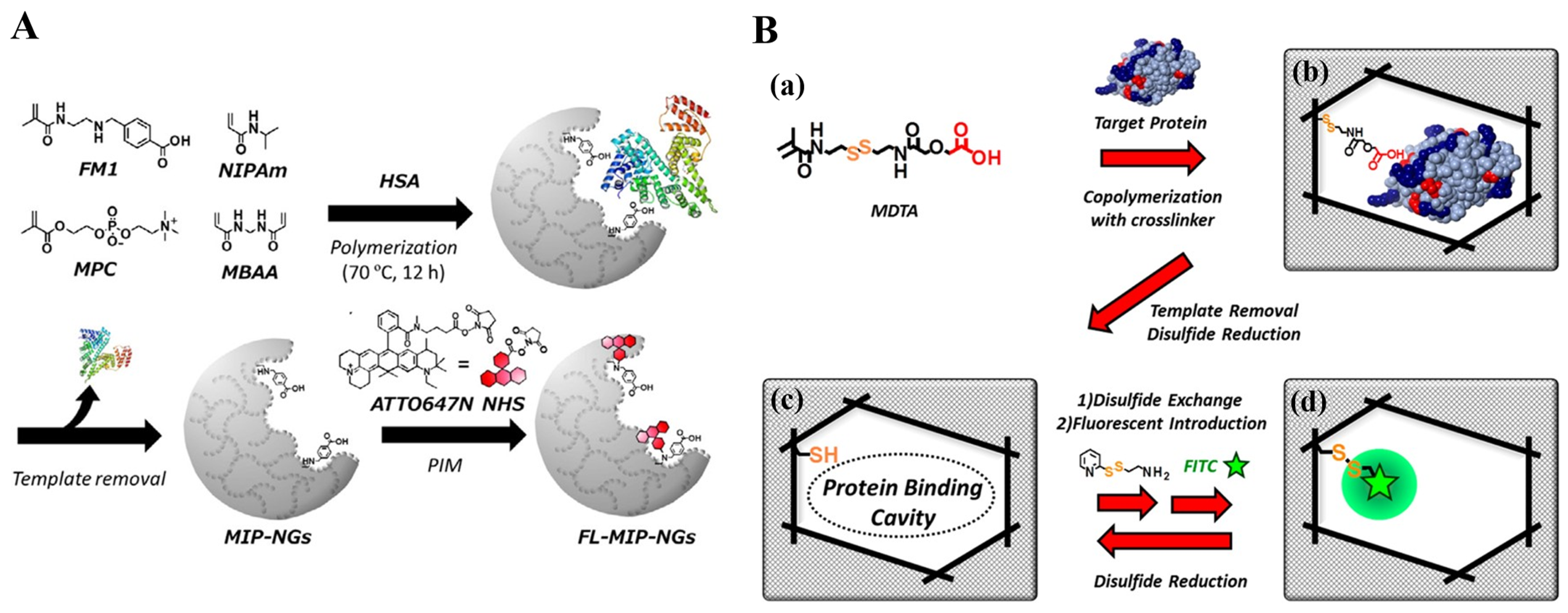
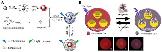
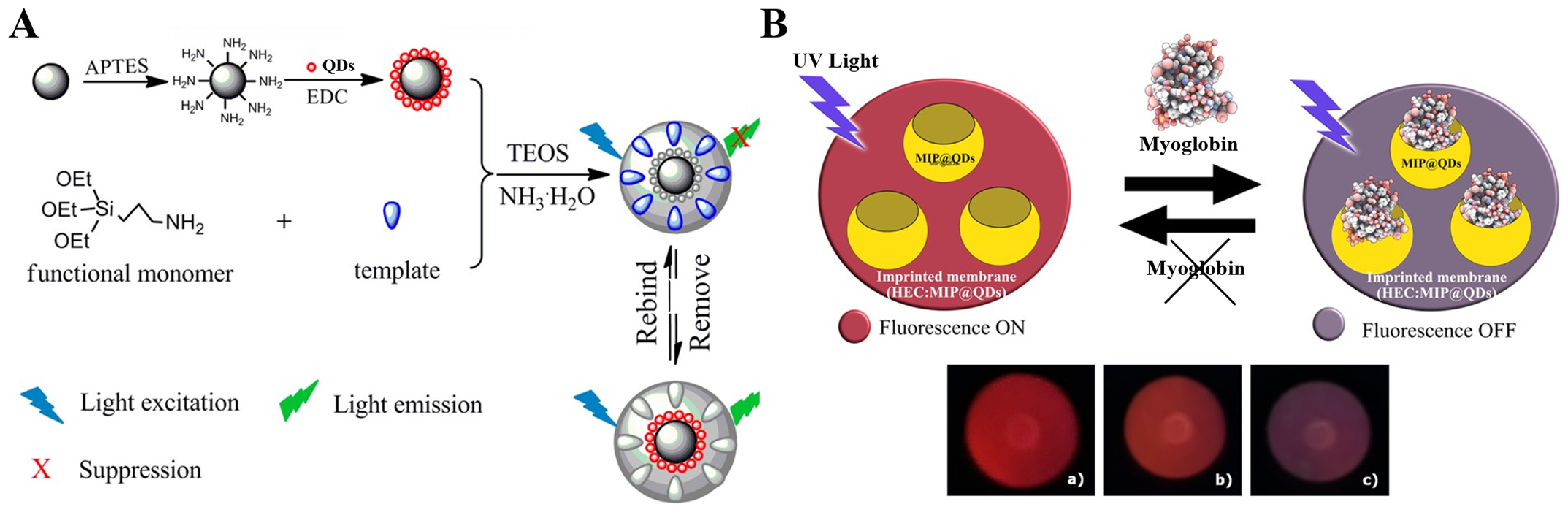
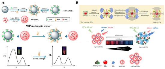
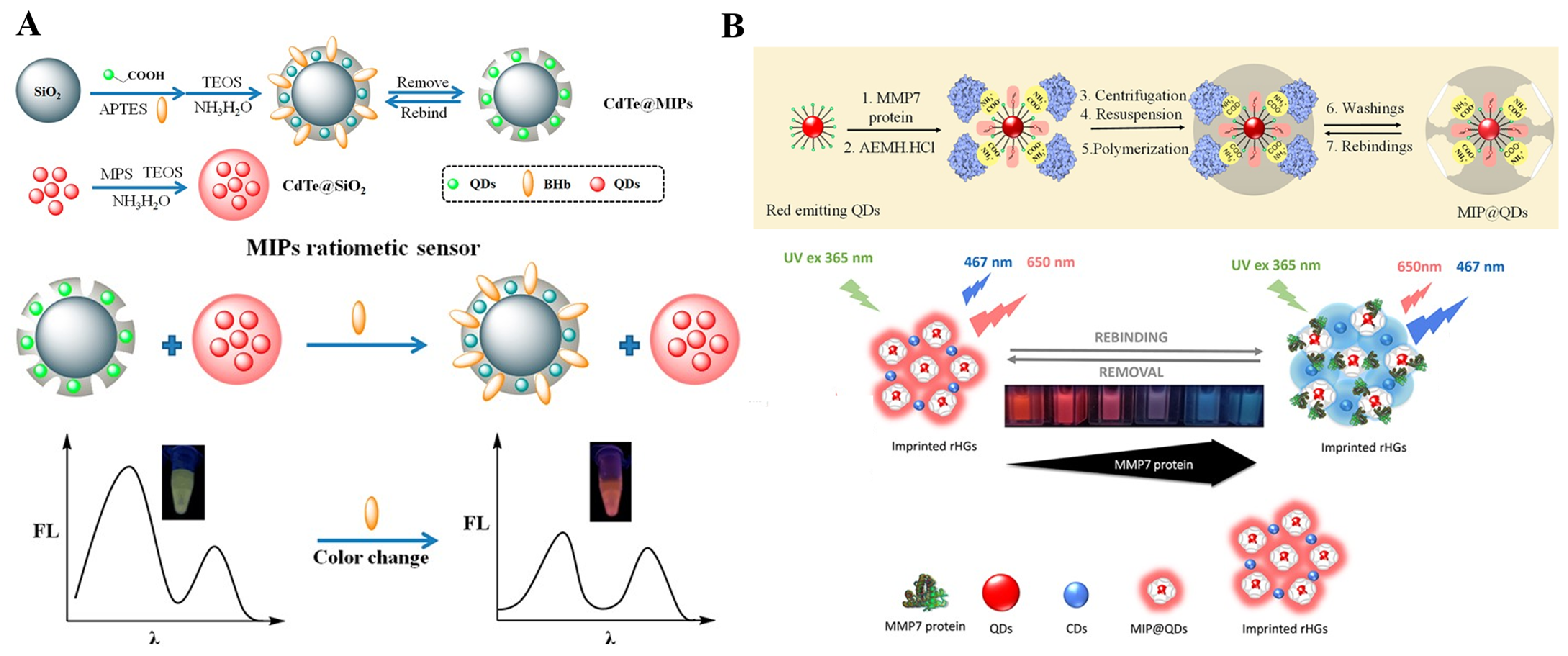
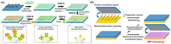
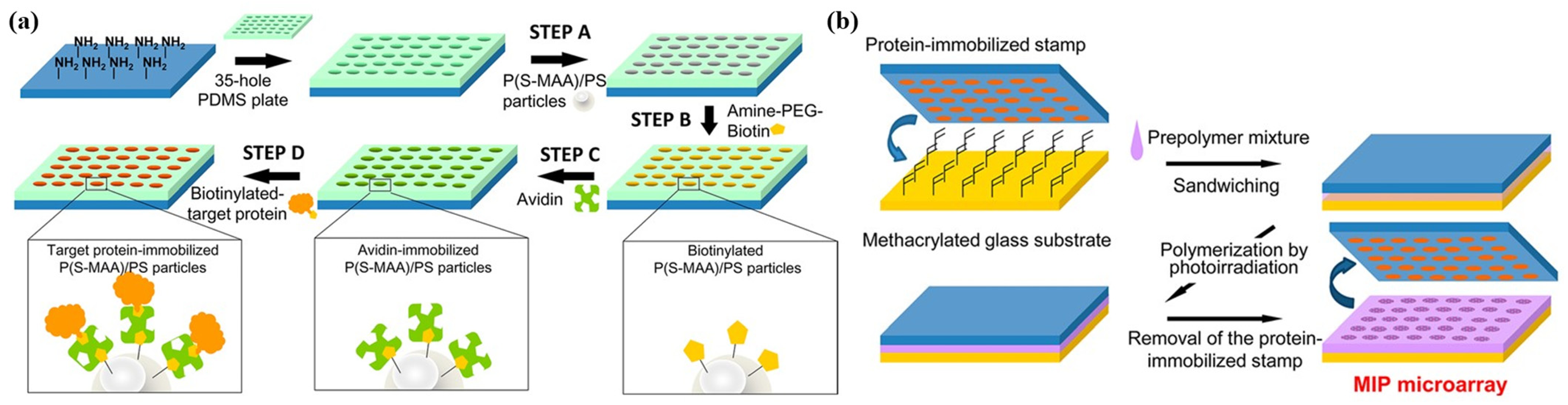
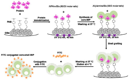
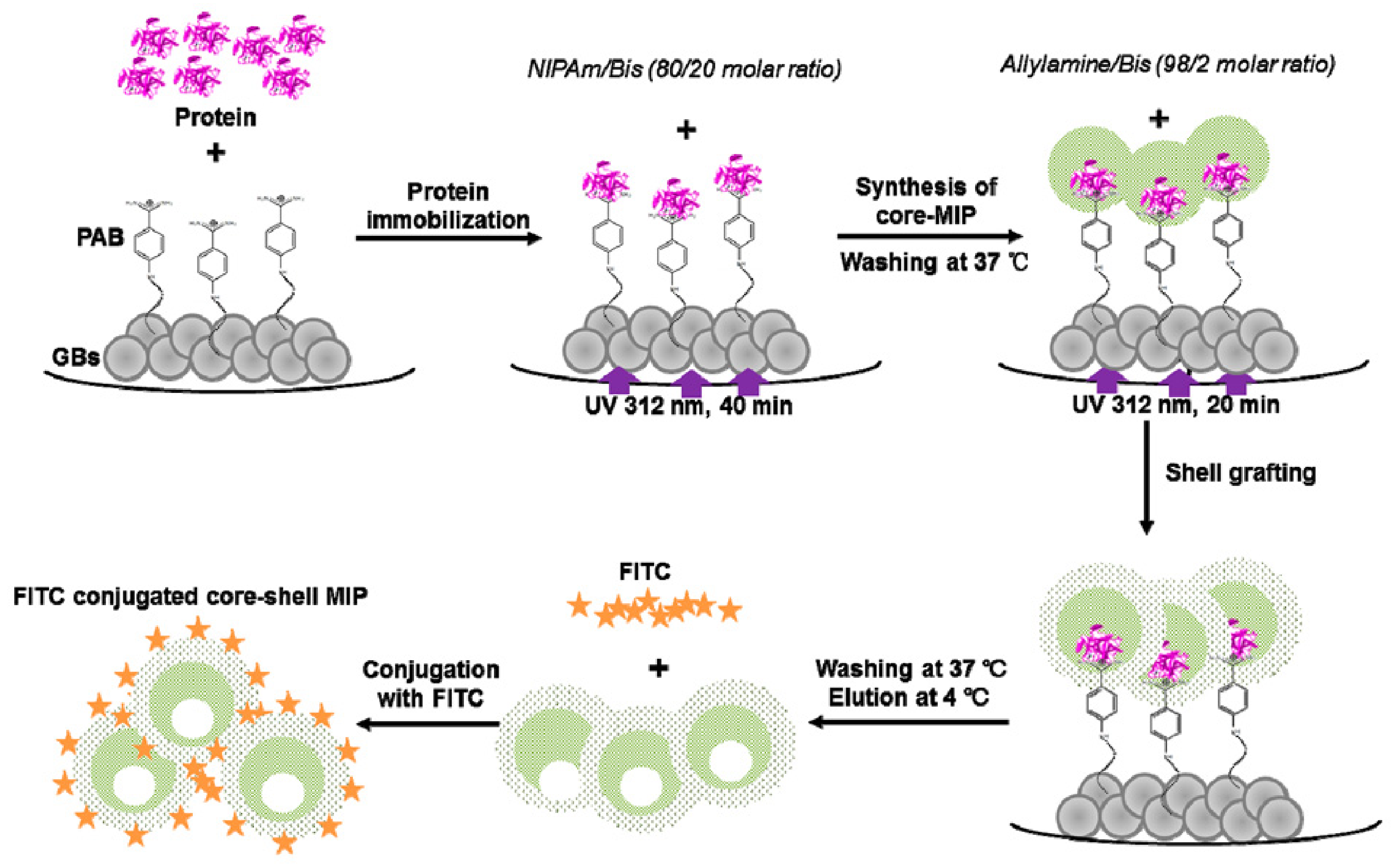
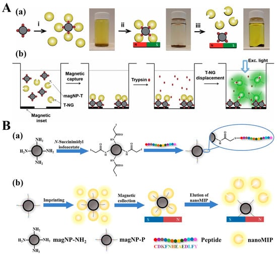
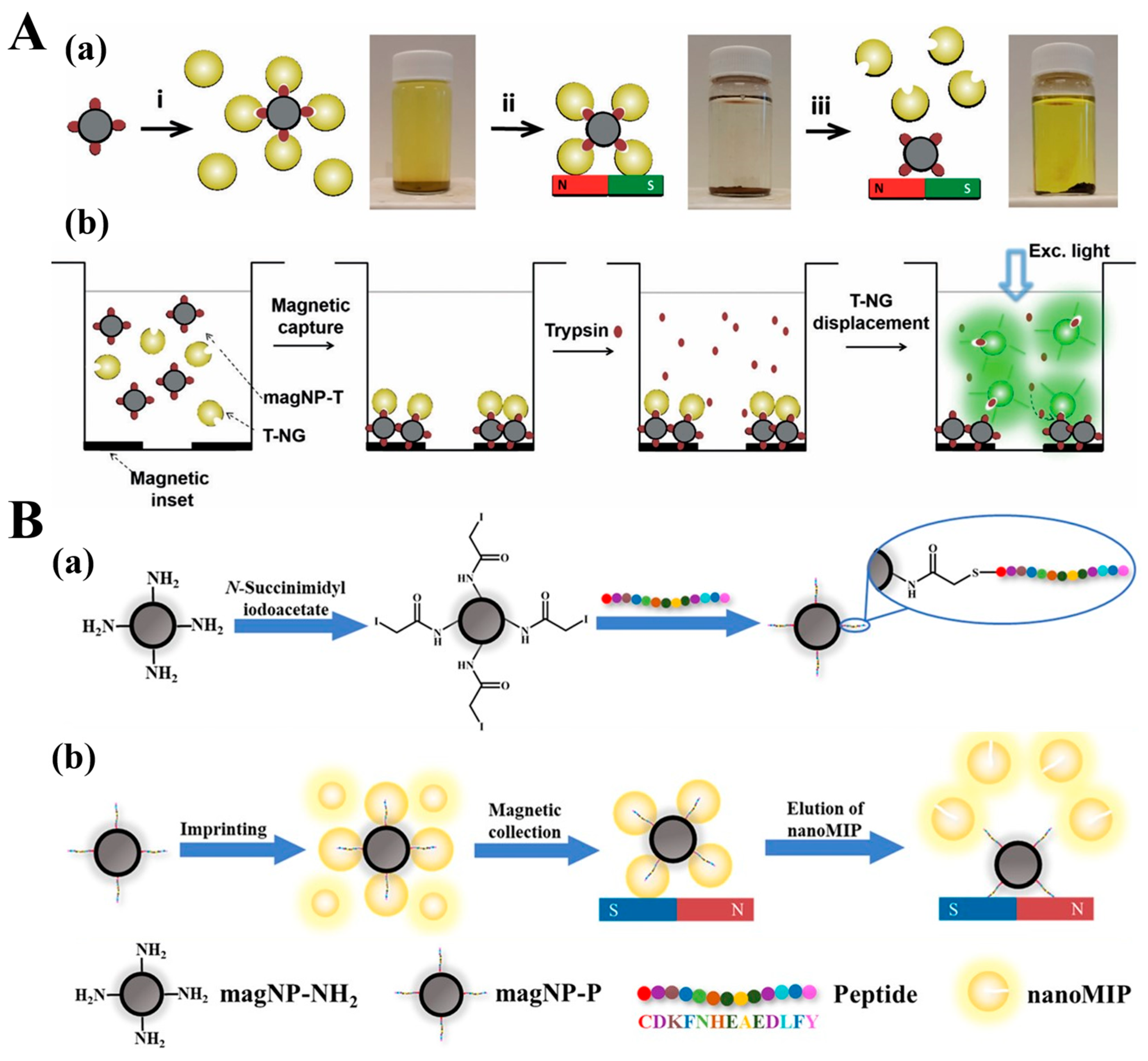
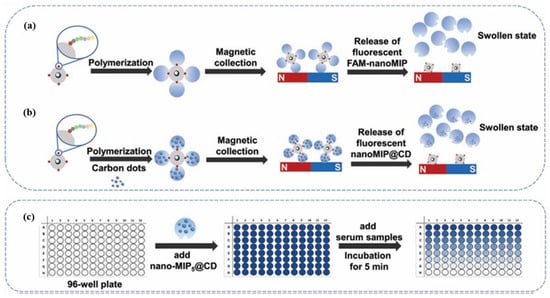
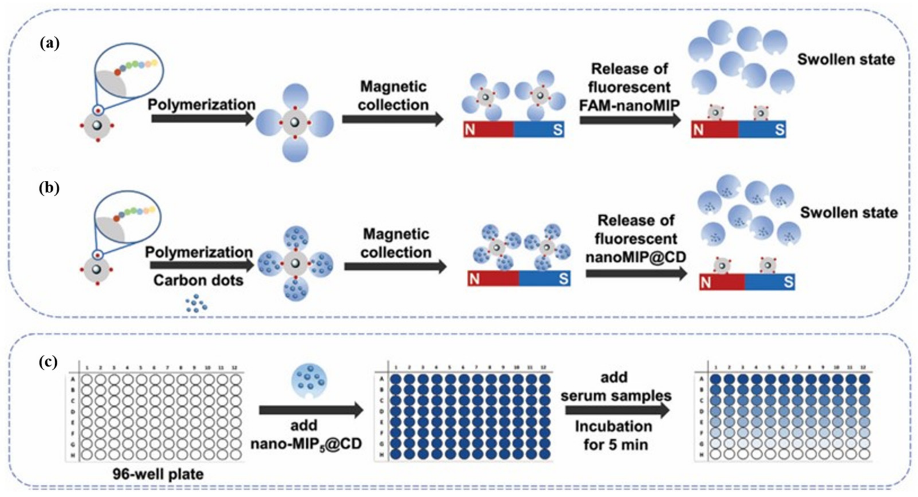
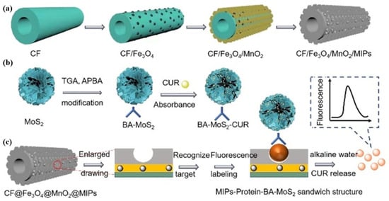

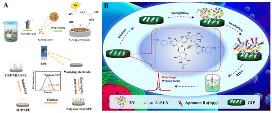
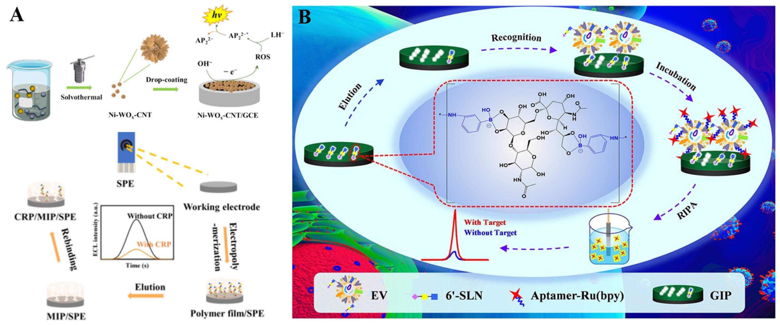
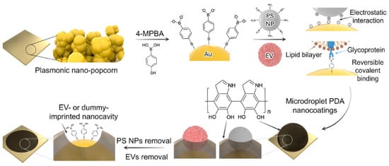
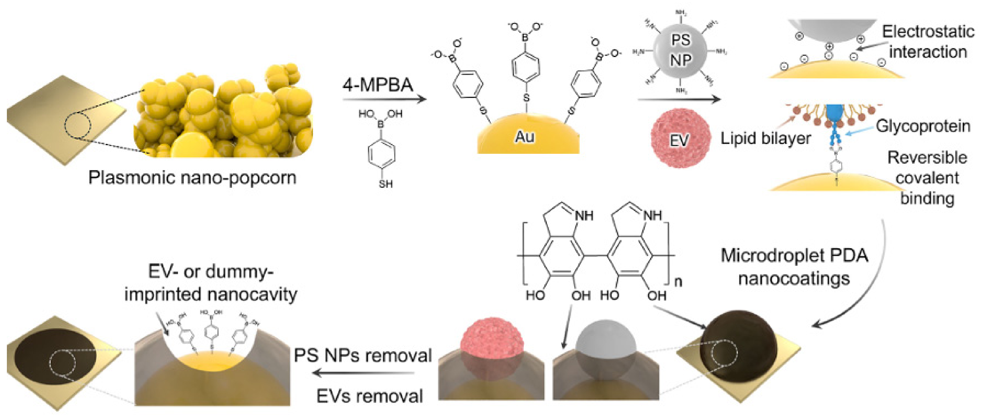
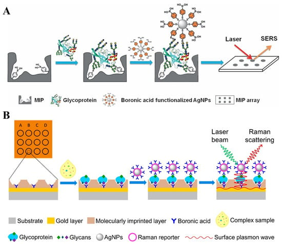
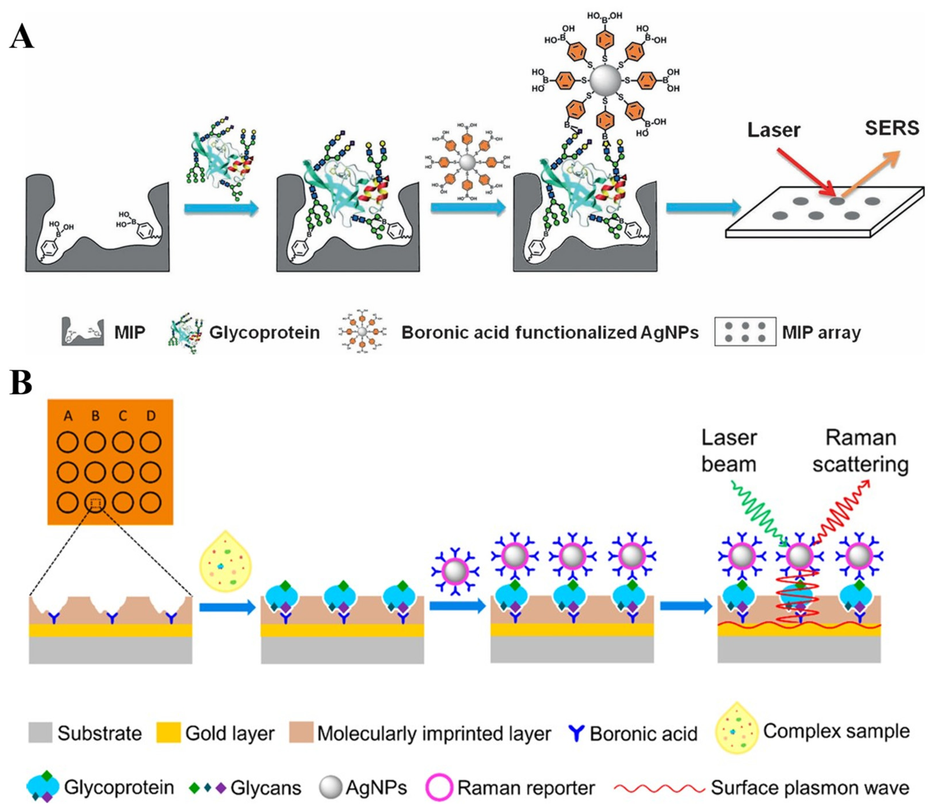
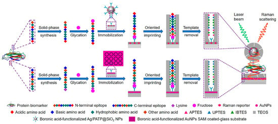
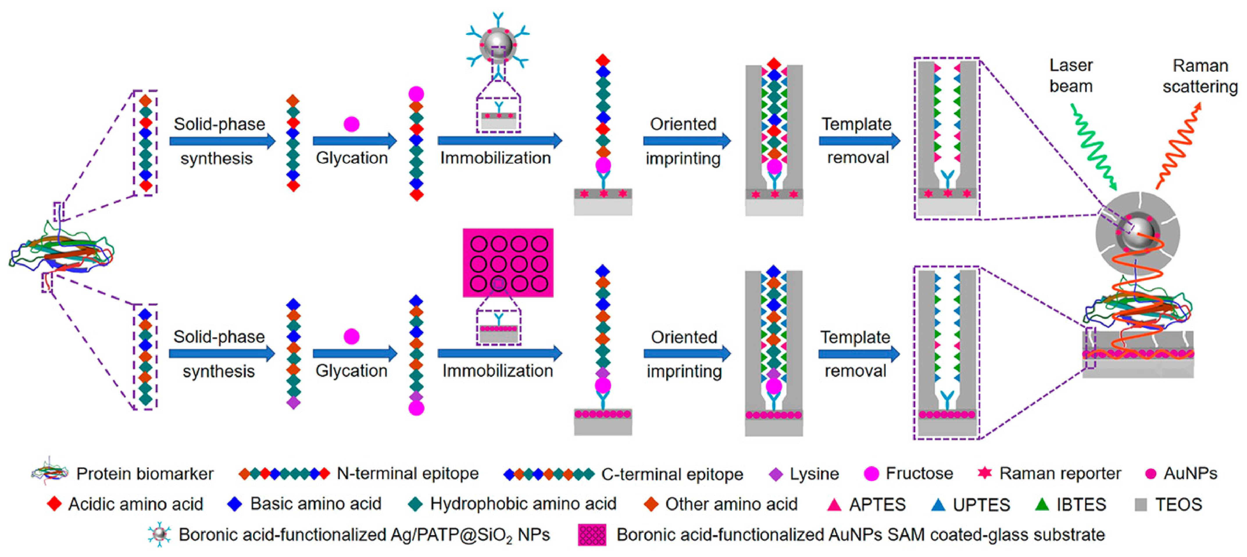
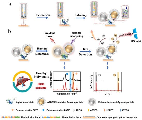
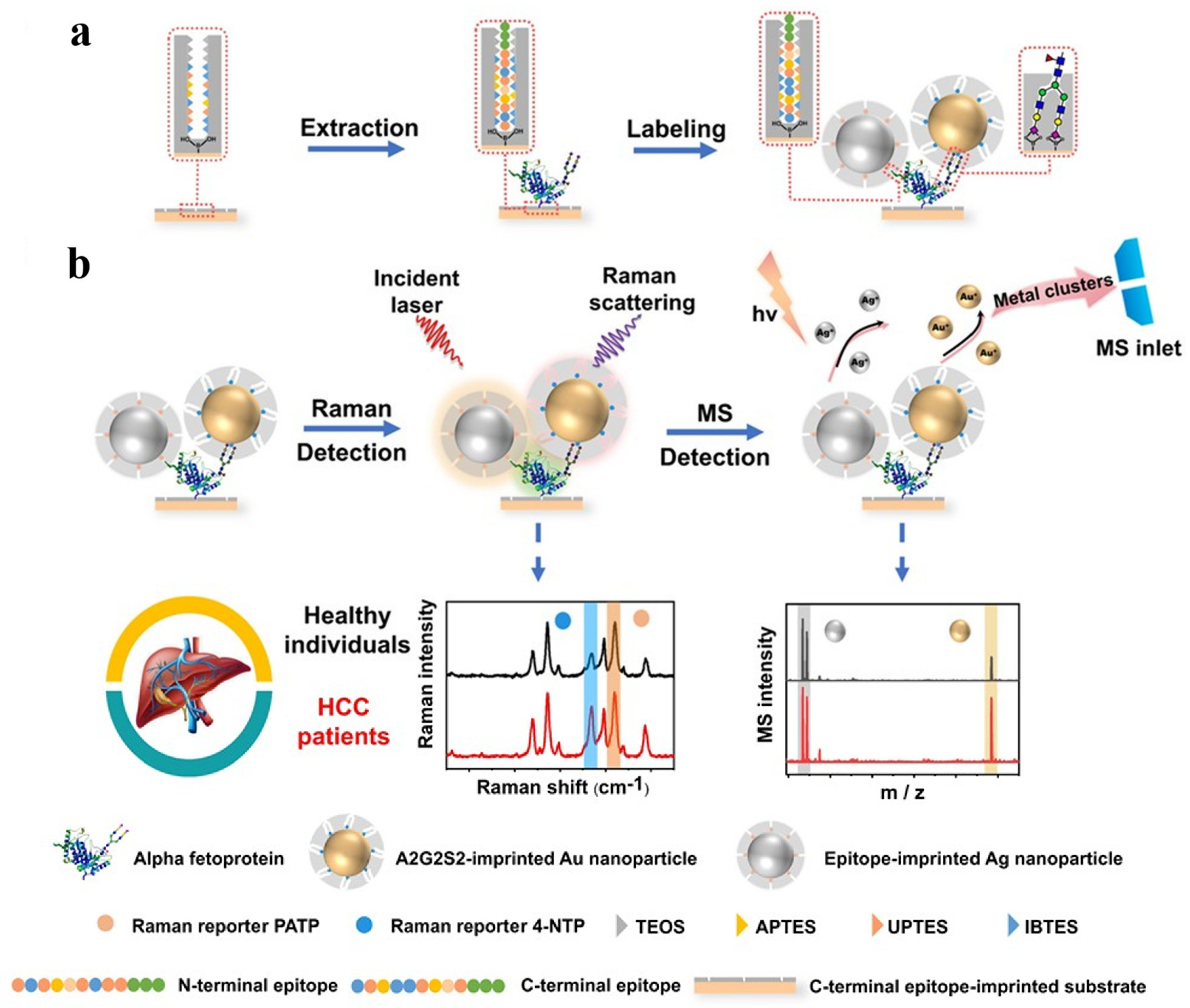
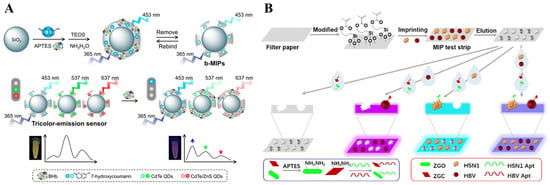
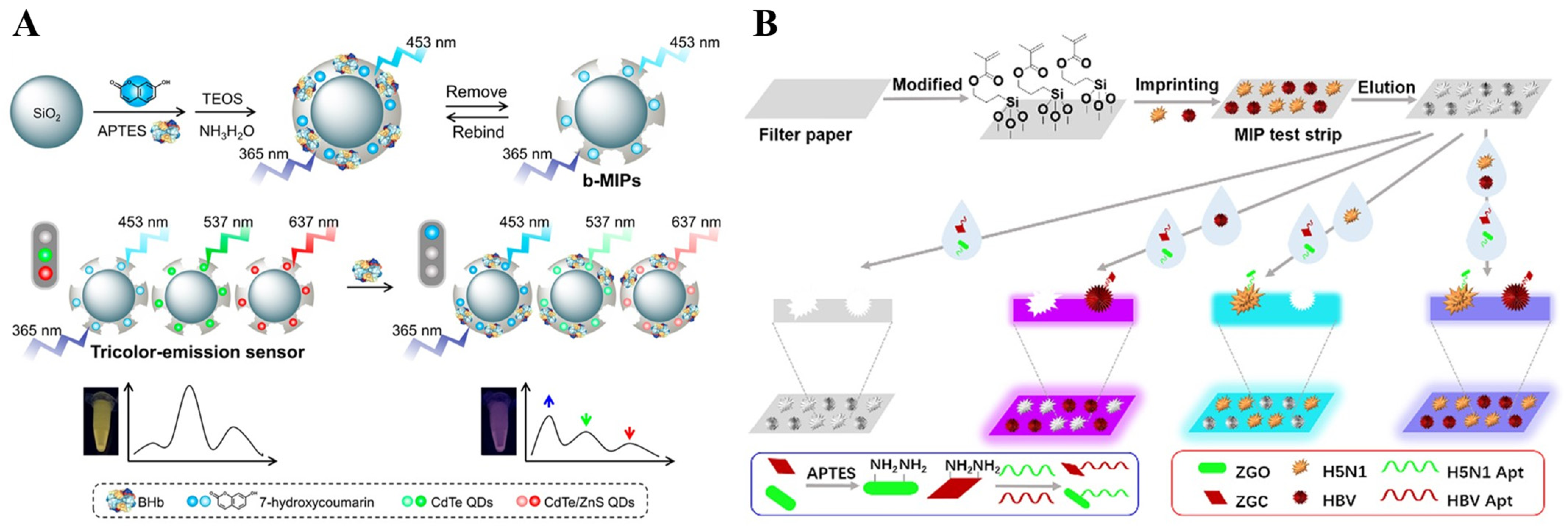
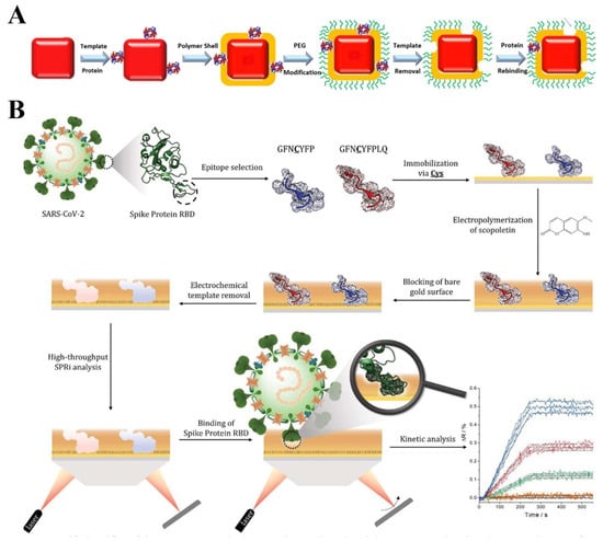
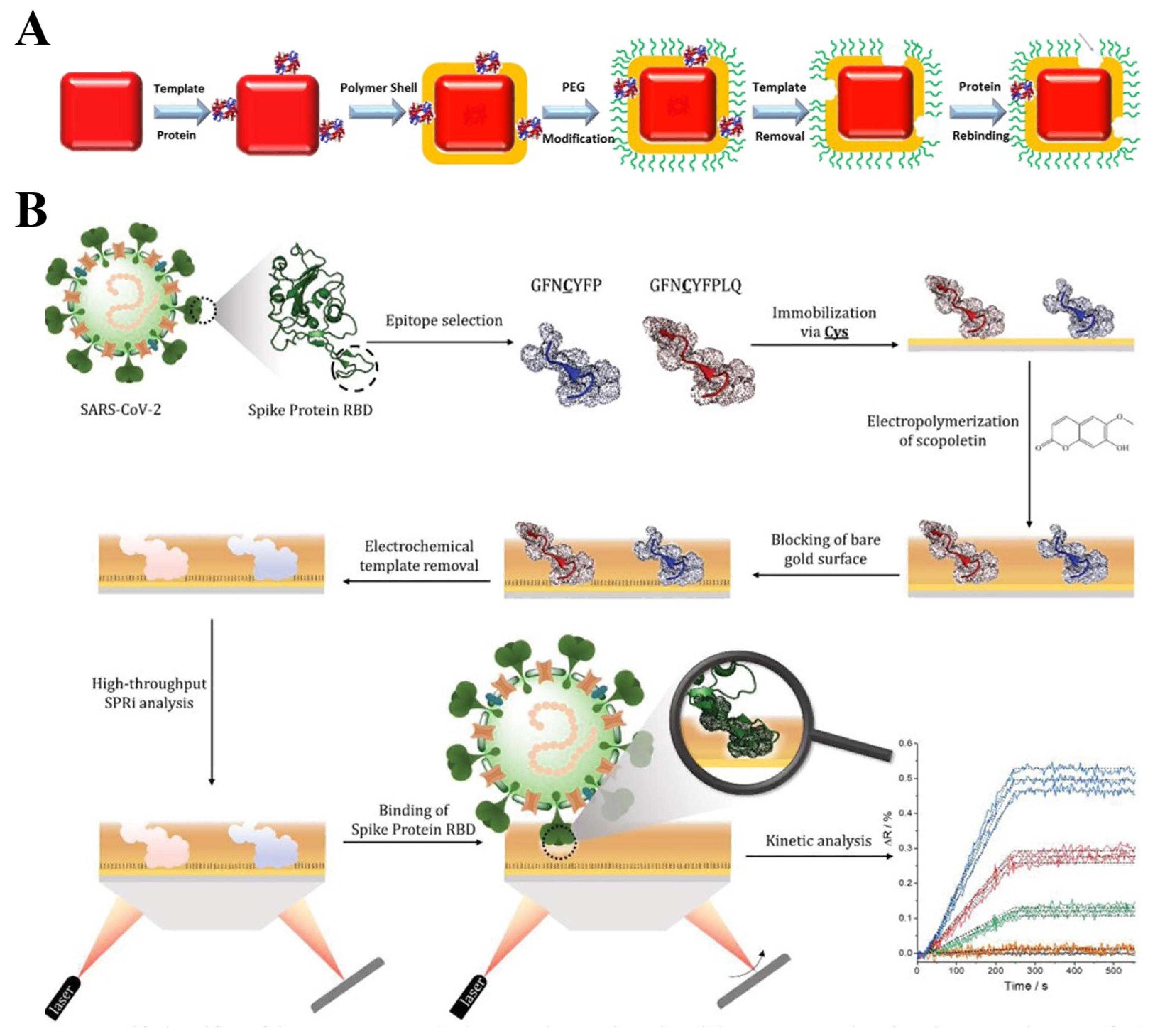
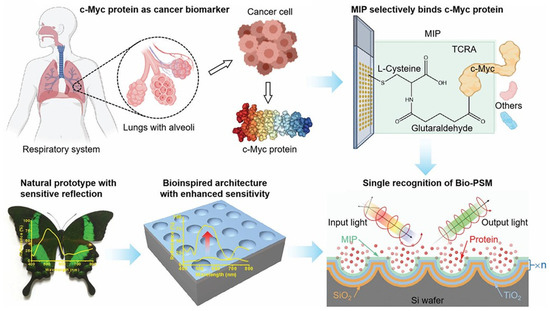
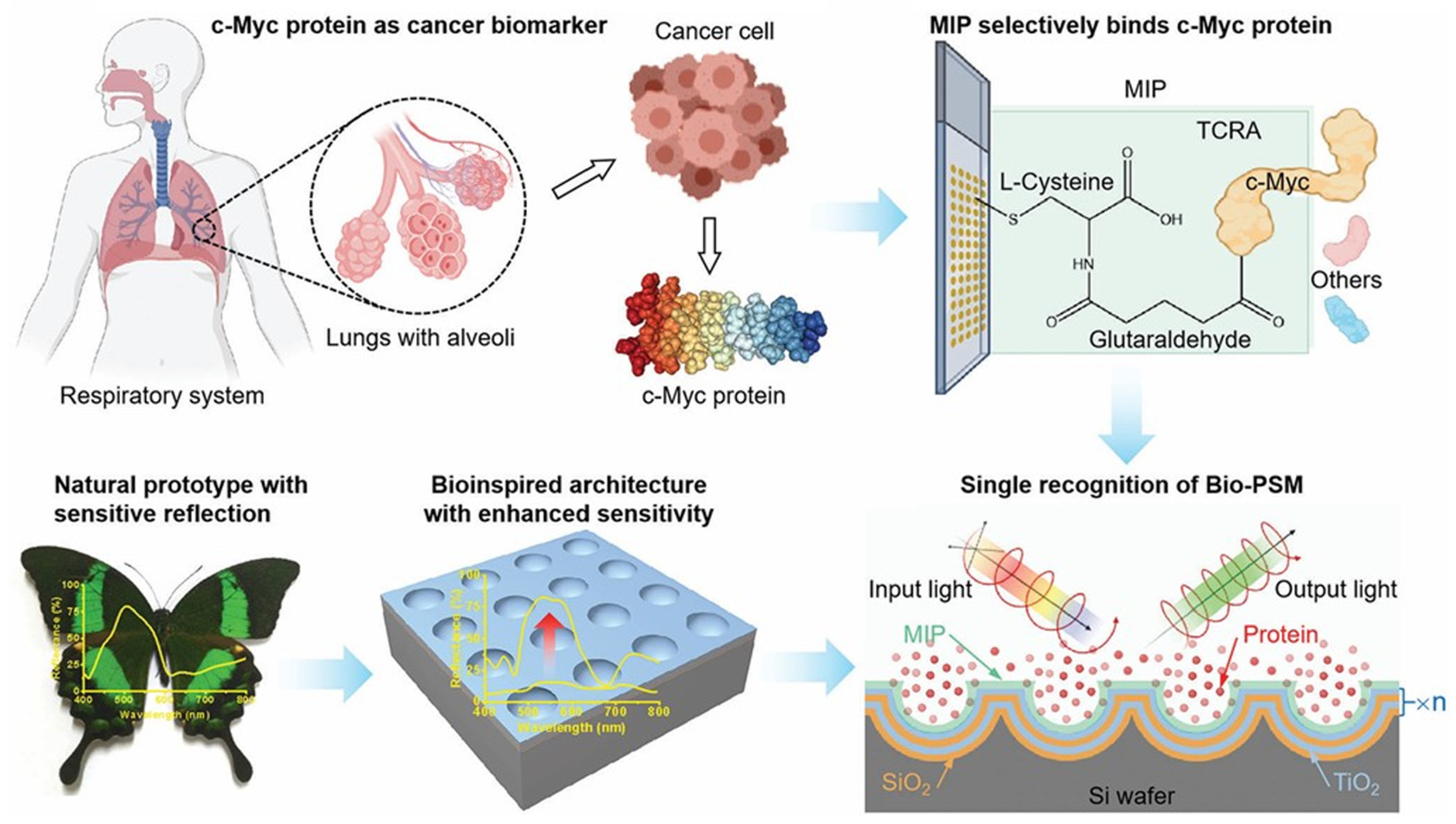
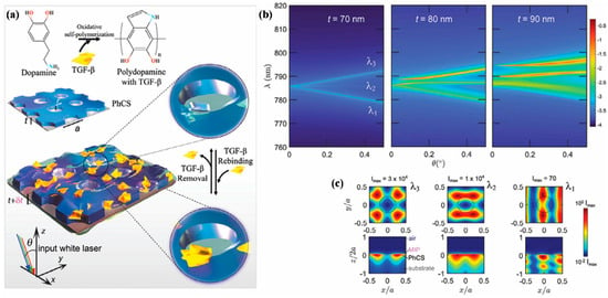
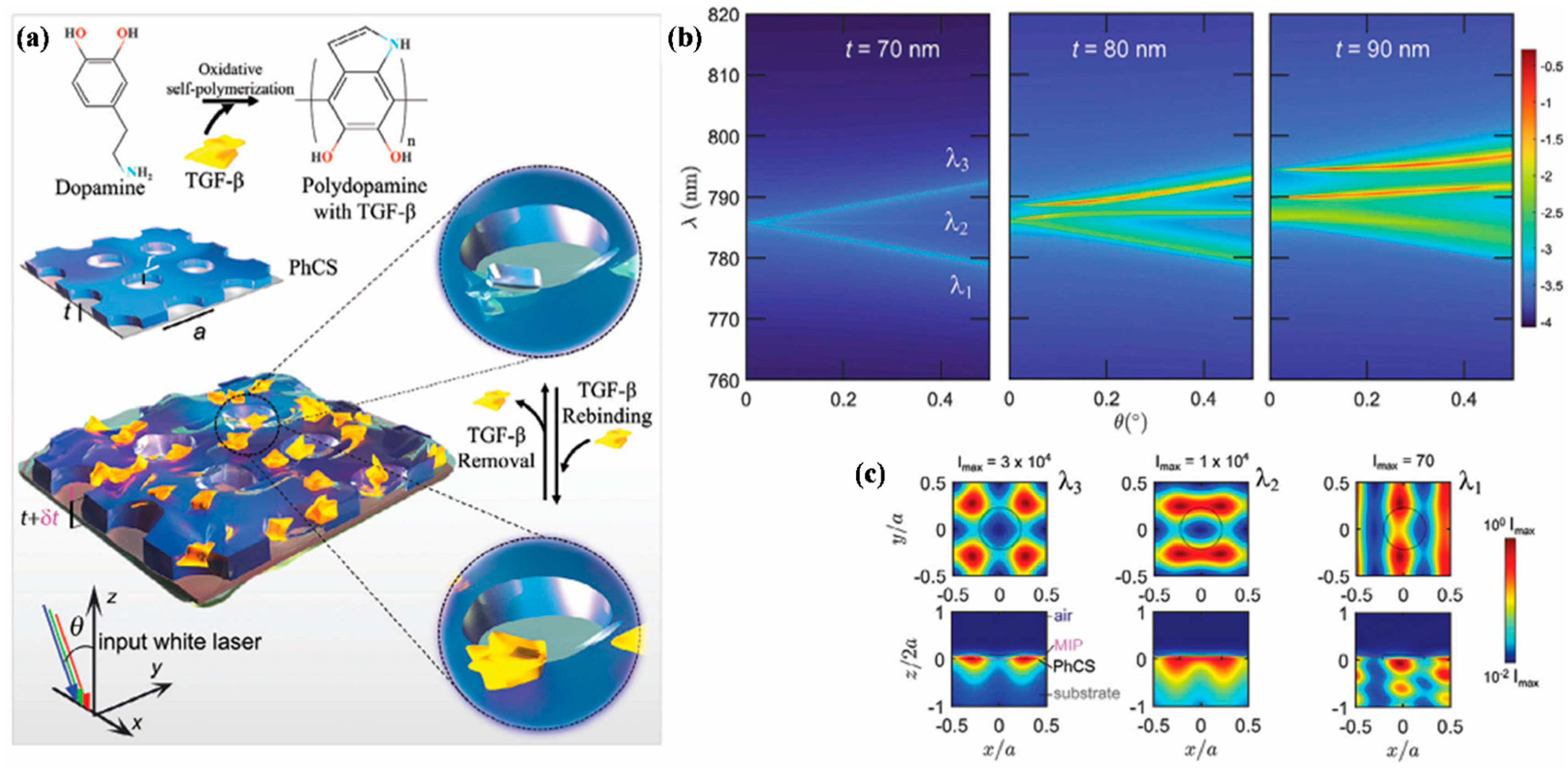

Disclaimer/Publisher’s Note: The statements, opinions and data contained in all publications are solely those of the individual author(s) and contributor(s) and not of MDPI and/or the editor(s). MDPI and/or the editor(s) disclaim responsibility for any injury to people or property resulting from any ideas, methods, instructions or products referred to in the content. |
© 2026 by the authors. Licensee MDPI, Basel, Switzerland. This article is an open access article distributed under the terms and conditions of the Creative Commons Attribution (CC BY) license.