


Abstract
Microwave sensing technology is rapidly advancing and increasingly finding its way into biomedical applications, promising significant improvements for medical care. Concurrently, the rise of artificial intelligence (AI) is enabling significant enhancements in the biomedical domain. Close scrutiny of the recent literature reveals intense activity in both fields, with particularly impactful outcomes deriving from the combined use of advanced microwave techniques and AI for biomedical monitoring. In this review, an up-to-date compilation, from the perspective of the authors, of the most significant works published on these topics in recent years is given, focusing on their integration and current challenges. With the objective of analyzing the current landscape, we survey and compare state-of-the-art biosensors and imaging systems at all healthcare levels, from outpatient contexts to specialized medical equipment and laboratory analysis tools. We also delve into the relevant applications of AI in medicine for processing microwave-derived data. As our core focus, we analyze the synergistic integration of AI in the design of microwave devices and the processing of the acquired data, which have shown notable performances, opening new avenues for compact, affordable, and multi-functional medical devices. We conclude by synthesizing the prevailing technical, algorithmic, and translational challenges that must be addressed to realize this potential.
1. Introduction
The current landscape of medicine and biomedical science is undergoing a profound transformation driven by the ever-increasing integration of sophisticated electronic technologies. This pervasive integration manifests across the entire healthcare continuum, fundamentally boosting diagnostic capabilities, therapeutic interventions, research methodologies, and patient management paradigms. The digitalization of health records has established the foundation for interconnected data ecosystems, while advanced diagnosis tools leverage complex electronics for unprecedented precision and early disease detection. Current hardware possibilities range from high-resolution digital imaging equipment and miniaturized wearable biosensors to point-of-care genetic sequencers [1,2]. This technological progression finds a particularly compelling expression in microwave sensing devices, which are garnering significant interest due to their unique combination of non-ionizing safety, deep-tissue penetration, and innate sensitivity to the dielectric properties of biological tissues, crucial factors for differentiating pathological states and monitoring physiological processes [3].
Furthermore, telemedicine platforms, powered by robust communication networks and secure data protocols, are dissolving geographical barriers to specialist access and enabling remote patient monitoring, thereby enhancing healthcare delivery efficiency and reach. This data-centric paradigm creates both an opportunity and a bottleneck for advanced analysis. The resulting proliferation of diverse biomedical data has spurred the development of advanced biosensors and AI-driven analysis tools to manage and interpret this information [4,5,6].
Concurrently, the convergence of microwave sensing systems with computational power, particularly through artificial intelligence (AI) techniques, is revolutionizing the analysis of the now massive biomedical datasets, thereby accelerating key aspects of modern medicine such as drug discovery, personalizing treatment strategies, and predicting patient outcomes with increasing accuracy. In recent years, in addition to the expected progress associated with AI, such as early detection of subtle pathologies through AI-driven medical image analysis or predictive analytics for optimized and individualized interventions, AI has also proved beneficial for improving the development of the microwave tools themselves. We are now witnessing the synergistic integration of hardware and software at all stages of design and implementation, with exceptional promise.
As a result, the significant potential of these technologies has led to intense research activity in the realms of AI techniques and microwave sensing applied to biomedicine, as well as their intersections. Indeed, at the time of writing this manuscript a quick search on Scopus database with “artificial”, “intelligence”, “electronic”, and “medical” as keywords yields 6181 results, from which over 77% are from the last 5 years. This prolific academic production demonstrates the interest and potential of these technologies, the boundaries between them becoming progressively blurred. Some recent reviews on these topics can be found. To name a few, we discovered [3] exclusively focused on microwaves in medicine; Ref. [7] focused on electromagnetic technologies for blood glucose level monitoring; Ref. [8] focused on microwave medical imaging techniques; Ref. [9] focused on different techniques and technologies for developing wearable healthcare devices; or Refs. [10,11], focused on different uses and applications of AI in medicine. However, to the best of our knowledge, no works have been published exploring the specific combination of microwave sensing technology and AI for biomedical monitoring and imaging, despite their evident relationship. This gap is notable because microwave sensing presents unique signal processing challenges (e.g., low signal-to-noise ratio, complex dielectric interactions) where AI techniques can be particularly transformative.
In these circumstances, an updated review of the main literature focused on this intersection seems of high relevance. Not aiming at an extensive compilation of the vast available literature, this narrative review has three core objectives:
- (1)
- To survey and critically compare microwave-based devices for physiological monitoring and imaging (Section 2).
- (2)
- To review AI paradigms suited for analyzing microwave-derived data (Section 3).
- (3)
- To analyze the integration of AI at the system level, from algorithm design to embedded implementation (Section 4).
We conclude by synthesizing the key limitations, challenges, and future directions at this interdisciplinary frontier (Section 5). For clarity, this review focuses specifically on diagnostic monitoring and imaging applications, excluding therapeutic microwave uses and implantable systems.
2. Microwave Sensing and Imaging Technology in Medicine
Electronics underpin almost all modern fields, and medicine is no exception. From screening instruments to therapy tools, the advances in medicine have been closely linked to advances in technologies over the last several decades. The landscape of electronic biosensors is broadly divided between general health monitoring [12,13,14] and the management of specific chronic conditions [15], including behavioral and psychiatric disorders [16,17]. For general wellness, dominant technologies include hydrogel-based electrodes for biopotentials such as electrocardiography (ECG) or electroencephalography (EEG) [18], wearable iontronic and triboelectric sensors for blood pressure monitoring [19,20], and optoelectronic systems like photoplethysmography (PPG) for vital signs [21]. These same modalities, along with measures of electrodermal activity (EDA) and heart rate variability (HRV), are leveraged for disease-specific management, from guiding rehabilitation processes [22] to adjusting therapies for behavioral disorders [23,24,25] and screening for depression and anxiety.
While this established ecosystem excels at measuring surface-level electrical, optical, and chemical signals, it provides limited direct insight into the internal dielectric properties of tissues. This fundamental limitation defines a compelling niche for microwave-based sensing, which probes tissue composition and deep-tissue physiology to offer unique, complementary data on conditions like blood composition analysis, deep tissue health or detection of specific cells or nanoparticles, as explored in the following subsections. The unique attributes of microwave sensing, including its contactless operation, sensitivity to deep-tissue hydration and composition, and ability to monitor physiological movements, are creating new paradigms for non-invasive, continuous health monitoring. A comparative summary of representative microwave-based devices and systems discussed in this section is provided in Table 1. It should be noted that a comprehensive review of the most significant works on these topics would be lengthy and unwieldy. Instead, we collect a representative selection of proposals and applications drawing significant interest within the research community.
2.1. Biomarkers Monitoring
Biomarkers monitoring, with a general emphasis on wearable and outpatient technologies, represents an intensely active field of research, motivated by the generally low-cost nature of implementations and a wide variety of potential applications. This domain has been bolstered by advancements in fabrication techniques, such as flexible and textile-based electronics [26,27,28]. While extensive reviews cover the broader landscape of wearable biosensors [9,29,30], this section focuses specifically on the emerging potential of microwave-based systems. The unique attributes of microwave sensing, including its contactless operation, sensitivity to deep-tissue hydration and composition, and ability to monitor physiological movements, are creating new paradigms for non-invasive, continuous health monitoring.
During the last decades we have witnessed considerable advancements in the development of sensors and instruments for non-invasive measurement of the concentration or presence of certain solutes or compounds, whether in solutions or blood. These are of interest in the biomedical realm, particularly in the measurement of glucose concentration [31,32]. Such sensors find enormous uptake in the management of diabetes and its complications, since they ideally allow for continuous non-invasive monitoring of patients’ blood glucose level, thereby facilitating the right treatment and actions at opportune times [33].
Based on microwave technology, the fundamentals of these sensors were studied in [34]. Their operating principle relies on the influence of glucose on the dielectric permittivity of the medium. The dielectric permittivity is a characteristic of each material that depends on its chemical composition, and that determines the strength of the interaction of the material with electromagnetic fields over a wide range of frequencies. It has been shown repeatedly that, for liquid solutions containing glucose (such as water solutions or blood), a change in the glucose concentration entails a change in the dielectric permittivity of the solution [35,36,37,38,39]. Consequently, dielectric permittivity sensors, which have non-invasive capabilities, could potentially act as practical glucose concentration sensors. These sensors are usually made with planar microwave resonators, the response of which depends on the dielectric permittivity in their vicinity. Functional devices were thoroughly analyzed from a general point of view in [40,41].
Following these principles, numerous works have demonstrated glucose concentration measurement capabilities through different metrics derived from the response of the sensor [42], such as the resonance frequency (fr) [43], the insertion/return losses [44], the phase of the scattering parameters [45], or the unloaded quality factor (Qu) [46]. For example, the glucose measurement capabilities of Qu-based sensors with ternary blood plasma solutions were assessed in [47], a certified biocompatible implementation was proposed in [48], and clinical trials were conducted in [49]. In terms of fr-based approaches, wireless implementations were proposed in [50,51], a stepped impedance resonator was used in [52], and a dual band split ring resonator was tested in [53] with good resolution. A return loss-based dumbbell defective ground resonator was analyzed in [54], while a plasmonic microwave resonator was assessed with human trials in [55]. The measurement concept of these sensors is illustrated in Figure 1.
Figure 1.
Measurement concept for planar microwave glucose sensors, where the interaction between the electric field lines (red arrows) and the liquid sample (in blue) is seen (VNA = vector network analyzer). Reprinted from [47] under CC license.
In addition to glucose concentration sensing, this technology has been applied to other biosensing scenarios. A planar microwave sensor for the detection of sweat and urine leakage in underwear was studied in [56]. Complementary split-ring resonators were used as blood urea concentration sensors for enhanced dialysis adequacy [57,58]. The denaturation of bovine serum albumin protein, due to the action of urea, was detected through a differential microfluidic sensor based on slot resonators [59]. Lactate monitoring, of interest for hyperlactatemia and aerobic fitness assessment, was achieved in [60,61]. A substrate-integrated waveguide resonator was combined with the lipase enzyme as a bioreceptor for successful estimation of triglycerides mixture in blood serum for heart health monitoring [62]. A sensor based on a split-ring resonator for detecting several types of amino acids for cell cultures and nutrition studies was shown in [63]. Skin hydration was monitored with a resonant sensing tag [64]. Continued progress in microwave biosensors has permitted the accurate detection of nanoparticles [65,66,67], proteins [68], emerging contaminants [69], and bacteria [70,71,72], all of high biomedical interest. Measurement of biosignals and vital signs through microwave devices has also been achieved [50], such as heart beat, in several configurations [73,74], among others [75,76].
Despite the latest advancements, these microwave biosensors will still face several challenges in the near future. Firstly, the high detection standards usually required in medicine translate to considerably high device sensitivities. Ongoing research is starting to yield impressive sensitivity boosting strategies through different approaches, such as coupled-resonator sensors [77,78,79] or so-called lossy sensors [80,81,82], that may lead to unprecedented biosensing capabilities. Secondly, in a complex environment such as the biomedical one, these sensors will have to demonstrate selectivity or specificity high enough for robust, trustable operation, not only against multi-component variations [83], but also against the influence of external parameters such as temperature [84,85] or pressure [86]. This is indeed a challenging issue considering that the effective permittivity is a global parameter for the medium. In this regard, recent progress has been made with differential sensing [87], multi-frequency multi-phenomena sensing [61,88], or even multi-parameter simultaneous sensing [89]. The use of artificial intelligence techniques is expected to have a particularly pronounced impact here, as will be discussed in Section 4. Also, other alternative technologies are under current research, such as glucose measurement from sweat [90,91] by means of an enzymatic graphene sponge [92] or on gel electrolyte [93], for instance. Glucose measurement from saliva has also been investigated [94] by means of optical [95], electrochemical [96], or enzymatic techniques [97,98].
2.2. Microbiology- and Cell Biology-Related Equipment
Microbiology and cell biology testing have also been of interest for researchers developing new sensors. A high selectivity can be obtained by capturing single cells using AC dielectrophoresis [99], allowing for the study of intracellular properties. Although cell microscopy using chemical stains or fluorescent dyes to study and sort cells presents several advantages (i.e., high specificity, sensitivity, and efficiency), the required equipment is generally expensive, and the used chemicals may interact with the biomolecules under test, affecting the extracted parameters.
The use of microwave sensing removes the need of chemicals or solutions that can have an impact on the cells under study. Different approaches have been presented over the years, such the integration of microstrip lines and capacitive gaps to determine the dielectric properties of the sample under test [100,101,102]. More recently, significant advances in microwave circuit integration with microfluidics have allowed probing biological samples at microwave and millimeter wave frequencies. For example, in [103], a microfluidic biosensor was implemented using a microwave SIW cavity resonator with a resonant frequency of 13.34 GHz. This sensor can detect fibroblast cells by measuring the frequency shift of the cavity, as shown in Figure 2. Additionally, in [104], a design using an interdigitated capacitor loaded coplanar waveguide was presented to differentiate dead and living cancer cells by determining their dielectric properties at 40 GHz. This integration can even allow scientists to study individual cells as conducted in [105,106] where the principle of label-free cell probing with microwaves in a microfluidic channel is described. A further comparison of microwave and conventional cellular analysis techniques is presented in [107]. The use of these microwave sensors increases the capability of cell differentiation given their morphological similarities.
Figure 2.
Design of a SIW cavity microfluidic biosensor for fibroblast cells detection while flowing through the cavity. Reprinted from [103] under CC license.
Other successful examples of integration between microfluidics and microwave sensing strategies make use of coplanar capacitive gaps [108], and coplanar waveguides in frequency ranges between 40 MHz and 40 GHz [109,110,111]. As the frequency of operation of these sensors increase, a higher resolution can be achieved, allowing the characterization of the cell nucleus [112,113]. The successful implementation of microwave sensors allowing cell characterization to a nucleus level allows researchers to better identify and differentiate these cells in details that were not imaginable years ago.
2.3. Medical Imaging
Medical imaging equipment consists of non-invasive tools used to visualize the interior of the human body, either for clinical analysis or to detect abnormalities. Traditional imaging methods that provide information about tissues, such as X-rays, computed tomography (CT), magnetic resonance imaging (MRI), or diagnostic echography, among others, have evolved over time, improving their capabilities [114]. However, these methods still present various limitations, including their complexity, high cost due to maintenance and implementation requirements, bulkiness, portability challenges, low sensitivity to tissue changes, and exposure to harmful radiation and nephrotoxic contrast agents in certain cases. On the other hand, they offer the significant advantage of providing high resolution and precision in images, creating a strong demand for medical imaging systems that are safe, reliable, and cost-effective [115,116,117]. In addition to the above-mentioned limitations, in systems based on ionizing radiation, the permissible exposure dose is also limited. Consequently, significant efforts have been devoted to finding a reliable medical imaging technology that utilizes non-ionizing radiation while remaining user-friendly and cost-effective. One such alternative technology for image reconstruction is based on electromagnetic energy, specifically in the microwave frequency range [116].
Microwave imaging (MWI) is a non-invasive and non-destructive technique aimed at detecting materials or targets in order to obtain their physical properties and/or information about the conditions of the structures under study [118]. These techniques typically rely on near-field measurement systems capable of capturing the electromagnetic field resulting from the interaction between incident waves and the materials or targets in question [118]. Medical MWI uses electromagnetic waves, typically in the range of 1 to 10 GHz. The frequency range of these systems is usually selected to ensure appropriate signal penetration depth and spatial resolution [119,120]. MWI is an alternative to traditional imaging methods due to its numerous and significant advantages, such as the use of low-power, non-ionizing signals, making it a low-risk health method that can be safely repeated more frequently compared to conventional imaging systems [121]. Additionally, it is a cost-effective technique, since the equipment used is generally inexpensive and can even be made portable, opening the door to outpatient usage.
However, MWI techniques also present some disadvantages and technical challenges, such as limited spatial resolution [115,116]. Medical MWI systems comprise a hardware component responsible for data acquisition and a software component to process the data. Over the years, research on these medical MWI systems has been motivated by its significant potential in monitoring various pathological conditions [115]. Examples of applications of these systems include breast cancer detection [122,123,124], monitoring of bone fractures [125,126], portable knee imaging systems [127], neck tumor [128] and skin tumor [129] detection, human torso scanners [130], lung cancer detection [124,131], human heartbeat detection systems [132,133], congestive heart failure detection [134], dynamic imaging for cardiovascular systems [135], muscle tear imaging [136], and detection of brain abnormalities such as strokes [137,138], brain tumors [124,139], or intracranial hemorrhages [140,141], among others.
The basic principle of microwave medical imaging is the contrast in tissues due to their different dielectric constants, which is used to reconstruct the backscattered signals and to generate the corresponding images by means of processing algorithms [142]. These algorithms vary depending on whether the system is tomographic or radar-based [115]. Different tissues through which the electromagnetic signals travel exhibit varying dielectric properties. Due to these differences, image reconstruction algorithms can display the tissues the signal travelled through and even locate specific elements within the body, such as malignant or tumor tissues, which tend to scatter signals more intensely than healthy tissues [115]. This is a result of healthy and malignant biological tissues being morphologically distinct, a direct consequence of variations in total cell water content and changes in cell membrane properties [120].
In the literature, three methods of medical MWI systems have been explored, conventionally classified as passive, hybrid, and active methods. The passive method uses a radiometry device to measure temperature differences between healthy and malignant tissues, since malignant tissues exhibit higher temperatures compared to surrounding normal tissues. The hybrid method, also known as the thermoacoustic method, employs microwave sensors and ultrasonic transducers to detect malignant tissues. In this case, tissues are illuminated by electromagnetic waves at microwave frequencies, which are absorbed by the tissue. Malignant tissues absorb more energy than normal tissues and emit stronger acoustic waves due to their higher electrical conductivity compared to surrounding normal tissues. An ultrasonic transducer then measures the reflected acoustic signals and processes them to construct an image. The active method uses microwave signals to illuminate tissues, and the variation in dielectric properties between normal and malignant tissues is measured within a specific microwave frequency range. The backscattered signals from the tissues are estimated to construct an image indicating the presence of malignant tissue based on the reflected signals, which are stronger than those from healthy tissues [143].
Techniques for microwave imaging using active methods can be divided into two main groups: microwave tomography and radar-based imaging. Microwave tomography applies microwave signals to illuminate the tissue under study and provides quantitative information about the dielectric properties of the tissue to identify malignant tissues. This is achieved by generating a permittivity and conductivity map through the inversion of these signals. This method typically employs iterative image reconstruction algorithms and can be represented as a nonlinear inverse problem, which is time-consuming due to the complexity of the computational process, requiring significant computational resources to obtain a solution. If these inverse scattering approaches are poorly formulated, the results may lack uniqueness, necessitating regularization to achieve convergence toward a meaningful solution. Images of the tissue under study can be constructed from microwave data using inverse scattering algorithms, which estimate the constitutive parameters of tissues by analyzing absorbed and reflected microwave signals [143,144]. Examples of medical imaging systems using microwave tomography include breast cancer detection [145,146,147,148], stroke detection [137,149], brain imaging [150], shoulder injury detection [151], lung and cardiovascular activity measurement [152], and axillary lymph node detection [153], among others. Some of these systems are illustrated in Figure 3.
Figure 3.
Examples of microwave tomography systems: (a) system for cerebrovascular accidents detection, reprinted from [137]; (b) brain imaging system, reprinted from [150]; and (c) system for assessing lung and cardiovascular activity, reprinted from [152]. All images are reprinted under CC license.
Regarding radar-based techniques, these ones utilize an external microwave source to illuminate the object of interest with wideband signals. The reflected signals from the object are then used to detect differences in dielectric properties between normal and malignant tissues, enabling the identification and localization of potential malignant tissues [143,154]. The radar-based method, by employing wideband signals, satisfies the resolution requirement while maintaining adequate signal penetration as tissue conductivity increases with frequency. The optimal frequency range for such systems usually lies between 1 and 10 GHz. When the transmitting antenna irradiates short-duration microwave energy bursts toward the object under study, any dielectric discontinuity encountered by the wave will cause a reflection. These reflected signals are captured by the receiving antenna, which estimates the existence, location, and characteristics of the structure causing that discontinuity [154]. This method is both more convenient and safer than microwave tomography [144]. Examples of radar-based medical imaging systems can be found in various monitoring applications, such as breast cancer detection [123,155,156,157], brain-shift detection [158], brain anomaly detection [159,160], knee injury imaging [127], abdominal aortic aneurysm detection [161], bone fracture detection [162], or cardiovascular activity detection [135], to name a few. Two of these systems are illustrated in Figure 4.
Figure 4.
Examples of microwave radar-based medical imaging systems: (a) system for breast cancer detection, reprinted from [123]; (b) system for detection of abdominal aortic aneurysm, reprinted from [161]. All images are reprinted under CC license.
Table 1.
Summary of microwave-based devices and systems for biomedical monitoring and imaging.
3. Artificial Intelligence in Medicine
In 1955, John McCarthy and colleagues coined the term Artificial Intelligence (AI), defining it as “the science and engineering of making intelligent machines” [163]. AI, therefore, can be broadly understood as the ability of a machine to imitate intelligent human behavior. Today, AI is increasingly integrated into almost all aspects of daily life, with medicine being one of the fields where its impact is most significant. AI applications in healthcare primarily aim to enhance patient care and improve diagnostic accuracy.
In the medical field, knowledge and experience are the most critical factors for providing effective patient care. The more a physician learns about and treats their patients, the better the quality of care they are able to provide. This process implies that greater experience and access to data lead to more informed, knowledge-based decisions. Such data can be derived from evidence-based medical sources, including textbooks and peer-reviewed research articles, whereas experience is gained through real-world patient outcomes and treatment decisions. However, the human mind is constrained by time and cognitive capacity, limiting their ability to process vast amounts of information efficiently. Thus, AI can play a pivotal role by leveraging extensive medical databases and transforming raw data into actionable knowledge for clinical decision-making and diagnosis [164].
3.1. Principal AI Pathways in Medicine
The use of AI in medicine can be classified into two main branches: virtual and physical [165]. The virtual branch is represented by mathematical algorithms that enhance learning through experience. AI has driven and continues to drive significant discoveries in genetics and molecular medicine. For instance, protein–protein interaction algorithms have facilitated the identification of new therapeutic targets by employing adaptive clustering methods [166]. Other virtual applications of AI in medicine include electronic health records, where specific algorithms can be applied to identify individuals with a family history of hereditary diseases, assess high risks of developing chronic conditions, or aid in the detection of rare diseases [167].
AI is also extensively utilized in radiology, particularly due to advances in image recognition and processing technologies. With the increasing availability of digital medical records and enhanced computational power, AI is now at the forefront of medical imaging research. Numerous research groups are actively working on the development of image processing and computer vision algorithms to enable faster diagnoses [168,169], improve pathology visualization [170,171], issue alerts in emergency situations [172], and address healthcare workforce shortages [173]. While these techniques are supported with the different available medical imaging technologies, AI-driven microwave-based medical imaging is a currently burgeoning field with exciting future prospects [174].
Moreover, AI has demonstrated significant utility in oncology. For instance, AI-based diagnostic models have shown superior accuracy compared to human interpretation in the diagnosis and staging of breast cancer [175]. In lung cancer detection, machine learning algorithms have also proved to be more effective than human readings. Findings from [176] suggest that AI can predict lung cancer prognosis with high accuracy, enabling more precise and personalized oncological treatment plans. AI-boosted MWI technologies are also increasingly desirable for the early detection of certain cancers, such as breast cancer [177,178]. AI applications are also prevalent in other medical fields, including cardiology [179], gastroenterology [180], surgery [181], and ophthalmology [182]. While AI-based diagnostics may never achieve absolute certainty, the combination of AI and human medical expertise reliably enhances patient care and clinical decision-making process.
On the other hand, the physical branch of AI involves the use of tangible hardware, medical devices, or even robots, which are becoming increasingly sophisticated and can actively contribute to patient care [183]. One example is the use of emotionally supportive robotic devices (capable of estimating a person’s emotional state) to assist elderly individuals suffering from cognitive decline or limited mobility. In this field, recent research has shown the potential of AI-driven microwave devices for posture and biomechanical monitoring [184], emerging as an excellent complement for these applications.
This category also includes devices designed to improve the quality of life for individuals with neurocognitive disorders, integrating AI-based functionalities. For instance, a real-time cardiovascular monitoring system using video imaging and fuzzy inference rules is described in [185]. The proposed system consists of a transparent mirror equipped with a camera that detects the user’s face. The recorded frames are processed using photoplethysmography to estimate various physiological parameters, which are then analyzed to predict the risk of cardiovascular disease. A schematic of this system is shown in Figure 5. Leveraging their wireless and non-invasive capabilities, microwave sensors are also positioning themselves as good candidates for neurocognitive applications [186]. Furthermore, research making use of mechanical devices has also been published. A system utilizing displacement monitoring devices and machine learning algorithms is presented in [187]. This system comprises devices that track user movement during gait and stimulate muscle nerves via electrical stimulation through electrodes. The process begins with data collection, followed by the training of a recurrent neural network-based model. Ultimately, the trained model predicts user movement during gait and controls the stimulation signal accordingly. In this regard, AI-driven microwave sensors have proved outstanding displacement detection capabilities [188] with interesting application possibilities, such as osteoporosis and bone fracture diagnosis and monitoring [189].
Figure 5.
Schematic of the hardware architecture for a cardiovascular monitoring system based on fuzzy logic analysis shown in [185]: (1) A see-through mirror; (2) a wood frame with two LED strips on the sides for appropriate lighting; (3) a monitor behind the mirror used to show vital parameters given by the system after processing; (4) a high-definition 1080 p camera for video recording; (5) a Raspberry Pi processor board acting as client in a client/server architecture; and (6) a computationally powerful desktop computer acting as server, which receives the video frames from the client, processes them, and sends the results back to the client. Reprinted from [185] under CC license.
Another application of AI in medicine consists of combining machine learning algorithms with computer vision to develop control and assistance tools for clinical personnel. For instance, in [190], a study presents the development of an algorithm designed to detect surgical hemorrhages during laparoscopic procedures. The model analyzes each frame captured by laparoscopic cameras to classify pixels corresponding to blood. This may be one of the most impactful areas of AI in medicine, leveraging the convolutional neural networks that have revolutionized image analysis. AI models trained on vast datasets can now detect anomalies in medical images with accuracy comparable to or exceeding that of human radiologists. Applications include early detection of cancers [168], automated segmentation of brain tumors [191], and AI-assisted radiology workflows [192], the microwave technology showing enticing prospects in all these fields [193]. The effective merger of these AI capabilities with sensing hardware, particularly for real-time and embedded applications, introduces a distinct set of engineering challenges related to system architecture, computational efficiency, and reliability.
3.2. Trends and Future Directions
The rapid advancements in machine learning, particularly in deep learning and deep convolutional neural networks, have strongly fed progress in computer vision, as well as in image analysis and interpretation. Tasks that have traditionally been complex or analytically intractable, such as medical image classification, segmentation, and the identification and recognition of objects of interest, have now become considerably less challenging [194]. These advancements are also reshaping predictive modeling in omics (biology, such as genomics, transcriptomics, proteomics, metabolomics, metagenomics, and phenomics), where the exponential growth of high-throughput sequencing data presents both opportunities and challenges. Traditional machine learning approaches have been partially successful in analyzing omics data but struggle to capture complex relationships for accurate predictions. Deep learning, particularly convolutional neural networks, offers a transformative solution by enabling more effective feature extraction [195]. Methods like DeepInsight convert tabular omics data into image-like representations, allowing convolutional neuronal networks to uncover latent patterns and enhance predictive accuracy [196]. Moreover, the use of transfer learning reduces computational costs while improving performance. However, integrating convolutional neural networks into omics analysis comes with challenges, including model interpretability, data heterogeneity, and scalability [195]. Addressing these issues requires a multidisciplinary effort involving experts from machine learning, bioinformatics, and medical research.
AI-driven predictive models are also reshaping personalized medicine by analyzing patient-specific data to tailor treatments. Deep learning techniques, such as recurrent neural networks and transformer-based architectures, are improving drug discovery [197] and treatment response prediction [198]. This is a particularly active field where microwave technology is enabling notable advancements as for both drug discovery and improved drug delivery [199,200,201].
The implementation of AI in clinical decision support systems has become increasingly important, too, providing real-time assistance to clinicians by analyzing patient data and offering evidence-based recommendations. Natural language processing enables AI models to extract insights from electronic health records, assisting in disease risk assessment [202] and automated patient triage [203]. These advancements have been complemented by AI-powered robotics, electronics, and automation in healthcare, where robotic systems assist in surgical procedures and patient care. Robotic platforms, such as the Da Vinci surgical system, enhance precision in minimally invasive procedures. AI-driven humanoid robots have been developed to address caregiver shortages, assisting the elderly with daily activities and monitoring health conditions. Despite these innovations, AI in medicine faces challenges related to data privacy, model interpretability, and ethical deployment. Ensuring unbiased AI systems and maintaining patient confidentiality remain paramount, frameworks such as “future-AI” have been proposed to guide the trustworthy and ethical implementation of AI in healthcare, emphasizing principles like fairness, transparency, and robustness [204].
AI in medicine is therefore poised for spectacular further growth, with increasing adoption of AI agents to automate administrative tasks and support clinical workflows. Companies are developing AI agents capable of enrolling participants in clinical trials, compiling patient histories, and scheduling appointments, with the goal of reducing physician burnout and improving patient care. In contemporary clinical settings, AI has firmly transitioned from experimental to operational, with widespread deployment across hospitals and health systems. For example, in the United States, approximately two-thirds of radiology departments now utilize AI tools (such as the over 340 FDA-approved applications for detecting tumors, strokes, and hemorrhages) to prioritize urgent cases and expedite diagnostic workflows while maintaining radiologist oversight [205]. Recent pilot studies demonstrate that AI-assisted drafting of radiology reports can significantly reduce reporting time (by nearly 25%), without increasing clinically significant errors, highlighting the tangible efficiency gains of AI integration in routine practice [206]. However, the integration of AI in medicine requires addressing multiple challenges, including model interpretability, data heterogeneity, and scalability (all in the context of societal acceptability and environmental stability, as for any AI system). In this regard, a vast amount of data must be available for trustable AI algorithms to train and use, and microwave devices can potentially become instrumental in data gathering [121].
Beyond hospital operations and diagnostics, AI is increasingly embedded in patient-centered and remote care. Advanced anomaly detection systems like “AI on the Pulse” continuously monitor individuals via wearable sensors, autonomously learning personalized physiological patterns and generating real-time alerts for proactive home-based intervention [207]. Moreover, AI-powered frameworks combining sensor fusion and multivariate analytics have enhanced remote patient monitoring capabilities, enabling early detection of health deterioration, personalized monitoring, and real-time insights, while maintaining data reliability and scalability [208,209]. Again, their wireless, compact, non-invasive capabilities position microwave devices as key components for these systems [121], including applications such as AI-driven microwave-based early detection of Alzheimer’s disease [210].
In summary, AI has evolved from a theoretical concept to a transformative force across virtually all domains of medicine. It now plays a dual role: enhancing clinical decision-making and diagnostic accuracy while simultaneously improving operational efficiency and patient engagement. By supporting physicians with real-time data analysis, predictive modeling, and decision support, AI complements human expertise and contributes to more precise, timely, and personalized care. At the same time, its integration into administrative workflows and patient self-management tools reflects a broader shift toward efficiency and preventive medicine. Although challenges remain regarding transparency, ethics, and clinical validation, the current trajectory confirms that AI is not a future promise but an essential component of modern healthcare practice. For AI to be successfully embedded within next-generation microwave-based diagnostic systems, specific technical hurdles must be addressed, such as optimizing algorithms for edge deployment, ensuring real-time processing, and guaranteeing system safety. These implementation considerations form the focus of the next section. Powered by modern sensing and monitoring technologies, where microwave devices can play a pivotal role, the possibilities of AI in the biomedical realm are limitless.
4. Synergistic Combination of Artificial Intelligence and Microwave Technology in Medicine
Microwave technology and AI, as outlined in Section 2 and Section 3, exhibit powerful synergies for biomedical applications. Section 2 showed the immense potential of research on microwave technology for biomedical applications, while Section 3 demonstrated achievable performance gains of off-the-shelf devices and conventional procedures through different AI techniques, particularly machine learning (ML) and deep learning (DL) algorithms. From this perspective, the interest in conducting research involving the simultaneous synergistic combination of advanced microwave devices and AI analysis/processing becomes evident. This has been acknowledged by a growing part of the scientific community in recent years. We now review some of the most relevant works combining these techniques within the biomedical realm.
4.1. AI-Enabled Performance Boost
Among the many benefits of AI, two may be singled out as especially relevant for sensing and measurement applications. Firstly, AI can lead to notable increases in the accuracy and robustness of the measurement of certain quantities. For example, advancements have been reported in the measurement of body temperature in wearable or outpatient scenarios (which became especially relevant during and after the COVID-19 pandemic) through linear regression-based ML algorithms with high linearity and low errors [211], including Internet of Things (IoT) capabilities. Microwave radiometry techniques have especially shown performance boost when combined with ML techniques for internal body temperature measurement [212,213], as well as other interesting applications such as skin hydration sensing [214]. Secondly, AI techniques bring new possibilities for the measurement or discernment of new variables, so far blinded or masked. An example is the determination of behavioral, mental, or neural parameters from appropriate processing of physiological variables such as heart rate, pulse rate variability, breath rate, PPG, ECG, EEG, or EDA, among others. For example, while still remaining within a wearable and outpatient setup, mental distress was quantified and classified with decision tree ML techniques with good results [21]. Microwave devices and their novel detection paradigms, propelled by the power of AI, doubtlessly bring new possibilities in this regard [215], such as contactless sleep quality monitoring [216,217,218], for instance.
Another particularly active field of research is solute concentration measurement for biomedical applications, mostly through microwave sensors, allowing the convenient determination of dielectric permittivity properties at these frequencies, as outlined in Section 2.1. Combining the remote or non-invasive capabilities of this technology with the potential of AI leads to intriguing possibilities both for industry-related [219] and biomedical applications [220,221]. In this field, the typically low selectivity is one of the current key challenges [83], to be potentially addressed through AI techniques. Discerning selected variables when multiple changes simultaneously occur in the sample has been proved feasible with analytical approaches provided that the number of varying parameters remains low (i.e., two or three) [61,88,89,222,223,224]. However, as the complexity of the application increases, the analytical extraction of the desired variables becomes unmanageable. At this point, microwave sensors and AI can make a great team.
Exploring this direction, recent works have shown enhanced selective capabilities for multi-solute concentration with microwave sensors and ML techniques, such as principal components regression [225]. These techniques seem particularly relevant for glucose concentration sensing applications, currently a remarkably active field. Microwave sensors combined with AI techniques have been tested in human trials, involving DL algorithms such as Long Short-Term Memory (LSTM) [226,227] or Super-Resolution Generative Adversarial Network (SRGAN) [228], as well as ML algorithms such as Light Gradient-Boosting Machine (LightGBM) [229], Random Forests Regressor (RF) [230] or MultiLayer Perceptron (MLP) [231]. All these works have shown notably enhanced glucose measurement capabilities with high specificity for glucose level challenges, proving their merit in addressing the selectivity challenge. As a limitation, the human trials in these studies have, to date, involved too few volunteers (less than 10 in all cases), which is not enough for rigorous validation.
Other efforts have led to proposed AI techniques for resolution enhancement of microwave sensors in microfluidic contexts, with DL algorithms such as CycleGAN [232], Convolutional Neural Networks (CNN) [233] or Artificial Neural Networks (ANN) [234], or ML algorithms such as General Regression Neural Network (GRNN) [235], all of which have notably improved sensor accuracies. Techniques other than microwaves, such as optical sensors, have also been combined with AI processing for glucose sensing, with ML algorithms such as Support Vector Machine (SVM) [236], although less frequently. Finally, microwave biosensors have been combined with AI techniques for other types of sensing, such as oedema severity classification through ML SVM processing [237] or bacteria detection in body fluids by means of ML RF [220].
4.2. AI-Driven Optimized Design
As shown, AI has proved to be broadly effective for computing applications in complex environments or scenarios. This is clearly the case with free space propagation, reflection, and scattering of electromagnetic waves through different media, which is the basis for MWI and antenna-related applications in general [238]. The burgeoning research activity at the intersection of AI, antenna design, and electromagnetic wave propagation methodologies for biosensing applications is expected to continue into the foreseeable future [239,240].
Importantly, AI techniques can not only boost system performance in many applications, but also help in optimizing the hardware design itself. For instance, automated algorithms based on Boolean transformations, evolutionary algorithms, and gradient-based optimizers were proposed for unsupervised design of highly sensitive microwave resonant sensors [241], or optimization algorithms were combined with DL techniques based on a U-Net architecture for optimizing the design of the pattern of the antennas in a microwave imaging system [242], to name a few. Similar techniques have also been studied for other biosensing scenarios. Ultra-Wideband (UWB) antennas were used to monitor vital signs, such as heart rate, respiratory rate, and even lung water level by means of several ML algorithms [243], and radar apparatuses were powered with DL processing for respiratory monitoring [244], to name a few. These advancements also find application in non-biomedical industries, such as the control of food and beverage products. Here, the ML SVM technique has been deployed in conjunction with a tunnel-shaped antenna assembly for a conveyor belt monitoring system [245], for example.
4.3. Impact of AI in Medical Imaging
Within the field of medicine, MWI systems are particularly well poised for improvement with AI techniques. AI addresses core limitations of MWI across two main fronts: image reconstruction and image analysis.
In image reconstruction, AI mitigates the inherent computational complexity and ill-posed nature of inverse scattering problems. Traditional iterative algorithms for microwave tomography are computationally intensive and sensitive to noise and model inaccuracies. DL approaches, particularly CNNs and physics-informed neural networks (PINNs), learn direct mappings from scattered field data to dielectric property maps or to initial estimates that significantly accelerate convergence [150,246]. These methods can implicitly regularize the solution, enhancing robustness to measurement noise and reducing the need for extensive calibration, as demonstrated in noise-resilient stroke imaging (see Figure 6).
Figure 6.
Reconstructed brain-stroke screening images under different intensities of noise (4%, 6%, 8%, and 10% in (top left), (top right), (bottom left), and (bottom right), respectively) according to the technique in [150], where the red circle shows the true position. Reprinted from [150] under CC license.
In image analysis, AI excels at the automated detection and classification of pathologies within reconstructed MWI data. Here, the challenge shifts from solving a physics-based inverse problem to a pattern recognition task within often low-contrast or noisy images. DL architectures, such as U-Net for segmentation [246,247] and CNNs for classification [248,249,250], are trained to identify the subtle, complex signatures of malignancies (e.g., in breast tissue [251]) or anomalies (e.g., strokes, tumors) that may be indistinct to human observers. This capability is crucial for translating MWI into a practical diagnostic tool, providing quantitative, user-independent assessments.
As shown, the scientific community has reached a general consensus on the suitability of DL in MWI, the vast majority of reports being based on these techniques. The prevailing research trajectory involves moving from purely data-driven models to hybrid approaches that incorporate electromagnetic physical constraints, aiming to improve generalizability, reduce data requirements, and provide physically plausible outputs, which remain a key concern for clinical trustworthiness.
4.4. Implementation Architectures and System-Level Challenges
The integration of AI into functional microwave-based medical devices introduces critical engineering decisions and challenges beyond algorithmic performance. The choice of implementation architecture, spanning the continuum from cloud-based processing to fully embedded edge AI, directly impacts system utility, safety, and clinical viability.
A primary architectural decision involves the computing paradigm. For data-intensive tasks like high-resolution microwave image reconstruction or training of complex models, cloud-based processing offers virtually unlimited computational power and ease of updates [205,252]. However, it necessitates constant, high-bandwidth connectivity, introduces latency, and raises concerns about data privacy and transmission security [253]. Conversely, for real-time, continuous monitoring applications (e.g., vital sign tracking, fall detection, and glucose alerting), embedded edge AI is preferable. Here, lightweight models are deployed directly on the sensor hardware or a local gateway, enabling immediate inference, operation without connectivity, and enhanced user privacy [207,254]. The development of TinyML and model optimization techniques (e.g., pruning and quantization) is crucial to fit performant models into the constrained memory and power budgets of wearable microwave sensors.
Meeting the real-time processing demands of medical monitoring, such as detecting cardiac arrhythmias or apnea events with sub-second latency, requires careful co-design of the AI model and the microwave sensor system. This involves trading off model complexity (accuracy) for inference speed and power efficiency on edge hardware, which must also be designed for reliable operation in ambulatory environments, managing issues such as motion artifacts, thermal management, and battery life [208].
Beyond performance, the deployment of AI in medical systems mandates exceptional reliability and safety. Models must be robust to noisy, out-of-distribution, or adversarial input data. Explainability and interpretability of AI decisions remain a significant hurdle for clinical adoption and trust [204,255]. Fail-safe mechanisms are essential, and systems should be designed to default to a safe state or provide clear uncertainty estimates when AI confidence is low, rather than providing a potentially erroneous output. Establishing rigorous validation frameworks and standards for AI-enabled medical devices is an ongoing priority for regulatory bodies [204].
In summary, transitioning from proof-of-concept AI algorithms to deployed clinical systems requires solving a multi-faceted set of implementation challenges. Future work must focus on developing resource-efficient AI models tailored for microwave hardware, creating robust and interpretable systems, and establishing clear regulatory pathways for these convergent technologies.
5. Challenges, Limitations, and Future Directions at the Microwave-AI Frontier
The convergence of microwave sensing and artificial intelligence, as surveyed in this manuscript, holds immense promise for transforming biomedical monitoring and imaging. However, translating this promise into widespread clinical practice requires a clear-eyed assessment of the persistent challenges and limitations that span technical, algorithmic, and practical domains. A comparative evaluation of the primary technology paradigms discussed in this review is presented in Table 2, summarizing their key advantages, inherent limitations, and the corresponding role of AI. This section subsequently synthesizes these cross-cutting hurdles and outlines critical pathways for future research.
Table 2.
Comparative evaluation of microwave–AI integration paradigms for biomedical applications.
5.1. Technical and Physical Limitations of Microwave Sensing
The fundamental physics of microwave-tissue interaction presents inherent trade-offs. While advantageous for non-ionizing, deep-tissue penetration, it also leads to limited spatial resolution compared to modalities like X-ray or MRI. Furthermore, the dielectric properties of biological tissues are non-specific; changes due to a target biomarker (e.g., glucose) can be masked or mimicked by confounding variables such as temperature fluctuations, hydration level, or the presence of other solutes [83,85,86]. This creates a fundamental selectivity challenge, especially for continuous monitoring applications. At the system level, designing hardware that is simultaneously sensitive, miniaturized, power-efficient, and robust to motion artifacts or environmental interference remains a significant engineering obstacle. While strategies like coupled resonators [77,78,79] and lossy sensors [80,81,82] enhance sensitivity, integrating them into stable, wearable form factors is an ongoing effort.
5.2. Data and Algorithmic Hurdles for AI Integration
The effectiveness of AI is contingent on the quality and scale of data. A primary limitation across the field is the scarcity of large, curated, and clinically validated datasets specific to microwave-based biosensing and imaging. Many promising studies, particularly in non-invasive glucose monitoring, are validated on prohibitively small volunteer cohorts [226,227,231], limiting statistical power and generalizability. Future work must prioritize the creation of open, benchmark datasets.
Algorithmically, the predominance of complex deep learning models exacerbates issues of interpretability and explainability. Deploying “black-box” models in safety-critical medical applications is problematic. Techniques such as SHAP (SHapley Additive exPlanations) analysis [256] should be leveraged to unravel the model decisions and build clinician trust. Furthermore, models must be designed for efficiency, utilizing techniques like pruning and quantization to enable real-time inference on edge hardware with limited power and memory, a necessity for wearable and point-of-care devices [207].
Finally, algorithmic bias and data privacy are paramount concerns within the Internet of Medical Things (IoMT) framework. Ensuring that models are trained on diverse datasets and deploying privacy-preserving techniques like federated learning will be essential for equitable and secure AI deployment [253,255,257].
5.3. Clinical Translation and Practical Deployment Challenges
Beyond laboratory performance, successful translation requires overcoming a distinct set of barriers [258]. Clinically, the ultimate benchmark consists in improving patient outcomes. This demands rigorous, large-scale clinical trials that move beyond technical feasibility to demonstrate diagnostic accuracy, cost-effectiveness, and workflow integration compared to standard of care.
The choice of implementation architecture (viz. cloud versus edge computing) presents practical trade-offs. Cloud-based processing facilitates complex model updates and heavy computation but introduces latency, connectivity dependence, and data security risks [254]. Edge AI enables real-time, private analysis but faces strict hardware constraints [208]. Hybrid models may offer a viable path forward.
Regulatory pathways for these convergent AI-enabled medical devices are still evolving. Agencies require robust evidence of safety, efficacy, and algorithmic transparency [204]. Concurrently, addressing ethical frameworks, ensuring equitable access to avoid exacerbating health disparities, and designing fail-safe operational protocols are non-technical but critical prerequisites for clinical adoption [255,257].
5.4. Concluding Synthesis and Emerging Pathways
The synergistic combination of microwave technology and AI is not merely an incremental improvement but a paradigm shift towards intelligent, pervasive, and personalized healthcare monitoring. As this review illustrates, progress has been rapid across multiple fronts: from AI-enhanced sensor selectivity [225,226,227,228,229,230,231] and optimized hardware design [241,242] to revolutionary advances in medical image reconstruction [246,247,248,249,250,251].
The path forward is inherently multidisciplinary. Future research must tightly couple advances in microwave engineering (developing more selective and robust sensors), data science (creating explainable and efficient algorithms), and clinical medicine (designing outcome-driven validation studies). Prioritizing the creation of shared datasets, developing standards for performance reporting, and fostering collaboration across engineering, computer science, and clinical domains will be crucial. By systematically addressing the technical limitations, data scarcity, algorithmic opacity, and clinical translation barriers outlined here, the field can mature from producing compelling proofs-of-concept to delivering trustworthy, accessible, and life-saving diagnostic tools that fulfill the transformative potential of this powerful technological convergence.
6. Conclusions and Perspectives
This article has analyzed the burgeoning convergence of microwave sensing technology and artificial intelligence for biomedical monitoring and imaging. The surveyed literature reveals intense, parallel advancements in both fields, which are now merging into a synergistic and transformative research frontier. Significant progress is evident across the stack: from the development of sensitive, miniaturized microwave hardware to the application of sophisticated AI algorithms for signal interpretation, hardware optimization, and image reconstruction. Looking ahead, this convergence is poised to enable a new generation of medical technologies, including AI-optimized microwave biosensors for continuous biomarker tracking, predictive personal health monitoring systems, and highly accessible, AI-enhanced diagnostic imaging devices.
However, as detailed in the discussion of challenges, the path from compelling research to clinical adoption is non-trivial. Realizing this future requires concerted, multidisciplinary efforts to overcome fundamental technical hurdles (e.g., selectivity and miniaturization), critical data and algorithmic bottlenecks (e.g., dataset scarcity, and model explainability), and pragmatic translation barriers (e.g., clinical validation, regulatory pathways, and ethical deployment). Success will hinge on the tight co-design of microwave engineering, data science, and clinical medicine. Ultimately, the fusion of microwave technology and AI represents more than an incremental improvement: it heralds a paradigm shift toward intelligent, pervasive, and personalized healthcare. By systematically addressing the outlined challenges, the field can transition from producing innovative proofs-of-concept to delivering robust, trustworthy, and equitable diagnostic tools, thereby unlocking the full potential of this powerful synergy to improve patient outcomes and redefine biomedical monitoring.
Funding
The work of Carlos G. Juan was funded by “Ramón y Cajal” program through grant RYC2022-036257-I funded by MICIU/AEI/10.13039/501100011033 and by ESF+. This publication is part of the grant PID2022-139783OB-I00 funded by MICIU/AEI/10.13039/501100011033 and by ERDF/EU. This publication is part of the grant PID2024-160413OA-I00 funded by MICIU/AEI/10.13039/501100011033 and by ERDF/EU.
Data Availability Statement
No new data were created or analyzed in this study.
Conflicts of Interest
The authors declare no conflicts of interest. The funders had no role in the design of the study; in the collection, analyses, or interpretation of data; in the writing of the manuscript; or in the decision to publish the results.
References
- Coquart, P.; El Haddad, A.; Koutsouras, D.A.; Bolander, J. Organic Bioelectronics in Microphysiological Systems: Bridging the Gap Between Biological Systems and Electronic Technologies. Biosensors 2025, 15, 253. [Google Scholar] [CrossRef] [Scilit]
- Chang, Z.; Fu, Q.; Wang, M.; Duan, D. Advances of Nanozyme-Driven Multimodal Sensing Strategies in Point-of-Care Testing. Biosensors 2025, 15, 375. [Google Scholar] [CrossRef] [Scilit]
- Chiao, J.-C.; Li, C.; Lin, J.; Caverly, R.H.; Hwang, J.C.M.; Rosen, H.; Rosen, A. Applications of Microwaves in Medicine. IEEE J. Microw. 2023, 3, 134–169. [Google Scholar] [CrossRef] [Scilit]
- Qureshi, R.; Irfan, M.; Ali, H.; Khan, A.; Nittala, A.S.; Ali, S.; Shah, A.; Gondal, T.M.; Sadak, F.; Shah, Z.; et al. Artificial Intelligence and Biosensors in Healthcare and Its Clinical Relevance: A Review. IEEE Access 2023, 11, 61600–61620. [Google Scholar] [CrossRef] [Scilit]
- Kokabi, M.; Tahir, M.N.; Singh, D.; Javanmard, M. Advancing Healthcare: Synergizing Biosensors and Machine Learning for Early Cancer Diagnosis. Biosensors 2023, 13, 884. [Google Scholar] [CrossRef] [Scilit] [PubMed]
- Islam, T.; Washington, P. Non-Invasive Biosensing for Healthcare Using Artificial Intelligence: A Semi-Systematic Review. Biosensors 2024, 14, 183. [Google Scholar] [CrossRef] [Scilit]
- Dutt, S.; Thakur, R.; Kandwal, A.; Kumar, P.; Jasrotia, R.; Lakshmaiya, N.; Liu, L.W.; Kumar, S.; Luadang, B.; Chan, C.K. Emerging EM wave sensors for non-invasive glucose monitoring: Review, techniques and developments. Sens. Actuators Rep. 2025, 9, 100282. [Google Scholar] [CrossRef] [Scilit]
- Abbosh, Y.M.; Sultan, K.; Guo, L.; Abbosh, A. Synthetic Microwave Focusing Techniques for Medical Imaging: Fundamentals, Limitations, and Challenges. Biosensors 2024, 14, 498. [Google Scholar] [CrossRef] [Scilit]
- Luo, X.; Tan, H.; Wen, W. Recent Advances in Wearable Healthcare Devices: From Material to Application. Bioengineering 2024, 11, 358. [Google Scholar] [CrossRef] [Scilit]
- Liu, P.-R.; Lu, L.; Zhang, J.-Y.; Huo, T.-T.; Liu, S.-X.; Ye, Z.-W. Application of Artificial Intelligence in Medicine: An Overview. Curr. Med. Sci. 2021, 41, 1105–1115. [Google Scholar] [CrossRef] [Scilit]
- Gou, F.; Liu, J.; Xiao, C.; Wu, J. Research on Artificial-Intelligence-Assisted Medicine: A Survey on Medical Artificial Intelligence. Diagnostics 2024, 14, 1472. [Google Scholar] [CrossRef] [Scilit]
- Hua, J.; Su, M.; Sun, X.; Li, J.; Sun, Y.; Qiu, H.; Shi, Y.; Pan, L. Hydrogel-Based Bioelectronics and Their Applications in Health Monitoring. Biosensors 2023, 13, 696. [Google Scholar] [CrossRef] [Scilit]
- Vicente, J.M.; Ávila-Navarro, E.; Juan, C.G.; García, N.; Sabater-Navarro, J.M. Design of Wearable Bio Patch for Monitoring Patient’s Temperature. In Proceedings of the 2016 38th Annual International Conference of the IEEE Engineering in Medicine and Biology Society (EMBC), Orlando, FL, USA, 16–20 August 2016; pp. 4792–4795. [Google Scholar]
- Zambrana-Vinaroz, D.; Vicente-Samper, J.M.; Juan, C.G.; Esteve-Sala, V.; Sabater-Navarro, J.M. Non-Invasive Device for Blood Pressure Wave Acquisition by Means of Mechanical Transducer. Sensors 2019, 19, 4311. [Google Scholar] [CrossRef] [Scilit] [PubMed]
- Shah, A.J.; Althobiani, M.A.; Saigal, A.; Ogbonnaya, C.E.; Hurst, J.R.; Mandal, S. Wearable technology interventions in patients with chronic obstructive pulmonary disease: A systematic review and meta-analysis. npj Digit. Med. 2023, 6, 222. [Google Scholar] [CrossRef] [Scilit] [PubMed]
- Zamkah, A.; Hui, T.; Andrews, S.; Dey, N.; Shi, F.; Sherratt, R.S. Identification of Suitable Biomarkers for Stress and Emotion Detection for Future Personal Affective Wearable Sensor. Biosensors 2020, 10, 40. [Google Scholar] [CrossRef] [Scilit]
- Wu, J.-Y.; Ching, C.T.-S.; Wang, H.-M.D.; Liao, L.-D. Emerging Wearable Biosensor Technologies for Stress Monitoring and Their Real-World Applications. Biosensors 2022, 12, 1097. [Google Scholar] [CrossRef] [Scilit]
- Liu, X.; Chen, X.; Chi, X.; Feng, Z.; Yang, C.; Gao, R.; Li, S.; Zhang, C.; Chen, X.; Huang, P.; et al. Biomimetic integration of tough polymer elastomer with conductive hydrogel for highly stretchable, flexible electronic. Nano Energy 2022, 92, 106735. [Google Scholar] [CrossRef] [Scilit]
- Ahmed, S.A.; Liu, Y.; Xiong, T.; Zhao, Y.; Xie, B.; Pan, C.; Ma, W.; Yu, P. Iontronic Sensing Based on Confined Ion Transport. ACS Anal. Chem. 2024, 96, 8056−8077. [Google Scholar]
- Zhang, T.; Yao, C.; Xu, X.; Liu, Z.; Liu, Z.; Sun, T.; Huang, S.; Huang, X.; Farah, S.; Shi, P.; et al. Nanopores-templated CNT/PDMS Microcolumn Substrate for the Fabrication of Wearable Triboelectric Nanogenerator Sensors to Monitor Human Pulse and Blood Pressure. Adv. Mater. Technol. 2025, 10, 2400749. [Google Scholar] [CrossRef] [Scilit]
- Zangróniz, R.; Martínez-Rodrigo, A.; López, M.T.; Pastor, J.M.; Fernández-Caballero, A. Estimation of Mental Distress from Photoplethysmography. Appl. Sci. 2018, 8, 69. [Google Scholar] [CrossRef] [Scilit]
- Burridge, J.H.; Lee, A.C.W.; Turk, R.; Stokes, M.; Whitall, J.; Vaidyanathan, R.; Clatworthy, P.; Hughes, A.-M.; Meagher, C.; Franco, E.; et al. Telehealth, Wearable Sensors, and the Internet: Will They Improve Stroke Outcomes Through Increased Intensity of Therapy, Motivation, and Adherence to Rehabilitation Programs? J. Neurol. Phys. Ther. 2017, 41, S32–S38. [Google Scholar] [CrossRef] [Scilit]
- Benedicto, G.; Juan, C.G.; Fernández-Caballero, A.; Fernandez, E.; Ferrández, J.M. Unravelling the Robot Gestures Interpretation by Children with Autism Spectrum Disorder During Human-Robot Interaction. In Artificial Intelligence for Neuroscience and Emotional Systems. IWINAC 2024; Ferrández Vicente, J.M., Val Calvo, M., Adeli, H., Eds.; Lecture Notes in Computer Science; Springer: Cham, Switzerland, 2024; Volume 14674, pp. 342–355. [Google Scholar] [CrossRef] [Scilit]
- Benedicto-Rodríguez, G.; Bosch, F.; Juan, C.G.; Bonomini, M.P.; Fernandez-Caballero, A.; Fernandez-Jover, E.; Ferrandez-Vicente, J.M. Understanding Robot Gesture Perception in Children with Autism Spectrum Disorder during Human–Robot Interaction. Int. J. Neural Syst. 2025, 35, 2550026. [Google Scholar] [CrossRef] [Scilit]
- Benedicto-Rodríguez, G.; Hongn, A.; Juan, C.G.; Garrigós-Guerrero, J.; Bonomini, M.P.; Fernández-Jover, E.; Ferrández-Vicente, J.M. Physiological Response in Children with Autism Spectrum Disorder (ASD) During Social Robot Interaction. Int. J. Neural Syst. 2025, 35, 2550066. [Google Scholar] [CrossRef] [Scilit] [PubMed]
- Phan, D.T.; Nguyen, C.H.; Nguyen, T.D.P.; Tran, L.H.; Park, S.; Choi, J.; Lee, B.; Oh, J. A flexible, wearable, and wireless biosensor paatch with Internet of Medical Things applications. Biosensors 2022, 12, 139. [Google Scholar] [CrossRef] [Scilit]
- Song, Z.; Zhou, S.; Qin, Y.; Xia, X.; Sun, Y.; Han, G.; Shu, T.; Hu, L.; Zhang, Q. Flexible and Wearable Biosensors for Monitoring Health Conditions. Biosensors 2023, 13, 630. [Google Scholar] [CrossRef] [Scilit]
- Li, S.; Li, H.; Lu, Y.; Zhou, M.; Jiang, S.; Du, X.; Guo, G. Advanced Textile-Based Wearable Biosensors for Healthcare Monitoring. Biosensors 2023, 13, 909. [Google Scholar] [CrossRef] [Scilit]
- Yetisen, A.K.; Martinez-Hurtado, J.L.; Ünal, B.; Khademhosseini, A.; Butt, H. Wearables in Medicine. Adv. Mater. 2018, 30, 1706910. [Google Scholar] [CrossRef] [Scilit]
- Rodriguez-León, C.; Villalonga, C.; Munoz-Torres, M.; Ruiz, J.R.; Banos, O. Mobile and Wearable Technology for the Monitoring of Diabetes-Related Parameters: Systematic Review. JMIR Mhealth Uhealth 2021, 9, e25138. [Google Scholar] [CrossRef] [Scilit] [PubMed]
- Shokrekhodaei, M.; Quinones, S. Review of Non-Invasive Glucose Sensing Techniques: Optical, Electrical and Breath Acetone. Sensors 2020, 20, 1251. [Google Scholar] [CrossRef] [Scilit]
- Jang, C.; Lee, H.-J.; Yook, J.-G. Radio-Frequency Biosensors for Real-Time and Continuous Glucose Detection. Sensors 2021, 21, 1843. [Google Scholar] [CrossRef] [Scilit]
- Cano, M.; del Valle, M. The Personal Glucose Meter as the Measurement Principle in Point-of-Care Applications. Biosensors 2025, 15, 121. [Google Scholar] [CrossRef] [Scilit]
- Juan, C.G. Designing Microwave Sensors for Glucose Concentration Detection in Aqueous and Biological Solutions: Towards Non-Invasive Glucose Sensing; Springer: Cham, Switzerland, 2021; ISBN 978-3-030-76178-3. [Google Scholar]
- Potelon, B.; Quendo, C.; Carré, J.-L.; Chevalier, A.; Person, C.; Queffelec, P. Electromagnetic signature of glucose in aqueous solutions and human blood. In Proceedings of the MEMSWAVE Conference, La Rochelle, France, 30 June–2 July 2014. [Google Scholar]
- Lin, T.; Gu, S.; Lasri, T. Highly sensitive characterization of glucose aqueous solution with low concentration: Application to broadband dielectric spectroscopy. Sens. Actuators A Phys. 2017, 267, 318–326. [Google Scholar] [CrossRef] [Scilit]
- Juan, C.G.; Bronchalo, E.; Torregrosa, G.; Ávila, E.; García, N.; Sabater-Navarro, J.M. Dielectric characterization of water glucose solutions using a transmission/reflection line method. Biomed. Signal Process. Control. 2017, 31, 139–147. [Google Scholar] [CrossRef] [Scilit]
- Turgul, V.; Kale, I. Permittivity Extraction of Glucose Solutions Through Artificial Neural Networks and Non-invasive Microwave Glucose Sensing. Sens. Actuators A Phys. 2018, 277, 65–72. [Google Scholar] [CrossRef] [Scilit]
- Dima, R.; Costanzo, S.; Solimene, R. Dielectric Models of Blood-Glucose Solutions: A Systematic Literature Review. IEEE Access 2024, 12, 163056–163077. [Google Scholar] [CrossRef] [Scilit]
- Martín, F.; Vélez, P.; Muñoz-Enano, J.; Su, L. Planar Microwave Sensors; Wiley-IEEE Press: Hoboken, NJ, USA, 2023; ISBN 978-1-119-81105-3. [Google Scholar]
- Juan, C.G.; Grenier, K.; Zarifi, M.H.; Ebrahimi, A.; Martín, F. Planar Microwave Sensors: State of the Art and Applications. IEEE Access 2025, 13, 143985–144038. [Google Scholar] [CrossRef] [Scilit]
- Juan, C.G.; Potelon, B.; Quendo, C.; Bronchalo, E. Microwave Planar Resonant Solutions for Glucose Concentration Sensing: A Systematic Review. Appl. Sci. 2021, 11, 7018. [Google Scholar] [CrossRef] [Scilit]
- Govind, G.; Akhtar, J. Design of an ELC resonator-based reusable RF microfluidic sensor for blood glucose estimation. Sci. Rep. 2020, 10, 18842. [Google Scholar] [CrossRef] [Scilit]
- Chretiennot, T.; Dubuc, D.; Grenier, K. Microwave-Based Microfluidic Sensor for Non-Destructive and Quantitative Glucose Monitoring in Aqueous Solution. Sensors 2016, 16, 1733. [Google Scholar] [CrossRef] [Scilit]
- Huang, S.; Omkar; Yoshida, Y.; Garcia, A.; Chia, X.; Mu, W.; Meng, Y.; Yu, W. Microstrip Line-based Glucose Sensor for Noninvasive Continuous Monitoring using the Main Field for Sensing and Multivariable Crosschecking. IEEE Sens. J. 2019, 19, 535–547. [Google Scholar] [CrossRef] [Scilit]
- Juan, C.G.; Bronchalo, E.; Potelon, B.; Quendo, C.; Ávila-Navarro, E.; Sabater-Navarro, J.M. Concentration Measurement of Microliter-Volume Water-Glucose Solutions Using Q Factor of Microwave Sensors. IEEE Trans. Instrum. Meas. 2019, 68, 2621–2634. [Google Scholar] [CrossRef] [Scilit]
- Juan, C.G.; Bronchalo, E.; Potelon, B.; Quendo, C.; Sabater-Navarro, J.M. Glucose concentration measurement in human blood plasma solutions with microwave sensors. Sensors 2019, 19, 3779. [Google Scholar] [CrossRef] [Scilit]
- Juan, C.G.; Potelon, B.; Quendo, C.; García-Martínez, H.; Ávila-Navarro, E.; Bronchalo, E.; Sabater-Navarro, J.M. Study of Qu-Based Resonant Microwave Sensors and Design of 3-D-Printed Devices Dedicated to Glucose Monitoring. IEEE Trans. Instrum. Meas. 2021, 70, 8005716. [Google Scholar] [CrossRef] [Scilit]
- Juan, C.G.; García, H.; Ávila-Navarro, E.; Bronchalo, E.; Galiano, V.; Moreno, O.; Orozco, D.; Sabater-Navarro, J.M. Feasibility Study of Portable Microwave Microstrip Open-Loop Resonator for Noninvasive Blood Glucose Level Sensing: Proof of Concept. Med. Biol. Eng. Comput. 2019, 57, 2389–2405. [Google Scholar] [CrossRef] [Scilit] [PubMed]
- Hassan, R.S. Wireless and Battery-Free Biosensor Based on Parallel Resonators for Monitoring a Wide Range of Biosignals. IEEE Trans. Microw. Theory Tech. 2022, 70, 4566–4578. [Google Scholar] [CrossRef] [Scilit]
- Hassan, R.S.; Lee, Y. Fully Passive Sensor Coated with Phenylboronic Acid Hydrogel for Continuous Wireless Glucose Monitoring. IEEE Sens. J. 2024, 24, 12025–12033. [Google Scholar] [CrossRef] [Scilit]
- Yunos, M.F.A.M.; Manczak, R.; Guines, C.; Mansor, A.F.M.; Mak, W.C.; Khan, S.; Ramli, N.A.; Pothier, A.; Nordin, A.N. RF remote blood glucose sensor and a microfluidic vascular phantom for sensor validation. Biosensors 2021, 11, 494. [Google Scholar] [CrossRef] [Scilit] [PubMed]
- Mohammadi, P.; Mohammadi, A.; Kara, A. Dual-Frequency Microwave Resonator for Noninvasive Detection of Aqueous Glucose. IEEE Sens. J. 2023, 23, 21246–21253. [Google Scholar] [CrossRef] [Scilit]
- Li, C.; Li, H.; Wang, L.; Fan, X.; Xiong, Y.; Qian, L.; Li, D.; Miao, L.; Wang, J.; Li, H.; et al. Microwave Microfluidic Glucose Sensor Based on A Single-port Dumbbell Defective Ground Structure. IEEE Sens. J. 2024, 24, 25670–25679. [Google Scholar] [CrossRef] [Scilit]
- Soltanian, F.; Nosrati, M.; Mobayen, S.; Li, C.-C.; Pan, T.; Ke, M.-T.; Skruch, P. On-body non-invasive glucose monitoring sensor based on high figure of merit (FoM) surface plasmonic microwave resonator. Sci. Rep. 2023, 13, 17527. [Google Scholar] [CrossRef] [Scilit]
- Su, L.; Vélez, P.; Casacuberta, P.; Muñoz-Enano, J.; Martín, F. Microwave Humidity Sensor for Early Detection of Sweat and Urine Leakage. Electronics 2023, 12, 2276. [Google Scholar] [CrossRef] [Scilit]
- Hosseinzadeh, S.; Yousefi, M. A Permittivity and Conductivity Sensor Based on Microstrip Transmission Line With Defective Ground for Detection of Urea in Saline. IEEE Trans. Instrum. Meas. 2024, 73, 8003411. [Google Scholar] [CrossRef] [Scilit]
- Charvadeh, S.R.; Ghalibafan, J.; Abbasi, A.; Haidary, A.; Salimi, E. Utilization of the dialysate to characterize dialysis adequacy by using microwave urea biosensor based on complementary split-ring resonator (CSRR). Measurement 2025, 241, 115703. [Google Scholar] [CrossRef] [Scilit]
- Muñoz-Enano, J.; Peytral-Rieu, O.; Vélez, P.; Dubuc, D.; Grenier, K.; Martín, F. Characterization of the Denaturation of Bovine Serum Albumin (BSA) Protein by Means of a Differential-Mode Microwave Microfluidic Sensor Based on Slot Resonators. IEEE Sens. J. 2022, 22, 14075–14083. [Google Scholar] [CrossRef] [Scilit]
- Baghelani, M.; Abbasi, Z.; Daneshmand, D.; Light, P.E. Non-Invasive Lactate Monitoring System Using Wearable Chipless Microwave Sensors With Enhanced Sensitivity and Zero Power Consumption. IEEE Trans. Biomed. Eng. 2022, 69, 3175–3182. [Google Scholar] [CrossRef] [Scilit] [PubMed]
- Nguenouho, O.S.B.; Chevalier, A.; Potelon, B.; Quendo, C.; Benedicto, J.; Simon, B. Electromagnetic Characterization of Aqueous Sodium Lactate Solutions and Its Application for Enhanced Concentration Monitoring. IEEE Trans. Instrum. Meas. 2023, 72, 8005308. [Google Scholar] [CrossRef] [Scilit]
- Azad, P.; Kumari, A.; Akhtar, M.J. Lipase Assisted Improved RF Biosensor for Triglycerides Level Detection in Blood Serum. IEEE J. Electromagn. RF Microw. Med. Biol. 2024, 8, 265–272. [Google Scholar] [CrossRef] [Scilit]
- Dehning, K.J.; Hitzemann, M.; Gossmann, A.; Zimmermann, S. Split-Ring Resonator Based Sensor for the Detection of Amino Acids in Liquids. Sensors 2023, 23, 645. [Google Scholar] [CrossRef] [Scilit] [PubMed]
- Chowdhury, S.; Ebrahimi, A.; Ghorbani, K.; Tovar-Lopez, F. Skin Hydration Monitoring Using a Microwave Sensor: Design, Fabrication, and In Vivo Analysis. Sensors 2025, 25, 3445. [Google Scholar] [CrossRef] [Scilit] [PubMed]
- Grzegorzewski, L.; Zierold, R.; Blick, R.H. Coupling-Based Sensing with a Microwave Resonator for Single Nanoscale Particles Detection. IEEE Sens. J. 2023, 23, 24452–24464. [Google Scholar] [CrossRef] [Scilit]
- Peytral-Rieu, O.; Dubuc, D.; Grenier, K. Microwave-Based Sensor for the Noninvasive and Real-Time Analysis of 3-D Biological Microtissues: Microfluidic Improvement and Sensitivity Study. IEEE Trans. Microw. Theory Tech. 2023, 71, 4996–5003. [Google Scholar] [CrossRef] [Scilit]
- Bourdiol, F.; Dubuc, D.; Grenier, K.; Mouchet, F.; Gauthier, L.; Flahaut, E. Quantitative detection of carbon nanotubes in biological samples by an original method based on microwave permittivity measurements. Carbon 2015, 81, 535–545. [Google Scholar] [CrossRef] [Scilit]
- Sakib, S.; Hoque, A.; Islam, M.T.; Islam, M.S.; Soliman, M.S.; Alsaif, H. Protein dielectric characterization using a tetra square-shaped DNG metamaterial as a differential microwave sensor. Int. J. Optomechatron. 2025, 19, 2432675. [Google Scholar] [CrossRef] [Scilit]
- González-Zea, A.; Arbelaez, A.; Chávez-Ramírez, A.U.; Olvera-Cervantes, J.-L.; Herrera-Celis, J. A High-Sensitivity Microwave Resonator for Triclosan and Glyphosate Detection in Water. IEEE Trans. Instrum. Meas. 2025, 74, 8005208. [Google Scholar] [CrossRef] [Scilit]
- Piekarz, I.; Sorocki, J.; Górska, S.; Bartsch, H.; Rydosz, A.; Smolarz, R.; Wincza, K.; Gruszczynski, S. High sensitivity and selectivity microwave biosensor using biofunctionalized differential resonant array implemented in LTCC for Escherichia coli detection. Measurement 2023, 208, 112473. [Google Scholar] [CrossRef] [Scilit]
- Piekarz, I.; Sorocki, J.; Gruszczynski, S.; Wincza, K.; Gorska, S.; Bartsch, H.; Wang, H.; Shamim, A. Highly sensitive space-filling planar microwave biosensors for label-free E. coli bacteria sensing with enhanced detection capability. Measurement 2026, 257, 119028. [Google Scholar] [CrossRef] [Scilit]
- Piekarz, I.; Sorocki, J.; Skowroński, W.; Gruszczynski, S.; Wincza, K.; Casacuberta, P.; Velez, P.; Martin, F. Low-Cost and Highly Sensitive Reflective-Mode Phase-Variation Sensor for Bacteria Detection. IEEE Access 2025, 13, 111567–111576. [Google Scholar] [CrossRef] [Scilit]
- Asano, T.; Izumi, S.; Kawaguchi, H. Heartbeat detection and personal authentication using a 60 GHz Doppler sensor. Front. Digit. Health 2025, 7, 1570144. [Google Scholar] [CrossRef] [Scilit]
- Karami-Horestani, A.; Paredes, F.; Adolphs-Saura, K.; Martín, F. Monitoring Wrist Pulse Wave With a Cantilever-Type Microwave Capacitive Sensor. IEEE J. Electromagn. RF Microw. Med. Biol. 2025, 9, 517–524. [Google Scholar] [CrossRef] [Scilit]
- Jonnalagadda, S.V.S.T.; Mishra, D.P.; Behera, S.K. Chipless RF Identification Sensors for Vital Signs Monitoring. IEEE Microw. Mag. 2023, 24, 53–70. [Google Scholar] [CrossRef] [Scilit]
- Ebrahim, M.P.; Tom, N.; Redoute, J.-M.; Yuce, M.R. A Low-Frequency Portable Continuous Wave Radar System for Vital Signs Monitoring. IEEE Sens. J. 2023, 23, 8876–8886. [Google Scholar] [CrossRef] [Scilit]
- Juan, C.G.; Potelon, B.; Quendo, C.; Bronchalo, E.; Sabater-Navarro, J.M. Highly-Sensitive Glucose Concentration Sensor Exploiting Inter-resonators Couplings. In Proceedings of the 49th European Microwave Conference (EuMC), Paris, France, 29 September–4 October 2019; pp. 662–665. [Google Scholar]
- Potelon, B.; Bronchalo, E.; Juan, C.G.; Quendo, C.; Chevalier, A. Solute Concentration Sensing in Aqueous Solutions with Coupled Microstrip Resonators. In Coupled Structures for Microwave Sensing; Martín, F., Bronchalo, E., Eds.; Lecture Notes in Electrical Engineering, Vol. 1150; Springer: Cham, Switzerland, 2024; pp. 375–425. [Google Scholar] [CrossRef] [Scilit]
- Kusumah, M.N.; Alam, S.; Surjati, I.; Sari, L.; Ningsih, Y.K.; Sari, F.K.; Firmansyah, T.; Shairi, N.A.; Zakaria, Z. Low-frequency Dual-Port Microwave Sensor Based on CSRR and Electric Field Coupled for Precise Permittivity Detection in Biological Samples. Prog. Electromagn. Res. M 2025, 134, 87–98. [Google Scholar]
- Ve, P.; Canalias, X.; Mun, J.; Casacuberta, P.; Su, L.; Marti, F. Effects of Losses on the Sensitivity of Reflective-Mode Phase-Variation Liquid Sensors. IEEE Trans. Microw. Theory Tech. 2024, 72, 903–918. [Google Scholar] [CrossRef] [Scilit]
- Mun, J.; Ve, P.; Casacuberta, P.; Su, L.; Marti, F. Reflective-Mode Phase-Variation Permittivity Sensor Based on a Step-Impedance Microstrip Line Terminated With a Slot Resonator for Solid and Liquid Characterization. IEEE Trans. Microw. Theory Tech. 2024, 72, 2519–2533. [Google Scholar] [CrossRef] [Scilit]
- Casacuberta, P.; Vélez, P.; Su, L.; Canalias, X.; Martín, F. Highly Sensitive Lossy Tunable Permittivity Sensor. IEEE Microw. Wirel. Technol. Lett. 2024, 34, 687–690. [Google Scholar] [CrossRef] [Scilit]
- Juan, C.G.; Bronchalo, E.; Potelon, B.; Quendo, C.; Muñoz, V.F.; Ferrández-Vicente, J.M.; Sabater-Navarro, J.M. On the Selectivity of Planar Microwave Glucose Sensors with Multicomponent Solutions. Electronics 2023, 12, 191. [Google Scholar] [CrossRef] [Scilit]
- Juan, C.G.; Bronchalo, E.; Potelon, B.; Álvarez-Pastor, J.; Sabater-Navarro, J.M. Use of coplanar quarter-wave resonators for glucose sensing in aqueous solutions. In Proceedings of the 2020 IEEE MTT-S International Microwave Biomedical Conference (IMBioC), Toulouse, France, 14–17 December 2020. [Google Scholar]
- Kumari, S.; Samanta, K. The Effect of Temperature and Additives on the Dielectric Behavior of Human Whole Blood, Its Different Components, and Cell Suspensions. IEEE Trans. Instrum. Meas. 2022, 71, 1001809. [Google Scholar] [CrossRef] [Scilit]
- Turgul, V.; Kale, I. Sensitivity of non-invasive RF/microwave glucose sensors and fundamental factors and challenges affecting measurement accuracy. In Proceedings of the 2018 IEEE International Instrumentation and Measurement Technology Conference (I2MTC), Houston, TX, USA, 14–17 May 2018. [Google Scholar] [CrossRef] [Scilit]
- Woo, H.; Lee, H.-J.; Yook, J.-G. Non-Invasive Detection of Glucose and NaCl Solutions With Environment Correction Using a Dual IDC-Based Microwave Sensor. IEEE Sens. J. 2024, 24, 19039–19049. [Google Scholar] [CrossRef] [Scilit]
- Nguenouho, O.B.; Chevalier, A.; Potelon, B.; Benedicto, J.; Quendo, C. Dielectric characterization and modelling of aqueous solutions involving sodium chloride and sucrose and application to the design of a bi-parameter RF-sensor. Sci. Rep. 2022, 12, 7209. [Google Scholar] [CrossRef] [Scilit]
- Juan, C.G.; Potelon, B.; Aquino, A.; García-Martínez, H.; Quendo, C. Multi-parameter simultaneous extraction with a novel microwave sensor based on coupled resonators. Sci. Rep. 2024, 14, 23076. [Google Scholar] [CrossRef] [Scilit] [PubMed]
- Zhou, W.; He, Q.; Ye, H.; Ye, C.; Wu, X.; Chu, J. Recent advances in flexible sweat glucose biosensors. J. Phys. D Appl. Phys. 2021, 54, 423001. [Google Scholar] [CrossRef] [Scilit]
- Zafar, H.; Channa, A.; Jeoti, V.; Stojanović, G.M. Comprehensive Review on Wearable Sweat-Glucose Sensors for Continuous Glucose Monitoring. Sensors 2022, 22, 638. [Google Scholar] [CrossRef] [Scilit]
- Li, B.; Wu, X.; Shi, C.; Dai, Y.; Zhang, J.; Liu, W.; Wu, C.; Zhang, Y.; Huang, X.; Zeng, W. Flexible enzymatic biosensor based on graphene sponge for glucose detection in human sweat. Surf. Interfaces 2023, 36, 102525. [Google Scholar] [CrossRef] [Scilit]
- Gao, N.; Cai, Z.; Chang, G.; He, Y. Non-invasive and wearable glucose biosensor based on gel electrolyte for detection of human sweat. J. Mater. Sci. 2023, 58, 890–901. [Google Scholar] [CrossRef] [Scilit]
- Du, Y.; Zhang, W.; Wang, M.L. Sensing of Salivary Glucose Using Nano-Structured Biosensors. Biosensors 2016, 6, 10. [Google Scholar] [CrossRef] [Scilit] [PubMed]
- Singh, A.K.; Jha, S.K. Fabrication and Validation of a Handheld Non-Invasive, Optical Biosensor for Self-Monitoring of Glucose Using Saliva. IEEE Sens. J. 2019, 19, 8332–8339. [Google Scholar] [CrossRef] [Scilit]
- Liu, M.; Yang, M.; Wang, M.; Wang, H.; Cheng, J. A Flexible Dual-Analyte Electrochemical Biosensor for Salivary Glucose and Lactate Detection. Biosensors 2022, 12, 210. [Google Scholar] [CrossRef] [Scilit]
- Wang, M.; Wang, H.; Cheng, J. An Enzyme-Encapsulated Metal-Organic Frameworks Nanomesh Biosensor for Salivary Glucose Detection. Adv. Mater. Technol. 2024, 9, 2301678. [Google Scholar] [CrossRef] [Scilit]
- Yang, L.; Tian, H.; Liu, C.; Jiang, F.; Dong, Y.; Sheng, Y.; Sun, Y.; Fei, Y.; Xu, J.; Jiang, D. A fluorescent biosensor based on glucose oxidase-DNAzyme/substrate complex synergy for salivary glucose monitoring. Anal. Bioanal. Chem. 2025, 417, 5303–5313. [Google Scholar] [CrossRef] [Scilit]
- Bao, X.; Ocket, I.; Bao, J.; Doijen, J.; Zheng, J.; Kil, D.; Puers, B.; Schreurs, D.; Nauwelaers, B. Broadband Dielectric Spectroscopy of Cell Cultures. IEEE Trans. Microw. Theory Tech. 2018, 66, 5750–5759. [Google Scholar] [CrossRef] [Scilit]
- Leroy, J.; Dalmay, C.; Landoulsi, A.; Hjeij, F.; Mélin, C.; Bessette, B.; Puch, C.B.M.D.; Giraud, S.; Lautrette, C.; Battu, S.; et al. Microfluidic biosensors for microwave dielectric spectroscopy. Sens. Actuators A Phys. 2015, 229, 172–181. [Google Scholar] [CrossRef] [Scilit]
- Meyne, N.; Fuge, G.; Zeng, A.-P.; Jacob, A.F. Resonant Microwave Sensors for Picoliter Liquid Characterization and Nondestructive Detection of Single Biological Cells. IEEE J. Electromagn. RF Microw. Med. Biol. 2017, 1, 98–104. [Google Scholar] [CrossRef] [Scilit]
- Song, X.; Vandenbosch, G.A.E.; Yan, S. An Enhanced Microwave Differential Microfluidic Sensing System for Detecting Small Variation of Dielectric Properties. IEEE Sens. J. 2025, 25, 9608–9617. [Google Scholar] [CrossRef] [Scilit]
- Salim, A.; Kim, S.-H.; Park, J.Y.; Lim, S. Microfluidic Biosensor Based on Microwave Substrate-Integrated Waveguide Cavity Resonator. J. Sens. 2018, 2018, 324145. [Google Scholar] [CrossRef] [Scilit]
- Dubuc, D.; Mazouffre, O.; Llorens, C.; Taris, T.; Poupot, M.; Fournie, J.-J.; Begueret, J.-B.; Grenier, K. Microwave-based biosensor for on-chip biological cell analysis. Analog. Integr. Circuits Signal Process. 2013, 77, 135–142. [Google Scholar] [CrossRef] [Scilit]
- Ning, Y.; Multari, C.; Luo, X.; Palego, C.; Cheng, X.; Hwang, J.C.M.; Denzi, A.; Merla, C.; Apollonio, F.; Liberti, M. Broadband Electrical Detection of Individual Biological Cells. IEEE Trans. Microw. Theory Tech. 2014, 62, 1905–1911. [Google Scholar] [CrossRef] [Scilit]
- Grenier, K.; Dubuc, D.; Chen, T.; Artis, F.; Chretiennot, T.; Poupot, M.; Fournie, J.-J. Recent Advances in Microwave-Based Dielectric Spectroscopy at the Cellular Level for Cancer Investigations. IEEE Trans. Microw. Theory Tech. 2013, 61, 2023–2030. [Google Scholar] [CrossRef] [Scilit]
- Artis, F.; Chen, T.; Chretiennot, T.; Fournie, J.-J.; Poupot, M.; Dubuc, D.; Grenier, K. Microwaving Biological Cells: Intracellular Analysis with Microwave Dielectric Spectroscopy. IEEE Microw. Mag. 2015, 16, 87–96. [Google Scholar] [CrossRef] [Scilit]
- Chen, T.; Artis, F.; Dubuc, D.; Fournié, J.-J.; Poupot, M.; Grenier, K. Microwave biosensor dedicated to the dielectric spectroscopy of a single alive biological cell in its culture medium. In Proceedings of the 2013 IEEE MTT-S International Microwave Symposium Digest (MTT), Seattle, WA, USA, 2–7 June 2013. [Google Scholar]
- Palego, C.; Merla, C.; Ning, Y.; Multari, C.R.; Cheng, X.; Molinero, D.G.; Ding, G.; Luo, X.; Hwang, J.C.M. Broadband microchamber for electrical detection of live and dead biological cells. In Proceedings of the 2013 IEEE MTT-S International Microwave Symposium Digest (MTT), Seattle, WA, USA, 2–7 June 2013. [Google Scholar]
- Li, H.; Multari, C.; Palego, C.; Ma, X.; Du, X.; Ning, Y.; Buceta, J.; Hwang, J.C.M.; Cheng, X. Differentiation of live and heat-killed E. coli by microwave impedance spectroscopy. Sens. Actuators B Chem. 2018, 255, 1614–1622. [Google Scholar] [CrossRef] [Scilit]
- Du, X.; Ladegard, C.; Ma, X.; Cheng, X.; Hwang, J.C.M. Broadband Electrical Sensing of Nucleus Size in a Live Cell From 900 Hz to 40 GHz. In Proceedings of the 2020 IEEE MTT-S International Microwave Biomedical Conference (IMBioC), Toulouse, France, 14–17 December 2020. [Google Scholar]
- Du, X.; Ferguson, C.; Ma, X.; Cheng, X.; Hwang, J.C.M. Ultra-Wideband Impedance Spectroscopy of the Nucleus in a Live Cell. IEEE J. Electromagn. RF Microw. Med. Biol. 2022, 6, 267–272. [Google Scholar] [CrossRef] [Scilit]
- Jin, X.; Farina, M.; Wang, X.; Fabi, G.; Cheng, X.; Hwang, J.C.M. Quantitative Scanning Microwave Microscopy of the Evolution of a Live Biological Cell in a Physiological Buffer. IEEE Trans. Microw. Theory Tech. 2019, 67, 5438–5445. [Google Scholar] [CrossRef] [Scilit]
- Dimaridis, I.; Sridharan, P.; Ntziachristos, V.; Karlas, A.; Hadjileontiadis, L. Image Quality Improvement Techniques and Assessment Adequacy in Clinical Optoacoustic Imaging: A Systematic Review. Biosensors 2022, 12, 901. [Google Scholar] [CrossRef] [Scilit] [PubMed]
- de Azevedo-Marques, P.M.; Mencalni, A.; Salmeri, M.; Rangayyan, R.M. Medical Image Analysis and Informatics: Computer-Aided Diagnosis and Therapy; CRC Press: Boca Raton, FL, USA, 2018; ISBN 9781351230827. [Google Scholar]
- Alibakhshikenari, M.; Virdee, B.S.; Shukla, P.; Parchin, N.O.; Azpilicueta, L.; See, C.H.; Abd-Alhameed, R.A.; Falcone, F.; Huynen, I.; Limiti, E. Metamaterial-Inspired Antenna Array for Application in Microwave Breast Imaging Systems for Tumor Detection. IEEE Access 2020, 8, 174667–174678. [Google Scholar] [CrossRef] [Scilit]
- Gopalakrishnan, K.; Adhikari, A.; Pallipamu, N.; Singh, M.; Nusrat, T.; Gaddam, S.; Samaddar, P.; Rajagopal, A.; Cherukuri, A.S.S.; Yadav, A.; et al. Applications of Microwaves in Medicine Leveraging Artificial Intelligence: Future Perspectives. Electronics 2023, 12, 1101. [Google Scholar] [CrossRef] [Scilit]
- Pastorino, M.; Randazzo, A. Microwave Imaging Methods and Applications; Artech House: Norwood, MA, USA, 2018; ISBN 9781630815264. [Google Scholar]
- Barnes, F.S.; Greenebaum, B. Handbook of Biological Effects of Electromagnetic Fields, 3rd ed.; CRC Press: Boca Raton, FL, USA, 2006; ISBN 9781032338644. [Google Scholar]
- Borja, B.; Tirado-Méndez, J.A.; Jardon-Aguilar, H. An Overview of UWB Antennas for Microwave Imaging Systems for Cancer Detection Purposes. Prog. Electromagn. Res. B 2018, 80, 173–198. [Google Scholar] [CrossRef] [Scilit]
- Origlia, C.; Rodriguez-Duarte, D.O.; Vasquez, J.A.T.; Bolomey, J.-C.; Vipiana, F. Review of Microwave Near-Field Sensing and Imaging Devices in Medical Applications. Sensors 2024, 24, 4515. [Google Scholar] [CrossRef] [Scilit]
- Bourqui, J.; Kuhlmann, M.; Kurrant, D.J.; Lavoie, B.R.; Fear, E.C. Adaptive Monostatic System for Measuring Microwave Reflections from the Breast. Sensors 2018, 18, 1340. [Google Scholar] [CrossRef] [Scilit]
- Blanco-Angulo, C.; Martínez-Lozano, A.; Gutiérrez-Mazón, R.; Juan, C.G.; García-Martínez, H.; Arias-Rodríguez, J.; Sabater-Navarro, J.M.; Ávila-Navarro, E. Non-Invasive Microwave-Based Imaging System for Early Detection of Breast Tumours. Biosensors 2022, 12, 752. [Google Scholar] [CrossRef] [Scilit] [PubMed]
- Hamza, M.H.; Koziel, S.; Dabrowska, A.P. Design and experimental validation of a metamaterial based sensor for microwave imaging in breast, lung, and brain cancer detection. Sci. Rep. 2024, 14, 16177. [Google Scholar] [CrossRef] [Scilit]
- Boologam, A.V.; Krishnan, K.; Palaniswamy, S.K.; Kumar, S.; Bhowmik, S.; Sharma, N.; Vaish, D.; Chatterjee, S. On the Design and Development of Planar Monopole Antenna for Bone Crack/Void Detection. Int. J. Antennas Propag. 2022, 2022, 663488. [Google Scholar] [CrossRef] [Scilit]
- Rizwan, S.; Kumar, K.V.P.; Alazemi, A.J. A compact textile monopole antenna for monitoring the healing of bone fractures using un-supervised machine learning algorithm. IEEE Access 2023, 11, 101195–101204. [Google Scholar] [CrossRef] [Scilit]
- Sultan, K.S.; Mohamed, B.; Manoufali, M.; Abbosh, A. Portable electromagnetic knee imaging system. IEEE Trans. Antennas Propag. 2021, 69, 6824–6837. [Google Scholar] [CrossRef] [Scilit]
- Dachena, C.; Fedeli, A.; Fanti, A.; Lodi, M.B.; Fumera, G.; Randazzo, A.; Pastorino, M. Microwave imaging of the neck by means of artificial neural networks for tumor detec5on. IEEE Open J. Antennas Propag. 2021, 2, 1044–1056. [Google Scholar] [CrossRef] [Scilit]
- Bai, J.; Guo, H.; Lai, X. An Electronically Controlled Resonant Microwave Sensor Array for Skin Tumor Detection. IEEE Access 2025, 13, 102445–102461. [Google Scholar] [CrossRef] [Scilit]
- Rezaeieh, S.A.; Zamani, A.; Bialkowski, K.S.; Macdonald, G.A.; Abbosh, A.M. Three-Dimensional Electromagnetic Torso Scanner. Sensors 2019, 19, 1015. [Google Scholar] [CrossRef] [Scilit] [PubMed]
- Zamani, A.; Rezaeieh, S.A.; Bialkowski, K.S.; Abbosh, A.M. Boundary Estimation of Imaged Object in Microwave Medical Imaging Using Antenna Resonant Frequency Shift. IEEE Trans. Antennas Propag. 2018, 66, 927–936. [Google Scholar] [CrossRef] [Scilit]
- Brovoll, S.; Berger, T.; Paichard, Y.; Aardal, Ø.; Lande, T.S.; Hamran, S.-E. Time-Lapse Imaging of Human Heart Motion With Switched Array UWB Radar. IEEE Trans. Biomed. Circuits Syst. 2014, 8, 704–715. [Google Scholar] [CrossRef] [Scilit] [PubMed]
- Seguin, M.; Bourqui, J.; Fear, E.; Okoniewski, M. Monitoring the heart with ultra-wideband microwave signals: Evaluation with a semi-dynamic heart model. Biomed. Phys. Eng. Express 2016, 2, 035011. [Google Scholar] [CrossRef] [Scilit]
- Rezaeieh, S.A.; Abbosh, A.; Wang, Y. Wideband Unidirectional Antenna of Folded Structure in Microwave System for Early Detection of Congestive Heart Failure. IEEE Trans. Antennas Propag. 2014, 62, 5375–5381. [Google Scholar] [CrossRef] [Scilit]
- Lauteslager, T.; Tømmer, M.; Lande, T.S.; Constandinou, T.G. Dynamic Microwave Imaging of the Cardiovascular System Using Ultra-Wideband Radar-on-Chip Devices. IEEE Trans. Biomed. Eng. 2022, 69, 2935–2946. [Google Scholar] [CrossRef] [Scilit]
- Orozco, L.G.; Peterson, L.; Fhager, A. Microwave Antenna System for Muscle Rupture Imaging with a Lossy Gel to Reduce Multipath Interference. Sensors 2022, 22, 4121. [Google Scholar] [CrossRef] [Scilit]
- Karadima, O.; Rahman, M.; Sotiriou, I.; Ghavami, N.; Lu, P.; Ahsan, S.; Kosmas, P. Experimental Validation of Microwave Tomography with the DBIM-TwIST Algorithm for Brain Stroke Detection and Classification. Sensors 2020, 20, 840. [Google Scholar] [CrossRef] [Scilit]
- Rodriguez-Duarte, D.O.; Origlia, C.; Vasquez, J.A.T.; Scapaticci, R.; Crocco, L.; Vipiana, F. Experimental assessment of real-time brain stroke monitoring via a microwave imaging scanner. IEEE Open J. Antennas Propag. 2022, 3, 824–835. [Google Scholar] [CrossRef] [Scilit]
- Hossain, A.; Islam, M.T.; Beng, G.K.; Kashem, S.B.A.; Soliman, M.S.; Misran, N.; Chowdhury, M.E.H. Microwave brain imaging system to detect brain tumor using metamaterial loaded stacked antenna array. Sci. Rep. 2022, 12, 16478. [Google Scholar] [CrossRef] [Scilit]
- Mobashsher, A.T.; Bialkowski, K.S.; Abbosh, A.M.; Crozier, S. Design and Experimental Evaluation of a Non-Invasive Microwave Head Imaging System for Intracranial Haemorrhage Detection. PLoS ONE 2016, 11, e0152351. [Google Scholar] [CrossRef] [Scilit]
- Li, Q.; Liu, Z.; Zhao, Y.; Zhao, R.; Fan, M.; Zhang, G.; Yu, M.; Han, P. A Portable Microwave Intracranial Hemorrhage Imaging System Based on PSO-MCKD- CEEMDAN Method. IEEE Trans. Microw. Theory Tech. 2023, 71, 773–783. [Google Scholar] [CrossRef] [Scilit]
- Chandra, R.; Zhou, H.; Balasingham, I.; Narayanan, R.M. On the Opportunities and Challenges in Microwave Medical Sensing and Imaging. IEEE Trans. Biomed. Eng. 2015, 62, 1667–1682. [Google Scholar] [CrossRef] [Scilit]
- Aldhaeebi, M.A.; Alzoubi, K.; Almoneef, T.S.; Bamatraf, S.M.; Attia, H.; Ramahi, O.M. Review of Microwaves Techniques for Breast Cancer Detection. Sensors 2020, 20, 2390. [Google Scholar] [CrossRef] [Scilit] [PubMed]
- Wang, L. Microwave Sensors for Breast Cancer Detection. Sensors 2018, 18, 655. [Google Scholar] [CrossRef] [Scilit] [PubMed]
- Son, S.-H.; Simonov, N.; Kim, H.-J.; Lee, J.-M.; Jeon, S.-I. Preclinical prototype development of a microwave tomography system for breast cancer detection. ETRI J. 2010, 32, 901–910. [Google Scholar] [CrossRef] [Scilit]
- Kwon, K.-C.; Lim, Y.-T.; Kim, C.-H.; Kim, N.; Park, C.; Yoo, K.-H.; Son, S.-H.; Jeon, S.-I. Microwave Tomography Analysis System for Breast Tumor Detection. J. Med. Syst. 2012, 36, 1757–1767. [Google Scholar] [CrossRef] [Scilit]
- Grzegorczyk, T.M.; Meaney, P.M.; Kaufman, P.A.; diFlorio-Alexander, R.M.; Paulsen, K.D. Fast 3-D Tomographic Microwave Imaging for Breast Cancer Detection. IEEE Trans. Med. Imaging 2012, 31, 1584–1592. [Google Scholar] [CrossRef] [Scilit]
- Hosseinzadegan, S.; Fhager, A.; Persson, M.; Geimer, S.D.; Meaney, P.M. Discrete Dipole Approximation-Based Microwave Tomography for Fast Breast Cancer Imaging. IEEE Trans. Microw. Theory Tech. 2021, 69, 2741–2752. [Google Scholar] [CrossRef] [Scilit] [PubMed]
- Merunka, I.; Massa, A.; Vrba, D.; Fiser, O.; Salucci, M.; Vrba, J. Microwave tomography system for methodical testing of human brain stroke detection approaches. Int. J. Antennas Propag. 2019, 2019, 4074862. [Google Scholar] [CrossRef] [Scilit]
- Zuo, T.; Jiang, L.; Cheng, Y.; Yu, X.; Tao, X.; Zhang, Y.; Cao, R. Deep Learning-Based Electric Field Enhancement Imaging Method for Brain Stroke. Sensors 2024, 24, 6634. [Google Scholar] [CrossRef] [Scilit]
- Borzooei, S.; Tournier, P.-H.; Dolean, V.; Pichot, C.; Joachimowicz, N.; Roussel, H.; Migliaccio, C. Numerical Modeling for Shoulder Injury Detection Using Microwave Imaging. IEEE J. Electromagn. RF Microw. Med. Biol. 2024, 8, 282–289. [Google Scholar] [CrossRef] [Scilit]
- Gaikovich, K.P.; Maksimovitch, Y.S.; Badeev, V.A.; Bockeria, L.A.; Djitava, T.G.; Kakuchaya, T.T.; Kuular, A.M. Microwave Near-Field Dynamical Tomography of Thorax at Pulmonary and Cardiovascular Activity. Diagnostics 2023, 13, 1051. [Google Scholar] [CrossRef] [Scilit]
- Savazzi, M.; Karadima, O.; Duarte, D.O.R.; Vasquez, J.T.; Vipiana, F.; Kosmas, P.; Fernandes, C.A.; Felício, J.M.; Conceição, R.C. Experimental Assessment of Axillary Lymph Node Microwave Tomography Using Anthropomorphic Phantoms. IEEE J. Electromagn. RF Microw. Med. Biol. 2023, 7, 160–167. [Google Scholar] [CrossRef] [Scilit]
- AlSawaftah, N.; El-Abed, S.; Dhou, S.; Zakaria, A. Microwave Imaging for Early Breast Cancer Detection: Current State, Challenges, and Future Directions. J. Imaging 2022, 8, 123. [Google Scholar] [CrossRef] [Scilit] [PubMed]
- Bourqui, J.; Sill, J.M.; Fear, E.C. A Prototype System for Measuring Microwave Frequency Reflections from the Breast. Int. J. Biomed. Imaging 2012, 2012, 851234. [Google Scholar] [CrossRef] [Scilit]
- Porter, E.; Bahrami, H.; Santorelli, A.; Gosselin, B.; Rusch, L.; Popović, M. A wearable microwave antenna array for time-domain breast tumor screening. IEEE Trans. Med. Imaging 2016, 35, 1501–1509. [Google Scholar] [CrossRef] [Scilit]
- Oloumi, D.; Winter, R.; Kordzadeh, A.; Boulanger, P.; Rambabu, K. Microwave imaging of breast tumor using time-domain UWB circular-SAR technique. IEEE Trans. Med. Imaging 2020, 39, 934–943. [Google Scholar] [CrossRef] [Scilit]
- Martínez-Lozano, A.; Blanco-Angulo, C.; Rodríguez-Martínez, A.; Juan, C.G.; García-Martínez, H.; Sabater-Navarro, J.M.; Ávila-Navarro, E. Toward Intraoperative Brain-Shift Detection Through Microwave Imaging System. IEEE Trans. Instrum. Meas. 2023, 72, 4011411. [Google Scholar] [CrossRef] [Scilit]
- Naghavi, A.H.; Hassani, H.R.; Oloumi, D. Investigation and Analysis of EM Pulse Propagation Inside Human Head for High-Resolution UWB Elliptical SAR Imaging. IEEE J. Electromagn. RF Microw. Med. Biol. 2022, 6, 485–493. [Google Scholar] [CrossRef] [Scilit]
- Naghavi, A.H.; Hassani, H.R.; Oloumi, D. Improving the Probability of Stroke Detection inside the Human Head Using Wideband Polarimetric Synthetic Aperture Radar Imaging. Int. J. RF Microw. Comput. Aided Eng. 2023, 2023, 5644220. [Google Scholar] [CrossRef] [Scilit]
- Martínez-Lozano, A.; Gutierrez, R.; Juan, C.G.; Blanco-Angulo, C.; García-Martínez, H.; Torregrosa, G.; Sabater-Navarro, J.M.; Ávila-Navarro, E. Microwave Imaging System Based on Signal Analysis in a Planar Environment for Detection of Abdominal Aortic Aneurysms. Biosensors 2024, 14, 149. [Google Scholar] [CrossRef] [Scilit]
- Santos, K.C.; Fernandes, C.A.; Costa, J.R. Validation of a Compact Microwave Imaging System for Bone Fracture Detection. IEEE Access 2023, 11, 63690–63700. [Google Scholar] [CrossRef] [Scilit]
- McCarthy, J.; Minsky, M.; Rochester, N.; Shannon, C. A proposal for the Dartmouth summer research project on artificial intelligence. AI Magazine, 31 August 1955.
- Mintz, Y.; Brodie, R. Introduction to artificial intelligence in medicine. Minim. Invasive Ther. Allied Technol. 2019, 28, 73–81. [Google Scholar] [CrossRef] [Scilit]
- Hamet, P.; Tremblay, J. Artificial intelligence in medicine. Metab. Clin. Exp. 2017, 69, S36–S40. [Google Scholar] [CrossRef] [Scilit]
- Theofilatos, K.; Pavlopoulou, N.; Papasavvas, C.; Likothanassis, S.; Dimitrakopoulos, C.; Georgopoulos, E.; Moschopoulos, C.; Mavroudi, S. Predicting protein complexes from weighted protein–protein interaction graphs with a novel unsupervised methodology: Evolutionary enhanced Markov clustering. Artif. Intell. Med. 2015, 63, 181–189. [Google Scholar] [CrossRef] [Scilit] [PubMed]
- Brasil, S.; Pascoal, C.; Francisco, R.; Ferreira, V.D.R.; Videira, P.A.; Valadão, G. Artificial Intelligence (AI) in Rare Diseases: Is the Future Brighter? Genes 2019, 10, 978. [Google Scholar] [CrossRef] [Scilit]
- Esteva, A.; Kuprel, B.; Novoa, R.A.; Ko, J.; Swetter, S.M.; Blau, H.M.; Thrun, S. Dermatologist-level classification of skin cancer with deep neural networks. Nature 2017, 542, 115–118. [Google Scholar] [CrossRef] [Scilit]
- Teare, P.; Fishman, M.; Benzaquen, O.; Toledano, E.; Elnekave, E. Malignancy Detection on Mammography Using Dual Deep Convolutional Neural Networks and Genetically Discovered False Color Input Enhancement. J. Digit. Imaging 2017, 30, 499–505. [Google Scholar] [CrossRef] [Scilit]
- Li, R.; Zhang, W.; Suk, H.I.; Wang, L.; Li, J.; Shen, D.; Ji, S. Deep Learning Based Imaging Data Completion for Improved Brain Disease Diagnosis. In Medical Image Computing and Computer-Assisted Intervention—MICCAI 2014. MICCAI 2014; Golland, P., Hata, N., Barillot, C., Hornegger, J., Howe, R., Eds.; Lecture Notes in Computer Science, Vol. 8675; Springer: Cham, Switzerland, 2014; pp. 305–312. [Google Scholar] [CrossRef] [Scilit]
- Gulshan, V.; Peng, L.; Coram, M.; Stumpe, M.C.; Wu, D.; Narayanaswamy, A.; Venugopalan, S.; Widner, K.; Madams, T.; Cuadros, J.; et al. Development and Validation of a Deep Learning Algorithm for Detection of Diabetic Retinopathy in Retinal Fundus Photographs. JAMA J. Am. Med. Assoc. 2016, 316, 2402–2410. [Google Scholar] [CrossRef] [Scilit]
- Prevedello, L.M.; Erdal, B.S.; Ryu, J.L.; Little, K.J.; Demirer, M.; Qian, S.; White, R.D. Automated Critical Test Findings Identification and Online Notification System Using Artificial Intelligence in Imaging. Radiology 2017, 285, 923–931. [Google Scholar] [CrossRef] [Scilit] [PubMed]
- Rosenkrantz, A.B.; Hughes, D.R.; Duszak, R. The U.S. Radiologist Workforce: An Analysis of Temporal and Geographic Variation by Using Large National Datasets. Radiology 2015, 279, 175–184. [Google Scholar] [CrossRef] [Scilit] [PubMed]
- Khalid, N.; Zubair, M.; Mehmood, M.Q.; Massoud, Y. Emerging paradigms in microwave imaging technology for biomedical applications: Unleashing the power of artificial intelligence. npj Imaging 2024, 2, 13. [Google Scholar] [CrossRef] [Scilit] [PubMed]
- Bejnordi, B.; Veta, M.; Van Diest, P.J.; Van Ginneken, B.; Karssemeijer, N.; Litjens, G.; Van Der Laak, J.A.W.M.; Hermsen, M.; Manson, Q.F.; Balkenhol, M.; et al. Diagnostic Assessment of Deep Learning Algorithms for Detection of Lymph Node Metastases in Women With Breast Cancer. JAMA J. Am. Med. Assoc. 2017, 318, 2199–2210. [Google Scholar] [CrossRef] [Scilit]
- Yu, K.H.; Zhang, C.; Berry, G.J.; Altman, R.B.; Ré, C.; Rubin, D.L.; Snyder, M. Predicting non-small cell lung cancer prognosis by fully automated microscopic pathology image features. Nat. Commun. 2016, 7, 12474. [Google Scholar] [CrossRef] [Scilit]
- Wang, L. Microwave Imaging and Sensing Techniques for Breast Cancer Detection. Micromachines 2023, 14, 1462. [Google Scholar] [CrossRef] [Scilit]
- Aragão, A.d.J.; Carvalho, D.; Sanches, B.; Van Noije, W.A.M. A Review On Microwave Imaging Systems for Breast Cancer Detection. IEEE Access 2024, 12, 190611–190628. [Google Scholar] [CrossRef] [Scilit]
- Slomka, P.J.; Dey, D.; Sitek, A.; Motwani, M.; Berman, D.S.; Germano, G. Cardiac imaging: Working towards fully-automated machine analysis & interpretation. Expert Rev. Med. Devices 2016, 14, 197–212. [Google Scholar]
- Misawa, M.; Kudo, S.-E.; Mori, Y.; Cho, T.; Kataoka, S.; Kataoka, S.; Yamauchi, A.; Ogawa, Y.; Maeda, Y.; Takeda, K.; et al. Artificial Intelligence-Assisted Polyp Detection for Colonoscopy: Initial Experience. Gastroenterology 2018, 154, 2027–2029. [Google Scholar] [CrossRef] [Scilit]
- Szold, A.; Bergamaschi, R.; Broeders, I.; Dankelman, J.; Forgione, A.; Langø, T.; Melzer, A.; Mintz, Y.; Morales-Conde, S.; Rhodes, M.; et al. European association of endoscopic surgeons (EAES) consensus statement on the use of robotics in general surgery. Surg. Endosc. 2015, 29, 253–288. [Google Scholar] [CrossRef] [Scilit]
- Lin, W.C.; Chen, J.S.; Chiang, M.F.; Hribar, M.R. Applications of Artificial Intelligence to Electronic Health Record Data in Ophthalmology. Transl. Vis. Sci. Technol. 2020, 9, 13. [Google Scholar] [CrossRef] [Scilit] [PubMed]
- Cornet, G. Robot companions and ethics: A pragmatic approach of ethical design. J. Int. De Bioethique 2013, 24, 49–58. [Google Scholar] [CrossRef] [Scilit]
- Yu, H.; Song, L.; Xiao, C.-Y.-K.; Peng, M.-G. Multilayer perceptron Algorithm-Enabled flexible Laser-Induced graphene microwave sensor for intelligent biomechanical monitoring of sedentary behaviors. Measurement 2025, 255, 118084. [Google Scholar] [CrossRef] [Scilit]
- Casalino, G.; Castellano, G.; Pasquadibisceglie, V.; Zaza, G. Contact-Less Real-Time Monitoring of Cardiovascular Risk Using Video Imaging and Fuzzy Inference Rules. Information 2019, 10, 9. [Google Scholar] [CrossRef] [Scilit]
- Wakiya, F.; Furuichi, T.; Suematsu, N. Feasibility Study of WSN System for Wireless EEG Using Sub-cm Sized 2-GHz Domino-Resonator. In Proceedings of the 2025 IEEE MTT-S International Microwave Biomedical Conference (IMBioC), Kaohsiung, Taiwan, 15–17 April 2025. [Google Scholar]
- Tang, S.Y.; Hoang, N.S.; Chui, C.K.; Lim, J.H.; Chua, M.C.H. Development of Wearable Gait Assistive Device Using Recurrent Neural Network. In Proceedings of the IEEE/SICE International Symposium on System Integration (SII), Paris, France, 14–16 January 2019. [Google Scholar]
- Li, S.; Alsalman, O.; Huang, J.; Zhu, C. In-Plane and Out-of-Plane 2-D Microdisplacement Sensor Based on a Single Microwave Resonator With Machine Learning. IEEE Trans. Microw. Theory Tech. 2025, 73, 3939–3952. [Google Scholar] [CrossRef] [Scilit]
- Hossen, M.S.; Islam, M.T.; Hoque, A.; Alenezi, A.M.; Kirawanich, P.; Baharuddin, M.H.; Faouri, Y.S.; Rahman, M.A. Revolutionizing Osteoporosis and Bone Fracture Diagnostics: The Emergence of Microwave Antenna Technology. IEEE Access 2024, 12, 160418–160440. [Google Scholar] [CrossRef] [Scilit]
- Garcia-Martinez, A.; Vicente-Samper, J.; Sabater-Navarro, J. Automatic detection of surgical haemorrhage using computer vision. Artif. Intell. Med. 2017, 78, 55–60. [Google Scholar] [CrossRef] [Scilit]
- Menze, B.; Jakab, A.; Bauer, S.; Kalpathy-Cramer, J.; Farahani, K.; Kirby, J.; Burren, Y.; Porz, N.; Slotboom, J.; Wiest, R.; et al. The Multimodal Brain Tumor Image Segmentation Benchmark (BRATS). IEEE Trans. Med. Imaging 2015, 34, 1993–2024. [Google Scholar]
- Ardila, D.; Kiraly, A.P.; Bharadwaj, S.; Choi, B.; Reicher, J.J.; Peng, L.; Tse, D.; Etemadi, M.; Ye, W.; Corrado, G.; et al. End-to-end lung cancer screening with three-dimensional deep learning on low-dose chest computed tomography. Nat. Med. 2019, 25, 954–961. [Google Scholar]
- Cataldo, A.; Cino, L.; Distante, C.; Maietta, G.; Masciullo, A.; Mazzeo, P.L.; Schiavoni, R. Integrating microwave reflectometry and deep learning imaging for in-vivo skin cancer diagnostics. Measurement 2024, 235, 114911. [Google Scholar] [CrossRef] [Scilit]
- Elyan, E.; Vuttipittayamongkol, P.; Johnston, P.; Martin, K.; McPherson, K.; Moreno-García, C.F.; Jayne, C.; Sarker, M.M.K. Computer vision and machine learning for medical image analysis: Recent advances, challenges, and way forward. Artif. Intell. Surg. 2022, 2, 24–45. [Google Scholar] [CrossRef] [Scilit]
- Sharma, A.; Lysenko, A.; Jia, S.; Boroevich, K.A.; Tsunoda, T. Advances in AI and machine learning for predictive medicine. J. Hum. Genet. 2024, 69, 487–497. [Google Scholar] [CrossRef] [Scilit] [PubMed]
- Sharma, A.; Vans, E.; Shigemizu, D.; Boroevich, K.A.; Tsunoda, T. DeepInsight: A methodology to transform a non-image data to an image for convolution neural network architecture. Sci. Rep. 2019, 9, 11399. [Google Scholar] [CrossRef] [Scilit] [PubMed]
- Zhavoronkov, A.; Ivanenkov, Y.A.; Aliper, A.; Veselov, M.S.; Aladinskiy, V.A.; Aladinskaya, A.V.; Terentiev, V.A.; Polykovskiy, D.A.; Kuznetsov, M.D.; Asadulaev, A.; et al. Deep learning enables rapid identification of potent DDR1 kinase inhibitors. Nat. Biotechnol. 2019, 37, 1038–1040. [Google Scholar] [CrossRef] [Scilit] [PubMed]
- Li, P.; Jiang, Z.; Liu, T.; Liu, X.; Qiao, H.; Yao, X. Improving drug response prediction via integrating gene relationships with deep learning. Brief. Bioinform. 2024, 25, bbae153. [Google Scholar]
- Javahershenas, R.; Makarem, A.; Klika, K.D. Recent advances in microwave-assisted multicomponent synthesis of spiro heterocycles. RSC Adv. 2024, 14, 5547–5565. [Google Scholar] [CrossRef] [Scilit]
- Majhi, S.; Mitra, P.; Mondal, P.K. Green Synthesis of Thiazoles and Thiadiazoles having Anticancer Activities under Microwave Irradiation. Curr. Microw. Chem. 2024, 11, 74–94. [Google Scholar] [CrossRef] [Scilit]
- Matini, A.; Naghib, S.M. Microwave-assisted Natural Gums for Drug Delivery Systems: Recent Progresses and Advances over Emerging Biopolymers and Technologies. Curr. Med. Chem. 2025, 32, 2547–2571. [Google Scholar] [CrossRef] [Scilit]
- Rajkomar, A.; Oren, E.; Chen, K.; Dai, A.M.; Hajaj, N.; Hardt, M.; Liu, P.J.; Liu, X.; Marcus, J.; Sun, M.; et al. Scalable and accurate deep learning with electronic health records. Nat. npj Digit. Med. 2018, 1, 18. [Google Scholar] [CrossRef] [Scilit] [PubMed]
- Miotto, R.; Li, L.; Kidd, B.A.; Dudley, J.T. Deep Patient: An Unsupervised Representation to Predict the Future of Patients from the Electronic Health Records. Sci. Rep. 2016, 6, 26094. [Google Scholar] [CrossRef] [Scilit] [PubMed]
- Lekadir, K.; Frangi, A.F.; Porras, A.R.; Glocker, B.; Cintas, C.; Langlotz, C.P.; Weicken, E.; Asselbergs, F.W.; Prior, F.; Collins, G.S.; et al. FUTURE-AI: International consensus guideline for trustworthy and deployable artificial intelligence in healthcare. BMJ 2025, 388, e081554. [Google Scholar] [CrossRef] [Scilit]
- American College of Radiology Data Science Institute. ACR Data Science Institute. Available online: https://www.acr.org/Data-Science-and-Informatics/ACR-Data-Science-Institute (accessed on 27 August 2025).
- Bluethgen, C. Generative Artificial Intelligence Increases Radiograph Reporting Efficiency. Am. J. Roentgenol. 2025. Online ahead of print. [Google Scholar] [CrossRef] [Scilit]
- Jeong, J.-W.; Lee, W.; Kim, Y.-J. A Real-Time Wearable Physiological Monitoring System for Home-Based Healthcare Applications. Sensors 2022, 22, 104. [Google Scholar] [CrossRef] [Scilit]
- Yammouri, G.; Lahcen, A.A. AI-Reinforced Wearable Sensors and Intelligent Point-of-Care Tests. J. Pers. Med. 2024, 14, 1088. [Google Scholar] [CrossRef] [Scilit]
- Ghadi, Y.Y.; Shah, S.F.A.; Waheed, W.; Mazhar, T.; Ahmad, W.; Saeed, M.M.; Hamam, H. Integration of wearable technology and artificial intelligence in digital health for remote patient care. J. Cloud Comput. 2025, 14, 39. [Google Scholar] [CrossRef] [Scilit]
- Cardinali, L.; Mariano, V.; Rodriguez-Duarte, D.O.; Vasquez, J.A.T.; Scapaticci, R.; Crocco, L.; Vipiana, F. Early Detection of Alzheimer’s Disease via Machine Learning-Based Microwave Sensing: An Experimental Validation. Sensors 2025, 25, 2718. [Google Scholar] [CrossRef] [Scilit]
- Mata-Romero, M.E.; Simental-Martínez, O.A.; Simental-Martínez, H.A.; Luque-Vega, L.F.; Lopez-Neri, E.; Ornelas-Vargas, G.; Castañeda-Miranda, R.; Martínez-Blanco, M.D.R.; Nava-Pintor, J.A.; García-Vázquez, F. A Low-Cost Wearable Device to Estimate Body Temperature Based on Wrist Temperature. Sensors 2024, 24, 1944. [Google Scholar] [CrossRef] [Scilit]
- Villa, E.; Aja, B.; de la Fuente, L.; Artal, E.; Arteaga-Marrero, N.; Ramos, G.; Ruiz-Alzola, J. Multifrequency Microwave Radiometry for Characterizing the Internal Temperature of Biological Tissues. Biosensors 2023, 13, 25. [Google Scholar] [CrossRef] [Scilit]
- Wu, J.; Liu, J. Review of the Capacity to Accurately Detect the Temperature of Human Skin Tissue Using the Microwave Radiation Method. Biosensors 2024, 14, 221. [Google Scholar] [CrossRef] [Scilit]
- Trovarello, S.; Afif, O.; Di Renzo, A.D.F.; Masotti, D.; Tartagni, M.; Costanzo, A. A Non-Invasive, Machine Learning Assisted Skin- Hydration Microwave Sensor. In Proceedings of the 54th European Microwave Conference (EuMC), Paris, France, 24–26 September 2024; pp. 932–935. [Google Scholar] [CrossRef] [Scilit]
- Singh, D.; Särestöniemi, M.; Myllylä, T. Microwave Technique for Brain Monitoring: State of the Art, Promising Applications, Challenges, and Future Directions. IEEE Access 2025, 13, 35183–35202. [Google Scholar] [CrossRef] [Scilit]
- Geng, D.; Yang, D.; Cai, M.; Zheng, L. A novel microwave treatment for sleep disorders and classification of sleep stages using multi-scale entropy. Entropy 2020, 22, 347. [Google Scholar] [CrossRef] [Scilit] [PubMed]
- Xu, X.; Zhang, D.; Lu, Z.; Chen, J.; Wu, Z.; Geng, R.; Sun, Q.; Chen, Y. Contactless Nighttime Stress Monitoring with mmWave Radar. In Proceedings of the 2025 IEEE International Conference on Acoustics, Speech and Signal Processing (ICASSP), Hyderabad, India, 6–11 April 2025. [Google Scholar] [CrossRef] [Scilit]
- Farooq, M.; Saleemi, B.; Ishabakaki, P.; Shah, S.A.; Taha, A.; Imran, M.; Abbasi, Q.H.; Abbas, H.T. Contactless Sleep Quality Monitoring Exploiting Radar Signal. In Proceedings of the 2nd International Conference on Microwave, Antennas & Circuits (ICMAC), Islamabad, Pakistan, 16–17 April 2025. [Google Scholar] [CrossRef] [Scilit]
- Casacuberta, P.; Niknahad, F.; Gargari, A.M.; Martín, F.; Zarifi, M.H. AI-Driven Battery-Free Wireless Sensing of Hazardous Liquid Spills via a Frequency-Selective Surface in a Monostatic Antenna Configuration. IEEE Microw. Wirel. Technol. Lett. Early Access 2025, 35, 1093–1096. [Google Scholar] [CrossRef] [Scilit]
- Yang, S.; Wang, Y.; Jiang, Y.; Qiang, T. An Integrated Microfluidic Microwave Array Sensor with Machine Learning for Enrichment and Detection of Mixed Biological Solution. Biosensors 2025, 15, 45. [Google Scholar] [CrossRef] [Scilit]
- Casacuberta, P.; Vélez, P.; Paredes, F.; Martín, F. Artificial Intelligence Assisted Measurement of Glucose, Sodium, and Potassium Concentrations in Diluted Aqueous Solutions Using Microwaves. IEEE Sens. Lett. 2025, 9, 3503804. [Google Scholar] [CrossRef] [Scilit]
- Aquino, A.; Juan, C.G.; Potelon, B.; Quendo, C. Dielectric Permittivity Sensor Based on Planar Open-Loop Resonator. IEEE Sens. Lett. 2021, 5, 3500204. [Google Scholar] [CrossRef] [Scilit]
- Islam, M.R.; Islam, M.T.; Hoque, A.; Bais, B.; Alsaif, H.; Islam, M.S.; Soliman, M.S. Quad-band split ring resonator- based sensor for microwave sensing application. Sci. Rep. 2025, 15, 6888. [Google Scholar] [CrossRef] [Scilit]
- Yong, J.L.; Li, P.; He, X.L.; Tong, M.S.; Guo, L.T.; Zhang, Y.J. Multifrequency Microwave Liquid Identification Sensor Based on Interdigital Split Ring Resonators. IEEE Trans. Instrum. Meas. 2025, 74, 8001710. [Google Scholar] [CrossRef] [Scilit]
- Mertens, M.; Trouillon, R.; Markovic, T.; Wu, K.; Schreurs, D. Principal Component Regression for Broadband Microwave-Microfluidic Chemometrics on Small Sample Counts. IEEE J. Electromagn. RF Microw. Med. Biol. 2025, 9, 117–125. [Google Scholar] [CrossRef] [Scilit]
- Kazemi, N.; Abdolrazzaghi, M.; Light, P.E.; Musilek, P. In–human testing of a non-invasive continuous low–energy microwave glucose sensor with advanced machine learning capabilities. Biosens. Bioelectron. 2023, 241, 115668. [Google Scholar] [CrossRef] [Scilit]
- Wang, Z.; Xiao, X.; Pang, Y.; Su, W.; Kikkawa, T. Noninvasive, Intelligent Blood Glucose Monitoring on Fingertip Using Dual-Band Fusion and LSTM-R Network. IEEE Sens. J. 2024, 24, 3465–3476. [Google Scholar] [CrossRef] [Scilit]
- Kazemi, N.; Musilek, P. Resolution enhancement of microwave sensors using super-resolution generative adversarial network. Expert Syst. Appl. 2023, 213, 119252. [Google Scholar] [CrossRef] [Scilit]
- Klyve, D.; Lowe, S.; Currie, K.; Anderson, J.H., Jr.; Ward, C.; Shelton, B. Noninvasive Blood Glucose Measurement Using RF Spectroscopy and a LightGBM AI Model. IEEE Sens. J. 2024, 24, 28049–28055. [Google Scholar] [CrossRef] [Scilit]
- Sun, Y.; Kosmas, P. Non-Invasive Glucose Prediction System Enhanced by Mixed Linear Models and Meta-Forests for Domain Generalization. arXiv 2024. [Google Scholar] [CrossRef] [Scilit]
- Harnsoongnoen, S.; Srisai, S.; Kongkeaw, P.; Buranrat, B. Detection and classification of glucose solution concentration and blood sugar levels at the fingertip using a novel planar microwave sensor and deep learning techniques. Sens. Actuators B Chem. 2025, 430, 137322. [Google Scholar] [CrossRef] [Scilit]
- Kazemi, N.; Musilek, P. Enhancing Microwave Sensor Performance With Ultrahigh Q Features Using CycleGAN. IEEE Trans. Microw. Theory Tech. 2022, 70, 5369–5382. [Google Scholar] [CrossRef] [Scilit]
- Lee, S.; Kim, K.; Yang, Y.; Seong, J.; Jung, C.; Lee, H.-J.; Rho, J. Deep Learning-Drive Robust Glucose Sensing and Fruit Brix Estimation Using a Single Microwave Spli tRing Resonator. Laser Photonics Rev. 2024, 18, 2300768. [Google Scholar] [CrossRef] [Scilit]
- Srisai, S.; Kongkeaw, P.; Harnsoongnoen, S. Noncontact and Noninvasive Detection of Glucose Concentration Using a Single-Port Microwave Sensor Coupled With Artificial Neural Networks. IEEE Trans. Instrum. Meas. 2025, 74, 9510910. [Google Scholar] [CrossRef] [Scilit]
- Wang, Z.; Xiao, X.; Yang, C.; Kikkawa, T. Combined Approach to Estimate Blood Glucose Level in Noninvasive Monitoring: Ultra-Wide Band Microwave and Cascaded General Regression Neural Network. IEEE Trans. Ind. Inform. 2022, 18, 5105–5114. [Google Scholar] [CrossRef] [Scilit]
- Shokrekhodaei, M.; Cistola, D.P.; Roberts, R.C.; Quinones, S. Non-Invasive Glucose Monitoring Using Optical Sensor and Machine Learning Techniques for Diabetes Applications. IEEE Access 2021, 9, 73029–73045. [Google Scholar] [CrossRef] [Scilit]
- Khasnobish, A.; Mazumder, A.; Bharadwaj, V.K.; Chakravarty, T.; Akhtar, M.J. Novel Microwave-Based NonContact Oedema Severity Monitoring with Machine Learning. IEEE Sens. Lett. 2024, 8, 6015104. [Google Scholar] [CrossRef] [Scilit]
- Balanis, C.A. Evolution of Antenna Technology: Apertures, waveguides, horns, and Vivaldi. IEEE Antennas Propag. Mag. 2024, 66, 29–36. [Google Scholar] [CrossRef] [Scilit]
- Mosavirik, T.; Nayyeri, V.; Hashemi, M.; Soleimani, M.; Ramahi, O.M. Direct permittivity reconstruction from power mea- surements using a machine learning aided method. IEEE Trans. Microw. Theory Tech. 2023, 70, 4437–4448. [Google Scholar] [CrossRef] [Scilit]
- Kumari, A.; Govind, G.; Akhtar, M.J. Microwave Subsurface Imaging, Sensing, and Nondestructive Testing Using Metamaterial-Inspired Planar Sensors. IEEE Microw. Mag. 2024, 25, 18–31. [Google Scholar] [CrossRef] [Scilit]
- Haq, T.; Koziel, S.; Pietrenko-Dabrowska, A. Unsupervised design and geometry optimization of high-sensitivity ring-resonator-based sensors. Sci. Rep. 2025, 15, 17986. [Google Scholar] [CrossRef] [Scilit]
- Chiu, C.-C.; Li, C.-L.; Chen, P.-H.; Cheng, H.-M.; Jiang, H. Whale Optimization Algorithm with Machine Learning for Microwave Imaging. Electronics 2024, 13, 4342. [Google Scholar] [CrossRef] [Scilit]
- Elsheakh, D.N.; El-Hameed, A.S.A.; Elashrya, G.M.; Fahmy, O.M.; Abdallah, E.A. Coupled and radiated microwave sensors for vital signs and lung water level monitoring. Measurement 2025, 240, 115535. [Google Scholar] [CrossRef] [Scilit]
- Wang, S.; Han, C.; Guo, J.; Sun, L. MM-FGRM: Fine-Grained Respiratory Monitoring Using MIMO Millimeter Wave Radar. IEEE Trans. Instrum. Meas. 2023, 73, 4000913. [Google Scholar] [CrossRef] [Scilit]
- Darwish, A.; Ricci, M.; Vasquez, J.A.T.; Migliaccio, C.; Vipiana, F. Near field microwave sensing technology enhanced with machine learning for the non destructive evaluation of packaged food and beverage products. Sci. Rep. 2024, 14, 13413. [Google Scholar] [CrossRef] [Scilit]
- Movafagh, M.; Ghavami, N.; Tiberi, G.; Taghipour-Gorjikolaie, M.; Cecchi, P.; Cosottini, M.; Dudley-McEvoy, S.; Ghavami, M. Enhancing Radar-Based Stroke Detection: A Novel Approach Integrating Huygens’ Principle and Deep Learning Techniques. IEEE Sens. J. 2024, 24, 36085–36098. [Google Scholar] [CrossRef] [Scilit]
- Mousavi, S.S.S.; Majedi, M.S. Reconstruction and Classification of Brain Strokes Using Deep learning-based microwave imaging. IEEE Access 2025, 13, 27024–27036. [Google Scholar] [CrossRef] [Scilit]
- Hossain, A.; Islam, M.T.; Chowdhury, M.S.I.M.E.H.; Almutairi, A.F.; Razouqi, Q.A.; Misran, N. A YOLOv3 Deep Neural Network Model to Detect Brain Tumor in Portable Electromagnetic Imaging System. IEEE Access 2021, 9, 82647–82660. [Google Scholar] [CrossRef] [Scilit]
- Hossain, A.; Islam, M.T.; Almutairi, A.F. A deep learning model to classify and detect brain abnormalities in portable microwave based imaging system. Sci. Rep. 2022, 12, 6319. [Google Scholar] [CrossRef] [Scilit]
- Hossain, A.; Islam, M.T.; Rahman, T.; Chowdhury, M.E.H.; Tahir, A.; Kiranyaz, S.; Mat, K.; Beng, G.K.; Soliman, M.S. Brain Tumor Segmentation and Classification from Sensor-Based Portable Microwave Brain Imaging System Using Lightweight Deep Learning Models. Biosensors 2023, 13, 302. [Google Scholar] [CrossRef] [Scilit] [PubMed]
- Ghosh, M.; Basu, B. Detection of malignant breast tissue using SAR observation with microwave imaging and convolutional neural network. Comput. Electr. Eng. 2024, 119, 109620. [Google Scholar] [CrossRef] [Scilit]
- Vicente-Samper, J.M.; Ávila, E.; Sabater-Navarro, J.M. Data acquisition devices towards a system for monitoring sensory processing disorders. IEEE Access 2020, 8, 183596–183605. [Google Scholar] [CrossRef] [Scilit]
- Abounassar, E.M.; El-Kafrawy, P.; El-Latif, A.A.A. Security and Interoperability Issues with Internet of Things (IoT) in Healthcare Industry: A Survey. In Security and Privacy Preserving for IoT and 5G Networks; El-Latif, A.A.A., Abd-El-Atty, B., Venegas-Andraca, S.E., Mazurczyk, W., Gupta, B.B., Eds.; Studies in Big Data, Vol. 95; Springer: Cham, Switzerland, 2022; pp. 159–189. [Google Scholar] [CrossRef] [Scilit]
- Pradyumna, G.R.; Hedge, R.B.; Bommegowda, K.B.; Jan, T.; Naik, G.R. Empowering Healthcare With IoMT: Evolution, Machine Learning Integration, Security, and Interoperability Challenges. IEEE Access 2024, 12, 20603–20623. [Google Scholar] [CrossRef] [Scilit]
- Goktas, P.; Grzybowski, A. Shaping the Future of Healthcare: Ethical Clinical Challenges and Pathways to Trustworthy AI. J. Clin. Med. 2025, 14, 1605. [Google Scholar] [CrossRef] [Scilit] [PubMed]
- Lundberg, S.; Lee, S.-I. A Unified Approach to Interpreting Model Predictions. arXiv 2017, arXiv:1705.07874. [Google Scholar] [CrossRef] [Scilit]
- Hoagland, A.; Kipping, S. Challenges in Promoting Health Equity and Reducing Disparities in Access Across New and Established Technologies. Can. J. Cardiol. 2024, 40, 1154–1167. [Google Scholar] [CrossRef] [Scilit]
- Taylor, D.L.; Panhuis, M.I.H. Self-Healing Hydrogels. Adv. Mater. 2016, 28, 9060–9093. [Google Scholar] [CrossRef] [Scilit] [PubMed]
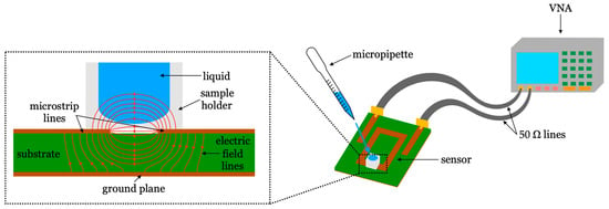
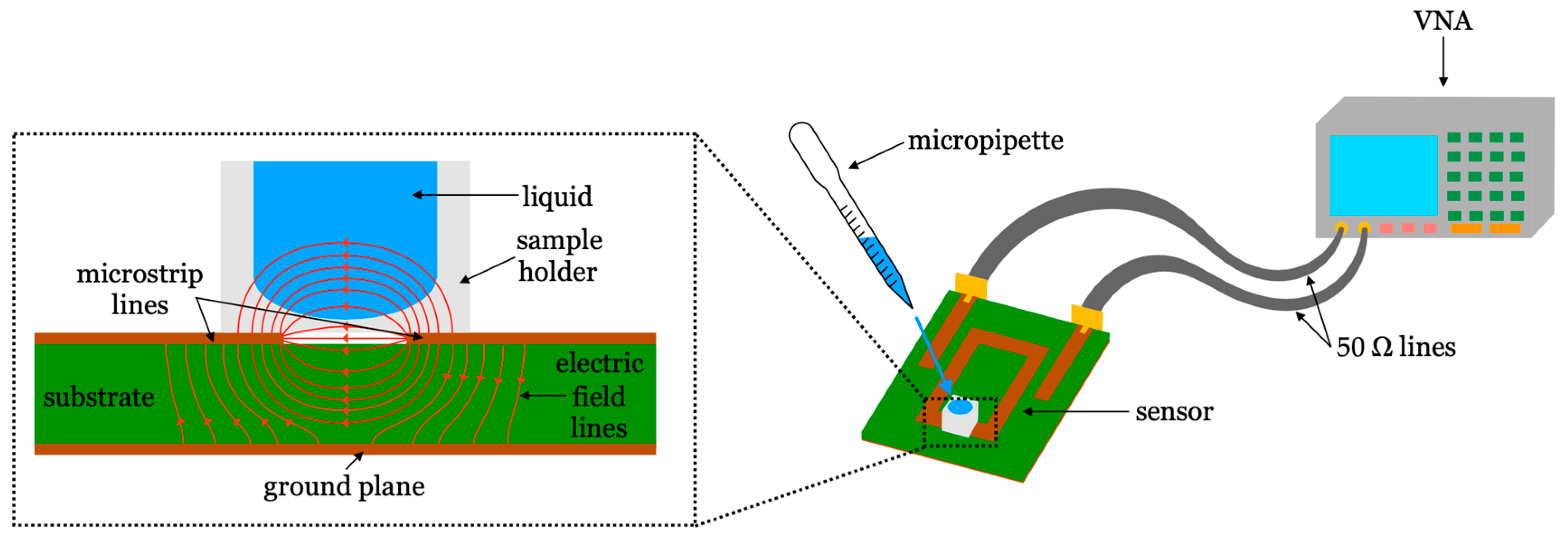
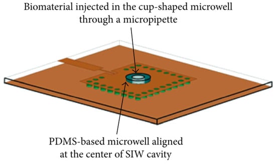
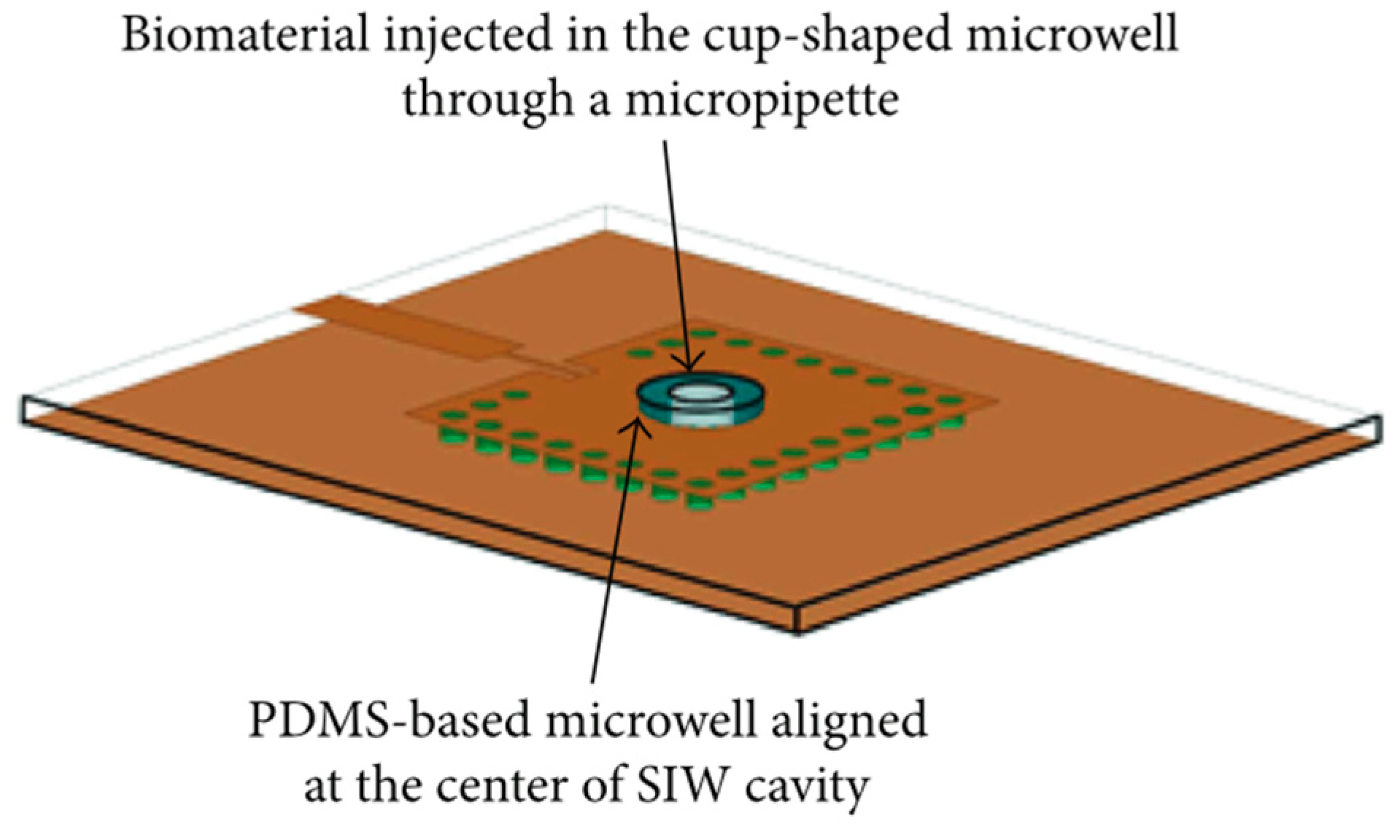
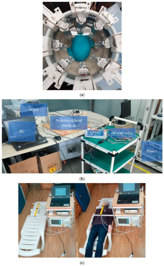
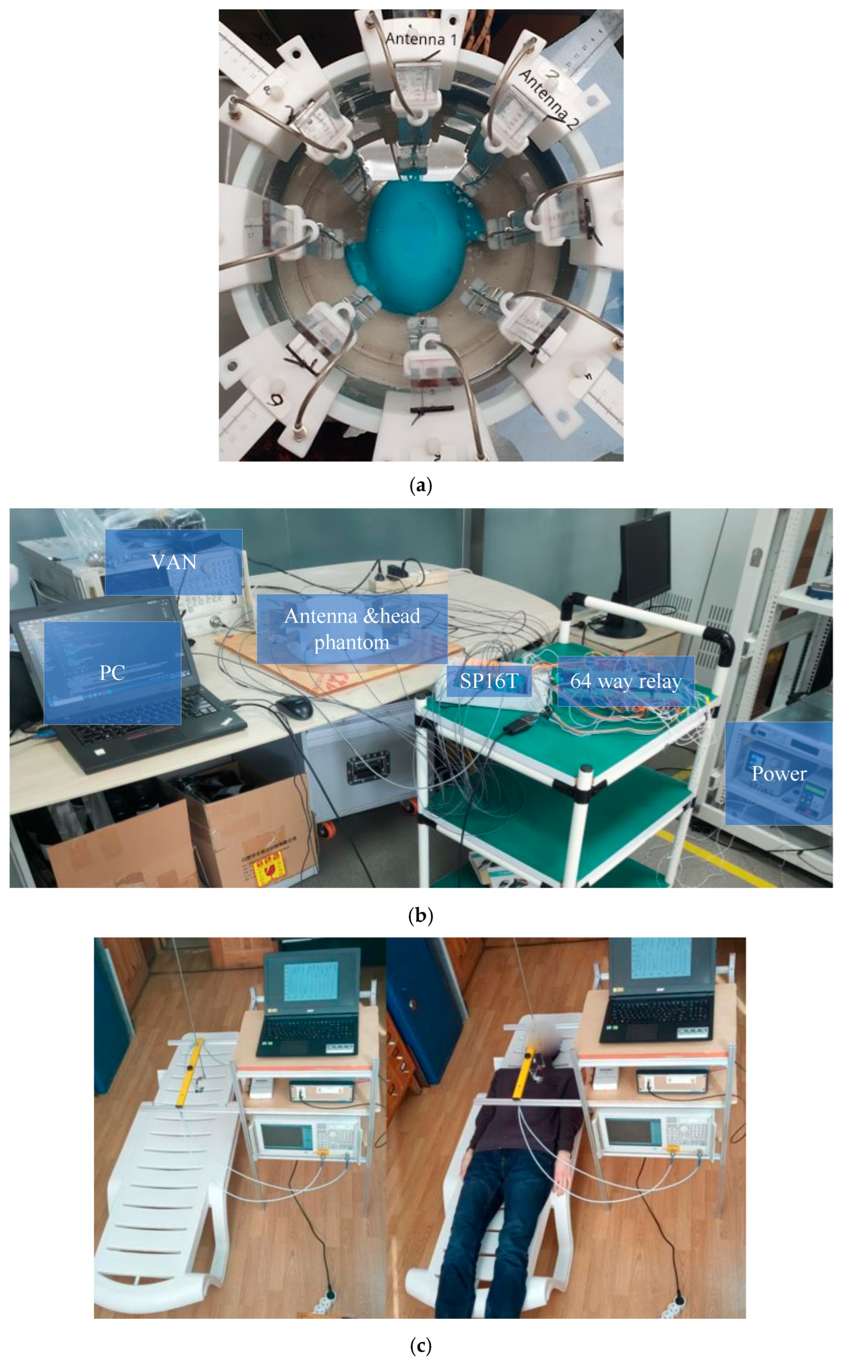
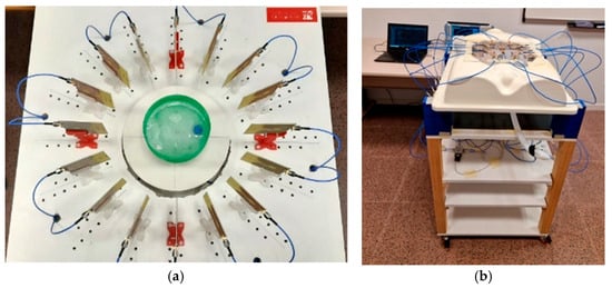
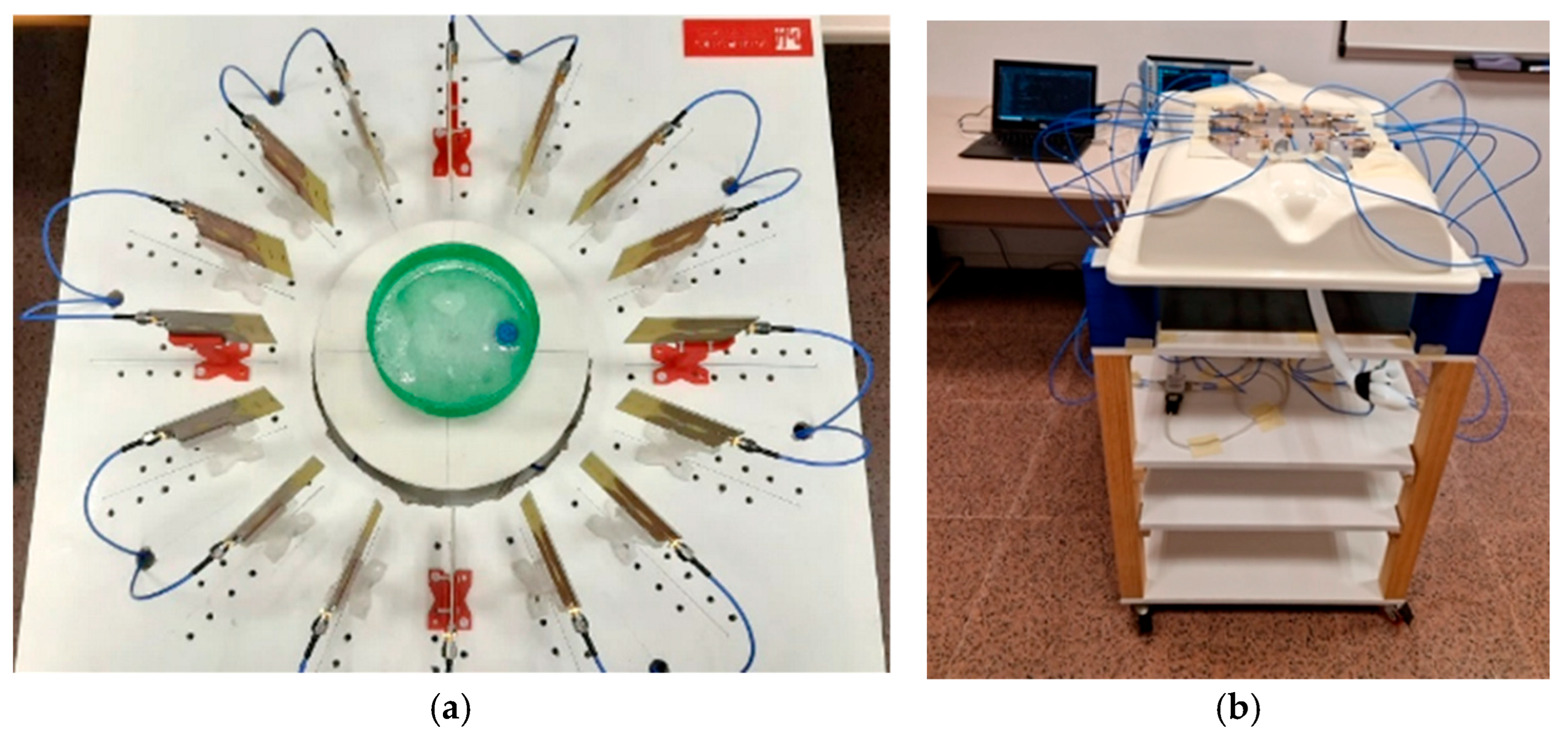
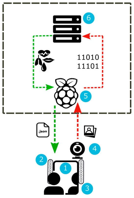
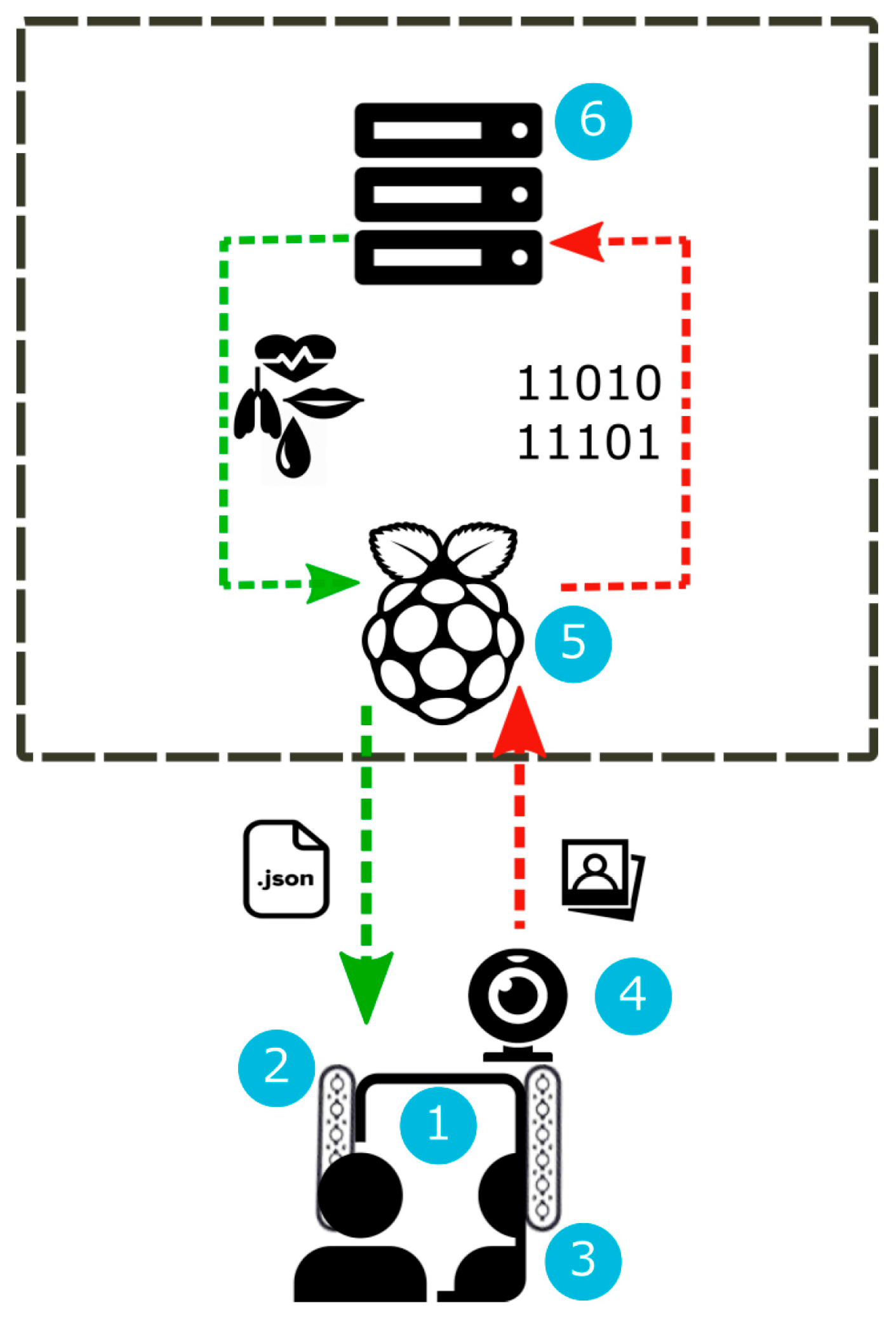
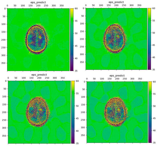
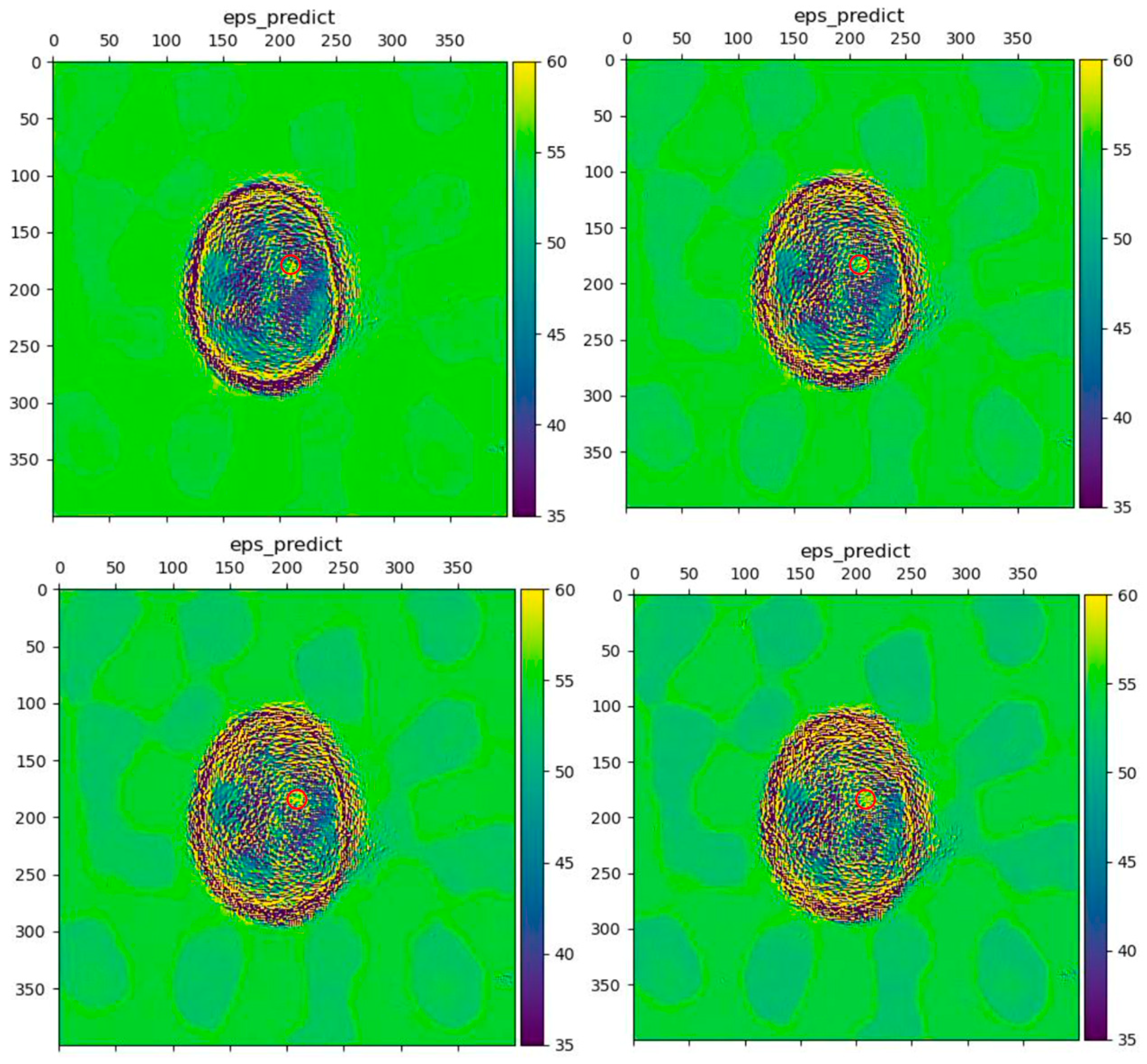
Disclaimer/Publisher’s Note: The statements, opinions and data contained in all publications are solely those of the individual author(s) and contributor(s) and not of MDPI and/or the editor(s). MDPI and/or the editor(s) disclaim responsibility for any injury to people or property resulting from any ideas, methods, instructions or products referred to in the content. |
© 2026 by the authors. Licensee MDPI, Basel, Switzerland. This article is an open access article distributed under the terms and conditions of the Creative Commons Attribution (CC BY) license.