Article

Staging of Skin Cancer Based on Hyperspectral Microscopic Imaging and Machine Learning


1
School of Optoelectronic Engineering, Xidian University, Xi’an 710071, China
2
CAS Key Laboratory of Spectral Imaging Technology, Xi’an Institute of Optics and Precision Mechanics, Chinese Academy of Sciences, Xi’an 710119, China
3
Sino-German College of Intelligent Manufacturing, Shenzhen Technology University, Shenzhen 518118, China
4
College of Physics and Optoelectronic Engineering, Shenzhen University, Shenzhen 518060, China
*
Authors to whom correspondence should be addressed.
Biosensors 2022, 12(10), 790; https://doi.org/10.3390/bios12100790
Submission received: 15 July 2022 / Revised: 28 August 2022 / Accepted: 19 September 2022 / Published: 25 September 2022

Abstract

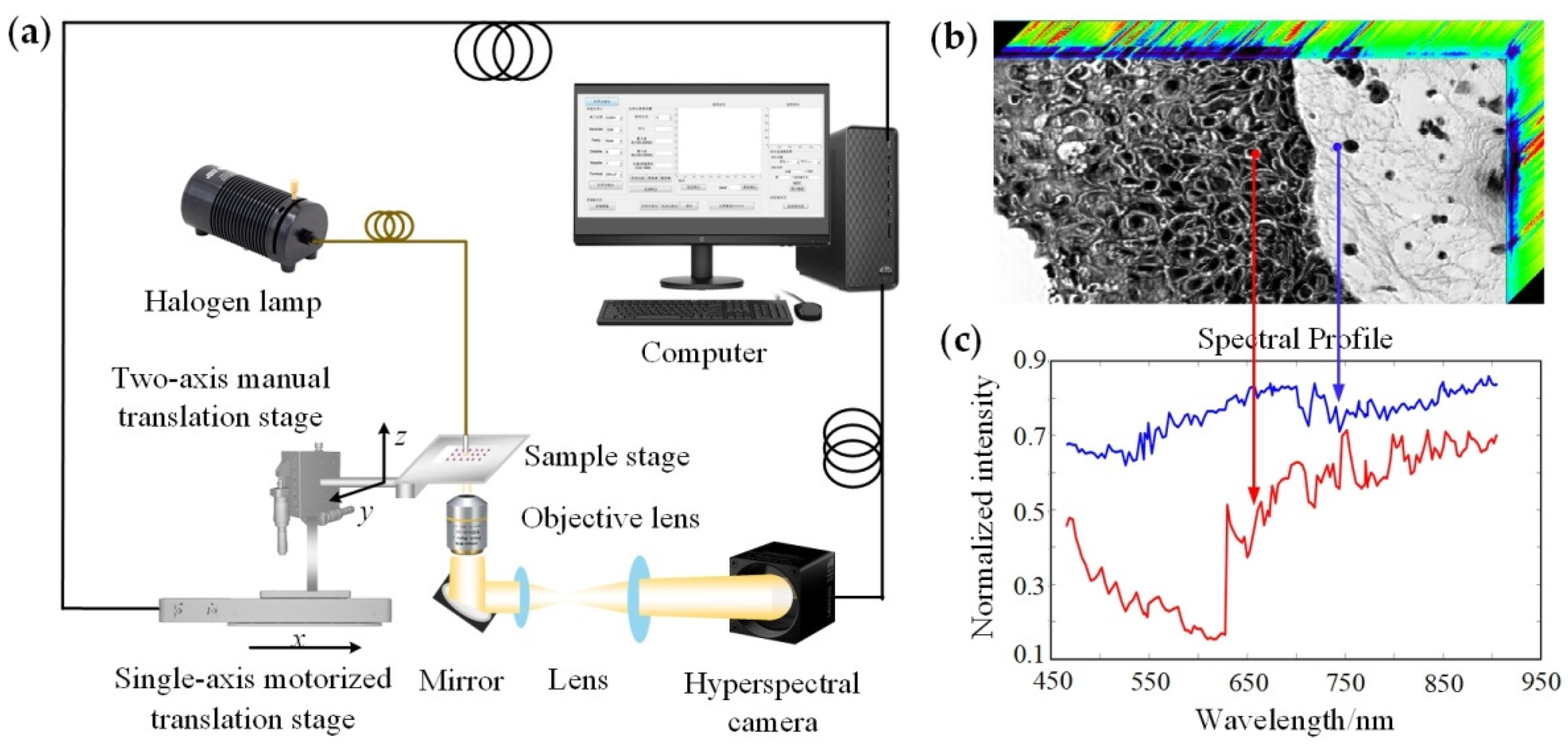
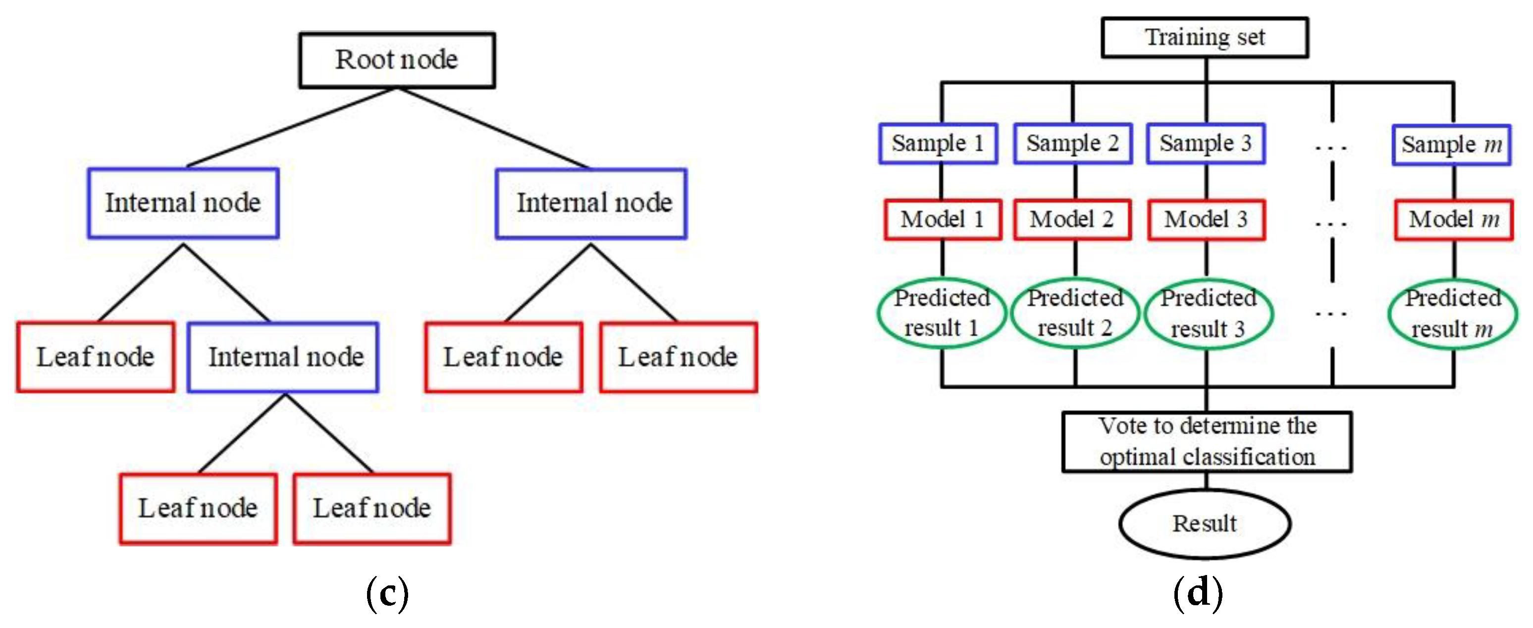
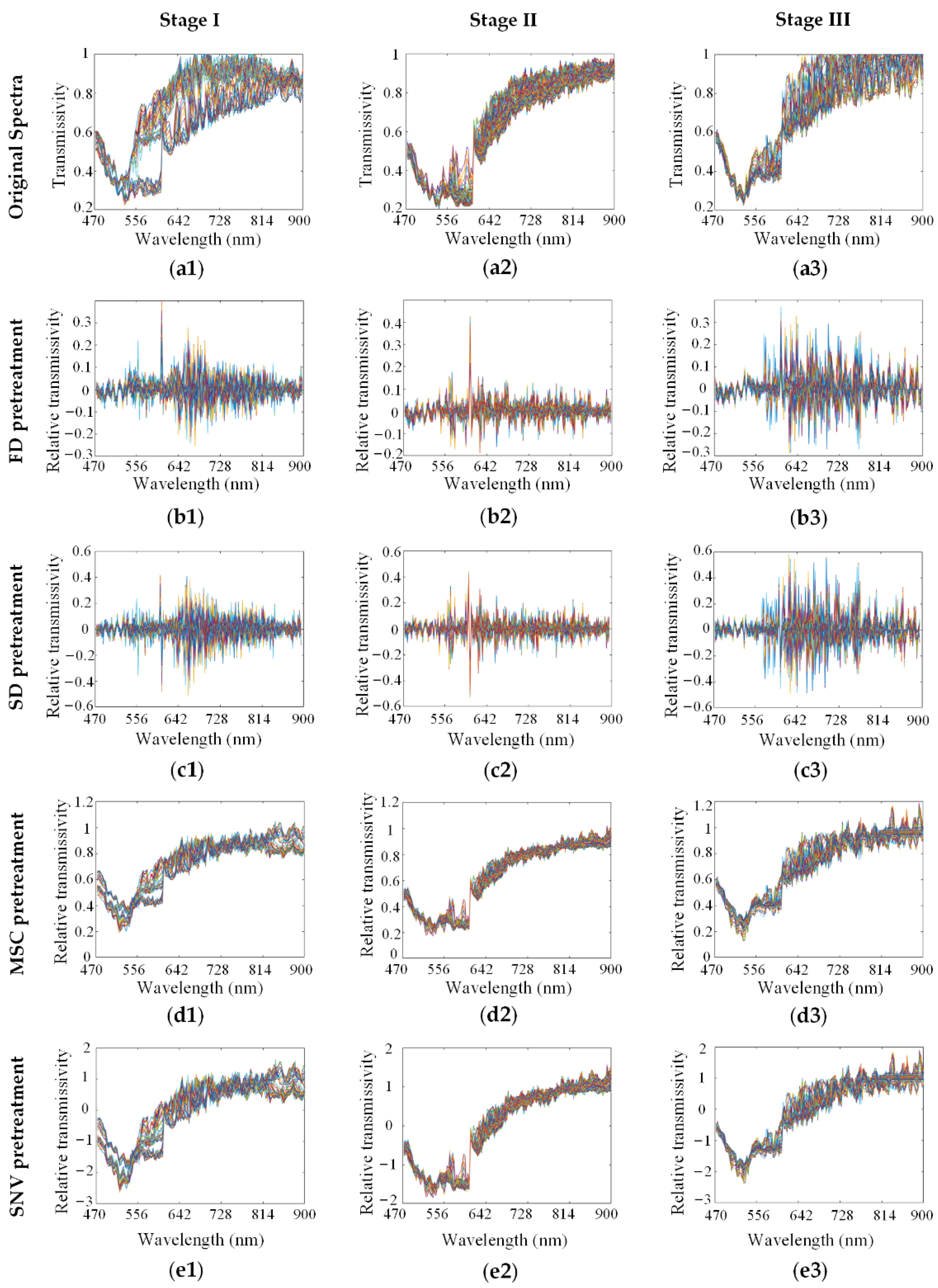
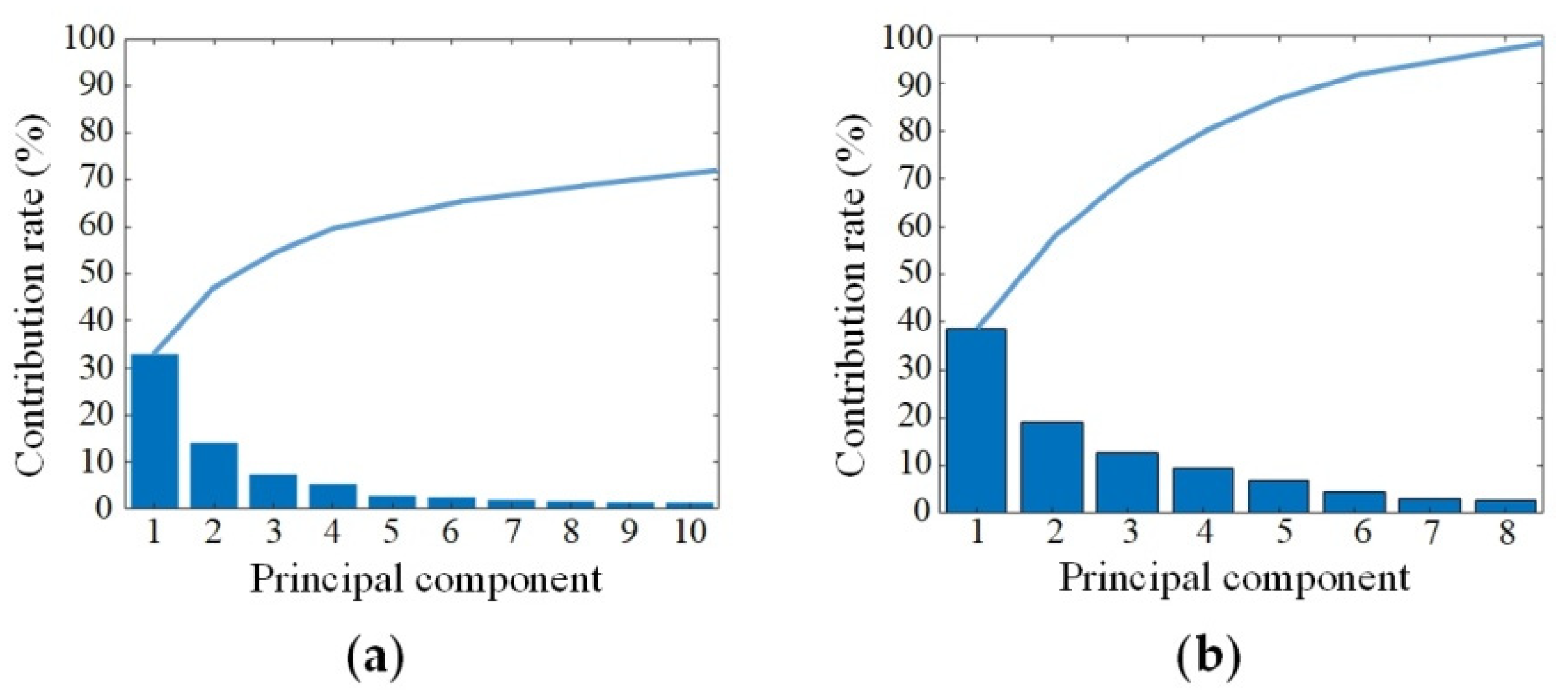
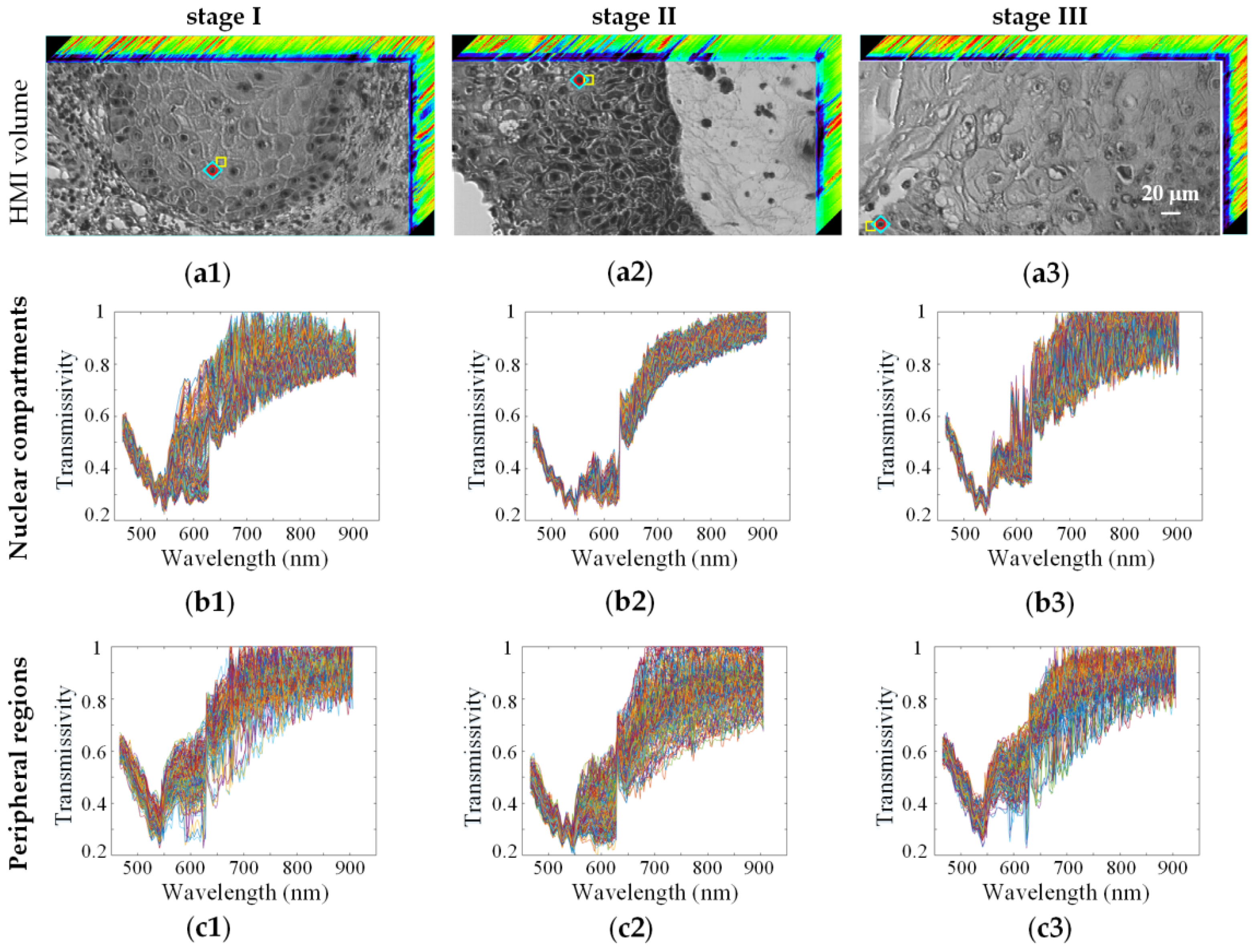
Skin cancer, a common type of cancer, is generally divided into basal cell carcinoma (BCC), squamous cell carcinoma (SCC) and malignant melanoma (MM). The incidence of skin cancer has continued to increase worldwide in recent years. Early detection can greatly reduce its morbidity and mortality. Hyperspectral microscopic imaging (HMI) technology can be used as a powerful tool for skin cancer diagnosis by reflecting the changes in the physical structure and microenvironment of the sample through the differences in the HMI data cube. Based on spectral data, this work studied the staging identification of SCC and the influence of the selected region of interest (ROI) on the staging results. In the SCC staging identification process, the optimal result corresponded to the standard normal variate transformation (SNV) for spectra preprocessing, the partial least squares (PLS) for dimensionality reduction, the hold-out method for dataset partition and the random forest (RF) model for staging identification, with the highest staging accuracy of 0.952 ± 0.014, and a kappa value of 0.928 ± 0.022. By comparing the staging results based on spectral characteristics from the nuclear compartments and peripheral regions, the spectral data of the nuclear compartments were found to contribute more to the accurate staging of SCC.
Keywords: hyperspectral microscopic imaging technology; machine learning; skin cancer; cancer classification; staging identification hyperspectral microscopic imaging technology; machine learning; skin cancer; cancer classification; staging identification