


Abstract
Autologous platelet concentrates have emerged as promising biomaterials for tissue regeneration; however, differences in preparation protocols may result in distinct biological properties. This study compared the biological properties of standard platelet-rich fibrin (S-PRF) and progressive platelet-rich fibrin (PRO-PRF) using human dental pulp cells (HDPCs). S-PRF and PRO-PRF were prepared from peripheral blood collected from six healthy donors. Growth factor release was quantified by enzyme-linked immunosorbent assay at 6, 24, 48, 72, and 240 h, while cellular metabolic activity, wound closure, and cytoskeletal organization were assessed using MTT, wound-healing, and immunofluorescence assays, respectively. Histomorphometric analysis revealed a higher fibrin-positive area in S-PRF than in PRO-PRF (18.66 ± 1.27 vs. 18.08 ± 1.73; p = 0.036), although qualitative assessment showed a denser and more organized fibrin network in PRO-PRF. S-PRF showed higher early release of TGF-β1, EGF, and PDGF and greater HDPC metabolic activity than PRO-PRF at both conditioning times (p ≤ 0.0001). In contrast, PRO-PRF promoted significantly greater wound closure at 24 h (p ≤ 0.05), with no significant difference between the PRF groups at 48 h. These findings demonstrate that S-PRF and PRO-PRF exhibit distinct fibrin architectures and biological profiles, with potential implications for the development of PRF-based regenerative strategies in vital pulp therapy.
1. Introduction
Endodontic treatment of teeth with incomplete root development remains a clinical challenge because of their thin dentinal walls, which may be divergent or parallel, making canal preparation and filling procedures more difficult and increasing the risk of root fracture [1]. In immature necrotic teeth, regenerative endodontic therapies (RETs) have become an increasingly preferred treatment approach over conventional therapies, particularly following traumatic dental injuries, although their use has also been reported in mature teeth [2]. The use of blood-derived concentrates, especially platelet-rich plasma (PRP) and platelet-rich fibrin (PRF), has attracted growing interest because of their potential to promote tissue regeneration [3,4]. However, preserving pulp vitality should remain the primary goal in immature teeth, even in teeth presenting with signs and symptoms consistent with irreversible pulpitis, allowing dentin deposition and continued root development until complete root maturation [4].
Pulpotomy is the treatment of choice for pulp exposures caused by traumatic crown fractures as well as deep carious lesions. Although calcium hydroxide, mineral trioxide aggregate (MTA), and, more recently, calcium silicate-based materials are the materials most commonly recommended for this procedure, increasing evidence from both in vitro and in vivo studies has explored the regenerative potential of PRF as an adjunct to vital pulp therapy (VPT) [5,6].
Platelet concentrates are abundant sources of autologous platelets, leukocytes, cytokines, chemokines, and growth factors [7]. Platelet-rich fibrin (PRF) has emerged as a second-generation autologous platelet concentrate that overcomes several limitations of platelet-rich plasma (PRP) by entrapping platelets and leukocytes within a three-dimensional fibrin meshwork [4]. This benefit arises from the slow, sustained release of growth factors enabled by the fibrin-clot structure of PRF [7].
Alternative preparation protocols for PRF have been proposed in recent years to increase biological performance, structural integrity, and therapeutic outcomes of PRF matrices [8]. Recently, our research group investigated a novel centrifugation protocol using progressively increasing centrifugation speeds and additive-free plastic tubes for PRF production and demonstrated that it yields fibrin networks and cellular morphologies comparable to those obtained using silica-coated plastic tubes, regardless of the centrifugation protocol [9]. This protocol, known as the progressive PRF protocol (PRO-PRF), showed promising results for tensile strength and cellular distribution [10].
In an RET context, PRF acts as a bioactive three-dimensional scaffold, providing a sustained release of growth factors that support stem cell recruitment, stimulate angiogenesis, and modulate the local inflammatory response [1,3]. Studies have shown that PRF can enhance the proliferation, migration, and differentiation of stem cells from the human apical papilla (SCAPs) and dental pulp cells (HDPCs), cell types crucial for the success of regeneration in immature teeth [11]. Recent clinical evidence supports these experimental findings. Darwish et al. (2025) demonstrated that S-PRF and advanced platelet-rich fibrin (A-PRF), when used as intracanal scaffolds in RET, promoted periapical healing and functional recovery, thereby reinforcing the clinical applicability of platelet-based matrices [12]. To date, the PRO-PRF protocol has not been evaluated for its applicability in pulpotomy.
Given the clinical relevance of PRF protocols in RETs, it is crucial to investigate the properties of newly developed PRF protocols using relevant cellular models. Therefore, this study aimed to (1) compare in vitro the release of growth factors from the standard PRF (S-PRF) and PRO-PRF protocols over time; (2) evaluate the histomorphometric characteristics of the fibrin network for both protocols; and (3) examine the effects on human dental pulp cell (HDPC) viability, morphology, and wound healing in conditioned media with platelets. The null hypothesis states that there is no significant difference between the protocols tested.
2. Materials and Methods
SigmaPlot software (v. 12, Systat Software) was used to calculate the sample size. One-way ANOVA was used with a probability of error of α = 0.05 and a power (probability of error 1 − β) = 0.8. The specific sample size was calculated using a previous study [13]. Seven individuals were included in accordance with the inclusion criteria.
Female volunteers who met the following inclusion criteria were selected: aged 20 to 40 years, healthy, non-smokers, and not using anticoagulants, anti-inflammatory drugs, corticosteroids, immunosuppressants, hormonal contraceptives, or antibiotics in the previous three months. The study included six biological donors, whose samples were used across all experimental assays. For each donor, both S-PRF and PRO-PRF samples were evaluated. The number of technical replicates, wells, and histological sections or regions of interest varied according to the assay and is specified in the corresponding subsection. Technical replicates and multiple measurements obtained from the same donor were not considered independent biological samples. Throughout all experimental procedures, all samples were coded to maintain donor-specific identification. Donor identification was maintained in the experimental plate layouts, histological sections, and data sheets used for statistical analysis, allowing all technical replicates and repeated measurements to be traced back to the corresponding biological donor. The study was approved by the Institutional Ethics Committee (CAAE: 74692623.3.0000.5152) and conducted in accordance with the Declaration of Helsinki. Informed consent was obtained from all participants included in the study.
2.1. Preparation and Cultivation of PRF
Peripheral venous blood was collected from the antecubital fossa and immediately processed using two distinct centrifugation protocols. For the S-PRF group, samples were centrifuged in a fixed-angle rotor centrifuge set at 45° (SpinPlus Titan, Daiki, Ribeirão Preto, SP, Brazil). In contrast, the PRO-PRF group was processed using a fixed-angle rotor centrifuge at 55° (CS TOUCH, Driller, Carapicuíba, SP, Brazil). Each volunteer contributed 10 blood samples (9 mL per tube), with five tubes allocated to each PRF protocol. Blood was collected in additive-free plastic tubes (Vacuette® Z, No Additive, Greiner Bio-One GmbH, Kremsmünster, Austria).
Centrifugation was performed at the following relative centrifugal force parameters: S-PRF (200× g for 14 min) and PRO-PRF (60× g for 5 min, 200× g for 5 min, and 700× g for 5 min). Following centrifugation, 3 mL of PRF liquid was aspirated from each tube under sterile conditions using a 40/12 needle within a laminar flow hood. The aspirate was transferred to glass tubes and allowed to rest for 40 min to promote clot formation. Each tube yielded one fibrin clot, which was then gently removed. For each donor and protocol, one clot was allocated to histomorphometric analysis, two clots were used for growth factor release assessment, and the remaining two clots were used to prepare conditioned media for the cellular assays. The clots designated for growth factor release assessment were placed individually in wells of 6-well culture plates (Greiner Bio-One International, Kremsmünster, Austria), each containing 5 mL of serum-free Dulbecco’s Modified Eagle Medium (DMEM; Embriolife Vitrocell, Campinas, SP, Brazil) supplemented with L-glutamine and 1% penicillin/streptomycin.
The cultures were incubated at 37 °C in a humidified atmosphere containing 5% CO2 for up to 10 days to evaluate growth factor release. Supernatants were harvested at 6, 24, 48, 72, and 240 h, aliquoted, and stored at −80 °C. At each collection point, the medium was aspirated and replaced with 5 mL of fresh DMEM.
A second blood collection was performed at a later time to obtain additional PRF clots for the preparation of conditioned media used in the cellular assays. Four tubes were collected, with two allocated to each protocol, and each tube yielded one clot. These clots were placed individually in wells containing 5 mL of serum-free DMEM and incubated for 24 h without medium replacement. The resulting supernatants were collected separately, without pooling, and stored at −80 °C until use. Thus, these conditioned media represented the cumulative release of PRF-derived components over the entire 24-h conditioning period and were used in the immunofluorescence and wound healing assays.
2.2. Measurement of Growth Factors
PRF supernatants were collected at different time points (6, 24, 48, 72, and 240 h). We used human enzyme-linked immunosorbent assay (ELISA) kits to measure growth factors per the manufacturer’s protocol (R&D Systems, Minneapolis, MN, USA): platelet-derived growth factor subunit B (PDGF-BB; #DY220, assay range: 31.2–2000 pg/mL); transforming growth factor beta 1 (TGF-β1; #DY240-05, 31.2–2000 pg/mL); vascular endothelial growth factor (VEGF; #DY293B-05, 31.2–2000 pg/mL); and epidermal growth factor (EGF; #DY236-05, 3.91–250 pg/mL).
Absorbance was recorded at 450 and 570 nm with a microplate spectrophotometer (Eon™, BioTek Instruments, Inc., Winooski, VT, USA). Positive concentration estimates generated by the curve-fitting software below the stated assay quantification ranges were retained in the original statistical analysis, without substitution by zero or the lower assay limit. These values represent estimates derived from the standard curve rather than reliably quantified concentrations and were therefore interpreted with caution. Results below the quantification range do not necessarily indicate the absence of the analyte. All measurements were performed in duplicate, and each assay was repeated in three independent experiments for both platelet concentrates. Supernatants from six donors were analyzed in duplicate at each sampling time point, resulting in 12 wells per time point for each PRF protocol in each experimental run. The complete assay was performed in three experimental runs using supernatants from the same six donors.
2.3. Histomorphometric Analysis
After polymerization, the PRF clots were carefully removed from the glass tubes and compressed in a PRF-BOX to obtain membranes, following the protocol described by Saboia-Dantas et al. (2023) [10]. For each of the six donors, one membrane was prepared using the S-PRF protocol and another using the PRO-PRF protocol. Ten histological sections were evaluated per membrane for each protocol. The membranes were fixed in 10% phosphate-buffered formaldehyde for 24 h at room temperature, then embedded in paraffin and semi-serially sectioned at 5 µm using a microtome (#RM2245, Leica Biosystems, Nussloch, Germany). The sections were stained with hematoxylin and eosin (H&E).
Histological images were acquired at 40× magnification using an optical microscope (Leica Microsystems, Wetzlar, Germany) and digitized with Aperio ImageScope software (version 12.4.6, Leica Biosystems Imaging). The images were visualized and organized in the Aperio ImageScope software for detailed analysis. For each membrane, six histological slides were selected, and ten regions of interest (ROIs) were defined (n = 60).
Histomorphometric analysis was performed semi-automatically using ImageJ (version 1.52, National Institutes of Health, USA). Fibrin network density was quantified by binarized image analysis after thresholding and expressed as the percentage of fibrin-positive area within standardized regions of interest. In addition, platelet aggregates and cellular morphological characteristics were evaluated based on previously defined morphological criteria. A single, previously calibrated examiner conducted all analyses to ensure data consistency and reproducibility.
2.4. Cell Culture Experiments
2.4.1. Cell Isolation
Primary HDPCs were established following the protocol described by Bonvicini et al. (2022) [14]. Pulp tissue was obtained from freshly extracted third molars. Cells were cultured using the explant outgrowth method in DMEM supplemented with 10% fetal bovine serum (FBS; Gibco, Grand Island, NY, USA) and 1% penicillin/streptomycin. Cells were incubated at 37 °C in a humidified atmosphere of 5% CO2 and expanded through the sixth passage before being used in the experiments.
2.4.2. Conditioned Media
Conditioned media from S-PRF and PRO-PRF were prepared using fibrin clots obtained from six donors. For each donor and PRF protocol, two clots were individually placed in separate wells of 6-well plates containing 5 mL of DMEM medium without fetal bovine serum (FBS). The cultures were incubated at 37 °C in a humidified atmosphere containing 5% CO2 for up to 10 days to evaluate the release of growth factors. Supernatants were collected at 6, 24, 48, 72, and 240 h, aliquoted, and stored at −80 °C. At each collection time point, the medium was aspirated and replaced with 5 mL of fresh DMEM.
For the cell-based experiments, two additional clots from each donor and protocol were placed individually into separate wells of 6-well plates, each containing 5 mL of DMEM medium without FBS. For the 6-h condition, the supernatant was collected after incubation, and the fibrin clots were discarded. For the 24-h condition, the clots remained incubated in DMEM medium without FBS for 24 h without medium replacement; following the collection of the supernatant, the clots were also discarded. The collected supernatants were stored at −80 °C until use. Both 6-h and 24-h conditioned media were used in the immunofluorescence and MTT assays, whereas only the 24-h conditioned media were used in the wound healing assay.
The selection of these conditioning periods was based on the growth factor release analysis, which demonstrated distinct release profiles at 6 and 24 h. Unless otherwise stated, cells were exposed to the conditioned media for 24 h.
2.4.3. MTT Assay
Cells were treated with PRO-PRF and S-PRF conditioned media collected after 6 and 24 h, as previously described. Untreated cells cultured in serum-free DMEM were used as controls. After 24 h of treatment, cell metabolic activity was assessed using the methyl thiazolyl tetrazolium (MTT) assay (Sigma-Aldrich). The experiment was performed in triplicate using 96-well plates, with one well per donor for each PRF protocol and conditioning time in each experimental run. Each well received 90 µL of serum-free DMEM and 10 µL of MTT solution (5 mg/mL in PBS). Following 4 h of incubation at 37 °C, the medium was replaced with 100 µL of dimethyl sulfoxide (DMSO; LGC Biotecnologia, Cotia, SP, Brazil) to solubilize the formazan crystals. Absorbance was measured at 570 nm using a spectrophotometer (Thermo Plate, Shenzhen, China). Cell viability was calculated by normalizing the absorbance values of the experimental groups to those of the untreated control, which was set at 100% viability.
2.4.4. Wound Healing Assay
For migration analysis, HDPCs were seeded into migration-specific culture wells containing removable silicone inserts to create two separate compartments (Culture-Insert 2 Well 24, ibiTreat, Ibidi, Martinsried, Germany). Cells were seeded at 2 × 10⁵ cells/mL by dispensing 70 μL of suspension into each compartment and cultured for 24 h. Inserts were then removed, producing a standardized gap of 500 ± 50 µm. Three random fields per well were imaged immediately, and treatments were applied according to the experimental groups (Control, S-PRF, and PRO-PRF). For the experimental groups, the 24-h conditioned media were used. The experiment was performed in triplicate, with one well per donor for each experimental group. Three regions of interest were imaged per well. Additional images were captured at 24 and 48 h. Gap closure was analyzed using a specific ImageJ plugin, following the protocol described by Suarez-Arnedo et al. (2020) [15].
2.4.5. Immunofluorescence Analysis
HDPCs were seeded onto sterile circular glass coverslips (13 mm) in 24-well plates at a density of 2 × 10⁵ cells per well until they reached 70–80% confluence. Cells were then treated with PRF-conditioned media collected after 6 and 24 h, as previously described, and incubated for 24 h at 37 °C. Cells were then fixed in 4% paraformaldehyde for 30 min at room temperature and incubated with Phalloidin-tetramethylrhodamine B isothiocyanate (TRITC, 1:200; Sigma-Aldrich, St. Louis, MO, USA) for 30 min, followed by TO-PRO-3 iodide (1 µM; 1:1000 dilution from stock solution; Thermo Fisher Scientific, Waltham, MA, USA) for 60 min. All procedures were carried out at room temperature, protected from light. Imaging was performed using a confocal laser scanning microscope (TCS SP8, Leica Microsystems, Wetzlar, Germany) with acquisition settings standardized for TRITC (excitation 540–550 nm; emission 565–580 nm) and TO-PRO-3 (excitation 642 nm; emission 661 nm). The experiment was performed in duplicate, with one well per donor for each PRF protocol and conditioning time in each experimental run. Five regions of interest were captured per well.
2.5. Statistical Analysis
Statistical analyses were performed using GraphPad Prism (version 9.0.2; GraphPad Software, Boston, MA, USA). Data normality was assessed using the Shapiro–Wilk test. Growth factor release data were analyzed using a mixed-effects model with restricted maximum likelihood (REML), with matching by donor in a stacked format to account for repeated measurements from the same donors. Tukey’s post hoc test was used for multiple comparisons. Comparisons between time points were performed using a separate two-way ANOVA with Šídák’s correction, where applicable. For histomorphometric analysis, technical replicates obtained from each donor were averaged to generate a single value per protocol per donor (n = 6 biological replicates). S-PRF and PRO-PRF were compared using a paired t-test, with donor identity defining the pairing. Statistical significance was set at p < 0.05.
For all assays, donor identity was retained throughout data organization and statistical analysis. Multiple wells, histological sections, and regions of interest derived from the same donor were regarded as technical replicates or subsamples, as appropriate, and were not considered independent biological replicates.
3. Results
Seven participants were initially enrolled based on the sample size calculation. One participant withdrew during blood collection and was excluded from the study, resulting in a final sample of six participants aged 25–34 years.
3.1. Growth Factor Release
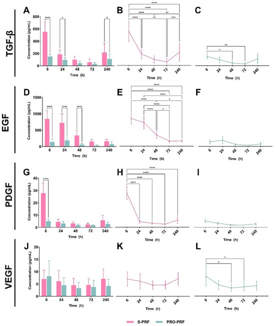
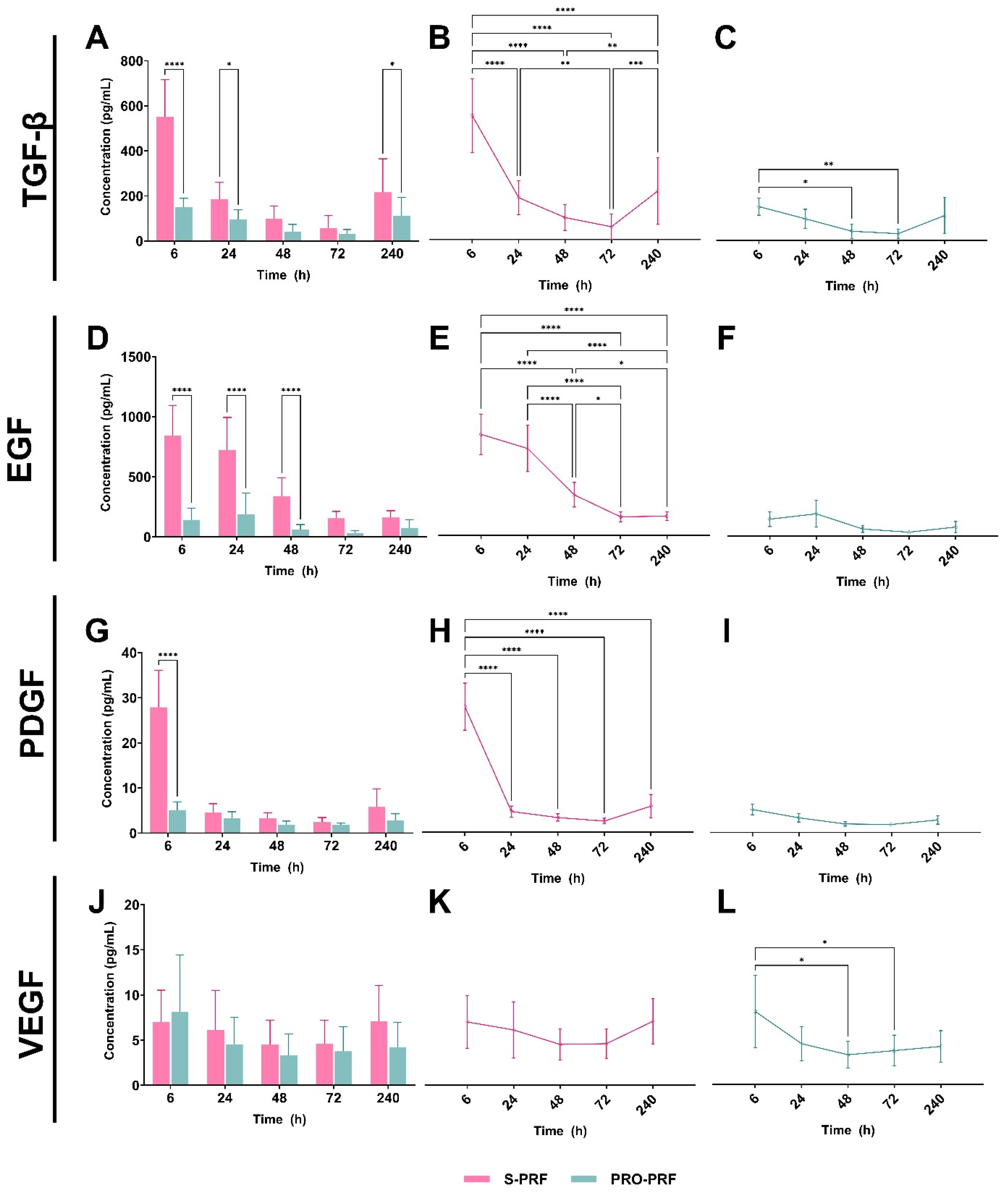
Growth factor concentrations measured in the collected supernatants over 240 h are summarized in Figure 1. The intergroup comparison demonstrated that the concentrations of TGF-β1, EGF, and PDGF at the early time point (6 h) were significantly higher in the S-PRF group (p ≤ 0.05, Figure 1A,D,G). The S-PRF group also showed significantly higher concentrations of TGF-β1 and EGF at 24 h (p ≤ 0.05, Figure 1A,D). In contrast, no significant difference in PDGF concentration was observed between S-PRF and PRO-PRF at this time point (p > 0.05, Figure 1G). S-PRF also showed significantly higher concentrations of EGF at 48 h and TGF-β1 at 240 h (p ≤ 0.0001, Figure 1D; p ≤ 0.05, Figure 1A, respectively). Most VEGF estimates were below the stated assay quantification range. Although the original statistical analysis did not identify significant differences between protocols or sampling time points, these comparisons should be interpreted cautiously, as the predominance of below-range estimates limits the reliability of quantitative comparisons. Therefore, the observed numerical patterns should be considered exploratory and do not support firm conclusions regarding VEGF release kinetics or differences between protocols (p > 0.05, Figure 1J).
Figure 1.
Quantification of growth factor release from S-PRF and PRO-PRF at different time intervals (6, 24, 48, 72, and 240 h). (A–C) TGF-β concentrations. (D–F) EGF concentrations. (G–I) PDGF concentrations. (J–L) VEGF concentrations. Bar graphs (A,D,G,J) represent mean concentrations (pg/mL) ± SD. * p ≤ 0.05, ** p ≤ 0.005, *** p ≤ 0.0005, **** p ≤ 0.0001.
The intragroup comparison demonstrated that S-PRF showed a decline in TGF-β1 release over time (6–72 h) (p ≤ 0.0001), with a second peak at 240 h (p ≤ 0.05, Figure 1B). The amount of EGF released at each time point peaked at 6 and 24 h (p > 0.05), with a significant progressive reduction over time (p ≤ 0.05; Figure 1E). The peak release of PDGF in the S-PRF group was observed at 6 h, followed by a decline at 24 h (p ≤ 0.0001), and persistently low levels throughout the remaining experimental periods (Figure 1H). VEGF estimates remained low throughout the evaluated period in the S-PRF group, with no statistically significant differences among time points (p > 0.05, Figure 1K). In the PRO-PRF group, the numerical estimates suggested higher VEGF levels at 6 h, followed by lower estimates at 48 and 72 h (Figure 1L). However, because most VEGF measurements were below the assay quantification range, this apparent temporal pattern should be considered exploratory rather than evidence of a reliable release kinetic profile. The amounts of EGF and PDGF released by the PRO-PRF group were low across all experimental time points, showing no statistically significant variations over time (p > 0.05, Figure 1F,I). The PDGF-BB comparisons should be interpreted cautiously because they included estimates below the stated assay range, particularly at later sampling intervals.
3.2. Fibrin Network Density Evaluation
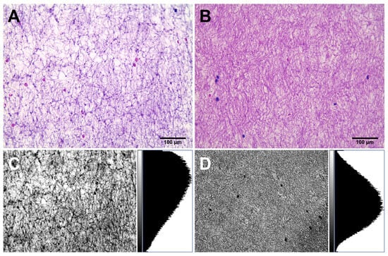
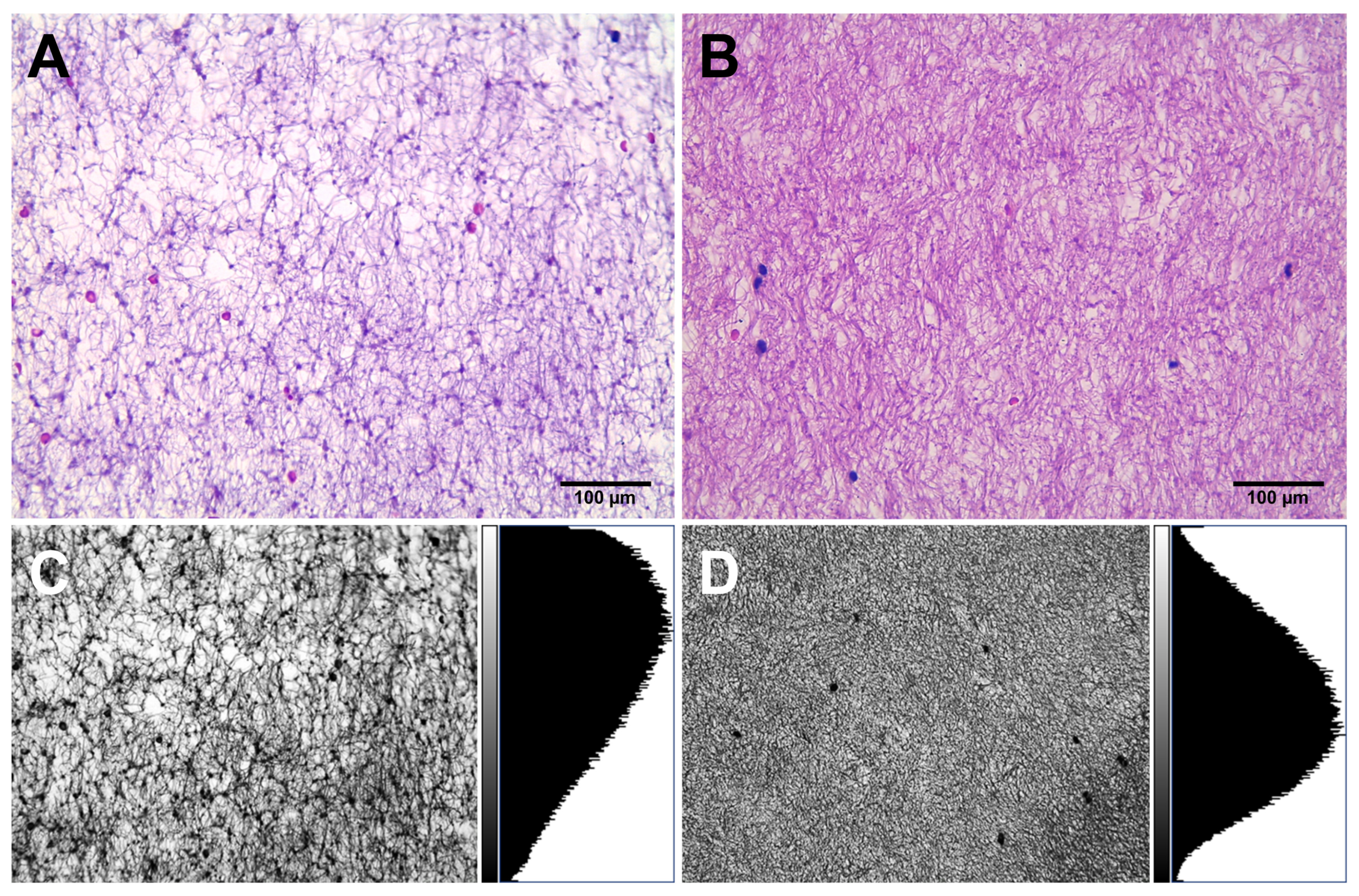
Qualitative histomorphometric analysis demonstrated that the fibrin network of membranes prepared using the PRO-PRF protocol exhibited greater structural density—characterized by a more compact and organized fibrin architecture—compared to S-PRF membranes (p ≤ 0.05). Quantitative analysis revealed that S-PRF showed a larger fibrin-positive area (18.66 ± 1.27) than PRO-PRF (18.08 ± 1.73). A paired t-test confirmed that this difference was statistically significant (p = 0.036). Qualitative assessment showed that S-PRF samples displayed a looser fibrin arrangement, with wider interfibrillar spacing (Figure 2A). In contrast, PRO-PRF membranes exhibited a denser and more organized fibrin network, with reduced interfibrillar spaces (Figure 2B).
Figure 2.
Representative hematoxylin and eosin (H&E)-stained sections showing the morphological fibrin network density obtained from (A) S-PRF and (B) PRO-PRF protocols (40× magnification). Images were binarized to quantify fibrin distribution and structural density. The corresponding histograms for (C) S-PRF and (D) PRO-PRF show the distribution of black/white pixels, where black pixels indicate fibrin-rich or cell-dense regions.
3.3. Cell Culture Experiments
3.3.1. MTT Assay
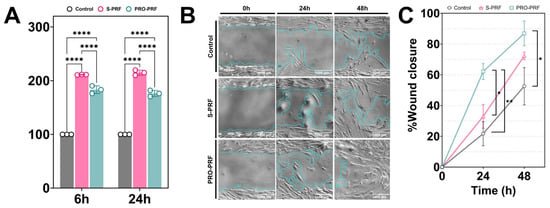
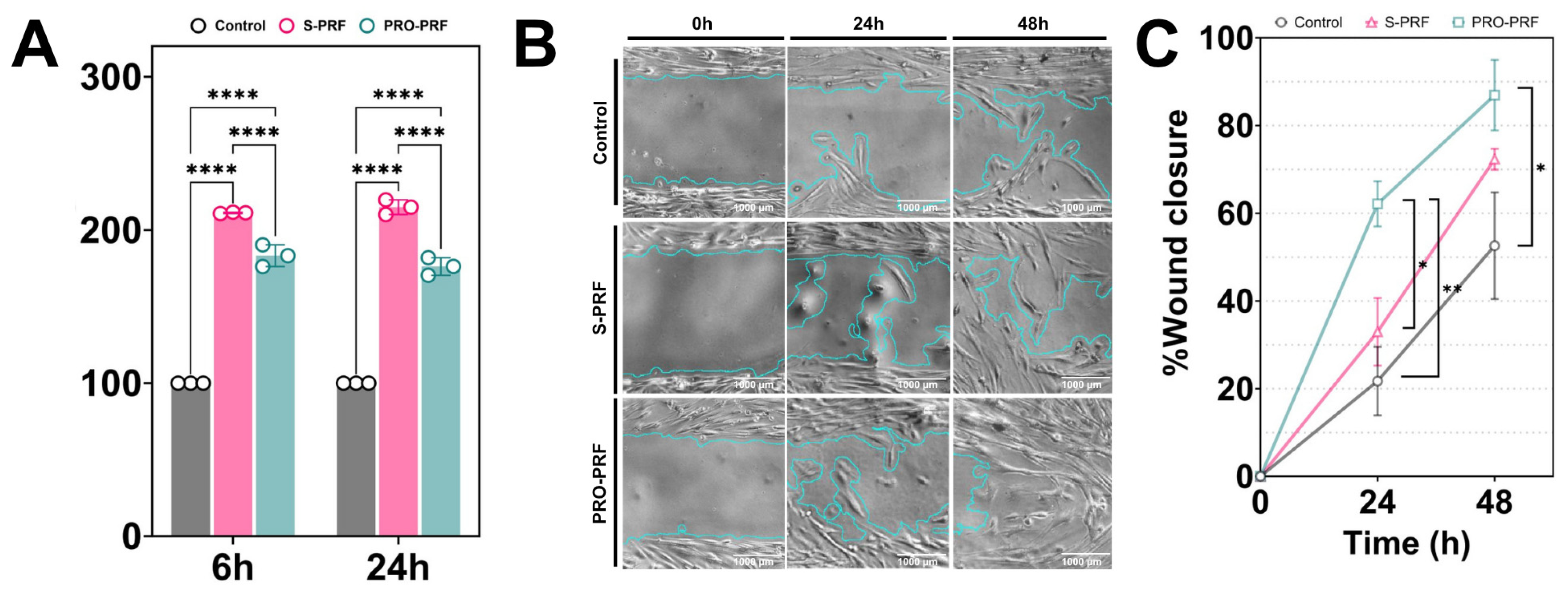
Cell metabolic activity, assessed by the MTT assay, was significantly higher in all PRF-treated groups compared with the control when cells were exposed to conditioned media collected after both 6 and 24 h (p ≤ 0.0001; Figure 3A). Among the experimental groups, S-PRF induced significantly greater metabolic activity than PRO-PRF at both time points (p ≤ 0.0001).
Figure 3.
HDPCs’ behavior in response to PRF-related conditioned media. (A) Cell viability after 24 h of incubation in fresh medium (control), S-PRF, or PRO-PRF conditioned media. (B) Representative images of the wound-healing assay over 48 h using the 24-h conditioned media from each treatment. (C) Wound healing results are shown as the average percentage of wound closure in each well, based on wound area measured using a semi-automated ImageJ plugin. The average of three independent experiments was used for statistical analysis. * p ≤ 0.05, ** p ≤ 0.005, **** p ≤ 0.0001.
3.3.2. Wound Healing Assay
At 24 h, PRO-PRF exhibited significantly greater wound closure than both the control and S-PRF groups (p ≤ 0.05), whereas no significant difference was observed between the control and S-PRF groups (p ≥ 0.05; Figure 3B,C). At 48 h, PRO-PRF maintained significantly greater wound closure than the control (p ≤ 0.05), while no statistically significant difference was observed between PRO-PRF and S-PRF (p ≥ 0.05).
3.3.3. Cell Morphology
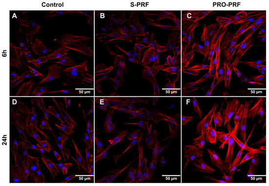
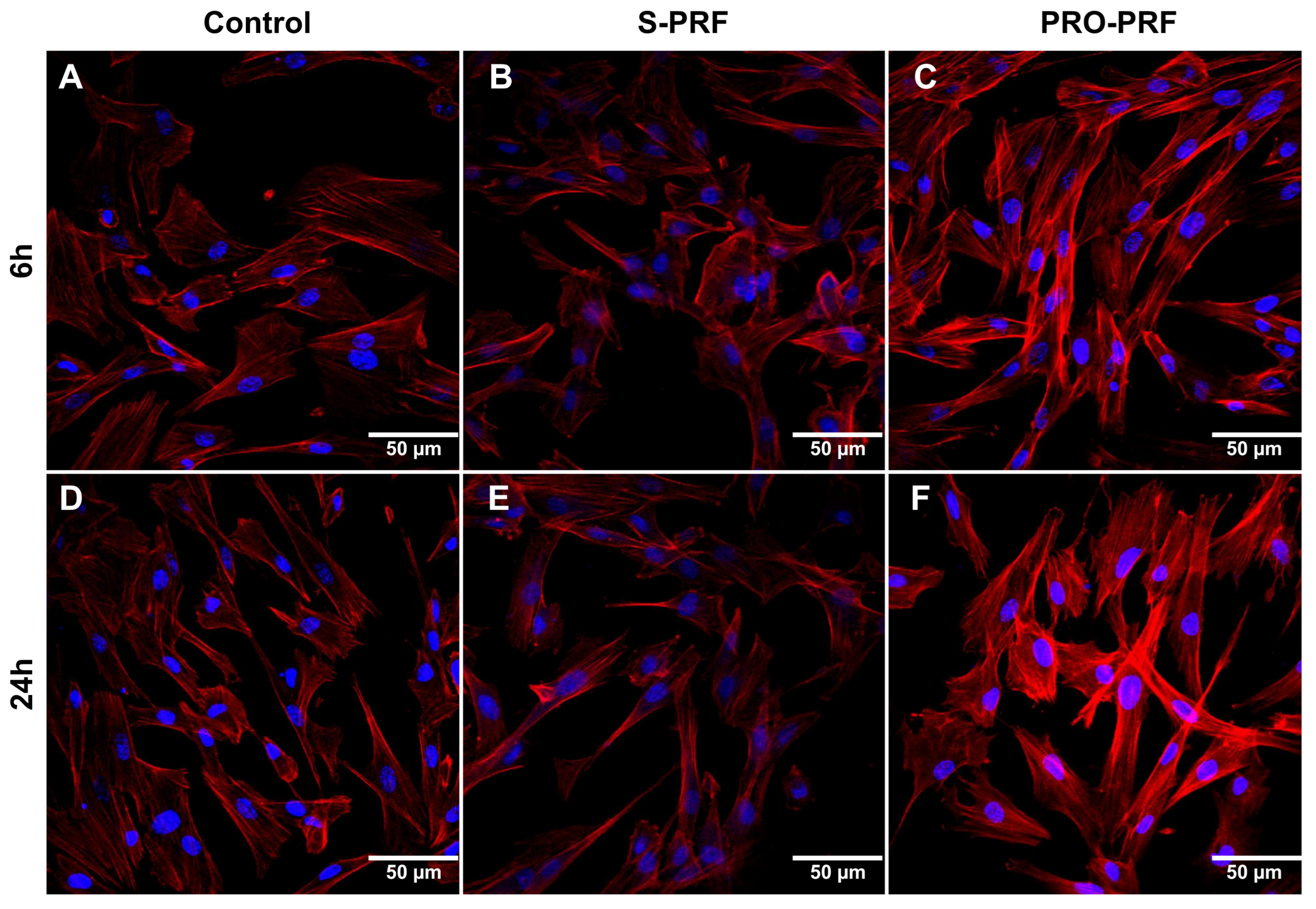
Regarding F-actin organization, after 6 h of exposure to PRF-conditioned media collected at different time points, HDPCs exhibited distinct cytoskeletal patterns (Figure 4). After 6 h of exposure to conditioned media, both the control and S-PRF groups exhibited fibroblast-like morphology, characterized by elongated, flattened cells with a relatively uniform actin distribution (Figure 4A,B). In contrast, cells treated with PRO-PRF-conditioned medium exhibited enhanced cytoskeletal organization and initial cytoplasmic projections (Figure 4C). This morphological pattern became more pronounced after 24 h of conditioned media treatment, particularly in the PRO-PRF group, which exhibited F-actin organization and prominent fusiform cytoplasmic extensions (Figure 4F). After 24 h of exposure to the conditioned media, control cells showed an aligned cytoskeletal arrangement. In contrast, cells treated with S-PRF exhibited a less uniform distribution of actin filaments and partial cell scattering on the surface (Figure 4D,E).
Figure 4.
Immunofluorescence images of HDPCs cultured with conditioned media collected after 6 h (A–C) and 24 h (D–F) of incubation with S-PRF, PRO-PRF, or control (DMEM). The F-actin cytoskeleton of HDPCs was stained with phalloidin-TRITC (red); the nucleus was stained with TO-PRO 3 iodide (blue).
4. Discussion
S-PRF was associated with greater early growth factor release and higher HDPC metabolic activity, whereas PRO-PRF induced greater wound closure at 24 h and was associated with more pronounced F-actin organization and elongated cytoplasmic projections. Both protocols showed significant differences, rejecting the null hypothesis. These findings suggest that the biological performance of PRF may be influenced not only by growth factor release profiles but also by other preparation-dependent properties, potentially including fibrin architecture. Taken together, these findings support the potential of PRF as a regenerative adjunct in VPT, suggesting that differences in fibrin architecture and growth factor dynamics may influence the cellular responses underlying pulp tissue repair and regeneration [16].
Different protocols have been proposed in the literature, exhibiting growth-factor release behavior similar to that observed in our study. A high initial release of growth factors is associated with A-PRF and Leukocyte–Platelet-Rich Fibrin (L-PRF), which are characterized by a less compact fibrin network and higher leukocyte content [13,17]. This release pattern has been attributed to a more porous fibrin architecture with larger interfibrillar spaces that reduce diffusion barriers, as well as to increased leukocyte-mediated enzymatic remodeling of the fibrin matrix [18,19]. In our study, a similar structural pattern was observed in the S-PRF group, which exhibited a looser fibrin architecture with larger interfibrillar spaces, consistent with its higher early growth factor release.
In contrast, PRO-PRF demonstrated a denser, more cohesive fibrin configuration, consistent with the concept of progressive PRF, which is designed to increase matrix stability, cell distribution, and handling characteristics [10]. Although this architecture is generally associated with a slower and more sustained release of growth factors [13,19,20], both protocols exhibited a characteristic release pattern, peaking at 6 h and declining until 72 h. This is consistent with previous reports that most PRF-mediated bioactivity occurs early in wound healing [7,21]. The slight increase at later time points may reflect fibrin remodeling and sampling intervals [8,9].
The release profiles observed in our study demonstrate that each growth factor contributes distinctly to the early and intermediate phases of the regenerative signaling cascade. The higher initial concentrations of PDGF and EGF detected in S-PRF are consistent with their recognized role in promoting early chemotactic recruitment and proliferative activation of pulp-derived progenitor cells, as previously described in platelet concentrate-based tissue engineering models [11,22]. TGF-β1, while also released in greater amounts during the initial period, plays a more complex role by directing odontoblastic differentiation and extracellular matrix deposition during later repair stages. TGF-β1 plays a fundamental role in odontoblastic differentiation and extracellular matrix deposition [23,24,25] and has been implicated in cell recruitment during pulp regeneration [25]. In addition, growth factor–mediated signaling pathways are known to regulate dental pulp cell migration through cytoskeletal reorganization and activation of migration-related pathways [26]. Although the numerical VEGF estimates appeared to decrease over time in both groups, the predominance of measurements below the assay quantification range limits the interpretation of this apparent pattern and does not allow reliable conclusions regarding its release kinetics. These findings suggest that S-PRF provides a stronger early signal for cell activation. In contrast, the denser fibrin network of PRO-PRF may influence cell–matrix interactions and the mechanical signals perceived by cells, potentially modulating cellular responses involved in pulp repair and regeneration [22,24].
From a clinical perspective, challenges in RET include achieving uniform scaffold distribution, limited control over scaffold structure, and reduced stability after placement, which may contribute to variable outcomes [1,3]. In this context, the structural characteristics of PRO-PRF may offer a potential advantage in terms of scaffold stability, although this possibility requires further investigation under clinically relevant conditions. Additionally, the liquid form of PRF may offer practical advantages during clinical application compared with preformed fibrin clots, which may require trimming to fit the dimensions of the treated area. This handling characteristic could potentially facilitate scaffold placement and adaptation to the pulp surface; however, these proposed advantages remain to be confirmed in clinical studies. The use of liquid PRF may therefore represent a potential approach for scaffold delivery in VPT, warranting further investigation.
Accordingly, S-PRF induced significantly greater metabolic activity in HDPCs. This finding may be associated with the higher early release of growth factors observed for S-PRF, particularly PDGF and EGF, which are known to promote cellular metabolic activity and chemotactic responses in pulp-derived cells [11,22]. Notably, the MTT assay reflects mitochondrial dehydrogenase activity rather than cell proliferation, indicating enhanced cellular metabolic activity rather than an increased cell number [27]. In this context, different PRF preparation protocols may modulate cellular responses through differences in growth factor release kinetics, with potential implications for cellular recruitment, metabolic activity, proliferation, and differentiation [28,29,30].
In contrast to the growth factor release profiles, PRO-PRF-conditioned medium induced significantly greater HDPC wound closure than both the control and S-PRF-conditioned medium at 24 h. At 48 h, wound closure remained significantly greater in the PRO-PRF group than in the control, whereas no significant difference was observed between PRO-PRF and S-PRF. These findings suggest that wound closure-related responses are not determined solely by the concentrations of soluble growth factors, but may also be influenced by other properties of the PRF preparations [10,31]. The more pronounced F-actin organization and elongated cytoplasmic projections observed in HDPCs exposed to PRO-PRF are consistent with cellular morphological changes associated with adhesion and migration [31]. However, the contribution of specific scaffold-related mechanisms, such as focal adhesion formation and mechanotransduction, remains to be investigated. Similarly, the higher early release of growth factors observed with S-PRF may contribute to initial cellular responses, although the present findings do not establish a direct relationship between growth factor release kinetics and migratory behavior [24]. Together, these findings suggest that the biological responses elicited by PRF preparations may reflect the combined influence of soluble factors and other preparation-dependent properties, warranting further investigation [31,32].
Although this study constitutes an initial in vitro evaluation of two specific PRF protocols in the context of RET, the findings provide mechanistic evidence regarding fibroblast responses to platelet concentrate-derived biomaterials. These observations contribute to the existing body of evidence on the biological effects of platelet concentrates and provide a rationale for further preclinical and clinical investigation.
Several limitations should be considered. Although the sample size was determined, the limited number of participants may have reduced the ability to capture interindividual biological variability. The exclusive inclusion of female participants was based on documented sex-dependent differences in platelet biology and activation, as female platelets exhibit greater responsiveness to agonists than male platelets [33]. In addition, estrogen has been shown to modulate platelet activation and surface receptor expression, providing a mechanistic basis for sex-related differences that may influence PRF composition and bioactivity [34]. Although this strategy improved biological homogeneity within the study population, it may limit the generalizability of the findings, and further studies including both sexes are warranted. Another limitation is that different centrifuges were used for the S-PRF and PRO-PRF protocols, in accordance with the equipment recommended or specifically designed for each protocol. Although this choice was intended to ensure standardized implementation, the potential influence of equipment-related factors on the observed biological differences cannot be entirely excluded. An analytical limitation concerns the inclusion of concentration estimates below the stated ELISA quantification ranges, particularly for VEGF and PDGF-BB. These estimates were generated by the curve-fitting software and retained in the original statistical analysis; however, their numerical values have limited quantitative reliability. Results below the quantification range do not necessarily indicate the absence of the analyte. Although the observed numerical patterns may suggest temporal variations, they should be considered exploratory and do not support firm conclusions regarding the magnitude of growth factor release or precise release kinetics. Consequently, comparisons involving these estimates, particularly those concerning VEGF and later PDGF-BB measurements, should be interpreted with caution, and the robustness of these findings remains to be established.
Human dental pulp cells (HDPCs) represent a relevant cellular model for investigating the biological effects of PRF in the context of VPT, as they comprise the resident cell population directly involved in pulp tissue homeostasis, repair, and regeneration. Previous studies have demonstrated that PRF can modulate the behavior of dental pulp-derived cells, including their metabolic activity and migratory capacity, supporting the biological relevance of these cells for evaluating PRF-based regenerative strategies [6,35]. Moreover, HDPCs play a pivotal role in orchestrating early reparative events within the pulp microenvironment, including cell recruitment, extracellular matrix remodeling, and cell signaling processes required for tissue regeneration [36].
Although these findings should be interpreted with caution, the more cohesive fibrin structure observed in PRO-PRF may have implications for its handling properties and structural behavior within the confined root canal environment. However, the potential clinical relevance of these structural characteristics remains to be established. In contrast, the higher early release of growth factors observed in S-PRF may favor initial cellular responses, highlighting the distinct biological profiles of the two protocols and their potential relevance at different stages of VPT. Future in vivo investigations and well-designed clinical trials, including larger cohorts of both sexes, are warranted to validate these observations and determine the most appropriate clinical indications for each PRF preparation protocol in RET.
5. Conclusions
The findings suggest that the biological behavior of PRF is influenced by the preparation protocol, with differences across cellular and growth-factor-related outcomes. Neither protocol demonstrated a consistent advantage across all parameters assessed, highlighting the importance of considering multiple biological properties when evaluating PRF for regenerative endodontic applications. Further research is needed to determine the clinical relevance of these differences and their implications for protocol selection in RET.
Author Contributions
Conceptualization, A.d.P.M., G.L.d.S. and P.D.; Methodology, A.d.P.M., C.M.M.L.d.S., C.R.B.L., P.D., G.L.d.S. and R.O.T.; Investigation, A.d.P.M., C.M.M.L.d.S. and C.R.B.L., J.R.M.e.S. and R.O.T.; Formal Analysis, A.d.P.M., D.C.F. and J.R.M.e.S.; Data Curation, A.d.P.M. and D.C.F.; Writing—Original Draft Preparation, A.d.P.M. and D.C.F.; Writing—Review and Editing, A.d.P.M., D.C.F., G.L.d.S. and C.C.G.M.; Visualization, A.d.P.M. and D.C.F.; Supervision, P.D. and C.C.G.M.; Project Administration, A.d.P.M., J.R.M.e.S. and P.D.; Funding Acquisition, J.R.M.e.S. All authors have read and agreed to the published version of the manuscript.
Funding
This study was conducted with the support of research funding partially provided by the Coordination for the Improvement of Higher Education Personnel—Brazil (CAPES)—Funding Code 001; the National Council for Scientific and Technological Development—CNPq (INCT Oral Health and Dentistry) 406840/2022-9; 406261/2023-7; and the Minas Gerais State Research Support Foundation—FAPEMIG, grant numbers APQ-02105-18, APQ-00927-23, APQ-04262-2, APQ-03238-24 and RED-00204-23. The ELISA kits were subsidized by the funds approved in process 406261/2023-7 CNPq.
Institutional Review Board Statement
The study was conducted in accordance with the Declaration of Helsinki and approved by the Institutional Ethics Committee of the Federal University of Uberlândia (protocol code CAAE: 74309722.3.0000.5152, 7 October 2023).
Informed Consent Statement
Written informed consent was obtained from the patients to publish this paper.
Data Availability Statement
The data presented in this study are available from the corresponding author upon reasonable request.
Conflicts of Interest
The authors declare no conflicts of interest. The funders had no role in the design of the study; in the collection, analyses, or interpretation of data; in the writing of the manuscript; or in the decision to publish the results.
Abbreviations
The following abbreviations are used in this manuscript:
| A-PRF | Advanced platelet-rich fibrin |
| DMEM | Dulbecco’s Modified Eagle Medium |
| DMSO | Dimethyl sulfoxide |
| EGF | Epidermal growth factor |
| ELISA | Enzyme-linked immunosorbent assay |
| FBS | Fetal bovine serum |
| HDPCs | Human dental pulp cells |
| MTA | Mineral trioxide aggregate |
| MTT | Methyl thiazolyl tetrazolium |
| PDGF | Platelet-derived growth factor |
| PRF | Platelet-rich fibrin |
| PRP | Platelet-rich plasma |
| PRO-PRF | Progressive platelet-rich fibrin |
| RET | Regenerative endodontic therapy |
| ROI | Region of interest |
| S-PRF | Standard platelet-rich fibrin |
| SCAPs | Stem cells from the apical papilla |
| TGF-β1 | Transforming growth factor beta 1 |
| TRITC | Tetramethylrhodamine B isothiocyanate |
| VEGF | Vascular endothelial growth factor |
| VPT | Vital pulp therapy |
References
- Sabeti, M.; Ghobrial, D.; Zanjir, M.; da Costa, B.R.; Young, Y.; Azarpazhooh, A. Treatment outcomes of regenerative endodontic therapy in immature permanent teeth with pulpal necrosis: A systematic review and network meta-analysis. Int. Endod. J. 2024, 57, 238–255. [Google Scholar] [CrossRef] [Scilit] [PubMed]
- Torabinejad, M.; Najafimakhsoos, N.; Bogen, G. Regenerative Endodontics in Teeth with Irreversible Pulpitis: A Scoping Review. Int. Endod. J. 2026, 59, 1792–1803. [Google Scholar] [CrossRef] [Scilit] [PubMed]
- Rebimbas Guerreiro, S.; Marto, C.M.; Paula, A.; Pereira, J.R.A.; Carrilho, E.; Marques-Ferreira, M.; Vicente Paulo, S. Platelet-Rich Plasma and Platelet-Rich Fibrin in Endodontics: A Scoping Review. Int. J. Mol. Sci. 2025, 26, 5479. [Google Scholar] [CrossRef] [Scilit] [PubMed]
- Ulusoy, A.T.; Turedi, I.; Cimen, M.; Cehreli, Z.C. Evaluation of Blood Clot, Platelet-rich Plasma, Platelet-rich Fibrin, and Platelet Pellet as Scaffolds in Regenerative Endodontic Treatment: A Prospective Randomized Trial. J. Endod. 2019, 45, 560–566. [Google Scholar] [CrossRef] [Scilit] [PubMed]
- Meza, G.; Urrejola, D.; Saint Jean, N.; Inostroza, C.; López, V.; Khoury, M.; Brizuela, C. Personalized Cell Therapy for Pulpitis Using Autologous Dental Pulp Stem Cells and Leukocyte Platelet-rich Fibrin: A Case Report. J. Endod. 2019, 45, 144–149. [Google Scholar] [CrossRef] [Scilit] [PubMed]
- Tran, D.N.; Nguyen, M.D.; Truong, N.H.; Pham, G.T.; Vo, T.V.; Lam, H.M.; Nguyen, M.T.; Tran, H.L.; Ngo, L.T.; Luong, K.N.; et al. Effects of A-PRF+ Extract at Different Concentrations on the Metabolic Activity and Migration of Dental Pulp Stem Cells in Irreversible Pulpitis: An In Vitro Study. Int. J. Dent. 2026, 2026, 3582890. [Google Scholar] [CrossRef] [Scilit] [PubMed]
- Miron, R.J.; Fujioka-Kobayashi, M.; Sculean, A.; Zhang, Y. Optimization of platelet-rich fibrin. Periodontol 2000 2024, 94, 79–91. [Google Scholar] [CrossRef] [Scilit] [PubMed]
- Miron, R.J.; Pinto, N.R.; Quirynen, M.; Ghanaati, S. Standardization of relative centrifugal forces in studies related to platelet-rich fibrin. J. Periodontol. 2019, 90, 817–820. [Google Scholar] [CrossRef] [Scilit] [PubMed]
- Silva, M.A.F.S.; Linhares, C.R.B.; Saboia-Dantas, C.J.; Limirio, P.H.J.O.; de Assis Costa, M.D.M.; de Oliveira, H.A.A.B.; Alves, R.N.; Dechichi, P. Fibrin Network and Platelets Densities in Platelet-Rich Fibrin (PRF) Membranes Produced from Plastic Tubes Without Additives: A New Approach to PRF Clinical Use. J. Maxillofac. Oral Surg. 2024, 23, 727–733. [Google Scholar] [CrossRef] [Scilit] [PubMed]
- Saboia-Dantas, C.J.; Limirio, P.H.J.O.; Costa, M.D.M.A.; Linhares, C.R.B.; Santana Silva, M.A.F.; Borges de Oliveira, H.A.A.; Dechichi, P. Platelet-Rich Fibrin Progressive Protocol: Third Generation of Blood Concentrates. J. Maxillofac. Oral Surg. 2023, 81, 80–87. [Google Scholar] [CrossRef] [Scilit] [PubMed]
- Bi, J.; Liu, Y.; Liu, X.M.; Lei, S.; Chen, X. Platelet-rich Fibrin Improves the Osteo-/Odontogenic Differentiation of Stem Cells from Apical Papilla via the Extracellular Signal-regulated Protein Kinase Signaling Pathway. J. Endod. 2020, 46, 648–654. [Google Scholar] [CrossRef] [Scilit] [PubMed]
- Darwish, O.B.; Aziz, S.M.A.; Sadek, H.S. Healing potentiality of blood clot, S-PRF and A-PRF as scaffold in treatment of non-vital mature single rooted teeth with chronic peri-apical periodontitis following regenerative endodontic therapy: Randomized clinical trial. BMC Oral Health 2025, 25, 50. [Google Scholar] [CrossRef] [Scilit] [PubMed]
- El Bagdadi, K.; Kubesch, A.; Yu, X.; Al-Maawi, S.; Orlowska, A.; Dias, A.; Booms, P.; Dohle, E.; Sader, R.; Kirkpatrick, C.J.; et al. Reduction of relative centrifugal forces increases growth factor release within solid platelet-rich-fibrin (PRF)-based matrices: A proof of concept of LSCC (low speed centrifugation concept). Eur. J. Trauma Emerg. Surg. 2019, 45, 467–479. [Google Scholar] [CrossRef] [Scilit] [PubMed]
- Bonvicini, J.F.S.; Basso, F.G.; de Souza Costa, C.A.; Soares, C.J.; Turrioni, A.P. Photobiomodulation effect of red LED (630 nm) on the free radical levels produced by pulp cells under stress conditions. Lasers Med. Sci. 2022, 37, 607–617. [Google Scholar] [CrossRef] [Scilit] [PubMed]
- Suarez-Arnedo, A.; Torres Figueroa, F.; Clavijo, C.; Arbeláez, P.; Cruz, J.C.; Muñoz-Camargo, C. An image J plugin for the high throughput image analysis of in vitro scratch wound healing assays. PLoS ONE 2020, 15, e0232565. [Google Scholar] [CrossRef] [Scilit] [PubMed]
- Pourmohammadi-Nejad, F.; Irannezhad, A.; Abazarpour, R.; Abedi, P.; Yousefi-Ahmadipour, A. A regenerative strategy for vital pulp therapy: Platelet-rich fibrin augments stem cell viability and pluripotency in bioactive cement environments. Biochem. Biophys. Rep. 2026, 47, 102739. [Google Scholar] [CrossRef] [Scilit] [PubMed]
- Fujioka-Kobayashi, M.; Miron, R.J.; Hernandez, M.; Kandalam, U.; Zhang, Y.; Choukroun, J. Optimized Platelet-Rich Fibrin with the Low-Speed Concept: Growth Factor Release, Biocompatibility, and Cellular Response. J. Periodontol. 2017, 88, 112–121. [Google Scholar] [CrossRef] [Scilit] [PubMed]
- Baca-Gonzalez, L.; Serrano Zamora, R.; Rancan, L.; González Fernández-Tresguerres, F.; Fernández-Tresguerres, I.; López-Pintor, R.M.; López-Quiles, J.; Leco, I.; Torres, J. Plasma rich in growth factors (PRGF) and leukocyte-platelet rich fibrin (L-PRF): Comparative release of growth factors and biological effect on osteoblasts. Int. J. Implant. Dent. 2022, 8, 39. [Google Scholar] [CrossRef] [Scilit] [PubMed]
- Dohan Ehrenfest, D.M.; Del Corso, M.; Diss, A.; Mouhyi, J.; Charrier, J.B. Three-dimensional architecture and cell composition of a Choukroun’s platelet-rich fibrin clot and membrane. J. Periodontol. 2010, 81, 546–555. [Google Scholar] [CrossRef] [Scilit] [PubMed]
- Saboia-Dantas, C.J.; Dechichi, P.; Fech, R.L.; de Carvalho Furst, R.V.; Raimundo, R.D.; Correa, J.A. Progressive Platelet Rich Fibrin tissue regeneration matrix: Description of a novel, low cost and effective method for the treatment of chronic diabetic ulcers-Pilot study. PLoS ONE 2023, 18, e0284701. [Google Scholar] [CrossRef] [Scilit] [PubMed]
- Dohan Ehrenfest, D.M.; Rasmusson, L.; Albrektsson, T. Classification of platelet concentrates: From pure platelet-rich plasma (P-PRP) to leucocyte- and platelet-rich fibrin (L-PRF). Trends Biotechnol. 2009, 27, 158–167. [Google Scholar] [CrossRef] [Scilit] [PubMed]
- Albuquerque, M.T.; Valera, M.C.; Nakashima, M.; Nör, J.E.; Bottino, M.C. Tissue-engineering-based strategies for regenerative endodontics. J. Den. Res. 2014, 93, 1222–1231. [Google Scholar] [CrossRef] [Scilit] [PubMed]
- Bai, Y.; Cheng, X.; Liu, X.; Guo, Q.; Wang, Z.; Fu, Y.; He, W.; Yu, Q. Transforming growth factor-β1 promotes early odontoblastic differentiation of dental pulp stem cells via activating AKT, Erk1/2 and p38 MAPK pathways. J. Dent. Sci. 2023, 18, 87–94. [Google Scholar] [CrossRef] [Scilit] [PubMed]
- Bai, Y.; Liu, X.; Li, J.; Wang, Z.; Guo, Q.; Xiao, M.; Cooper, P.R.; Yu, Q.; He, W. Stage-Dependent Regulation of Dental Pulp Stem Cell Odontogenic Differentiation by Transforming Growth Factor-β1. Stem Cells Int. 2022, 2022, 2361376. [Google Scholar] [CrossRef] [Scilit] [PubMed]
- Widbiller, M.; Rosendahl, A.; Wölflick, M.; Linnebank, M.; Welzenbach, B.; Hiller, K.A.; Buchalla, W.; Galler, K.M. Isolation of Endogenous TGF-β1 from Root Canals for Pulp Tissue Engineering: A Translational Study. Biology 2022, 11, 227. [Google Scholar] [CrossRef] [Scilit] [PubMed]
- Li, M.; Sun, X.; Ma, L.; Jin, L.; Zhang, W.; Xiao, M.; Yu, Q. SDF-1/CXCR4 axis induces human dental pulp stem cell migration through FAK/PI3K/Akt and GSK3β/β-catenin pathways. Sci. Rep. 2017, 7, 40161. [Google Scholar] [CrossRef] [Scilit] [PubMed]
- Riss, T.L.; Moravec, R.A.; Niles, A.L.; Duellman, S.; Benink, H.A.; Worzella, T.J.; Minor, L. Cell Viability Assays. 2013 May 1 [Updated 2016 Jul 1]. In Assay Guidance Manual [Internet]; Markossian, S., Grossman, A., Baskir, H., Eds.; Eli Lilly & Company and the National Center for Advancing Translational Sciences: Bethesda, MD, USA, 2004. Available online: https://www.ncbi.nlm.nih.gov/books/NBK144065/ (accessed on 20 September 2026).
- Miron, R.J.; Zucchelli, G.; Pikos, M.A.; Salama, M.; Lee, S.; Guillemette, V.; Fujioka-Kobayashi, M.; Bishara, M.; Zhang, Y.; Wang, H.-L.; et al. Use of platelet-rich fibrin in regenerative dentistry: A systematic review. Clin. Oral Investig. 2017, 21, 1913–1927. [Google Scholar] [CrossRef] [Scilit] [PubMed]
- Li, S.; Dan, X.; Chen, H.; Li, T.; Liu, B.; Ju, Y.; Li, Y.; Lei, L.; Fan, X. Developing fibrin-based biomaterials/scaffolds in tissue engineering. Bioact. Mater. 2024, 40, 597–623. [Google Scholar] [CrossRef] [Scilit] [PubMed]
- Hong, S.; Chen, W.; Jiang, B. A Comparative Evaluation of Concentrated Growth Factor and Platelet-rich Fibrin on the Proliferation, Migration, and Differentiation of Human Stem Cells of the Apical Papilla. J. Endod. 2018, 44, 977–983. [Google Scholar] [CrossRef] [Scilit] [PubMed]
- Jones, T.D.; Naimipour, H.; Sun, S.; Cho, M.; Alapati, S.B. Mechanical changes in human dental pulp stem cells during early odontogenic differentiation. J. Endod. 2015, 41, 50–55. [Google Scholar] [CrossRef] [Scilit] [PubMed]
- Engler, A.J.; Sen, S.; Sweeney, H.L.; Discher, D.E. Matrix elasticity directs stem cell lineage specification. Cell 2006, 126, 677–689. [Google Scholar] [CrossRef] [Scilit] [PubMed]
- Sabetta, A.; Lombardi, L.; Stefanini, L. Sex differences at the platelet-vascular interface. Intern. Emerg. Med. 2022, 17, 1267–1276. [Google Scholar] [CrossRef] [Scilit] [PubMed]
- Dupuis, M.; Severin, S.; Noirrit-Esclassan, E.; Arnal, J.F.; Payrastre, B.; Valéra, M.C. Effects of Estrogens on Platelets and Megakaryocytes. Int. J. Mol. Sci. 2019, 20, 3111. [Google Scholar] [CrossRef] [Scilit] [PubMed]
- Smeda, M.; Galler, K.M.; Woelflick, M.; Rosendahl, A.; Moehle, C.; Lenhardt, B.; Buchalla, W.; Widbiller, M. Molecular Biological Comparison of Dental Pulp- and Apical Papilla-Derived Stem Cells. Int. J. Mol. Sci. 2022, 23, 2615. [Google Scholar] [CrossRef] [Scilit] [PubMed]
- Stefańska, K.; Volponi, A.A.; Kulus, M.; Waśko, J.; Farzaneh, M.; Grzelak, J.; Azizidoost, S.; Mozdziak, P.; Bukowska, D.; Antosik, P.; et al. Dental pulp stem cells—A basic research and future application in regenerative medicine. Biomed. Pharmacother. 2024, 178, 116990. [Google Scholar] [CrossRef] [Scilit] [PubMed]
Disclaimer/Publisher’s Note: The statements, opinions and data contained in all publications are solely those of the individual author(s) and contributor(s) and not of MDPI and/or the editor(s). MDPI and/or the editor(s) disclaim responsibility for any injury to people or property resulting from any ideas, methods, instructions or products referred to in the content. |
© 2026 by the authors. Licensee MDPI, Basel, Switzerland. This article is an open access article distributed under the terms and conditions of the Creative Commons Attribution (CC BY) license.