


Abstract
Chitosan is a biopolymer with well-documented biological properties, making it a promising candidate for tissue regeneration; however, its combination with dentin extracellular matrix (ECM) remains largely unexplored. This study hypothesized that dentin ECM–chitosan hydrogels would exhibit composition-dependent gelation, chemical, and biological properties, with composition influencing antimicrobial efficacy while preserving biocompatibility. Hydrogels were fabricated by blending demineralized dentin and chitosan at volume ratios of 10:90, 50:50, and 90:10, with β-glycerophosphate as a thermogelation agent. All formulations were injectable through a 1 mL syringe with a 27-gauge needle; the 10:90 and 50:50 blends gelled within ~15 min at 37 °C, whereas 90:10 formed a weak gel. Fourier-transform infrared spectroscopy confirmed characteristic dentin ECM and chitosan functional groups in all formulations, with peak intensities varying by composition. Against Enterococcus faecalis, all formulations showed strong antibacterial effects, with 50:50 exhibiting the highest proportion of dead bacteria in biofilms at 48 h. Biocompatibility was assessed using murine odontoblast-like cells (OD-21) via Alamar Blue and Live/Dead assays. All dentin ECM–chitosan formulations supported cellular viability, with metabolic activity increasing over time and the 50:50 and 10:90 formulations reaching the highest levels by Day 6. Overall, gelation, antimicrobial, and biological responses depended on the ECM-to-chitosan ratio; among the three ratios tested, the 50:50 hydrogel showed the most balanced performance and warrants further study.
1. Introduction
Dentin is a unique mineralized dental tissue with a remarkable ability to preserve its structural and functional integrity even under pathophysiological challenges. Although dentin lacks an intrinsic cell-mediated mechanism for remodeling or repair, its extracellular matrix (ECM) remains structurally preserved and biologically active even after tooth extraction for therapeutic purposes [1,2,3,4]. Growing evidence indicates that dentin ECM is not merely a passive scaffold but rather a bioactive entity that provides three-dimensional structural support, delivers biophysical cues, and serves as a reservoir of growth factors and matrix-associated molecules that regulate cell behavior, differentiation, and tissue homeostasis [5,6,7,8]. Owing to its distinctive biochemical composition, hierarchical organization, and preserved nature, dentin ECM has emerged as a promising natural platform for biomaterial development, including applications in the regeneration of hard and soft oral tissues, such as bone and the dentin–pulp complex [9,10,11,12].
In parallel, chitosan-based biomaterials have also gained significant attention in tissue engineering and controlled drug delivery due to their biocompatibility, biodegradability, and inherent antimicrobial activity [13,14]. Nonetheless, chitosan exhibits limited solubility in aqueous environments, lacks cell recognition motifs, and possesses low mechanical strength, which collectively restricts its use as a standalone scaffold and necessitates blending with other polymers [15]. Accordingly, chitosan has been incorporated into or blended with a variety of natural and/or synthetic polymers (e.g., gelatin, hyaluronic acid, collagen, polycaprolactone, and polyethylene glycol) to enhance stability and biological performance [16,17]. Injectable, thermosensitive hydrogels combining demineralized dentin ECM and low-molecular-weight chitosan have recently been proposed, leveraging the complementary biological properties of these components [18,19].
Our previous studies established the feasibility of this system using dECM:CS ratios of 25:75 [18] and 25:75 and 50:50 [19], showing that the hydrogels were injectable, thermoresponsive, biodegradable, antibacterial, and cytobiocompatible. However, how the relative proportions of the two components govern the resulting properties across a broad compositional range has not been examined. The present study therefore compared the 50:50 formulation with the two compositional extremes, 10:90 (CS-dominant) and 90:10 (dECM-dominant), to screen how shifting the balance toward either component affects gelation, chemical signature, antimicrobial activity, and cytobiocompatibility. It was hypothesized that dentin ECM–chitosan hydrogels would exhibit composition-dependent gelation, biochemical, and antimicrobial properties, with composition modulating antimicrobial efficacy while preserving biocompatibility.
2. Materials and Methods
Unless otherwise stated, all chemicals and reagents were obtained from Fisher Scientific (Hampton, NH, USA) or Sigma-Aldrich (St. Louis, MO, USA). Extracted human teeth (n = 40; third molars), free of caries and restorative materials, were collected from dental clinics in the Chicagoland area. Based on determination by the Institutional Review Board of Midwestern University–IL (#25-0605), this study did not constitute human subjects research, as no identifying donor information was accessible or recorded.
2.1. Preparation of Root Dentin Powder
Dentin from all teeth was pooled into a single batch to minimize donor-to-donor variability. Root dentin was isolated by sectioning teeth with a diamond bur mounted on a high-speed dental handpiece under copious irrigation. The collected dentin fragments were flash-frozen, mechanically milled using a ball mill system (MM200, Retsch, Newtown, PA, USA), and subsequently sieved to obtain particles smaller than 106 µm [18,19].
2.2. Demineralization of Dentin Powder
The dentin powder was divided into equal 1 g aliquots, transferred into 15 mL conical tubes, and partially demineralized using 10% phosphoric acid (pH 1.0) under continuous agitation at 4 °C for 2 h. Following phosphoric acid treatment, the reaction was neutralized with 10× phosphate-buffered saline (PBS) for 1 h. The demineralized powder was then thoroughly washed with ultrapure water overnight at 4 °C, lyophilized, and stored at −20 °C until further use. The demineralization protocol was intentionally designed to produce a partially, rather than completely, demineralized dentin matrix. Residual mineral in dECM was estimated by FTIR as the phosphate (ν1, ν3 PO43−, ~900–1200 cm−1) to Amide I (~1585–1720 cm−1) integrated-area ratios, relative to mineralized dentin powder (Supplementary Material, Figure S1).
2.3. Preparation of Dentin ECM–Chitosan Polymer Blends
Lyophilized demineralized dentin ECM (dECM) was reconstituted at a concentration of 20 mg/mL in 0.1 M acetic acid (pH 5.2) to form Suspension A. Low-molecular-weight chitosan (CS; Sigma-Aldrich, CAS# 9012-76-4, Product# 448869, MW: ~50,000–190,000 Da, ≥75% deacetylation), previously sieved to <106 µm, was dissolved in 0.02 M acetic acid to obtain a 2 wt/vol% solution (pH ≈ 6.0) (Suspension B). Suspensions A and B were combined at dECM:CS volume ratios of 10:90, 50:50, or 90:10. Because both suspensions contained 20 mg/mL solids, these volume ratios correspond to identical dry-mass ratios of dECM and CS. Sodium β-glycerophosphate (BGP) was added as a thermogelation agent to a final concentration of 5.8 wt/vol%, and each mixture was allowed to degas for 24 h at 4 °C. The resulting formulations (i.e., pre-hydrogels) were dialyzed (MWCO 12 kDa) for 4 days to remove residual acid and unbound BGP and then freeze-dried [18,19].
2.4. Chemical Characterization and Rheological Properties of dECM–CS Polymer Blends
Following lyophilization, each material was directly applied onto the attenuated total reflectance (ATR) crystal of a Fourier-transform infrared (FTIR) spectrometer (Spectrum Two, PerkinElmer, Waltham, MA, USA) for chemical characterization. Spectra were collected in triplicate to assess the chemical composition and functional group interactions within the hydrogel scaffolds. Mineralized dentin powder, dECM, CS, and BGP were used as reference materials [18,19].
For the rheology assessments, aliquots (100 mg) of dECM–CS blends were suspended in 1.0 mL of pre-cooled PBS, and the pH was adjusted to 7.4 ± 0.1 (Orion meter Pro Star PH 211, Thermo Fisher Scientific, Waltham, MA, USA) using 10 µL of 0.1 N NaOH. Samples (n = 3 per material) were maintained on ice to prevent premature gelation and transferred onto a rheometer (Discovery Hybrid Rheometer HR-20, TA Instruments, New Castle, DE, USA) fitted with a 20 mm parallel-plate geometry, using a pipette suitable for viscous fluids (Finnpipette F2 single-channel, Thermo Fisher Scientific). Rheology measurements were conducted in dynamic mechanical analysis mode using the same rheometer equipped with a temperature-controlled chamber (DHR AR Smart Swap Advanced Peltier Plate), initially set to 15 °C. Samples were allowed to equilibrate on the plate for 5 min prior to testing. An oscillatory temperature ramp was performed from 15 °C to 37 °C at a rate of 2 °C/min, using 1% strain (within the linear viscoelastic region) and a frequency of 1 Hz. The storage modulus (G′), loss modulus (G″), and loss tangent (tan δ) were continuously recorded as a function of temperature to evaluate the thermoresponsive and gelation behavior of the formulations.
2.5. Antibacterial Activity of dECM–CS Scaffolds
Antibacterial activity was assessed using Enterococcus faecalis (E. faecalis), the species most frequently recovered from persistent endodontic root-canal infections; its ability to colonize dentinal tubules and tolerate alkaline conditions makes it a stringent, standard model for materials intended for contact with dentin and pulp [20], and it allows direct comparison with our previous work [19]. The antimicrobial performance of the dECM–CS blends was evaluated against a hydraulic calcium silicate-based dental cement used as a reference material (EndoSequence® BC Sealer™, [CaSi], Brasseler, Savannah, GA, USA) [19].
E. faecalis suspensions (1 × 10⁵ CFU/mL) were added to 96-well plates containing pre-cast specimens of each experimental material (n = 3 wells per material) and incubated at 37 °C. After 24 or 48 h, bacterial suspensions were serially diluted in PBS (10−1 to 10−7), and 5 µL aliquots of the 10−6 or 10−7 dilutions were spotted onto tryptic soy agar (TSA) plates. Following overnight incubation at 37 °C, colonies were counted, and bacterial viability was calculated as CFU/mL using the equation [19]: CFU/mL = number of colonies × dilution factor × 200. Antibacterial activity was expressed as the percentage of viable bacteria relative to the untreated E. faecalis control, which was defined as 100% viability.
Additionally, the antibacterial activity of the hydrogels and CaSi cement against E. faecalis biofilms was evaluated using the LIVE/DEAD BacLight Bacterial Viability Kit (Invitrogen, Waltham, MA, USA). Briefly, 150 µL of an E. faecalis suspension (1 × 10⁵ CFU/mL) was added to biomaterial specimens previously cast in 96-well plates (n = 3 wells per group). Untreated controls (bacterial suspension in Tryptic soy broth [TSB] only) and background controls (biomaterials in TSB without bacterial inoculation) were included. Following incubation at 37 °C under aerobic conditions for 24 or 48 h, the supernatant was carefully aspirated to remove planktonic bacteria. The specimens were gently rinsed once with sterile PBS, and the biofilm was dislodged by pipetting. The bacterial suspension was then stained according to the manufacturer’s instructions and incubated in the dark for 15 min at room temperature. Live (green-stained) and dead (red/magenta-stained) bacterial cells were visualized using a fluorescence microscope (Nikon Eclipse Ti2-E, Melville, NY, USA) at 20× magnification. Representative images were acquired from at least three wells for each experimental condition. ImageJ software (Fiji/ImageJ 2.16.0/1.54p, National Institutes of Health, Bethesda, MD, USA) was used to analyze fluorescence images and quantify the number of live and dead bacterial cells based on their respective fluorescence signals.
2.6. Biocompatibility of dECM–CS Scaffolds
Fresh PBS-suspended dECM–CS blends were injected into wells of either 96-well or 48-well culture plates. The samples were incubated at 37 °C for 30 min to allow thermosetting and hydrogel formation. Here, again, CaSi cement (EndoSequence® BC Sealer™) was used as the reference material. Following confirmation of dECM–CS hydrogel formation, all samples were exposed to ultraviolet light for 30 min to achieve surface sterilization.
Murine undifferentiated odontoblast-like cells (OD-21) were seeded onto the scaffold samples at defined densities (5 × 10³ cells per well in 96-well plates and 1 × 10⁴ cells per well in 48-well plates). Cells were cultured in Dulbecco’s Modified Eagle Medium (DMEM) supplemented with 10% fetal bovine serum and 1% penicillin–streptomycin and maintained at 37 °C in a humidified atmosphere containing 5% CO2.
Cell metabolic activity was quantified using the alamarBlue™ assay (Thermo Fisher Scientific; excitation/emission: 560/590 nm) with a microplate reader (GloMax®, Promega, Madison, WI, USA) over a period of 6 days. Cell viability was assessed using the Live/Dead® assay (Invitrogen, Waltham, MA, USA). Stained samples were imaged on days 2 and 6 using an inverted fluorescence microscope (Nikon Eclipse Ti2-E, Melville, NY, USA). Experiments were performed in triplicate and independently repeated three times. Viability was expressed as a percentage relative to the control group (OD-21 cells cultured in untreated wells), which was defined as 100% [18,19].
For both antibacterial (Section 2.5) and biocompatibility (Section 2.6) assays, pure CS and pure dECM samples were tested in separate experiments using the same protocols. Because they were not run concurrently with the blends, the data are reported descriptively (Supplementary Material, Figures S3 and S4) and were not included in the statistical comparisons.
2.7. Data Analyses
For ATR-FTIR measurements, comparisons among dECM–CS hydrogels were performed for selected fingerprint peaks associated with dECM (~3300 cm−1: Amide A, N–H/O–H stretching; ~2920 cm−1: C–H stretching; ~1633–1650 cm−1: Amide I, C=O stretching; ~1540–1550 cm−1: Amide II, N–H bending/C–N stretching; ~1400 cm−1: CH2 bending overlapping carbonate; ~1250–1350 cm−1: Amide III; ~1000–1080 cm−1: phosphate ν3; ~930–970 cm−1: phosphate ν1; ~650 and ~550 cm−1: phosphate bending) and CS (~3200–3500 cm−1: O–H/N–H stretching; ~2900 cm−1: C–H stretching; ~1640 cm−1: Amide I: ~1540 cm−1; Amide II: ~1250 cm−1, and ~1020–1150 cm−1: C–O–C/C–O stretching), as well as for the ratios of Amide I (~1640 cm−1)/Amide II (~1545 cm−1), Amides II/carbohydrates (~1050 and 1105 cm−1), and Amide I/1400 cm−1. Each parameter was individually analyzed using one-way ANOVA followed by Dunnett’s post hoc test. Rheological measurements were performed in triplicate, and G′, G″, and δ values were analyzed descriptively and are reported as mean ± SD. Antimicrobial and biocompatibility assays were conducted in triplicate (n = 3 wells per group) and repeated across three independent experiments to ensure reproducibility, with data from the three experiments pooled for analysis. After confirming normality, antimicrobial and biocompatibility outcomes were evaluated using two-way repeated-measures analysis of variance (RM-ANOVA), followed by Tukey’s post hoc tests for multiple comparisons, with data reported as mean ± SE. Statistical significance was set at α = 0.05, and all analyses were performed using SPSS (version 29.0; IBM Corp., Armonk, NY, USA) and GraphPad Prism (version 11.0.1 GraphPad Software Inc., Boston, MA, USA).
3. Results
3.1. Macroscopic Appearance and Chemical Characterization of dECM-CS Polymer Blends
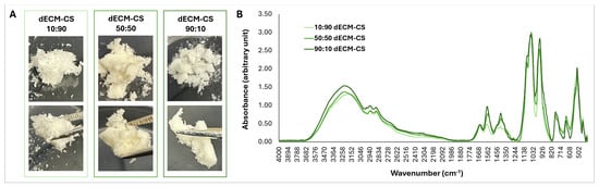
The dECM–CS blends exhibited subtle differences in macroscopic appearance. Following the reaction and lyophilization of raw materials (i.e., dECM and CS), the 10:90 formulation formed a sponge-like, slightly electrostatic compound. As the dECM content increased, the blends became progressively drier, more compact, and aggregated, displaying a more granular texture (Figure 1A).
Figure 1.
Macroscopic appearance and Chemical characterization dECM-CS blends. (A) Dentin ECM-CS blends exhibited subtle differences in macroscopic appearance following lyophilization. The 10:90 formulation displayed a sponge-like structure with slight electrostatic properties, whereas formulations with increasing dentin ECM content appeared progressively drier, more compact, and aggregated, exhibiting a more granular texture. (B) Mean ATR-FTIR spectra (n = 3/formulation) demonstrated similar overall spectral profiles among dECM-CS blends, characterized by common fingerprint regions, while also revealing differences in absorbance intensity and peak positions for selected bands. Peak-intensity and peak-ratio comparisons are provided in Figure S2.
ATR-FTIR analysis revealed ratio-dependent spectral variations among the dECM–CS blends (Figure 1B and Supplementary Material Figure S2A,B), with all formulations exhibiting characteristic vibrational bands of both dECM and CS, consistent with their effective combination. The 10:90 blend was predominantly characterized by CS-associated carbohydrate features, including a broad absorption band between 3200 and 3500 cm−1 attributed to overlapping –OH and –NH vibrations, a low-intensity but well-defined peak at ~2900 cm−1 attributed to symmetric and asymmetric C–H stretching of CH2 and CH3 groups in the pyranose ring, and intense bands in the 1020–1150 cm−1 region corresponding to C–O–C and C–O stretching vibrations [19]. Additionally, Amide I (~1640–1650 cm−1) and Amide II (~1540–1550 cm−1) bands were observed, primarily arising from chitosan’s acetylated units, with presumably lower contributions from dentin matrix components such as collagen and proteoglycans (Figure 1B and Supplementary Material Figure S2A,B) [19,21,22]. The most prominent dECM contribution in this formulation was associated with phosphate vibrations (~1000–1050, ~960, ~650, and ~550 cm−1), attributable to hydroxyapatite, which is abundantly present in partially demineralized dentin.
Increasing dECM content induced progressive spectral changes, as observed in the 50:50 blend, which retained strong chitosan-related carbohydrate bands while exhibiting increased Amide I and II intensities (common to both dECM and CS), indicating a greater contribution of dentin matrix components to these bands. This formulation also showed enhanced contributions from other dentin matrix constituents, including CH2 bending (scissoring) vibrations of the organic collagen matrix, overlapping with carbonate (CO32−) vibrations of hydroxyapatite at ~1400 cm−1 [19,23]. Notably, phosphate-related absorptions in the ~960 and 1000–1050 cm−1 regions remained of high intensity but were comparatively lower than those observed in the 10:90 blend, particularly in the context of the simultaneous increase in Amide I and II vibrations [24,25] (Supplementary Material Figure S2A,B).
The spectral profile of the 90:10 blend appeared more defined and uniform compared to the other two formulations, exhibiting carbohydrate bands attributable not only to chitosan (~1020–1150 cm−1) but also to dentin collagen-associated proteoglycans and glycosaminoglycans (980–1140 cm−1) [26,27]. This was accompanied by a marked increase in Amide II intensity, higher absorption near ~1400 cm−1 (attributed to aliphatic CH2 groups present in the amino acid side chains of collagen) [28], and more well-defined phosphate vibrations [24,25]. Overall, the FTIR profiles demonstrated composition-dependent shifts in carbohydrate-, protein-, and phosphate-related bands, reflecting distinct biochemical signatures across the dECM–CS blends (Figure 1B and Supplementary Material Figure S2A,B).
3.2. Macroscopic Appearance and Viscoelastic Properties of PBS-Suspended dECM-CS Blends
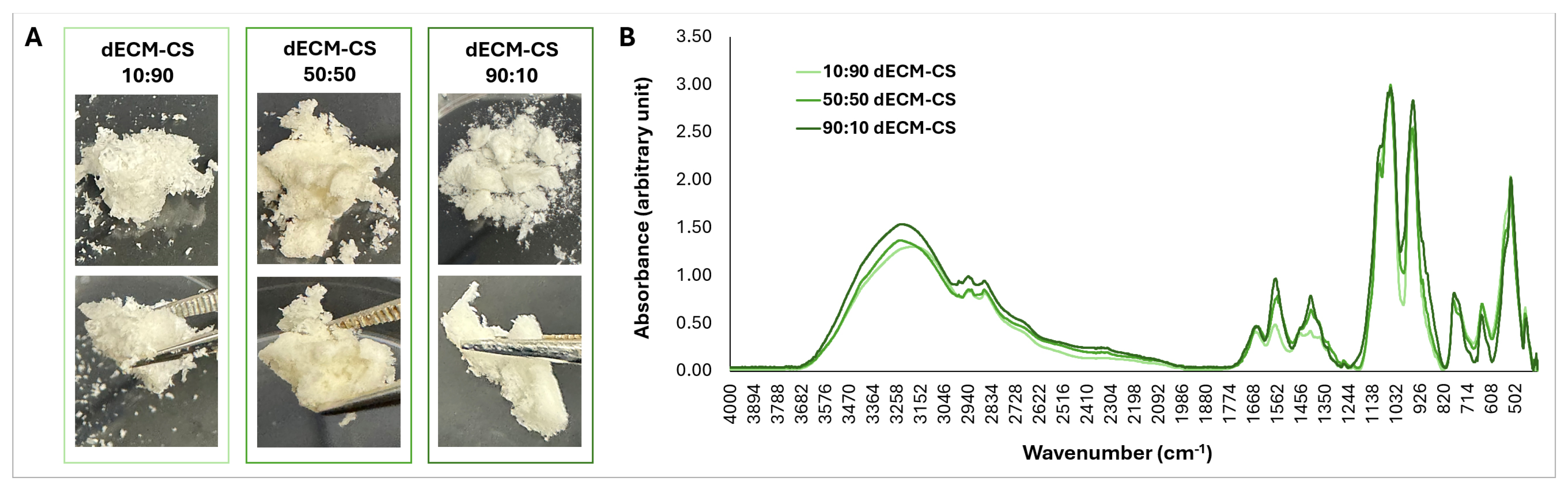
When suspended in PBS, all dECM–CS blends were injectable through a 1-mL syringe fitted with a 27-gauge needle (not shown) and via pipettes suitable for viscous fluids. At 4 °C, the 10:90 and 50:50 formulations appeared more homogeneous than the 90:10 blend (Figure 2A). Following incubation at 37 °C and pH adjustment, the 10:90 (pH 7.3) and 50:50 (pH 7.4) formulations showed obvious gel formation after approximately 15 and 12 min, respectively. In contrast, the 90:10 formulation (pH 7.5) showed limited gelation and remained relatively weak/less consolidated after approximately 25 min (Figure 2A).
Figure 2.
Macroscopic appearance and viscoelastic properties of dECM-CS hydrogel precursor solutions and hydrogels. (A) dECM-CS precursor solutions at 4 °C and neutral pH (upper and middle panels) and after incubation at 37 °C (lower panel). (B) Storage modulus (G′), (C) loss modulus (G″), and (D) tan δ (G″/G′) during a 15–37 °C temperature ramp. Note the y-axis break for the 90:10 formulation in (B,C). (E) Mean G′, G″, and Tan δ during a 600-s time sweep at 37 °C. Data are presented as mean ± SD, n = 3 independent samples per group.
To characterize the viscoelastic behavior of the dECM–CS blends under physiologically relevant applications, rheological properties were evaluated as a function of temperature and time. Rheological analysis across the temperature range of 15–37 °C revealed distinct, composition-dependent viscoelastic behaviors among the dECM-CS hydrogels (Figure 2B–D). The storage modulus (G′), indicative of the elastic component of the hydrogel response, showed a temperature-dependent increase for all formulations. G′ increased from 172 ± 0.4 Pa at 15 °C to 263 ± 9.4 Pa at 37 °C for the 10:90 formulation and from 162 ± 3.5 Pa to 276 ± 5.5 Pa for the 50:50 formulation. In contrast, the 90:10 formulation displayed markedly lower G′ values, ranging from 2.6 ± 0.3 to 3.7 ± 0.5 Pa (Figure 2B), consistent with the limited gelation observed visually.
The loss modulus (G″), corresponding to the viscous component of the viscoelastic response, also differed among the formulations with increasing temperature (Figure 2C). The 50:50 formulation displayed the highest G″ values (24.1 ± 0.6 Pa at 15 °C to 24.3 ± 0.8 Pa at 37 °C), followed by the 10:90 formulation (21.7 ± 1.9 Pa at 15 °C to 18.5 ± 1.5 Pa at 37 °C). On the other hand, the G″ of the dECM–CS 90:10 formulation remained substantially lower, ranging from 0.30 ± 0.01 Pa at 15 °C to 0.24 ± 0.04 Pa at 37 °C. These results indicate that the 50:50 formulation exhibited the greatest viscous contribution, whereas the 90:10 formulation showed minimal viscous resistance, consistent with its relatively weak and incompletely developed gel network.
The loss tangent (tan δ; G″/G′) was then evaluated to compare the relative viscous and elastic contributions of each of the formulations (Figure 2D). As the temperature increased from 15 to 37 °C, the 10:90 formulation showed tan δ values ranging from 0.12 ± 0.01 to 0.05 ± 0.003 Pa, while the 50:50 formulation showed values ranging from 0.15 ± 0.006 to 0.06 ± 0.001 Pa. The 90:10 formulation showed tan δ values ranging from 0.14 ± 0.03 at 15 °C to 0.04 ± 0.001 Pa at 37 °C. The relatively low tan δ values indicate that the elastic component was greater than the viscous component in all three formulations, although the 90:10 formulation had much lower overall G′ and G″ values.
To further examine the behavior of the dECM–CS hydrogels at physiological temperature, rheological measurements were performed at 37 °C for 600 s (Figure 2E). The 50:50 formulation had the highest G′ throughout the time sweep, increasing from 283 ± 5.3 to 371 ± 12.1 Pa. Its G″ remained relatively stable, ranging from approximately 24 ± 0.7 to 26 ± 1.4 Pa, while tan δ decreased from approximately 0.09 ± 0.001 to 0.07 ± 0.001 Pa. The 10:90 formulation also showed an increase in G′ over time, with G′ increasing from approximately 267 ± 10.5 to 311 ± 15.6 Pa. G″ remained relatively stable at approximately 18.4–18.5 Pa, and tan δ decreased from approximately 0.07 ± 0.003 to 0.06 ± 0.005 Pa. In comparison, the 90:10 formulation had much lower G′ and G″ values throughout the measurement. Its G′ increased from approximately 3.6 ± 0.6 to 4.8 ± 0.8 Pa, while tan δ showed greater variation during the time sweep. Collectively, these findings demonstrate that the dECM to CS ratio strongly influenced the rheological properties of the hydrogels. At the end of the 37 °C time sweep, G′ of the 50:50 formulation was higher than that of 10:90, whereas the two showed overlapping values during the temperature ramp.
3.3. Antibacterial Activity
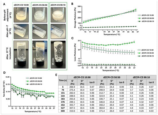
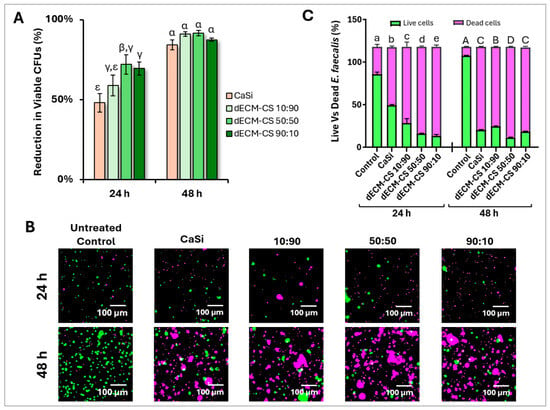
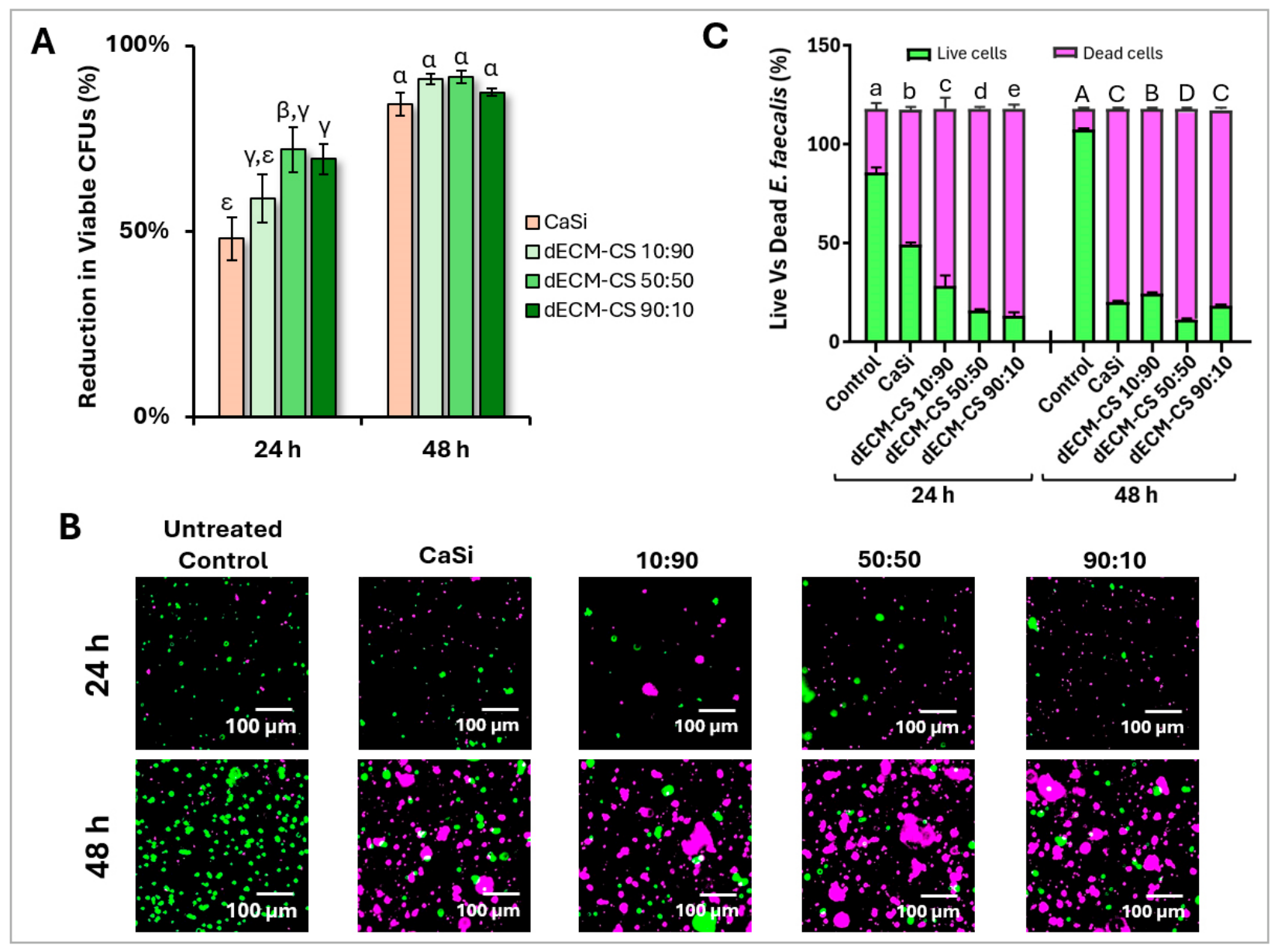
The antibacterial activity of the dECM–CS hydrogels against E. faecalis was evaluated after 24 and 48 h using a colony-forming unit (CFU) assay (Figure 3A). The untreated control was considered to represent 100% bacterial viability, and antibacterial activity was expressed as the percentage reduction in viable bacterial counts relative to the untreated control. At 24 h, CFU reduction differed significantly among the tested groups. Both the dECM–CS 50:50 (72%) and dECM–CS 90:10 (69%) formulations achieved significantly greater CFU reduction than CaSi (48%) (p < 0.05), while the dECM-CS 10:90 formulation (59%) did not differ significantly from CaSi.
Figure 3.
Antimicrobial activity of dECM-CS hydrogels against E. faecalis. (A) Reduction in colony-forming units (CFU/mL) of planktonic E. faecalis relative to the untreated control (100% viability) at 24 and 48 h. (B) Representative fluorescence microscopy images (20×, scale bar = 100 μm) of Live/Dead-stained E. faecalis biofilms on the untreated control, CaSi, and dECM-CS hydrogels at 24 and 48 h; live bacteria fluoresce green and dead bacteria red/magenta. (C) Proportions (%) of live and dead bacteria within biofilms, quantified by ImageJ. Data are presented as mean ± SE of three independent experiments (n = 3 wells per group per experiment). Different Greek letters (A) and lowercase/uppercase letters (C; 24 h/48 h) indicate statistically significant differences (p < 0.05).
After 48 h, CFU reduction increased to 91%, 92%, and 87% for the 10:90, 50:50, and 90:10 formulations, respectively, compared with 84% for CaSi. Within each dECM-CS hydrogel, the reduction in viable bacterial counts was significantly greater at 48 h than at 24 h (p < 0.0001 for the 10:90 formulation, p < 0.001 for the 50:50 and 90:10 formulations). Although the 50:50 formulation showed the highest numerical reduction at both time points, no significant differences were observed among the three dentin dECM–CS blends or between any blend and CaSi at 48 h.
The antibacterial activity of the dECM–CS hydrogels was further evaluated using an E. faecalis biofilm model. Bacteria were incubated directly on the scaffolds to allow biofilm formation, followed by live/dead fluorescence staining to assess bacterial viability based on membrane integrity, with membrane-intact bacteria classified as live and membrane-compromised bacteria classified as dead (Figure 3B). At 24 h, the untreated control biofilm consisted of 73% live and 27% dead bacteria, whereas CaSi resulted in 42% live and 58% dead bacteria. The dECM–CS hydrogels demonstrated a further shift toward the dead bacterial portion, with 24% live and 76% dead bacteria for the 10:90 formulation, 14% live and 86% dead bacteria for the 50:50 formulation, and 11% live and 89% dead bacteria for the 90:10 formulation (Figure 3C). All three hydrogel formulations resulted in significantly lower proportions of live bacteria and higher proportions of dead bacteria within the biofilm compared with the untreated control at 24 h (p < 0.0001). The three dECM–CS formulations also differed significantly from one another (p < 0.0001), with dead-bacteria proportions ranking, from lowest to highest, as 10:90 (76%) < 50:50 (86%) < 90:10 (89%).
At 48 h, the untreated control biofilm comprised 91% live and 9% dead bacteria, whereas all treated groups showed a markedly higher proportion of dead bacteria. The 10:90, 50:50, and 90:10 dECM–CS formulations resulted in 21%, 10%, and 16% live bacteria (79%, 90%, and 84% dead bacteria, respectively), compared with 17% live and 83% dead bacteria for CaSi (Figure 3C). The 50:50 formulation showed a significantly higher proportion of dead bacteria than CaSi (p < 0.0001), whereas the 10:90 formulation showed a significantly lower proportion of dead bacteria than CaSi (p < 0.01), and the 90:10 formulation did not differ significantly from CaSi (p = 0.595). The three dECM–CS formulations also differed significantly from one another (p < 0.001), with dead-bacteria proportions ranking, from lowest to highest, as 10:90 (79%) < 90:10 (84%) < 50:50 (90%).
Taken together, the CFU and live/dead analyses demonstrated substantial antibacterial activity of the dECM–CS hydrogels against both planktonic E. faecalis and hydrogel-associated E. faecalis biofilms. Although increased CS content generally correlated with enhanced antibacterial activity, the reduced efficacy of the 10:90 blend relative to the 50:50 blend suggests that the relationship is not strictly concentration-dependent, partially supporting the initial hypothesis.
In a separate experiment, pure CS and pure dECM samples were shown to reduce viable E. faecalis by 63% and 58% at 24 h and 94% and 82% at 48 h, respectively, relative to their untreated control (Supplemental Materials, Figure S3).
3.4. Biocompatibility
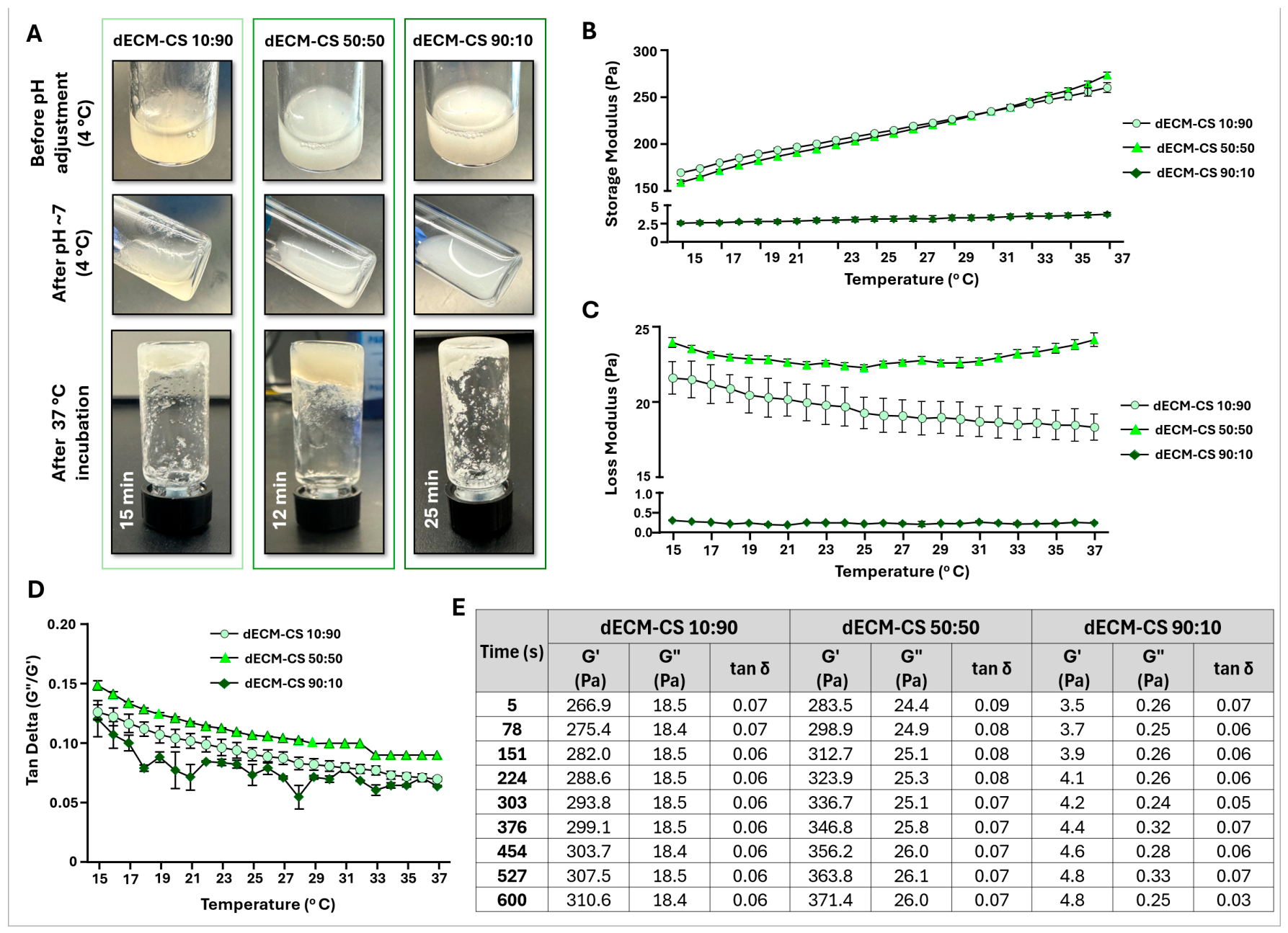
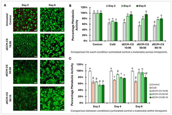
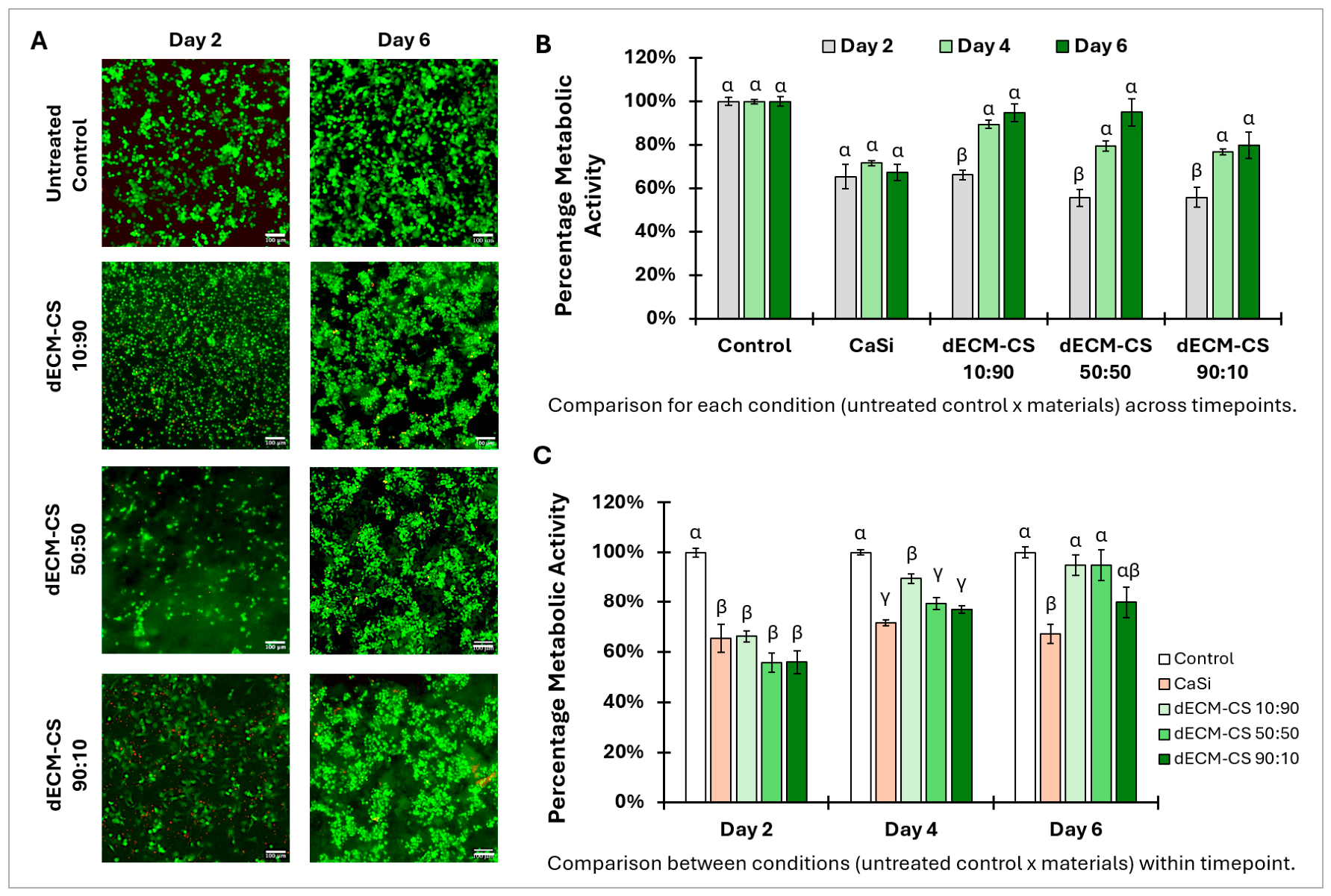
Live/Dead merged fluorescent (green/red) staining demonstrated a favorable cell viability profile for dECM–CS scaffolds, with an overall increase over time (Figure 4A). At day 2, viability ranged from ~85% to 108% across dECM–CS formulations, with the 10:90 scaffold showing the highest viability, while the 50:50 and 90:10 blends exhibited viability comparable to or slightly lower than the control (~83% and 85%, respectively). By day 6, viability remained stable for the 10:90 formulation (~105%) and 90:10 (~86%) formulations but increased significantly for the 50:50 (~110%) compared to the control; however, the 90:10 blend differed significantly from both the 10:90 (p < 0.01) and 50:50 (p < 0.0001) blends. CaSi scaffolds appeared optically opaque under live/dead staining, preventing reliable fluorescence imaging; however, spectrophotometric analysis indicated ~61% and 58% cell viability at day 2 and day 6, respectively.
Figure 4.
Biocompatibility of dECM–CS scaffolds with OD-21 cells. (A) Representative fluorescence microscopy images (20× magnification; scale bar = 100 μm) of merged Live/Dead (green/red) staining on days 2 and 6. (B,C) Metabolic activity (%) compared across time points for each condition (B) and among conditions within each time point (C). Different Greek letters indicate statistically significant differences (p < 0.001 in (B) and p < 0.01 in (C)). Data are presented as mean ± SE of three independent experiments (n = 3 wells per group per experiment).
Cell metabolic activity assessed by the alamarBlue™ assay increased over time for dECM-CS scaffolds (Figure 4B,C). Figure 4B shows the within-material time course, while Figure 4C compares materials at each time point. On day 2, metabolic activity in cells cultured on dECM–CS scaffolds and CaSi was ~56–66% of the control (p < 0.001), indicating an initial adaptation to the scaffold microenvironment. Among the dECM-CS formulations, the 50:50 blend showed the largest overall increase from day 2 to day 6, with metabolic activity increasing significantly from day 2 to day 4 (p < 0.01) and from day 2 to day 6 (p < 0.0001), then plateauing between day 4 and day 6. By day 6, metabolic activity increased across all dECM–CS scaffolds, reaching 80–95% of the untreated control. The 10:90 and 50:50 blends reached the highest levels (95% each), with the 50:50 blend significantly exceeding CaSi (p < 0.05) but not differing significantly from the 10:90 blend, the 90:10 blend, or the control. In contrast, cells cultured on CaSi showed stable metabolic activity (~65–72%) throughout the experiment.
Collectively, the results indicate that cell metabolic activity and viability were high for all dECM–CS hydrogels and were only modestly influenced by the dECM-to-CS ratio. Overall, the formulation containing equal parts of dECM and CS (dECM-CS 50:50) demonstrated the most consistent and progressive outcomes; however, differences were not consistently pronounced across all analyses. Thus, the hypothesis that all formulations would exhibit comparable biocompatibility regardless of composition is supported by the present findings.
In a separate experiment, pure CS and pure dECM samples reached 100% and 108% metabolic activity relative to their untreated control at day 6 (Supplemental Materials, Figure S4).
4. Discussion
This study advances the concept of dentin substrate recycling by demonstrating that this mineralized tissue, typically discarded as biological waste following tooth extraction, can be repurposed through integration with chitosan to generate a functional, cytobiocompatible hydrogel scaffold for regenerative dentistry [18,19]. The results indicate that the physicochemical and biological behavior of dECM–CS blends depends on their compositional ratio; within the three ratios tested, these properties did not change proportionally with composition, although intermediate ratios are needed to confirm this trend. This was most evident in the antimicrobial results, where efficacy did not scale directly with chitosan content—the chitosan-richest formulation (10:90) was among the weakest performers at the earlier time point, while formulations spanning a much wider compositional range performed comparably well, pointing to a meaningful contribution from dECM components alongside CS established antibacterial activity. Accordingly, the hypothesis that dECM–CS hydrogel scaffolds would display composition-dependent handling, biochemical, and antimicrobial properties was supported, but the more specific prediction that increasing chitosan content would directly and proportionally enhance antimicrobial efficacy was not; biocompatibility, by contrast, was preserved across all formulations as hypothesized.
Macroscopic differences observed following lyophilization and resuspension of dECM–CS blends reflect fundamental distinctions in the intermolecular interactions governing scaffold assembly, as previously reported [19,29]. The faster gelation kinetics and greater homogeneity exhibited by the 10:90 and 50:50 dECM–CS formulations are consistent with CS-driven network formation, in which electrostatic interactions, hydrogen bonding, and polymer chain entanglement dominate gelation under physiological pH and temperature [16,30]. In contrast, the delayed gelation and increased heterogeneity of the 90:10 blend suggest reduced availability of CS chains, which limits rapid network percolation and results in weaker physical crosslinking [30,31]. The progressive increase in gelation time observed with higher dECM content further implies that dentin ECM components modulate CS self-association, likely through competitive hydrogen bonding and steric hindrance [32]. Although dentin ECM proteins and proteoglycans contribute to important biological functionality, their comparatively low intrinsic gel-forming capacity appears to diminish network cohesion when present at high proportions, thereby affecting injectability and structural uniformity [23,33].
Molecular-level insights provided by FTIR analysis further reinforce this composition-influenced behavior. The dominance of CS-associated carbohydrate bands in the 10:90 formulation confirms that polysaccharide chemistry governs the structural backbone of CS-rich blends [13,27]. As dECM content increased, the progressive emergence and intensification of collagen-related Amide I and Amide II bands, together with phosphate-associated vibrations, reflect a gradual transition toward ECM-dominated biochemical features [19,24,25]. The coexistence and modulation of carbohydrate, protein, and phosphate bands across all formulations indicate that dECM and CS are not merely physically blended but rather interact through non-covalent mechanisms [30,32]. Electrostatic attraction between CS amine groups and anionic dECM components, such as collagen carboxylates and phosphate groups, likely underlies the observed spectral shifts, supporting the formation of a composition-dependent hybrid network rather than a phase-separated system [30,32]. These interactions are expected to play a central role in regulating network stability, degradation behavior, and the presentation of biologically relevant cues [17,33].
Consistent with the macroscopic gelation behavior and FTIR-based molecular evidence described above, quantitative oscillatory rheology further demonstrates that CS content, rather than dECM content, is the primary determinant of network stiffness in this system. While all three formulations transitioned from a translucent, low-viscosity sol at 4 °C to a cohesive gel upon warming, consistent with the classic thermogelling behavior of CS-based systems [34], the magnitude of that transition differed by nearly two orders of magnitude depending on composition. The dECM–CS 90:10 formulation, containing only 10% CS, showed minimal stiffening across both the temperature ramp and the isothermal hold (G′ < 5 Pa throughout), indicating that dECM alone is insufficient to support a mechanically robust network and likely contributes primarily to bioactive rather than structural function—consistent with the established role of demineralized dentin matrix as a reservoir of matrix-bound bioactive factors rather than a load-bearing scaffold component [2,6]. In contrast, both the 10:90 and 50:50 dECM–CS formulations reached storage moduli in the hundreds of Pascals, indicating that a substantially higher chitosan content is required for effective physical network formation in this system; the large gap between 10% and 50% CS suggests this requirement is met somewhere within that range, though additional intermediate compositions would be needed to define it more precisely.
Notably, the two CS-rich formulations behaved differently under sustained isothermal conditions at 37 °C: whereas 10:90 and 50:50 dECM–CS reached comparable moduli by the end of the temperature ramp, the 50:50 formulation continued to stiffen substantially during the subsequent 600 s hold, ultimately surpassing 10:90 (Figure 2C). This divergence suggests that the 50:50 ratio supports more extensive or sustained network consolidation at physiological temperature, potentially reflecting a more favorable balance between CS’ network-forming capacity and dECM-mediated crosslinking interactions, whereas the CS-majority 10:90 formulation may reach a more kinetically arrested state earlier [35]. Together, these findings indicate that intermediate dECM:CS ratios may offer an advantageous combination of adequate mechanical reinforcement and continued in situ maturation for applications requiring stable gelation at body temperature, while also underscoring that neither maximizing dECM content (for bioactivity) nor maximizing CS content (for stiffness) alone is sufficient to optimize both properties simultaneously.
All dECM–CS blends exhibited pronounced antibacterial activity against E. faecalis, underscoring the intrinsic antimicrobial potential of CS-containing systems [13,22]. The enhanced efficacy observed at 48 h relative to 24 h across all formulations suggests sustained antibacterial action, potentially mediated by continued bacterial membrane disruption and interference with microbial metabolism [19,22,36,37]. Notably, antibacterial efficacy did not scale directly with CS content, particularly at the earlier 24 h time point. Despite containing the highest proportion of CS, the 10:90 formulation performed no better than CaSi and was statistically indistinguishable from it, while both 50:50 and 90:10—spanning a much wider range of dECM:CS ratios—achieved significantly greater and statistically comparable reductions in bacterial viability. This indicates that CS concentration alone is not the primary determinant of early antimicrobial efficacy in this system and that factors such as polymer accessibility, charge distribution, and diffusion of chitosan chains within the hydrogel network likely play a more dominant role than total chitosan content [38,39]. The particularly strong early performance of 90:10—the formulation with the lowest chitosan content but the highest proportion of dECM—further suggests a meaningful contribution from dentin ECM components to the antimicrobial response, potentially through modulation of scaffold porosity, matrix-bound bioactive factors, or sustained exposure of cationic functional groups [19]. By 48 h, these compositional differences were fully resolved, with no significant differences observed among any of the dECM–CS formulations or CaSi, consistent with a time-dependent convergence in antimicrobial performance across all groups.
Cellular assays further highlighted the importance of compositional balance in governing biological responses. Rather than mechanical stiffness alone dictating cytocompatibility, all three dECM–CS formulations, including the mechanically weakest 90:10 ratio, supported robust cell viability and metabolic activity over the 6-day culture period, with predominantly live cells and minimal cytotoxicity by live/dead staining regardless of composition. This suggests that the compositional trade-off identified in the rheological characterization, whereby dECM content favors bioactivity and CS content favors network stiffness, does not translate into a straightforward biological trade-off.
The metabolic activity data reinforces this interpretation while also revealing a composition-dependent gradient in the magnitude of the proliferative response. All three dECM–CS formulations outperformed the CaSi comparator by day 4 and continued to diverge through day 6, suggesting that the dentin-derived matrix component confers a bioactive advantage over a standard calcium silicate-based material, although the specific contribution of dECM beyond supporting cell survival remains to be established [2,6,19]. Notably, however, the 10:90 and 50:50 formulations demonstrated the greatest recovery in metabolic activity by day 6, while the 90:10 formulation showed a more moderate recovery. The strong recovery observed with the 50:50 formulation, together with its performance during the isothermal rheological hold, suggests that this intermediate ratio may represent a particularly favorable balance between the structural and bioactive contributions of chitosan and dentin ECM, respectively. The transient difference in cell morphology observed for the 90:10 formulation at day 2—a more spread, spindle-like phenotype compared to the rounded morphology seen in the stiffer formulations—is consistent with substrate-stiffness-dependent cell spreading behavior described elsewhere [40], though this was not directly investigated here and would benefit from further study.
This study has several limitations that warrant further investigation. First, FTIR indicated that the 2 h phosphoric acid treatment left residual hydroxyapatite; thus, the dECM used here was partially demineralized. Residual mineral likely contributed to the phosphate bands, gelation behavior, and potentially the antimicrobial and cellular responses. Quantitative characterization of mineral content (e.g., XRD or calcium analysis) would strengthen these observations. Second, only three dECM:CS ratios were evaluated. While sufficient to demonstrate that composition influences rheological, antimicrobial, and biological properties, additional intermediate formulations would help more precisely define these transitions. Third, antimicrobial and biocompatibility testing were limited to a single microorganism (E. faecalis) and a single cell line (OD-21), respectively; although both are relevant to the dentin–pulp complex, broader microbial and cellular models would enhance clinical relevance. Fourth, all evaluations were conducted in vitro and over relatively short timeframes, and therefore the long-term degradation, mechanical stability, and bioactivity of these hydrogels under physiological conditions remain to be characterized, ideally including in vivo validation. In addition, single-component controls were not tested alongside blend formulations, handling properties were assessed qualitatively, and parameters such as injection force, swelling, porosity, microstructure, gel fraction, degradation, and osmolarity were not evaluated. Although pooling teeth reduces donor-specific effects, donor variability was not characterized, and comparison with a defined collagen–hydroxyapatite composite could provide additional context. Finally, while FTIR results and macroscopic gelation support the proposed non-covalent interactions between dECM and CS, the underlying mechanisms were not directly measured; future studies evaluating binding affinity, crosslink density, degradation kinetics, and gelation dynamics would provide a more comprehensive mechanistic understanding.
5. Conclusions
Collectively, these findings underscore the importance of compositional tuning in the design of dentin ECM-based hydrogel scaffolds. In particular, among the three ratios tested, the dECM–CS 50:50 formulation represents a promising compromise, combining efficient gelation, robust antimicrobial activity, and favorable biocompatibility. The relationships observed between composition and function across the rheological, antimicrobial, and biological assays further suggest that performance depends on the balance between dentin ECM and chitosan, rather than maximal incorporation of either component alone, although intermediate ratios are needed to define this relationship more precisely. Such insights are critical for the rational development of injectable, bioactive hydrogels intended for pulp protection and regeneration of the dentin–pulp complex, where both antimicrobial defense and cellular compatibility are essential design criteria.
Supplementary Materials
The following supporting information can be downloaded at https://www.mdpi.com/article/10.3390/jfb17110586/s1. Figure S1. ATR-FTIR characterization of mineralized and partially demineralized dentin. (A) Representative ATR-FTIR spectra of mineralized and partially demineralized dentin, highlighting characteristic spectral features (black solid arrows) associated with phosphate (ν1, ν3 PO43−, ~900–1200 cm−1) and the organic matrix (Amide I, ~1645 cm−1). (B) Comparison of the phosphate/Amide I peak ratio between mineralized and partially demineralized dentin. Figure S2. FTIR peak-intensity and peak-ratio comparisons among dECM–CS blends. (A) Intensity (absorbance, arbitrary units) of key FTIR peaks and bands. The 90:10 blend showed higher absorbance in the ~3200–3500, ~2900, ~2850, and ~1056 cm−1 regions than the other formulations (p < 0.01), whereas the 10:90 blend showed lower absorbance in the ~1540–1550 and ~1400 cm−1 regions (p < 0.01). (B) Amide I/Amide II, Amide I/1400 cm−1, and Amide II/carbohydrate peak ratios. The 10:90 and 90:10 blends differed significantly in Amide I/Amide II (p = 0.005), Amide I/1400 cm−1 (p = 0.013), Amide II/carbohydrate at 1049 cm−1 (p = 0.0005), and Amide II/carbohydrate at 1105 cm−1 (p = 0.0145). Figure S3. Antibacterial activity of pure CS and pure dECM against E. faecalis (separate experiment). (A) Bacterial viability by CFU assay at 24 and 48 h, expressed as a percentage of the untreated control. Viability increased significantly from 24 to 48 h in both groups (p < 0.05). (B) Live/dead bacterial proportions in biofilms at 24 and 48 h (Live/Dead staining). Data are mean ± SEM (n = 3 wells/group; 3 independent experiments). Samples were not tested concurrently with the dECM-CS blends and are shown for descriptive purposes only. Figure S4. Metabolic activity of OD-21 cells cultured on pure CS and pure dECM materials (separate experiment). Metabolic activity was assessed by alamarBlue over 6 days and expressed as a percentage of the untreated control. Data are mean ± SD (n = 5 wells/group). No significant differences were observed between groups at any time point, and both 2% CS and 2% dECM maintained >90% viability throughout the experiment. Samples were not tested concurrently with the dECM-CS blends and are shown for descriptive purposes only.
Author Contributions
Conceptualization, M.R.C., S.S. and Z.S.; Methodology, S.S., Z.S., B.O., N.T., A.V., I.S., F.G. and M.R.C.; Software, M.R.C., S.S. and Z.S.; Validation, Z.S., S.S., B.O., N.T., A.V., F.G., I.S. and M.R.C.; Formal Analysis, M.R.C., S.S. and Z.S.; Investigation, S.S., Z.S., B.O., N.T., A.V., I.S., F.G. and M.R.C.; Resources, M.R.C., B.O. and N.T.; Data Curation, M.R.C., S.S. and Z.S.; Writing—Original Draft Preparation, S.S., Z.S., B.O., N.T., A.V., I.S., F.G. and M.R.C.; Writing—Review and Editing, S.S., Z.S., B.O., N.T., A.V., I.S., F.G. and M.R.C.; Visualization, S.S., Z.S., B.O., N.T., A.V., I.S., F.G. and M.R.C.; Supervision, M.R.C., F.G. and I.S.; Project Administration, M.R.C.; Funding Acquisition, M.R.C., Z.S. and S.S. contributed equally to this study. All authors have contributed substantially to the reported work. The authors have reviewed and edited the final manuscript and take full responsibility for the content of this publication. All authors have read and agreed to the published version of the manuscript.
Funding
This study was supported by an MWU-CDMI intramural Dean’s Student Fellowship (B.O. and N.T.) and by the National Institute of Dental and Craniofacial Research under grant R15DE032836 (M.R.C.).
Institutional Review Board Statement
This study utilized extracted human teeth that would otherwise have been discarded. According to a determination by the Institutional Review Board of Midwestern University, Illinois (Protocol #25-0605), this project did not constitute human subjects research because no identifiable donor information was accessed, collected, or recorded.
Informed Consent Statement
Not applicable.
Data Availability Statement
The data presented in this study and the supporting analytical methods used for data analysis will be made available by the authors upon publication.
Acknowledgments
The authors thank Harold Haering Jr. (MWU-CDMI), Ellen Kohlmeir (MWU-IL Core Facility), and Joshua Z. Gasiorowski (MWU-CGS-IL and ORSP-IL) for their technical and scientific support, as well as John C. Mitchell (MWU-CDMA) for the generous gift of the OD-21 cells. AI-assisted tools were used for language editing and proofreading only.
Conflicts of Interest
The authors declare no conflicts of interest. The funders had no role in the design of the study; in the collection, analyses, or interpretation of data; in the writing of the manuscript; or in the decision to publish the results.
Abbreviations
The following abbreviations are used in this manuscript:
| ATR | Attenuated total reflectance |
| BGP | Beta-glycerol phosphate |
| CDMA | College of Dental Medicine-Arizona |
| CDMI | College of Dental Medicine-Illinois |
| CGS | College of Graduate Studies |
| CS | Chitosan |
| dECM | Demineralized dentin extracellular matrix |
| ECM | Extracellular matrix |
| FTIR | Fourier-transform infrared spectroscopy |
| MWU | Midwestern University |
| OD-21 | Murine undifferentiated pulp-derived cells |
| PBS | Phosphate buffer solution |
References
- Goldberg, M.; Kulkarni, A.B.; Young, M.; Boskey, A. Dentin: Structure, composition and mineralization. Front. Biosci. (Elite Ed.) 2011, 3, 711–735. [Google Scholar] [CrossRef] [Scilit] [PubMed]
- Smith, A.J.; Scheven, B.A.; Takahashi, Y.; Ferracane, J.L.; Shelton, R.M.; Cooper, P.R. Dentine as a bioactive extracellular matrix. Arch. Oral Biol. 2012, 57, 109–121. [Google Scholar] [CrossRef] [Scilit] [PubMed]
- Bertassoni, L.E. Dentin on the nanoscale: Hierarchical organization, mechanical behavior and bioinspired engineering. Dent. Mater. 2017, 33, 637–649. [Google Scholar] [CrossRef] [Scilit] [PubMed]
- Reis, M.; Alania, Y.; Leme-Kraus, A.; Free, R.; Joester, D.; Ma, W.; Irving, T.; Bedran-Russo, A.K. The stoic tooth root: How the mineral and extracellular matrix counterbalance to keep aged dentin stable. Acta Biomater. 2022, 138, 351–360. [Google Scholar] [CrossRef] [Scilit] [PubMed]
- Sloan, A.; Smith, A. Stem cells and the dental pulp: Potential roles in dentine regeneration and repair. Oral Dis. 2007, 13, 151–157. [Google Scholar] [CrossRef] [Scilit] [PubMed]
- Schmalz, G.; Widbiller, M.; Galler, K.M. Signaling molecules and pulp regeneration. J. Endod. 2017, 43, S7–S11. [Google Scholar] [CrossRef] [Scilit] [PubMed]
- Athirasala, A.; Lins, F.; Tahayeri, A.; Hinds, M.; Smith, A.J.; Sedgley, C.; Ferracane, J.; Bertassoni, L.E. A novel strategy to engineer pre-vascularized full-length dental pulp-like tissue constructs. Sci. Rep. 2017, 7, 3323. [Google Scholar] [CrossRef] [Scilit] [PubMed]
- Li, J.; Yang, H.; Lu, Q.; Chen, D.; Zhou, M.; Kuang, Y.; Ying, S.; Song, J. Proteomics and N-glycoproteomics analysis of an extracellular matrix-based scaffold-human treated dentin matrix. J. Tissue Eng. Regen. Med. 2019, 13, 1164–1177. [Google Scholar] [CrossRef] [Scilit] [PubMed]
- Wen, B.; Huang, Y.; Qiu, T.; Huo, F.; Xie, L.; Liao, L.; Tian, W.; Guo, W. Reparative dentin formation by dentin matrix proteins and small extracellular vesicles. J. Endod. 2021, 47, 253–262. [Google Scholar] [CrossRef] [Scilit] [PubMed]
- Cunha, D.; Souza, N.; Moreira, M.; Rodrigues, N.; Silva, P.; Franca, C.; Horsophonphong, S.; Sercia, A.; Subbiah, R.; Tahayeri, A.; et al. 3D-printed microgels supplemented with dentin matrix molecules as a novel biomaterial for direct pulp capping. Clin. Oral Investig. 2023, 27, 1215–1225. [Google Scholar] [CrossRef] [Scilit] [PubMed]
- Wu, X.; Peng, W.; Liu, G.; Wang, S.; Duan, B.; Yu, J.; Yang, H.; Huang, C. Extrafibrillarly demineralized dentin matrix for bone regeneration. Adv. Healthc. Mater. 2023, 12, e2202611. [Google Scholar] [CrossRef] [Scilit] [PubMed]
- Xie, Z.; Jiang, W.; Liu, H.; Chen, L.; Xuan, C.; Wang, Z.; Shi, X.; Lin, Z.; Gao, X. Antimicrobial peptide- and dentin matrix-functionalized hydrogel for vital pulp therapy via synergistic bacteriostasis, immunomodulation, and dentinogenesis. Adv. Healthc. Mater. 2024, 13, e2303709. [Google Scholar] [CrossRef] [Scilit] [PubMed]
- Dash, M.; Chiellini, F.; Ottenbrite, R.M.; Chiellini, E. Chitosan—A versatile semi-synthetic polymer in biomedical applications. Prog. Polym. Sci. 2011, 36, 981–1014. [Google Scholar] [CrossRef] [Scilit]
- Li, A.; Ma, B.; Hua, S.; Ping, R.; Ding, L.; Tian, B.; Zhang, X. Chitosan-based injectable hydrogel with multifunction for wound healing: A critical review. Carbohydr. Polym. 2024, 333, 121952. [Google Scholar] [CrossRef] [Scilit] [PubMed]
- Abdel-Mohsen, A.M.; Abdel-Rahman, R.M.; Skotnicova, K. Recent advances and applications of chitin and chitosan hydrogel scaffolds in tissue engineering. Gels 2026, 12, 427. [Google Scholar] [CrossRef] [Scilit] [PubMed]
- Kołodziejska, M.; Jankowska, K.; Klak, M.; Wszoła, M. Chitosan as an underrated polymer in modern tissue engineering. Nanomaterials 2021, 11, 3019. [Google Scholar] [CrossRef] [Scilit] [PubMed]
- Kocak, F.Z.; Yar, M.; Rehman, I.U. In vitro degradation, swelling, and bioactivity performances of in situ forming injectable chitosan-matrixed hydrogels for bone regeneration and drug delivery. Biotechnol. Bioeng. 2024, 121, 2767–2779. [Google Scholar] [CrossRef] [Scilit] [PubMed]
- Arbeed, S.; Osman, M.; Gao, F.; Suchy, S.; Sharmin, Z.; Gasiorowski, J.Z.; Kaminski, A.; Sigar, I.M.; Carrilho, M.R. Bioengineered injectable hydrogel based on the dentin extracellular matrix and chitosan. ACS Omega 2025, 10, 9210–9223. [Google Scholar] [CrossRef] [Scilit] [PubMed]
- Osman, M.; Sharmin, Z.; Suchy, S.; Gao, F.; Kaminski, A.; Mitchell, J.C.; Sigar, I.M.; Carrilho, M.R. Bioinspired smart dentin ECM-chitosan hydrogels for dentin-pulp complex regeneration. J. Dent. 2025, 159, 105811. [Google Scholar] [CrossRef] [Scilit] [PubMed]
- de Lima, F.S.F.; Facury, A. Enterococcus faecalis and endodontic treatment failure: A qualitative systematic review of its role and elimination strategies. Aust. Endod. J. 2026, 52, 585–605. [Google Scholar] [CrossRef] [Scilit] [PubMed]
- Kwon, H.-J.; Cho, H.-H.; Lee, C.W. Chitosan-bridged amide networks in waterborne PVGA films: Structure-surface-barrier coupling and a moisture-retention demonstration. Int. J. Biol. Macromol. 2026, 364, 152067. [Google Scholar] [CrossRef] [Scilit] [PubMed]
- Sousa, J.M.; Braz, E.M.A.; Bezerra, R.D.S.; Morais, A.I.S.; Vieira, A.C.C.; Costa, M.P.; Rizzo, M.S.; Chaves, L.L.; Barreto, H.M.; Osajima, J.A.; et al. Study of the antibacterial and cytotoxic activity of chitosan and its derivatives chemically modified with phthalic anhydride and ethylenediamine. Int. J. Biol. Macromol. 2024, 263, 130292. [Google Scholar] [CrossRef] [Scilit] [PubMed]
- Khurshid, Z.; Cooper, P.R.; Khan, A.S.; Dias, G.; Mazher, J.; Moin, S.F.; Ratnayake, J. Demineralization of camel dentin for dental tissue engineering. Int. Dent. J. 2026, 76, 109620. [Google Scholar] [CrossRef] [Scilit] [PubMed]
- Paschalis, E.P.; DiCarlo, E.; Betts, F.; Sherman, P.; Mendelsohn, R.; Boskey, A.L. FTIR microspectroscopic analysis of human osteonal bone. Calcif. Tissue Int. 1996, 59, 480–487. [Google Scholar] [CrossRef] [PubMed]
- Pires, M.; Mendes Soares, I.P.; Fernandes, L.O.; Anselmi, C.; Gibin, M.S.; Baesso, M.L.; Scheffel, D.L.S.; de Souza Costa, C.A.; Hebling, J. Mineral-to-matrix ratio of caries-affected dentin treated with biomimetic analogs using the dentin fluid and a remineralizing solution as ion donors. J. Dent. 2025, 161, 105993. [Google Scholar] [CrossRef] [Scilit] [PubMed]
- Saarakkala, S.; Julkunen, P. Specificity of fourier transform infrared (FTIR) microspectroscopy to estimate depth-wise proteoglycan content in normal and osteoarthritic human articular cartilage. Cartilage 2010, 1, 262–269. [Google Scholar] [CrossRef] [Scilit] [PubMed]
- Hosney, A.; Ullah, S.; Barčauskaitė, K. A review of the chemical extraction of chitosan from shrimp wastes and prediction of factors affecting chitosan yield by using an artificial neural network. Mar. Drugs 2022, 20, 675. [Google Scholar] [CrossRef] [Scilit] [PubMed]
- Nisar, S.; Liu, H.; Hass, V.; Wang, Y. Dual-functional etchants that simultaneously demineralize and stabilize dentin render collagen resistant to degradation for resin bonding. Dent. Mater. 2023, 39, 1004–1012. [Google Scholar] [CrossRef] [Scilit] [PubMed]
- Alia, A.; Fuks, B.; Sharmin, Z.; Suchy, S.; Venigalla, A.; Gao, F.; Gasiorowski, J.Z.; Pfeifer, C.S.; Carrilho, M.R. Dentin beyond its ontology: A platform to produce dental adhesives. J. Dent. Res. 2026, 220345261461515. [Google Scholar] [CrossRef]
- Sacco, P.; Furlani, F.; De Marzo, G.; Marsich, E.; Paoletti, S.; Donati, I. Concepts for developing physical gels of chitosan and of chitosan derivatives. Gels 2018, 4, 67. [Google Scholar] [CrossRef] [Scilit] [PubMed]
- Fletes-Vargas, G.; Espinosa-Andrews, H.; Cervantes-Uc, J.M.; Limón-Rocha, I.; Luna-Bárcenas, G.; Vázquez-Lepe, M.; Morales-Hernández, N.; Jiménez-Ávalos, J.A.; Mejía-Torres, D.G.; Ramos-Martínez, P.; et al. Porous chitosan hydrogels produced by physical crosslinking: Physicochemical, structural, and cytotoxic properties. Polymers 2023, 15, 2203. [Google Scholar] [CrossRef] [Scilit] [PubMed]
- Khoshdouni Farahani, Z. A unified mechanistic perspective on protein-polysaccharide interactions underlying hybrid gel formation: A mini review. Curr. Res. Food Sci. 2026, 12, 101436. [Google Scholar] [CrossRef] [Scilit] [PubMed]
- Pires Figueiredo, M.; Rodríguez-Fernández, S.; Copes, F.; Mantovani, D. Review of collagen type I-based hydrogels: Focus on composition-structure-properties relationships. npj Biomed. Innov. 2025, 2, 16. [Google Scholar] [CrossRef] [Scilit] [PubMed]
- Chenite, A.; Chaput, C.; Wang, D.; Combes, C.; Buschmann, M.D.; Hoemann, C.D.; Leroux, J.C.; Atkinson, B.L.; Binette, F.; Selmani, A. Novel injectable neutral solutions of chitosan form biodegradable gels in situ. Biomaterials 2000, 21, 2155–2161. [Google Scholar] [CrossRef] [Scilit] [PubMed]
- Kohl, M.; Capellmann, R.F.; Laurati, M.; Egelhaaf, S.U.; Schmiedeberg, M. Directed percolation identified as equilibrium pre-transition towards non-equilibrium arrested gel states. Nat. Commun. 2016, 7, 11817. [Google Scholar] [CrossRef] [Scilit] [PubMed]
- Raafat, D.; von Bargen, K.; Haas, A.; Sahl, H.G. Insights into the mode of action of chitosan as an antibacterial compound. Appl. Environ. Microbiol. 2008, 74, 3764–3773. [Google Scholar] [CrossRef] [Scilit] [PubMed]
- Mirbagheri, V.S.; Alishahi, A.; Ahmadian, G.; Hashemi Petroudi, S.H.; Ojagh, S.M.; Romanazzi, G. Toward understanding the antibacterial mechanism of chitosan: Experimental approach and in silico analysis. Food Hydrocoll. 2023, 147, 109382. [Google Scholar] [CrossRef] [Scilit]
- Kong, M.; Chen, X.G.; Xing, K.; Park, H.J. Antimicrobial properties of chitosan and mode of action: A state of the art review. Int. J. Food Microbiol. 2010, 144, 51–63. [Google Scholar] [CrossRef] [Scilit] [PubMed]
- Ke, C.L.; Deng, F.S.; Chuang, C.Y.; Lin, C.H. Antimicrobial actions and applications of chitosan. Polymers 2021, 13, 904. [Google Scholar] [CrossRef] [Scilit] [PubMed]
- Di, X.; Gao, X.; Peng, L.; Ai, J.; Jin, X.; Qi, S.; Li, H.; Wang, K.; Luo, D. Cellular mechanotransduction in health and diseases: From molecular mechanism to therapeutic targets. Signal Transduct. Target. Ther. 2023, 8, 282. [Google Scholar] [CrossRef] [Scilit] [PubMed]
Disclaimer/Publisher’s Note: The statements, opinions and data contained in all publications are solely those of the individual author(s) and contributor(s) and not of MDPI and/or the editor(s). MDPI and/or the editor(s) disclaim responsibility for any injury to people or property resulting from any ideas, methods, instructions or products referred to in the content. |
© 2026 by the authors. Licensee MDPI, Basel, Switzerland. This article is an open access article distributed under the terms and conditions of the Creative Commons Attribution (CC BY) license.