


Abstract
Bone tissue engineering has emerged as a promising alternative to conventional grafting approaches for treating critical-sized bone defects. To overcome the limitations of conventional biomaterials, considerable effort has been directed toward the development of bioactive nanocomposite hydrogels. Among the nanomaterials used in these systems, Laponite (LAP), a synthetic nanoclay composed of disk-shaped nanosilicate platelets, stands out due to its unique physicochemical properties, including high surface area, shear-thinning behavior, ionic dissolution products, and strong interactions with polymers, proteins, and cells. Across diverse polymer systems, incorporation of LAP consistently improves rheological and mechanical performance, facilitating injectable formulations while enhancing structural stability and shape fidelity for extrusion-based bioprinting. However, the magnitude and biological consequences of these effects depend on polymer chemistry, LAP concentration and dispersion, crosslinking, and construct architecture. LAP nanocomposite hydrogels also exhibit pronounced osteoinductive capacity, promoting osteogenic differentiation of mesenchymal stem cells through sustained release of bioactive ions, including silicon, magnesium, and lithium, and activation of bone-related signaling pathways. In addition, LAP-containing hydrogels influence key components of the regenerative microenvironment by supporting angiogenesis and modulating immune responses. Preclinical studies across multiple animal defect models generally demonstrate enhanced bone formation, mineralization, vascularization, and defect healing compared with LAP-free controls. Despite these encouraging findings, important translational challenges remain. Long-term biocompatibility, degradation behavior, biodistribution of degradation products, manufacturing reproducibility, scalability, and regulatory requirements have not yet been comprehensively addressed. Furthermore, most evidence is derived from short- to medium-term preclinical studies, with limited data from large-animal models and no established clinical applications to date. Collectively, this review provides a balanced assessment of LAP nanocomposite hydrogels, highlighting both their considerable potential and the key scientific, safety, and regulatory barriers that must be overcome to enable successful clinical translation.
1. Introduction
The repair of bone defects caused by trauma, disease, or surgical resection remains a major clinical challenge. Conventional therapeutic approaches, such as autologous bone grafts, are limited by donor site morbidity and availability [1]. Although widely used clinically, allogeneic and synthetic grafts generally possess limited intrinsic osteoinductive potential compared with autografts [1,2]. Injectable and minimally invasive biomaterial strategies have therefore attracted increasing attention, as they enable conformal filling of irregular defects and localized delivery of bioactive cues while reducing surgical burden [3,4,5]. Among these approaches, hydrogel-based scaffolds have been widely investigated for bone tissue engineering due to their high water content, tunable physicochemical properties, and ability to mimic aspects of the native extracellular matrix [6,7,8]. However, many conventional polymeric hydrogels suffer from insufficient mechanical strength, uncontrolled degradation, and limited intrinsic bioactivity, which restrict their effectiveness in supporting osteogenesis and long-term bone regeneration [7,9,10].
To address these limitations, nanocomposite hydrogels incorporating inorganic components have been developed to enhance mechanical performance and biological functionality [11,12,13]. Laponite (LAP), a synthetic nanosilicate clay composed of disk-shaped nanoplatelets, has emerged as a particularly promising nanofiller for hydrogel systems due to its ability to act as a physical crosslinker and to release bioactive ions such as silicon, magnesium, and lithium [13,14,15]. Incorporation of LAP into polymeric hydrogels has been reported to significantly improve rheological behavior, injectability, shear-thinning properties, and shape fidelity, enabling minimally invasive delivery and advanced fabrication techniques such as extrusion-based 3D bioprinting [16,17,18]. Beyond mechanical reinforcement, LAP nanocomposite hydrogels have demonstrated intrinsic osteoinductive properties, promoting the adhesion, proliferation, and osteogenic differentiation of mesenchymal stem cells (MSCs) and osteoblasts without the need for high doses of exogenous growth factors [7,13,19].
Several studies have further shown that LAP nanocomposite hydrogels can modulate the local biological microenvironment by sustaining growth factor release, enhancing angiogenesis, and regulating immune responses via macrophage polarization, all of which contribute to improved bone regeneration in vivo [14,20,21]. Moreover, in vivo investigations using critical-sized bone defect models have demonstrated that LAP nanocomposite hydrogel systems can significantly enhance new bone formation, mineralization, and defect healing compared to LAP-free controls [9,11,22]. Collectively, these findings indicate that LAP nanocomposite hydrogels represent a versatile and multifunctional platform for bone tissue engineering, integrating mechanical reinforcement, controlled bioactivity, and biological signaling within a single injectable or printable system [18,23,24].
Despite the rapid expansion of research in this field, current studies remain highly fragmented, with limited critical discussion linking material design, physicochemical behavior, and biological performance. In particular, there is a lack of comprehensive analysis specifically addressing how LAP influences hydrogel functionality, osteogenic mechanisms, injectability, printability, and translational potential in bone regeneration. To address this gap, we critically examine recent advances in LAP nanocomposite hydrogel systems for bone tissue engineering, focusing on material design strategies, biological interactions, and mechanisms of action, as well as current limitations, translational challenges, and future research directions.
LAP nanocomposite hydrogels have been widely explored for drug delivery applications owing to their excellent biocompatibility, biodegradability, and ability to precisely regulate drug release kinetics by modulating hydrogel swelling behavior and mechanical properties [25,26]. Furthermore, the physicochemical properties of LAP have been extensively exploited to develop tunable pH-responsive drug delivery systems, in which changes in the local microenvironment can influence nanoparticle–drug interactions and release profiles [25]. As a result, LAP-based hydrogels have attracted considerable attention as versatile drug delivery platforms across numerous biomedical applications [25,26]. Several comprehensive reviews have already summarized the design principles, mechanisms, and therapeutic applications of LAP-mediated drug delivery systems; therefore, readers interested in these broader aspects are referred to these publications [25,26,27,28]. In this review, we focus on the application of LAP nanocomposite hydrogels in bone tissue engineering and regenerative medicine.
This review primarily considers systems in which an aqueous polymer network constitutes the continuous phase, and LAP is incorporated as a rheological modifier, physical crosslinker, bioactive component, or therapeutic depot. LAP-containing nanofibers, thermoplastic scaffolds, ceramic constructs, and other non-hydrogel materials are discussed only when they provide a relevant mechanistic comparison or when they are integrated with an LAP-containing hydrogel in a multilevel composite. To facilitate comparison, the preclinical systems are considered as three broad categories: acellular LAP nanocomposite hydrogels, LAP-containing therapeutic delivery systems, and mechanically reinforced or hard–soft composite constructs. These categories should not be considered equivalent because the relative contribution of LAP, the polymer network, incorporated biologics, and rigid structural components differ substantially among them.
2. Fundamentals of Laponite
LAP is a synthetic layered clay material with a structural formula Na+0.7[Si8Mg5.5Li0.3O20(OH)4]0.7− originally developed by Laporte Industries Ltd. (currently, BYK-Chemie GmbH, Germany) [29,30]. Its crystal structure consists of an octahedral layer sandwiched between two tetrahedral layers, the so-called 2:1 or TOT (tetrahedral-octahedral-tetrahedral) structure (Figure 1). In aqueous suspensions, LAP exhibits extremely rich behavior due to the interplay of electrostatic interactions, which will be discussed below. The TOT layer is negatively charged due to partial substitution of divalent Mg2+ with Li+ in the octahedral sheet. In a dry state, negatively charged TOT layers are stacked together into tactoids since Na+ ions in the interlayer space compensate for the charge and link layers together. On the other hand, in the wet state, hydration of Na+ ions first increases the interlayer spacing, and then, as Na+ ions diffuse into bulk water, electrostatic repulsion of negatively charged basal surfaces leads to separation of tactoids into platelike crystals with an average diameter of 25 nm and a thickness of approximately 1 nm [31]. While the basal planes are negatively charged, the edges expose -OH groups that can be protonated or deprotonated depending on pH. With the isoelectric point at about pH 11, the edges carry a positive charge at neutral pH, which is about 10% of the negative charge of the basal planes (60 vs. 700 electron charges) [32]. It should also be noted that LAP is not inert in aqueous solutions: prolonged exposure to neutral to low pH conditions leads to gradual dissolution of LAP particles with the release of Na+, Mg2+, Li+, and soluble silicon species [29,33].
Figure 1.
The structure, dispersion, and charge characteristics of Laponite (LAP) nanodisks. LAP is composed of stacked disk-shaped platelets formed by layered silicate sheets. Upon dispersion in aqueous media, these stacks can delaminate into individual or partially exfoliated nanodisks, with the extent of exfoliation and dispersion depending on factors such as medium composition, ionic strength, pH, concentration, and aging time. Each nanodisk exhibits an anisotropic charge distribution, with negatively charged basal surfaces and positively charged edges. This dual-charge nature, combined with van der Waals forces, leads to complex interparticle interactions that are essential for hydrogel formation and reinforcement in biomedical applications. Figure designed using BioRender.com.
The presence of opposite charges on the basal plane and edges, on the one hand, and a highly anisotropic crystal shape, on the other hand, leads to the formation of complex percolating networks in LAP dispersions, which can be based on face-to-edge (house-of-cards) and parallel, partially overlapped local arrangements [34]. Depending on the ionic strength and LAP concentration, this leads to a complex phase diagram containing attractive and repulsive glass phases and gels, as well as thixotropic rheological behavior due to destruction and recovery of the network under shear stress [32,34].
3. Laponite Nanocomposite Hydrogels
LAP-based hydrogels are commonly designed as nanocomposite systems in which synthetic nanosilicate platelets are incorporated into polymeric matrices to enhance structural integrity and functional performance [7,18,35]. LAP acts as a physical crosslinker through electrostatic interactions, hydrogen bonding, and supramolecular assembly with polymer chains such as gelatin, alginate, chitosan, hyaluronic acid, and silk fibroin, thereby stabilizing hydrogel networks without the need for high crosslinker concentrations [7,19,22,36]. Several studies demonstrated that LAP nanocomposite hydrogels exhibit enhanced shape fidelity and printability compared to LAP-free systems, allowing the fabrication of stable three-dimensional scaffolds under mild processing conditions [17,37]. From a mechanical standpoint, LAP addition significantly increases compressive modulus, tensile strength, and overall mechanical stability of hydrogels, addressing the inherent weakness of many soft polymeric matrices used in bone tissue engineering [9,11,13].
The mechanical reinforcement imparted by LAP has been attributed to nanosheet-induced strengthening and the formation of dense nanocomposite networks, enabling hydrogels to withstand substantial deformation while maintaining structural integrity [38,39]. LAP concentration plays a critical role in tuning physicochemical properties, as increasing nanosilicate content generally enhances mechanical strength while reducing swelling ratio and water content of the hydrogel systems [6,36,40]. In addition to mechanical reinforcement, LAP incorporation modulates hydrogel degradation behavior, with several studies reporting slower degradation rates and improved long-term stability as LAP content increases [11,17,36].
Importantly, LAP concentration alone does not determine nanocomposite performance. The magnitude of reinforcement depends on the affinity between the polymer and the charged LAP surfaces, the extent of nanoparticle exfoliation and dispersion, the ionic composition of the medium, the crosslinking mechanism, and the architecture of the resulting construct [26,41]. Polymers containing ionic or hydrogen-bonding groups can form strong reversible interactions with LAP and may show substantial increases in viscosity, yield stress, and mechanical stability [26,42]. However, excessive polymer–particle association or LAP loading can reduce swelling and mesh size, impede injection, retard degradation, and restrict cell-mediated remodeling. Conversely, insufficient dispersion or weak polymer–LAP compatibility can result in particle aggregation and heterogeneous mechanical and biological properties. Thus, greater LAP loading should not be interpreted as universally beneficial, and the optimal formulation represents a balance among processability, structural reinforcement, mass transport, degradation, ion exposure, and cellular permissiveness.
At the microstructural level, it is important to distinguish between the hydrogel mesh size and the scaffold porosity. The hydrogel mesh size, which exists on the nanometer scale, governs the diffusion of water, nutrients, ions, proteins, and other soluble factors through the polymer network and can be influenced by polymer crosslinking density and LAP content. In contrast, scaffold porosity refers to the micron-scale pore architecture that supports cell infiltration, vascular ingrowth, and tissue integration. Several LAP nanocomposite hydrogels have been reported to exhibit interconnected porous or mesoporous structures that facilitate regenerative processes within bone defects [37,43,44]. Both mesh size and pore size distribution can be tailored through polymer composition, fabrication strategy, and nanosilicate concentration, enabling the design of scaffold architectures optimized for bone tissue regeneration [43,44].
Thermal stability of hydrogel systems is also enhanced by LAP incorporation, as demonstrated by thermogravimetric analyses showing increased resistance to thermal degradation in nanocomposite hydrogels [6,11]. In addition to providing structural reinforcement, LAP can release potentially bioactive dissolution products; the biological significance and proposed mechanisms of this ion release are discussed in Section 4 [13,20,45]. Overall, the effects of LAP on injectability, mechanical stability, degradation, and bioactivity depend on LAP concentration, polymer chemistry, particle dispersion, and crosslinking strategy.
LAP incorporation can enhance the mechanical and physicochemical performance of composite scaffolds, although the magnitude of reinforcement is strongly dependent on the surrounding matrix and fabrication strategy. For example, the incorporation of LAP into polycaprolactone-based 3D-printed scaffolds increased mechanical strength, specifically yielding a compressive strength ranging from 2 to 12 MPa and a compressive modulus between 0.05 and 0.5 GPa, while simultaneously improving bioactivity and cell viability, demonstrating synergistic structural and biological benefits [46]. Similarly, LAP-containing nanofiber membranes exhibited up to a 2.5-fold enhancement in tensile strength (increasing from ~160 kPa to ~400 kPa) and a four-fold increase in Young’s modulus (from 2.56 MPa to 9.20 MPa) compared to pristine polymer systems, highlighting the strong reinforcing capability of nanosilicates [47]. In porous scaffold systems, LAP increases compressive modulus and structural stability, with reported Young’s modulus values ranging from 190 to 260 kPa, while maintaining an interconnected porosity of approximately 75% and ensuring long-term stability by retaining over 60% of the scaffold mass after 40 days [48]. Additionally, LAP has been shown to improve hydrophilicity and surface bioactivity, thereby enhancing cell adhesion and proliferation [49,50].
Injectability is a defining feature of many LAP nanocomposite hydrogels developed for bone tissue engineering, as it enables minimally invasive delivery and conformal filling of irregular bone defects [4,22,51]. The incorporation of LAP nanoplatelets has been widely reported to significantly enhance the rheological properties of hydrogel systems, particularly by increasing viscosity and imparting pronounced shear-thinning behavior [16,17,18]. Shear-thinning behavior allows LAP nanocomposite hydrogels to flow under applied stress during injection or extrusion while rapidly recovering their solid-like structure after stress removal, which is critical for in situ retention at bone defect sites [7,51]. Several studies have demonstrated that LAP functions as a physical crosslinker via electrostatic interactions between negatively charged clay surfaces and positively charged polymer segments, enabling injectable gel formation without harsh chemical crosslinking [7,19,36]. Rheological analyses showed that LAP nanocomposite hydrogels exhibit rapid recovery of storage modulus after high-strain deformation, supporting repeated injectability and structural self-recovery [18,39,52].
Injectable formulations containing LAP have been successfully developed using a wide range of polymer matrices, including gelatin, alginate, hyaluronic acid, silk fibroin, poly(N-isopropylacrylamide), and methacrylated polymers, indicating the versatility of nanosilicate-mediated rheological control [4,7,17,19,22,36]. The injectability of LAP nanocomposite hydrogels has also been shown to be tunable by adjusting nanosilicate concentration, polymer composition, and ionic environment, allowing optimization of flow behavior and mechanical stability for specific bone regeneration applications [6,36,40]. Several studies reported that increasing LAP content enhances yield stress and structural stability but may reduce injectability if excessive nanosilicate loading is used, highlighting the importance of balancing rheology and handling properties [36,40]. In extrusion-based 3D bioprinting, LAP-mediated rheological behavior has been shown to protect encapsulated cells from excessive shear stress during printing while maintaining high shape fidelity of printed constructs [16,17,18]. Injectable LAP nanocomposite hydrogels have also demonstrated the ability to retain position within actively bleeding or irregular bone defects, which is attributed to their shear-thinning and rapid solidification characteristics [22,51]. Beyond conventional injection, advanced strategies such as cryoprinting and thermosensitive gelation have been combined with LAP reinforcement to create injectable systems with shape-memory behavior and enhanced post-injection stability [53,54]. Collectively, current evidence indicates that LAP plays a central role in enabling injectable, shear-thinning, and self-recovering hydrogel systems, making rheological control a key advantage of LAP-based materials for minimally invasive bone tissue engineering [16,18,51].
The most straightforward approach to generating LAP nanocomposite hydrogels involves mixing LAP nanodisks into aqueous polymer solutions and allowing gelation to occur through electrostatic interactions between the charged clay surfaces and oppositely charged polymer chains [55]. This method requires minimal processing steps and avoids the use of organic solvents, thereby reducing cytotoxicity concerns. More advanced fabrication strategies have incorporated photopolymerizable synthetic macromers to generate LAP-based nanocomposite hydrogels. For example, osteoblast-laden 3D bioprinted constructs were developed using a two-channel system consisting of a polyethylene glycol diacrylate (PEGDA)–LAP nanocomposite hydrogel and a hyaluronic acid phase for cell encapsulation [13]. The PEG–clay component was subsequently cross-linked via UV-induced photopolymerization, forming a mechanically stable scaffold while maintaining high osteoblast viability within the construct [13]. While photopolymerization enables rapid gelation and efficient stabilization of printed structures, its use in this system primarily serves to preserve construct fidelity and cell viability during fabrication rather than to achieve complex spatial cross-linking patterning.
Interpenetrating network (IPN) formation is an important strategy for preparing LAP nanocomposite hydrogels [56,57]. Unlike simple mixing approaches, where LAP particles often remain as aggregates within the polymer matrix, IPN systems allow LAP nanoparticles to first self-assemble into a percolated “house-of-cards” network before integration with the polymer phase [56]. This results in a dual organic–inorganic network in which the LAP and polymer networks interpenetrate, leading to significantly improved mechanical strength, shear-thinning behavior, and structural stability even at low LAP concentrations [56,57]. Such IPN architectures are particularly advantageous for injectable hydrogels and extrusion-based bioprinting applications requiring enhanced shape fidelity and mechanical robustness. Emerging approaches have extended LAP use to microgel and granular hydrogel systems, where nanosilicate incorporation enhanced osteogenic differentiation and mineral deposition while enabling tunable mechanical properties at the microscale level [58]. Core–shell and zonal bioprinting strategies using LAP have enabled spatially controlled delivery of differentiation factors, supporting the creation of osteogenic and chondrogenic compartments within a single construct [59].
Among the various strategies for fabricating LAP nanocomposite hydrogels, supramolecular coassembly between positively charged peptide amphiphiles (PAs) and negatively charged LAP nanodisks has emerged as an effective approach for constructing structurally robust three-dimensional hydrogel networks (Figure 2) [12]. In this system, electrostatic interactions promote the organization of PA nanofibers around disk-shaped LAP nanoparticles, forming an interconnected organic–inorganic hybrid architecture in which LAP functions as a physical crosslinker to reinforce the hydrogel matrix. This coassembled structure not only enhances mechanical stability but also creates a highly anisotropic and hierarchical nanoscale environment that could be used to guide biomineralization or de novo bone tissue formation [12].
Figure 2.
Schematic illustration of the supramolecular coassembly between negatively charged Laponite (LAP) nanodisks and positively charged peptide amphiphiles (PAs) for the fabrication of bioinspired three-dimensional hydrogels that support hydroxyapatite mineralization. (a) Molecular structure of the PA molecules, highlighting their functional domains alongside the structure of a LAP nanodisk. (b) Electrostatic interactions between PAs and LAP drive the formation of a supramolecular network, resulting in (c) mechanically stable organic–inorganic hybrid hydrogels composed of interconnected PA nanofibers physically reinforced by LAP nanodisks. Upon exposure to mineralizing ions, (d) ionic diffusion through the hydrogel matrix initiates (e) hydroxyapatite nucleation followed by (f) hierarchical crystal growth into high-aspect-ratio nanorod assemblies organized as spherical mineral clusters. (g) Chemical structures of histidine-functionalized peptide amphiphiles used in the system. Reproduced from [12] under the CC BY 4.0 license.
Some studies explored LAP as one component among multiple bioactive materials, where its role is synergistic rather than dominant (Table 1). For instance, composite bioinks incorporating multiple nanoparticles alongside LAP demonstrated improved mechanical properties and enhanced stem cell proliferation and differentiation, although other materials such as hydroxyapatite or whitlockite contributed more strongly to osteogenic outcomes [60]. The combination of LAP nanocomposite hydrogels with bioactive components such as MSCs, platelet-rich fibrin, extracellular vesicles, or growth factors has been shown to further improve in vivo bone regeneration outcomes compared with acellular or single-component systems [22,61,62,63].
Table 1.
Representative effects of LAP incorporation on the physicochemical and processing properties of hydrogel and composite systems.
Attribution of improved biological performance specifically to LAP also requires appropriate controls. The strongest evidence for an LAP-specific effect is provided by studies comparing otherwise equivalent formulations with and without LAP. In multicomponent systems containing therapeutic proteins, platelet derivatives, extracellular vesicles, cells, or rigid frameworks, the observed response reflects interactions among several components and should not be assigned to LAP alone. In such cases, LAP may principally influence rheology, protein retention, ion availability, or interfacial properties, while another component provides the dominant osteogenic or mechanical stimulus. Accordingly, results from complex composite systems should be interpreted as evidence of formulation-level synergy unless factorial or component-specific controls establish the individual contribution of LAP.
4. Laponite-Based Advanced Fabrication Techniques
Advanced fabrication strategies have increasingly been employed to exploit the rheological and physicochemical properties of LAP nanocomposite hydrogels to create complex, functional bone tissue engineering constructs. Extrusion-based 3D bioprinting is one of the most widely reported approaches, as the shear-thinning and rapid recovery behavior imparted by LAP enables high-fidelity printing of cell-laden and acellular hydrogel scaffolds under mild conditions [16,17,37,64]. LAP incorporation has been shown to improve printability, filament stability, and shape retention of bioinks composed of polymers such as alginate, gelatin methacrylate, methylcellulose, and hyaluronic acid, allowing the fabrication of free-standing three-dimensional constructs suitable for skeletal applications [16,17,65,66]. Several studies reported that LAP-based bioinks support high cell viability during and after printing, enabling the direct fabrication of osteogenic constructs containing mesenchymal stem cells or osteoblasts [13,17].
Beyond conventional printing, cryoprinting and dual-crosslinking strategies have been developed to produce injectable, shape-memory, and macroporous LAP nanocomposite hydrogels that recover their structure after injection and support cell infiltration [53,54]. Multifunctional LAP nanocomposite hydrogels have also been engineered by combining nanosilicates with bioactive polymers, peptides, or adhesive motifs to impart additional properties such as self-healing, tissue adhesion, and mechanical reinforcement [19,21,38]. Several advanced systems reported the use of supramolecular or dynamic crosslinking mechanisms involving LAP nanoplatelets to generate hydrogels with injectability, self-recoverability, and spatial adaptability for irregular bone defects [19,38,52]. LAP has also been incorporated into composite and multi-level scaffold designs, where a rigid, load-bearing framework is combined with a softer LAP-reinforced hydrogel phase to simultaneously provide mechanical support and a cell-friendly microenvironment [9,67].
The multifunctionality of LAP-based systems has been further enhanced by integrating growth factor delivery, oxygen-releasing components, antimicrobial agents, or immunomodulatory molecules within the hydrogel matrix [14,52,54]. In these systems, LAP nanoplatelets play a dual role by reinforcing the hydrogel structure and regulating the sustained release of incorporated bioactive agents, thereby improving therapeutic efficacy and spatial localization in bone defect sites [14,20,63]. Several studies demonstrated that multifunctional LAP nanocomposite hydrogels capable of simultaneously promoting osteogenesis, angiogenesis, and immunomodulation lead to superior bone regeneration outcomes compared with single-function materials [21,52,61]. Therefore, advanced fabrication strategies combined with multifunctional material design have enabled LAP nanocomposite hydrogels to evolve from simple injectable fillers into sophisticated, bioactive platforms capable of addressing the mechanical, biological, and spatial requirements of bone tissue engineering [16,23,24].
5. Laponite for In Vitro Osteogenic Induction
LAP promotes osteogenesis through multiple, interconnected physicochemical and biological mechanisms (Figure 3) [68]. Even when dispersed in cell-culture media, LAP has demonstrated dose-dependent enhancement of osteogenic markers, including RUNX2, BGLAP, and SPARC expression, together with increased ALP activity, type I collagen synthesis, and calcium phosphate deposition [69,70]. Importantly, these effects have been observed in the absence of conventional osteogenic supplements, indicating that LAP can provide intrinsic osteoinductive cues [68]. However, its osteogenic activity should not be attributed exclusively to the release of soluble ions. Instead, it likely results from the combined influence of dissolution products, particle–cell and particle–protein interactions, and, when LAP is incorporated into a scaffold, LAP-induced changes in the physicochemical and mechanical properties of the cellular microenvironment.
Figure 3.
Proposed cellular mechanisms and biological effects of Laponite in osteogenesis and bone regeneration. Upon exposure to physiological conditions, silicate-based materials undergo dissolution, releasing ionic species that interact with cell membranes and are internalized via endocytosis. These ions modulate ion channels and activate intracellular signaling pathways, including Wnt/β-catenin signaling through Li+-mediated inhibition of GSK-3β, and promote the colocalization of enzymes and substrates. Intracellular uptake of particles and their dissolution products can upregulate osteogenic transcription factors, including RUNX2, thereby increasing the expression of osteogenic markers (ALP, OCN, OPN, and COL1A1). Collectively, these processes can enhance extracellular matrix mineralization by promoting calcium deposition, hydroxyapatite nucleation, and accelerated mineral maturation. The released ions and cellular responses can stimulate osteogenic differentiation (e.g., increased ALP activity and early lineage commitment), improve cell adhesion and proliferation (including enhanced cell spreading), promote angiogenesis via upregulation of VEGF and vascular network formation, and modulate the immune microenvironment by reducing inflammation and supporting M2 macrophage polarization, thereby creating a pro-regenerative environment. Figure designed using BioRender.com.
Dissolution of LAP can release bioactive species, including orthosilicic acid [Si(OH)4], Mg2+, and Li+, which have been associated with collagen synthesis, integrin-related signaling, mineralization, and regulation of pathways such as Wnt/β-catenin [13,20,45,68]. Nevertheless, the relative contribution of each ion remains incompletely resolved. For example, lithium supplied at concentrations equivalent to those released from LAP did not reproduce the full osteogenic response of intact LAP particles, suggesting that particle-associated properties and other dissolution products are also important [71]. This finding contrasts with reports that lithium released from lithium-doped calcium phosphate cement can activate Wnt/β-catenin signaling and promote osteogenic differentiation and bone regeneration [72]. This apparent discrepancy is likely related to differences in lithium concentration and delivery context. Whereas lithium was incorporated into calcium phosphate cement at therapeutically active concentrations, the amount released from osteogenically active LAP was estimated to be substantially lower and did not independently reproduce the effects of intact LAP [71]. Thus, although lithium can promote osteogenesis at appropriate concentrations, LAP-induced osteogenesis should not be attributed primarily to lithium release.
LAP may adsorb serum and extracellular-matrix proteins onto its highly charged surfaces, altering their local concentration, conformation, and presentation to cell-surface receptors. LAP may also facilitate the local accumulation and transport of calcium and phosphate species and modulate intracellular signaling through clay–protein and clay–membrane interactions [68,73]. Experimental evidence indicating that lithium alone does not account for LAP-induced osteogenesis supports this broader, multifactorial interpretation [71].
In LAP-containing hydrogels and scaffolds, additional matrix-mediated mechanisms become relevant. The incorporation of LAP typically increases stiffness, surface roughness, hydrophilicity, mineral nucleation capacity, and protein adsorption, although the magnitude of these effects depends on LAP concentration, polymer chemistry, crosslinking, and scaffold architecture [50,58]. Increased matrix stiffness and altered ligand presentation can enhance focal-adhesion formation and cytoskeletal tension, potentially activating mechanosensitive regulators such as YAP/TAZ and their downstream osteogenic programs [74,75]. These mechanical effects must be balanced against cellular permissiveness, because excessive stiffening or particle loading may restrict cell spreading, migration, and matrix remodeling [76]. LAP dissolution products may also partially buffer acidic degradation products generated by some polymer matrices, thereby maintaining a local environment more favorable for cell activity and apatite formation [50]. This buffering effect is formulation-dependent and should not be assumed for every LAP-containing material. Studies of polymer–LAP composites similarly attribute improved cellular responses to a combination of ion release, surface roughness, hydrophilicity, apatite formation, and regulation of the local acid–base environment rather than to ion delivery alone [50,77].
Transcriptomic analyses of human MSCs exposed to LAP nanosilicates have provided further insight into cellular uptake and biological activity [78]. Gene expression profiling indicates that LAP nanoparticles are primarily internalized via clathrin-mediated endocytosis upon interacting with the cell membrane [78]. After internalization, the particles are trafficked into endosomal and lysosomal compartments, where progressive acidification promotes protonation of reactive groups at the platelet edges and destabilization of the magnesium-containing octahedral layer [79]. This process facilitates the leaching of Mg2+ and Li+, disrupts the layered silicate structure, and progressively converts the silica component into soluble orthosilicic acid [Si(OH)4] [78,80]. LAP degradation has therefore been proposed to generate Mg2+, Li+, and orthosilicic acid within acidic intracellular compartments, although the rate and completeness of dissolution depend on particle concentration, aggregation state, exposure time, and local chemical conditions. At physiologically relevant exposure levels, these soluble degradation products are generally regarded as biocompatible and can enter normal metabolic or excretory pathways, with orthosilicic acid being readily filtered and excreted in urine [50,81]. Nevertheless, describing LAP as undergoing immediate or invariably complete dissolution would be too definitive, because its aqueous stability is strongly dependent on concentration, ionic composition, aggregation, and the surrounding microenvironment [82]. Thus, intracellular dissolution and ion release represent important components of LAP bioactivity, but they act alongside protein adsorption, membrane interactions, mineral-ion recruitment, local pH regulation, and matrix-mediated changes in mechanical properties and mechanotransduction to produce the overall osteogenic response [50,74,83].
LAP nanocomposite hydrogels have been widely reported to promote osteogenic differentiation of MSCs and osteoblast lineage cells, highlighting their intrinsic bioactivity beyond mechanical reinforcement [7,13,22]. Multiple in vitro studies demonstrated that incorporation of LAP into hydrogel matrices significantly enhances the expression of osteogenic markers, including alkaline phosphatase (ALP), runt-related transcription factor-2 (RUNX2), osteocalcin (OCN), osteopontin, and type I collagen, compared with LAP-free controls [6,7,84,85]. The osteoinductive capacity of LAP nanocomposite hydrogels has been observed across a wide range of polymer systems, including gelatin, alginate, silk fibroin, hyaluronic acid, and methacrylated hydrogels, indicating that the osteogenic effects are largely attributable to the nanosilicate component rather than the polymer matrix alone [7,17,18].
Several studies reported that LAP nanocomposite hydrogels can induce osteogenic differentiation in the absence of exogenously supplied osteogenic growth factors, suggesting that the material itself provides sufficient biochemical and mechanical cues to direct cell fate [13,19,69]. A key mechanism underlying this osteogenic activity is the gradual release of bioactive ions from LAP nanoplatelets during hydrogel degradation [13,45,86]. These released ions have been shown to enhance the proliferation and osteogenic differentiation of bone marrow mesenchymal stem cells by modulating intracellular signaling pathways involved in bone formation [40,45,61]. In particular, silicon and magnesium ions released from LAP were reported to promote osteogenic differentiation and matrix mineralization, thereby improving bone-forming capacity in both in vitro and in vivo models [13,45,54]. Lithium ions released from LAP nanocomposite hydrogels have also been implicated in osteogenic regulation, as lithium is known to influence signaling pathways related to bone metabolism and stem cell differentiation [20,45].
Further mechanistic insights indicate that LAP-mediated osteogenesis is strongly dependent on ion release and scaffold composition. LAP-loaded coaxial fiber scaffolds promoted endochondral ossification in critical-sized defects through sustained release of Si and Mg ions and activation of Hippo, Wnt, and Notch signaling pathways [87]. Transcriptomic and mechanistic investigations revealed that LAP-mediated osteogenesis involves activation of multiple signaling pathways, including MAPK/ERK, PI3K-AKT, TGF-β/Smads, Wnt/β-catenin, and AMPK/mTOR pathways [19,40,61,88]. For example, hydrogel composites containing 2% LAP were reported to enhance osteogenic differentiation of bone marrow mesenchymal stem cells through activation of the MAPK/ERK pathway [40]. However, this apparent optimum should not be considered a universal value, as the biological effects of LAP are highly formulation-dependent. The concentration of LAP influences scaffold stiffness, ion-release profiles, porosity, and cell permissiveness, and the balance among these factors ultimately determines osteogenic outcomes. In certain hydrogel systems, 2% LAP may offer a favorable balance of mechanical support and sustained ion delivery without excessively restricting cell spreading or matrix remodeling, whereas alternative formulations may require different LAP concentrations to achieve optimal performance.
Consistent with this concept, hydrogels containing 2% LAP have been shown to yield optimal osteogenic outcomes, with significantly increased expression of RUNX2, ALP, and COL1, as well as enhanced bone regeneration in vivo [89]. This concentration should not be interpreted as a universal optimum because LAP-dependent responses vary with polymer composition, crosslinking, particle dispersion, and culture conditions. Additionally, LAP-containing microcarriers and scaffolds demonstrated enhanced alkaline phosphatase activity and matrix mineralization, confirming direct stimulation of osteogenic differentiation [90]. LAP nanocomposite hydrogels have also been reported to synergize with incorporated biological components, such as platelet-rich fibrin, extracellular vesicles, or growth factors, to further amplify osteogenic differentiation via ion-mediated and signaling-dependent mechanisms [20,61,63]. Collectively, the in vitro evidence indicates that LAP can contribute to osteogenic differentiation through a combination of ion release, particle–protein and particle–cell interactions, and matrix-mediated signaling. However, the relative contribution of these mechanisms remains dependent on LAP concentration, polymer chemistry, crosslinking, and culture conditions [13,23,24].
LAP has also been shown to modulate the local physicochemical microenvironment, thereby favoring bone regeneration. For instance, LAP degradation produces alkaline products that can neutralize the acidic degradation byproducts of polymer matrices, thereby creating a mildly alkaline microenvironment conducive to osteogenesis [50]. Additionally, LAP incorporation significantly enhances apatite mineralization and promotes calcium phosphate deposition, further supporting bone matrix formation [46].
In addition to their osteoinductive capacity, LAP nanocomposite hydrogels have been increasingly reported to actively modulate the immune microenvironment, which plays a critical role in successful bone regeneration [21,52,61]. Several studies have demonstrated that incorporating LAP into hydrogel systems promotes macrophage polarization toward the anti-inflammatory M2 phenotype while suppressing pro-inflammatory M1 responses, thereby creating a pro-regenerative immune environment [21,52,61,91]. This immunomodulatory effect has been observed in both in vitro and in vivo models, in which LAP nanocomposite hydrogels reduced inflammatory marker expression and enhanced tissue repair outcomes compared with LAP-free controls [21,44,52]. Mechanistic investigations indicate that LAP-mediated macrophage polarization is associated with activation of signaling pathways such as PI3K-AKT, JAK1-STAT1, and calcium-related signaling cascades, which subsequently influence osteogenic differentiation of bone marrow stromal cells [52,88]. However, the immunomodulatory effects of LAP are not consistent across all experimental systems. Direct exposure of RAW264.7 macrophages to dispersed LAP was reported to induce a concentration-dependent M1-associated response, including increased reactive oxygen species and pro-inflammatory cytokine production. Conditioned medium from macrophages exposed to higher LAP concentrations also impaired the osteogenic differentiation of bone marrow stromal cells, whereas the presence of stromal cells shifted the macrophage response toward an M2-associated phenotype and improved bone formation in vivo [92]. These findings suggest that macrophage polarization depends on LAP concentration, particle presentation, the surrounding matrix, and interactions with other cell types. Therefore, the M2-associated responses reported for multicomponent LAP hydrogels should not necessarily be attributed to LAP alone.
Beyond immune regulation, LAP nanocomposite hydrogels have shown a strong capacity to support angiogenesis, which is essential for nutrient transport, oxygen supply, and coupling of vascularization with osteogenesis during bone healing [14,20,54]. The angiogenic potential of these systems is partly attributed to the ability of LAP nanoplatelets to bind, stabilize, and sustain the release of pro-angiogenic factors such as vascular endothelial growth factor (VEGF), platelet-derived growth factors, and platelet lysate components [14,20,93]. Several studies reported that LAP incorporation significantly delayed the release of VEGF and other growth factors from hydrogel matrices, resulting in prolonged bioactivity and enhanced endothelial cell recruitment and tubule formation [14,20,93,94]. In vitro experiments demonstrated that LAP nanocomposite hydrogels promote endothelial cell proliferation, migration, and vascular network formation, indicating a direct contribution to angiogenic processes [20,54,93]. In vivo studies further confirmed that enhanced angiogenesis induced by LAP nanocomposite hydrogels translates into increased neovascularization within bone defect sites, which correlates with improved bone formation and defect healing [9,14,21].
The release of bioactive ions from LAP, particularly silicon and magnesium ions, has also been implicated in promoting angiogenesis and vascular maturation, thereby supporting the coupling of vascular and bone tissue regeneration [13,20,54]. Notably, several multifunctional LAP nanocomposite hydrogels have been designed to simultaneously regulate immune responses and angiogenesis, demonstrating synergistic effects on osteogenic differentiation and overall bone regeneration outcomes [21,52,61,94]. These findings indicate that LAP nanocomposite hydrogels actively orchestrate the bone regenerative microenvironment by integrating immunomodulatory and pro-angiogenic functions, which complement their osteoinductive and mechanical roles in bone tissue engineering applications [14,21,24]. Comparative studies have indicated that although LAP exhibits osteogenic potential, other bioceramics such as tricalcium phosphate may provide superior osteogenic performance under certain conditions, emphasizing the importance of careful material selection and combination strategies [95]. Therefore, LAP may be most effective when integrated into synergistic composite systems tailored to specific applications.
6. Preclinical Data on Laponite Nanocomposite Hydrogels
The bone regenerative potential of LAP nanocomposite hydrogels has been evaluated in a range of preclinical animal models, including rat and rabbit critical-sized bone defect models, to assess their capacity for bone regeneration [14,22]. Critical-sized calvarial defect models in rats are among the most commonly employed systems, with multiple studies reporting significantly enhanced bone formation and defect closure compared with LAP-free hydrogel controls [9,20,22]. Additional critical-sized defect studies have reported increased mineral deposition and bone formation in LAP-containing systems relative to LAP-free or polymer-only comparators, although the magnitude of improvement depends on the formulation, defect model, and comparator used [9,11,22]. In these models, injectable LAP nanocomposite hydrogels demonstrated good defect conformity, in situ retention, and integration with surrounding bone tissue, supporting their suitability for minimally invasive repair strategies [20,22,51]. Rabbit models have also been employed to validate the translational potential of LAP nanocomposite hydrogels, where improved bone bridging, vascularization, and mechanical stability were observed when LAP was incorporated into hydrogel formulations or composite scaffolds [38,96]. Several in vivo studies reported that LAP nanocomposite hydrogels promote robust new bone formation characterized by increased mineral deposition, higher bone volume fraction, and improved micro-computed tomography parameters relative to control groups [9,11,20]. In addition to calvarial defects, femoral and tibial defect models have been used to assess the regenerative performance of LAP-based systems, demonstrating enhanced bone repair, osteointegration, and defect bridging in load-bearing environments [4,9,11].
Ex vivo chick chorioallantoic membrane (CAM) assays have been used to assess vascular infiltration and early mineralization in LAP-containing biomaterials [97]. These models further confirmed the ability of LAP nanocomposite hydrogels to stimulate vascular infiltration and mineralization, providing rapid screening evidence of their pro-regenerative performance [12,65,66,98]. Although CAM assays offer a cost-effective and physiologically relevant platform for assessing angiogenic and osteogenic potential, findings should be interpreted alongside mammalian in vivo studies, as differences in immune responses, tissue remodeling, and long-term regenerative outcomes may limit direct translation to clinical applications. In vivo angiogenesis within bone defects treated with LAP nanocomposite hydrogels has been frequently reported, with enhanced neovascularization correlating with improved bone formation and defect healing [14,20,21]. However, LAP-specific angiogenic effects can be established only when otherwise equivalent LAP-free controls are included, particularly in systems that also contain VEGF or other pro-angiogenic components.
Preclinical studies of LAP nanocomposite hydrogels can be broadly classified into three groups according to the principal function of LAP within the construct: (i) acellular LAP nanocomposite hydrogels in which the nanoclay provides intrinsic osteogenic, mechanical, or immunomodulatory activity; (ii) LAP-based hydrogels used as local delivery depots for biologics or therapeutic cells; and (iii) LAP incorporated into mechanically reinforced, printed, or otherwise rigid composite frameworks. This classification is useful because the biological and translational performance of these systems depends not only on LAP-mediated ion release, but also on whether regeneration is driven primarily by the material itself, by a delivered therapeutic payload, or by a load-supporting multiscale architecture.
6.1. Acellular LAP Nanocomposite Systems
Acellular systems provide the clearest evidence that LAP can contribute directly to tissue regeneration without requiring transplantation of exogenous cells or supplementation with recombinant growth factors. Nevertheless, LAP is rarely used as the sole constituent of an implant. Instead, it is typically incorporated into natural or synthetic polymer networks, where its intrinsic bioactivity is combined with the handling and degradability of the hydrogel phase. Early in vivo evidence was obtained with a thermally responsive pNIPAM-co-DMAc hydrogel crosslinked by LAP and supplemented with hydroxyapatite nanoparticles [4]. Following injection into rat femoral defects, the material was well tolerated, with no overt local inflammatory response, organ toxicity, or systemic toxicity. Although the response varied according to animal age and the inclusion of mesenchymal stromal cells, the acellular formulation promoted host–cell infiltration, integration with adjacent bone, collagen type I deposition, and Runx2 expression in aged animals. Importantly, adding transplanted rat mesenchymal stromal cells did not improve healing in young animals and was associated with a weaker response than the sham-operated control, illustrating that greater biological complexity does not necessarily produce better regeneration [4]. More direct evidence of material-driven repair has been generated using LAP-polymer nanocomposites without exogenous cells or therapeutic proteins. A photocrosslinked PEGDA (polyethylene glycol diacrylate)–LAP hydrogel promoted intramembranous bone formation in tibial defects in osteopenic rats, with the LAP-containing formulation demonstrating regenerative potential in a compromised bone environment [11]. Similarly, an injectable regenerated silk fibroin/LAP hydrogel significantly increased new bone formation in a rat calvarial-defect model. Mechanistic experiments linked its osteogenic activity to activation of protein kinase B (AKT) signalling, while increasing LAP content enhanced alkaline phosphatase activity and the expression of osteogenic markers in bone marrow mesenchymal stromal cells [99]. LAP concentration must be carefully optimized because increasing nanoclay content alters water content, stiffness, degradation, and ion exposure [40].
Acellular LAP nanocomposite hydrogels have also been designed to regulate the inflammatory and vascular components of bone repair. An injectable GelMA/dopamine-grafted alginate hydrogel containing polydopamine-functionalized LAP promoted M2 macrophage polarization, endogenous stem-cell recruitment, neovascularization, and bone formation in a rat critical-sized calvarial defect model without exogenous cells or growth factors [21]. However, because this was a multicomponent formulation, the individual contribution of LAP should be interpreted according to the comparator groups included in the study. In another growth-factor-free system, LAP was co-assembled with hyaluronic acid-tyramine and a copper-containing peptide amphiphile. The resulting multicomponent hydrogel improved mechanical stability, reduced swelling and degradation, and produced greater bone formation than Bio-Oss (Geistlich) in a rabbit maxillary sinus-floor augmentation model [24]. These findings indicate that an acellular construct can stimulate repair through the combined presentation of material-derived osteogenic, angiogenic, and immunomodulatory signals rather than by acting solely as a passive scaffold. Promising results have also been reported outside conventional calvarial models. An enzymatically crosslinked silk fibroin-LAP hydrogel promoted simultaneous regeneration of hyaline-like cartilage and subchondral bone after implantation into full-thickness rabbit osteochondral defects [100]. A bilayered alginate-polyacrylamide construct containing LAP in its bone-facing layer similarly improved mechanical performance, promoted M2-associated macrophage responses, and supported osteochondral repair over 12 weeks in a rat model [91]. For minimally invasive bone augmentation, an injectable hydroxyapatite/LAP/alginate hydrogel promoted osteogenic differentiation of periosteum-derived stromal cells and generated new bone after subperiosteal administration in rats [101]. Collectively, these studies support the preclinical potential of cell-free, LAP-containing constructs, although the specific contribution of LAP is difficult to isolate in multicomponent systems and the evidence remains largely limited to small-animal models and short observation periods.
6.2. LAP-Based Systems for the Delivery of Biologics or Therapeutic Cells
The high surface area and anisotropic charge distribution of LAP facilitate the adsorption of proteins, extracellular vesicles, small molecules, and other therapeutic agents [50,74,83]. Incorporation into a hydrogel can therefore reduce burst release, protect bioactivity, and extend local exposure. This depot function is particularly relevant to potent osteogenic growth factors, for which high systemic or local doses may cause adverse effects [25,26]. One of the earliest demonstrations showed that self-assembled LAP gels localized BMP-2 and induced ectopic bone formation in mice at concentrations approximately 3000-fold lower than those used clinically [14]. Subsequent studies combined LAP with polymer networks to improve both localization and handling. Bisphosphonate-functionalized hyaluronic acid interacted selectively with positively charged LAP edge sites, preserving negatively charged surface sites for protein adsorption and maintaining localized BMP-2 activity for more than six weeks in vivo [62]. Injectable self-healing polysaccharide hydrogels containing LAP-BMP-2 complexes likewise preserved BMP-2 bioactivity, extended release beyond four weeks, and improved bone regeneration in the rat calvarial bone defect model compared with LAP or BMP-2 alone [102]. These studies demonstrate that LAP can reduce the amount of freely diffusible protein while sustaining an osteogenic stimulus at the implantation site. The depot approach has since expanded to angiogenic and disease-specific biologics. A GelMA-LAP hydrogel providing sustained release of PDGF-BB restored stromal-cell proliferation, migration, and osteogenic differentiation under high-glucose conditions and repaired calvarial defects in diabetic rats [45]. Similarly, a GelMA hydrogel containing LAP and VEGF-A delayed VEGF release, promoted endothelial progenitor-cell recruitment and neovascularization, and improved healing of critical-sized rat calvarial defects [20]. Because both systems contained potent therapeutic proteins, the regenerative outcomes should be attributed to the complete delivery system rather than to LAP alone.
Platelet-derived preparations provide a broad spectrum of endogenous growth factors. GelMA microspheres containing platelet lysate and LAP sustained growth-factor availability for up to 28 days and enhanced microvascular formation and pulp-like tissue development after subcutaneous implantation in nude mice [93]. More recently, 3D-printed hydrogels containing platelet-rich plasma or injectable platelet-rich fibrin incorporated LAP as part of the delivery matrix. These multicomponent constructs were associated with vascularization, M2 macrophage polarization, endogenous stromal-cell recruitment, and bone regeneration in rat defect models [61,94]. However, the relative contributions of LAP and the platelet-derived components were not readily separable. LAP-based depots have also been used for non-protein therapeutic cargo. Supramolecular LAP-chitosan hydrogels enabled hierarchical co-delivery of simvastatin and demineralized bone matrix, improving osteogenesis and calvarial defect repair in mice [103]. A rosmarinic acid-releasing hyaluronic acid/LAP hydrogel combined antibacterial and antibiofilm activity with macrophage reprogramming and osteogenic differentiation, accelerating repair in a Staphylococcus aureus-infected rat skull-defect model [52]. In dental applications, a LAP/gelatin hydrogel loaded with epigallocatechin gallate-copper complexes suppressed Porphyromonas gingivalis, shifted macrophages away from an M1 phenotype, and promoted alveolar and periodontal tissue regeneration in a rat periodontitis model [104].
Extracellular vesicles represent another relevant cargo class. GelMA-LAP hydrogels improved the controlled release and local activity of epigenetically enhanced osteoblast-derived extracellular vesicles, thereby increasing stromal cell migration, matrix production, and mineralization in vitro, although this study did not yet establish therapeutic efficacy in an orthotopic bone defect model [63]. A more recent formulation delivered LAP-primed apoptotic vesicles from a thiol-ene hydrogel. In a rat femoral-defect model, sustained vesicle delivery promoted osteogenesis and angiogenesis while supporting anti-inflammatory macrophage polarization and mitochondrial metabolic recovery [105]. This study supports the use of LAP in vesicle-based delivery systems, although the respective contributions of LAP priming, the vesicles, and the hydrogel carrier require appropriate component-specific controls. In parallel, several studies have used LAP-containing hydrogels as vehicles for therapeutic cells. A gelatin/alginate/LAP injectable hydrogel maintained the survival and proliferation of encapsulated rat bone marrow mesenchymal stromal cells, and the cell-loaded formulation generated more bone than the corresponding acellular hydrogel in an 8-mm critical-sized rat calvarial defect [22]. Osteoblast-laden, 3D-bioprinted PEGDA/LAP/hyaluronic acid constructs maintained high viability and promoted osteogenic differentiation through the release of magnesium and silicate ions. The scaffold also demonstrated excellent osteogenic potential, promoting tibial bone repair and ectopic bone formation in the rat model [13]. Likewise, 3D-bioprinted nano-LAP hydrogels carrying bone marrow mesenchymal stromal cells improved bone formation in rat critical-sized cranial defects through activation of PI3K/AKT signalling [86]. A silk fibroin-based shape-memory cryogel containing LAP supported stem cell proliferation and osteogenic differentiation in vitro, and demonstrated excellent tissue compatibility, host cell invasion, and integration following subcutaneous implantation in mice [53]. However, the mixed response to cell addition in earlier work and the additional manufacturing, regulatory, and viability constraints associated with living products indicate that cell delivery should be justified against simpler acellular alternatives rather than assumed to be intrinsically superior.
6.3. LAP-Based Mechanically Reinforced, Printed, or Otherwise Rigid Composite Systems
Hydrogels alone generally provide limited load-bearing capacity and may lose their intended geometry after implantation. A third group of preclinical strategies, therefore, uses LAP within 3D-printed constructs, mineral-filled matrices, or hard-soft composite implants [94,98]. In these systems, LAP can improve ink rheology and shape fidelity, enhance interfacial bonding, stimulate osteogenesis, or serve as a depot within a mechanically stable framework. An important proof of concept was provided by extrusion-printed LAP-gellan gum scaffolds [65]. These constructs exhibited stable geometry, restricted swelling, sustained protein retention, and supported cell viability. When VEGF was incorporated into the printing system, the scaffolds promoted preferential vascular infiltration in an ex vivo chick CAM model [65]. A related GelMA-LAP bioink supported human bone marrow stromal-cell proliferation and osteogenic differentiation while improving VEGF retention and vascular penetration ex vivo [66]. Subsequent subcutaneous implantation studies in mouse models showed that nanoclay-based printed scaffolds carrying human bone marrow stromal cells or adsorbed BMP-2 generated mineralized tissue in vivo, with the cell-laden printed constructs outperforming acellular and cast comparators [98]. These studies established the multifunctional role of LAP as a rheological modifier, osteogenic component, and local protein-binding phase within printed scaffolds.
Greater mechanical reinforcement has been achieved by integrating LAP-containing hydrogels with thermoplastic or ceramic frameworks. PRP-GelMA/AlgMA-LAP bioink was printed layer by layer with polycaprolactone to create a porous composite scaffold that sustained growth factor release, promoted vascular ingrowth, and improved bone regeneration in rat femoral condyle defects [94]. In a multilevel implant, a hard porous bioceramic framework was combined with a softer LAP-reinforced alginate phase. The cell-free construct achieved approximately 84% repair of a critical-sized rat bone defect after eight weeks, supporting the concept that load-bearing architecture and a cell-instructive hydrogel microenvironment can be integrated within the same implant [9]. LAP has also been combined with calcium phosphate cement in gelatin to produce an injectable hybrid whose degradation rate could be adjusted to match new bone formation. The formulation induced ectopic ossification in the mouse model and promoted ligament-graft osseointegration in the rabbit model of tibial graft-bone integration, demonstrating transformation of the implant into host bone rather than persistence as an inert filler [10].
More recent rigid composite designs have focused on coordinating osteogenesis with vascularization. A polycaprolactone/nano-hydroxyapatite/LAP framework containing a VEGF-functionalized GelMA phase maintained a stable interconnected architecture, supported osteogenic differentiation, provided sustained VEGF availability, and enhanced vascularized bone regeneration in vivo [67]. Likewise, bioactive glass macrobeads incorporated into a cellulose-based hydrogel containing LAP generated more pronounced bone formation and fixation than free bioactive glass beads in rabbit calvarial and femoral defect models [96]. Although these systems are often described as hydrogels, their therapeutic performance is derived from the integration of the hydrogel with a substantially stiffer thermoplastic, cement, glass, or ceramic phase and should therefore be distinguished from injectable soft nanocomposites.
Taken together, the available evidence does not support a universally beneficial effect of increasing LAP content or construct complexity. Greater LAP loading can improve rheological stability and mechanical properties but may simultaneously compromise injectability, degradation, or cellular remodeling. Similarly, the addition of cells, growth factors, bioceramics, or rigid frameworks may improve selected outcomes but makes the specific contribution of LAP more difficult to establish. These trade-offs, together with major differences in experimental design, currently prevent direct ranking of LAP formulations across studies.
Overall, preclinical evidence indicates that LAP can fulfil three complementary roles: an intrinsically bioactive nanomaterial in acellular hydrogels, an adsorption-based depot for local delivery of cells and biologics, and a reinforcing or biofunctional phase in rigid composite frameworks [4,11,52]. Acellular formulations offer the simplest translational route and demonstrate that bone repair can be achieved without living cells or supraphysiological growth-factor doses. Delivery systems may provide stronger or more targeted effects in compromised, infected, or poorly vascularized defects, but introduce additional manufacturing and regulatory complexity. Rigid and multilevel composites provide superior architectural stability and are likely to be more relevant for large or mechanically demanding defects. However, comparisons across studies remain limited by differences in LAP grade and concentration, polymer chemistry, defect site, animal species, observation time, and outcome measures. Future preclinical studies should therefore establish dose–response relationships, ion-release and degradation profiles, long-term biodistribution, systemic safety, mechanical performance under physiological loading, and efficacy in large-animal models before clinical translation is considered.
7. Drug and Growth Factor Delivery for Osteogenic Induction
One of the major limitations of conventional hydrogels is their highly porous structure, which often results in rapid burst release of loaded therapeutics, thereby reducing local bioactivity and increasing the risk of systemic side effects [106]. In this context, LAP has emerged as an attractive nanomaterial for controlled therapeutic delivery due to its high surface area, discotic charge distribution, and strong electrostatic interactions with biomolecules [26,107]. LAP provides multiple binding and loading sites, including external surfaces, platelet edges, interlayer galleries, and interparticle spaces. These diverse interaction domains contribute to high loading efficiencies and enable the incorporation of a wide range of therapeutic agents [28,108]. The negatively charged basal surfaces and positively charged edge regions generate heterogeneous charge distributions that facilitate electrostatic interactions with molecules of varying physicochemical characteristics [26,107]. In addition, the abundance of surface oxygen atoms, hydroxyl groups, and exchangeable interlayer cations promotes ion exchange, hydrogen bonding, van der Waals interactions, and adsorption processes [108]. These properties enable sustained, localized, and spatiotemporally controlled delivery of growth factors, small-molecule drugs, and extracellular vesicles, thereby enhancing osteogenic induction and tissue regeneration [107].
For example, GelMA–LAP hydrogels delivering platelet-derived growth factor-BB (PDGF-BB) have been shown to restore stem cell migration and osteogenic activity under diabetic conditions, effectively overcoming the inhibitory effects of high glucose environments on bone regeneration [45]. Among osteogenic growth factors, bone morphogenetic protein-2 (BMP-2) has been extensively investigated in LAP-based systems. LAP nanoplatelets form stable electrostatic complexes with BMP-2, preserving its bioactivity while extending release for more than four weeks [102]. Sustained BMP-2 presentation promoted cell spreading and osteogenic differentiation, resulting in successful ectopic bone formation in vivo using BMP-2 concentrations significantly lower than those typically required in clinical collagen sponge systems [14]. These findings highlight the ability of LAP to localize growth factor activity and reduce the need for supra-physiological doses, thereby reducing the side effects of growth factor use.
Beyond osteogenesis, LAP-based hydrogels have also been explored for the delivery of angiogenic factors. Incorporation of LAP into alginate-methylcellulose bioinks slowed the release of vascular endothelial growth factor (VEGF), thereby prolonging angiogenic stimulation compared to LAP-free controls [16]. Similarly, VEGF-loaded LAP–gellan gum constructs demonstrated significantly enhanced vascular infiltration and integration in chick chorioallantoic membrane models, confirming the pro-angiogenic effect of nanosilicate-mediated growth factor delivery [65]. To improve spatial control over therapeutic delivery, coaxial core–shell bioprinting approaches have been developed in which LAP-containing cores function as localized depots for osteogenic and chondrogenic factors such as BMP-2 and TGF-β3, enabling spatially defined cell differentiation within engineered constructs [59]. This strategy minimized factor diffusion between adjacent regions and enabled targeted osteogenic and chondrogenic differentiation within osteochondral constructs.
In addition to growth factors, LAP has shown considerable potential for sustained delivery of therapeutic small molecules and cytokines. For example, injectable RGD-alginate/LAP hydrogels enabled prolonged release of interleukin-12 (IL-12), promoting BM-MSC proliferation and the secretion of hematopoietic growth factors through activation of the IL-12R-PI3K/AKT signaling pathway [109]. LAP has also been used to deliver hydrophobic osteogenic agents, such as simvastatin, in which drug encapsulation within the interlayer galleries of nanosilicates enabled sustained release and enhanced BMP-2 expression, thereby promoting bone regeneration [103].
Recent studies have further expanded the therapeutic applications of LAP-based delivery systems toward cartilage regeneration, antioxidative therapy, and extracellular vesicle delivery. Kartogenin-loaded LAP-containing scaffolds promoted chondrogenic differentiation of hBMSCs for osteochondral repair applications [110], while injectable LAP-based antioxidant hydrogels enabled sustained release of rosmarinic acid, reducing bacterial growth and supporting osteogenesis [52]. Moreover, LAP nanocomposite hydrogels have demonstrated the capacity to regulate the release kinetics of extracellular vesicles, improving stem cell proliferation, migration, collagen production, and local metabolic activity during bone regeneration [63,105]. Collectively, these findings demonstrate that LAP nanocomposite hydrogels function not only as structural biomaterials but also as multifunctional therapeutic delivery platforms.
In addition to osteogenic and angiogenic factors, LAP-based delivery systems have been employed for antibacterial and anti-inflammatory applications. For example, antibiotic-loaded LAP nanoplates demonstrated sustained release over extended periods, effective inhibition of bacterial growth, and promotion of osteogenic differentiation [111]. Moreover, multifunctional hydrogels combining LAP with antibacterial agents or metallic nanoparticles exhibited both antimicrobial activity and enhanced tissue regeneration potential [112].
Despite these promising therapeutic capabilities, careful consideration of safety remains essential when designing LAP-based delivery systems. The prolonged local retention of growth factors and bioactive molecules, while beneficial for sustained tissue regeneration, may increase the risk of adverse effects, including ectopic mineralized tissue formation, excessive bone growth, or aberrant angiogenesis, if release kinetics and dosing are not tightly controlled [113,114]. Therefore, future studies should focus on optimizing spatiotemporal delivery profiles, therapeutic dosing windows, and long-term biosafety to maximize regenerative efficacy while minimizing potential off-target tissue responses.
8. Challenges and Limitations
Although LAP is generally considered biocompatible at concentrations commonly used in hydrogel formulations, several studies have reported concentration-dependent cytotoxic effects associated with free nanosilicates [115,116]. High LAP concentrations may induce membrane damage, lysosomal stress, reactive oxygen species generation, and impaired cell viability following cellular internalization [55,115,117]. Importantly, these effects appear highly dependent on particle concentration, exposure duration, cell type, and whether LAP is freely dispersed or immobilized within a hydrogel matrix [26,115]. Current evidence suggests that maintaining LAP within an optimized therapeutic window is essential to preserve its osteogenic benefits while avoiding adverse cellular responses [35,118]. Further studies are needed to establish standardized safety thresholds and dose–response relationships relevant to clinical translation.
Despite the promising in vitro and in vivo performance of LAP nanocomposite hydrogels, several studies, whether explicit or implicit, highlight limitations that must be addressed before clinical translation can be realized [4,107,119]. One frequently reported challenge is that hydrogel systems, including those reinforced with LAP, may still lack sufficient mechanical strength for application in large or load-bearing bone defects when used as standalone materials [4,9]. As a result, several studies have proposed composite or multi-level designs combining LAP-reinforced hydrogels with rigid frameworks or bioceramic components to overcome mechanical limitations and improve load-bearing capacity [9,67]. Another limitation concerns the need to precisely control LAP concentration within hydrogel systems, as excessive nanosilicate content can reduce water content, alter degradation behavior, or negatively affect injectability and handling properties [36,40]. In addition, variations in LAP content significantly influence mechanical properties, degradation behavior, and biological outcomes, emphasizing the need for precise formulation control [48,89]. Indeed, transcriptomic and in vivo studies indicate that osteogenic efficacy is highly dependent on achieving an optimal LAP proportion, with suboptimal concentrations resulting in diminished bone-promoting effects [40].
Scalability and manufacturing consistency are also noted as potential barriers to translation, especially for complex multifunctional or 3D-printed LAP-based constructs that require precise control over material composition and fabrication parameters [120]. From a regulatory perspective, incorporating multiple functional components such as growth factors or immunomodulatory molecules into LAP nanocomposite hydrogels may increase translational complexity despite their enhanced regenerative performance [14,52,54]. Nevertheless, several studies emphasize that the intrinsic osteoinductive and immunomodulatory properties of LAP allow reduced reliance on supraphysiological growth factor doses, which is viewed as a significant advantage for clinical safety and regulatory approval [20,24]. While challenges remain in mechanical optimization, long-term evaluation, and manufacturing scalability, LAP nanocomposite hydrogels represent a highly promising and versatile platform for bone tissue engineering [107,119].
One reported example of the successful incorporation of LAP into a commercial medical product is Obsidio GEM™, a vascular embolization device [121,122]. This ready-to-use device consists of a prehydrated mixture of bioresorbable gelatin and layered silicate (LAP), together with tantalum powder for radiopacity. It is delivered through an intravascular catheter to form a mechanical barrier that occludes blood flow within targeted vessels. Obsidio GEM received U.S. Food and Drug Administration 510(k) clearance on 1 July 2022 for the embolization of hypervascular tumors and peripheral blood vessels to control bleeding or hemorrhage [122]. Its regulatory clearance represents an important example of the clinical translation of an LAP-containing biomaterial and supports the feasibility of incorporating LAP into medical devices intended for human use.
Despite encouraging preclinical evidence, the clinical translation of LAP nanocomposite hydrogels remains in its early stages. Key barriers include the lack of standardized material characterization and reporting protocols, batch-to-batch reproducibility, sterilization validation, long-term biosafety assessment, scalable GMP-compliant manufacturing processes, and regulatory challenges associated with multifunctional constructs [121]. Future studies should prioritize standardized characterization frameworks that report LAP grade and source; dispersion and mixing protocols; physicochemical properties; rheological behavior; degradation kinetics; endotoxin levels; sterility testing; ionic composition; pH; and biological performance metrics to facilitate comparisons across studies and accelerate clinical translation.
9. Future Perspectives
Future research on LAP nanocomposite hydrogels is expected to focus on further optimizing material composition and nanosilicate concentration to precisely balance injectability, mechanical stability, degradation behavior, and osteogenic efficacy, as several studies indicate that biological outcomes are highly sensitive to LAP content [6,36,40]. Identifying optimal formulation windows for different clinical indications, such as non-load-bearing versus load-bearing defects, will be critical for advancing LAP-based systems toward practical applications [4,9]. The integration of LAP nanocomposite hydrogels with hierarchical or multi-level scaffold designs is likely to remain an important research direction, as composite systems combining rigid frameworks with nanosilicate-reinforced hydrogels have demonstrated superior in vivo bone regeneration compared to single-phase materials [9,67].
Several studies highlight the potential of advanced biofabrication strategies, including extrusion-based bioprinting, cryoprinting, and dual-crosslinking approaches, to enable patient-specific and spatially controlled delivery of LAP nanocomposite hydrogels for complex bone defects [16,17,53]. The multifunctional nature of LAP nanocomposite hydrogels, particularly their ability to simultaneously regulate osteogenesis, angiogenesis, and immunomodulation, suggests that future efforts will focus on designing single-platform materials capable of addressing multiple biological requirements of bone healing [21,52,61]. Extensive research focusing on the sustained delivery of growth factors, extracellular vesicles, or bioactive ions indicates that future systems may increasingly rely on the LAP nanocomposite hydrogels for controlled release mechanisms, thereby reducing dependence on supraphysiological doses of exogenous biologics [14,20,63].
The intrinsic osteoinductive properties of LAP also support the continued development of cell-free regenerative strategies, which have already demonstrated effective bone regeneration in vivo and may simplify regulatory and translational pathways [9,14,20]. Future translational studies are expected to place greater emphasis on long-term in vivo evaluation, large-animal models, and standardized assessment of biodegradation and host response, as current studies are largely limited to short-term small-animal models [4,11,107]. Scalability of LAP nanocomposite hydrogels, particularly for complex multifunctional or printed constructs, is also likely to be a key focus area for future research aimed at bridging laboratory development and clinical implementation [16,18,107]. The available evidence indicates that continued advances in material design, fabrication strategies, and biological integration will position LAP nanocomposite hydrogels as a highly adaptable and promising platform for bone tissue engineering applications [24,107,119].
Despite extensive preclinical evidence supporting LAP nanocomposite hydrogels, no LAP-based bone regeneration product has yet to achieve clinical translation. Several factors may contribute to this gap, including limited long-term safety data, uncertainties in the biodistribution and clearance of nanosilicate degradation products, challenges in large-scale manufacturing and quality control, and the absence of standardized regulatory pathways for multifunctional nanocomposite biomaterials. Furthermore, much of the current evidence is derived from small-animal studies, while data from large-animal models and clinically relevant defect scenarios remain relatively scarce.
From a translational perspective, acellular LAP-based biomaterials may represent the most realistic near-term clinical opportunity, as they can potentially be regulated as Class III medical devices, provided their primary mode of action remains structural or physicochemical [123,124]. In contrast, LAP systems incorporating therapeutic drugs, growth factors, or extracellular vesicles are likely to be regulated as combination products, thereby introducing additional requirements for pharmacokinetics, efficacy, and safety. The incorporation of living cells, particularly stem cells, further increases regulatory complexity and may result in classification as biologics or advanced therapy medicinal products, substantially increasing manufacturing, quality assurance, and clinical validation requirements. Consequently, while multifunctional LAP platforms offer significant therapeutic advantages, simpler acellular systems may provide a more practical pathway for initial clinical translation, with increasingly complex drug- or cell-based strategies emerging as safety, manufacturing, and regulatory frameworks mature [124]. Future studies should therefore not only focus on enhancing biological performance but also on generating the long-term safety, reproducibility, manufacturability, and regulatory evidence required for successful clinical adoption.
10. Conclusions
LAP nanocomposite hydrogels represent a versatile class of biomaterials for bone tissue engineering, but their reported advantages are strongly dependent on formulation and application. The most consistently supported function of LAP is its ability to modify hydrogel rheology through reversible polymer–particle and particle–particle interactions, which can improve yield behavior, shear thinning, recovery, and shape retention. LAP can also provide bioactive dissolution products and adsorption sites for proteins and therapeutic molecules. However, the magnitude of these effects varies with LAP grade and concentration, particle dispersion, polymer chemistry, ionic environment, crosslinking, and scaffold architecture.
Evidence from in vitro and small-animal studies supports a potential role for LAP in osteogenic differentiation and bone regeneration. Nevertheless, LAP-specific effects are difficult to establish in multicomponent systems containing cells, growth factors, bioceramics, extracellular vesicles, or rigid structural frameworks. Ion release is likely to contribute to osteogenic activity, but it does not fully explain the observed response, which may also involve protein adsorption, mineral-ion recruitment, surface-mediated interactions, local pH changes, matrix stiffening, and mechanotransduction. Evidence for direct and reproducible immunomodulatory and angiogenic effects remains less extensive and is frequently derived from multifunctional formulations in which LAP is not the only active component.
Several limitations remain unresolved. Higher LAP loading may improve stiffness and structural stability while modifying swelling, degradation, cell migration, or matrix remodeling properties. Free or highly concentrated LAP may also produce concentration- and exposure-dependent cytotoxic effects. Furthermore, comparisons among published studies are limited by inconsistent reporting of LAP grade, dispersion protocols, formulation variables, defect models, follow-up periods, and outcome measures. Most in vivo evidence is derived from short-duration rodent studies, with limited evaluation under physiological loading and little information concerning long-term biodistribution and clearance. Future studies should therefore prioritize LAP-specific controls, dose–response analysis, standardized physicochemical and biological characterization, long-term safety evaluation, and validation in clinically relevant large-animal models. Acellular formulations may offer the most practical initial translational route, whereas cell-, drug-, or growth-factor-containing systems are likely to face greater manufacturing and regulatory complexity. Thus, LAP nanocomposite hydrogels are promising but should be regarded as formulation-dependent platforms rather than as a universally osteoinductive or mechanically reinforcing class of materials.
Author Contributions
Conceptualization, K.S. and C.P.P.; methodology, K.S.; investigation, K.S., S.A.B. and S.M.; writing—original draft preparation, K.S., S.A.B. and L.G.; writing—review and editing, K.S., S.A.B., S.M., L.G. and C.P.P.; visualization, K.S. and L.G.; supervision, K.S. and C.P.P.; project administration, K.S. and C.P.P.; funding acquisition, K.S. and C.P.P. All authors have read and agreed to the published version of the manuscript.
Funding
This research was funded by the European Union’s Horizon Europe program through a Marie Skłodowska-Curie Postdoctoral Fellowship (Grant Agreement ID: 101207455) for the project “BONEGEL: Development of a novel bone-adapting injectable smart hydrogel for bone tissue engineering”.
Institutional Review Board Statement
Not applicable.
Informed Consent Statement
Not applicable.
Data Availability Statement
No new data were created or analyzed in this study. Data sharing is not applicable to this article.
Acknowledgments
All authors would like to acknowledge the support provided by their respective institutes and universities for facilitating this work. The authors also gratefully acknowledge all members of the BONEGEL project for their valuable contributions, collaborative efforts, and continuous scientific support throughout the development of this research. During the preparation of this manuscript, the author(s) used Microsoft 365 Copilot (Version number: 2.20260527.37.0) for the purposes of editing language and grammar. AI was also used to generate some of the illustration icons incorporated into the figures. The authors have reviewed and edited the output and take full responsibility for the content of this publication.
Conflicts of Interest
The authors declare no conflicts of interest.
Abbreviations
The following abbreviations are used in this manuscript:
| ALP | Alkaline Phosphatase |
| AMPK | AMP-Activated Protein Kinase |
| BGLAP | Bone Gamma-Carboxyglutamate Protein (Osteocalcin Gene) |
| BM-MSC | Bone Marrow-Derived Mesenchymal Stem Cell |
| BMP-2 | Bone Morphogenetic Protein-2 |
| CAM | Chorioallantoic Membrane |
| COL1A1 | Collagen Type I Alpha 1 Chain |
| ECM | Extracellular Matrix |
| GelMA | Gelatin Methacryloyl |
| GSK-3β | Glycogen Synthase Kinase 3 Beta |
| IL-12 | Interleukin-12 |
| IPN | Interpenetrating Network |
| JAK1 | Janus Kinase 1 |
| LAP | Laponite |
| Li+ | Lithium Ion |
| MAPK/ERK | Mitogen-Activated Protein Kinase/Extracellular Signal-Regulated Kinase |
| Mg2+ | Magnesium Ion |
| MSC | Mesenchymal Stem Cell |
| mTOR | Mammalian Target of Rapamycin |
| OCN | Osteocalcin |
| OPN | Osteopontin |
| PAs | Peptide Amphiphiles |
| PDGF-BB | Platelet-Derived Growth Factor-BB |
| PEGDA | Polyethylene Glycol Diacrylate |
| PI3K | Phosphoinositide 3-Kinase |
| RUNX2 | Runt-Related Transcription Factor 2 |
| Si4+ | Silicon Ion |
| SPARC | Secreted Protein Acidic and Rich in Cysteine |
| STAT1 | Signal Transducer and Activator of Transcription 1 |
| TGF-β | Transforming Growth Factor Beta |
| UV | Ultraviolet |
| VEGF | Vascular Endothelial Growth Factor |
References
- Wang, W.; Yeung, K.W.K. Bone Grafts and Biomaterials Substitutes for Bone Defect Repair: A Review. Bioact. Mater. 2017, 2, 224–247. [Google Scholar] [CrossRef] [Scilit]
- Movaniya, P.; Chhatbar, R.; Bhatt, S.; Sing, N.J.; Arya, A.; Parmar, H. Comparison of Osteoinductive Potential of Autografts vs. Allografts in Mandibular Defect Models. J. Pharm. Bioallied Sci. 2025, 17, S1399–S1401. [Google Scholar] [CrossRef] [Scilit]
- Thorpe, A.A.; Creasey, S.; Sammon, C.; Le Maitre, C.L. Hydroxyapatite Nanoparticle Injectable Hydrogel Scaffold to Support Osteogenic Differentiation of Human Mesenchymal Stem Cells. Eur. Cells Mater. 2016, 32, 1–23. [Google Scholar] [CrossRef] [Scilit]
- Thorpe, A.A.; Freeman, C.; Farthing, P.; Callaghan, J.; Hatton, P.V.; Brook, I.M.; Sammon, C.; Le Maitre, C.L. In Vivo Safety and Efficacy Testing of a Thermally Triggered Injectable Hydrogel Scaffold for Bone Regeneration and Augmentation in a Rat Model. Oncotarget 2018, 9, 18277–18295. [Google Scholar] [CrossRef] [Scilit]
- Parvin, N.; Joo, S.W.; Mandal, T.K. Injectable Biopolymer-Based Hydrogels: A Next-Generation Platform for Minimally Invasive Therapeutics. Gels 2025, 11, 383. [Google Scholar] [CrossRef] [Scilit]
- Andrade, D.B.; Soares, L.L.S.; Cardoso, F.L.A.; de Lima, I.S.; Silva, J.G.V.; Carvalho, M.A.M.; Fonseca, M.G.; Brito, G.C.; Santos, F.E.P.; Osajima, J.A.; et al. Hydrogel Based on Nanoclay and Gelatin Methacrylate Polymeric Matrix as a Potential Osteogenic Application. J. Funct. Biomater. 2023, 14, 74. [Google Scholar] [CrossRef] [Scilit]
- Su, D.; Jiang, L.; Chen, X.; Dong, J.; Shao, Z. Enhancing the Gelation and Bioactivity of Injectable Silk Fibroin Hydrogel with Laponite Nanoplatelets. ACS Appl. Mater. Interfaces 2016, 8, 9619–9628. [Google Scholar] [CrossRef] [Scilit]
- Cota Quintero, J.L.; Ramos-Payán, R.; Romero-Quintana, J.G.; Ayala-Ham, A.; Bermúdez, M.; Aguilar-Medina, E.M. Hydrogel-Based Scaffolds: Advancing Bone Regeneration Through Tissue Engineering. Gels 2025, 11, 175. [Google Scholar] [CrossRef] [Scilit]
- Keshavarz, M.; Alizadeh, P.; Kadumudi, F.B.; Orive, G.; Gaharwar, A.K.; Castilho, M.; Golafshan, N.; Dolatshahi -Pirouz, A. Multi-Leveled Nanosilicate Implants Can Facilitate Near-Perfect Bone Healing. ACS Appl. Mater. Interfaces 2023, 15, 21476–21495. [Google Scholar] [CrossRef] [Scilit]
- Li, Y.; Xiao, L.; Wei, D.; Liu, S.; Zhang, Z.; Lian, R.; Wang, L.; Chen, Y.; Jiang, J.; Xiao, Y.; et al. Injectable Biomimetic Hydrogel Guided Functional Bone Regeneration by Adapting Material Degradation to Tissue Healing. Adv. Funct. Mater. 2023, 33, 2213047. [Google Scholar] [CrossRef] [Scilit]
- Magalhaes, L.S.S.M.; Andrade, D.B.; Bezerra, R.D.S.; Morais, A.I.S.; Oliveira, F.C.; Rizzo, M.S.; Silva-Filho, E.C.; Lobo, A.O. Nanocomposite Hydrogel Produced from PEGDA and Laponite for Bone Regeneration. J. Funct. Biomater. 2022, 13, 53. [Google Scholar] [CrossRef] [Scilit]
- Okesola, B.O.; Mendoza-Martinez, A.K.; Cidonio, G.; Derkuş, B.; Boccorh, D.K.; Osuna de la Peña, D.; Elsharkawy, S.; Wu, Y.; Dawson, J.I.; Wark, A.W.; et al. De Novo Design of Functional Coassembling Organic-Inorganic Hydrogels for Hierarchical Mineralization and Neovascularization. ACS Nano 2021, 15, 11202–11217. [Google Scholar] [CrossRef] [Scilit]
- Zhai, X.; Ruan, C.; Ma, Y.; Cheng, D.; Wu, M.; Liu, W.; Zhao, X.; Pan, H.; Lu, W.W. 3D-Bioprinted Osteoblast-Laden Nanocomposite Hydrogel Constructs with Induced Microenvironments Promote Cell Viability, Differentiation, and Osteogenesis Both In Vitro and In Vivo. Adv. Sci. 2018, 5, 1700550. [Google Scholar] [CrossRef] [Scilit]
- Gibbs, D.; Black, C.; Hulsart-Billström, G.; Shi, P.; Scarpa, E.; Oreffo, R.O.C.; Dawson, J.I. Bone Induction at Physiological Doses of BMP through Localization by Clay Nanoparticle Gels. Biomaterials 2016, 99, 16–23. [Google Scholar] [CrossRef] [Scilit]
- Tomás, H.; Alves, C.S.; Rodrigues, J. Laponite®: A Key Nanoplatform for Biomedical Applications? Nanomedicine 2018, 14, 2407–2420. [Google Scholar] [CrossRef] [Scilit]
- Ahlfeld, T.; Cidonio, G.; Kilian, D.; Duin, S.; Akkineni, A.R.; Dawson, J.I.; Yang, S.; Lode, A.; Oreffo, R.O.C.; Gelinsky, M. Development of a Clay Based Bioink for 3D Cell Printing for Skeletal Application. Biofabrication 2017, 9, 034103. [Google Scholar] [CrossRef] [Scilit]
- Dong, L.; Bu, Z.; Xiong, Y.; Zhang, H.; Fang, J.; Hu, H.; Liu, Z.; Li, X. Facile Extrusion 3D Printing of Gelatine Methacrylate/Laponite Nanocomposite Hydrogel with High Concentration Nanoclay for Bone Tissue Regeneration. Int. J. Biol. Macromol. 2021, 188, 72–81. [Google Scholar] [CrossRef] [Scilit]
- Zandi, N.; Sani, E.S.; Mostafavi, E.; Ibrahim, D.M.; Saleh, B.; Shokrgozar, M.A.; Tamjid, E.; Weiss, P.S.; Simchi, A.; Annabi, N. Nanoengineered Shear-Thinning and Bioprintable Hydrogel as a Versatile Platform for Biomedical Applications. Biomaterials 2021, 267, 120476. [Google Scholar] [CrossRef] [Scilit]
- Zhang, X.; Fan, J.; Lee, C.-S.; Kim, S.; Chen, C.; Lee, M. Supramolecular Hydrogels Based on Nanoclay and Guanidine-Rich Chitosan: Injectable and Moldable Osteoinductive Carriers. ACS Appl. Mater. Interfaces 2020, 12, 16088–16096. [Google Scholar] [CrossRef] [Scilit]
- Mi, B.; Zhang, J.; Meng, H.; Xu, Y.; Xie, J.; Hao, D.; Shan, L. Laponite Modified Methacryloyl Gelatin Hydrogel with Controlled Release of Vascular Endothelial Growth Factor a for Bone Regeneration. Biochem. Biophys. Res. Commun. 2024, 733, 150714. [Google Scholar] [CrossRef] [Scilit]
- Wu, M.; Wang, Y.; Liu, H.; Chen, F.; Zhang, Y.; Wu, P.; Deng, Z.; Cai, L. Engineering Mussel-Inspired Multifunctional Nanocomposite Hydrogels to Orchestrate Osteoimmune Microenvironment and Promote Bone Healing. Mater. Des. 2023, 227, 111705. [Google Scholar] [CrossRef] [Scilit]
- Liu, B.; Li, J.; Lei, X.; Miao, S.; Zhang, S.; Cheng, P.; Song, Y.; Wu, H.; Gao, Y.; Bi, L.; et al. Cell-Loaded Injectable Gelatin/Alginate/LAPONITE® Nanocomposite Hydrogel Promotes Bone Healing in a Critical-Size Rat Calvarial Defect Model. RSC Adv. 2020, 10, 25652–25661. [Google Scholar] [CrossRef] [Scilit]
- Erezuma, I.; Lukin, I.; Desimone, M.; Zhang, Y.S.; Dolatshahi-Pirouz, A.; Orive, G. Progress in Self-Healing Hydrogels and Their Applications in Bone Tissue Engineering. Biomater. Adv. 2023, 146, 213274. [Google Scholar] [CrossRef] [Scilit]
- Okesola, B.O.; Ni, S.; Derkuş, B.; Galeano, C.C.; Hasan, A.; Wu, Y.; Ramis, J.; Buttery, L.; Dawson, J.I.; DʼEste, M.; et al. Growth-Factor Free Multicomponent Nanocomposite Hydrogels That Stimulate Bone Formation. Adv. Funct. Mater. 2020, 30, 1906205. [Google Scholar] [CrossRef] [Scilit]
- Kiaee, G.; Dimitrakakis, N.; Sharifzadeh, S.; Kim, H.-J.; Avery, R.K.; Moghaddam, K.M.; Haghniaz, R.; Yalcintas, E.P.; Barros, N.R.d; Karamikamkar, S.; et al. Laponite-Based Nanomaterials for Drug Delivery. Adv. Healthc. Mater. 2022, 11, e2102054. [Google Scholar] [CrossRef] [Scilit]
- Stealey, S.T.; Gaharwar, A.K.; Zustiak, S.P. Laponite-Based Nanocomposite Hydrogels for Drug Delivery Applications. Pharmaceuticals 2023, 16, 821. [Google Scholar] [CrossRef] [Scilit]
- Gonzalez-Pujana, A.; Igartua, M.; Hernandez, R.M.; Santos-Vizcaino, E. Laponite Nanoclays for the Sustained Delivery of Therapeutic Proteins. Eur. J. Pharm. Sci. 2024, 201, 106858. [Google Scholar] [CrossRef] [Scilit]
- Onojah, J.; Gebrehiwot, W.; Raghavan, D. Advances of LAPONITE®-Based Nanocarrier in Drug Delivery Applications. Biomater. Sci. 2025, 13, 4858–4884. [Google Scholar] [CrossRef] [Scilit]
- Thompson, D.W.; Butterworth, J.T. The Nature of Laponite and Its Aqueous Dispersions. J. Colloid Interface Sci. 1992, 151, 236–243. [Google Scholar] [CrossRef] [Scilit]
- Suman, K.; Joshi, Y.M. Microstructure and Soft Glassy Dynamics of an Aqueous Laponite Dispersion. Langmuir 2018, 34, 13079–13103. [Google Scholar] [CrossRef] [Scilit]
- Cummins, H.Z. Liquid, Glass, Gel: The Phases of Colloidal Laponite. J. Non-Cryst. Solids 2007, 353, 3891–3905. [Google Scholar] [CrossRef] [Scilit]
- Martin, C.; Pignon, F.; Piau, J.-M.; Magnin, A.; Lindner, P.; Cabane, B. Dissociation of Thixotropic Clay Gels. Phys. Rev. E 2002, 66, 021401. [Google Scholar] [CrossRef] [Scilit]
- Mourchid, A.; Levitz, P. Long-Term Gelation of Laponite Aqueous Dispersions. Phys. Rev. E 1998, 57, R4887–R4890. [Google Scholar] [CrossRef] [Scilit]
- Ruzicka, B.; Zaccarelli, E. A Fresh Look at the Laponite Phase Diagram. Soft Matter 2011, 7, 1268. [Google Scholar] [CrossRef] [Scilit]
- Macêdo, L.; Costa, M.; Nascimento, J.; Carvalho, V.; Macedo, L.; Costa, A.C.F.M.; Soares-Sobrinho, J.; Silva, D. Trends of Laponite® Clay Mineral as an Excipient in Pharmaceuticals, Cosmetics and Medical Devices: Patent Analysis. J. Pharm. Innov. 2026, 22, 163. [Google Scholar] [CrossRef] [Scilit]
- Yu, C.; Dou, X.; Meng, L.; Feng, X.; Gao, C.; Chen, F.; Tang, X. Structure, Rheological Properties, and Biocompatibility of Laponite® Cross-Linked Starch/Polyvinyl Alcohol Hydrogels. Int. J. Biol. Macromol. 2023, 253, 127618. [Google Scholar] [CrossRef] [Scilit]
- Valchanov, P.; Dukov, N.; Pavlov, S.; Kontny, A.; Dikova, T. 3D Printing, Histological, and Radiological Analysis of Nanosilicate-Polysaccharide Composite Hydrogel as a Tissue-Equivalent Material for Complex Biological Bone Phantom. Gels 2023, 9, 547. [Google Scholar] [CrossRef] [Scilit]
- Cheng, J.; Wang, H.; Li, M.; Gao, J.; Liu, X.; Zhang, C.; Chi, P.; Li, B.; Xve, Y.; Fan, D.; et al. Dual-Biomimetic Bone Adhesive with Osteoimmunomodulatory Capabilities for Anatomical Reconstruction of Comminuted Fractures. Adv. Sci. 2025, 12, e01108. [Google Scholar] [CrossRef] [Scilit]
- Shen, K.; Liu, Z.; Xie, R.; Zhang, Y.; Yang, Y.; Zhao, X.; Zhang, Y.; Yang, A.; Cheng, Y. Nanocomposite Conductive Hydrogels with Robust Elasticity and Multifunctional Responsiveness for Flexible Sensing and Wound Monitoring. Mater. Horiz. 2023, 10, 2096–2108. [Google Scholar] [CrossRef] [Scilit]
- Miao, S.; Zhou, J.; Lei, X.; Wang, T.; Cheng, P.; Pei, G.; Bi, L. Optimal Proportion of Laponite in Hydrogel for Promoting Bone Regeneration via MAPK-Erk Pathway. Mater. Des. 2025, 257, 114446. [Google Scholar] [CrossRef] [Scilit]
- Brunchi, C.-E.; Morariu, S. Laponite®-From Dispersion to Gel-Structure, Properties, and Applications. Molecules 2024, 29, 2823. [Google Scholar] [CrossRef] [Scilit]
- Gaharwar, A.K.; Avery, R.K.; Assmann, A.; Paul, A.; McKinley, G.H.; Khademhosseini, A.; Olsen, B.D. Shear-Thinning Nanocomposite Hydrogels for the Treatment of Hemorrhage. ACS Nano 2014, 8, 9833–9842. [Google Scholar] [CrossRef] [Scilit]
- Chen, M.; Yu, R.; Mu, C.; Wang, Z.; Li, J.; Shen, X.; He, Y.; Cai, B.; Zheng, X. Facile Design of Biofunctionalized Nanocomposite Hydrogel to Potentiate Angiogenesis and Osteogenesis for the Skull Regeneration. Appl. Mater. Today 2024, 40, 102401. [Google Scholar] [CrossRef] [Scilit]
- Filiz, Y.; Saglam-Metiner, P.; Ersoy, S.; Yesil-Celiktas, O. Supercritical Carbon Dioxide Dried Double Layer Laponite XLS and Alginate/Polyacrylamide Construct and Immune Response. Tissue Cell 2022, 74, 101712. [Google Scholar] [CrossRef] [Scilit]
- Li, Y.; Liu, Z.; Zhao, C.; Xu, C.; Shin, A.; Wu, J.; Li, D.; Lin, K.; Liu, J. A Sustained-Release PDGF-BB Nanocomposite Hydrogel for DM-Associated Bone Regeneration. J. Mater. Chem. B 2022, 11, 974–984. [Google Scholar] [CrossRef] [Scilit]
- Janmohammadi, M.; Bahraminasab, M.; Nourbakhsh, M.S. Enhanced Bone Tissue Engineering by 3D-Printed Host-Guest Polycaprolactone Scaffolds Stuffed with Chitosan/Laponite. Int. J. Biol. Macromol. 2025, 330, 148295. [Google Scholar] [CrossRef] [Scilit]
- Qiu, X.; Luo, W.; Qu, X.; Li, Y.; Liu, C. Fabrication and Properties of PLLA/Laponite Composite Nanofiber Membranes via Airbrushing. J. Funct. Polym. 2019, 32, 454–462. [Google Scholar] [CrossRef]
- Karamesouti, A.; Chatzinikolaidou, M. Effect of Sulfated Polysaccharides and Laponite in Composite Porous Scaffolds on Osteogenesis. Biomolecules 2026, 16, 80. [Google Scholar] [CrossRef] [Scilit]
- Ren, Y.; Lin, Y.; Ding, Y.; Tang, R.; Yuan, M.; Li, H.; Chuan, Y. Preparation and Properties of PLCL/OM-Laponite Materials with Potential Applications in Orthopedic Bandage. J. Polym. Eng. 2025, 45, 187–199. [Google Scholar] [CrossRef] [Scilit]
- Tang, L.; Wei, W.; Wang, X.; Qian, J.; Li, J.; He, A.; Yang, L.; Jiang, X.; Li, X.; Wei, J. LAPONITE® Nanorods Regulating Degradability, Acidic-Alkaline Microenvironment, Apatite Mineralization and MC3T3-E1 Cells Responses to Poly(Butylene Succinate) Based Bio-Nanocomposite Scaffolds. RSC Adv. 2018, 8, 10794–10805. [Google Scholar] [CrossRef] [Scilit]
- Niu, Y.; Yang, Z.; Yang, Y.; Wang, X.; Zhang, P.; Lv, L.; Wang, S.; Liu, Y.; Liu, Y.; Zhou, Y. Alkaline Shear-Thinning Micro-Nanocomposite Hydrogels Initiate Endogenous TGFβ Signaling for in Situ Bone Regeneration. npj Regen. Med. 2023, 8, 56. [Google Scholar] [CrossRef] [Scilit]
- Du, J.; Chu, Y.; Hu, Y.; Liu, J.; Liu, H.; Wang, H.; Yang, C.; Wang, Z.; Yu, A.; Ran, J. A Multifunctional Self-Reinforced Injectable Hydrogel for Enhancing Repair of Infected Bone Defects by Simultaneously Targeting Macrophages, Bacteria, and Bone Marrow Stromal Cells. Acta Biomater. 2024, 189, 232–253. [Google Scholar] [CrossRef] [Scilit]
- Wang, Y.; Zhou, X.; Zhu, S.; Wei, X.; Zhou, N.; Liao, X.; Peng, Y.; Tang, Y.; Zhang, L.; Yang, X.; et al. Cryoprinting of Nanoparticle-Enhanced Injectable Hydrogel with Shape-Memory Properties. Mater. Des. 2022, 223, 111120. [Google Scholar] [CrossRef] [Scilit]
- Xu, Y.; Zheng, S.; Tang, Z.; Zhong, Q.; Chen, R.; Wang, P.; Fu, J.; Xie, J.; Ning, Y.; Lei, M.; et al. Injectable, Oxygen-Releasing, Thermosensitive Hydrogel Promotes Vascularized Bone Formation with Prolonged Oxygen Delivery and Improved Osteoinductivity. Mater. Today Bio 2024, 29, 101267. [Google Scholar] [CrossRef] [Scilit]
- Gaharwar, A.K.; Schexnailder, P.J.; Kline, B.P.; Schmidt, G. Assessment of Using Laponite Cross-Linked Poly(Ethylene Oxide) for Controlled Cell Adhesion and Mineralization. Acta Biomater. 2011, 7, 568–577. [Google Scholar] [CrossRef] [Scilit]
- Boyer, C.; Figueiredo, L.; Pace, R.; Lesoeur, J.; Rouillon, T.; Visage, C.L.; Tassin, J.-F.; Weiss, P.; Guicheux, J.; Rethore, G. Laponite Nanoparticle-Associated Silated Hydroxypropylmethyl Cellulose as an Injectable Reinforced Interpenetrating Network Hydrogel for Cartilage Tissue Engineering. Acta Biomater. 2018, 65, 112–122. [Google Scholar] [CrossRef] [Scilit]
- Golafshan, N.; Rezahasani, R.; Tarkesh Esfahani, M.; Kharaziha, M.; Khorasani, S.N. Nanohybrid Hydrogels of Laponite: PVA-Alginate as a Potential Wound Healing Material. Carbohydr. Polym. 2017, 176, 392–401. [Google Scholar] [CrossRef] [Scilit]
- Jalandhra, G.K.; Hung, T.-T.; Kilian, K.A. Laponite Nanoclay-Loaded Microgel Suspensions as Supportive Matrices for Osteogenesis. Adv. NanoBiomed Res. 2024, 4, 2400024. [Google Scholar] [CrossRef] [Scilit]
- Kilian, D.; Cometta, S.; Bernhardt, A.; Taymour, R.; Golde, J.; Ahlfeld, T.; Emmermacher, J.; Gelinsky, M.; Lode, A. Core-Shell Bioprinting as a Strategy to Apply Differentiation Factors in a Spatially Defined Manner inside Osteochondral Tissue Substitutes. Biofabrication 2022, 14, 014108. [Google Scholar] [CrossRef] [Scilit]
- Bharadwaj, T.; Chrungoo, S.; Jenamani, A.N.D.S.; Pradhan, S.S.; Verma, D. Osteogenic Evaluation of BM-MSCs in Thermosensitive Chitosan Bioinks Incorporating Gelatin–Carrageenan Polyelectrolyte Complex SNAs and Nanoparticles. Carbohydr. Polym. 2026, 375, 124791. [Google Scholar] [CrossRef] [Scilit]
- Cao, B.; Zhang, K.; Zuo, R.; Kang, Z.; Lin, J.; Kang, Z.; Luo, D.; Chai, Y.; Xu, J.; Kang, Q.; et al. 3D-Bioprinted Functional Scaffold Based on Synergistic Induction of i-PRF and Laponite Exerts Efficient and Personalized Bone Regeneration via miRNA-Mediated TGF-β/Smads Signaling. Int. J. Surg. 2025, 111, 3193–3211. [Google Scholar] [CrossRef] [Scilit]
- Kim, Y.-H.; Yang, X.; Shi, L.; Lanham, S.A.; Hilborn, J.; Oreffo, R.O.C.; Ossipov, D.; Dawson, J.I. Bisphosphonate Nanoclay Edge-Site Interactions Facilitate Hydrogel Self-Assembly and Sustained Growth Factor Localization. Nat. Commun. 2020, 11, 1365. [Google Scholar] [CrossRef] [Scilit]
- Man, K.; Barroso, I.A.; Brunet, M.Y.; Peacock, B.; Federici, A.S.; Hoey, D.A.; Cox, S.C. Controlled Release of Epigenetically-Enhanced Extracellular Vesicles from a GelMA/Nanoclay Composite Hydrogel to Promote Bone Repair. Int. J. Mol. Sci. 2022, 23, 832. [Google Scholar] [CrossRef] [Scilit]
- Valchanov, P.; Pavlov, S.; Chervenkov, T. Composite Hydrogels and Their Application for 3D Bioprinting in the Regenerative Medicine. In Proceedings of the International Conference on Biomedical Innovations and Applications (BIA); Markova, V., Ganchev, T., Eds.; Institute of Electrical and Electronics Engineers Inc.: New York, NY, USA, 2020; pp. 25–28. [Google Scholar]
- Cidonio, G.; Cooke, M.; Glinka, M.; Dawson, J.I.; Grover, L.; Oreffo, R.O.C. Printing Bone in a Gel: Using Nanocomposite Bioink to Print Functionalised Bone Scaffolds. Mater. Today Bio 2019, 4, 100028. [Google Scholar] [CrossRef] [Scilit]
- Cidonio, G.; Alcala-Orozco, C.R.; Lim, K.S.; Glinka, M.; Mutreja, I.; Kim, Y.-H.; Dawson, J.I.; Woodfield, T.B.F.; Oreffo, R.O.C. Osteogenic and Angiogenic Tissue Formation in High Fidelity Nanocomposite Laponite-Gelatin Bioinks. Biofabrication 2019, 11, 035027. [Google Scholar] [CrossRef] [Scilit]
- Shao, T.; Hu, G.; Bai, Y.; Li, Y.; Xiao, H.; Han, Y.; Zeng, S.; Ma, Y.; Zhang, L. Engineering a Vascularized-Osteogenic Microenvironment to Enhance Bone Regeneration via a 3D-Printed Composite Scaffold with Progressive-Release Bio-Factors. J. Transl. Med. 2026, 24, 522. [Google Scholar] [CrossRef] [Scilit]
- Mousa, M.; Evans, N.D.; Oreffo, R.O.C.; Dawson, J.I. Clay Nanoparticles for Regenerative Medicine and Biomaterial Design: A Review of Clay Bioactivity. Biomaterials 2018, 159, 204–214. [Google Scholar] [CrossRef] [Scilit]
- Gaharwar, A.K.; Mihaila, S.M.; Swami, A.; Patel, A.; Sant, S.; Reis, R.L.; Marques, A.P.; Gomes, M.E.; Khademhosseini, A. Bioactive Silicate Nanoplatelets for Osteogenic Differentiation of Human Mesenchymal Stem Cells. Adv. Mater. 2013, 25, 3329–3336. [Google Scholar] [CrossRef] [Scilit]
- Mihaila, S.M.; Gaharwar, A.K.; Reis, R.L.; Khademhosseini, A.; Marques, A.P.; Gomes, M.E. The Osteogenic Differentiation of SSEA-4 Sub-Population of Human Adipose Derived Stem Cells Using Silicate Nanoplatelets. Biomaterials 2014, 35, 9087–9099. [Google Scholar] [CrossRef] [Scilit]
- Mousa, M.; Milan, J.A.; Kelly, O.; Doyle, J.; Evans, N.D.; Oreffo, R.O.C.; Dawson, J.I. The Role of Lithium in the Osteogenic Bioactivity of Clay Nanoparticles. Biomater. Sci. 2021, 9, 3150–3161. [Google Scholar] [CrossRef] [Scilit]
- Li, L.; Peng, X.; Qin, Y.; Wang, R.; Tang, J.; Cui, X.; Wang, T.; Liu, W.; Pan, H.; Li, B. Acceleration of Bone Regeneration by Activating Wnt/β-Catenin Signalling Pathway via Lithium Released from Lithium Chloride/Calcium Phosphate Cement in Osteoporosis. Sci. Rep. 2017, 7, 45204. [Google Scholar] [CrossRef] [Scilit]
- Shih, Y.-R.V.; Hwang, Y.; Phadke, A.; Kang, H.; Hwang, N.S.; Caro, E.J.; Nguyen, S.; Siu, M.; Theodorakis, E.A.; Gianneschi, N.C.; et al. Calcium Phosphate-Bearing Matrices Induce Osteogenic Differentiation of Stem Cells through Adenosine Signaling. Proc. Natl. Acad. Sci. USA 2014, 111, 990–995. [Google Scholar] [CrossRef] [Scilit]
- Dupont, S.; Morsut, L.; Aragona, M.; Enzo, E.; Giulitti, S.; Cordenonsi, M.; Zanconato, F.; Le Digabel, J.; Forcato, M.; Bicciato, S.; et al. Role of YAP/TAZ in Mechanotransduction. Nature 2011, 474, 179–183. [Google Scholar] [CrossRef] [Scilit]
- Engler, A.J.; Sen, S.; Sweeney, H.L.; Discher, D.E. Matrix Elasticity Directs Stem Cell Lineage Specification. Cell 2006, 126, 677–689. [Google Scholar] [CrossRef] [Scilit]
- Wei, Q.; Wang, S.; Han, F.; Wang, H.; Zhang, W.; Yu, Q.; Liu, C.; Ding, L.; Wang, J.; Yu, L.; et al. Cellular Modulation by the Mechanical Cues from Biomaterials for Tissue Engineering. Biomater. Transl. 2021, 2, 323–342. [Google Scholar] [CrossRef] [Scilit]
- Liu, Z.; Tang, Q.; Liu, R.-T.; Yu, M.-Z.; Peng, H.; Zhang, C.-Q.; Zhu, Z.-Z.; Wei, X.-J. Laponite Intercalated Biomimetic Multilayer Coating Prevents Glucocorticoids Induced Orthopedic Implant Failure. Bioact. Mater. 2023, 22, 60–73. [Google Scholar] [CrossRef] [Scilit]
- Carrow, J.K.; Cross, L.M.; Reese, R.W.; Jaiswal, M.K.; Gregory, C.A.; Kaunas, R.; Singh, I.; Gaharwar, A.K. Widespread Changes in Transcriptome Profile of Human Mesenchymal Stem Cells Induced by Two-Dimensional Nanosilicates. Proc. Natl. Acad. Sci. USA 2018, 115, E3905–E3913. [Google Scholar] [CrossRef] [Scilit]
- Mousa, M.; Kim, Y.-H.; Evans, N.D.; Oreffo, R.O.C.; Dawson, J.I. Tracking Cellular Uptake, Intracellular Trafficking and Fate of Nanoclay Particles in Human Bone Marrow Stromal Cells. Nanoscale 2023, 15, 18457–18472. [Google Scholar] [CrossRef] [Scilit]
- Iturrioz-Rodríguez, N.; Martín-Rodríguez, R.; Renero-Lecuna, C.; Aguado, F.; González-Legarreta, L.; González, J.; Fanarraga, M.L.; Perdigón, A.C. Free-Labeled Nanoclay Intracellular Uptake Tracking by Confocal Raman Imaging. Appl. Surf. Sci. 2021, 537, 147870. [Google Scholar] [CrossRef] [Scilit]
- Jurkić, L.M.; Cepanec, I.; Pavelić, S.K.; Pavelić, K. Biological and Therapeutic Effects of Ortho-Silicic Acid and Some Ortho-Silicic Acid-Releasing Compounds: New Perspectives for Therapy. Nutr. Metab. 2013, 10, 2. [Google Scholar] [CrossRef] [Scilit]
- Jatav, S.; Joshi, Y.M. Chemical Stability of Laponite in Aqueous Media. Appl. Clay Sci. 2014, 97–98, 72–77. [Google Scholar] [CrossRef] [Scilit]
- Wang, S.; Castro, R.; An, X.; Song, C.; Luo, Y.; Shen, M.; Tomás, H.; Zhu, M.; Shi, X. Electrospun Laponite-Doped Poly(Lactic-Co-Glycolic Acid) Nanofibers for Osteogenic Differentiation of Human Mesenchymal Stem Cells. J. Mater. Chem. 2012, 22, 23357. [Google Scholar] [CrossRef] [Scilit]
- Wang, R.; He, X.; Su, S.; Bai, J.; Liu, H.; Zhou, F. Multifunctional Tannic Acid-Based Nanocomposite Methacrylated Silk Fibroin Hydrogel with the Ability to Scavenge Reactive Oxygen Species and Reduce Inflammation for Bone Regeneration. Int. J. Biol. Macromol. 2024, 266, 131357. [Google Scholar] [CrossRef] [Scilit]
- Wang, R.; He, X.; Chen, Z.; Su, S.; Bai, J.; Liu, H.; Zhou, F. A Nanoparticle Reinforced Microporous Methacrylated Silk Fibroin Hydrogel to Promote Bone Regeneration. Biomater. Sci. 2024, 12, 2121–2135. [Google Scholar] [CrossRef] [Scilit]
- Miao, S.; Zhou, J.; Liu, B.; Lei, X.; Wang, T.; Hao, X.; Cheng, P.; Wu, H.; Song, Y.; Pei, G.; et al. A 3D Bioprinted Nano-Laponite Hydrogel Construct Promotes Osteogenesis by Activating PI3K/AKT Signaling Pathway. Mater. Today Bio 2022, 16, 100342. [Google Scholar] [CrossRef] [Scilit]
- Yuan, L.; Wei, J.; Xiao, S.; Jin, S.; Xia, X.; Liu, H.; Liu, J.; Hu, J.; Zuo, Y.; Li, Y.; et al. Nano-Laponite Encapsulated Coaxial Fiber Scaffold Promotes Endochondral Osteogenesis. Regen. Biomater. 2024, 11, rbae080. [Google Scholar] [CrossRef] [Scilit]
- Zhou, L.; Zhang, C.; Shi, T.; Wu, D.; Chen, H.; Han, J.; Chen, D.; Lin, J.; Liu, W. Functionalized 3D-Printed GelMA/Laponite Hydrogel Scaffold Promotes BMSCs Recruitment through Osteoimmunomodulatory Enhance Osteogenic via AMPK/mTOR Signaling Pathway. Mater. Today Bio 2024, 29, 101261. [Google Scholar] [CrossRef] [Scilit]
- Miao, S.; Zhou, J.; Liu, B.; Hao, X.; Pei, G.; Bi, L. Preparation and Osteogenic Properties of Hydrogel Scaffolds with Different Concentrations of Laponite. Chin. J. Orthop. Trauma 2022, 24, 522–527. [Google Scholar] [CrossRef]
- Han, H.; Zhou, Z.; Shang, T.; Li, S.; Shen, X.; Fang, J.; Cui, L. Silk Fibroin-Laponite Porous Microspheres as Cell Microcarriers for Osteogenic Differentiation. Tissue Eng. Part A 2025, 31, 255–266. [Google Scholar] [CrossRef] [Scilit]
- Saygili, E.; Saglam-Metiner, P.; Cakmak, B.; Alarcin, E.; Beceren, G.; Tulum, P.; Kim, Y.-W.; Gunes, K.; Eren-Ozcan, G.G.; Akakin, D.; et al. Bilayered Laponite/Alginate-Poly(Acrylamide) Composite Hydrogel for Osteochondral Injuries Enhances Macrophage Polarization: An in Vivo Study. Biomater. Adv. 2022, 134, 112721. [Google Scholar] [CrossRef] [Scilit]
- Li, T.; Liu, Z.L.; Xiao, M.; Yang, Z.Z.; Peng, M.Z.; Li, C.D.; Zhou, X.J.; Wang, J.W. Impact of Bone Marrow Mesenchymal Stem Cell Immunomodulation on the Osteogenic Effects of Laponite. Stem Cell Res. Ther. 2018, 9, 100. [Google Scholar] [CrossRef] [Scilit]
- Zhang, Q.; Yang, T.; Zhang, R.; Liang, X.; Wang, G.; Tian, Y.; Xie, L.; Tian, W. Platelet Lysate Functionalized Gelatin Methacrylate Microspheres for Improving Angiogenesis in Endodontic Regeneration. Acta Biomater. 2021, 136, 441–455. [Google Scholar] [CrossRef] [Scilit]
- Cao, B.; Lin, J.; Tan, J.; Li, J.; Ran, Z.; Deng, L.; Hao, Y. 3D-Printed Vascularized Biofunctional Scaffold for Bone Regeneration. Int. J. Bioprinting 2023, 9, 702. [Google Scholar] [CrossRef] [Scilit]
- Patrawalla, N.Y.; Kajave, N.S.; Kishore, V. A Comparative Study of Bone Bioactivity and Osteogenic Potential of Different Bioceramics in Methacrylated Collagen Hydrogels. J. Biomed. Mater. Res. Part A 2023, 111, 224–233. [Google Scholar] [CrossRef] [Scilit]
- Choi, W.; Kang, S.; Nham, E.; Go, S.-H.; Lee, D.-Y.; Kim, B.-H.; Oh, J.-K. CaO–SiO2–P2O5–B2O3-Based Bioactive Glass (BGS-7) Macrobeads Incorporated in Hydrogels Aid Bone Regeneration: Evaluation in Rabbit Calvarial and Femoral Defect Models. Materials 2026, 19, 309. [Google Scholar] [CrossRef] [Scilit]
- Kim, Y.-H.; Kanczler, J.M.; Lanham, S.; Rawlings, A.; Roldo, M.; Tozzi, G.; Dawson, J.I.; Cidonio, G.; Oreffo, R.O.C. Biofabrication of Nanocomposite-Based Scaffolds Containing Human Bone Extracellular Matrix for the Differentiation of Skeletal Stem and Progenitor Cells. Bio-Des. Manufact. 2024, 7, 121–136. [Google Scholar] [CrossRef] [Scilit]
- Cidonio, G.; Glinka, M.; Kim, Y.-H.; Kanczler, J.M.; Lanham, S.A.; Ahlfeld, T.; Lode, A.; Dawson, J.I.; Gelinsky, M.; Oreffo, R.O.C. Nanoclay-Based 3D Printed Scaffolds Promote Vascular Ingrowth Ex Vivo and Generate Bone Mineral Tissue in Vitro and in Vivo. Biofabrication 2020, 12, 035010. [Google Scholar] [CrossRef] [Scilit]
- Sun, L.; Lu, M.; Chen, L.; Zhao, B.; Yao, J.; Shao, Z.; Chen, X.; Liu, Y. Silk-Inorganic Nanoparticle Hybrid Hydrogel as an Injectable Bone Repairing Biomaterial. J. Funct. Biomater. 2023, 14, 86. [Google Scholar] [CrossRef] [Scilit]
- Zhang, W.; Zhang, Y.; Zhang, A.; Ling, C.; Sheng, R.; Li, X.; Yao, Q.; Chen, J. Enzymatically Crosslinked Silk-Nanosilicate Reinforced Hydrogel with Dual-Lineage Bioactivity for Osteochondral Tissue Engineering. Mater. Sci. Eng. C Mater. Biol. Appl. 2021, 127, 112215. [Google Scholar] [CrossRef] [Scilit]
- Li, Y.; Zhao, D.; Wang, Z.; Meng, Y.; Liu, B.; Li, L.; Liu, R.; Dong, S.; Wei, F. Minimally Invasive Bone Augmentation through Subperiosteal Injectable Hydroxylapatite/Laponite/Alginate Nanocomposite Hydrogels. Int. J. Biol. Macromol. 2023, 231, 123232. [Google Scholar] [CrossRef] [Scilit]
- Zhang, Y.; Chen, M.; Dai, Z.; Cao, H.; Li, J.; Zhang, W. Sustained Protein Therapeutics Enabled by Self-Healing Nanocomposite Hydrogels for Non-Invasive Bone Regeneration. Biomater. Sci. 2020, 8, 682–693. [Google Scholar] [CrossRef] [Scilit]
- Zhang, X.; Fan, J.; Chen, C.; Aghaloo, T.; Lee, M. Co-Delivery of Simvastatin and Demineralized Bone Matrix Hierarchically from Nanosheet-Based Supramolecular Hydrogels for Osteogenesis. J. Mater. Chem. B 2021, 9, 7741–7750. [Google Scholar] [CrossRef] [Scilit]
- Hu, Y.; Xu, W.; Sun, L.; Ma, X.; Zhou, P.; Zhang, C.; Cai, R.; Wang, X.; Yang, H.; Tao, G.; et al. Multifunctional Injectable Hydrogel Incorporating EGCG-Cu Complexes for Synergistic Antibacterial, Immunomodulatory, and Osteogenic Therapy in Periodontitis. Mater. Today Bio 2025, 32, 101907. [Google Scholar] [CrossRef] [Scilit]
- Li, Y.; Li, S.; Wu, J.; Li, J.; Chen, H.; Jia, J.; Hong, J.; Guo, Q.; Xiao, J.; Wei, J. Bioengineered Apoptotic Vesicles Overcome Energy Crisis in Bone Regeneration through Mitochondrial Metabolic Activation. Bioact. Mater. 2026, 56, 115–135. [Google Scholar] [CrossRef] [Scilit]
- Vigata, M.; Meinert, C.; Hutmacher, D.W.; Bock, N. Hydrogels as Drug Delivery Systems: A Review of Current Characterization and Evaluation Techniques. Pharmaceutics 2020, 12, 1188. [Google Scholar] [CrossRef] [Scilit]
- Shadi, M.; Vafaei-Nezhad, S.; Ahangar, A. Nanoclay-Engineered Scaffolds for the Controlled Delivery of Biomolecules in Regenerative Medicine. Asia-Pac. J. Chem. Eng. 2026, 21, e70229. [Google Scholar] [CrossRef] [Scilit]
- Rouf, T.B.; Schmidt, G.; Kokini, J.L. Zein–Laponite Nanocomposites with Improved Mechanical, Thermal and Barrier Properties. J. Mater. Sci. 2018, 53, 7387–7402. [Google Scholar] [CrossRef] [Scilit]
- Lin, C.; Xiang, Y.; Zhang, Y.; Yang, Z.; Chen, N.; Zhang, W.; Hu, L.; Chen, J.; Luo, Y.; Wang, X.; et al. Interleukin-12 Sustained Release System Promotes Hematopoietic Recovery after Radiation Injury. MedComm 2024, 5, e704. [Google Scholar] [CrossRef] [Scilit]
- Zhang, W.; Shi, K.; Yang, J.; Li, W.; Yu, Y.; Mi, Y.; Yao, T.; Ma, P.; Fan, D. 3D Printing of Recombinant Collagen/Chitosan Methacrylate/Nanoclay Hydrogels Loaded with Kartogenin Nanoparticles for Cartilage Regeneration. Regen. Biomater. 2024, 11, rbae097. [Google Scholar] [CrossRef] [Scilit]
- Orafa, Z.; Bakhshi, H.; Arab-Ahmadi, S.; Irani, S. Laponite/Amoxicillin-Functionalized PLA Nanofibrous as Osteoinductive and Antibacterial Scaffolds. Sci. Rep. 2022, 12, 6583. [Google Scholar] [CrossRef] [Scilit]
- Long, L.; Ma, X.; Zhang, H.; Lan, C. Novel Preparation of Laponite Based Theranostic Silver Nanocomposite for Drug Delivery, Radical Scavenging and Healing Efficiency for Wound Care Management after Surgery. Regen. Ther. 2025, 28, 235–245. [Google Scholar] [CrossRef] [Scilit]
- Geuze, R.E.; Theyse, L.F.H.; Kempen, D.H.R.; Hazewinkel, H.A.W.; Kraak, H.Y.A.; Oner, F.C.; Dhert, W.J.A.; Alblas, J. A Differential Effect of Bone Morphogenetic Protein-2 and Vascular Endothelial Growth Factor Release Timing on Osteogenesis at Ectopic and Orthotopic Sites in a Large-Animal Model. Tissue Eng. Part A 2012, 18, 2052–2062. [Google Scholar] [CrossRef] [Scilit]
- James, A.W.; LaChaud, G.; Shen, J.; Asatrian, G.; Nguyen, V.; Zhang, X.; Ting, K.; Soo, C. A Review of the Clinical Side Effects of Bone Morphogenetic Protein-2. Tissue Eng. Part B Rev. 2016, 22, 284–297. [Google Scholar] [CrossRef] [Scilit]
- Cardoso, F.L.A.; Soares, L.L.S.; Silva, D.B.A.; Argôlo Neto, N.M.; Silva Filho, E.C.; Silva, A.S.; Bezerra, D.O.; Cruz, M.S.P.; Nunes, F.C.; Melo, W.G.G.; et al. Cytotoxicity and Genotoxic Impacts of LAPONITE® on Murine Adipose Stem Cells. Appl. Clay Sci. 2024, 261, 107572. [Google Scholar] [CrossRef] [Scilit]
- Gaharwar, A.K.; Cross, L.M.; Peak, C.W.; Gold, K.; Carrow, J.K.; Brokesh, A.; Singh, K.A. 2D Nanoclay for Biomedical Applications: Regenerative Medicine, Therapeutic Delivery, and Additive Manufacturing. Adv. Mater. 2019, 31, e1900332. [Google Scholar] [CrossRef] [Scilit]
- Li, C.; Hou, Y.; He, M.; Lv, L.; Zhang, Y.; Sun, S.; Zhao, Y.; Liu, X.; Ma, P.; Wang, X.; et al. Laponite Lights Calcium Flickers by Reprogramming Lysosomes to Steer DC Migration for An Effective Antiviral CD8+ T-Cell Response. Adv. Sci. 2023, 10, 2303006. [Google Scholar] [CrossRef] [Scilit]
- Ghadiri, M.; Chrzanowski, W.; Lee, W.H.; Fathi, A.; Dehghani, F.; Rohanizadeh, R. Physico-Chemical, Mechanical and Cytotoxicity Characterizations of Laponite®/Alginate Nanocomposite. Appl. Clay Sci. 2013, 85, 64–73. [Google Scholar] [CrossRef] [Scilit]
- Erezuma, I.; Eufrasio-da-Silva, T.; Golafshan, N.; Deo, K.; Mishra, Y.K.; Castilho, M.; Gaharwar, A.K.; Leeuwenburgh, S.; Dolatshahi-Pirouz, A.; Orive, G. Nanoclay Reinforced Biomaterials for Mending Musculoskeletal Tissue Disorders. Adv. Healthc. Mater. 2021, 10, 2100217. [Google Scholar] [CrossRef] [Scilit]
- Agarwal, T.; Onesto, V.; Banerjee, D.; Guo, S.; Polini, A.; Vogt, C.; Viswanath, A.; Esworthy, T.; Cui, H.; O’Donnell, A.; et al. 3D Bioprinting in Tissue Engineering: Current State-of-the-Art and Challenges towards System Standardization and Clinical Translation. Biofabrication 2025, 17, 042003. [Google Scholar] [CrossRef] [Scilit]
- Hwang, H.S.; Lee, C.-S. Nanoclay-Composite Hydrogels for Bone Tissue Engineering. Gels 2024, 10, 513. [Google Scholar] [CrossRef] [Scilit]
- 510(k) Premarket Notification. Available online: https://www.accessdata.fda.gov/scripts/cdrh/cfdocs/cfpmn/pmn.cfm?ID=K213385 (accessed on 24 September 2026).
- Burdick, J.A.; Mauck, R.L.; Gorman, J.H.; Gorman, R.C. Acellular Biomaterials: An Evolving Alternative to Cell-Based Therapies. Sci. Transl. Med. 2013, 5, 176ps4. [Google Scholar] [CrossRef] [Scilit]
- Shi, L.; Xuan, D.; Jakovljevic, M. A Review on the Evolving Environment of Medical Device Real-World Evidence Regulation on Market Access in the USA. Cost. Eff. Resour. Alloc. 2024, 22, 75. [Google Scholar] [CrossRef] [Scilit]
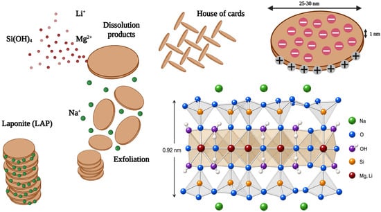
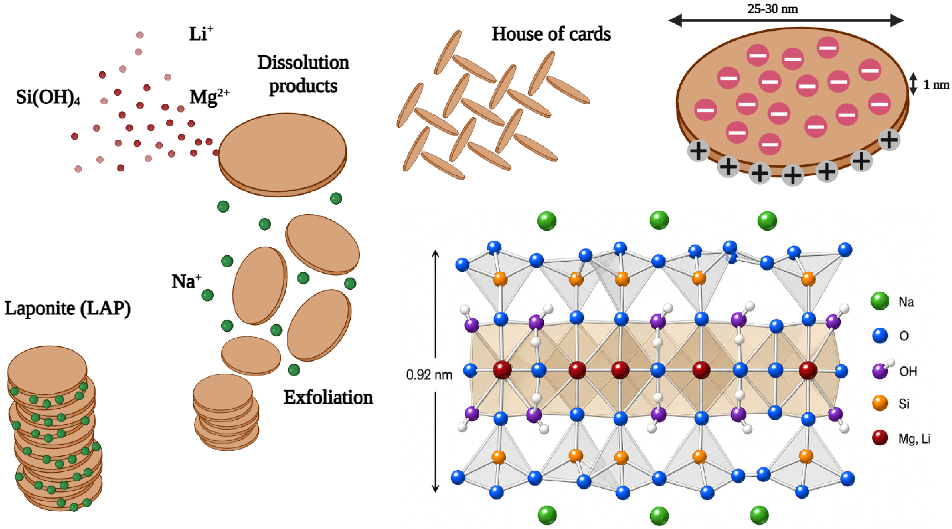
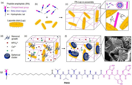
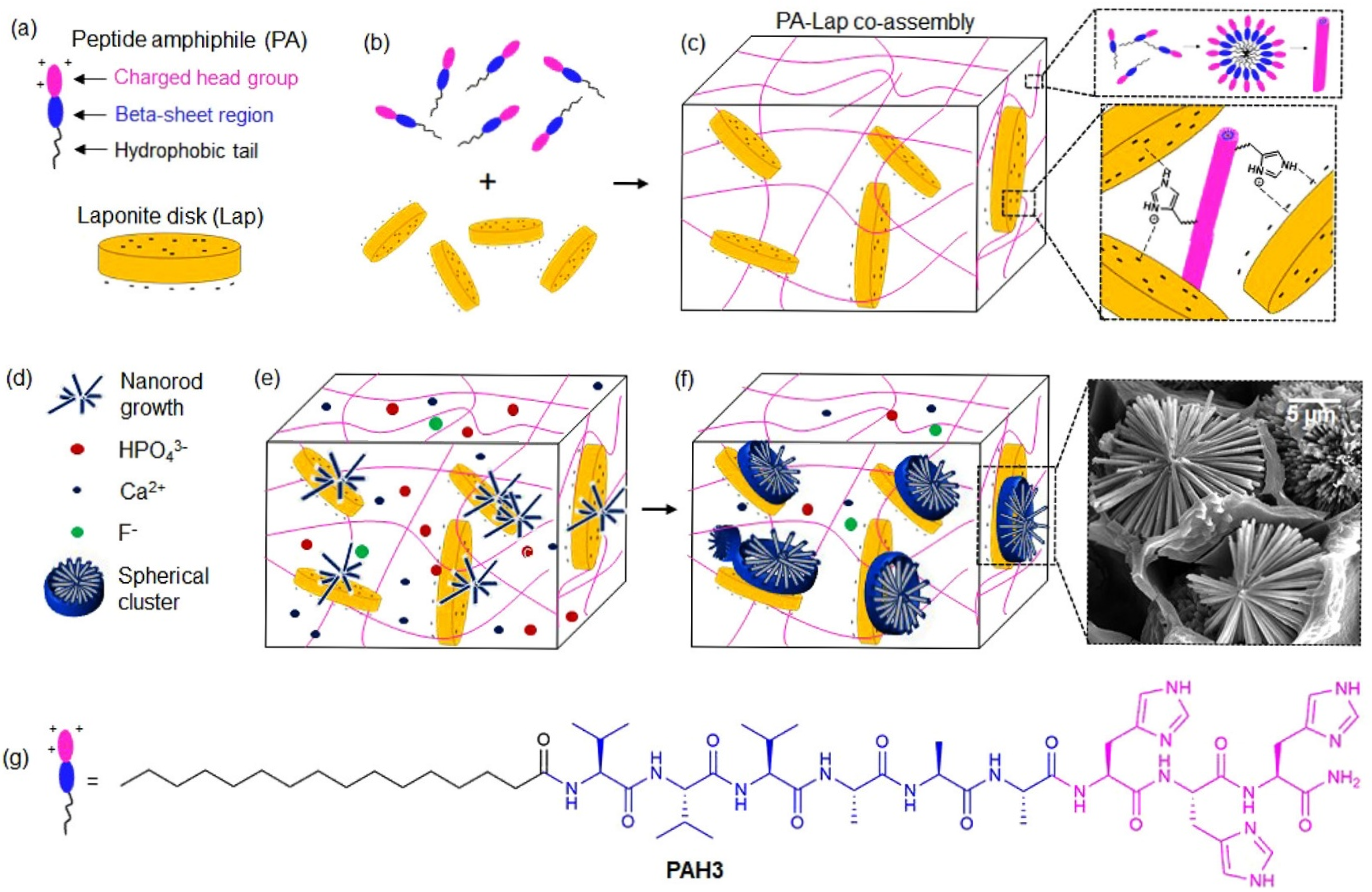
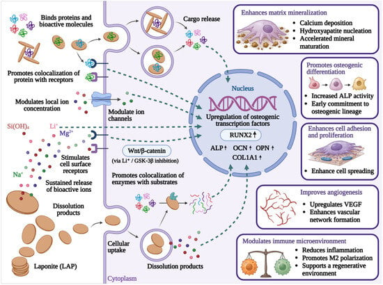
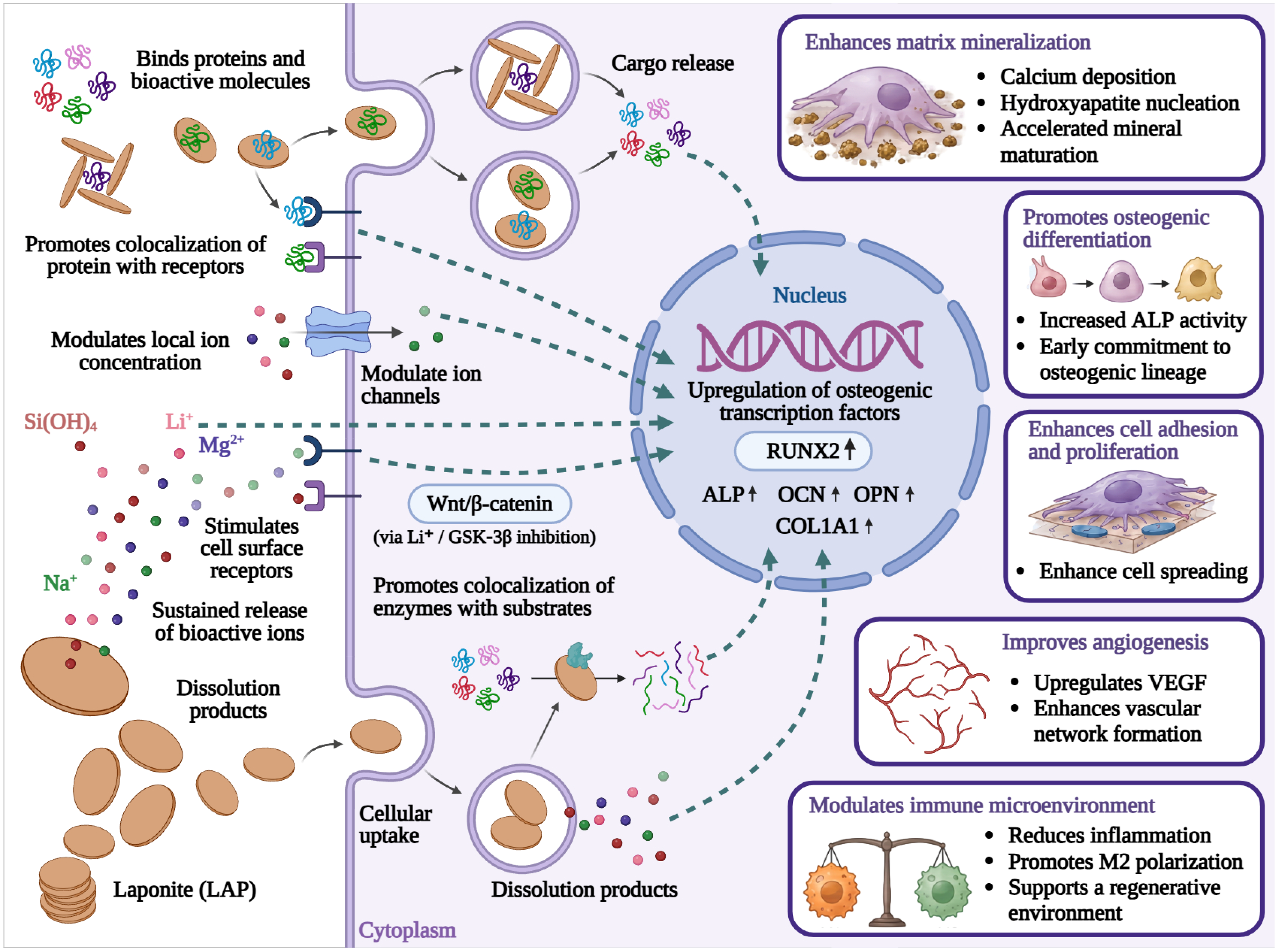

Disclaimer/Publisher’s Note: The statements, opinions and data contained in all publications are solely those of the individual author(s) and contributor(s) and not of MDPI and/or the editor(s). MDPI and/or the editor(s) disclaim responsibility for any injury to people or property resulting from any ideas, methods, instructions or products referred to in the content. |
© 2026 by the authors. Licensee MDPI, Basel, Switzerland. This article is an open access article distributed under the terms and conditions of the Creative Commons Attribution (CC BY) license.