Next Issue
Volume 12, September
Previous Issue
Volume 12, March


J. Funct. Biomater., Volume 12, Issue 2 (June 2021) – 20 articles

Cover Story (view full-size image): 
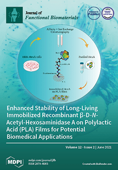
β-d-N-acetyl-hexosaminidase A (HexA) is an acid hydrolase that is capable of degrading many biological substrates; additionally, it can play an important role in the biomedical field for the treatment of Tay–Sachs and Sandhoff diseases. A device with a stable enzyme was obtained with a method of covalent immobilization on polylactic acid (PLA) films. The results obtained showed an improvement in terms of kinetic parameters and stability to heat for the enzyme with an unexpected ability to maintain its functionality for a long period of time (over a year). The stability and functionality of the enzyme in its immobilized form are, therefore, extremely promising for potential biotechnological and biomedical applications. View this paper
  • Issues are regarded as officially published after their release is announced to the table of contents alert mailing list.
  • You may sign up for e-mail alerts to receive table of contents of newly released issues.
  • PDF is the official format for papers published in both, html and pdf forms. To view the papers in pdf format, click on the "PDF Full-text" link, and use the free Adobe Reader to open them.
Order results
Result details
Section
Select all
Export citation of selected articles as:
17 pages, 1626 KiB  
Review
Electrical Stimulation to Enhance Wound Healing
by Saranya B. Rajendran, Kirsty Challen, Karen L. Wright and John G. Hardy
J. Funct. Biomater. 2021, 12(2), 40; https://doi.org/10.3390/jfb12020040 - 19 Jun 2021
Cited by 34 | Viewed by 17143
Abstract
Electrical stimulation (ES) can serve as a therapeutic modality accelerating the healing of wounds, particularly chronic wounds which have impaired healing due to complications from underlying pathology. This review explores how ES affects the cellular mechanisms of wound healing, and its effectiveness in [...] Read more.
Electrical stimulation (ES) can serve as a therapeutic modality accelerating the healing of wounds, particularly chronic wounds which have impaired healing due to complications from underlying pathology. This review explores how ES affects the cellular mechanisms of wound healing, and its effectiveness in treating acute and chronic wounds. Literature searches with no publication date restrictions were conducted using the Cochrane Library, Medline, Web of Science, Google Scholar and PubMed databases, and 30 full-text articles met the inclusion criteria. In vitro and in vivo experiments investigating the effect of ES on the general mechanisms of healing demonstrated increased epithelialization, fibroblast migration, and vascularity around wounds. Six in vitro studies demonstrated bactericidal effects upon exposure to alternating and pulsed current. Twelve randomized controlled trials (RCTs) investigated the effect of pulsed current on chronic wound healing. All reviewed RCTs demonstrated a larger reduction in wound size and increased healing rate when compared to control groups. In conclusion, ES therapy can contribute to improved chronic wound healing and potentially reduce the financial burden associated with wound management. However, the variations in the wound characteristics, patient demographics, and ES parameters used across studies present opportunities for systematic RCT studies in the future. Full article
Show Figures

Figure 1

12 pages, 2724 KiB  
Article
Bioactivity of an Experimental Dental Implant with Anodized Surface
by Maria Fernanda Lima Villaça-Carvalho, Juliani Caroline Ribeiro de Araújo, Juliana Mariano Beraldo, Renata Falchete do Prado, Mari Eli Leonelli de Moraes, Luiz Roberto Coutinho Manhães Junior, Eduardo Norberto Codaro, Heloisa Andrea Acciari, João Paulo Barros Machado, Natal Nerímio Regone, Anderson Oliveira Lobo, Fernanda Roberta Marciano and Luana Marotta Reis de Vasconcellos
J. Funct. Biomater. 2021, 12(2), 39; https://doi.org/10.3390/jfb12020039 - 07 Jun 2021
Cited by 2 | Viewed by 2858
Abstract
Background: Several studies proved that anodic oxidation improves osseointegration. This study aimed to optimize osseointegration through anodization in dental implants, obtaining anatase phase and controlled nanotopography. Methods: The division of the groups with 60 titanium implants was: control (CG); sandblasted (SG); anodized (AG): [...] Read more.
Background: Several studies proved that anodic oxidation improves osseointegration. This study aimed to optimize osseointegration through anodization in dental implants, obtaining anatase phase and controlled nanotopography. Methods: The division of the groups with 60 titanium implants was: control (CG); sandblasted (SG); anodized (AG): anodized pulsed current (duty cycle 30%, 30 V, 0.2 A and 1000 Hz). Before surgery, surface characterization was performed using Atomic Force Microscopy (AFM), Scanning Electron Microscopy (SEM), X-ray Dispersive Energy Spectroscopy (EDS) and Raman Spectroscopy. For in vivo tests, 10 New Zealand white rabbits received an implant from each group. The sacrifice period was 2 and 6 weeks (n = 5) and the specimens were subjected to computed microtomography (μCT) and reverse torque test. Results: AFM and SEM demonstrated a particular nanotopography on the surface in AG; the anatase phase was proved by Raman spectroscopy. In the μCT and in the reverse torque test, the AG group presented better results than the other groups. Conclusion: The chemical composition and structure of the TiO2 film were positively affected by the anodizing technique, intensifying the biological characteristics in osseointegration. Full article
(This article belongs to the Special Issue Advanced Functional Biomaterials for Dental Implants)
Show Figures

Figure 1

14 pages, 9344 KiB  
Article
The Effect of Bottom Profile Dimples on the Femoral Head on Wear in Metal-on-Metal Total Hip Arthroplasty
by J. Jamari, Muhammad Imam Ammarullah, Amir Putra Md Saad, Ardiyansyah Syahrom, Mohammad Uddin, Emile van der Heide and Hasan Basri
J. Funct. Biomater. 2021, 12(2), 38; https://doi.org/10.3390/jfb12020038 - 06 Jun 2021
Cited by 104 | Viewed by 5399
Abstract
Wear and wear-induced debris is a significant factor in causing failure in implants. Reducing contact pressure by using a textured surface between the femoral head and acetabular cup is crucial to improving the implant’s life. This study presented the effect of surface texturing [...] Read more.
Wear and wear-induced debris is a significant factor in causing failure in implants. Reducing contact pressure by using a textured surface between the femoral head and acetabular cup is crucial to improving the implant’s life. This study presented the effect of surface texturing as dimples on the wear evolution of total hip arthroplasty. It was implemented by developing finite element analysis from the prediction model without dimples and with bottom profile dimples of flat, drill, and ball types. Simulations were carried out by performing 3D physiological loading of the hip joint under normal walking conditions. A geometry update was initiated based on the patient’s daily routine activities. Our results showed that the addition of dimples reduced contact pressure and wear. The bottom profile dimples of the ball type had the best ability to reduce wear relative to the other types, reducing cumulative linear wear by 24.3% and cumulative volumetric wear by 31% compared to no dimples. The findings demonstrated that surface texturing with appropriate dimple bottom geometry on a bearing surface is able to extend the lifetime of hip implants. Full article
Show Figures

Figure 1

17 pages, 7753 KiB  
Article
Physico-Chemical Properties and Biocompatibility of Thermosensitive Chitosan Lactate and Chitosan Chloride Hydrogels Developed for Tissue Engineering Application
by Katarzyna Pieklarz, Grzegorz Galita, Michał Tylman, Waldemar Maniukiewicz, Ewa Kucharska, Ireneusz Majsterek and Zofia Modrzejewska
J. Funct. Biomater. 2021, 12(2), 37; https://doi.org/10.3390/jfb12020037 - 20 May 2021
Cited by 17 | Viewed by 3927
Abstract
Recently, the modification of the initial structure of biopolymers, mainly chitosan, has been gaining importance with a view to obtain functional forms with increased practicality and specific properties enabling their use in tissue engineering. Therefore, in this article, the properties (structural and biological) [...] Read more.
Recently, the modification of the initial structure of biopolymers, mainly chitosan, has been gaining importance with a view to obtain functional forms with increased practicality and specific properties enabling their use in tissue engineering. Therefore, in this article, the properties (structural and biological) of thermosensitive hydrogels obtained from chitosan lactate/chloride and two types of crosslinking agents (β-glycerol phosphate disodium salt pentahydrate and uridine 5′-monophosphate disodium salt) are discussed. The aim of the research is to identify changes in the structure of the biomaterials during conditioning in water. Structural investigations were carried out by FTIR spectroscopy. The crystallinity of gels was determined by X-ray diffraction analysis. The biocompatibility (evaluation of cytotoxicity and genotoxicity) of chitosan hydrogels was investigated by contact with human colon adenocarcinoma cell line for 48 h. The cytotoxicity was verified based on the colorimetric resazurin assay, and the genotoxicity was checked by the comet assay (percentage of DNA in the comet tail). The conducted research showed that the analyzed types of chitosan hydrogels are non-cytotoxic and non-genotoxic materials. The good biocompatibility of chitosan hydrogels surfaces makes them interesting scaffolds with clinical potential in tissue regeneration engineering. Full article
(This article belongs to the Special Issue Bioinspired Materials for Medical and Biotechnological Applications)
Show Figures

Graphical abstract

15 pages, 1928 KiB  
Article
TiAl6V4 Alloy Surface Modifications and Their Impact on Biofilm Development of S. aureus and S. epidermidis
by Astrid H. Paulitsch-Fuchs, Lukas Wolrab, Nicole Eck, Nigel P. Dyer, Benjamin Bödendorfer and Birgit Lohberger
J. Funct. Biomater. 2021, 12(2), 36; https://doi.org/10.3390/jfb12020036 - 18 May 2021
Cited by 7 | Viewed by 3013
Abstract
One of the most serious complications following joint replacement surgeries are periprosthetic infections (PIs) arising from the adhesion of bacteria to the artificial joint. Various types of titanium–aluminum–vanadium (TiAl6V4) alloy surface modifications (coatings with silver (Ag), titanium nitride (TiN), pure titanium (cpTi), combinations [...] Read more.
One of the most serious complications following joint replacement surgeries are periprosthetic infections (PIs) arising from the adhesion of bacteria to the artificial joint. Various types of titanium–aluminum–vanadium (TiAl6V4) alloy surface modifications (coatings with silver (Ag), titanium nitride (TiN), pure titanium (cpTi), combinations of cpTi and hydroxyapatite (HA), combinations of cpTi and tricalcium phosphate (TCP), and a rough-blasted surface of TiAl6V4) have been investigated to assess their effects on biofilm development. Biofilms were grown, collected, and analyzed after 48 h to measure their protein and glucose content and the cell viability. Biofilm-associated genes were also monitored after 48 h of development. There was a distinct difference in the development of staphylococcal biofilms on the surfaces of the different types of alloy. According to the findings of this study, the base alloy TiAl6V4 and the TiN-coated surface are the most promising materials for biofilm reduction. Rough surfaces are most favorable when it comes to bacterial infections because they allow an easy attachment of pathogenic organisms. Of all rough surfaces tested, rough-blasted TiAl6V4 was the most favorable as an implantation material; all the other rough surfaces showed more distinct signs of inducing the development of biofilms which displayed higher protein and polysaccharide contents. These results are supported by RT-qPCR measurements of biofilm associated genes for Staphylococcus aureus (icaA, icaC, fnbA, fnbB, clfB, atl) and Staphylococcus epidermidis (atle, aap). Full article
Show Figures

Figure 1

8 pages, 1215 KiB  
Article
Aesthetic Outcomes and Peri-Implant Health of Angled Screw Retained Implant Restorations Compared with Cement Retained Crowns: Medium Term Follow-Up
by Livia Nastri, Ludovica Nucci, Vincenzo Grassia and Rino Miraldi
J. Funct. Biomater. 2021, 12(2), 35; https://doi.org/10.3390/jfb12020035 - 16 May 2021
Cited by 6 | Viewed by 2956
Abstract
Single tooth implant restorations in the aesthetic area are a demanding challenge. If a complete osseointegration is mandatory, the final result has to result in a higher standard of biomimetic and soft tissue health among natural teeth. This outcome is traditionally pursued by [...] Read more.
Single tooth implant restorations in the aesthetic area are a demanding challenge. If a complete osseointegration is mandatory, the final result has to result in a higher standard of biomimetic and soft tissue health among natural teeth. This outcome is traditionally pursued by cementing crowns over individualized abutments. However, in recent years, the need for controlling peri-implant health and the preference towards a retrievable solution has led to an increase in screw-retained crowns, which is not always applicable when the implant axis is not ideal. In the aesthetic area, the use of a novel technical solution represented by the angled screw channel (ASC) of the abutment has been proposed in order to match the advantages of the screwed solution with the aesthetic demands. The aim of this study was to compare ASC crowns to cemented crowns (CC) in single implant restorations using the white esthetic score (WES) and pink esthetic score (PES) at the crown delivery and at a follow-up of a minimum of 2 years. Peri-implant health and marginal bone loss (MBL) were also evaluated. The mean follow-up was 44.3 months, with a mean MBL of 0.22 mm in the ASC group and 0.29 mm in the CC group. The total WES/PES score was 16.6 for ASC, compared with 17.3 for CC at baseline, and 16.2 and 17.1, respectively, at follow-up. Both of the groups reached a high WES/PES, and this was maintained over time, without signs of peri-implant diseases or bone loss, regardless of the choice of connection. In conclusion, ASC can be adopted in cases where the implant axis is not ideal, with aesthetic and functional results that are comparable to implants restored by cemented crowns. Full article
(This article belongs to the Special Issue Advanced Functional Biomaterials for Dental Implants)
Show Figures

Figure 1

18 pages, 1916 KiB  
Article
EtoGel for Intra-Articular Drug Delivery: A New Challenge for Joint Diseases Treatment
by Maria Chiara Cristiano, Antonia Mancuso, Elena Giuliano, Donato Cosco, Donatella Paolino and Massimo Fresta
J. Funct. Biomater. 2021, 12(2), 34; https://doi.org/10.3390/jfb12020034 - 16 May 2021
Cited by 17 | Viewed by 2836
Abstract
Ethosomes® have been proposed as potential intra-articular drug delivery devices, in order to obtain a longer residence time of the delivered drug in the knee joint. To this aim, the conventional composition and preparation method were modified. Ethosomes® were prepared by [...] Read more.
Ethosomes® have been proposed as potential intra-articular drug delivery devices, in order to obtain a longer residence time of the delivered drug in the knee joint. To this aim, the conventional composition and preparation method were modified. Ethosomes® were prepared by using a low ethanol concentration and carrying out a vesicle extrusion during the preparation. The modified composition did not affect the deformability of ethosomes®, a typical feature of this colloidal vesicular topical carrier. The maintenance of sufficient deformability bodes well for an effective ethosome® application in the treatment of joint pathologies because they should be able to go beyond the pores of the dense collagen II network. The investigated ethosomes® were inserted in a three-dimensional network of thermo-sensitive poloxamer gel (EtoGel) to improve the residence time in the joint. Rheological experiments evidenced that EtoGel could allow an easy intra-articular injection at room temperature and hence transform itself in gel form at body temperature into the joint. Furthermore, EtoGel seemed to be able to support the knee joint during walking and running. In vitro studies demonstrated that the amount of used ethanol did not affect the viability of human chondrocytes and nanocarriers were also able to suitably interact with cells. Full article
Show Figures

Figure 1

10 pages, 6210 KiB  
Article
Endothelial Cell Responses to a Highly Deformable Titanium Alloy Designed for Vascular Stent Applications
by Raluca Ion, Gaëtan Cabon, Doina-Margareta Gordin, Elena Ionica, Thierry Gloriant and Anisoara Cimpean
J. Funct. Biomater. 2021, 12(2), 33; https://doi.org/10.3390/jfb12020033 - 14 May 2021
Cited by 5 | Viewed by 2354
Abstract
Titanium alloys are widely used for biomedical applications due to their good biocompatibility. Nevertheless, they cannot be used for balloon expandable stents due to a lack of ductility compared to cobalt-chromium (Co-Cr) alloys and stainless steels. In this study, a new highly deformable [...] Read more.
Titanium alloys are widely used for biomedical applications due to their good biocompatibility. Nevertheless, they cannot be used for balloon expandable stents due to a lack of ductility compared to cobalt-chromium (Co-Cr) alloys and stainless steels. In this study, a new highly deformable Ti-16Nb-8Mo alloy was designed for such an application. However, the biological performance of a stent material is strongly influenced by the effect exerted on the behavior of endothelial cells. Therefore, the cellular responses of human umbilical vein endothelial cells (HUVECs), including morphological characteristics, cell viability and proliferation, and functional markers expression, were investigated to evaluate the biocompatibility of the alloy in the present study. The in vitro results demonstrated the suitability of this alloy for use as endovascular stents. Full article
Show Figures

Figure 1

17 pages, 4491 KiB  
Article
Enhanced Stability of Long-Living Immobilized Recombinant β-d-N-Acetyl-Hexosaminidase A on Polylactic Acid (PLA) Films for Potential Biomedical Applications
by Eleonora Calzoni, Alessio Cesaretti, Nicolò Montegiove, Alessandro Di Michele and Carla Emiliani
J. Funct. Biomater. 2021, 12(2), 32; https://doi.org/10.3390/jfb12020032 - 11 May 2021
Cited by 6 | Viewed by 2525
Abstract
β-d-N-acetyl-hexosaminidase (Hex, EC 3.2.1.52) is an acid hydrolase that catalyzes the cleavage of the β-1,4 bond in N-acetyl-d-galactosamine (Gal-NAc) and N-acetyl-d-glucosamine (Glc-NAc) from the non-reducing end of oligosaccharides and glycoconjugates. It is widely [...] Read more.
β-d-N-acetyl-hexosaminidase (Hex, EC 3.2.1.52) is an acid hydrolase that catalyzes the cleavage of the β-1,4 bond in N-acetyl-d-galactosamine (Gal-NAc) and N-acetyl-d-glucosamine (Glc-NAc) from the non-reducing end of oligosaccharides and glycoconjugates. It is widely expressed in both the prokaryotic and eukaryotic world, where it performs multiple and important functions. Hex has antifungal activity in plants, is capable of degrading many biological substrates, and can play an important role in the biomedical field for the treatment of Tay-Sachs and Sandhoff diseases. With the aim being able to obtain a device with a stable enzyme, a method of covalent immobilization on polylactic acid (PLA) films was developed for the A isoform of the β-d-N-acetyl-hexosaminidase enzyme (HexA), produced in a recombinant way from Human Embryonic Kidney-293 (HEK-293) cells and suitably purified. An in-depth biochemical characterization of the immobilized enzyme was carried out, evaluating the optimal temperature, thermal stability, pH parameters, and Km value. Moreover, the stability of the enzymatic activity over time was assessed. The results obtained showed an improvement in terms of kinetic parameters and stability to heat for the enzyme following immobilization and the presence of HexA in two distinct immobilized forms, with an unexpected ability for one of them to maintain its functionality for a long period of time (over a year). The stability and functionality of the enzyme in its immobilized form are therefore extremely promising for potential biotechnological and biomedical applications. Full article
Show Figures

Graphical abstract

27 pages, 5289 KiB  
Article
Functionalization with a Polyphenol-Rich Pomace Extract Empowers a Ceramic Bone Filler with In Vitro Antioxidant, Anti-Inflammatory, and Pro-Osteogenic Properties
by Giorgio Iviglia, Elisa Torre, Clara Cassinelli and Marco Morra
J. Funct. Biomater. 2021, 12(2), 31; https://doi.org/10.3390/jfb12020031 - 05 May 2021
Cited by 5 | Viewed by 2782
Abstract
Oral diseases and periodontitis in particular are a major health burden worldwide, because of their association with various systemic diseases and with conditions such as peri-implantitis. Attempts have been made over the years to reverse bone loss due to the host disproportionate inflammatory [...] Read more.
Oral diseases and periodontitis in particular are a major health burden worldwide, because of their association with various systemic diseases and with conditions such as peri-implantitis. Attempts have been made over the years to reverse bone loss due to the host disproportionate inflammatory response and to prevent failure of dental implants. To this end, the use of biomaterials functionalized with molecules characterized by anti-inflammatory and antioxidant properties could represent a new frontier for regenerating functional periodontal tissues. In this study, a new ceramic granulated biomaterial, named Synergoss Red (SR), functionalized with a polyphenolic mixture extracted from pomace of the Croatina grape variety, is introduced. Following a preliminary in-depth characterization of the extract by HPLC analysis and of the biomaterial surface and composition, we performed evaluations of cytocompatibility and a biological response through in vitro assays. The anti-inflammatory and antioxidant properties of the identified phenolic molecules contained in SR were shown to downregulate inflammation in macrophages, to stimulate in osteoblast-like cells the expression of genes involved in deposition of the early bone matrix, and to mitigate bone remodeling by decreasing the RANKL/OPG ratio. Thanks to its cytocompatibility and assorted beneficial effects on bone regeneration, SR could be considered an innovative regenerative approach in periodontal therapy. Full article
(This article belongs to the Special Issue Recent Advances in Dental Implants and Biomaterials)
Show Figures

Figure 1

11 pages, 3282 KiB  
Article
Survival and Proliferation under Severely Hypoxic Microenvironments Using Cell-Laden Oxygenating Hydrogels
by Shabir Hassan, Berivan Cecen, Ramon Peña-Garcia, Fernanda Roberta Marciano, Amir K. Miri, Ali Fattahi, Christina Karavasili, Shikha Sebastian, Hamza Zaidi and Anderson Oliveira Lobo
J. Funct. Biomater. 2021, 12(2), 30; https://doi.org/10.3390/jfb12020030 - 02 May 2021
Cited by 6 | Viewed by 3532
Abstract
Different strategies have been employed to provide adequate nutrients for engineered living tissues. These have mainly revolved around providing oxygen to alleviate the effects of chronic hypoxia or anoxia that result in necrosis or weak neovascularization, leading to failure of artificial tissue implants [...] Read more.
Different strategies have been employed to provide adequate nutrients for engineered living tissues. These have mainly revolved around providing oxygen to alleviate the effects of chronic hypoxia or anoxia that result in necrosis or weak neovascularization, leading to failure of artificial tissue implants and hence poor clinical outcome. While different biomaterials have been used as oxygen generators for in vitro as well as in vivo applications, certain problems have hampered their wide application. Among these are the generation and the rate at which oxygen is produced together with the production of the reaction intermediates in the form of reactive oxygen species (ROS). Both these factors can be detrimental for cell survival and can severely affect the outcome of such studies. Here we present calcium peroxide (CPO) encapsulated in polycaprolactone as oxygen releasing microparticles (OMPs). While CPO releases oxygen upon hydrolysis, PCL encapsulation ensures that hydrolysis takes place slowly, thereby sustaining prolonged release of oxygen without the stress the bulk release can endow on the encapsulated cells. We used gelatin methacryloyl (GelMA) hydrogels containing these OMPs to stimulate survival and proliferation of encapsulated skeletal myoblasts and optimized the OMP concentration for sustained oxygen delivery over more than a week. The oxygen releasing and delivery platform described in this study opens up opportunities for cell-based therapeutic approaches to treat diseases resulting from ischemic conditions and enhance survival of implants under severe hypoxic conditions for successful clinical translation. Full article
(This article belongs to the Special Issue Fibrous Scaffolds for Tissue Engineering Application)
Show Figures

Figure 1

18 pages, 5752 KiB  
Article
Analysis of Chemisorbed Tribo-Film for Ceramic-on-Ceramic Hip Joint Prostheses by Raman Spectroscopy
by Risha Rufaqua, Martin Vrbka, Dušan Hemzal, Dipankar Choudhury, David Rebenda, Ivan Křupka and Martin Hartl
J. Funct. Biomater. 2021, 12(2), 29; https://doi.org/10.3390/jfb12020029 - 01 May 2021
Cited by 4 | Viewed by 3475
Abstract
To understand the possible lubricant mechanism in ceramic-on-ceramic hip joint prostheses, biochemical reactions of the synovial fluid and the corresponding frictional coefficients were studied. The experiments were performed in a hip joint simulator using the ball-on-cup configuration with balls and cups made from [...] Read more.
To understand the possible lubricant mechanism in ceramic-on-ceramic hip joint prostheses, biochemical reactions of the synovial fluid and the corresponding frictional coefficients were studied. The experiments were performed in a hip joint simulator using the ball-on-cup configuration with balls and cups made from two types of ceramics, BIOLOX®forte and BIOLOX®delta. Different lubricants, namely albumin, γ-globulin, hyaluronic acid and three model synovial fluids, were studied in the experiments and Raman spectroscopy was used to analyze the biochemical responses of these lubricants at the interface. BIOLOX®delta surface was found less reactive to proteins and model fluid lubricants. In contrast, BIOLOX®forte ball surface has shown chemisorption with both proteins, hyaluronic acid and model fluids imitating total joint replacement and osteoarthritic joint. There was no direct correlation between the measured frictional coefficient and the observed chemical reactions. In summary, the study reveals chemistry of lubricant film formation on ceramic hip implant surfaces with various model synovial fluids and their components. Full article
(This article belongs to the Special Issue Advanced Bioceramics)
Show Figures

Figure 1

20 pages, 4538 KiB  
Article
Rheological Behavior of a New Mucoadhesive Oral Formulation Based on Sodium Chondroitin Sulfate, Xyloglucan and Glycerol
by Tiziana Maria Grazia Pecora, Barbara Ragazzo, Walter Bertin, Alessia Ragonese, Marco Mascagni, Paola Maffei and Rosario Pignatello
J. Funct. Biomater. 2021, 12(2), 28; https://doi.org/10.3390/jfb12020028 - 28 Apr 2021
Cited by 8 | Viewed by 2815
Abstract
Background: The study aimed at assessing the mucoadhesive properties and the barrier effect of a formulation, labelled as AL2106, containing sodium chondroitin sulfate (ChS), xyloglucan from tamarind seed extract, and glycerol, by evaluating the capacity to adhere to a layer of mucin, [...] Read more.
Background: The study aimed at assessing the mucoadhesive properties and the barrier effect of a formulation, labelled as AL2106, containing sodium chondroitin sulfate (ChS), xyloglucan from tamarind seed extract, and glycerol, by evaluating the capacity to adhere to a layer of mucin, the rheological synergism and the barrier effect in comparison to the marketed Esoxx One medical device. AL2106 is a medical device distributed by Alfasigma SpA, Italy with REF FTP57 (Manufacturer: Labomar SpA); it is analogous to Esoxx One medical device: the two products are drinkable solutions that, after swallowing, adhere to the esophageal mucosa, protecting it from the corrosive effect of the gastric acid reflux. AL2106 has been conceived to be better performing in terms of duration of the barrier effect compared to Esoxx One. Methods: The mucoadhesive properties, rheological behavior, buffering capacity against acidity, and film-forming ability with the resultant protecting effect on esophagus mucosa (caffeine permeation test) was compared between the two products. Results: The mucoadhesivity of the formulations was shown in vitro: both remained adherent to a mucin layer, also when the support was rotated by 90°, and when the film layer was washed with water, intended to simulate the washout due to swallowing. AL2106 showed a good buffering efficacy, being able to absorb at least 50% of its weight of 0.03 M HCl while maintaining the pH above 4. The film-forming effect and barrier properties of AL2106 and Esoxx One were confirmed by an in vitro study on reconstructed human esophageal epithelium. A greater film-forming efficacy of AL2106, lasting for at least 5 h, than Esoxx One was observed. Noteworthy, the barrier function of esophageal tissues was shown to be preserved after the application of both formulations. Conclusions: The combination of ChS with the mucoadhesive glycerol−xyloglucan complex and other excipients, which contribute to the barrier effect and to mucoadhesion, contained in AL2106, allowed a longer-lasting protective effect than Esoxx One, proving its effectivity and safety for oral use. Full article
Show Figures

Figure 1

13 pages, 8114 KiB  
Article
Comparative Study of Technologies for Tubule Occlusion and Treatment of Dentin Hypersensitivity
by Camilla Berg, Erik Unosson, Håkan Engqvist and Wei Xia
J. Funct. Biomater. 2021, 12(2), 27; https://doi.org/10.3390/jfb12020027 - 27 Apr 2021
Cited by 7 | Viewed by 4164
Abstract
This study aimed to evaluate the occluding/remineralization performance and resistance to acid attacks of the mineralization layer formed by a tooth-desensitizing gel containing amorphous calcium magnesium phosphate (ACMP) particles and compare it to six other desensitizing products available on the market. Similar comprehensive [...] Read more.
This study aimed to evaluate the occluding/remineralization performance and resistance to acid attacks of the mineralization layer formed by a tooth-desensitizing gel containing amorphous calcium magnesium phosphate (ACMP) particles and compare it to six other desensitizing products available on the market. Similar comprehensive studies are few and there is especially a lack of studies that are up to date. A dentin-disc model was used for in vitro evaluation of the desensitizing toothpastes/gels. Application of the products was performed twice daily for seven days. One set of specimens were evaluated using scanning electron microscopy (SEM) directly after the final treatment and another set was evaluated after an acid challenge, exposing specimens to 2 wt% citric acid. The ACMP desensitizing gel was the only product resulting in complete occlusion by the formation of mineralized material on the dentin surface and inside the tubules. Particle deposition was dominant after treatment with the other desensitizing products, with little or no mineralization, resulting in partial occlusion only. Sensodyne Repair & Protect and Oral-B Pro-Expert showed the highest resistance toward acid attacks. Material inside the tubules remained relatively unaffected by acid attacks in all specimens. The results in this study indicated a great variability among the occluding agents in terms of occlusion and acid resistance of the mineralization layer. The high degree of occlusion and intra-tubular mineralization that could mitigate the effect of acid solubilization indicate that the ACMP desensitizing gel may be a superior option for the treatment of dentin hypersensitivity. Full article
(This article belongs to the Special Issue Recent Advances in Dental Implants and Biomaterials)
Show Figures

Figure 1

32 pages, 6894 KiB  
Review
Advances in Fabricating the Electrospun Biopolymer-Based Biomaterials
by Sebastian Wilk and Aleksandra Benko
J. Funct. Biomater. 2021, 12(2), 26; https://doi.org/10.3390/jfb12020026 - 16 Apr 2021
Cited by 29 | Viewed by 4540
Abstract
Biopolymers formed into a fibrous morphology through electrospinning are of increasing interest in the field of biomedicine due to their intrinsic biocompatibility and biodegradability and their ability to be biomimetic to various fibrous structures present in animal tissues. However, their mechanical properties are [...] Read more.
Biopolymers formed into a fibrous morphology through electrospinning are of increasing interest in the field of biomedicine due to their intrinsic biocompatibility and biodegradability and their ability to be biomimetic to various fibrous structures present in animal tissues. However, their mechanical properties are often unsatisfactory and their processing may be troublesome. Thus, extensive research interest is focused on improving these qualities. This review article presents the selection of the recent advances in techniques aimed to improve the electrospinnability of various biopolymers (polysaccharides, polynucleotides, peptides, and phospholipids). The electrospinning of single materials, and the variety of co-polymers, with and without additives, is covered. Additionally, various crosslinking strategies are presented. Examples of cytocompatibility, biocompatibility, and antimicrobial properties are analyzed. Special attention is given to whey protein isolate as an example of a novel, promising, green material with good potential in the field of biomedicine. This review ends with a brief summary and outlook for the biomedical applicability of electrospinnable biopolymers. Full article
(This article belongs to the Special Issue Bioinspired Materials for Medical and Biotechnological Applications)
Show Figures

Figure 1

28 pages, 4822 KiB  
Article
Human Adipose Stem Cells (hASCs) Grown on Biodegradable Microcarriers in Serum- and Xeno-Free Medium Preserve Their Undifferentiated Status
by Francesco Muoio, Stefano Panella, Valentin Jossen, Matias Lindner, Yves Harder, Michele Müller, Regine Eibl and Tiziano Tallone
J. Funct. Biomater. 2021, 12(2), 25; https://doi.org/10.3390/jfb12020025 - 16 Apr 2021
Cited by 7 | Viewed by 3793
Abstract
Human adipose stem cells (hASCs) are promising candidates for cell-based therapies, but they need to be efficiently expanded in vitro as they cannot be harvested in sufficient quantities. Recently, dynamic bioreactor systems operated with microcarriers achieved considerable high cell densities. Thus, they are [...] Read more.
Human adipose stem cells (hASCs) are promising candidates for cell-based therapies, but they need to be efficiently expanded in vitro as they cannot be harvested in sufficient quantities. Recently, dynamic bioreactor systems operated with microcarriers achieved considerable high cell densities. Thus, they are a viable alternative to static planar cultivation systems to obtain high numbers of clinical-grade hASCs. Nevertheless, the production of considerable biomass in a short time must not be achieved to the detriment of the cells’ quality. To facilitate the scalable expansion of hASC, we have developed a new serum- and xeno-free medium (UrSuppe) and a biodegradable microcarrier (BR44). In this study, we investigated whether the culture of hASCs in defined serum-free conditions on microcarriers (3D) or on planar (2D) cell culture vessels may influence the expression of some marker genes linked with the immature degree or the differentiated status of the cells. Furthermore, we investigated whether the biomaterials, which form our biodegradable MCs, may affect cell behavior and differentiation. The results confirmed that the quality and the undifferentiated status of the hASCs are very well preserved when they grow on BR44 MCs in defined serum-free conditions. Indeed, the ASCs showed a gene expression profile more compatible with an undifferentiated status than the same cells grown under standard planar conditions. Full article
Show Figures

Figure 1

11 pages, 2118 KiB  
Article
Increased Elasticity Modulus of Polymeric Materials Is a Source of Surface Alterations in the Human Body
by Matthias Kapischke, Igor Erlichman and Alexandra Pries
J. Funct. Biomater. 2021, 12(2), 24; https://doi.org/10.3390/jfb12020024 - 16 Apr 2021
Viewed by 2175
Abstract
The introduction of alloplastic materials (meshes) in hernia surgery has improved patient outcome by a radical reduction of hernia recurrence rate, but discussion about the biocompatibility of these implanted materials continues since observations of surface alterations of polypropylene and other alloplastic materials were [...] Read more.
The introduction of alloplastic materials (meshes) in hernia surgery has improved patient outcome by a radical reduction of hernia recurrence rate, but discussion about the biocompatibility of these implanted materials continues since observations of surface alterations of polypropylene and other alloplastic materials were published. This study intends to investigate if additives supplemented to alloplastic mesh materials merge into the solution and become analyzable. Four polypropylene and one polyester alloplastic material were incubated in different media for three weeks: distilled water, saline solution, urea solution, formalin, and hydrogen peroxide. No swelling or other changes were observed. Infrared spectroscopy scanning of incubated alloplastic materials and NMR studies of extracted solutions were performed to investigate loss of plasticizers. The surface of the mesh materials did not show any alterations independent of the incubation medium. FT-IR spectra before and after incubation did not show any differences. NMR spectra showed leaching of different plasticizers (PEG, sterically hindered phenols, thioester), of which there was more for polypropylene less for polyester. This could be the reason for the loss of elasticity of the alloplastic materials with consecutive physically induced surface alterations. A mixture of chemical reactions (oxidative stress with additive leaching from polymer fiber) in connection with physical alterations (increased elasticity modulus by loss of plasticizers) seem to be a source of these PP and PE alterations. Full article
Show Figures

Figure 1

14 pages, 38744 KiB  
Article
Personalized Biomechanical Analysis of the Mandible Teeth Behavior in the Treatment of Masticatory Muscles Parafunction
by Denis Gribov, Mikhail Antonik, Denis Butkov, Alexandr Stepanov, Pavel Antonik, Yaser Kharakh, Anton Pivovarov and Sergey Arutyunov
J. Funct. Biomater. 2021, 12(2), 23; https://doi.org/10.3390/jfb12020023 - 09 Apr 2021
Cited by 4 | Viewed by 3478
Abstract
A 3D finite element model of the mandible dentition was developed, including 14 teeth, a periodontal ligament (PDL), and a splint made of polymethylmethacrylate (PMMA). The study considered three design options: 1—the case of splint absence; 2—the case of the splint presence installed [...] Read more.
A 3D finite element model of the mandible dentition was developed, including 14 teeth, a periodontal ligament (PDL), and a splint made of polymethylmethacrylate (PMMA). The study considered three design options: 1—the case of splint absence; 2—the case of the splint presence installed after manufacture; and 3—the case of splint presence installed after correction (grinding) performed to ensure a uniform distribution of occlusal force between the teeth. For cases of absence and presence of splint, three measurements of the functional load were performed using the T-Scan III software and hardware complex (TekScan, Boston, MA, USA). It was found that the presence of a splint led to a decrease in the total value of the occlusive load and to a uniform distribution between all the mandible teeth. The occlusal force was considered as a static vertical force evenly distributed between the nodes belonging to the occlusive surface of the corresponding tooth for the first design option and the occlusal surface of the splint for the second and third ones, respectively. As a result of the study, it was concluded that the splint usage was effective in order to change the distribution of the functional load during the treatment of proved masticatory muscles’ parafunction; the safety of using a splint for teeth and surrounding tissues under the influence of the considered functional load was shown; the potential applicability of PMMA as a structural material of a splint that had been used for the treatment of masticatory muscles’ parafunction was established. Full article
Show Figures

Figure 1

24 pages, 2613 KiB  
Review
Osteogenic Peptides and Attachment Methods Determine Tissue Regeneration in Modified Bone Graft Substitutes
by George Bullock, Joss Atkinson, Piergiorgio Gentile, Paul Hatton and Cheryl Miller
J. Funct. Biomater. 2021, 12(2), 22; https://doi.org/10.3390/jfb12020022 - 31 Mar 2021
Cited by 13 | Viewed by 3587
Abstract
The inclusion of biofunctional molecules with synthetic bone graft substitutes has the potential to enhance tissue regeneration during treatment of traumatic bone injuries. The clinical use of growth factors has though been associated with complications, some serious. The use of smaller, active peptides [...] Read more.
The inclusion of biofunctional molecules with synthetic bone graft substitutes has the potential to enhance tissue regeneration during treatment of traumatic bone injuries. The clinical use of growth factors has though been associated with complications, some serious. The use of smaller, active peptides has the potential to overcome these problems and provide a cost-effective, safe route for the manufacture of enhanced bone graft substitutes. This review considers the design of peptide-enhanced bone graft substitutes, and how peptide selection and attachment method determine clinical efficacy. It was determined that covalent attachment may reduce the known risks associated with growth factor-loaded bone graft substitutes, providing a predictable tissue response and greater clinical efficacy. Peptide choice was found to be critical, but even within recognised families of biologically active peptides, the configurations that appeared to most closely mimic the biological molecules involved in natural bone healing processes were most potent. It was concluded that rational, evidence-based design of peptide-enhanced bone graft substitutes offers a pathway to clinical maturity in this highly promising field. Full article
Show Figures

Graphical abstract

16 pages, 5409 KiB  
Article
Synthesis and Characterization of Polymer-Based Coatings Modified with Bioactive Ceramic and Bovine Serum Albumin
by Wioletta Florkiewicz, Dagmara Słota, Angelika Placek, Klaudia Pluta, Bożena Tyliszczak, Timothy E. L. Douglas and Agnieszka Sobczak-Kupiec
J. Funct. Biomater. 2021, 12(2), 21; https://doi.org/10.3390/jfb12020021 - 30 Mar 2021
Cited by 15 | Viewed by 3123
Abstract
This study involves the synthesis of hydroxyapatite and describes the preparation and characterization of polymer coatings based on poly(ethylene glycol) diacrylate and poly(ethylene glycol) and modified with bovine serum albumin and hydroxyapatite. Hydroxyapatite was obtained by wet chemical synthesis and characterized by X-ray [...] Read more.
This study involves the synthesis of hydroxyapatite and describes the preparation and characterization of polymer coatings based on poly(ethylene glycol) diacrylate and poly(ethylene glycol) and modified with bovine serum albumin and hydroxyapatite. Hydroxyapatite was obtained by wet chemical synthesis and characterized by X-ray diffraction and FTIR spectroscopy, and its Ca/P molar ratio was determined (1.69 ± 0.08). The ceramic and bovine serum albumin were used in the preparation of composite materials with the polymeric matrix. The chemical composition of coatings was characterized with FTIR spectroscopy, and their morphology was recorded with SEM imaging. Moreover, the measurements of surface roughness parameters and stereometric research were performed. The prepared coatings were subjected to in vitro studies in simulated body fluid and artificial saliva. Changes in chemical composition and morphology after immersion were examined with FTIR spectroscopy and SEM imaging. Based on the conducted research, it can be stated that applied modifiers promote the biomineralization process. The roughness analysis confirmed prepared materials were characterized by the micrometer-scale topography. The materials morphology and roughness, and the morphology of the newly formed apatite deposit, were dependent on the type of the used modifier, and the artificial fluid used in in vitro studies. Full article
(This article belongs to the Special Issue Bioinspired Materials for Medical and Biotechnological Applications)
Show Figures

Figure 1

Previous Issue
Back to TopTop