


Abstract
Brown spot on the leaf sheath is an emerging disease of sweet corn (Zea mays L.) in Sinaloa, Mexico, with an unknown etiology. This study aimed to identify the causal agent of the disease and assess its pathogenicity on commercial sweet corn hybrids. Bacterial strains were isolated from symptomatic leaf sheaths collected from commercial fields. Identification was performed through biochemical profiling (API 50CHB/E), pathogenicity tests on alternative hosts (potato, onion, celery), and molecular analysis (16S rRNA and recA genes sequencing and phylogenetic reconstruction). Pathogenicity and virulence were confirmed by inoculating four sweet corn hybrids in a greenhouse. The strains were Gram-negative rods, identified as Burkholderia gladioli based on biochemical profiles and molecular data (99% 16S rRNA+ recA similarity; phylogenetic clustering within the B. gladioli clade). In greenhouse trials, the strains induced brown spot lesions on the leaf sheaths of all tested hybrids, replicating field symptoms fulfilling Koch’s postulates. This is the first report of B. gladioli as the causal agent of brown spot on the leaf sheath of sweet corn in Mexico. The pathogen’s broad host range highlights its potential as an emerging threat to horticultural crops in the region.
1. Introduction
Sweet corn belongs to the Poaceae family and originated in the tropical environments of the American continent [1]. It is cultivated under diverse climatic conditions and has a growth cycle of 75 to 90 days. Used exclusively for human consumption, whether fresh or processed, its genetic improvement has primarily focused on increasing sugar content. Sweet corn contains 5–6% sugar, 10–11% starch, 3% water-soluble polysaccharides, and moderate levels of protein, vitamins, and potassium [2]. It has been reported that cooking sweet corn ears at 115 °C for 25 min increases antioxidant levels by 44–54%, although vitamin C content is reduced by 25% [3]. Due to its nutritional value and health benefits [4], sweet corn is a crop of global importance, leading to a gradual expansion of its cultivation area in Sinaloa, Mexico. During the 2022–2023 growing season, 71,000 hectares were planted in this region [5]. However, the limited genetic diversity of sweet corn hybrids makes them susceptible to abiotic stresses, which in turn predisposes the crop to fungal diseases that limit both yield and quality [6].
In dent corn, annual grain production is constrained by plant pathogens, which not only reduce economic returns but also increase management costs associated with mitigating potential losses. Across the United States and Ontario, estimated yield losses from 2020 to 2023 averaged 3.0%, with the most significant losses attributed to tar spot (Phyllachora maydis), Fusarium stalk rot (Fusarium spp.), and plant-parasitic nematodes [7]. In Mexico, yield losses due to biotic diseases range from 7.5% to 38% [8]. F. temperatum was associated with ear rot in sweet corn in the United States [9]. In Australia, Watson et al. [10] identified F. verticillioides, F. proliferatum, and F. subglutinans affecting stalks, peduncles, and kernels of sweet corn ears. Other fungi, such as Rhizoctonia, Diplodia, Penicillium, and Aspergillus, are implicated in seed rot and reduced germination. Additionally, Colletotrichum graminicola and Diplodia cause stalk and root rot in sweet corn, often leading to premature plant death, particularly toward the end of the growing cycle [6].
Although bacterial diseases are less frequently reported in sweet corn than in dent corn, notable cases do occur. These include leaf streak caused by Xanthomonas vasicola pv. vasculorum [11] and Stewart’s wilt, caused by Pantoea stewartii subsp. stewartii (syn. Erwinia stewartii), which is considered one of the most significant bacterial diseases of sweet and dent corn in the United States [12]. Pantoea ananatis has been identified as the causal agent of leaf streak in maize in several countries, including Argentina [13], Ecuador [14], China [15,16], South Africa [17], Mexico [18], and Poland [19]. Furthermore, B. gladioli has been reported to cause stalk rot and leaf streak in dent corn in Mexico [20] and similar symptoms in Thailand [21]. This bacterium causes panicle blight and grain rot in rice, symptoms similar to those induced by B. glumae [22]. In dent corn in Mexico, disease symptoms caused by B. gladioli included necrotic stripes with occasional reddening of the affected leaves. Rot occurs at the stems and base of the stems. The flag leaf exhibits basal rot and necrosis, accompanied by inward rolling and desiccation. Additionally, necrosis develops at the base of the corn ears, resulting in arrested growth [20]. Similarly, in sweet corn in Thailand, B. gladioli caused circular water-soaked lesions on the stem, as well as rot and necrotic tissue at the base of the stalk, but no symptoms were reported on leaves or on the corn ears [21].
Burkholderia was originally classified within the Pseudomonas genus; however, based on analyses of 16S rRNA sequences, DNA-DNA homology, fatty acid composition, and phenotypic characteristics, it was reclassified as a distinct genus [23]. Burkholderia is a bifunctional genus, as some species engage in mutualistic associations with plants, while others are pathogenic to economically important crops, animals, and humans.
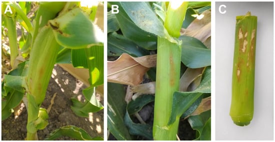
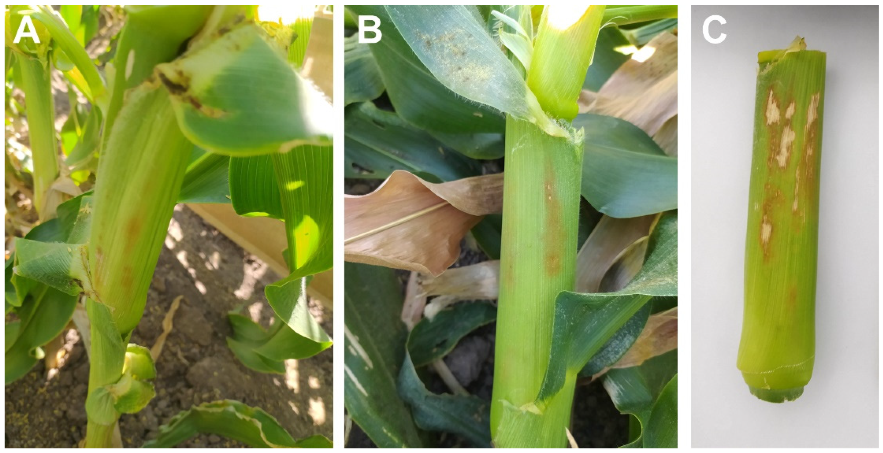
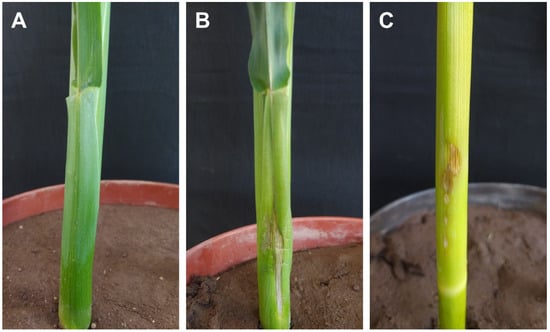
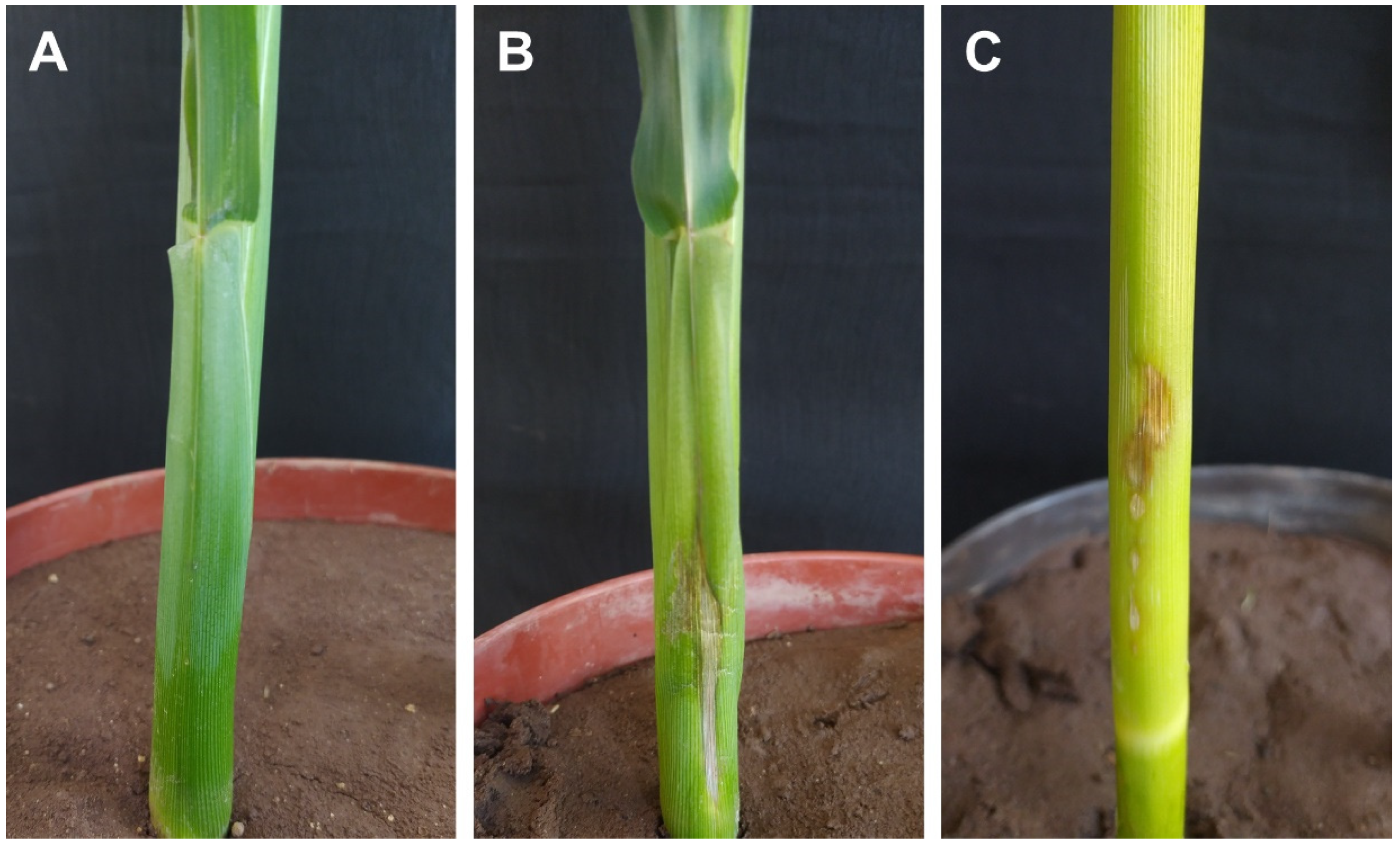
Despite these reports, bacterial diseases in sweet corn have often been underestimated by farmers and field advisors in Sinaloa, Mexico. However, in the last four growing seasons, a brown spot affecting the basal leaf sheath of sweet corn has been observed, and its etiology remains unknown. The symptoms appear after flowering. The initial symptoms consist of light brown spots with diffuse margins on the basal leaf sheath (Figure 1A). As the spots increased in size, they acquired an irregular, elongated shape and turned dark brown with a slightly sunken appearance (Figure 1B). In more advanced stages, the lesions developed a straw-colored to whitish coloration with reddish margins (Figure 1C). In the field, no completely desiccated sheaths directly associated with the symptom, nor leaves affected as a consequence of sheath damage, were observed.
Figure 1.
Symptoms of brown spot of leaf sheath in sweet corn observed during the 2022–2023 growing season in commercial fields in Sinaloa, Mexico. (A) Initial symptoms. (B) Intermediate symptoms. (C) Advanced symptoms depicting a lesion with a whitish center surrounded by a dark brown margin.
Preliminary studies indicate that an unidentified bacterium is consistently associated with these symptoms. Although the current damage does not appear to affect yield or quality, identifying the pathogen is fully justified, as it may represent an emerging disease linked to climate change and could become economically significant in the short or medium term. Given the interest from growers and field advisors in identifying the causal agent, and because this disease has not been previously reported in the region, the objectives of this study were to (a) identify the bacterial species associated with the disease using biochemical, physiological, and molecular techniques, and (b) determine the pathogenicity of the bacteria in slices of potato, onion, and celery in the laboratory and in four sweet corn hybrids under greenhouse conditions.
2. Materials and Methods
2.1. Isolation of the Bacterium Associated with the Disease
Sweet corn plants exhibiting brown spot symptoms on the leaf sheath were sampled from commercial fields in the municipalities of Juan José Ríos and Ahome, Sinaloa, Mexico, during the 2023–2024 growing season. Throughout the sampling period, the temperature ranged from 8.5 to 33 °C. The average monthly maximum temperatures were 1 to 3 °C higher than those recorded during the same months of the 1961–1990 baseline period. Average monthly minimum temperatures over the same study period also rose, increasing by 2.6 to 4.7 °C relative to the baseline average. For context, during the 1961–1990 period, the highest average minimum temperature occurred in December, while the lowest was observed in January and May. The accumulated rainfall was 154.5 mm [24].
To evaluate disease incidence, the “five-point” sampling method was used, which consisted of selecting five points per sampling site (four corners and the center). At each point, five consecutive plants were evaluated to determine the presence or absence of characteristic disease symptoms. From the symptomatic plants, two leaf sheaths were collected per site, obtaining a total of ten samples per plot for subsequent analysis. Disease incidence at harvest ranged from 45–50%, with 5–10% of the basal leaf sheath area affected per plant. No symptoms were observed on the stems beneath the symptomatic sheaths. Two composite samples, each consisting of three randomly selected symptomatic sheaths, were placed in an ice chest (10–12 °C), transported to the laboratory, and processed for bacterial isolation within 24 h, following standard procedures [25]. Initially, 33 bacterial isolates from nine commercial sweet corn fields were obtained during the sampling period. Next, after considering colony characteristics (shape, color, elevation, margin, texture, pigment production), as well as Gram and positive KOH string tests, nine morphotypes were delineated. Afterwards, a preliminary pathogenicity test was performed on the sweet corn hybrid ‘Accentuate’ under greenhouse conditions. The results indicated that only four bacterial isolates were pathogenic; from these, isolates A and D from the municipalities of Juan Jose Rios (25°51′17.4′′ N, 108°45′27.5′′ W and 25°47′52.4′′ N, 108°46′47.8′′ W, respectively) were selected for subsequent studies.
2.2. Biochemical Identification
Bacterial strains were characterized biochemically and physiologically using Gram staining, the KOH string test, detection of diffusible pigments on Nutrient Agar (NA), oxidation/fermentation tests, catalase activity, growth in 5% NaCl, and growth at 40 °C, all performed as described by Schaad et al. [25]. Additionally, strains were tested using API 50 CHB/E medium strips (bioMérieux, Durham, NC, USA) according to the manufacturer’s instructions. Briefly, a bacterial suspension was inoculated into each tube of the strip and incubated for 48 h at 30 °C. Fermentation of carbohydrates was indicated by a color change due to acid production and pH drop. The resulting biochemical profile was analyzed using the manufacturer’s identification software, and the results were compared with previously published profiles for B. gladioli [26].
2.3. Hypersensitivity Reaction to Bacterial Strains on Tobacco
Colonies grown on NA for 48 h at 25 °C were suspended in sterile distilled water and adjusted to a concentration of 1 × 10⁵ CFU mL−1. Suspensions were infiltrated into the abaxial epidermis of tobacco leaves (Nicotiana tabacum cv. Xanthi) using a 3 mL syringe. Tissue necrosis observed 24–48 h after infiltration was recorded as a positive hypersensitive reaction (HR) [25].
2.4. Molecular Characterization of the Bacterial Strains
2.4.1. DNA Extraction
Bacterial strains were incubated in nutrient broth at 25 °C for 24 h on a Yamato orbital shaker (SOU-300, Monterrey, N.L, Mexico). The cultures were centrifuged at 13,000 rpm for 2 min, and the broth was decanted to collect the bacterial cells. Genomic DNA extraction was performed using the CTAB method, modified from Doyle [27]. A total of 600 µL of CTAB buffer solution (4% of 0.5 M EDTA, 10% Tris-HCl, 8% molecular-grade NaCl, 2% CTAB) and 6 µL of β-mercaptoethanol were added. The DNA was rehydrated with 30 µL of ultrapure water and stored at −20 °C. DNA quality and concentration were verified using a NanoDrop spectrophotometer 2000 (Thermo Scientific, Wilmington, DE, USA).
2.4.2. PCR Amplification
The 16S ribosomal RNA (16S rRNA) and recombinase A (recA) genes were amplified using primers F2C/C and BUR1/BUR2, respectively [28,29]. PCR was carried out in a TC1000-G thermocycler (DLAB Scientific, Monterrey, N.L. México) with a total reaction volume of 25 µL. Each reaction contained 2 µL of DNA (50 ng/µL), 1x reaction buffer, 1.5 mM MgCl2, 0.2 mM dNTPs, 0.5 µM of each primer, and 0.25 U of Taq DNA polymerase. The amplification conditions were as follows: 16S: an initial denaturation at 95 °C for 4 min, followed by 34 cycles of denaturation at 95 °C for 1 min, annealing at 60 °C for 1 min, and extension at 72 °C for 1.5 min, with a final extension at 72 °C for 5 min; recA: an initial denaturation at 94 °C for 5 min, followed by 30 cycles of denaturation at 94 °C for 30 s, annealing at 60 °C for 30 s, and extension at 72 °C for 45 s, with a final extension at 72 °C for 5 min. PCR products were sequenced at Macrogen (Seoul, Republic of Korea).
2.4.3. Phylogenetic Analysis
Sequences were edited using BioEdit v.7.7.1 software [30] and compared using the NCBI’s BlastN algorithm. The bacterial sequences were aligned with reference sequences from Burkholderia type specimens [31] and one Paraburkholderia acidisoli (DHF22) sequence as an outgroup. Individual gene sequence alignments were performed using the MUSCLE algorithm and concatenated in MEGA v.12.0.10 [32]. The best-fit nucleotide substitution model for the combined dataset 16S + recA was selected based on the corrected Akaike information criterion in MEGA. The phylogenetic tree was constructed using the Maximum Likelihood (ML) and the Bayesian Inference (BI) methods. The ML analysis was performed in RAxML v 7.2.8 [33] using the GTRGAMMAI model and 1000 bootstrap replicates. Mr. Bayes 3.2.7 [34] was used for IB phylogeny with the GTR + I + G model, two independent Metropolis Markov-chain Monte Carlo runs, 1 million generations, sampling every 1000 trees, and a burn-in of 20%. Final tree editing was performed in iTOL v6 [35].
2.5. Rotting of Potato, Onion, and Celery Slices
Potato tubers, onion bulbs, and celery petioles were surface-disinfected by immersion in 0.5% sodium hypochlorite for 5 min, rinsed with sterile distilled water, and aseptically cut into 0.8 cm thick slices. Slices were placed in Petri dishes on sterile moist filter paper to maintain 100% relative humidity (RH) at 25 °C. Each slice was wounded with a sterile scalpel and inoculated with a loopful of bacteria taken from 48 h NA cultures. Control slices were treated similarly, but only with sterile water. After 72 h incubation at 25 °C, symptoms were recorded. Bacteria were re-isolated from symptomatic tissues and compared biochemically with the original strains. The experiment was conducted twice.
2.6. Pathogenicity Tests of Strains Obtained from Symptomatic Leaf Sheaths
Sweet corn seeds of hybrids Accentuate, 8909MRW, 1760MR and BSS1075 (Syngenta Agro, S.A. de C.V., Ciudad de Mexico, Mexico) were disinfected with 1.5% NaClO, sown in plastic pots containing 5 kg of pasteurized clay/loam/sand mix (at the ratio of 28.1:27.0:44.9, pH 7.0), and fertilized regularly with Miracle Gro fertilizer (The Scotts Company LLC; Marysville, OH, USA) and irrigated as needed. One-month-old plants were inoculated by applying 25 µL of bacterial suspension of isolates A and D (1 × 10⁵ CFU mL−1) between the stem and the first basal leaf sheath. Prior to placing the inoculum between the leaf sheaths and the stems of the plants, three perforations were made in the leaf sheath with a sterilized 3 mL syringe. Control plants received sterile water. Plants were arranged in a completely randomized design with six replicates (six plants; one per pot) per strain and covered with transparent polyethylene bags for 48 h to maintain high RH (94–96%), followed by a 12 h daily high-RH period for five days, after which they remained uncovered for 7 days (RH 50–75%). Disease severity was assessed 14 days post-inoculation (dpi) as leaf sheath diseased area (LSDA; cm2). To determine the LSDA, a grid of dots 0.5 cm apart on a 7 × 7 cm piece of clear polyethylene sheet was placed on the lesions. The number of dots that touched the lesion was counted, and this number was divided by 4 to estimate the lesion size in cm2 [36]. The experiment was run twice. In experiments 1 and 2, the temperature ranged from 16 to 29 °C and from 17 to 28 °C, respectively. Bacteria were re-isolated from symptomatic tissues to fulfill Koch’s postulates, and their identity was confirmed using biochemical and molecular techniques.
2.7. Statistical Analysis of Data
All statistical analyses were performed in R Studio version 1.4.1106.
Data of LSDA were tested for homogeneity of variances and normality using Levene’s and Shapiro–Wilk test, respectively, using the car package. A Kruskal–Wallis [37] (α = 0.05) test was performed to assess differences between the two experiment replications; since no differences were detected, data from both experiments were pooled for subsequent analysis (n = 12). Pairwise comparison between control and infected plants for each maize hybrid was performed with the Wilcoxon signed-rank test (α = 0.05). The effect of bacterial strain and sweet corn hybrid on LSDA was evaluated by a Kruskal–Wallis [37] test (α = 0.05), followed by a Bonferroni correction of Fisher’s least significant difference (LSD) test, using the agricolae package. Effect size was calculated with the Epsilon squared () formula. The box plots were created using the ggplot2 package in R Studio.
3. Results
3.1. Incidence and Isolation
The incidence of the disease at harvest ranged from 45 to 50%, while lesion size varied from 2 to 4.5 cm in length by 0.5 to 1.5 cm in width across the different sweet corn hybrids. In total, 33 bacterial isolates were obtained from symptomatic sweet corn tissue collected from nine commercial sweet corn lots during the sampling period.
Using Gram staining and the KOH string test, rod-shaped, Gram-negative isolates were selected. Based on colony phenotypic characteristics, 23 isolates were grouped into nine morphotypes. In a preliminary pathogenicity study on the sweet corn hybrid Accentuate, morphotypes A and D harbored 3 and 4 pathogenic isolates, respectively. Subsequently, one isolate from each morphotype was used for further study.
3.2. Biochemical and Physiological Characteristics
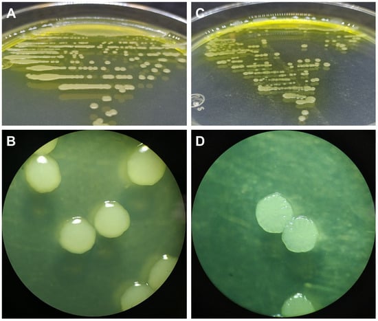
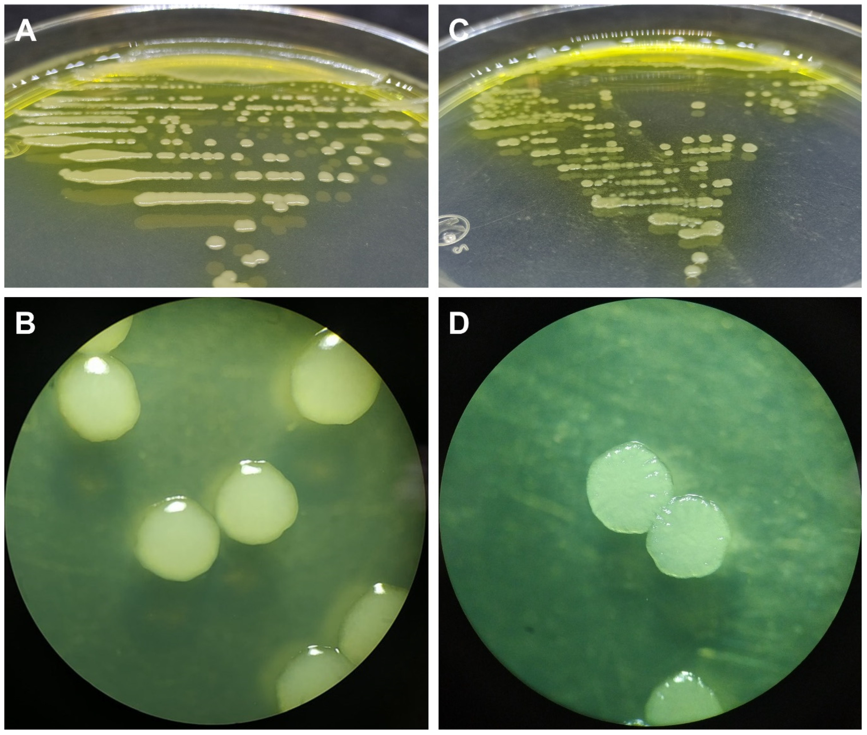
Two strains, designated A and D, obtained from symptomatic leaf sheaths for detailed biochemical and physiological characterization, were included in this study. Both strains were Gram-negative rods, positive in the KOH string test, and formed circular, cream-colored colonies with smooth to corrugated surfaces and undulate edges (Figure 2). They did not grow in 5% NaCl nutrient broth, produced yellow diffusible pigments on NA, and induced a hypersensitive response in tobacco (var. Xanthi). Both strains were catalase- and oxidase-negative but capable of growth at 40 °C.
Figure 2.
Colonies of Burkholderia gladioli of 72 h of age on NA. (A) Strain A producing a yellow diffusing pigment in the medium. (B) Shiny colonies, slightly corrugated and undulated margin of strain A under the stereomicroscope. (C) Colonies of strain D producing yellow diffusing pigment in the medium. (D) Colonies of strain D flat, corrugated with undulated margins under the stereomicroscope.
In API 50CHB/E tests, strain A was negative for glycerol, erythritol, D-arabinose, L-xylose, D-adonitol, methyl β-D-xylopyranoside, L-sorbose, L-rhamnose, methyl α-D-mannopyranoside, methyl α-D-glucopyranoside, N-acetylglucosamine, arbutin, salicin, D-maltose, D-lactose, D-saccharose, inulin, D-melezitose, D-raffinose, starch, glycogen, D-turanose, D-lyxose, L-arabitol, D-tagatose, potassium gluconate, potassium 2-ketogluconate, and potassium 5-ketogluconate; and positive for L-arabinose, D-ribose, D-xylose, D-galactose, D-glucose, D-fructose, D-mannose, dulcitol, inositol, D-mannitol, D-sorbitol, amygdalin, aesculin ferric citrate, D-cellobiose, D-melibiose, D-trehalose, xylitol, gentiobiose, D-fucose, L-fucose, and D-arabitol. Strain D showed a similar profile to strain A but differed by being positive for glycerol, D-arabinose, D-adonitol, D-lyxose, and D-tagatose. These results closely matched those previously reported by Palleroni [26] for B. gladioli (Table 1).
Table 1.
Phenotypic characterization of strains A and D using API®50 CH carbohydrates as compared to Burkholderia gladioli.
3.3. Pathogenicity Tests on Potato, Onion, and Celery
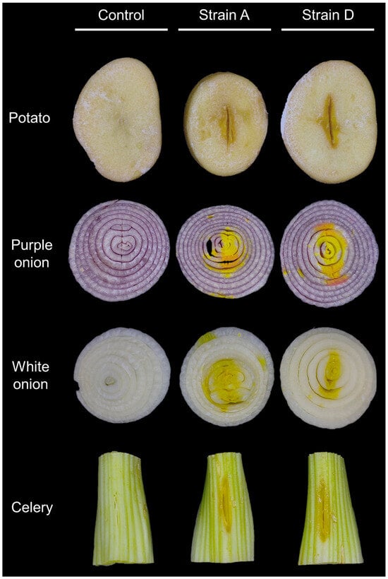
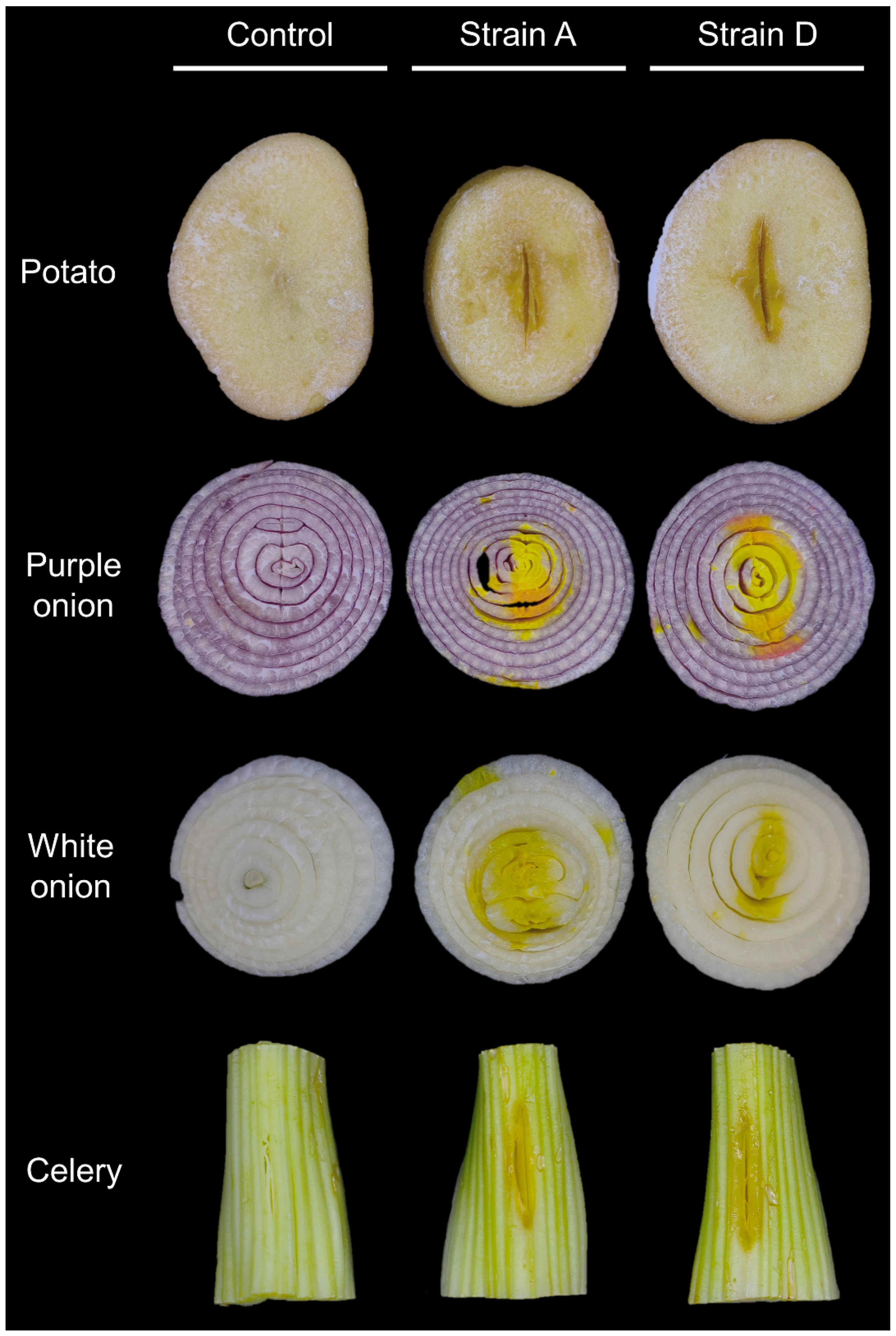
Both strains induced a slight soft rot in potato slices, characterized by darkening of the inoculated area. In contrast, more pronounced maceration was observed on slices of white onion, purple onion, and celery. Additionally, a diffusible yellow pigment was produced on all three host tissues following inoculation (Figure 3).
Figure 3.
Soft rot in potato, purple onion, white onion, and celery slices 72 h post-inoculation with strains A and D of Burkholderia gladioli.
3.4. Molecular Identification
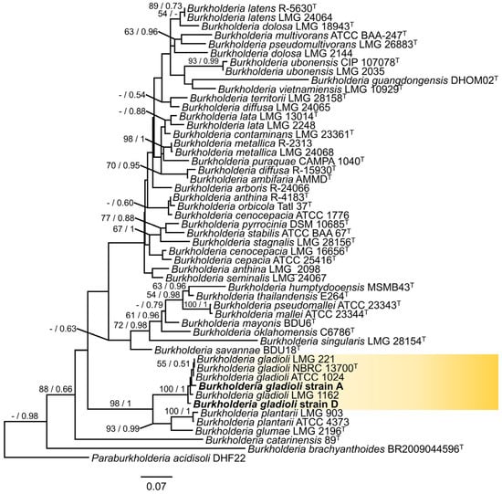
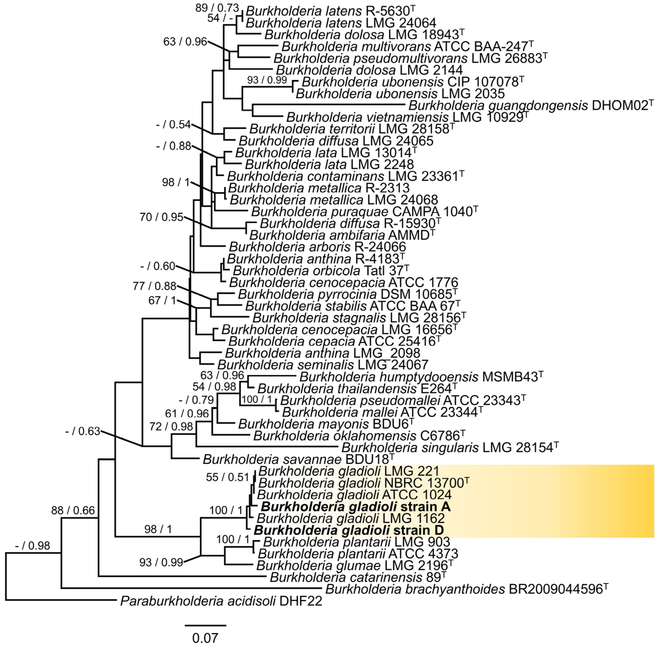
Sequence comparison revealed that both strains shared 99% similarity with reference sequences of B. gladioli (accessions: 16S, CP002600 and KP306792; recA, CP054468 and CP033430). The sequences were deposited in GenBank: strain A, PX794909 (16S), PZ247305 (recA); strain D, PX794908 (16S), PZ247304 (recA). The phylogenetic analyses using Maximum Likelihood and Bayesian Inference placed strains A and D within a well-supported clade containing several B. gladioli type strains, with a bootstrap value of 100% and a posterior probability of 1 (Figure 4). This molecular confirmation aligns with biochemical and physiological identification.
Figure 4.
Maximum likelihood tree of the combined 16S rRNA + recA gene data for Burkholderia. The sequence of Paraburkholderia acidisoli type strain DHF22 was used as an outgroup. Strains from this study are shown in bold. Bootstrap values higher than 50% and posterior probabilities greater than 0.5 are shown above the branches. The scale bar indicates the number of nucleotide substitutions. T indicate ex-type strains.
3.5. Pathogenicity Tests on Sweet Corn Leaf Sheaths
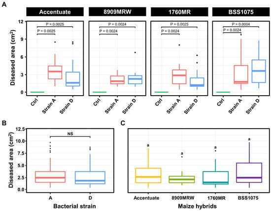
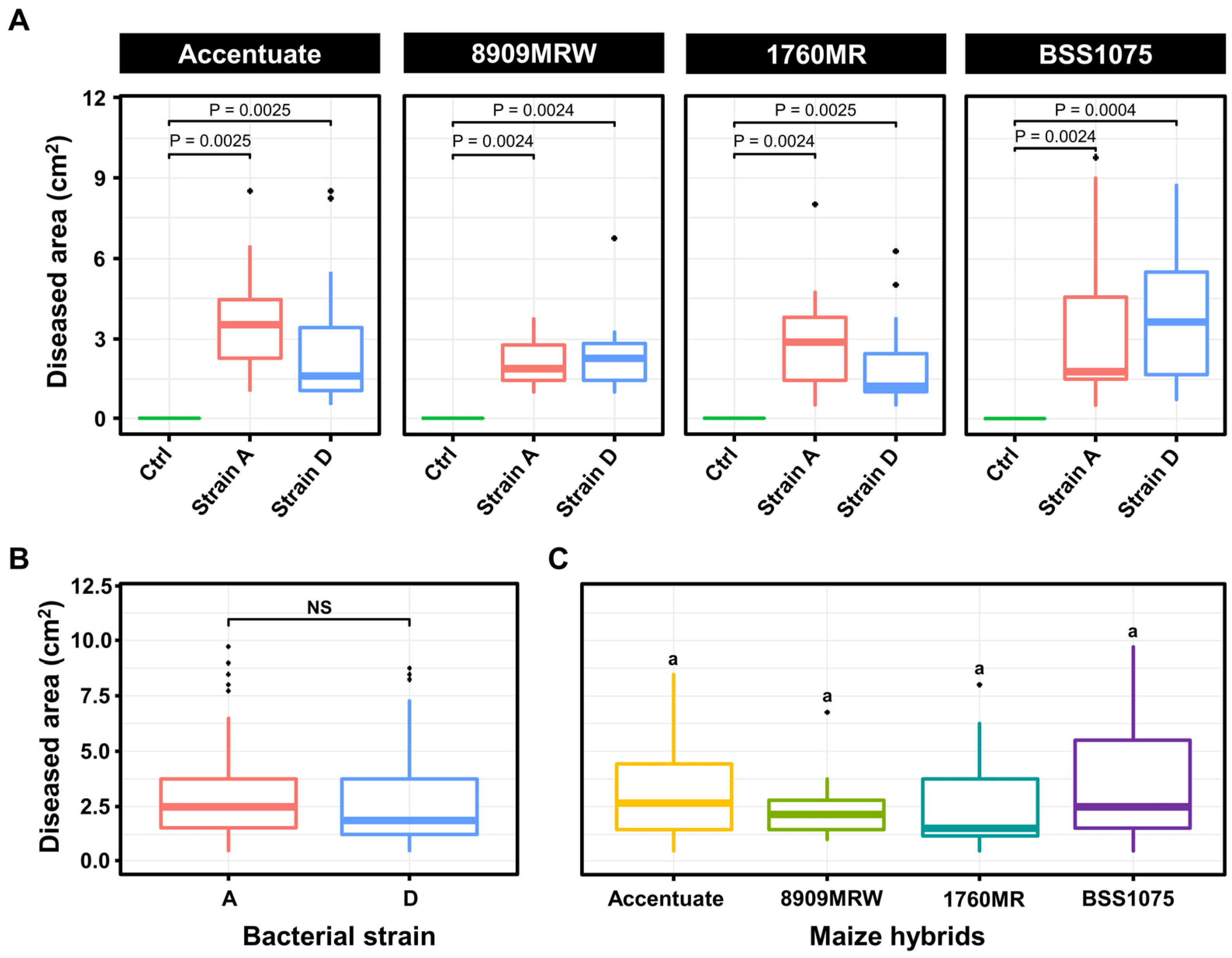
Strains A and D of B. gladioli induced brown spot symptoms on the leaf sheaths of all four sweet corn hybrids tested, closely resembling field observations (Figure 5B,C). Initial symptoms appeared 72 h post-inoculation as small, clear brown lesions at the inoculation site. By day 14, symptoms were consistent with those observed in commercial fields. Non-inoculated plants remained symptom-free throughout the experiment. Pairwise comparison between control and infected plants was significant (p < 0.001) in all maize hybrids (Figure 6).
Figure 5.
Non-inoculated plant and lesions caused after 14 days of inoculation in the leaf sheaths of sweet corn with Burholderia gladioli. (A) Non-inoculated plant. (B) Symptoms on the leaf sheath caused by strain A. (C) Symptoms on the leaf sheath caused by strain D.
Figure 6.
Pathogenicity test of Burkholderia gladioli strains A and D expressed as leaf sheath diseased area (LSDA; cm2). All data are shown as medians (25–75 percentiles). (A) Significant pairwise comparisons of LSDA between non-inoculated (control) and inoculated plants of different sweet corn hybrids, based on the Wilcoxon signed-rank test (B) Non-significant effect of bacterial strains, and (C) sweet corn hybrids on LSDA after Kruskal–Wallis [37] test (p > 0.05). NS stands for non-significant difference. Same letters above the boxes indicate non-significant differences among treatments
The Kruskal–Wallis [37] test of LSDA revealed no significant differences between strains A and D (X2 = 0.68, p = 0.4087; Figure 6). Damage caused by strain A across all sweet corn hybrids ranged from 1.40 to 3.73 cm2, while strain D caused a LSDA ranging from 2.27 to 4.41 cm2. The effect of the sweet corn hybrid was not significant (X2 = 3.43, p = 0.3286; Figure 6), suggesting that all hybrids are equally susceptible to bacterial infection, under greenhouse conditions. Koch’s postulates were fulfilled by re-isolating strains with identical cultural, biochemical, and molecular profiles from symptomatic tissues.
The Kruskal–Wallis [37] test of LSDA revealed no significant differences between strains A and D (X2 = 0.68, p = 0.4087; ε2 = 0.0071; Figure 6). Damage caused by strain A across all sweet corn hybrids ranged from 1.40 to 3.73 cm2, while strain D caused an LSDA ranging from 2.27 to 4.41 cm2. The effect of the sweet corn hybrid was not significant (X2 = 3.43, p = 0.3286; ε2= 0.0361; Figure 6), which suggests that all hybrids are equally susceptible to bacterial infection under the evaluated conditions. Koch’s postulates were fulfilled by re-isolating strains with identical cultural, biochemical, and molecular profiles from symptomatic tissues.
4. Discussion
The high percentage of biochemical test results from our strains, isolated from sweet corn leaf sheaths, aligned with those reported for B. gladioli. Notably, slight metabolic variations were observed between strains A and D; for instance, strain D was positive for glycerol, D-arabinose, D-adonitol, D-xylose, and D-tagatose utilization, whereas strain A was negative for these carbon sources. Furthermore, both strains failed to utilize N-acetyl-glucosamine, salicin, potassium 2-ketogluconate, and potassium 5-ketogluconate, in contrast to the typical B. gladioli profile described by Palleroni [26]. These findings confirm the metabolic versatility of B. gladioli, which is known to utilize a broad spectrum of carbon sources [26]. The observed intraspecific variation in substrate utilization is consistent with patterns reported in other bacterial genera, such as Pectobacterium within the Enterobacteriaceae [38,39].
The integrated biochemical, physiological, and pathogenicity data from this study fully support the molecular identification of the two B. gladioli strains obtained from symptomatic sweet corn plants in Sinaloa, Mexico. Both strains (A and D) successfully reproduced symptoms under greenhouse conditions similar to those observed in commercial fields (Figure 4). Notably, while the bacteria consistently induced brown spots on leaf sheaths, no stem symptoms were detected during this investigation. The four bacterial isolates originally obtained from sweet corn are not epiphytic because they were obtained from symptomatic tissue on the leaf sheath of sweet corn; it can be said that the representative isolates A and D included in the present study should be characterized as weak pathogens because they induced symptoms on four sweet corn hybrids under greenhouse conditions but did not colonize the leaf sheath severely under both field conditions and in artificial greenhouse inoculations.
The strains, originally isolated from naturally infected sweet corn, proved pathogenic on the commercial hybrids Accentuate, 8909MRW, 1760MR, and BSS1075. Interestingly, under field conditions, the damage appeared confined to the leaf sheath, without progressing to leaves or stems, a pattern that contrasts with reports of B. gladioli causing leaf stripe and stem rot in dent corn in other regions of Mexico [20]. First isolated from gladiolus bulbs, B. gladioli has since been identified in a wide range of hosts, including iris, saffron, maize, rice [40] and various orchids [41]. Its pathogenicity extends to multiple crops, where it has been associated with soft rot in purple onion bulbs, leek stalks, broccoli inflorescences, cactus cladodes, carrot roots, ginger rhizomes, and mushroom basidiocarps, and has been shown to colonize bean plants in greenhouse studies [42]. This is consistent with pathogenicity tests conducted on potato, onion, and celery using the isolated strains in this study.
Globally, B. gladioli has been implicated in garlic bulb decay and shown high pathogenicity on cactus, gladiolus, onion, and potato in Iran [43], as well as dry rot of saffron in the same country [44]. In Mexico, previous reports include onion bulb rot [45] and leaf stripe and stem rot in dent corn [20]. The bacterium has also been associated with ear soft rot of corn in the United States [46]. To our knowledge, this study is the first to report B. gladioli as the causal agent of brown spot on sweet corn leaf sheaths in Mexico. The broad and expanding host range of this bacterium highlights its potential as an emerging phytopathogen in the region, particularly for crops cultivated during the fall–winter season in Sinaloa. Although garlic and onion are not uncommon, dent corn is the predominant crop in the sampled region and in the state of Sinaloa, and even crop rotation between these crops occurs, implying a potential risk of a greater incidence of the disease in these crops. This work establishes a critical foundation for future studies on the host range, epidemiology, and management of this pathogen. Special attention should be given to dent corn, which occupies approximately 500,000 hectares in Sinaloa during the fall–winter cycle and in which B. gladioli has already been reported elsewhere in Mexico [20]. The potential economic impact on this major crop and on vegetable production in the state warrants careful monitoring and proactive research. Regarding crop rotation disease management, B. gladioli has a wide host range [42] and the genus contains biologically heterogeneous strains that can survive in diverse habitats such as soil, water and diseased plant material [47]. Therefore, further studies on the host range of the pathogen should be conducted and should focus on those that are not susceptible to the bacterium.
5. Conclusions
This study identified B. gladioli as the causal agent of the emerging brown spot disease on sweet corn leaf sheaths in Sinaloa, Mexico. The identification was confirmed through a polyphasic approach that integrated biochemical profiles consistent with the species, pathogenicity tests on alternative potential hosts and sweet corn plants, and molecular analysis (16S rRNA + recA gene sequencing and phylogenetic inference). The bacterium successfully reproduced field symptoms in the greenhouse, fulfilling Koch’s postulates. This represents the first report of B. gladioli causing this specific symptomatology on sweet corn in Mexico, expanding its documented host range in Mexico and geographic distribution as a phytopathogen.
Given the bacterium’s broad host range, which includes economically important crops like onion, garlic, and dent corn cultivated in the region, B. gladioli should be considered a potential emerging threat to horticultural production in Sinaloa. Future research should focus on determining the epidemiological conditions favorable for disease development.
Author Contributions
Conceptualization, R.F.-G.; methodology, R.F.-G., J.R.E.-C., K.Y.L.-M., I.E.M.-M. and G.H.-R.; formal analysis, R.F.-G., K.Y.L.-M. and G.H.-R.; investigation R.F.-G. and J.R.E.-C.; resources, R.F.-G. and K.Y.L.-M.; data curation, J.R.E.-C., K.Y.L.-M. and G.H.-R.; writing—original draft preparation, R.F.-G., K.Y.L.-M. and G.H.-R.; writing—review and editing, R.F.-G., K.Y.L.-M. and G.H.-R.; visualization, R.F.-G., K.Y.L.-M., I.E.M.-M. and G.H.-R.; supervision, R.F.-G. and G.H.-R.; funding acquisition, R.F.-G. All authors have read and agreed to the published version of the manuscript.
Funding
This research was funded by the Graduate Division of the Autonomous University of the West in Sinaloa, Mexico, within the framework of the PIFIP-2023 Research Project.
Data Availability Statement
The original data presented in the study are openly available in National Center for Biotechnology Information (NCBI) database at PX794909 and PX794908.
Acknowledgments
The authors acknowledge the Autonomous University of the West in Sinaloa, Mexico for the financial support to conduct the present research.
Conflicts of Interest
The authors declare no conflicts of interest.
References
- Pajic, Z.; Rodosavlevic, M.; Eric, U. The utilizable value of sweet corn and popcorn hybrids. Agro Knowl. J. 2004, 5, 53–60. [Google Scholar]
- Oktem, A.; Oktem, A. Effect of nitrogen and intra spaces on sweet corn (Zea mays Sachharata Sturt) ear characteristics. Indian J. Plant Sci. 2005, 4, 361–363. [Google Scholar]
- Dewanto, V.; Wu, X.; Liu, R.H. Processed sweet corn has higher antioxidant activity. J. Agric. Food Chem. 2002, 50, 4959–4964. [Google Scholar] [CrossRef] [Scilit] [PubMed]
- Swapna, G.; Jadesha, G.; Mahadevu, P. Sweet Corn—A future healthy human nutrition food. Int. J. Curr. Microbiol. Appl. Sci. 2020, 9, 3859–3865. [Google Scholar] [CrossRef] [Scilit]
- SIVASA, Sistema de Validación y Seguimiento Agrícola. 2024. Avance de Permisos Únicos de Siembra del Otoño-Invierno 2023–2024. Available online: https://www.sivasa.mx/ (accessed on 2 September 2024).
- Parera, C.A. Producción de Maíz Dulce; Primera Edición; Ediciones INTA: Buenos Aires, Argentina, 2017; 65p. [Google Scholar]
- Sikora, E.; Faske, T.; Meyer, R.; Betts, A.; Kemerait, B.; Camiletti, B.; Telenko, D.; Robertson, A.; Mueller, D.; Sisson, A.; et al. Corn Disease Loss Estimates from the United States and Ontario, Canada—2024. Crop Protection Network. CPN-2007-24. 2025. Available online: https://cropprotectionnetwork.org/publications/corn-disease-loss-estimates-from-the-united-states-and-ontario-canada-2024 (accessed on 18 March 2026).
- Castañón, N.G.; Latournerie, L.M. Performance of S1 maize (Zea mays L.) families in different soil pH. Bragantia 2004, 63, 63–72. [Google Scholar]
- Ridout, M.E.; Godfrey, B.; Newcombe, G. Effects of Antagonists on mycotoxins of seedborne Fusarium spp. in sweet corn. Toxins 2019, 11, 438. [Google Scholar] [CrossRef] [Scilit]
- Watson, A.; Burgess, L.W.; Summerell, B.A.; O’Keeffe, K. Fusarium species associated with cob rot of sweet corn and maize in New South Wales. Australas. Plant Dis. Notes 2014, 9, 1–5. [Google Scholar] [CrossRef] [Scilit]
- Malvick, D.K.; Rohwe, C.L.; Floyd, C.M. Effects of hybrids, foliar treatments, and Infection Conditions on Bacterial Leaf Streak of Sweet Corn. Plant Dis. 2024, 108, 2906–2912. [Google Scholar] [CrossRef] [Scilit]
- Roper, M.C. Pantoea stewartii subsp. stewartii: Lessons learned from a xylem-dwelling pathogen of sweet corn. Mol. Plant Pathol. 2011, 12, 628–637. [Google Scholar] [CrossRef] [Scilit]
- Alippi, A.M.; López, A.C. First Report of Leaf spot disease of maize caused by Pantoea ananatis in Argentina. Plant Dis. 2010, 94, 487. [Google Scholar] [CrossRef] [Scilit]
- Toaza, A.; Caiza, R.B.; Garrido, A.D.; Moreno, C.R.; Guevara, J.; Regalado, H.; Flores, F.J.; Ramos, L.A.; Garrido, P.A. First report of Pantoea ananatis causing leaf spot in maize in Ecuador. Plant Dis. 2021, 105, 3286. [Google Scholar] [CrossRef] [Scilit]
- Cui, L.; Zou, C.; Zhang, Z.; Duan, L.; Huang, J.; Wang, L.; Xiao, W.; Li, W.; Yang, X.; Xiang, Y.; et al. First report of maize white spot disease caused by Pantoea ananatis in China. Plant Dis. 2023, 107, 210. [Google Scholar] [CrossRef] [Scilit]
- Zhang, N.; Li, X.F.; Li, Y.L.; Lu, W.J.; Xie, L.H.; Li, G.K. First report of bacterial leaf streak in sweet corn caused by Pantoea ananatis in Southeast China. Plant Dis. 2023, 107, 1214. [Google Scholar] [CrossRef] [Scilit]
- Goszczynska, T.; Botha, W.J.; Venter, S.N.; Coutinho, T.A. Isolation and identification of the causal agent of brown stalk rot, a new disease of maize in South Africa. Plant Dis. 2007, 91, 711–718. [Google Scholar] [CrossRef] [Scilit] [PubMed]
- Pérez-Terrón, R.; Villegas, M.C.; A Cuellar, A.; Muñoz-Rojas, J.; Castañeda-Lucio, M.; Hernández-Lucas, I.; Bustillos-Cristales, R.; Bautista-Sosa, L.; Munive, J.A.; Caicedo-Rivas, R.; et al. Detection of Pantoea ananatis, causal agent of leaf spot disease of maize, in Mexico. Australas. Plant Dis. Notes 2009, 4, 96–99. [Google Scholar] [CrossRef] [Scilit]
- Krawczyk, K.; Foryś, J.; Nakonieczny, M.; Tarnawska, M.; Bereś, P. Transmission of Pantoea ananatis, the causal agent of leaf spot disease of maize (Zea mays), by western corn by western corn rootworm (Diabrotica virgifera virgifera LeConte). Crop Prot. 2021, 141, 105431. [Google Scholar] [CrossRef] [Scilit]
- Gijón-Hernandez, A.; Téliz-Ortiz, D.; Cárdenas-Soriano, E.; De León, C.; Mora-Aguilera, A.; Mejía-Sánchez, D.; De La Torre-Almaraz, R. Leaf stripe and stem rot caused by Burkholderia gladioli, a new disease of maize in México. Plant Dis. 2008, 92, 1249. [Google Scholar] [CrossRef] [Scilit] [PubMed]
- Chiangkhaek, O.; Akarapisan, A. Characterization and identification of Burkholderia gladioli causing stalk rot of sweet corn in Thailand. J. Phytopathol. 2024, 172, e13314. [Google Scholar] [CrossRef] [Scilit]
- Nandakumar, R.; Shahjahan, A.K.; Yuan, X.L.; Dickstein, E.R.; Groth, D.E.; Clark, C.A.; Cartwright, R.D.; Rush, M.C. Burkholderia glumae and B. gladioli cause bacterial panicle blight in rice in the southern United States. Plant Dis. 2009, 93, 896–905. [Google Scholar] [CrossRef] [Scilit]
- Yabuuchi, E.; Kosako, Y.; Oyaizu, H.; Yano, I.; Hotta, H.; Hashimoto, Y.; Ezaki, T.; Arakawa, M. Proposal of Burkholderia gen. nov. and transfer of seven species of the genus Pseudomonas homology Group II to the new genus, with the Type Species Burkholderia cepacia (Palleroni and Holmes 1981) Comb. Nov. Microbiol. Immunol. 1992, 36, 1251–1275. [Google Scholar] [CrossRef] [Scilit]
- Servicio Meteorologico Nacional, México. Normales Climatologicas por Estado. Available online: https://smn.conagua.gob.mx/es/climatologia/informacion-climatologica/normales-climatologicas-por-estado (accessed on 16 March 2026).
- Schaad, N.W.; Jones, J.B.; Chun, W. (Eds.) Laboratory Guide for Identification of Plant Pathogenic Bacteria, 2nd ed.; The American Phytopathological Society (APS): St. Paul, MN, USA, 2000; pp. 1–16. [Google Scholar]
- Palleroni, N.J. Genus Burkholderia. In Bergy’s Manual of Systematics of Archea and Bacteria; John Wiley and Sons, Inc.: Hoboken, NJ, USA, 2015. [Google Scholar] [CrossRef] [Scilit]
- Doyle, J.J. Isolation of plant DNA from fresh tissue. Focus 1990, 12, 13–15. [Google Scholar]
- Shi, T.; Reeves, R.H.; Gilichinsky, D.A.; Friedmann, E.I. Characterization of viable bacteria from Siberian permafrost by 16S rDNA Sequencing. Microb. Ecol. 1997, 33, 169–179. [Google Scholar] [CrossRef] [Scilit]
- Payne, G.W.; Vandamme, P.; Morgan, S.H.; Lipuma, J.J.; Coenye, T.; Weightman, A.J.; Jones, T.H.; Mahenthiralingam, E. Development of a recA gene-based identification approach for the entire Burkholderia genus. Appl. Environ. Microbiol. 2005, 71, 3917–3927. [Google Scholar] [CrossRef] [Scilit] [PubMed]
- Hall, T.A. BioEdit: A user-friendly biological sequence alignment editor and analysis program for Windows 95/98/NT. Nucleic Acids Symp. Ser. 1999, 41, 95–98. Available online: https://www.academia.edu/download/29520866/1999hall1.pdf (accessed on 1 February 2026).
- Chalita, M.; Kim, Y.O.; Park, S.; Oh, H.S.; Cho, J.H.; Moon, J.; Baek, N.; Moon, C.; Lee, K.; Yang, J.; et al. EzBioCloud: A genome-driven database and platform for microbiome identification and discovery. Int. J. Syst. Evol. Microbiol. 2024, 74, 006421. [Google Scholar] [CrossRef] [Scilit]
- Kumar, S.; Stecher, G.; Suleski, M.; Sanderford, M.; Sharma, S.; Tamura, K. Molecular evolutionary genetics analysis version 12 for adaptive and green computing. Mol. Biol. Evol. 2024, 41, 1–9. [Google Scholar] [CrossRef] [Scilit]
- Stamatakis, A. RAxML-VI-HPC: Maximum likelihood-based phylogenetic analyses with thousands of taxa and mixed models. Bioinformatics 2006, 22, 2688–2690. [Google Scholar] [CrossRef] [Scilit]
- Ronquist, F.; Teslenko, M.; van der Mark, P.; Ayres, D.L.; Darling, A.; Höhna, S.; Larget, B.; Liu, L.; Suchard, M.A.; Huelsenbeck, J.P. MrBayes 3.2: Efficient Bayesian phylogenetic inference and model choice across a large model space. Syst. Biol. 2012, 61, 539–542. [Google Scholar] [CrossRef] [Scilit] [PubMed]
- Letunic, I.; Bork, P. Interactive Tree of Life (iTOL) v6: Recent updates to the phylogenetic tree display and annotation tool. Nucleic Acids Res. 2024, 52, W78–W82. [Google Scholar] [CrossRef] [Scilit] [PubMed]
- Pryor, B.M.; Davis, R.M.; Gilbertson, R.L. A toothpick inoculation method for evaluation carrot cultivars for resistance to Alternaria radicina. HortScience 2002, 35, 1099–1102. [Google Scholar] [CrossRef] [Scilit]
- Kruskal, W.H.; Wallis, W.A. Use of Ranks in One-Criterion Variance Analysis. J. Am. Stat. Assoc. 1952, 47, 583–621. [Google Scholar] [CrossRef]
- Li, X.; Ma, Y.; Liang, S.; Tian, Y.; Yin, S.; Xie, S.; Xie, H. Comparative genomics of 84 Pectobacterium genomes reveals the variations related to a pathogenic lifestyle. BMC Genom. 2018, 19, 889. [Google Scholar] [CrossRef] [Scilit]
- Chen, C.; Li, X.; Bo, Z.; Du, W.; Fu, L.; Tian, Y.; Cui, S.; Shi, Y.; Xie, H. Occurrence, characteristics, and PCR-based detection of Pectobacterium polaris causing soft rot of Chinese cabbage in China. Plant Dis. 2021, 105, 2880–2887. [Google Scholar] [CrossRef] [Scilit]
- Mirghasempour, S.A.; Huang, S.; Xie, G.L. First report of Burkholderia gladioli causing rice panicle blight and grain discoloration in China. Plant Dis. 2018, 102, 2635. [Google Scholar] [CrossRef] [Scilit]
- Keith, L.; Thammakijjawat, P. Detection of Burkholderia glalioli in orchids. In Detection of Plant-Pathogenic Bacteria in Seed and Other Planting Material, 2nd ed.; Fatme, M., Walcott, R.R., Schaad, N.W., Eds.; The American Phytopathological Society (APS) Press: St. Paul, MN, USA, 2019; pp. 355–360. [Google Scholar] [CrossRef] [Scilit]
- Serret-Lopez, M.; Aranda-Ocampo, S.; Espinosa-Victoria, D.; Ortiz-Martínez, L.E.; Ramírez-Razo, K. Polyphasic characterization of Burkholderia gladioli isolated from onion and evaluation of its potential pathogenicity for other crops. Mex. J. Phytopathol. 2021, 39, 21–40. [Google Scholar] [CrossRef] [Scilit]
- Abachi, H.; Moallem, M.; Taghavi, M.; Hamidizade, M.; Soleimani, A.; Fazliarab, A.; Portier, P.; Osdaghi, E. Garlic Bulb Decay and Soft Rot Caused by the Cross-Kingdom Pathogen Burkholderia gladioli. Plant Dis. 2024, 108, 684–693. [Google Scholar] [CrossRef] [Scilit] [PubMed]
- Karimi-Shahri, M.R.; Zaki-Aghl, M. Prevalence and characterization of Burkholderia gladioli in Iran, from bacterial dry rot of saffron corms (Crocus sativus L.). Phytopathol. Mediterr. 2024, 63, 283–294. [Google Scholar] [CrossRef] [Scilit]
- Félix-Gastélum, R.; Maldonado-Mendoza, I.E.; Olivas-Peraza, N.G.; Brito-Vega, H.; Peñuelas-Rubio, O.; Longoria-Espinoza, R.M. First report of slippery skin caused by Burkholderia gladioli in stored onion bulbs in Mexico. Plant Dis. 2017, 101, 1030. [Google Scholar] [CrossRef] [Scilit]
- Lu, S.E.; Henn, R.A.; Nagel, D.H. First report of ear soft rot of corn (Zea mays) caused by Burkholderia gladioli in the United States. Plant Dis. 2007, 11, 1514. [Google Scholar] [CrossRef] [Scilit]
- Hamidizade, M.; Taghavi, S.M.; Soleimani, A.; Bouazar, M.; Abachi, H.; Portier, P.; Osdaghi, E. Wild mushrooms as potential reservoirs of plant pathogenic bacteria: A case study on Burkholderia gladioli. Microbiol. Spectr. 2024, 12, e03395-23. [Google Scholar] [CrossRef] [Scilit]

Disclaimer/Publisher’s Note: The statements, opinions and data contained in all publications are solely those of the individual author(s) and contributor(s) and not of MDPI and/or the editor(s). MDPI and/or the editor(s) disclaim responsibility for any injury to people or property resulting from any ideas, methods, instructions or products referred to in the content. |
© 2026 by the authors. Licensee MDPI, Basel, Switzerland. This article is an open access article distributed under the terms and conditions of the Creative Commons Attribution (CC BY) license.