


Abstract
The Tibetan chicken (TC) is a small indigenous breed native to the Qinghai–Tibet Plateau in China, exhibiting remarkable adaptation to the plateau’s extreme high-altitude environment. Its strong hypoxia tolerance is reflected in the ability to maintain normal embryonic cardiac structure and function during hypoxic incubation or high-altitude incubation. This study performed transcriptome sequencing of embryonic heart tissues from TC and White Leghorn (WL) incubated for 9, 11, and 16 days in Lhasa (altitude of 3650 m). A total of 1788 differentially expressed genes (DEGs) were identified through inter-breed comparison. Some DEGs were enriched in signaling pathways related to angiogenesis, apelin signaling, and myocardial contraction. Through integrating temporal expression analysis and weighted gene co-expression network analysis (WGCNA), we identified six key candidate DEGs (CREB3L2, MYH7B, CREB1, LOXL2, MICAL2, and AKAP13) that are involved in hypoxic response, myocardial structural remodeling, and regulation of signaling pathways. These genes likely represent core components of the molecular network underlying hypoxic adaptation in TC embryos. Overall, our findings provide a molecular basis for understanding the genetic mechanisms of hypoxic adaptation during embryonic cardiac development in chickens.
1. Introduction
Oxygen is an indispensable substrate for maintaining cardiac structural integrity and functional homeostasis. The sustained contraction and energy metabolism of cardiomyocytes are highly dependent on a stable oxygen supply environment [1,2,3]. When the body is exposed to hypoxic conditions, such as those induced by high altitude, an imbalance between oxygen supply and demand triggers a series of adaptive remodeling processes in the heart. These changes span multiple levels, including activation of molecular signaling pathways [4], cellular-level metabolic mode switching [5], and structural as well as functional adjustments at the organ level—a process collectively referred to as cardiac hypoxic adaptation. This adaptive capacity not only underlies the ability of high-altitude species to survive and thrive in low-oxygen environments, but also provides a critical entry point for elucidating the mechanisms of hypoxia-induced cardiovascular injury.
The Qinghai–Tibet Plateau is characterized by low atmospheric pressure and reduced partial pressure of oxygen, imposing a persistent hypoxic challenge on the cardiovascular system [6,7]. Humans and animals that have resided on the plateau for many generations have developed unique cardiac physiological traits through natural selection and genetic variation [8]. Tibetan chickens (TC), an important local economic breed in this region, contribute significantly to agricultural and pastoral production owing to their high-altitude adaptations, including cold tolerance, resistance to hypoxia, and strong disease resistance [9]. Chickens possess a highly efficient dual respiratory system typical of birds, which confers greater gas-exchange capacity compared with mammals. However, avian embryos develop ex utero, making them particularly sensitive to fluctuations in oxygen availability [10,11].
Previous studies have shown that cardiac development in chicken embryos incubated under hypoxia exhibits allometric growth patterns that differ from those of other organs [7,12]. Our previous studies demonstrated that TC embryos incubated under hypoxic conditions maintain relatively normal cardiac development, whereas low-altitude breeds display “emergency compensatory” characteristics such as thinning of the ventricular wall, enlargement of ventricular volume, and reduced pumping capacity [13]. These striking population-level differences strongly suggest fundamental divergences in regulatory mechanisms governing cardiac adaptation between TC and low-altitude chicken breeds [14].
In recent years, the molecular mechanisms underlying TC adaptation to hypoxia have been partially elucidated. Transcriptome studies of TC embryonic cardiac tissue under hypoxic conditions identified marked changes in gene expression relative to low-altitude breeds, implicating pathways related to cardiopulmonary development [15]. Furthermore, ChIP-seq analyses of HIF-1α binding sites in embryonic tissues revealed target genes and signaling pathways associated with vascular development, energy metabolism, and hypoxia-responsive signaling, including vascular endothelial growth factor (VEGF)and PI3K-Akt pathways, underscoring their importance in TC hypoxic adaptation [16]. Nonetheless, the dynamic patterns of gene expression during TC embryonic heart development, particularly under high-altitude hypoxia, remain largely unexplored.
To address this knowledge gap, we analyzed cardiac tissues from TC and White Leghorns (WL) at 9, 11 and 16 days of incubation at high altitude. Using RNA-sequencing, we characterized differential gene expression and functions between the two breeds, and combined temporal expression profiling with weighted gene co-expression network analysis (WGCNA) to identify key candidate genes associated with hypoxic adaptation in TC embryos. This work aims to provide theoretical and molecular support for elucidating the mechanisms of hypoxic adaptation in TC and, more broadly, vertebrate cardiac adaptation to chronic hypoxia.
2. Materials and Methods
2.1. Animals and Ethics Statement
WL and TC eggs were incubated in Lhasa, Tibet (mean altitude ~3650 m), under standardized conditions (37.8 °C, 70% relative humidity), with automatic turning 12 times per day. Embryos were harvested at incubation days 9, 11, and 16 from both breeds. In 2022, 10 embryos were collected for each breed at each time point. From each group, three embryos were randomly chosen for transcriptome sequencing (WL-E9, WL-E11, WL-E16, TC-E9, TC-E11, TC-E16; n = 3 per group). Cardiac tissues were excised, immediately snap-frozen in liquid nitrogen, and stored at −80 °C until further processing. All procedures were conducted in compliance with the Beijing Experimental Animal Production and Use License (Permit No. SYXK2024-0067).
2.2. Total RNA Extraction and Quality Control
Appropriate amounts of heart tissue were collected. After grinding with a grinding rod, the TRIzol assay was used to extract total RNA from the chicken embryo hearts. A NanoDropTM 2000 spectrophotometer (Thermo Fisher, Waltham, MA, USA) was used to detect the OD260/OD280 value to identify the concentration of the RNA samples. Agilent2100 Bioanalyzer (Agilent Technologies, Santa Clara, CA, USA)/LabChip GX System (Revvity, Waltham, MA, USA)was used to detect RNA integrity.
2.3. Library Construction and Sequencing
After the sample quality check, library construction was performed with three biological replicates per group. mRNA was first enriched using magnetic beads with Oligo (dT), followed by random fragmentation of the mRNA using Fragmentation Buffer (Illumina, San Diego, CA, USA). Fragmented mRNA was used as a template to synthesize the first-strand cDNA, followed by second-strand cDNA synthesis, after which the double-stranded cDNA was purified. The purified cDNA underwent end repair, addition of an A-tail, and ligation of sequencing adapters. Next, AMPure XP beads (Beckman Coulter, Brea, CA, USA) were used to select fragments approximately 380 bp in length. PCR amplification was then performed to obtain the cDNA sequencing library. After library construction, the concentration of the library was initially quantified using the Qubit 3.0 Fluorometer (Thermo Fisher Scientific, Waltham, MA, USA)to ensure it reached at least 1 ng·μL−1. The library insert size was then detected using the Qsep400 high-throughput analysis system. After passing quality control, the library was subjected to high-throughput sequencing in PE150 mode on the Illumina NovaSeq 6000 sequencing platform.
2.4. Gene Expression Quantification and Differential Analysis
The raw sequencing data were subjected to quality control to remove reads containing adapter sequences and low-quality reads, resulting in high-quality clean reads. Clean reads were then aligned to the chicken reference genome CRCg7b using the HISAT2 software (v2.2.1) for fast and accurate mapping [17]. Transcript assembly and expression quantification were performed using StringTie (v2.2.1) [18], and FPKM (Fragments Per Kilobase of transcript per Million mapped reads) normalization was applied [19]. Gene expression consistency between samples was evaluated by calculating Spearman’s correlation coefficient (r). White Laihang chicken was used as the control group, and differential expression analysis was conducted using the DESeq2 software (v1.36.0) [20], with Fold Change ≥ 2 and FDR < 0.05 as the criteria for selecting significantly differentially expressed genes.
2.5. Functional Analysis of Differentially Expressed Genes (DEGs)
Subsequent functional annotation was carried out for the identified DEGs. GO enrichment was conducted via the Biomarker platform (https://www.biocloud.net/), accessed on 19 June 2025, and KEGG pathway enrichment was performed using KOBAS (http://bioinfo.org/kobas), accessed on 19 June 2025. Enrichment results from both analyses were visualized in R (v4.6.0)using the “ggplot2” package.
2.6. Analysis of Gene Temporal Expression
The “ClusterGVis” R package (https://github.com/junjunlab/ClusterGVis), accessed on 25 June 2025 was applied to characterize temporal expression patterns of DEGs across the three stages and to partition genes into distinct trend-based clusters. Based on functional annotation, DEGs related to cardiovascular development were then extracted for each stage.
2.7. WGCNA
WGCNA was performed using the “WGCNA” R package to identify functionally similar genes and gene clusters associated with study features. This approach links gene expression to phenotypic variations, uncovering key modules and core genes influencing trait changes [21]. First, a series of soft thresholds (ranging from 1 to 30) were calculated based on the near-scaleless topology criterion, and an appropriate soft threshold was selected. Subsequently, a topological overlap matrix (TOM) was constructed with a minimum module size set to 30 and a dendrogram cutoff height of 0.25 for module merging. Next, genes were classified into modules based on weighted correlation coefficients between genes and their expression patterns. Using hypoxic adaptation as a trait specifically, whether chickens from different altitudes adapted to low-oxygen environments the correlation between hypoxic adaptation and module eigenvalues was evaluated. Modules significantly correlated with hypoxic adaptation (p < 0.05) were identified for further analysis.
2.8. Validation via qRT-PCR
Six DEGs were randomly selected: CREB1 (cAMP responsive element binding protein 1, CREB1), LOXL2 (lysyl oxidase like 2, LOXL2), CREB3L2 (cAMP responsive element binding protein 3 like 2, CREB3L2) and APAK13 (A-kinase anchoring protein 13, AKAP13). Primer sequences were designed using NCBI (Table 1) and synthesized by Beijing Liuhe BGI Technology Co., Ltd. (Beijing, China) HPRT1 served as the reference (housekeeping) gene. qRT-PCR was conducted on WL and TC samples to validate the RNA-seq results and the downstream analyses. Relative transcript abundance was calculated using the 2−ΔΔCt method, and figures were generated in GraphPad Prism 9.5.1 (La Jolla, CA, USA).
Table 1.
qRT-PCR primers.
2.9. Statistical Analysis
Data are presented as mean ± standard error of the mean (SEM). For comparisons among groups with normally distributed data and homogeneous variances, one-way analysis of variance (ANOVA) followed by Tukey’s multiple comparison test was used. For non-normally distributed data, non-parametric tests followed by Dunn’s multiple comparison test were applied. When data were normally distributed but variances were unequal, Brown–Forsythe and Welch ANOVA tests followed by Dunnett’s T3 multiple comparisons were used. Student’s paired t-test was used for comparisons between two groups where appropriate. Statistical analyses and graphing were performed using GraphPad Prism 9.5.1. A p-value < 0.05 was considered statistically significant (ns = not significant; * p < 0.05; ** p < 0.01; *** p < 0.001).
3. Results
3.1. Quality of Sequencing Data
Transcriptome sequencing data from 18 heart samples of WL and TC embryos passed stringent quality control, yielding a total of 110.79 Gb of clean data. For each sample, clean reads exceeded 5.97 Gb, with Q30 base percentages ≥ 91.28%, and mapping rates ranging from 91.97% to 94.20% (Table S1). These metrics indicate high-quality sequencing data suitable for downstream analyses.
3.2. DEGs and Functional Analysis
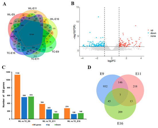
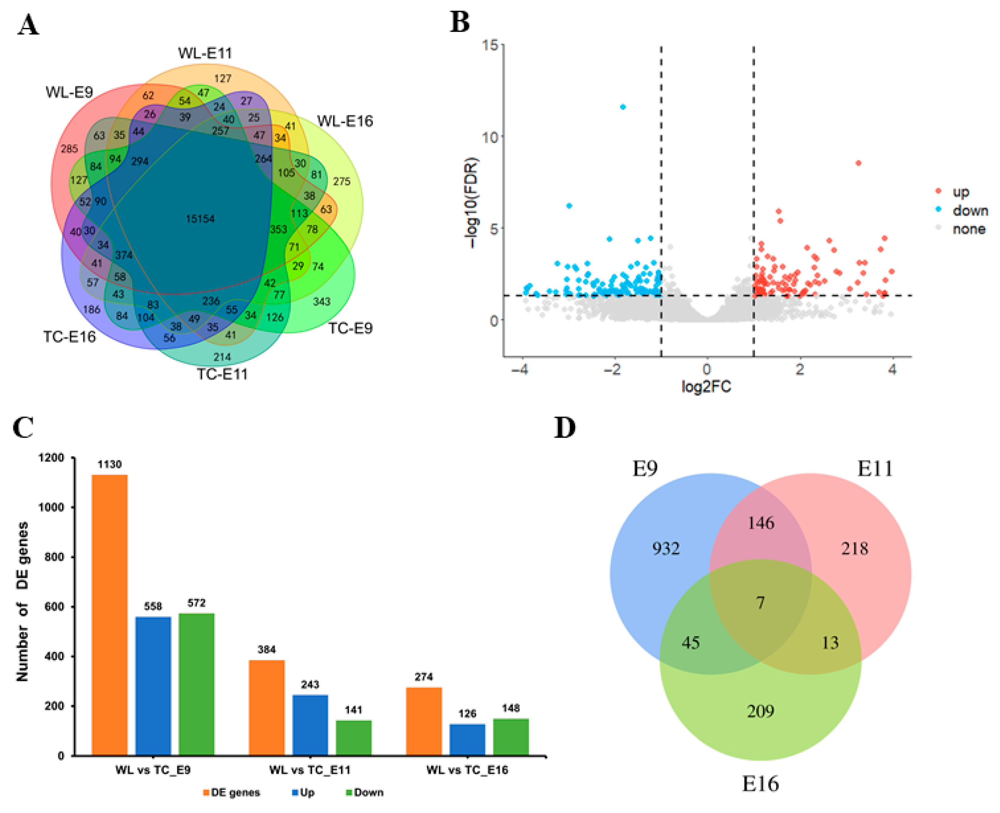
Gene expression levels were quantified based on the number of mapped reads per gene. In total, 21,226 genes were expressed in the embryonic heart tissues examined, of which 15,154 were detected in all six groups (Figure 1A).
Figure 1.
Differential gene analysis across three incubation stages. Note: (A) The Venn diagram of the number of positively expressed genes in each group at three time points. (B) A volcano diagram, with blue dots representing DEGs with down-regulated expression and red dots representing DEGs with up-regulated expression. (C) Histogram of the number of DEGs, upregulated genes, and downregulated genes at three time points. (D) Venn diagram of DEGs at three time points.
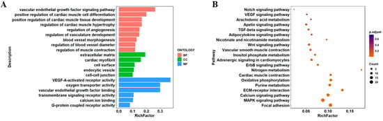
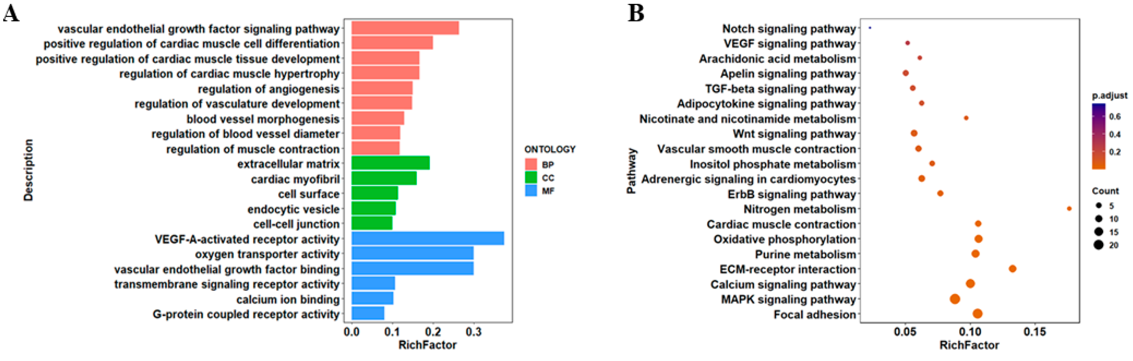
At day 9 of incubation, 1130 DEGs were identified in the myocardial tissues of TC compared with WL embryos, including 558 upregulated and 572 downregulated genes in TC (Figure 1B,C). These DEGs were primarily enriched in GO terms including regulation of angiogenesis, regulation of vasculature development, blood vessel morphogenesis, oxygen transporter activity, and calcium ion binding (Figure 2A). KEGG analysis showed that these DEGs were significantly enriched in the Apelin signaling pathway, cardiac muscle contraction, vascular smooth muscle contraction, and the VEGF signaling pathway, which are closely associated with cardiac morphogenesis and function (Figure 2B).
Figure 2.
Functional enrichment results of DEGs on E9. Note: (A) GO of DEGs. (B) KEGG of DEGs.
At day 11 of incubation, 384 DEGs were detected between TC and WL, of which 243 were upregulated and 141 were downregulated in TC (Supplementary Figure S1A). These genes were enriched in GO terms such as cardiac muscle atrophy, cardiac muscle cell proliferation, cardiac myofibril, and calcium-dependent phospholipid binding (Supplementary Figure S1B). KEGG analysis indicated that the DEGs were primarily involved in the Apelin signaling pathway, VEGF signaling pathway, and vascular smooth muscle contraction (Supplementary Figure S1C).
At 16 days post-incubation, 274 DEGs were identified in the cardiac muscle tissues of WL and TC embryos. Among these, 126 genes were significantly upregulated in Tibetan chickens, while 148 genes were significantly downregulated (Supplementary Figure S2A). The 274 DEGs were primarily enriched in GO terms including atrial cardiac muscle tissue development, blood vessel development, cardiovascular system development, and acetyl-CoA C-acetyltransferase activity (Supplementary Figure S2B). KEGG analysis revealed that these DEGs were primarily involved in pathways related to cardiac development, including cardiac muscle contraction, the apelin signaling pathway, vascular smooth muscle contraction, and the VEGF signaling pathway (Supplementary Figure S2C). Intersecting the DEGs across the three developmental stages identified seven genes that exhibited differential expression in all three periods (Figure 1D).
3.3. Temporal Analysis of Gene Expression
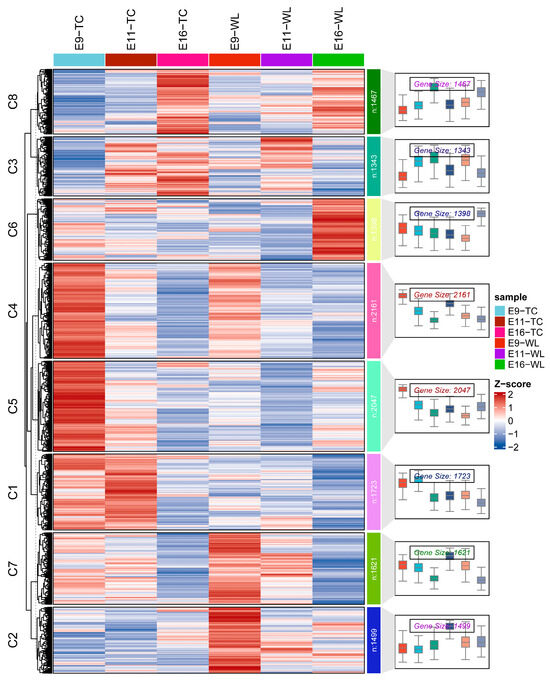
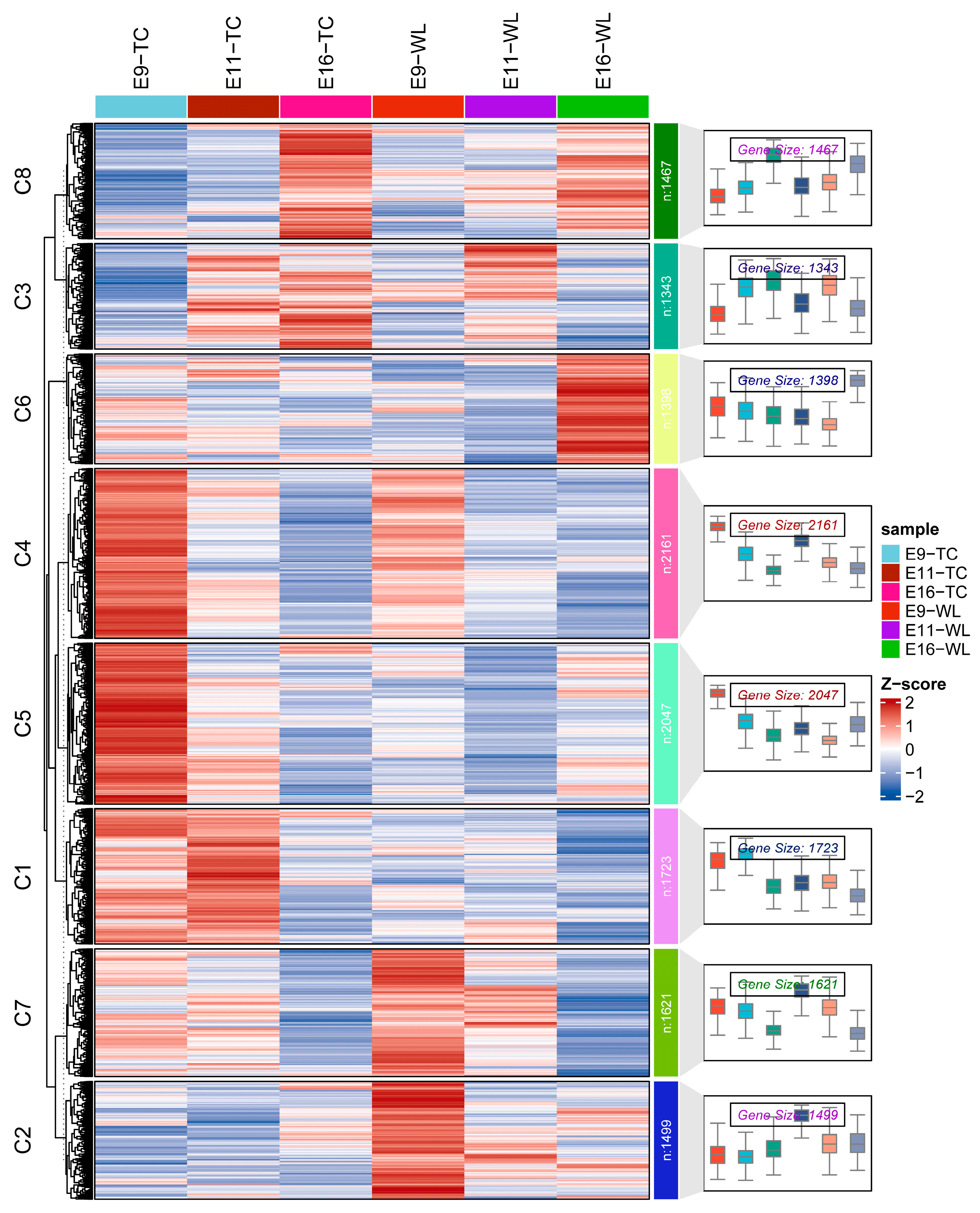
To identify co-expressed genes associated with hypoxic adaptation in TC, all genes across the three incubation stages were subjected to time-series clustering using the R package ClusterGVis. All genes were grouped into eight clusters based on their expression trajectories. Among these, cluster1, 2, and 5 (C1, C2 and C5) were of particular interest, as they displayed distinct expression patterns between TC and WL. C1 contained 1723 genes and C5 contained 2047 genes, both exhibiting higher expression in TC than in WL across the three stages. C2 comprised 1499 genes, which also showed elevated expression in TC relative to WL (Figure 3).
Figure 3.
Gene expression time series clustering and visualization.
3.4. Identification of Co-Expression Modules by WGCNA
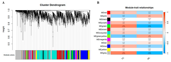
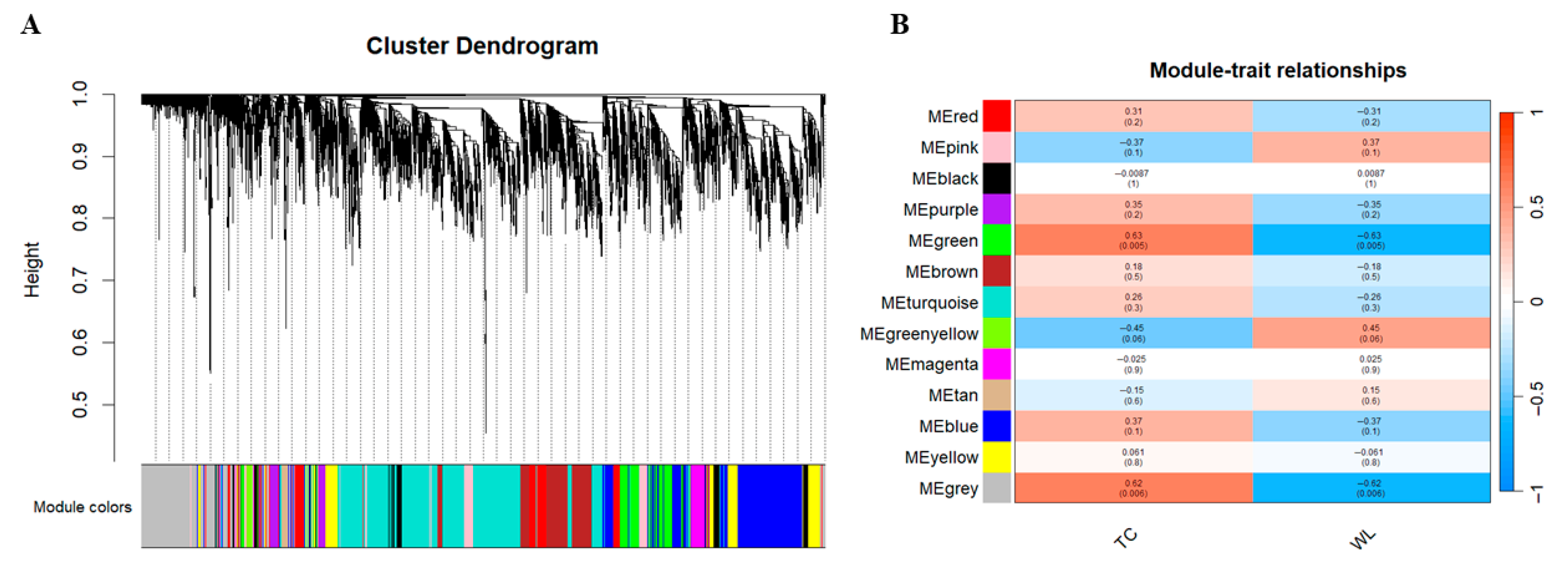
To further identify key candidate genes associated with TC adaptation to hypoxic environments, we performed WGCNA on genes selected through variance filtering. These genes were clustered into 13 co-expressed modules (Figure 4A). Module-trait correlation analysis revealed that one module showed significant association with breeds differences at high altitudes (p < 0.05). Specifically, the green module showed a significant positive correlation with TC and a significant negative correlation with WL (Figure 4B), suggesting that genes in this module are closely related to hypoxic adaptation.
Figure 4.
WGCNA to identify gene co-expression modules. Note: (A) Cluster Dendrogram. (B) Relationships between modules and chicken breeds. Correlation coefficients and significance p-values are marked within each cell.
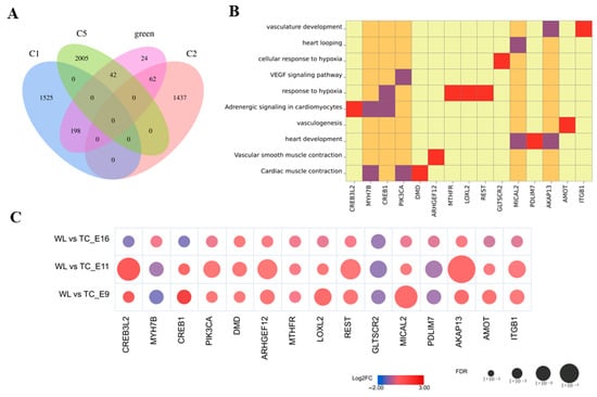
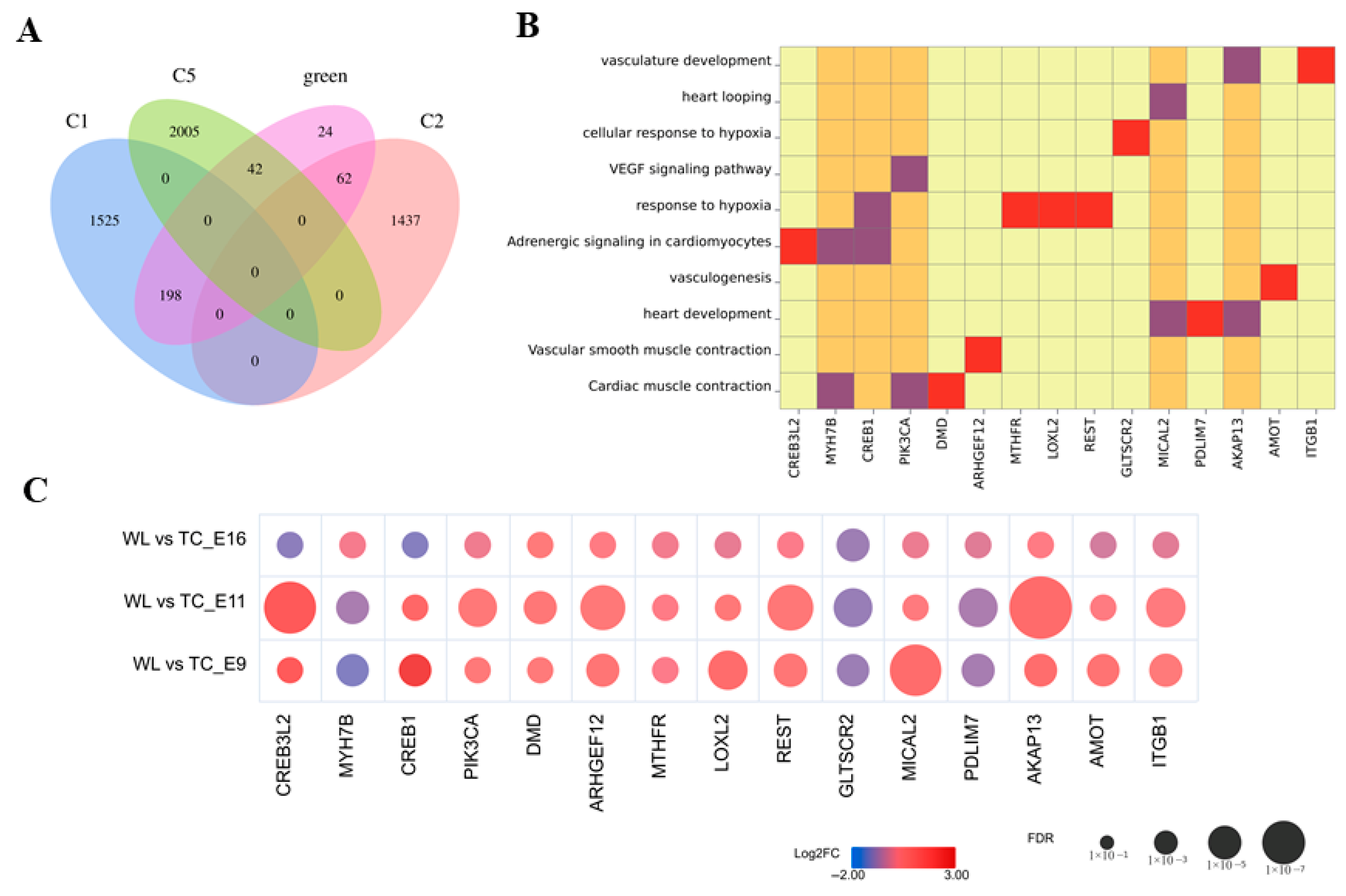
The green module contained 326 genes. Intersection of these genes with those from the three temporal clusters of interest (C1, C2, and C5) identified 302 overlapping genes (Figure 3 and Figure 5A). Functional enrichment and differential-expression analyses of these 302 genes highlighted 15 genes enriched in pathways related to cardiovascular development (Figure 5B). Among these, six genes (CREB3L2, MYH7B, CREB1, LOXL2, MICAL2, and AKAP13) exhibited differential expression across incubation stages. CREB3L2 and AKAP13 were significantly upregulated in TC cardiac tissue at day 11 of incubation, whereas CREB1, LOXL2, and MICAL2 were significantly upregulated at day 9. MYH7B showed significant downregulation in cardiac tissue at day 9 (Figure 5C). These six genes were selected as core candidate genes for TC adaptation to hypoxia.
Figure 5.
Candidate gene screening. Note: (A) Venn diagram of genes in C1, C2, C5, and the green module. (B) Functional enrichment results of 15 DEGs. (C) Temporal expression of 15 DEGs.
3.5. Validation of DEGs
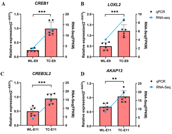
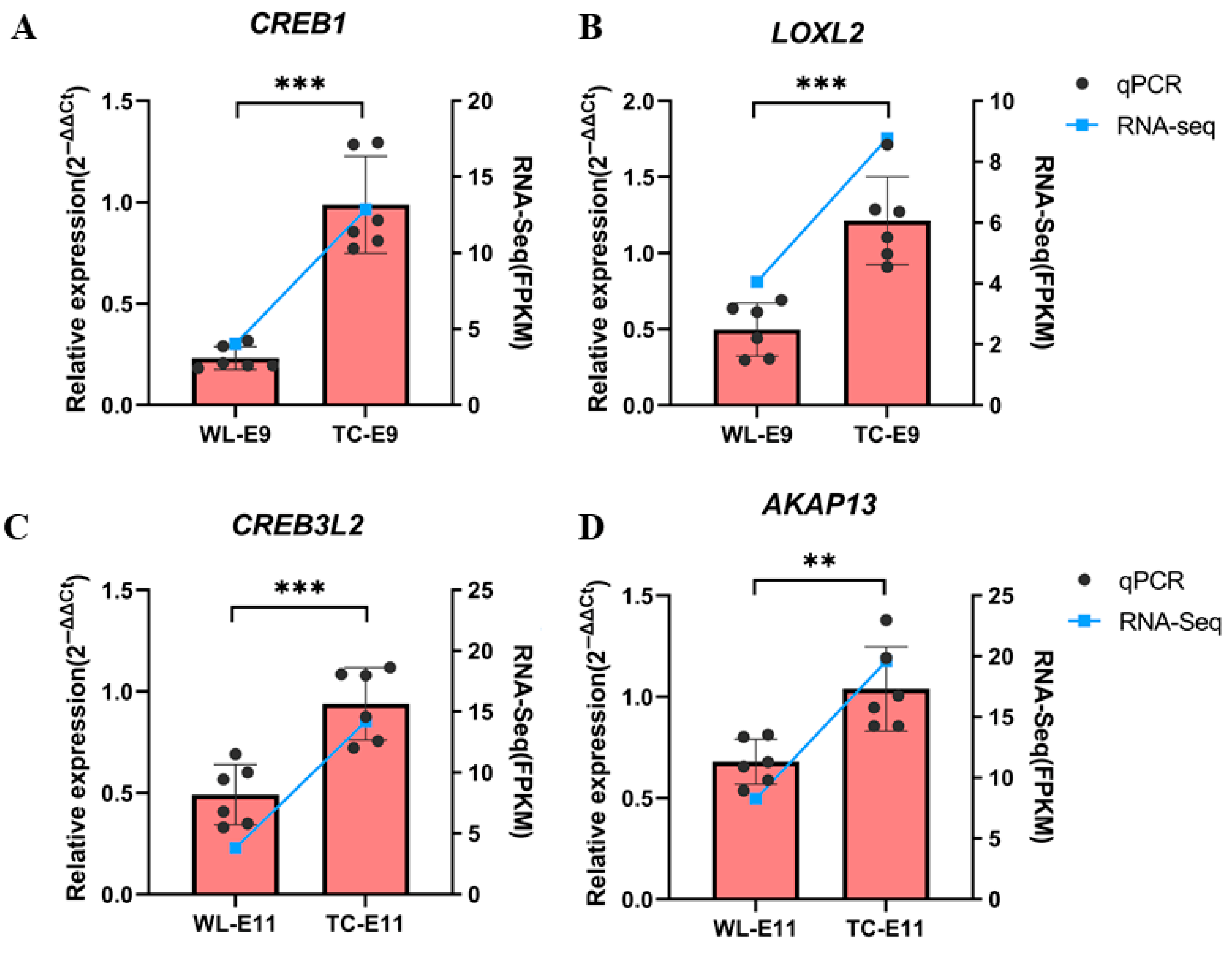
Four functionally significant DEGs were selected for qPCR validation using tissues from WL and TC. As shown in Figure 6, qRT-PCR results were consistent with the expression trends observed in the RNA-seq data, confirming the reliability and accuracy of the transcriptome sequencing and analysis.
Figure 6.
qRT-PCR and RNA-seq results. Notes: (A) The relative expression of CREB1. (B) The relative expression of LOXL2. (C) The relative expression of CREB3L2. (D) The relative expression of AKAP13. A bar chart displaying the results of the quantitative experiment, with a line plot representing the RNA-seq data. ** p < 0.01; *** p < 0.001.
4. Discussion
Hypoxia exerts profound effects on embryonic cardiac development, affecting both morphogenesis and functional maturation. During early embryogenesis, insufficient oxygen supply can lead to structural abnormalities such as impaired heart-tube looping, reduced ventricular size, and altered trabeculation, ultimately compromising cardiac output and contractile performance [22,23]. To cope with limited oxygen availability, embryos activate a series of molecular responses that maintain oxygen homeostasis and support normal cardiac development. In this study, GO enrichment analyses revealed that DEGs between TC and WL embryos were significantly associated with biological processes such as regulation of angiogenesis, vasculature development, blood vessel morphogenesis, oxygen transporter activity, and calcium ion binding. These results suggest that TC embryos initiate adaptive mechanisms to enhance oxygen delivery and cardiac function under hypoxic conditions.
Consistent with these observations, KEGG pathway enrichment analysis revealed significant involvement of the Apelin signaling pathway, cardiac muscle contraction, vascular smooth muscle contraction, and VEGF signaling pathways, all of which are crucial for cardiovascular development and hypoxic adaptation. VEGF is a well-characterized downstream target of HIF-1α that promotes angiogenesis and increases vascular density, thereby improving oxygen diffusion to embryonic tissues [23,24]. Activation of the Apelin signaling pathway enhances endothelial cell proliferation and myocardial contractility, functioning as a compensatory mechanism under low-oxygen conditions [25,26,27]. The enrichment of pathways related to vascular and smooth muscle contraction reflects adaptive adjustments in vascular tone and cardiac performance that facilitate efficient blood perfusion and oxygen transport [28]. Additionally, enrichment of calcium ion binding and cardiac muscle contraction pathways indicates modulation of excitation–contraction coupling, thereby helping to maintain stable cardiac output even under limited oxygen availability. Collectively, these findings demonstrate that the functional pathways identified in TC embryonic hearts are consistent with known mechanisms that support cardiac development and physiological adaptation to hypoxia. Such coordinated regulation of angiogenesis, vascular remodeling, and myocardial contraction likely contributes to the superior hypoxia tolerance observed in Tibetan chickens during embryonic development.
Building on these pathway-level findings, we further sought to pinpoint key molecular regulators that may underlie the enhanced hypoxia tolerance of TC embryos. From transcriptomic data of embryonic heart tissues from highland (TC) and lowland (WL) chickens, we identified six core candidate genes (CREB3L2, MYH7B, CREB1, LOXL2, MICAL2, and AKAP13) through differential expression and functional enrichment analyses, temporal expression profiling, and WGCNA. These genes are significantly enriched in pathways related to hypoxic responses, myocardial structural remodeling, and signaling regulation, and their expression patterns are consistent with the unique structural features of TC hearts under hypoxic incubation.
Among them, CREB3L2 is particularly notable. In this study, CREB3L2 expression remained consistently higher in TC cardiac tissue and was significantly elevated compared with WL. Its expression positively correlated with HSPA5, a marker of endoplasmic reticulum (ER) stress in cardiomyocytes [29]. As an ER stress–responsive transcription factor, CREB3L2 has been shown to promote cardiomyocyte tolerance to hypoxic stress by activating adaptive ER-stress pathways and modulating unfolded protein responses [30,31,32]. In this study, the high expression of CREB3L2 in TC and its low expression in WL suggest its pivotal role in maintaining myocardial cell homeostasis under hypoxic conditions [33].
CREB1 is a classic stress response transcription factor, exhibited significantly higher expression in TC than in WL, and its expression was positively correlated with CREB3L2 expression [34]. Functional enrichment analysis revealed CREB1 primarily participates in the “cAMP signaling pathway” and “adrenergic signaling in cardiomyocytes” [35,36]. Its elevated expression activates downstream anti-apoptotic genes B-cell Lymphoma 2 (BCL-2) through phosphorylation, enhancing cardiomyocyte resistance to hypoxia [37]. Recent studies indicate CREB1 also improves myocardial energy metabolism by regulating mitochondrial biogenesis-related genes PPARγ coactivator-1α (PGC-1α) [38].
As a component protein of cardiac myofibrils, MYH7B is closely associated with myocardial contractile function. Its specific expression modulates sarcomere contraction velocity to adapt to changes in myocardial workload under hypoxic conditions [39,40,41]. In the hypoxic environment of high altitudes, the heart must balance “oxygen-limited energy supply” with “maintaining pumping demands.” Reduced MYH7B expression may decrease sarcomere contraction speed, thereby lowering myocardial energy consumption per unit time and adapting to the weakened aerobic metabolism under hypoxia [42,43,44]. In this study, MYH7B expression in TC was significantly lower than in WL, suggesting it maintains cardiac function stability by optimizing myocardial contraction efficiency. Additionally, synergistic regulation of myosin heavy chain isoforms was observed in the myocardium. The downregulation of MYH7B in TC cardiac tissue may be accompanied by upregulation of other subunits. For instance, MYH7 and MYH5 were both upregulated in TC in this study. Through reconfiguring subunit ratios, this mechanism enhances energy utilization efficiency while maintaining contractile force [45].
LOXL2, belonging to the lysyl oxidase (LOX) family, was functionally enriched in the “response to hypoxia” pathway [46,47]. As a key collagen cross-linking enzyme, LOXL2 maintains structural stability of myocardial interstitium under hypoxic conditions by regulating collagen fiber maturation and arrangement, preventing increased myocardial stiffness due to excessive fibrosis [48,49]. In this study, LOXL2 expression was significantly higher in TC cardiac tissue compared to WL, suggesting its role in suppressing pathological myocardial fibrosis.
MICAL2 exhibits significantly higher expression in TC compared to WL. As a microtubule-modifying protein, it maintains cardiomyocyte morphology and organelle localization by regulating microtubule dynamic stability [50,51,52]. AKAP13 as a signal-anchoring molecule, anchors protein kinase A (PKA) to specific subcellular structures, precisely regulating the spatially specific activation of the cAMP signaling pathway [53,54]. In this study, TC exhibited significantly elevated AKAP13 expression, positively correlated with CREB1 levels, suggesting its involvement in hypoxic adaptation by enhancing precise activation of the CREB1 pathway [55].
The six candidate genes identified in this study constitute a “core regulatory network” for cardiac hypoxic adaptation in TC, whose expression patterns reflect a multi-tiered adaptive mechanism involving “stress response-structural remodeling-signal synergy.” The synergistic activation of CREB3L2 and CREB1 ensures myocardial cell survival and energy homeostasis under hypoxic stress. Adaptive regulation of MYH7B and LOXL2 maintains myocardial structural integrity and contractile function, while MICAL2 and AKAP13 achieve functional synergy through cytoskeletal remodeling and precise signal anchoring. Current research suggests that differential expressions of these genes may correlate with long-term natural selection in high-altitude species. For instance, specific SNP sites of certain genes exhibit significantly higher allele frequencies in Tibetan highland populations compared to lowland species, forming a genetic foundation for hypoxic adaptation [56]. By contrast, lowland breeds lacking such selection pressure may show insufficient coordination of stress response and remodeling pathways, partly explaining their poorer hypoxia tolerance. From a pathological perspective, imbalanced expression of these candidate genes in WL may contribute to the high mortality observed under hypoxic stress—for example, inadequate CREB3L2 expression may lead to excessive ER stress and cardiomyocyte apoptosis, whereas excessive MYH7B expression may cause maladaptive contractile remodeling and increased cardiac load. These findings also highlight potential molecular targets for clinical intervention in high-altitude–related cardiovascular disorders.
Naturally, this study has limitations. For example, the selection of candidate genes was primarily based on transcriptomic data and currently lacks corresponding phenotypic evidence and definitive validation. Future work could incorporate essential phenotyping to better link molecular changes with cardiac structure and function, such as heart weight (and the heart-to-body weight ratio) and basic quantitative histological indices (e.g., ventricular wall thickness and vascular/capillary density). Building on this, future studies could further integrate single-cell and spatial transcriptomics to systematically assess the cell-type specificity and spatial localization of candidate genes across distinct cardiac cell populations, such as cardiomyocytes and endothelial cells. In addition, experiments under varying hypoxic conditions (oxygen concentration gradients) could be conducted to investigate the dynamic expression changes in candidate genes across different oxygen levels.
5. Conclusions
This study reveals key molecular mechanisms underlying cardiac adaptation to hypoxia in Tibetan chicken embryos. Transcriptomic and functional analyses identified core pathways related to angiogenesis, vascular regulation, and myocardial contraction, supporting efficient oxygen delivery and cardiac performance under low-oxygen conditions. Six candidate genes (CREB3L2, MYH7B, CREB1, LOXL2, MICAL2, and AKAP13) form a coordinated regulatory network that promotes stress tolerance, structural remodeling, and signaling integration in the hypoxic heart. These findings suggest that long-term high-altitude selection has driven the evolution of efficient cardiac adaptive mechanisms in Tibetan chickens, providing valuable insights into vertebrate hypoxia adaptation.
Supplementary Materials
The following supporting information can be downloaded at: https://www.mdpi.com/article/10.3390/agriculture16020244/s1, Table S1: Sequence alignment between the sequenced samples and the reference genome; Figure S1: Functional enrichment results of DEGs on E11. Notes: (A) A volcano diagram, with blue dots representing differential genes with down-regulated expression and red dots representing differential genes with up-regulated expression. (B) GO of DEGs. (C) KEGG of DEGs; Figure S2: Functional enrichment results of DEGs on E16. Notes: (A) A volcano diagram, with blue dots representing differential genes with down-regulated expression and red dots representing differential genes with up-regulated expression. (B) GO of DEGs. (C) KEGG of DEGs.
Author Contributions
Conceptualization, X.C.; methodology, H.F.; validation, X.C. and H.F., resources, D.P.; writing—original draft preparation, X.C.; writing—review and editing, B.Z. and H.Z.; visualization, X.C.; supervision, D.P.; funding acquisition, B.Z. and H.Z. All authors have read and agreed to the published version of the manuscript.
Funding
This study was supported by the National Key Research and Development Program of China (2023YFF1002600), the Chinese Universities Scientific Fund (2025TC067, 2025TC135), the China Agriculture Research System (CARS-40).
Data Availability Statement
The original data presented in the study are openly available in NCBI (PRJNA1373377).
Conflicts of Interest
The authors declare no competing interests.
References
- Hu, Y.Q.; Lu, H.; Li, H.; Ge, J.B. Molecular basis and clinical implications of HIFs in cardiovascular diseases. Trends Mol. Med. 2022, 28, 916–938. [Google Scholar] [CrossRef] [Scilit]
- Okninska, M.; Zajda, K.; Zambrowska, Z.; Grzanka, M.; Paterek, A.; Mackiewicz, U.; Szczylik, C.; Kurzyna, M.; Piekielko-Witkowska, A.; Torbicki, A.; et al. Role of Oxygen Starvation in Right Ventricular Decompensation and Failure in Pulmonary Arterial Hypertension. JACC-Heart Fail. 2024, 12, 235–247. [Google Scholar] [CrossRef] [Scilit]
- She, P.L.; Gao, B.J.; Li, D.L.; Wu, C.; Zhu, X.J.; He, Y.; Mo, F.; Qi, Y.; Jin, D.Q.; Chen, Y.W.; et al. The transcriptional repressor HEY2 regulates mitochondrial oxidative respiration to maintain cardiac homeostasis. Nat. Commun. 2025, 16, 232. [Google Scholar] [CrossRef] [Scilit]
- Ruan, W.; Li, T.; Bang, I.H.; Lee, J.; Deng, W.K.; Ma, X.X.; Luo, C.; Du, F.; Yoo, S.H.; Kim, B.; et al. BMAL1-HIF2A heterodimer modulates circadian variations of myocardial injury. Nature 2025, 641, 1017–1028. [Google Scholar] [CrossRef] [Scilit]
- Tu, C.Y.; Caudal, A.; Liu, Y.; Gorgodze, N.; Zhang, H.; Lam, C.K.; Dai, Y.Q.; Zhang, A.E.L.; Wnorowski, A.; Wu, M.A.; et al. Tachycardia-induced metabolic rewiring as a driver of contractile dysfunction. Nat. Biomed. Eng. 2024, 8, 479–494. [Google Scholar] [CrossRef] [Scilit] [PubMed]
- Ge, R.L. Medical Problems of Chronic Hypoxia in Highlanders Living on the Tibetan Plateau. High Alt. Med. Biol. 2025, 26, 308–317. [Google Scholar] [CrossRef] [Scilit]
- Lock, M.C.; Ripley, D.M.; Smith, K.L.M.; Mueller, C.A.; Shiels, H.A.; Crossley, D.A., II; Galli, G.L.J. Developmental plasticity of the cardiovascular system in oviparous vertebrates: Effects of chronic hypoxia and interactive stressors in the context of climate change. J. Exp. Biol. 2024, 227, jeb245530. [Google Scholar] [CrossRef] [Scilit]
- Yang, Y.N.; Yuan, H.N.; Yao, B.Y.; Zhao, S.G.; Wang, X.R.; Xu, L.N.; Zhang, L.Y. Genetic Adaptations of the Tibetan Pig to High-Altitude Hypoxia on the Qinghai-Tibet Plateau. Int. J. Mol. Sci. 2024, 25, 11303. [Google Scholar] [CrossRef] [Scilit] [PubMed]
- Xue, M.M.; Yu, R.J.; Yang, L.X.; Xie, F.Y.; Fang, M.Y.; Tang, Q.G. Metabolomics and transcriptomics of embryonic livers reveal hypoxia adaptation of Tibetan chickens. BMC Genom. 2024, 25, 131. [Google Scholar] [CrossRef] [Scilit]
- Maina, J.N. Structure and function of the avian respiratory system. Philos. Trans. R. Soc. B-Biol. Sci. 2025, 380, 20230435. [Google Scholar] [CrossRef] [Scilit] [PubMed]
- Okur, N.; Eratalar, S.A.; Yigit, A.A.; Kutlu, T.; Kabakçi, R.; Özsoy, S.Y. Effects of incubator oxygen and carbon dioxide concentrations on hatchability of fertile eggs, some blood parameters, and histopathological changes of broilers with different parental stock ages in high altitude. Poult. Sci. 2022, 101, 101609. [Google Scholar] [CrossRef] [Scilit]
- Li, X.F.; Abdel-Moneim, A.M.E.; Hu, Z.Z.; Mesalam, N.M.; Yang, B. Effects of chronic hypoxia on the gene expression profile in the embryonic heart in three Chinese indigenous chicken breeds (Gallus gallus). Front. Vet. Sci. 2022, 9, 942159. [Google Scholar] [CrossRef] [Scilit]
- Zhang, Y.; Zheng, X.; Zhang, Y.; Zhang, H.; Zhang, X.; Zhang, H. Comparative transcriptomic and proteomic analyses provide insights into functional genes for hypoxic adaptation in embryos of Tibetan chickens. Sci. Rep. 2020, 10, 11213. [Google Scholar] [CrossRef] [Scilit]
- Zhong, H.A.; Zhu, L.; Kong, X.Y.; Zhang, K.; Tang, L.; Zhang, H.; Zhang, B.; Gou, X. Characterization and comparative transcriptomic analysis of high-altitude adaptation in Tibetan chicken using RNA-sequencing. Poult. Sci. 2025, 104, 104749. [Google Scholar] [CrossRef] [Scilit] [PubMed]
- Zhang, Q.; Gou, W.Y.; Wang, X.T.; Zhang, Y.W.; Ma, J.; Zhang, H.L.; Zhang, Y.; Zhang, H. Genome Resequencing Identifies Unique Adaptations of Tibetan Chickens to Hypoxia and High-Dose Ultraviolet Radiation in High-Altitude Environments. Genome Biol. Evol. 2016, 8, 765–776. [Google Scholar] [CrossRef] [Scilit] [PubMed]
- Zhang, Y.; Zhang, H.L.; Zhang, B.; Ling, Y.; Zhang, H. Identification of key HIF-1α target genes that regulate adaptation to hypoxic conditions in Tibetan chicken embryos. Gene 2020, 729, 144321. [Google Scholar] [CrossRef] [Scilit] [PubMed]
- Kim, D.; Landmead, B.; Salzberg, S.L. HISAT: A fast spliced aligner with low memory requirements. Nat. Methods 2015, 12, 357–360. [Google Scholar] [CrossRef] [Scilit]
- Pertea, M.; Pertea, G.M.; Antonescu, C.M.; Chang, T.C.; Mendell, J.T.; Salzberg, S.L. StringTie enables improved reconstruction of a transcriptome from RNA-seq reads. Nat. Biotechnol. 2015, 33, 290–295. [Google Scholar] [CrossRef] [Scilit]
- Trapnell, C.; Williams, B.A.; Pertea, G.; Mortazavi, A.; Kwan, G.; van Baren, M.J.; Salzberg, S.L.; Wold, B.J.; Pachter, L. Transcript assembly and quantification by RNA-Seq reveals unannotated transcripts and isoform switching during cell differentiation. Nat. Biotechnol. 2010, 28, 511. [Google Scholar] [CrossRef] [Scilit]
- Love, M.I.; Huber, W.; Anders, S. Moderated estimation of fold change and dispersion for RNA-seq data with DESeq2. Genome Biol. 2014, 15, 550. [Google Scholar] [CrossRef] [Scilit]
- Langfelder, P.; Horvath, S. WGCNA: An R package for weighted correlation network analysis. BMC Bioinform. 2008, 9, 559. [Google Scholar] [CrossRef] [Scilit]
- Haron, A.; Ruzal, M.; Shinder, D.; Druyan, S. Hypoxia during incubation and its effects on broiler’s embryonic development. Poult. Sci. 2021, 100, 100951. [Google Scholar] [CrossRef] [Scilit] [PubMed]
- Zhao, Y.C.; Xiong, W.D.; Li, C.F.; Zhao, R.Z.; Lu, H.; Song, S.; Zhou, Y.; Hu, Y.Q.; Shi, B.; Ge, J.B. Hypoxia-induced signaling in the cardiovascular system: Pathogenesis and therapeutic targets. Signal Transduct. Target. Ther. 2023, 8, 431. [Google Scholar] [CrossRef] [Scilit]
- Jiang, M.Z.; Fan, X.; Wang, Y.Q.; Sun, X.T. Effects of hypoxia in cardiac metabolic remodeling and heart failure. Exp. Cell Res. 2023, 432, 113763. [Google Scholar] [CrossRef] [Scilit]
- Liu, W.; Yan, J.L.; Pan, W.N.; Tang, M.J. Apelin/Elabela-APJ: A novel therapeutic target in the cardiovascular system. Ann. Transl. Med. 2020, 8, 243. [Google Scholar] [CrossRef] [Scilit] [PubMed]
- Chapman, F.A.; Maguire, J.J.; Newby, D.E.; Davenport, A.P.; Dhaun, N. Targeting the apelin system for the treatment of cardiovascular diseases. Cardiovasc. Res. 2023, 119, 2683–2696. [Google Scholar] [CrossRef] [Scilit] [PubMed]
- Tang, L.; Qiu, H.L.; Xu, B.; Su, Y.J.; Nyarige, V.; Li, P.S.; Chen, H.J.; Killham, B.; Liao, J.; Adam, H.; et al. Microparticle Mediated Delivery of Apelin Improves Heart Function in Post Myocardial Infarction Mice. Circ. Res. 2024, 135, 777–798. [Google Scholar] [CrossRef] [Scilit]
- Hernandez, I.; Baio, J.M.; Tsay, E.; Martinez, A.F.; Fuentes, T.I.; Bailey, L.L.; Hasaniya, N.W.; Kearns-Jonker, M. Short-term hypoxia improves early cardiac progenitor cell function in vitro. Am. J. Stem Cells 2018, 7, 1–17. [Google Scholar]
- Ha, D.P.; Shin, W.J.; Liu, Z.; Doche, M.E.; Lau, R.; Leli, N.M.; Conn, C.S.; Russo, M.; Lorenzato, A.; Koumenis, C.; et al. Targeting stress induction of GRP78 by cardiac glycoside oleandrin dually suppresses cancer and COVID-19. Cell Biosci. 2024, 14, 115. [Google Scholar] [CrossRef] [Scilit]
- Gao, Y.P.; Hu, C.; Hu, M.; Dong, W.S.; Li, K.; Ye, Y.J.; Hu, Y.X.; Zhang, X. CREB3 protein family: The promising therapeutic targets for cardiovascular and metabolic diseases. Cell Biol. Toxicol. 2024, 40, 103. [Google Scholar] [CrossRef] [Scilit]
- He, J.; Zhou, Y.; Sun, L.Q. Emerging mechanisms of the unfolded protein response in therapeutic resistance: From chemotherapy to Immunotherapy. Cell Commun. Signal. 2024, 22, 89. [Google Scholar] [CrossRef] [Scilit] [PubMed]
- Hamilton, S.; Terentyev, D. ER stress and calcium-dependent arrhythmias. Front. Physiol. 2022, 13, 1041940. [Google Scholar] [CrossRef] [Scilit]
- Bhattarai, K.R.; Riaz, T.A.; Kim, H.R.; Chae, H.J. The aftermath of the interplay between the endoplasmic reticulum stress response and redox signaling. Exp. Mol. Med. 2021, 53, 151–167. [Google Scholar] [CrossRef] [Scilit]
- Dinevska, M.; Widodo, S.S.; Cook, L.; Stylli, S.S.; Ramsay, R.G.; Mantamadiotis, T. CREB: A multifaceted transcriptional regulator of neural and immune function in CNS tumors. Brain Behav. Immun. 2024, 116, 140–149. [Google Scholar] [CrossRef] [Scilit]
- Dongdem, J.T.; Etornam, A.E.; Beletaa, S.; Alidu, I.; Kotey, H.; Wezena, C.A. The beta(3)-Adrenergic Receptor: Structure, Physiopathology of Disease, and Emerging Therapeutic Potential. Adv. Pharmacol. Pharm. Sci. 2024, 2024, 2005589. [Google Scholar] [CrossRef] [Scilit] [PubMed]
- Parichatikanond, W.; Duangrat, R.; Kurose, H.; Mangmool, S. Regulation of β-Adrenergic Receptors in the Heart: A Review on Emerging Therapeutic Strategies for Heart Failure. Cells 2024, 13, 1674. [Google Scholar] [CrossRef] [Scilit] [PubMed]
- Niu, R.Z.; Wang, L.Q.; Yang, W.; Sun, L.Z.; Tao, J.; Sun, H.; Mei, S.; Wang, W.J.; Feng, K.X.; Qian, D.L.; et al. MicroRNA-582-5p targeting Creb1 modulates apoptosis in cardiomyocytes hypoxia/reperfusion-induced injury. Immun. Inflamm. Dis. 2022, 10, e708. [Google Scholar] [CrossRef] [Scilit]
- Abu Shelbayeh, O.; Arroum, T.; Morris, S.; Busch, K.B. PGC-1alpha Is a Master Regulator of Mitochondrial Lifecycle and ROS Stress Response. Antioxidants 2023, 12, 1075. [Google Scholar] [CrossRef] [Scilit]
- Broadwell, L.J.; Smallegan, M.J.; Rigby, K.M.; Navarro-Arriola, J.S.; Montgomery, R.L.; Rinn, J.L.; Leinwand, L.A. Myosin 7b is a regulatory long noncoding RNA (lncMYH7b) in the human heart. J. Biol. Chem. 2021, 296, 100694. [Google Scholar] [CrossRef] [Scilit]
- Walklate, J.; Ferrantini, C.; Johnson, C.A.; Tesi, C.; Poggesi, C.; Geeves, M.A. Alpha and beta myosin isoforms and human atrial and ventricular contraction. Cell. Mol. Life Sci. 2021, 78, 7309–7337. [Google Scholar] [CrossRef] [Scilit]
- Barrick, S.K.; Greenberg, M.J. Cardiac myosin contraction and mechanotransduction in health and disease. J. Biol. Chem. 2021, 297, 101297. [Google Scholar] [CrossRef] [Scilit]
- Ng, R.; Sewanan, L.R.; Stankey, P.; Li, X.; Qyang, Y.; Campbell, S. Shortening Velocity Causes Myosin Isoform Shift in Human Engineered Heart Tissues. Circ. Res. 2021, 128, 281–283. [Google Scholar] [CrossRef] [Scilit]
- Cail, R.C.; Baez-Cruz, F.A.; Winkelmann, D.A.; Goldman, Y.E.; Ostap, E.M. Dynamics of beta-cardiac myosin between the super-relaxed and disordered-relaxed states. J. Biol. Chem. 2025, 301, 108412. [Google Scholar] [CrossRef] [Scilit]
- Nandwani, N.; Bhowmik, D.; Glaser, C.; Childers, M.C.; Goluguri, R.R.; Dawood, A.; Regnier, M.; Houdusse, A.; Spudich, J.A.; Ruppel, K.M. Hypertrophic cardiomyopathy mutations Y115H and E497D disrupt the folded-back state of human beta-cardiac myosin allosterically. Nat. Commun. 2025, 16, 8751. [Google Scholar] [CrossRef] [Scilit] [PubMed]
- Hoh, J.F.Y. Developmental, physiologic and phylogenetic perspectives on the expression and regulation of myosin heavy chains in mammalian skeletal muscles. J. Comp. Physiol. B 2023, 193, 355–382. [Google Scholar] [CrossRef] [Scilit]
- Poe, A.; Yus, M.M.; Wang, H.L.; Santhanam, L. Lysyl oxidase like-2 in fibrosis and cardiovascular disease. Am. J. Physiol.-Cell Physiol. 2023, 325, C694–C707. [Google Scholar] [CrossRef] [Scilit]
- Li, R.K.; Li, H.C.; Zhu, L.L.; Zhang, X.X.; Liu, D.J.; Li, Q.; Ni, B.; Hu, L.P.; Zhang, Z.G.; Zhang, Y.L.; et al. Reciprocal regulation of LOXL2 and HIF1α drives the Warburg effect to support pancreatic cancer aggressiveness. Cell Death Dis. 2021, 12, 1106. [Google Scholar] [CrossRef] [Scilit] [PubMed]
- Erasmus, M.; Samodien, E.; Lecour, S.; Cour, M.; Lorenzo, O.; Dludla, P.; Pheiffer, C.; Johnson, R. Linking LOXL2 to Cardiac Interstitial Fibrosis. Int. J. Mol. Sci. 2020, 21, 5913. [Google Scholar] [CrossRef] [Scilit]
- Perryman, L.; Findlay, A.; Baskar, J.; Charlton, B.; Foot, J.; Hamilton, R.; Hamprecht, D.; Joshi, A.; Stolp, J.; Turner, C.; et al. The small molecule LOXL2 inhibitor SNT-5382 reduces cardiac fibrosis and achieves strong clinical target engagement. Sci. Rep. 2025, 15, 22653. [Google Scholar] [CrossRef] [Scilit] [PubMed]
- Giarratana, N.; Conti, F.; La Rovere, R.; Gijsbers, R.; Carai, P.; Duelen, R.; Vervliet, T.; Bultynck, G.; Ronzoni, F.; Piciotti, R.; et al. MICAL2 is essential for myogenic lineage commitment. Cell Death Dis. 2020, 11, 654. [Google Scholar] [CrossRef] [Scilit]
- Conboy, J.P.; Isturiz Petitjean, I.; van der Net, A.; Koenderink, G.H. How cytoskeletal crosstalk makes cells move: Bridging cell-free and cell studies. Biophys. Rev. 2024, 5, 021307. [Google Scholar] [CrossRef] [Scilit]
- Caporizzo, M.A.; Prosser, B.L. The microtubule cytoskeleton in cardiac mechanics and heart failure. Nat. Rev. Cardiol. 2022, 19, 364–378. [Google Scholar] [CrossRef] [Scilit]
- Subramanian, H.; Nikolaev, V.O. A-Kinase Anchoring Proteins in Cardiac Myocytes and Their Roles in Regulating Calcium Cycling. Cells 2023, 12, 436. [Google Scholar] [CrossRef] [Scilit] [PubMed]
- Zhang, S.A.; Wang, H.Y.; Melick, C.H.; Jeong, M.H.; Curukovic, A.; Tiwary, S.; Lama-Sherpa, T.D.; Meng, D.L.; Servage, K.A.; James, N.G.; et al. AKAP13 couples GPCR signaling to mTORC1 inhibition. PLoS Genet. 2021, 17, e1009832. [Google Scholar] [CrossRef] [Scilit]
- Armandari, I.; Zomerman, W.W.; Plasschaert, S.L.A.; Smit, M.J.; Martini, T.E.I.; Magalhaes, E.S.D.; Hogeling, S.M.; Rozema-Huizinga, G.C.; Lourens, H.J.; Meeuwsen-de Boer, T.G.J.; et al. CREB signaling activity correlates with differentiation and survival in medulloblastoma. Sci. Rep. 2021, 11, 16077. [Google Scholar] [CrossRef] [Scilit] [PubMed]
- Zheng, W.S.; He, Y.X.; Guo, Y.B.; Yue, T.; Zhang, H.; Li, J.; Zhou, B.; Zeng, X.R.; Li, L.Y.; Wang, B.; et al. Large-scale genome sequencing redefines the genetic footprints of high-altitude adaptation in Tibetans. Genome Biol. 2023, 24, 73. [Google Scholar] [CrossRef] [Scilit] [PubMed]

Disclaimer/Publisher’s Note: The statements, opinions and data contained in all publications are solely those of the individual author(s) and contributor(s) and not of MDPI and/or the editor(s). MDPI and/or the editor(s) disclaim responsibility for any injury to people or property resulting from any ideas, methods, instructions or products referred to in the content. |
© 2026 by the authors. Licensee MDPI, Basel, Switzerland. This article is an open access article distributed under the terms and conditions of the Creative Commons Attribution (CC BY) license.