


Abstract
The ability of salicylic acid (SA) to control post-harvest fungal disease mango anthracnose caused by plant pathogen Colletotrichum gloeosporioides and the accompanying biochemical and metabolomic changes were studied. In vitro, C. gloeosporioides mycelial growth at 25 °C for 7 days was inhibited by SA at 2 and 5 mM concentrations. In vivo, mango fruits were treated with 2 mM SA for 10 min, then inoculated with C. gloeosporioides. The fruits were then stored at 25 °C and sampled every 2 days after inoculation. The anthracnose severity, infection index, malondialdehyde (MDA), antioxidant capacity (DPPH and FRAP), total phenolics (TPC), total flavonoids (TFC), ascorbic acid, and metabolomic changes in the peel were studied. Six days post inoculation, the SA-treated fruits showed reduced anthracnose severity, and the infection index was significantly reduced by SA (57%) compared to the control (67%). The SA-treated fruits exhibited decreased MDA, enhanced antioxidant capacity, and increased levels of TPC, TFC, and vitamin C in the peel. Compared with the control, SA treatment reduced MDA content by approximately 50% and increased TPC, TFC, and FRAP antioxidant capacity by 77%, 31%, and 29%, respectively. Vitamin C content and DPPH antioxidant activity were also significantly higher in SA-treated fruits. Untargeted metabolomics and a principal component analysis revealed a separation group between the SA-treated and control fruit. The SA treatment induced D-amino acids, phenylalanine pathways, and the biosynthesis of secondary metabolites such as phenolic acid. Together, these findings demonstrate that SA enhances mangoes’ resistance to post-harvest anthracnose through direct antifungal action and the activation of host defense by improving the antioxidant systems and stimulating the accumulation of secondary defense metabolites in mango fruit.
1. Introduction
Mango (Mangifera indica L.) is an economically significant tropical fruit grown in Thailand, and it is highly valued due to its high nutritional value and rich phytochemical content. The largest importers of mangoes from Thailand are South Korea and Malaysia. To help meet the strong demand for mangoes in international markets, mango cultivar Nam Dok Mai Si Thong is widely cultivated in southeast Asia [1]. However, as a climacteric fruit, mango is highly perishable and particularly susceptible to post-harvest losses, with the most significant post-harvest fungal disease being mango anthracnose, which is caused by plant pathogen Colletotrichum gloeosporioides.
In this context, post-harvest diseases—particularly anthracnose—represent a major constraint on the mango export industry. Anthracnose caused by C. gloeosporioides is one of the most destructive post-harvest diseases of mango worldwide [2]. The disease causes fruit spotting, decay, and rapid quality loss, resulting in substantial economic losses during storage, transportation, and marketing [3]. In light of the extended storage periods for mangoes and the strict quality standards in international markets, anthracnose poses a major challenge to export-oriented mango cultivars such as Nam Dok Mai Si Thong. Notably, C. gloeosporioides often establishes latent infections during the mango fruit’s development and remains quiescent until the fruit’s ripening, when physiological and biochemical changes favor rapid pathogen activation. This latent infection behavior complicates post-harvest disease management, as visible symptoms typically develop during storage and distribution rather than at harvest. Post-harvest management is essential in terms of maintaining fruit quality, safety, and nutritional value; extending shelf life; and reducing food losses. Various approaches, including refrigeration, controlled-atmosphere storage, chemical treatments, edible coatings, and ultraviolet-C radiation, have been developed to minimize post-harvest deterioration [4].
Chemical fungicides have traditionally been used in attempts to control anthracnose and other post-harvest diseases toward mitigation of the loss of post-harvest mangoes [5]. However, increasing restrictions on fungicide residues, strict maximum residue limits in importing countries, heightened consumer demand for safe and residue-free produce, and growing concerns regarding environmental impact, human health risks, and the development of pathogen resistance have collectively restricted the application of chemical fungicides. These limitations have intensified the need for effective, safe, and sustainable alternative post-harvest disease management strategies.
Salicylic acid (SA), also known as ortho-hydroxybenzoic acid, is an endogenous signaling molecule that modulates stress responses and plant physiological processes such as photosynthesis, heat production, stomatal conductance, transpiration, disease resistance, seed germination, crop yield, and glycolysis [6]. Salicylic acid has emerged as a promising and environmentally friendly option for post-harvest disease control [7]. The exogenous application of SA has been shown to delay senescence, enhance antioxidant capacity, and maintain post-harvest quality in a range of fruit crops. For example, SA treatment increased the total antioxidant activity in peach [8] and stimulated the accumulation of secondary metabolites including ascorbic acid, total phenolics, and flavonoids in apricot fruit (Prunus armeniaca L.) [9], suggesting the potential of SA treatment to enhance resistance to post-harvest pathogens such as C. gloeosporioides. Secondary metabolites, particularly phenolic and flavonoid compounds, are important components of plant defense responses because of their antioxidant and antimicrobial properties and their involvement in plant–pathogen interactions [10]. In addition, SA functions as a key signaling molecule in plant immunity, regulating local defense responses and systemic acquired resistance (SAR) against pathogen infection [11].
Despite these reported benefits, the mechanisms that underlie SA-induced disease resistance in mango remain poorly understood. Most of the prior relevant research focused on physiological and targeted biochemical responses, whereas comprehensive information on metabolite changes associated with SA treatment during anthracnose development remains limited. In particular, the manner in which SA modulates metabolite profiles associated with antioxidant capacity and defense responses during anthracnose infection in Nam Dok Mai Si Thong mango has not been fully elucidated. Untargeted metabolomics provides a powerful systems-level approach to capture complex metabolic changes associated with host defense; however, the application of untargeted metabolomics to characterize SA-mediated resistance mechanisms against post-harvest anthracnose in this commercially important mango cultivar has not yet been reported.
In the present study, an integrated approach combining disease assessment, antioxidant analysis, and untargeted metabolomics was used to identify key metabolic pathways involved in SA-induced disease resistance during mango storage. The study’s objectives were to evaluate the effectiveness of SA in controlling post-harvest anthracnose in Nam Dok Mai Si Thong mango fruit and elucidate the associated physiological, biochemical, and metabolic responses. The findings of this study provide mechanistic insights into SA-mediated defense regulation and offer practical guidance for the development of sustainable, residue-free post-harvest disease management strategies for mango.
2. Materials and Methods
2.1. Plant Material and Treatments
Mango fruits (Mangifera indica L. cv. Nam Dok Mai Si Thong) were harvested from a commercial orchard in Phichit Province, Thailand (16° N, 100° E; 43 m altitude), approximately 105 days after flowering, corresponding to commercial maturity. About 100 fruits were transported immediately to the Postharvest Technology Laboratory at Mae Fah Luang University, Chiang Rai, Thailand. Fruits weighing 250–300 g with uniform size, color, and maturity and free from visible defects, mechanical damage, and disease symptoms were selected for the experiment.
Selected fruits were immersed in 200 mg L−1 sodium hypochlorite (RCI Labscan Limited, Bangkok, Thailand) for 5 min, rinsed thoroughly with sterile distilled water, and air-dried at room temperature. They were then randomly allocated to a control group (0 mM salicylic acid [SA]; Ajax Finechem, Taren Point, NSW, Australia]) or an SA-treated group (2 mM SA). The 2 mM concentration was selected based on a previous report of its inhibitory effect against C. gloeosporioides in mango [12]. Both treatment solutions were prepared in distilled water containing 0.05% (v/v) Tween-80 (PanReac Química S.L.U., Castellar del Vallès, Spain).
Fruits were dipped in their respective solutions for 10 min and air-dried for 1 h. A single wound (3 mm deep × 5 mm wide) was then made at the equator of each fruit using a sterile cork borer, and 20 μL of a C. gloeosporioides conidial suspension (1 × 10⁶ spores mL−1) was applied to the wound. The inoculated fruits were held at 25 ± 1 °C and 85–90% relative humidity. The infection index was assessed 2, 4, and 6 days post inoculation. Each treatment had three replicates.
2.2. Pathogen Isolation and Inoculum Preparation
C. gloeosporioides was isolated from naturally infected mango fruits exhibiting typical anthracnose symptoms, then cultured on potato dextrose agar (PDA). A single-spore isolate was obtained to ensure genetic uniformity, subsequently re-cultured, and maintained on PDA slants at 4 °C until use. For the experimental assays, the pathogen was sub-cultured onto fresh PDA plates and incubated at 25 °C for 14 days to promote abundant sporulation, following the method described by Khan et al. [13].
For the preparation of the inoculum, conidia were harvested from actively growing fungal cultures by adding sterile distilled water containing 0.05% (v/v) Tween-80 to the plate surface and gently dislodging the spores with a sterile glass rod, as described by Khan et al. [13]. The resulting conidial suspension was filtered through four layers of sterile cheesecloth to remove mycelial fragments. The spore concentration was determined using a hemocytometer and adjusted to 1 × 10⁶ spores mL−1 with sterile distilled water prior to inoculation.
2.3. In Vitro Antifungal Activity
The inhibitory effect of SA on the in vitro mycelial growth of C. gloeosporioides was evaluated by the poisoned food technique. Appropriate volumes of SA stock solutions were added to molten PDA to obtain final concentrations of 0 (control), 0.5, 1.0, 2.0, and 5.0 mM, with a total medium volume of 20 mL per Petri dish (120-mm dia.). After thorough mixing, the medium was poured into sterile Petri plates and allowed to solidify at room temperature.
An 8 mm-diameter mycelial plug, excised from the actively growing margin of a 7-day-old C. gloeosporioides culture, was aseptically placed at the center of each PDA plate. The plates were incubated at 25 ± 1 °C for 7 days. Mycelial growth was assessed by measuring the colony diameter (mm). Each SA concentration was tested in three replicates.
2.4. Infection Index
The infection index was assessed as described by Corkidi, Balderas-Ruíz [14] 2, 4, and 6 days post inoculation. The severity of anthracnose on each fruit was visually evaluated and assigned a disease severity rating using a five-point scale based on the percentage of the fruit surface affected: 1 point = <1% (no visible disease), 2 points = 1–5% (slight disease), 3 points = 6–9% (moderate disease), 4 points = 10–49% (severe disease), and 5 points = 50–100% (very severe disease). Each fruit was assigned a disease severity rating from 1 to 5 according to the corresponding percentage of the fruit surface affected by anthracnose. The infection index (%) was then calculated from the disease severity ratings of all evaluated fruit using the following equation:
2.5. Malondialdehyde (MDA) Content
The malondialdehyde (MDA) content was determined as described by Duan et al. [15] with minor modifications. Briefly, 4.0 g of mango peel tissue was homogenized in 20 mL of 5% (w/v) trichloroacetic acid (TCA; Sigma-Aldrich, St. Louis, MO, USA) and centrifuged at 5000× g for 10 min. An aliquot of 1.0 mL of the supernatant was mixed with 3.0 mL of 0.5% (w/v) thiobarbituric acid (Sigma-Aldrich, St. Louis, MO, USA) prepared in 10% (w/v) TCA.
The mixture was incubated in a boiling water bath for 20 min, immediately cooled in an ice bath to terminate the reaction, and subsequently centrifuged at 8000× g for 10 min at 4 °C. The absorbance of the resulting supernatant was measured at 532 and 600 nm using a Multiskan GO spectrophotometer (Thermo Scientific, Vantaa, Finland). The MDA content was calculated using an extinction coefficient of 155 mM−1 cm−1, and the results are expressed as nmol g−1 fresh weight (FW).
2.6. Bioactive Compounds
2.6.1. Total Phenolic Content
The total phenolic content was determined using the Folin–Ciocalteu method as described by Salem et al. [16] with minor modifications. Briefly, 250 µL of sample extract was mixed with 1.25 mL of Folin–Ciocalteu reagent (Sigma-Aldrich, St. Louis, MO, USA), followed by 1.0 mL of 7.5% (w/v) sodium carbonate (Na2CO3; KEMAUS, Cherrybrook, NSW, Australia). The mixture was incubated at room temperature for 1 h, and absorbance was measured at 765 nm using a Multiskan GO spectrophotometer. A standard calibration curve was prepared using gallic acid (Sigma-Aldrich, St. Louis, MO, USA), and the results are expressed as µg gallic acid equivalents per g FW.
2.6.2. Total Flavonoid Content
The total flavonoid content was determined as described in [17], with slight modifications. Two grams of mango tissue (three replicates) were homogenized in 5 mL of acetone (Sigma-Aldrich, St. Louis, MO, USA), and the homogenate was centrifuged at 15,000× g for 30 min at 4 °C. A 0.5 mL aliquot of the supernatant was mixed with 2.2 mL of distilled water, 0.15 mL of 5% sodium nitrite (NaNO2; KEMAUS, Cherrybrook, NSW, Australia), and 0.15 mL of 10% aluminum chloride (AlCl3; KEMAUS, Cherrybrook, NSW, Australia). Six minutes later, 2 mL of 4% sodium hydroxide (NaOH; KEMAUS, Cherrybrook, NSW, Australia) was added, and the mixture was incubated at room temperature for 15 min. Absorbance was measured at 510 nm by the above-mentioned spectrophotometer. A standard calibration curve was prepared with catechin (Sigma-Aldrich, St. Louis, MO, USA), and the results are expressed as µg catechin equivalents per g FW.
2.6.3. Total Ascorbic Acid Content
The total ascorbic acid content was determined using the 2,4-dinitrophenylhydrazine method as described by Roe and Oesterling [18], with slight modifications. Briefly, 8 mL of sample extract was oxidized with 0.5 mL of 3% bromine water, followed by 0.25 mL of 10% thiourea (Sigma-Aldrich, St. Louis, MO, USA) to neutralize excess bromine. Subsequently, 2 mL of 2,4-dinitrophenylhydrazine (2,4-DNPH; Sigma-Aldrich, St. Louis, MO, USA) solution was added, and the mixture was incubated at 37 °C for 3 h. After cooling in an ice bath, 10 mL of chilled 85% sulfuric acid (H2SO4; Sigma-Aldrich, St. Louis, MO, USA) was added. Absorbance was measured at 510 nm with the above-mentioned spectrophotometer. A standard calibration curve was prepared with ascorbic acid, and the results are expressed as µg ascorbic acid (Sigma-Aldrich, St. Louis, MO, USA) equivalents per g FW.
2.7. Antioxidant Capacities
Antioxidant capacity was evaluated using the DPPH radical scavenging and ferric reducing antioxidant power (FRAP) assays. For the DPPH assay, a modified procedure based on the work of Moon and Shibamoto [19] was used. Sample extract (50 µL) was combined with 1950 µL of a 60 µmol DPPH (Sigma-Aldrich, St. Louis, MO, USA) solution prepared in methanol (RCI Labscan Limited, Bangkok, Thailand). After 30 min in the dark at room temperature, the absorbance was recorded at 517 nm. Activity was expressed as micromoles of Trolox (Sigma-Aldrich, St. Louis, MO, USA) equivalents (µmol TE) per g FW.
The FRAP assay was adapted from Benzie and Strain [20]. A 400 µL portion of extract was mixed with 2.6 mL of freshly prepared FRAP reagent and held at 37 °C for 30 min. Absorbance was then read at 595 nm. Results were calculated using an iron (II) sulfate (FeSO4; Sigma-Aldrich, St. Louis, MO, USA) standard curve and expressed as µmol FeSO4 per g FW.
2.8. Metabolomics Analysis
Peel tissue (2 g) was extracted in 10 mL of 70% methanol. After homogenization at 4 °C, the sample was sonicated for 10 min and centrifuged at 10,000× g for 10 min. The supernatant was cleaned using a Supel™ Sphere Carbon/NH2 cartridge (Merck Supelco, Darmstadt, Germany), then passed through a 0.22 μm membrane filter (Millipore, Billerica, MA, USA). This extraction procedure was adapted, with minor modifications, from Wang and Ren [21].
The filtered extract was analyzed on an Agilent G6545B LC-QTOF-MS/MS system (Agilent Technologies, Santa Clara, CA, USA). An ACE Excel-C18 AR column (100 × 2.1 mm, 1.7 μm) was held at 40 °C, and 5 μL of extract was injected per analysis. Water and acetonitrile (RCI Labscan Limited, Bangkok, Thailand), each containing 0.1% formic acid (RCI Labscan Limited, Bangkok, Thailand), served as mobile phases A and B, respectively. At a flow rate of 0.3 mL/min, the gradient increased from 5% B to 95% B over 25 min.
Data were collected in both positive and negative electrospray ionization modes across m/z 50–1200. Source settings were a capillary voltage of 3.5 kV, drying gas temperature of 325 °C, drying gas flow of 10 L/min, and nebulizer pressure of 45 psi. MassHunter Profinder version 10.0 (Agilent Technologies) was used to process the acquired data.
2.9. Data Processing
The raw metabolomics data were processed using the MassHunter Profinder software (version 10, Agilent Technology) (for peak extraction). The processing parameters included a mass tolerance of ±10 ppm, a minimum peak intensity of 400 counts, and a retention-time tolerance of ±0.15 min. The aligned peak list was exported to Mass Profiler Professional (MPP, ver. 15.1, Agilent Technologies) for further statistical analysis.
The dataset, comprising approx. 4000–6000 molecular features, was subjected to a one-way analysis of variance (ANOVA), with significance set at p < 0.05. Features with a fold change (FC) > 2.0 were selected for further investigation. Multivariate analyses, including a principal component analysis (PCA) and a partial least squares discriminant analysis (PLS-DA), were performed to visualize clustering patterns and maximize group separation. Metabolites with a variable importance in projection (VIP) score >1.0 were considered significant. Volcano plots were used to illustrate significantly altered metabolites and to highlight unique or overlapping features across sample groups.
Tentative metabolite identification was based on comparisons of accurate mass, retention time, and MS/MS spectra with public databases, including METLIN. The resulting annotated differential metabolites were subsequently subjected to metabolite set enrichment analysis and pathway impact analysis using MetaboAnalyst 6.0 to identify metabolite groups and metabolic pathways associated with SA treatment. Relevant metabolic pathways were further examined and visualized using the Kyoto Encyclopedia of Genes and Genomes (KEGG) pathway database. In particular, D-amino acid metabolism and phenylalanine metabolism were mapped to illustrate alterations in the corresponding metabolites in D6 SA-treated mango peel.
2.10. Statistical Analyses
All data are presented as the mean ± standard error (SE) of three biological replicates. The significance of differences among the treatment groups was evaluated by a one-way ANOVA followed by Tukey–Kramer post hoc tests at p ≤ 0.05. Pairwise comparisons were performed using Student’s t-test at a significance level of p ≤ 0.05. All analyses were conducted using the SAS statistical software package (SAS Institute, Cary, NC, USA).
3. Results and Discussion
3.1. The In Vitro Antifungal Activity of SA Against C. gloeosporioides
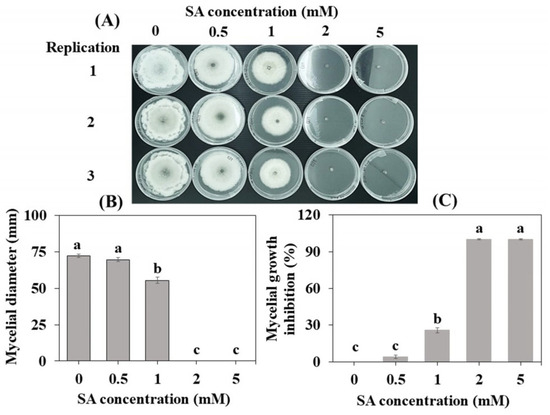
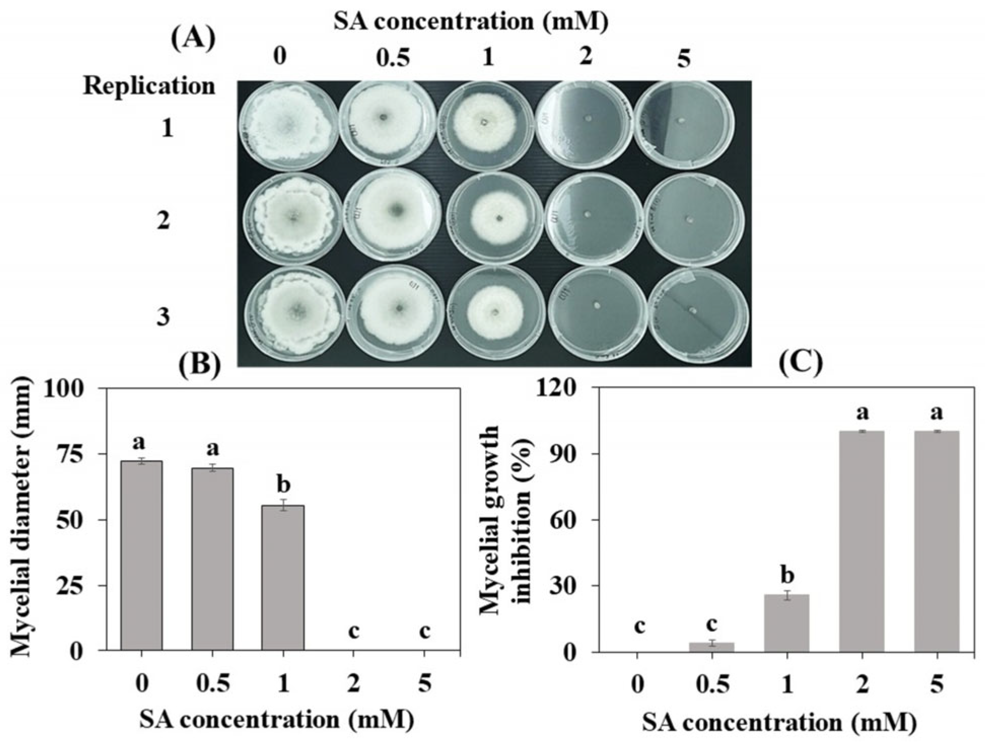
The effect of SA on mycelial growth in vitro is illustrated in Figure 1. The application of SA at the 1, 2, and 5 mM concentrations significantly decreased the C. gloeosporioides mycelial growth in vitro compared to the control and 0.5 mM concentration of SA. The 100% inhibition of mycelial development observed following the treatments with 2 mM SA and 5 mM SA was not significant (Figure 1C), indicating a strong fungitoxic effect. These results agree with those described by He et al. [10], who reported significant inhibition of C. gloeosporioides at SA concentrations above 0.5 mM.
Figure 1.
The in vitro effects of various salicylic acid (SA) concentrations on Colletotrichum gloeosporioides. (A) Representative mycelial growth images, (B) colony diameter, and (C) mycelial growth inhibition 7 days post incubation at 25 °C. Different letters indicate significant differences (p ≤ 0.05) to Tukey’s HSD post hoc test. The data are the mean ± SE of three replicates.
It has also been demonstrated that treatment with SA can reduce mycelial Alternaria alternata [22], Penicillium expansum [23], Rhizopus stolonifer [24], Aspergillus flavus [25], and Monilinia fructicola [26]. Salicylic acid not only directly inhibits the mycelial growth of these pathogens; it also enhances disease resistance in pear fruit [27]. The complete inhibition of C. gloeosporioides mycelial growth at 2 and 5 mM in the present study suggests that SA has a direct inhibitory effect on the colony growth of C. gloeosporioides in vitro.
3.2. The Effect of SA on Anthracnose Progression in Mango Fruit
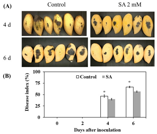
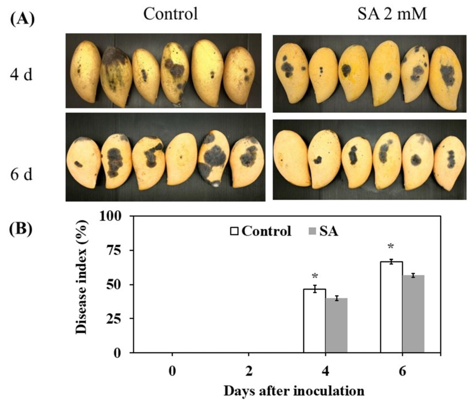
Four days post inoculation, symptoms of anthracnose disease were observed in both the SA and control groups (Figure 2A). Compared to the control fruit, the SA-treated fruit showed significantly decreased infection index values of mango fruit inoculated with C. gloeosporioides (Figure 3B); the infection index was 57% in the SA-treated fruit and 67% in the controls, with the values differing significantly between treatments 6 days post inoculation. These results agree with those reported by the authors of [28], who observed that SA treatment can reduce anthracnose disease in mango fruit. An investigation reported in [29] also revealed that pre- and post-harvest SA treatments for the management of latent infections of C. gloeosporioides significantly reduced the incidence of anthracnose in mangoes.
Figure 2.
The anthracnose symptoms (A) and infection index values 0, 2, 4, and 6 days post inoculation in Nam Dok Mai Si Thong mango fruit after SA treatment (B). * p ≤ 0.05, Student’s t-test. The data are the mean ± SE of three replicates.
Figure 3.
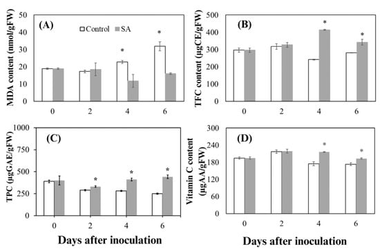
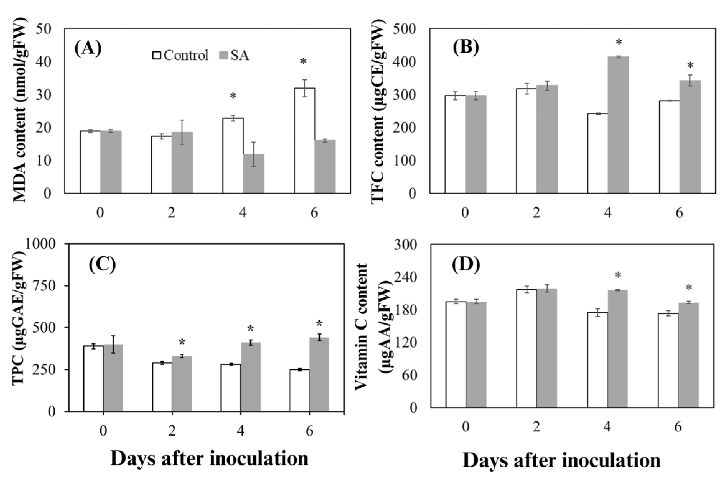
The changes in bioactive compounds and lipid peroxidation in mango peel after SA treatment. (A) Malondialdehyde content (MDA). (B) Total flavonoid content (TFC). (C) Total phenolic content (TPC). (D) Total ascorbic acid content. Measurements were taken 0, 2, 4, and 6 days post inoculation. * p ≤ 0.05, Student’s t-test. The data are the mean ± SE of three replicates.
3.3. Salicylic Acid Reduces Oxidative Stress in Mango Peel
MDA, which is produced from the peroxidation of membrane lipids, is often used as an indicator of oxidative stress and damage [30]. In the present study, the MDA levels in the SA-treated fruit decreased and those in the control group increased from 0 to 6 days post inoculation. The SA-treated fruit had significantly lower levels of MDA compared to the control fruit 4 and 6 days post inoculation (Figure 3A). The contrasting MDA patterns between the two treatments indicate that SA reduced membrane lipid peroxidation during anthracnose infection. The lower MDA accumulation in SA-treated fruit therefore suggests reduced oxidative damage and better preservation of cellular membrane integrity. Our results are consistent with those reported by Junmatong et al. [31], who noted that treatment with SA reduced the MDA content of Nam Dok Mai No. 4 mango fruit. It is also known that lower MDA levels indicate reduced membrane lipid peroxidation, helping maintain cellular integrity under pathogen challenge [32]. These findings suggest that SA treatment can delay the development of cell damage and help maintain membrane stability in mangoes during pathogen infection.
3.4. Salicylic Acid Enhances Bioactive Compounds in Mango Peel
Exogenous SA has been applied to increase bioactive compounds and antioxidants in many plant species [33]. Our present experiment revealed that the total flavonoid content in the SA-treated fruit increased from 2 to 4 days post inoculation, whereas the control group exhibited a decrease. The SA treatment resulted in significantly higher total flavonoid content in mango peels compared to the controls both 4 and 6 days post inoculation (Figure 3B). This result agrees with that of a 2 mg L−1 SA treatment of the medicinal plant Glycyrrhiza uralensis Fisch, which led to a 2.68-fold increase in the total flavonoid content [34]. It has also been reported that SA treatment enhanced the biosynthesis and accumulation of flavonoids in berries [35]. The higher flavonoid content observed in SA-treated mango peel may contribute to defense-associated responses because flavonoids are important secondary metabolites with antioxidant properties. The total phenolic content in the present study’s control group decreased, whereas it increased in the SA-treated fruit from days 2 to 6 post inoculation and was significantly higher than the control values in mango peels on all post-inoculation days (Figure 3C). In studies of other fruits, SA treatment significantly increased the antioxidant capacity and phenolics in pineapple (Ananas comosus L.) [36], pomegranates (Punica granatum) [37], and sweet cherry (Prunus avium) [38].
Phenolic compounds are secondary metabolites and include flavonoids and phenolic acids [39]. The biosynthesis of phenolics is involved in the phenylpropanoid pathway, a key defense mechanism in plants [28,40]. Phenolics act as substrates for polyphenol oxidase (PPO) and peroxidase (POD) to produce quinones and other antimicrobial compounds [41]. Therefore, the higher phenolic and flavonoid contents observed in SA-treated mango peel may strengthen antioxidant and defense-associated responses and thereby contribute to the limiting of pathogen development. In the present investigation, the total ascorbic acid levels in the control group decreased from 2 to 6 days post inoculation but increased in the SA-treated fruit. Notably, the SA treatment resulted in significantly higher total ascorbic acid levels compared to the control mango peels from 4 to 6 days post inoculation (Figure 3D). During pathogen infection, the total ascorbic acid participates in scavenging of the reactive oxygen species (ROS) that are generated during host–pathogen interactions, thereby protecting cellular structures from oxidative damage [42]. Thus, the higher ascorbic acid levels observed in SA-treated fruit may contribute to antioxidant protection against oxidative stress during anthracnose infection. Our results align with earlier studies indicating that SA treatment increases the total phenolic content while preserving the levels of chlorophyll, phenolics, flavonoids, and ascorbic acid in asparagus [43]. Taken together, the higher levels of phenolics, flavonoids, and ascorbic acid observed in SA-treated fruit indicate enhanced antioxidant and defense-related biochemical responses, which may contribute to the reduced anthracnose severity. This aligns with the work reported in [12], which demonstrated that SA improves mangoes’ resistance to post-harvest anthracnose by promoting antifungal activity and activating defense pathways.
3.5. Salicylic Acid Enhances Antioxidant Capacity
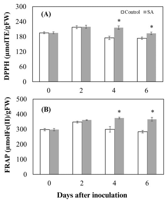
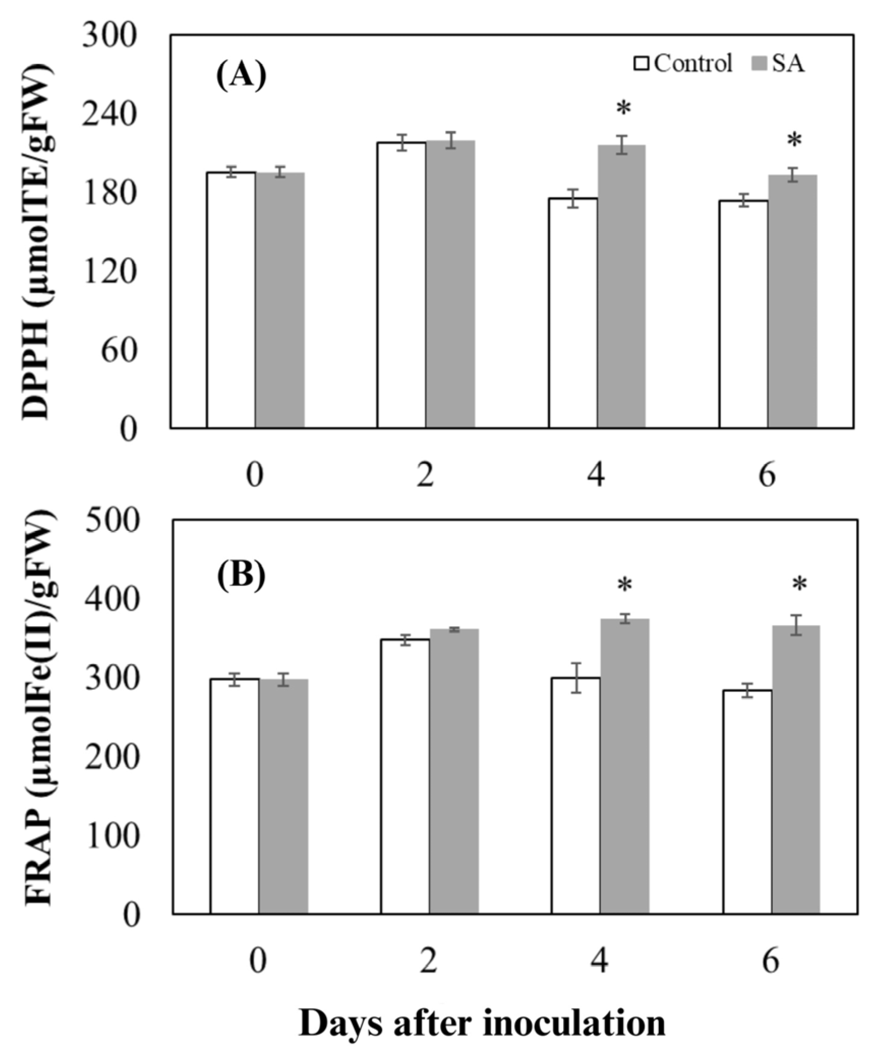
The plant antioxidant system regulates ROS to maintain redox homeostasis, and SA enhances this capacity, thereby reducing oxidative stress and improving post-harvest disease resistance [44]. Our present study’s DPPH and FRAP assays (Figure 4A,B) revealed that SA treatment significantly increased antioxidants compared to the control group, suggesting enhanced antioxidant potential during anthracnose development. The enhanced antioxidant capacity may be associated with the higher levels of phenolics, flavonoids, and ascorbic acid observed in SA-treated mango peel (Figure 3B–D), as these bioactive compounds contribute to antioxidant defense. These results agree with reports that SA treatment enhances the antioxidant capacity in asparagus (Asparagus officinalis) [43], bananas (Musa spp.) [45], papayas (Carica papaya) [46], apricots (Prunus armeniaca) [47], and grapes (Vitis vinifera) [48] during storage.
Figure 4.
The antioxidant capacity of mango peel following SA application. (A) DPPH radical scavenging activity. (B) Ferric reducing antioxidant power (FRAP). Measurements were taken 0, 2, 4, and 6 days post inoculation. * p ≤ 0.05, Student’s t-test. The data are the mean ± SE of three replicates.
We speculate that the enhanced antioxidant activity observed in SA-treated fruit may contribute to reduced anthracnose severity by limiting oxidative damage during pathogen infection. Improved redox regulation can help maintain membrane stability and reduce cellular damage during pathogen infection. These findings are consistent with the report in [48] that SA has the potential to enhance the DPPH free-radical scavenging capacity and the FRAP of grapes. Dokhanieh et al. [49] noted that treatment with SA significantly enhanced the DPPH scavenging activity of cornelian cherry fruits by increasing the concentrations of bioactive compounds such as phenolics.
3.6. Salicylic Acid-Associated Metabolomic Changes in Mango Peel
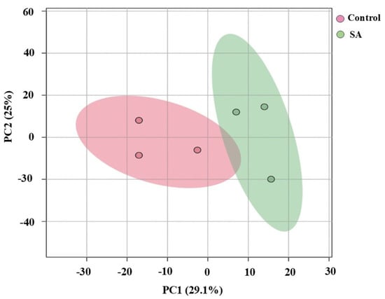
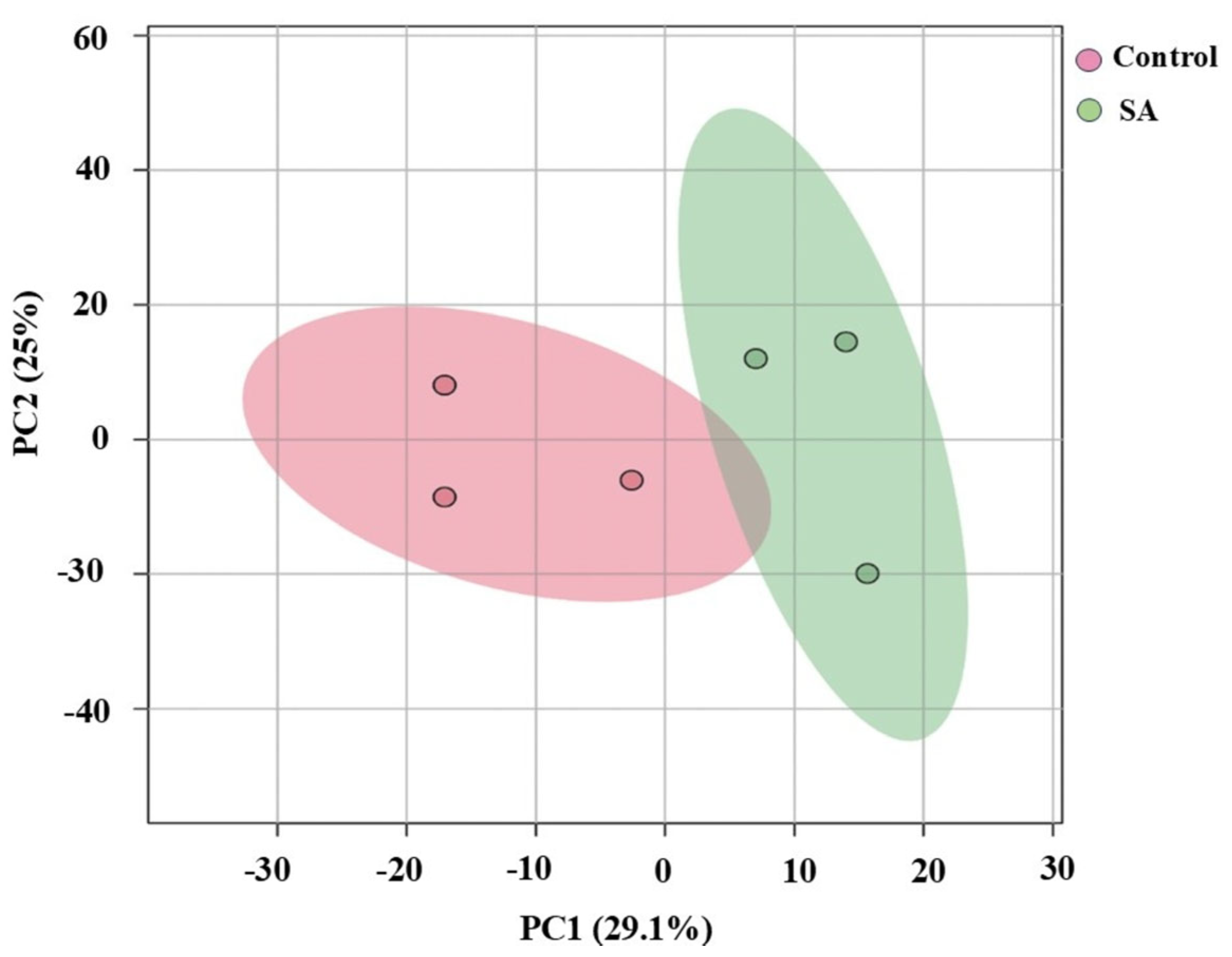
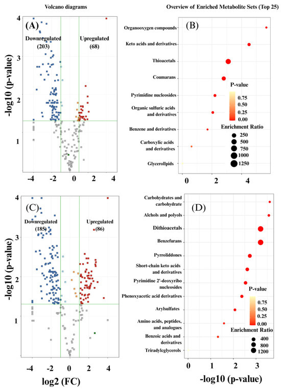
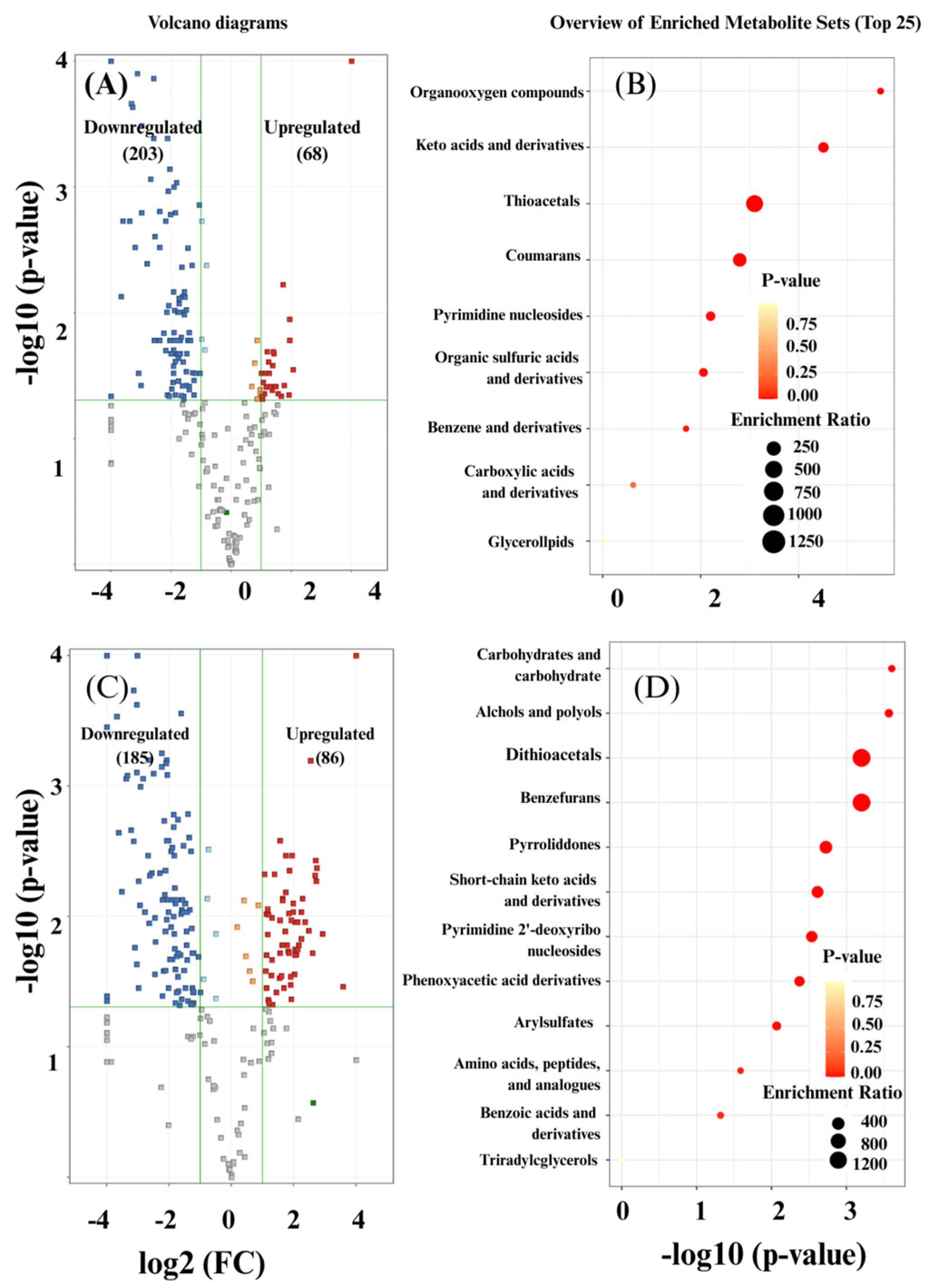
The results of the untargeted metabolomics analysis performed using PCA are presented in Figure 5. The PCA clearly distinguished SA-treated fruit 6 days post inoculation (D6SA) from untreated controls (D6C), with PC1 and PC2 accounting for 29.1% and 25.0% of the total variation, respectively (Figure 5). This result indicates that SA treatment was associated with changes in metabolite composition 6 days post inoculation. This metabolic separation was consistent with the biochemical responses observed in the present study, in which SA-treated fruit exhibited higher total phenolic content, total flavonoid content, ascorbic acid content, and antioxidant activity than the control fruit. These findings suggest that the distinct metabolomic profile of SA-treated fruit was accompanied by increased accumulation of antioxidant-related compounds and enhanced antioxidant capacity. The volcano plots revealed temporal changes in the metabolite profiles of both control and SA-treated mango peel between D0 and D6. The volcano plots for the D6 control group indicated a total of 68 upregulated metabolites (including thioacetals, coumarans, and keto acids) and 203 downregulated metabolites (Figure 6A). In contrast, the D6SA treatment resulted in 86 upregulated metabolites (including dithioacetals, benzofurans, and pyrrolidones) and 185 downregulated metabolites (Figure 6C). These results indicate substantial changes in metabolite profiles from D0 to D6 in both groups, with different temporal patterns observed between control and SA-treated fruit. Previous studies have reported that SA can influence the biosynthesis and accumulation of secondary metabolites in plants [50]. Plants possess diverse categories of secondary metabolites, which are classified into three major groups: terpenes or terpenoids, phenolics, and N-containing compounds [51].
Figure 5.
Principal component analysis (PCA) of mango peel metabolite profiles at 6 days post inoculation. Pink circles and shaded areas represent untreated control fruit (D6C), whereas green circles and shaded areas represent SA-treated fruit (D6SA). PC1 and PC2 explain 29.1% and 25.0% of the total variance, respectively.
Figure 6.
Changes in the metabolomic profiles of control and SA-treated mango peel between D0 and D6. (A) Volcano plot in the control group (D0 vs. D6 control). (B) Metabolite set enrichment analysis of the differential features identified in the control group (D0 vs. D6C). (C) Volcano plot in the SA-treated group (D0 vs. D6SA). (D) Metabolite set enrichment analysis of the differential features identified in the SA-treated group. Fold changes ≥2 and p ≤ 0.05 were considered significant. In panels (A) and (C), red and blue points indicate upregulated and downregulated metabolites, respectively. Green vertical and horizontal lines indicate the fold-change and p-value thresholds, respectively. Numbers in parentheses indicate the numbers of upregulated and downregulated metabolites. In panels (B) and (D), point color represents the p-value, and point size represents the enrichment ratio, as indicated in the legends. Analyses were based on three replicates per group. The enrichment analyses show the major metabolite classes associated with changes between D0 and D6 within each treatment group.
Secondary metabolites such as phenolics and flavonoids play a crucial role in plant defense against biotic stress by mediating antimicrobial activity and defense signaling [52]. Metabolite set enrichment analysis further revealed distinct temporal enrichment patterns within the two groups. Together, these results demonstrate that the two groups exhibited distinct patterns of metabolite changes over time. Thioacetals were predominantly enriched in the control fruit (Figure 6B), whereas dithioacetals were primarily enriched in the SA-treated fruit (Figure 6D). Although thioacetal-related metabolites were enriched in the control fruit, their specific biological functions in mango fruit during anthracnose development remain unclear. Therefore, their enrichment may represent temporal metabolic changes associated with fruit–pathogen interactions rather than a specific defense response. In contrast, Dithioacetals are regarded as functional structural motifs associated with plant defense responses [53]. The enrichment of dithioacetal-related metabolites observed in the D0 and D6 SA comparison may therefore reflect metabolic changes associated with defense responses during anthracnose development. We also observed that benzofuran-related metabolites were significantly enriched in the D0 and D6 SA comparison. Benzofuran derivatives have been reported to possess antifungal properties, inhibiting fungal growth and disrupting key metabolic processes that are necessary for pathogen development [54]. Thus, the enrichment of benzofuran-related metabolites indicates that SA may therefore be associated with defense-related metabolic responses and may provide additional metabolic context for the reduced anthracnose severity observed in SA-treated fruit.
3.7. The Effect of SA on Phenylalanine Metabolism Pathways
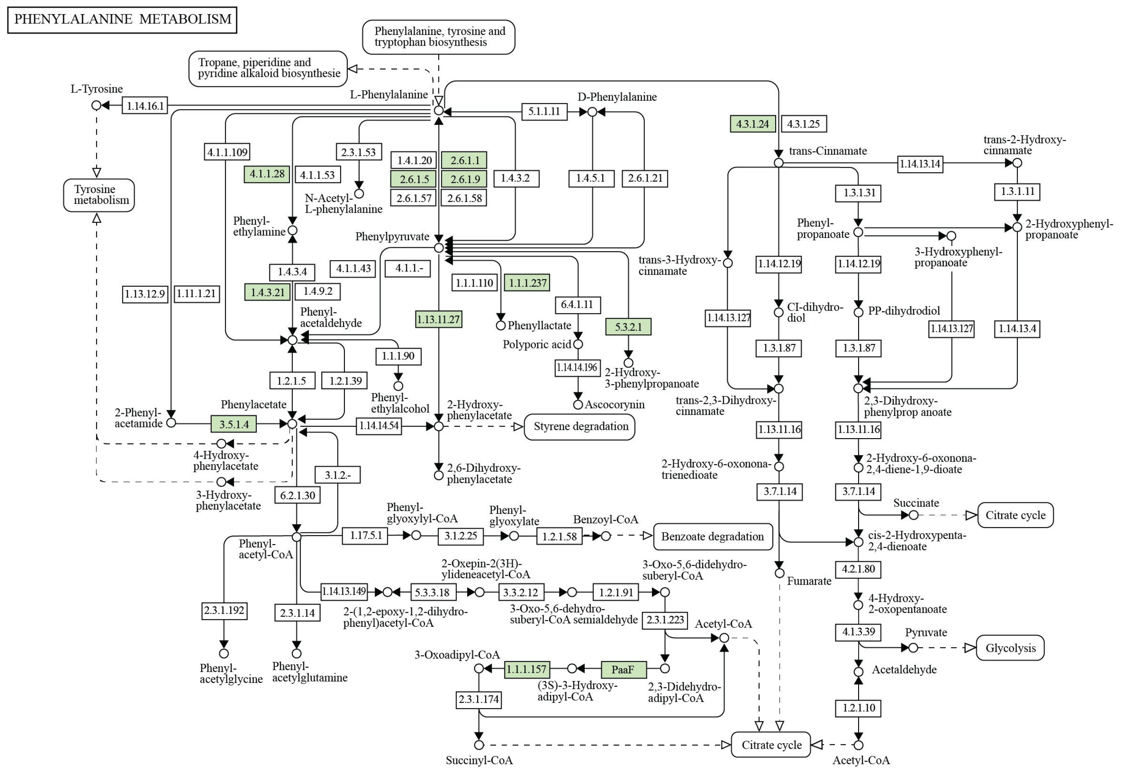
Salicylic acid is an important phytohormone for plant responses to biotic and abiotic stresses. Plants have evolved two pathways to produce SA: the isochorismate synthase pathway [55] and the phenylalanine ammonia lyase pathway [56]. Our pathway analysis indicated that D-amino acid metabolism was among the pathways showing temporal changes in SA-treated mango peel between D0 and D6 but was not identified in the corresponding D0 vs. D6 control analysis (Figure 7). D-amino acids play regulatory roles in plant stress signaling and immune responses [57]. KEGG pathway analysis further revealed alterations in phenylalanine metabolism in the D0 vs. D6 SA comparison (Figure 8 and Figure 9). Phenylalanine is an important precursor of the phenylpropanoid pathway, which produces phenolic and flavonoid compounds involved in plant defense. Thus, the observed alterations in phenylalanine metabolism may be associated with the increased accumulation of phenolics and flavonoid compounds observed in SA-treated fruit.
These results agree with reports that SA treatment induced the phenylalanine ammonia-lyase (PAL) enzyme in Capsicum chinense (habanero pepper) [58] as well as the activity and gene expression of PAL in the leaves of saffron, and it induced PAL gene expression in Salvia [59]. Phenylalanine is a key precursor for phenolic and flavonoid biosynthesis via the phenylpropanoid pathway, linking metabolite accumulation to enhanced defense and antioxidant capacity [60,61]. Multiple research groups have demonstrated that SA influences PAL activity and phenylpropanoid biosynthesis, thereby enhancing resistance against post-harvest pathogens [28]. In our previous study in ‘Nam Dok Mai Si Thong’ mango, SA treatment significantly increased the activities of defense-related enzymes polyphenol oxidase (PPO) and peroxidase (POD), whereas PAL activity was not significantly increased [62]. These findings suggest that SA-associated defense responses in this mango cultivar may involve multiple defense-related enzymatic processes and are not necessarily dependent on increased PAL activity alone.
The metabolomic results in this study are consistent with the biochemical measurements obtained herein. The SA-treated fruit exhibited significantly higher total phenolic and flavonoid contents than the control fruit at D6 (Figure 3B,C), which is consistent with the observed changes in phenylalanine-related metabolism. These compounds play essential roles in plant defense because they possess strong antioxidant and antimicrobial activities, which can inhibit fungal growth and reinforce host resistance against pathogens such as C. gloeosporioides.
Together with our previous observation that SA treatment increased PPO and POD activities in ‘Nam Dok Mai Si Thong’ mango, the present biochemical and metabolomic findings provide complementary evidence of defense-associated responses. The observed phenylalanine-related metabolic changes, together with higher phenolic and flavonoid contents and enhanced antioxidant capacity, provide additional metabolic context for the reduced anthracnose severity in SA-treated fruit.
Figure 7.
Pathway impact analysis of metabolic changes in SA-treated mango peel between D0 and D6. The analysis was based on differential metabolites identified from the D0 vs. D6 SA comparison. These pathways were detected only in the D0 vs. D6 SA pathway analysis and were not identified in the corresponding D0 vs. D6 control analysis. The x-axis represents pathway impact based on pathway topology analysis, whereas the y-axis represents pathway significance as −log10(p-value).
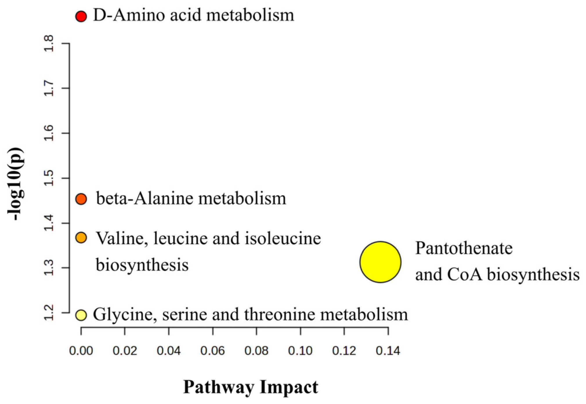
Figure 8.
D-Amino acid metabolism pathway (KEGG map00470) based on the KEGG database described by Kanehisa et al. [63]. Solid arrows indicate the direction of metabolic reactions, and dashed arrows indicate connections to other metabolic pathways or omitted intermediate steps. Rectangular boxes contain enzyme commission numbers (EC numbers), and green boxes highlight enzyme entries mapped in the D0 vs. D6 comparison of SA-treated mango peel.
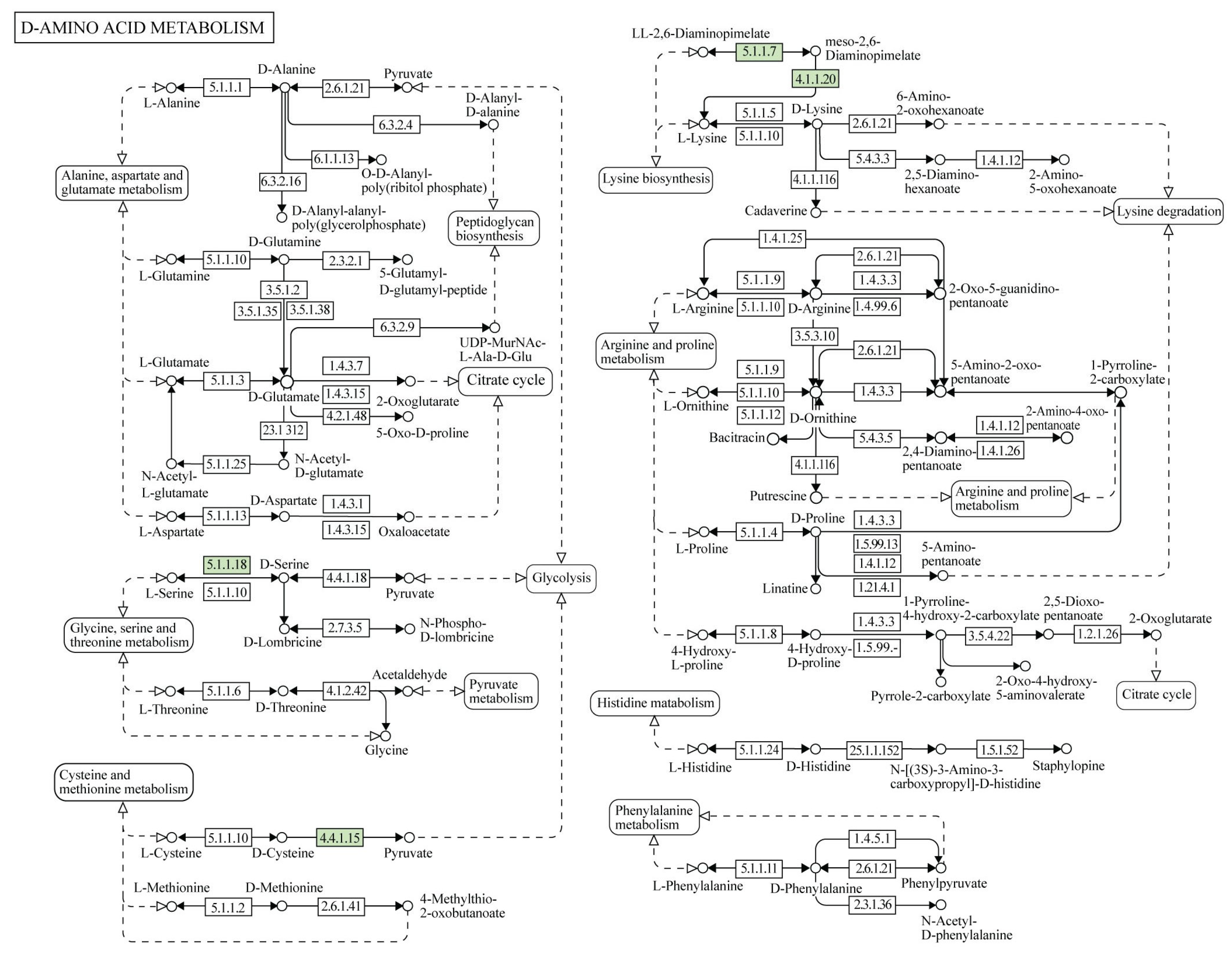
Figure 9.
Phenylalanine metabolism pathway (KEGG map00360), based on the KEGG database described by Kanehisa et al. [63]. Solid arrows indicate the direction of metabolic reactions, whereas dashed arrows indicate connections to other pathways or omitted intermediate steps. Green boxes (containing EC numbers) highlight enzyme entries mapped in the D0 vs. D6 comparison of SA-treated mango peel.
4. Conclusions
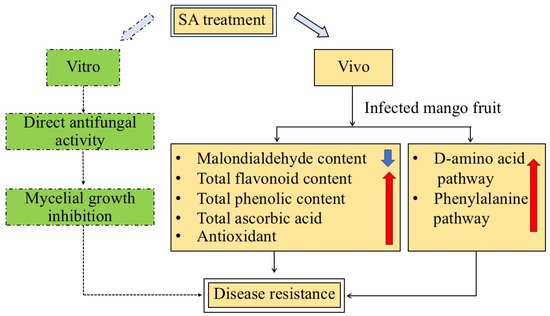
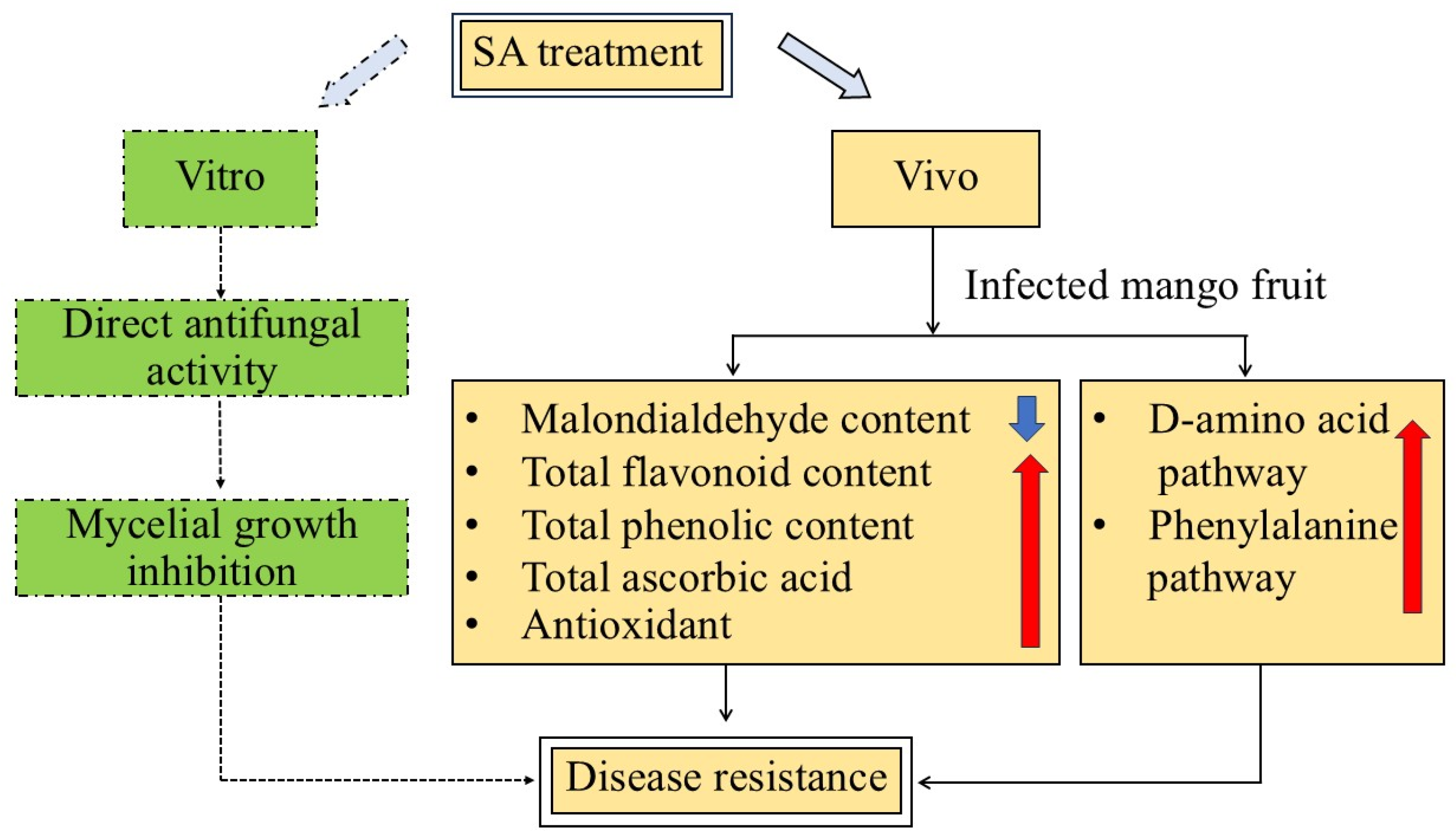
Treatment with salicylic acid decreased the degree of anthracnose disease caused by Colletotrichum gloeosporioides in Nam Dok Mai Si Thong mango fruit. Salicylic acid treatment reduced the disease severity and the fruits’ MDA content; it increased total phenolic content (TPC), total flavonoid content (TFC), ascorbic acid content, and antioxidant capacity; and untargeted metabolomic analysis further revealed distinct metabolite profiles between SA-treated and control fruit at D6, as well as changes in D-amino acid and phenylalanine-related metabolism in SA-treated fruit (Figure 10). Together, these findings indicate that SA treatment reduced anthracnose severity and was associated with biochemical and metabolic responses related to antioxidant and defense processes.
Figure 10.
Schematic diagram of salicylic acid (SA)-induced metabolic reprogramming and defense activation in mango peel. Exogenous SA treatment activates multiple defense-related pathways in mango fruit during post-harvest storage. The SA directly inhibits the growth of Colletotrichum gloeosporioides and simultaneously modulates key metabolic pathways, including phenylalanine metabolism and D-amino acid metabolism. Upregulated metabolites (e.g., phenolics, flavonoids, and benzofurans) enhance both the antioxidant capacity and cell membrane protection (lower MDA content). Dashed arrows indicate the in vitro antifungal response, whereas solid arrows indicate the proposed relationships among SA treatment, metabolic and biochemical responses, and disease resistance in vivo. Red upward arrows indicate increases, and the blue downward arrow indicates a decrease.
Author Contributions
A.K.: Conceptualization, Methodology, Investigation, Data analysis, Visualization, Writing—original draft, and Writing—review and editing. R.C.: Investigation and Data analysis. M.N.: Writing—review and editing. P.S.: Writing—review and editing. P.W.: Writing—review and editing. T.P.: Writing—review and editing. S.K. (Sunantha Ketnawa): Writing—review and editing. S.K. (Satoru Kondo): Conceptualization, Supervision, and Writing—review and editing. S.S.: Conceptualization, Methodology, Supervision, Funding acquisition, and Writing—review and editing. All authors have read and agreed to the published version of the manuscript.
Funding
This research was financially supported by Mae Fah Luang University under the National Science, Research and Innovation Fund (NSRF) 2024 (Grant No. 672A04016).
Institutional Review Board Statement
Ethical review and approval were waived for this study because it did not involve humans or animals.
Informed Consent Statement
Not applicable.
Data Availability Statement
The raw data supporting the conclusions of this article will be made available by the authors upon request.
Conflicts of Interest
The authors declare that they have no known competing financial interests or personal relationships that could have appeared to influence the work reported in this paper.
References
- Armand, A.; Absalan, S.; Khongphinitbunjong, K.; Maharachchikumbura, S.; Hyde, K.; Jayawardena, R. New species and new records of plant pathogenic fungi from northern Thailand. Asian J. Mycol. 2024, 7, 148–174. [Google Scholar] [CrossRef]
- Kankam, F.; Adomako, J.; Larbi-Koranteng, S.; Danso, Y.; Kwowura Kwodaga, J.; Akpatsu, I.B.; Sowley, E.N.K. Anthracnose Disease of Mango: Epidemiology, Impact and Management Options. In Current and Emerging Challenges in the Diseases of Trees; Bellé, C., Ed.; IntechOpen: London, UK, 2022. [Google Scholar] [CrossRef] [Scilit]
- Law, C.X.; Hashim, N.; Ismail, S.I.; Jahari, M.; Al Riza, D.F. A review on anthracnose disease caused by Colletotrichum spp. in fruits and advances in control strategies. Int. J. Food Microbiol. 2025, 442, 111397. [Google Scholar] [CrossRef] [Scilit]
- Ashraf, M.A.; Khan, A.S.; Azam, M.; Ayyub, S.; Kaleem, M.M.; Nawaz, S.; Abubakar, M.; Khalid, B.; Riaz, T.; Asim, M.; et al. Innovative technologies in postharvest management of fruits and vegetables: A review. Eur. Food Res. Technol. 2025, 251, 3445–3463. [Google Scholar] [CrossRef] [Scilit]
- Nunes, C.A. Biological control of postharvest diseases of fruit. Eur. J. Plant Pathol. 2012, 133, 181–196. [Google Scholar] [CrossRef] [Scilit]
- Klessig, D.F.; Malamy, J. The salicylic acid signal in plants. Plant Mol. Biol. 1994, 26, 1439–1458. [Google Scholar] [CrossRef] [Scilit]
- Aghdam, M.S.; Asghari, M.; Babalar, M.; Askari, M.A. Impact of Salicylic Acid on Postharvest Physiology of Fruits and Vegetables. In Eco-Friendly Technology for Postharvest Produce Quality; Academic Press: Cambridge, MA, USA, 2016; pp. 243–268. [Google Scholar] [CrossRef] [Scilit]
- Tareen, M.J.; Abbasi, N.A.; Hafiz, I.A. Postharvest application of salicylic acid enhanced antioxidant enzyme activity and maintained quality of peach cv. ‘Flordaking’ fruit during storage. Sci. Hortic. 2012, 142, 221–228. [Google Scholar] [CrossRef] [Scilit]
- Wang, Z.; Ma, L.; Zhang, X.; Xu, L.; Cao, J.; Jiang, W. The effect of exogenous salicylic acid on antioxidant activity, bioactive compounds and antioxidant system in apricot fruit. Sci. Hortic. 2015, 181, 113–120. [Google Scholar] [CrossRef] [Scilit]
- He, J.; Ren, Y.; Chen, C.; Liu, J.; Liu, H.; Pei, Y. Defense responses of salicylic acid in mango fruit against postharvest anthracnose, caused by Colletotrichum gloeosporioides and its possible mechanism. J. Food Saf. 2017, 37, e12294. [Google Scholar] [CrossRef] [Scilit]
- Ramaroson, M.-L.; Koutouan, C.; Helesbeux, J.-J.; Le Clerc, V.; Hamama, L.; Geoffriau, E.; Briard, M. Role of phenylpropanoids and flavonoids in plant resistance to pests and diseases. Molecules 2022, 27, 8371. [Google Scholar] [CrossRef] [Scilit]
- Cai, Y.; Ouyang, Y.; Zhang, X.; Chen, Y.; Zhang, J.; Bi, C.; Liu, Z.; Han, X.; Zhang, X.; Wang, W.; et al. How does salicylic acid activate plant immunity? New Crops 2026, 3, 100112. [Google Scholar] [CrossRef] [Scilit]
- Khan, M.; Chonhenchob, V.; Huang, C.; Suwanamornlert, P. Antifungal activity of propyl disulfide from neem (Azadirachta indica) in vapor and agar diffusion assays against anthracnose pathogens (Colletotrichum gloeosporioides and Colletotrichum acutatum) in mango fruit. Microorganisms 2021, 9, 839. [Google Scholar] [CrossRef] [Scilit]
- Corkidi, G.; Balderas-Ruíz, K.A.; Taboada, B.; Serrano-Carreón, L.; Galindo, E. Assessing mango anthracnose using a new three-dimensional image-analysis technique to quantify lesions on fruit. Plant Pathol. 2006, 55, 250–257. [Google Scholar] [CrossRef] [Scilit]
- Duan, X.; Liu, T.; Zhang, D.; Su, X.; Lin, H.; Jiang, Y. Effect of pure oxygen atmosphere on antioxidant enzyme and antioxidant activity of harvested litchi fruit during storage. Food Res. Int. 2011, 44, 1905–1911. [Google Scholar] [CrossRef] [Scilit]
- Salem, M.Z.M.; Ali, H.M.; El-Shanhorey, N.A.; Abdel-Megeed, A. Evaluation of extracts and essential oil from Callistemon viminalis leaves: Antibacterial and antioxidant activities, total phenolic and flavonoid contents. Asian Pac. J. Trop. Med. 2013, 6, 785–791. [Google Scholar] [CrossRef] [Scilit]
- Zhishen, J.; Mengcheng, T.; Jianming, W. The determination of flavonoid contents in mulberry and their scavenging effects on superoxide radicals. Food Chem. 1999, 64, 555–559. [Google Scholar] [CrossRef] [Scilit]
- Roe, J.H.; Oesterling, M.J. The determination of dehydroascorbic acid and ascorbic acid in plant tissues by the 2,4-dinitrophenylhydrazine method. J. Biol. Chem. 1944, 152, 511–517. [Google Scholar] [CrossRef] [Scilit]
- Moon, J.K.; Shibamoto, T. Antioxidant assays for plant and food components. J. Agric. Food Chem. 2009, 57, 1655–1666. [Google Scholar] [CrossRef] [Scilit]
- Benzie, I.F.; Strain, J.J. Ferric reducing/antioxidant power assay: Direct measure of total antioxidant activity of biological fluids and modified version for simultaneous measurement of total antioxidant power and ascorbic acid concentration. Methods Enzymol. 1999, 299, 15–27. [Google Scholar] [CrossRef] [Scilit]
- Wang, J.; Ren, C.; Wang, J.; Fu, J.; Yin, Q.; Huang, Y.; He, Z. Liquid chromatography quadrupole time-of-flight mass spectrometry-based metabolic characterization of mango ripened by different methods. Foods 2024, 13, 3548. [Google Scholar] [CrossRef] [Scilit]
- Qin, G.Z.; Tian, S.P.; Xu, Y.; Wan, Y.K. Enhancement of biocontrol efficacy of antagonistic yeasts by salicylic acid in sweet cherry fruit. Physiol. Mol. Plant Pathol. 2003, 62, 147–154. [Google Scholar] [CrossRef] [Scilit]
- Yu, T.; Zheng, X. Salicylic acid enhances biocontrol efficacy of the antagonist Cryptococcus laurentii in apple fruit. J. Plant Growth Regul. 2006, 25, 166–174. [Google Scholar] [CrossRef] [Scilit]
- Panahi Rad, S.; Zaare-Nahandi, F.; Safaralizadeh, R.; Alizadeh-Salteh, S. Postharvest control of Rhizopus stolonifer in peach (Prunus persica L. Batsch) fruits using salicylic acid. J. Food Saf. 2013, 32, 502–507. [Google Scholar] [CrossRef] [Scilit]
- Panahirad, S.; Zaare-Nahandi, F.; Mohammadi, N.; Alizadeh-Salteh, S.; Safaie, N. Effects of salicylic acid on Aspergillus flavus infection and aflatoxin B1 accumulation in pistachio (Pistacia vera L.) fruit. J. Sci. Food Agric. 2014, 94, 1758–1763. [Google Scholar] [CrossRef] [Scilit]
- Yao, H.; Tian, S. Effects of pre- and post-harvest application of salicylic acid or methyl jasmonate on inducing disease resistance of sweet cherry fruit in storage. Postharvest Biol. Technol. 2005, 35, 253–262. [Google Scholar] [CrossRef] [Scilit]
- Gao, J.; Wang, P.-Y.; Wen, M.-M.; Chen, C.; Zhang, J.; Wang, C.-L. Salicylic acid induces disease resistance of postharvest ‘Housui’ pear fruit by modulating reactive oxygen species metabolism and activating defense-related enzymes. Physiol. Mol. Plant Pathol. 2026, 142, 103126. [Google Scholar] [CrossRef] [Scilit]
- Zeng, K.; Cao, J.; Jiang, W. Enhancing disease resistance in harvested mango (Mangifera indica L. cv. ‘Matisu’) fruit by salicylic acid. J. Sci. Food Agric. 2006, 86, 694–698. [Google Scholar] [CrossRef] [Scilit]
- Zainuri, Z.; Joyce, D.; Wearing, H.; Coates, L.; Terry, L. Effects of phosphonate and salicylic acid treatments on anthracnose disease development and ripening of ‘Kensington Pride’ mango fruit. Aust. J. Exp. Agric. 2001, 41, 805–813. [Google Scholar] [CrossRef] [Scilit]
- Ohkawa, H.; Ohishi, N.; Yagi, K. Assay for lipid peroxides in animal tissues by thiobarbituric acid reaction. Anal. Biochem. 1979, 95, 351–358. [Google Scholar] [CrossRef] [Scilit]
- Junmatong, C.; Uthaibutra, J.; Boonyakiat, D.; Faiyue, B.; Saengnil, K. Reduction of chilling injury of ‘Nam Dok Mai No. 4’ mango fruit by treatments with salicylic acid and methyl jasmonate. J. Agric. Sci. 2012, 4, 126–136. [Google Scholar] [CrossRef] [Scilit]
- Hessini, K.; Martínez, J.P.; Gandour, M.; Albouchi, A.; Soltani, A.; Abdelly, C. Effect of water stress on growth, osmotic adjustment, cell wall elasticity and water-use efficiency in Spartina alterniflora. Environ. Exp. Bot. 2009, 67, 312–319. [Google Scholar] [CrossRef] [Scilit]
- Chen, C.; Sun, C.; Wang, Y.; Gong, H.; Zhang, A.; Yang, Y.; Guo, F.; Cui, K.; Fan, X.; Li, X. The preharvest and postharvest application of salicylic acid and its derivatives on storage of fruit and vegetables: A review. Sci. Hortic. 2023, 312, 111858. [Google Scholar] [CrossRef] [Scilit]
- Li, J.; Wang, J.; Li, J.; Li, J.; Liu, S.; Gao, W. Salicylic acid induces the change in the adventitious root of Glycyrrhiza uralensis Fisch.: Bioactive compounds and antioxidant enzymes. Res. Chem. Intermed. 2016, 42, 1503–1519. [Google Scholar] [CrossRef] [Scilit]
- Guan, C.; Song, X.; Ji, J.; Li, X.; Jin, C.; Guan, W.; Li, J.; Wang, G. Salicylic acid treatment enhances expression of chalcone isomerase gene and accumulation of corresponding flavonoids during fruit maturation of Lycium chinense. Eur. Food Res. Technol. 2014, 239, 857–865. [Google Scholar] [CrossRef] [Scilit]
- Sangprayoon, P.; Supapvanich, S.; Youryon, P.; Wongs-Aree, C.; Boonyaritthongchai, P. Efficiency of salicylic acid or methyl jasmonate immersions on internal browning alleviation and physicochemical quality of Queen pineapple cv. “Sawi” fruit during cold storage. J. Food Biochem. 2019, 43, e13059. [Google Scholar] [CrossRef] [Scilit]
- Koyuncu, M.A.; Erbas, D.; Onursal, C.E.; Secmen, T.; Guneyli, A.; Sevinc Uzumcu, S. Postharvest treatments of salicylic acid, oxalic acid and putrescine influences bioactive compounds and quality of pomegranate during controlled atmosphere storage. J. Food Sci. Technol. 2019, 56, 350–359. [Google Scholar] [CrossRef] [Scilit]
- Valero, D.; Díaz-Mula, H.M.; Zapata, P.J.; Castillo, S.; Guillén, F.; Martínez-Romero, D.; Serrano, M. Postharvest treatments with salicylic acid, acetylsalicylic acid or oxalic acid delayed ripening and enhanced bioactive compounds and antioxidant capacity in sweet cherry. J. Agric. Food Chem. 2011, 59, 5483–5489. [Google Scholar] [CrossRef] [Scilit]
- Oraei, M.; Panahirad, S.; Zaare-Nahandi, F.; Gohari, G. Pre-véraison treatment of salicylic acid to enhance anthocyanin content of grape (Vitis vinifera L.) berries. J. Sci. Food Agric. 2019, 99, 5946–5952. [Google Scholar] [CrossRef] [Scilit]
- Li, Q.; Liu, Z.; Jiang, Z.; Jia, M.; Hou, Z.; Dou, D.; Yu, J. Phenylalanine metabolism-dependent lignification confers rhizobacterium-induced plant resistance. Plant Physiol. 2025, 197, kiaf016. [Google Scholar] [CrossRef] [Scilit]
- Moosa, A.; Zulfiqar, F.; Siddique, K.H.M. Transcriptional and biochemical profiling of defense enzymes in Citrus sinensis during salicylic acid and cinnamon mediated suppression of green and blue mold. Front. Plant Sci. 2022, 13, 1048433. [Google Scholar] [CrossRef] [Scilit]
- Khan, T.; Mazid, M.; Mohammad, F. Role of ascorbic acid against pathogenesis in plants. J. Stress Physiol. Biochem. 2011, 7, 222–234. [Google Scholar]
- Wei, Y.; Liu, Z.; Su, Y.; Liu, D.; Ye, X. Effect of salicylic acid treatment on postharvest quality, antioxidant activities, and free polyamines of asparagus. J. Food Sci. 2011, 76, S126–S132. [Google Scholar] [CrossRef] [Scilit]
- Yang, W.; Liu, Y.; Sang, Y.; Ma, Y.; Guo, M.; Bai, G.; Cheng, S.; Chen, G. Influences of ice-temperature storage on cell wall metabolism and reactive oxygen metabolism in Xinjiang (Diaogan) apricot. Postharvest Biol. Technol. 2021, 180, 111614. [Google Scholar] [CrossRef] [Scilit]
- Fangxu, X.; Yefei, L.; Jie, X.; Liang, F. Influence of 1-methylcyclopropene (1-MCP) combined with salicylic acid (SA) treatment on the postharvest physiology and quality of bananas. J. Food Process. Preserv. 2019, 43, e13880. [Google Scholar] [CrossRef] [Scilit]
- Hanif, A.; Ahmad, S.; Shahzad, S.; Liaquat, M.; Anwar, R. Postharvest application of salicylic acid reduced decay and enhanced storage life of papaya fruit during cold storage. J. Food Meas. Charact. 2020, 14, 3078–3088. [Google Scholar] [CrossRef] [Scilit]
- Fan, X.; Du, Z.; Cui, X.; Ji, W.; Ma, J.; Li, X.; Wang, X.; Zhao, H.; Liu, B.; Guo, F.; et al. Preharvest methyl salicylate treatment enhance the chilling tolerance and improve the postharvest quality of apricot during low temperature storage. Postharvest Biol. Technol. 2021, 177, 111535. [Google Scholar] [CrossRef] [Scilit]
- Gomes, E.P.; Vanz Borges, C.; Monteiro, G.C.; Filiol Belin, M.A.; Minatel, I.O.; Pimentel Junior, A.; Tecchio, M.A.; Lima, G.P.P. Preharvest salicylic acid treatments improve phenolic compounds and biogenic amines in ‘Niagara Rosada’ table grape. Postharvest Biol. Technol. 2021, 176, 111505. [Google Scholar] [CrossRef] [Scilit]
- Dokhanieh, A.Y.; Aghdam, M.S.; Fard, J.R.; Hassanpour, H. Postharvest salicylic acid treatment enhances antioxidant potential of cornelian cherry fruit. Sci. Hortic. 2013, 154, 31–36. [Google Scholar] [CrossRef] [Scilit]
- Ali, B. Salicylic acid: An efficient elicitor of secondary metabolite production in plants. Biocatal. Agric. Biotechnol. 2021, 31, 101884. [Google Scholar] [CrossRef] [Scilit]
- Taiz, L.; Zeiger, E.; Møller, I.M.; Murphy, A. Plant Physiology and Development; Sinauer Associates: Sunderland, MA, USA, 2025. [Google Scholar]
- Bartwal, A.; Mall, R.; Lohani, P.; Guru, S.K.; Arora, S. Role of secondary metabolites and brassinosteroids in plant defense against environmental stresses. J. Plant Growth Regul. 2013, 32, 216–232. [Google Scholar] [CrossRef] [Scilit]
- Chen, J.; Shi, J.; Yu, L.; Liu, D.; Gan, X.; Song, B.; Hu, D. Design, synthesis, antiviral bioactivity, and defense mechanisms of novel dithioacetal derivatives bearing a strobilurin moiety. J. Agric. Food Chem. 2018, 66, 5335–5345. [Google Scholar] [CrossRef] [Scilit]
- Khanam, H.; Shamsuzzaman. Bioactive benzofuran derivatives: A review. Eur. J. Med. Chem. 2015, 97, 483–504. [Google Scholar] [CrossRef] [Scilit]
- Wildermuth, M.C.; Dewdney, J.; Wu, G.; Ausubel, F.M. Isochorismate synthase is required to synthesize salicylic acid for plant defence. Nature 2001, 414, 562–565. [Google Scholar] [CrossRef] [Scilit]
- Huang, W.; Wang, Y.; Li, X.; Zhang, Y. Biosynthesis and Regulation of Salicylic Acid and N-Hydroxypipecolic Acid in Plant Immunity. Mol. Plant 2020, 13, 31–41. [Google Scholar] [CrossRef] [Scilit]
- Kolukisaoglu, Ü. D-Amino Acids in Plants: Sources, Metabolism, and Functions. Int. J. Mol. Sci. 2020, 21, 5421. [Google Scholar] [CrossRef] [Scilit]
- Gutiérrez-Carbajal, M.G.; Monforte-González, M.; Miranda-Ham, M.d.L.; Godoy-Hernández, G.; Vázquez-Flota, F. Induction of capsaicinoid synthesis in Capsicum chinense cell cultures by salicylic acid or methyl jasmonate. Biol. Plant. 2010, 54, 430–434. [Google Scholar] [CrossRef] [Scilit]
- Ejtahed, R.S.; Radjabian, T.; Hoseini Tafreshi, S.A. Expression analysis of phenylalanine ammonia lyase gene and rosmarinic acid production in Salvia officinalis and Salvia virgata shoots under salicylic acid elicitation. Appl. Biochem. Biotechnol. 2015, 176, 1846–1858. [Google Scholar] [CrossRef] [Scilit]
- Vogt, T. Phenylpropanoid biosynthesis. Mol. Plant 2010, 3, 2–20. [Google Scholar] [CrossRef] [Scilit]
- Li, P.; Lan, L.; Xiong, L.; Tian, Y.; Wu, G.; Peng, D.; Lan, S.; Liu, Z.; Zhang, Q.; Ai, Y. Integrated transcriptome and metabolome analysis reveal a reprogramming of phenylpropanoid pathway and antioxidant activity in Cymbidium ensifolium resistance to anthracnose. J. Plant Physiol. 2025, 315, 154631. [Google Scholar] [CrossRef] [Scilit]
- Ampa, K.; Charatsaengphaibun, R.; Naradisorn, M.; Suthiluk, P.; Kondo, S.; Wongsa, P.; Ketnawa, S.; Saito, T.; Khewkhom, N.; Srilaong, V.; et al. The impact of hot water and salicylic acid treatments on metabolomic changes and defense mechanisms against anthracnose caused by Colletotrichum gloeosporioides in mango fruit. J. Agric. Food Res. 2026, 25, 102586. [Google Scholar] [CrossRef] [Scilit]
- Kanehisa, M.; Furumichi, M.; Sato, Y.; Matsuura, Y.; Ishiguro-Watanabe, M. KEGG: Biological systems database as a model of the real world. Nucleic Acids Res. 2025, 53, D672–D677. [Google Scholar] [CrossRef] [Scilit]
Disclaimer/Publisher’s Note: The statements, opinions and data contained in all publications are solely those of the individual author(s) and contributor(s) and not of MDPI and/or the editor(s). MDPI and/or the editor(s) disclaim responsibility for any injury to people or property resulting from any ideas, methods, instructions or products referred to in the content. |
© 2026 by the authors. Licensee MDPI, Basel, Switzerland. This article is an open access article distributed under the terms and conditions of the Creative Commons Attribution (CC BY) license.