


Abstract
Background/Objectives: Bone mineral density (BMD) is fundamental to osteoporosis assessment, but fracture risk is also shaped by fracture history, falls, frailty, secondary causes, age, sex, and treatment context. This narrative review examines clinically relevant information that modifies adult endocrine decision-making beyond the T-score. Methods: PubMed/MEDLINE was searched through 28 June 2026; recent guidelines, consensus statements, systematic reviews, meta-analyses, and pivotal fracture-endpoint trials were prioritized. The exact search strings, filters, eligibility criteria, selection process, and prioritized guidance documents are provided in PubMed MEDLINE search strategies. Results: BMD should be interpreted with fracture history and clinical risk factors, while the applicability of T-scores differs by age and sex. FRAX, vertebral imaging, and trabecular bone score can refine assessment in selected patients. Very-high-risk patients may warrant early osteoanabolic or dual-action therapy followed by antiresorptive treatment, whereas antiresorptives remain appropriate for many high-risk patients. Denosumab requires planned continuity and a follow-on strategy; advanced chronic kidney disease requires particular attention to mineral metabolism and hypocalcemia risk. Conclusions: Contemporary osteoporosis care expands rather than replaces densitometry. Endocrine management should integrate valid DXA interpretation, fracture-risk urgency, reversible contributors, drug-specific safety, treatment feasibility, monitoring, and the next treatment phase.
1. Introduction and Literature Search Strategy
Osteoporosis is a common metabolic bone disease whose clinical importance is determined principally by fragility fractures rather than by a densitometric threshold alone. Hip and vertebral fractures are associated with pain, loss of mobility and independence, subsequent fracture risk, and excess mortality. Despite the availability of dual-energy X-ray absorptiometry (DXA), validated fracture-risk tools, and effective antifracture therapies, underdiagnosis and undertreatment remain common, including after an established fragility fracture [1,2,3].
The T-score continues to have a central role in osteoporosis diagnosis and fracture-risk assessment, but its diagnostic use is age- and sex-dependent. In postmenopausal women and men aged 50 years or older, T-scores are preferred for densitometric classification. In females before menopause and males younger than 50 years, Z-scores are preferred; in particular, osteoporosis in a man younger than 50 years should not be diagnosed from BMD alone [4]. The limitation of the T-score is therefore not that it lacks clinical value, but that even when it is the correct densitometric metric, the same value can carry different implications in patients with different ages, fracture histories, fall risk, glucocorticoid exposure, renal function, or comorbidities. A recent vertebral fracture, for example, changes the urgency of treatment in a way that cannot be conveyed by BMD alone. Current guidance therefore places densitometry within a broader assessment that includes fracture history, clinical risk factors, and, when appropriate, FRAX, vertebral imaging, and trabecular bone score (TBS) [1,2,4,5,6,7,8,9].
This perspective is particularly relevant to endocrinology. Skeletal fragility may coexist with primary hyperparathyroidism, thyroid excess or iatrogenic TSH suppression, glucocorticoid exposure, diabetes, hypogonadism, malabsorption, chronic kidney disease, and other disorders in which the mechanism of bone loss influences both the diagnostic work-up and the choice of therapy [1,2,5,6].
This article was prepared as a targeted narrative review rather than a systematic review. PubMed/MEDLINE was searched using combinations of terms grouped around five clinical themes: fracture-risk assessment; care delivery; pharmacologic treatment and sequencing; secondary osteoporosis; and special adult populations. The search was initially completed on 3 June 2026 and checked for relevant updates through 28 June 2026. English-language guidelines, consensus statements, systematic reviews, meta-analyses, and pivotal randomized or extension trials were prioritized, with preference for recent guidance and higher-level evidence when available. Reference lists of major guidelines and consensus documents were checked to recover landmark fracture-endpoint trials that remained clinically relevant. Adult human populations were eligible when the source informed diagnosis, risk stratification, endocrine evaluation, treatment selection, sequencing, safety, or monitoring. Pediatric osteoporosis, oncology-specific bone loss, transplant bone disease, rare monogenic skeletal disorders, conference abstracts without a full report, and sources without direct clinical relevance to the prespecified themes were excluded. Screening and selection were not performed independently in duplicate. Consequently, there were no paired independent screening disagreements; citations of uncertain relevance were discussed within the author team and retained when consensus supported inclusion, without formal third-reviewer adjudication or an inter-reviewer agreement statistic. No formal risk-of-bias assessment, GRADE certainty rating, or quantitative pooling was undertaken. For major therapeutic statements, fracture-endpoint randomized evidence was distinguished from BMD-endpoint transition studies, observational data, regulatory safety information, and expert consensus when this distinction affected interpretation. The exact reproducible PubMed strings, date and language limits, eligibility criteria, selection process, and prioritized guideline and consensus documents are reported in Supplementary Table S1. These methodological boundaries are considered explicitly in Section 7.3.
The added value of this review is not a new risk score or a competing guideline. It is a single clinically oriented pathway that connects technical DXA validity, fracture recency and multiplicity, FRAX and TBS refinement, endocrine and metabolic contributors, treatment intensity, drug-specific safety, therapeutic sequence, and long-term continuity. These elements are commonly distributed across separate densitometry positions, disease-specific guidelines, regulatory documents, and pharmacologic reviews. Bringing them together clarifies when information beyond BMD can change diagnosis, urgency, treatment selection, safety assessment, monitoring, or the next treatment phase in adult endocrine practice.
2. Beyond the T-Score: Care Gaps, Fracture Risk, FRAX, TBS, and Imminent Risk
2.1. From Treatment Gap to Care Gap
The traditional term treatment gap describes the proportion of eligible patients who do not receive osteoporosis medication, but it does not capture the full sequence of missed opportunities that can occur before and after a prescription. Patients may not be screened when screening is appropriate, a low-trauma fracture may be treated without triggering osteoporosis evaluation, vertebral fractures may remain unrecognized, or a patient may discontinue treatment without a safe transition plan. Recent literature has therefore proposed the broader concept of an osteoporosis care gap [3].
Fracture liaison services (FLS) address one of the most important components of this gap by systematically identifying patients after fragility fracture and linking the acute event to secondary fracture prevention. Meta-analytic evidence supports improved secondary-prevention processes and a reduction in subsequent fractures in many FLS models; reported mortality benefits are less certain because program structure varies and much of the mortality evidence is observational [10]. In settings without a formal FLS, the same principle can be applied through local pathways that assign responsibility for fracture-risk assessment, secondary-cause evaluation, treatment initiation, adherence, and follow-up.
2.2. Risk Stratification Beyond BMD
BMD is strongly associated with fracture risk, but fracture probability is influenced by more than bone mass. Age, prior fracture, parental hip fracture, glucocorticoid exposure, smoking, alcohol use, falls, frailty, and selected comorbidities alter the clinical meaning of a measured T-score [1,2,5,6]. Risk stratification is useful because it links the severity and immediacy of fracture risk to treatment intensity. Several contemporary frameworks distinguish high from very high fracture risk, particularly when fractures are recent, multiple, vertebral or hip in location, occur during adherent therapy, or coexist with very low BMD [6,8,11].
The distinction is clinically relevant rather than semantic. For many patients at high risk, an oral or intravenous bisphosphonate or another antiresorptive agent is an appropriate initial treatment. In patients with very high or imminent fracture risk, rapid and substantial fracture-risk reduction becomes a priority, and an osteoanabolic or dual-action strategy may be preferred when available and clinically suitable [6,8,11,12,13,14].
There is, however, no single universally accepted definition of very high fracture risk. NOGG, AACE/ACE, ESCEO/IOF, the Endocrine Society, and the ASBMR/BHOF goal-directed framework overlap in emphasizing recent or multiple fractures, very low BMD, fracture during therapy, and other markers of imminent risk, but the exact criteria and intervention thresholds differ [6,8,11,12,15,16]. Table 1 summarizes these frameworks without attempting to harmonize them. The categories and treatment-intensity examples used in this review are illustrative clinical syntheses, not new recommendations. Eligibility for a specific treatment must follow the guideline, product license, contraindications, and reimbursement rules applicable in the clinician’s jurisdiction.
Table 1.
Comparison of high, very high, and imminent fracture-risk concepts across selected major frameworks.
2.3. FRAX and Its Limits
FRAX estimates 10-year probability of major osteoporotic fracture and hip fracture using age and selected clinical risk factors, with or without femoral-neck BMD. It is useful for translating multiple risk factors into an absolute probability and for supporting intervention decisions in health systems that use probability-based thresholds [5,6,11]. FRAX models and intervention thresholds are country- and healthcare-system-specific; a probability that triggers treatment in one setting may not do so in another. FRAX is a baseline risk-estimation tool rather than a validated instrument for monitoring treatment response, and its standard use is not a substitute for etiologic assessment in younger adults in whom conventional T-score-based algorithms may not apply. Falls are not directly incorporated, fracture recency and multiplicity are incompletely represented, and the effects of glucocorticoids and several comorbidities may require clinical adjustment. A recent major fragility fracture can therefore justify a more urgent treatment approach even when the 10-year estimate does not appear extreme [4,6,8,11,15].
2.4. Trabecular Bone Score and Vertebral Imaging
Before adding adjunctive measures, the DXA examination itself should be technically valid. Central DXA generally includes the PA lumbar spine and hip; the 33% (one-third) radius is particularly relevant when the hip or spine cannot be interpreted and in hyperparathyroidism [4]. Vertebrae affected by focal structural change or artifact should be excluded according to accepted densitometric rules, and diagnosis should not be based on a single remaining lumbar vertebra. Degenerative change, vertebral deformity, vascular calcification, positioning, and hardware can falsely alter lumbar-spine BMD. Longitudinal change should be interpreted against the facility-specific least significant change and, whenever possible, on the same or appropriately cross-calibrated device [4,17].
TBS is a texture-derived index calculated from lumbar-spine DXA images and provides information that is partly independent of areal BMD. The 2023 ISCD Official Positions state that TBS can refine fracture-risk stratification and can be used to adjust FRAX probability, particularly when a patient is close to a pharmacologic intervention threshold [9]. Its role should remain adjunctive: TBS should not be used as a stand-alone diagnosis of osteoporosis, as a substitute for hip and spine BMD, or as the sole reason to select a specific medication. TBS should be used only within the manufacturer’s recommended BMI range. L1–L4 may be retained in the presence of moderate degenerative change or chronic lumbar compression fracture, but TBS should not be reported when severe structural or pathological artifact invalidates interpretation, including vertebra plana, laminectomy, spinal hardware, or metastatic lesions [4,9]. Routine serial monitoring of TBS change is not currently recommended with the existing algorithm [4,9].
Vertebral fracture identification is equally important because a previously unrecognized vertebral fracture can materially reclassify risk. ISCD positions support vertebral fracture assessment (VFA) or lateral spine imaging in selected patients with low BMD and additional risk features, including advanced age, substantial historical height loss, an undocumented prior vertebral fracture, or sustained glucocorticoid exposure [4]. VFA is designed to detect vertebral fractures rather than to characterize every spinal abnormality; equivocal deformity, suspicious lytic or sclerotic change, or a finding that cannot be attributed confidently to osteoporosis should prompt conventional radiography or other clinically appropriate imaging [4].
2.5. Imminent Risk and Goal-Directed Treatment
Fracture risk is not constant over time. The period after a recent fragility fracture carries a particularly high probability of another fracture, which is why the term imminent fracture risk has become clinically useful [8,16]. The term does not have one universally fixed time window or threshold; in practice it generally refers to the early post-fracture period, with risk highest soon after the index fracture and remaining elevated over the following one to two years. The 2024 ASBMR/BHOF position statement on goal-directed osteoporosis treatment emphasizes rapid fracture-risk reduction for patients with recent fractures and other very-high-risk features and proposes individualized BMD goals as one component of long-term management [8]. This approach does not replace clinical judgment or national reimbursement criteria, but it reinforces the principle that the initial treatment should be chosen with the expected treatment trajectory in mind. The principal clinical modifiers and their management implications are summarized in Table 2.
Table 2.
Clinical features that modify fracture-risk interpretation beyond the T-score.
3. Endocrine Evaluation and Secondary Causes of Skeletal Fragility
The first osteoporosis consultation should establish whether the patient has predominantly postmenopausal or age-related osteoporosis, or whether a secondary disorder is contributing materially to skeletal fragility. The probability of a secondary cause is not the same in every patient; age, sex, fracture pattern, severity of BMD loss, biochemical abnormalities, medication exposure, and associated symptoms should determine the breadth of investigation [1,2,5,6].
A practical baseline evaluation commonly includes serum calcium, creatinine and estimated glomerular filtration rate, alkaline phosphatase, 25-hydroxyvitamin D, complete blood count, and selected liver and thyroid tests. Serum phosphate, parathyroid hormone, serum protein electrophoresis, celiac testing, urinary calcium, cortisol testing, testosterone in men, and other investigations should be added when the history, examination, or initial laboratory results justify them [1,2,5,6]. The aim is not to create a universal laboratory panel but to avoid missing a treatable disorder.
3.1. Hyperparathyroidism and Calcium-Phosphate Disorders
Primary hyperparathyroidism is particularly relevant because it can present with low BMD or fragility fracture and changes both the interpretation and management of osteoporosis. The Fifth International Workshop provides criteria for evaluation and management, including the role of serum calcium, PTH, renal assessment, nephrolithiasis, and skeletal involvement [21]. In this setting, cortical bone assessment is clinically relevant, and ISCD guidance supports measuring the 33% radius in patients with hyperparathyroidism [4]. In patients with unexplained hypercalcemia or a compatible pattern of bone loss, osteoporosis treatment should not proceed without clarifying the parathyroid disorder.
3.2. Thyroid, Adrenal, Gonadal, and Gastrointestinal Contributors
Overt hyperthyroidism increases bone turnover and is an established secondary cause of bone loss, whereas physiologic levothyroxine replacement that maintains an appropriate TSH should not be conflated with intentional or inadvertent chronic TSH suppression. Prolonged iatrogenic TSH suppression may adversely affect BMD in selected patients, especially postmenopausal women. These effects are clinically relevant in differentiated thyroid cancer, where the intensity and duration of suppression should be balanced against oncologic benefit and skeletal risk [22]. Hypercortisolism and chronic glucocorticoid therapy can cause rapid deterioration in bone strength, whereas hypogonadism in men and premature estrogen deficiency in women are important causes of secondary osteoporosis. Malabsorption, celiac disease, bariatric surgery, chronic liver disease, and nutritional deficiencies should be considered when the clinical history suggests them [1,2,5,6,20,23].
3.3. Why Mechanism Matters for Treatment
Identifying a secondary cause can change the treatment sequence, not merely add another diagnosis. Persistent hypercalcemia, advanced CKD-mineral and bone disorder, active malabsorption, severe vitamin D deficiency, or uncontrolled endocrine disease can alter drug safety, expected response, and monitoring. Treatment of the secondary cause and antifracture therapy are often complementary rather than mutually exclusive; in a patient with high or very high fracture risk, necessary endocrine evaluation should not create avoidable delays in fracture prevention.
3.4. Falls, Exercise, Nutrition, and Modifiable Risk
Fracture prevention is not purely pharmacologic. Falls are a major proximal event for many non-vertebral fractures, and a history of falls should prompt review of balance, lower-limb strength, vision, footwear, orthostatic symptoms, sedating or hypotensive medication, and environmental hazards. Exercise recommendations should be individualized rather than restricted reflexively because of a low T-score. A multidisciplinary UK consensus supports progressive resistance and impact exercise for bone strength, targeted strength and balance training for people at risk of falls, and spinal extension/postural work for people with vertebral fracture, with modification of impact level in frail patients or those with multiple low-trauma fractures [18].
Adequate calcium intake, correction of vitamin D deficiency, sufficient dietary protein, smoking cessation, moderation of excess alcohol intake, and preservation of muscle mass are supportive components of care [5,6,15]. Calcium and vitamin D supplementation should not be presented as substitutes for antifracture medication in a high-risk patient; their role is to correct deficiency and support safe, effective pharmacologic treatment. The practical objective is to reduce skeletal fragility and fall exposure at the same time.
4. Pharmacological Treatment: Antiresorptive, Osteoanabolic, and Dual-Action Strategies
4.1. Bisphosphonates
Bisphosphonates remain a mainstay of osteoporosis treatment. Oral alendronate or risedronate is appropriate for many patients when adherence, gastrointestinal tolerance, correct administration, and renal function are compatible with the product label. Renal thresholds are agent-specific rather than a class-wide interchangeable rule: current US labeling does not recommend alendronate when creatinine clearance is <35 mL/min and does not recommend risedronate when creatinine clearance is <30 mL/min [24,25]. For osteoporosis-dose intravenous zoledronic acid, serum creatinine and Cockcroft-Gault creatinine clearance should be assessed before each dose; treatment is contraindicated when creatinine clearance is <35 mL/min or acute renal impairment is present, and infusion should be withheld until normovolemia is restored if dehydration is suspected [26]. Local product information should take precedence. Intravenous zoledronic acid is useful when oral adherence is doubtful, gastrointestinal contraindications are present, or an infrequent parenteral regimen is preferable [5,6,12]. Bisphosphonate retention in bone allows consideration of a treatment holiday in selected patients, but a holiday is a risk-based decision rather than an automatic endpoint. Major guidelines commonly recommend reassessment after approximately five years of oral bisphosphonate therapy or three years of annual zoledronic acid in patients whose fracture risk has become low enough to consider interruption; continued treatment or a different strategy is appropriate when risk remains high [5,6,12,15]. A bisphosphonate holiday should therefore be distinguished from permanent treatment cessation, and the patient requires continued clinical reassessment during the interval.
4.2. Denosumab
Denosumab is a potent antiresorptive therapy with a clinical profile that differs fundamentally from bisphosphonates. In the FREEDOM trial, denosumab reduced vertebral, hip, and nonvertebral fractures in postmenopausal women with osteoporosis [27]. Because its effect is reversible and it is not retained in bone, six-monthly administration must be reliable. Interruption or discontinuation can lead to rapid rebound in bone turnover, loss of BMD, and vertebral fractures, particularly after longer exposure and in patients with previous vertebral fractures [28,29]. The future transition strategy should therefore be discussed when denosumab is started rather than only when it is stopped. Renal clearance is not required, but advanced CKD is not risk-free: severe hypocalcemia risk rises as kidney function worsens, especially in dialysis and CKD-MBD [30,31]. For CKD G4-G5D, a practical prevention protocol is to determine whether denosumab is appropriate with a clinician experienced in CKD-MBD; assess calcium, intact PTH, 25-hydroxyvitamin D, and 1,25-dihydroxyvitamin D and consider bone-turnover status before treatment; correct pre-existing hypocalcemia and manage CKD-MBD; provide adequate calcium and activated vitamin D; review concomitant calcimimetics or other calcium-lowering therapies; and educate the patient about symptoms requiring urgent assessment. Current US prescribing information recommends serum-calcium monitoring weekly for the first month after each injection and monthly thereafter in advanced CKD, with prompt treatment of hypocalcemia [31]. Supplement type and dose, dialysis timing, and additional phosphate or magnesium monitoring should be individualized with nephrology.
4.3. Teriparatide and Abaloparatide
Osteoanabolic therapy is most relevant when fracture risk is very high and a rapid, substantial skeletal response is desired. In the VERO trial, teriparatide reduced new vertebral and clinical fractures more effectively than risedronate in postmenopausal women with severe osteoporosis [32]. Abaloparatide also reduced new vertebral fractures in the ACTIVE trial [33]. These agents should be viewed as part of a treatment sequence because the gains achieved during the anabolic phase require subsequent antiresorptive therapy to be maintained [8,13,14].
4.4. Romosozumab
Romosozumab inhibits sclerostin and has both bone-forming and antiresorptive effects. FRAME demonstrated substantial vertebral-fracture reduction during the first year of romosozumab followed by denosumab, whereas ARCH showed lower fracture incidence with romosozumab followed by alendronate than with alendronate alone in a higher-risk population [34,35]. A numerical imbalance in serious cardiovascular events was observed during the first year of ARCH but not as a consistent signal across the trial program; this supports a precautionary risk assessment but does not by itself establish a simple causal relationship. Cardiovascular assessment should explicitly include prior myocardial infarction and stroke and should consider other major cardiovascular risk factors. Regulatory wording varies by jurisdiction: US labeling advises against initiation after myocardial infarction or stroke within the preceding year, whereas the European product information lists any previous myocardial infarction or stroke as a contraindication [36,37]. Local labeling therefore takes precedence. A recent meta-analysis supports the skeletal efficacy of romosozumab-based sequential treatment [38]. Antiresorptive treatment after the romosozumab course is required to preserve skeletal gains [12,13,34,38].
4.5. Selecting Treatment Intensity
An osteoanabolic-first strategy should not be presented as the default treatment for every patient with osteoporosis. Antiresorptive therapy remains appropriate for a large proportion of patients at high fracture risk. Osteoanabolic or dual-action treatment is most compelling when risk is very high, especially with recent major fracture, multiple vertebral fractures, very low BMD, or fracture despite appropriate treatment [6,8,11,13]. These treatment-intensity examples are illustrative and do not create new eligibility criteria. Drug choice must conform to locally applicable guidance, licensed indications, contraindications, and reimbursement, while also considering cardiovascular history, renal function, access, cost, patient preference, and the feasibility of the next treatment phase.
4.6. Other Selected Pharmacologic Options
The therapeutic discussion should not imply that contemporary osteoporosis care consists only of bisphosphonates, denosumab, and bone-forming agents. Selective estrogen-receptor modulators such as raloxifene or bazedoxifene can be considered in selected postmenopausal women, particularly when vertebral-fracture prevention is the main objective and venous-thromboembolic risk is low. Menopausal hormone therapy may be appropriate for fracture prevention in carefully selected younger postmenopausal women who also have menopausal symptoms and no major contraindication, but it is not a general substitute for osteoporosis-specific therapy in older or very-high-risk patients [5,12,15]. Drug selection therefore depends on fracture pattern, age, comorbidity, reproductive status, contraindications, route preference, and the expected duration and sequence of treatment. A practical comparison of these strategies is provided in Table 3, and selected pivotal fracture-endpoint trials are summarized in Table 4.
Table 3.
Practical comparison of major pharmacologic strategies used in adult osteoporosis care.
Table 4.
Selected pivotal fracture-endpoint trials that anchor the pharmacologic evidence base.
5. Sequential Therapy and Treatment Continuity
Osteoporosis is a chronic disease, but individual drug courses are often time-limited or require reassessment. The clinically relevant question is therefore not only which treatment to start, but what treatment is expected to follow it. This is especially important after osteoanabolic therapy and during denosumab discontinuation [8,13,14,28,29,42].
5.1. Osteoanabolic-to-Antiresorptive Sequencing
The sequence from an osteoanabolic agent to an antiresorptive is well supported biologically and clinically. Bone formation and BMD gains achieved during the anabolic phase can be consolidated with an antiresorptive agent. By contrast, prior exposure to potent antiresorptive therapy can modify the response to a subsequently administered osteoanabolic drug. DATA-Switch demonstrated that transition from teriparatide to denosumab continued to increase BMD, whereas transition from denosumab to teriparatide was associated with transient or progressive bone loss at several skeletal sites [42]. These findings illustrate why the order of treatment matters.
5.2. Denosumab Exit Strategies
Denosumab should not be stopped or materially delayed without a documented follow-on plan. Before the final intended injection, record the date on which the next dose would be due, reassess fracture risk and renal function, confirm calcium-vitamin D adequacy, and determine whether oral bisphosphonate administration and adherence are realistic. The ECTS position statement uses approximately six months after the final injection—the time the next denosumab dose would otherwise be due—as a practical anchor for initiating a potent antiresorptive [39]. Oral alendronate may be used when gastrointestinal tolerance, adherence, and renal function permit; intravenous zoledronic acid is commonly selected when oral treatment is unsuitable or adherence is uncertain. The choice is individualized according to denosumab duration, prior bisphosphonate exposure, renal function, and fracture risk [29,39].
After the transition, evaluate adherence and new fracture symptoms and reassess BMD, preferably on the same DXA system. Serum CTX, and sometimes PINP, can be checked where assays and local protocols are available; persistent elevation may support additional or repeated antiresorptive treatment, but proposed thresholds and schedules are not fully harmonized [29,39,43]. One zoledronic acid infusion does not suppress rebound reliably in every patient, particularly after longer denosumab exposure. If bone-turnover markers are unavailable, specialist protocols may use a planned second assessment or dose, but evidence remains consensus- and surrogate-endpoint based. Severe renal impairment may preclude usual bisphosphonate strategies and requires specialist planning rather than an untreated gap. New back pain or height loss should prompt evaluation for vertebral fracture. A missed denosumab injection is not a benign drug holiday [28,29,39].
5.3. Treatment Failure, Fracture on Therapy, and Reassessment
A fracture during therapy does not automatically prove pharmacologic failure. The first step is to verify adherence, administration technique, treatment duration, interval from treatment initiation, DXA comparability, and the presence of a new secondary cause or increased fall risk. A single fracture can occur despite effective therapy because no available treatment abolishes risk. Recurrent fractures, a significant BMD decline beyond least significant change, or persistently unfavorable bone-turnover-marker response despite adequate exposure provide stronger reasons to question efficacy or adherence and to reconsider treatment intensity [5,6,8,15]. In a patient who remains at very high risk, escalation or a change in treatment sequence may be appropriate.
5.4. What the Sequencing Evidence Does Not Yet Establish
The rationale for sequencing is strong, but the evidence should not be overstated. Fracture-endpoint randomized trials directly support the antifracture efficacy of individual agents and, for FRAME and ARCH, specific romosozumab-first sequences [34,35]. Other transition evidence, including DATA-Switch, is based primarily on BMD and bone-turnover outcomes rather than fractures [42]. Post-denosumab strategies rely on smaller interventional studies, observational cohorts, surrogate outcomes, and expert consensus [29,39]. Direct head-to-head fracture data for every possible sequence are not available. Most pivotal trials enrolled postmenopausal women; evidence is substantially thinner in men, advanced CKD, younger adults, and pregnancy-associated osteoporosis [16,23,42]. Table 5 labels the main evidentiary basis for each commonly discussed sequence so that randomized fracture evidence is not conflated with BMD outcomes or consensus.
Table 5.
Common treatment sequences and points that require active planning. The arrow symbol indicates the intended treatment transition or sequence.
6. Special Populations and Longitudinal Monitoring
6.1. Glucocorticoid-Induced Osteoporosis
Glucocorticoid-associated fracture risk can rise early after treatment begins and is not explained completely by BMD. Daily and cumulative dose, treatment duration, age, prior fracture, and the underlying inflammatory disorder all influence risk. The 2022 American College of Rheumatology guideline recommends early fracture-risk assessment and treatment according to risk category rather than waiting for advanced bone loss [20]. FRAX can contribute to assessment in age-appropriate adults, but glucocorticoid dose and the rapid early change in fracture risk require clinical interpretation rather than uncritical reliance on the calculated probability.
6.2. Diabetes Mellitus
Type 2 diabetes is a classic example of the limitation of a purely densitometric approach. Patients may experience increased fracture risk despite BMD values that are normal or only modestly reduced. Altered bone material properties, accumulation of advanced glycation end products, impaired bone turnover, neuropathy, visual impairment, sarcopenia, and falls all contribute to the skeletal phenotype [19]. BMD remains useful, but the clinician should recognize that standard risk estimates may not capture every diabetes-related contributor. TBS can be considered as an adjunctive risk-refinement measure when technically valid, but it should not be used as a diabetes-specific diagnostic test or as the sole determinant of treatment [9,19].
6.3. Thyroid Disease
Overt hyperthyroidism accelerates bone turnover and is an established secondary cause of bone loss. In differentiated thyroid cancer, chronic TSH suppression may also affect BMD, with the clearest concern in postmenopausal women. A systematic review and meta-analysis reported adverse BMD effects in this group, while the evidence is less consistent in premenopausal women and men [22]. The intensity of TSH suppression should therefore be individualized according to oncologic risk while skeletal risk is assessed separately.
6.4. Chronic Kidney Disease
In CKD, osteoporosis and CKD-mineral and bone disorder can coexist. In CKD G1-G3 without major biochemical abnormalities, osteoporosis assessment often resembles that used in the general population, while the distinction becomes increasingly important in G4-G5D because PTH abnormalities, altered bone turnover, mineral disturbances, and renal osteodystrophy can change both diagnosis and treatment safety [44]. The European consensus statement for CKD G4-G5D recommends integrating BMD and fracture history with calcium, phosphate, PTH, alkaline phosphatase, vitamin D status, and the clinical trajectory of kidney disease [44]. Drug choice also becomes more complex: bisphosphonate use is limited by renal function and evidence gaps, while denosumab can produce clinically important hypocalcemia in advanced CKD, with highest risk in dialysis and CKD-MBD [30]. Collaboration with nephrology is appropriate when advanced CKD or biochemical evidence of CKD-MBD is present.
6.5. Osteoporosis in Men
Osteoporosis in men remains underrecognized. In men aged 50 years or older, T-scores are appropriate for densitometric classification, whereas Z-scores are preferred in younger men and BMD alone should not establish the diagnosis of osteoporosis before age 50 [4]. Secondary causes are common and include hypogonadism, glucocorticoid use, alcohol excess, chronic pulmonary disease, malabsorption, renal disease, and hematologic disorders. The 2024 evidence-based guideline for osteoporosis in men emphasizes active case finding, evaluation for secondary causes, and pharmacologic treatment according to fracture risk [23]. However, male osteoporosis trials are fewer and often use BMD rather than fracture incidence as the primary endpoint; the fracture-endpoint evidence base and direct evidence for treatment sequences are therefore smaller than in postmenopausal women. Applying anabolic-first, transition, or goal-directed strategies to men should be described as extrapolation when the specific sequence has not been tested for fracture outcomes in male populations.
6.6. Pregnancy- and Lactation-Associated Osteoporosis
Pregnancy- and lactation-associated osteoporosis (PLO) is rare but clinically important because it affects young women and often presents with acute back pain and vertebral fractures during late pregnancy or early postpartum. Physiologic maternal bone loss during lactation is usually reversible; PLO represents a much less common fragility-fracture phenotype in which genetic predisposition, low pre-pregnancy bone mass, low BMI, nutritional factors, and other secondary contributors may be relevant [45,46,47]. Evidence for treatment is derived mainly from observational series and case reports rather than randomized trials. Evaluation should exclude secondary causes, ensure adequate calcium and vitamin D status, consider the timing of weaning and future pregnancy plans, and weigh fracture burden against limited reproductive safety data. Long skeletal retention is an additional consideration when bisphosphonates are contemplated in women who may become pregnant again, whereas experience with osteoanabolic and other agents remains limited and off-label in many jurisdictions. No single pharmacologic sequence can currently be presented as an evidence-based standard for all women with PLO [46,47].
6.7. Monitoring Treatment Response
Monitoring should answer a clinical question rather than follow a fixed calendar. The 2023 ISCD Official Positions recommend that follow-up DXA be performed with a predefined objective and that the interval be individualized according to age, sex, fracture risk, treatment history, and the likelihood that the result will change management [17]. Comparisons should be made on the same device when possible and interpreted using the facility-specific least significant change; change should not be inferred from small numerical differences between non-cross-calibrated instruments [4,17]. New height loss, new back pain, a suspected interval vertebral fracture, or continued high vertebral risk may justify repeat VFA or conventional spine imaging rather than relying on BMD alone [4].
Bone turnover markers can provide earlier information than BMD about treatment effect and adherence. The recent ESCEO/IOF/IFCC consensus supports the use of standardized markers, particularly serum PINP for bone formation and beta-CTX for bone resorption, while emphasizing standardized sample collection and assay interpretation [43]. Markers are complementary to clinical follow-up and DXA; they do not replace fracture surveillance or assessment of adherence and secondary causes.
6.8. Long-Term Safety, Adherence, and Treatment Feasibility
Long-term planning also requires discussion of uncommon but clinically important adverse events. Osteonecrosis of the jaw and atypical femoral fracture are rare at osteoporosis treatment doses, but their possibility influences dental counseling, duration of potent antiresorptive therapy, and evaluation of persistent thigh or groin pain [5,6,15]. ISCD positions recommend attention to cortical abnormalities on femur DXA images and support bilateral full-length femur imaging in selected patients with prolonged bisphosphonate or denosumab exposure, particularly when clinical suspicion for atypical femoral fracture exists [4]. Concern about rare adverse events should be placed in the context of the substantially greater absolute fracture risk faced by appropriately selected high-risk patients; unsupervised treatment interruption can itself be harmful, particularly with denosumab.
Adherence and feasibility are equally important. A theoretically optimal sequence will fail if the patient cannot reliably follow oral dosing instructions, attend six-monthly injections, afford or access the next drug, or accept the proposed plan. Route of administration, renal function, comorbidity, cognitive and functional status, cost, local reimbursement, and patient preference should therefore be considered before treatment begins, not only after adherence problems emerge. The additional assessment priorities for selected populations are summarized in Table 6.
Table 6.
Selected special populations: what changes in the endocrine assessment.
7. Practical Integration, Discussion, Limitations, and Conclusions
7.1. A Practical Endocrine Consultation
A useful osteoporosis consultation can be organized around a small number of decisions. First, confirm that the fracture history and DXA result are clinically and technically valid, including use of the correct T-score or Z-score framework for age and sex, appropriate skeletal sites, and recognition of artifact [4]. Second, establish prior fractures, including vertebral fractures that may not have been recognized clinically, and determine whether a recent fracture increases short-term risk. Third, assess falls, frailty, major clinical risk factors, and the need for FRAX or TBS refinement. Fourth, investigate secondary causes in a targeted manner and correct major nutritional or metabolic deficiencies. Fifth, classify current fracture risk using locally applicable guidance and select treatment intensity accordingly. Sixth, review drug-specific safety, renal function, cardiovascular history, reproductive context where relevant, route of administration, access, and patient preference. Seventh, address strength, balance, exercise, fall prevention, smoking/alcohol exposure, and adherence. Finally, define how efficacy and safety will be monitored and what therapy is expected to follow when the current course ends [4,5,6,8,9,11,15,16,17,18,19,20,22,23,30,39,43,44,45,46,47].
This approach avoids two opposite errors. One is undertreatment of a recently fractured or very-high-risk patient because the T-score does not appear sufficiently low. The other is overtreatment based on a densitometric threshold without considering competing risks, comorbidity, life expectancy, treatment feasibility, or patient preference. Risk stratification is therefore a method for improving treatment fit, not a reason to discard individualized judgment.
7.2. Discussion
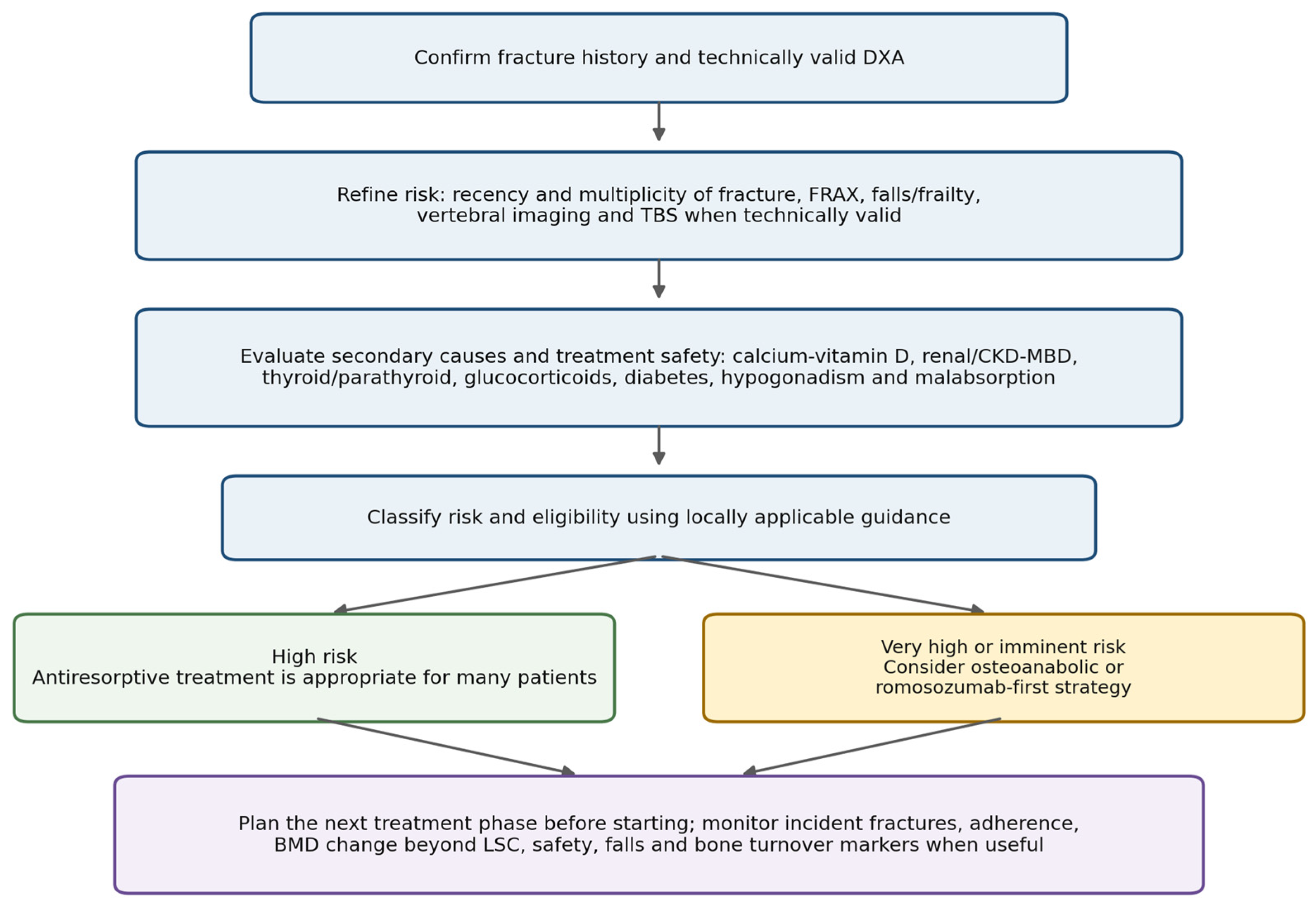
Figure 1 condenses the review into a layered clinical pathway: establish a valid skeletal phenotype, refine the urgency of fracture risk, identify secondary causes and safety constraints, apply a locally recognized risk category, then select treatment only after planning its continuation. The certainty of evidence differs across these steps. Fracture-endpoint randomized trials anchor statements about drug efficacy; BMD and bone-turnover outcomes inform several transition strategies; observational evidence contributes to care-delivery and uncommon-population questions; and expert consensus remains necessary for denosumab exit protocols, selected very-high-risk definitions, and several special populations. These categories should not be treated as evidentially interchangeable.
Figure 1.
Proposed clinical pathway from DXA and fracture history to risk refinement, locally applicable treatment selection, sequencing, and monitoring. BMD, bone mineral density; CKD-MBD, chronic kidney disease-mineral and bone disorder; DXA, dual-energy X-ray absorptiometry; LSC, least significant change; TBS, trabecular bone score. Blue boxes represent sequential assessment steps; green denotes high risk; amber denotes very high or imminent risk; and purple denotes treatment continuity and follow-up.
The contribution of endocrinology is particularly relevant because osteoporosis often represents the final skeletal expression of several interacting mechanisms. A postmenopausal woman with TSH suppression, diabetes, CKD, or glucocorticoid exposure may have the same T-score as a patient with uncomplicated postmenopausal osteoporosis, yet the diagnostic priorities, safety considerations, and follow-up can differ substantially. The value of going beyond the T-score is therefore not the addition of more tests for every patient; it is the use of the right additional information when it can change management.
Goal-directed treatment provides a useful conceptual extension of this reasoning. The desired clinical outcome is fracture prevention, while BMD targets, absence of new fractures, adherence, and control of modifiable risk factors can help determine whether treatment is moving in the intended direction [8]. The evidence for specific targets is still developing and remains strongest in postmenopausal women, so the approach should not be applied rigidly to every population.
7.3. Limitations
This review is a targeted narrative synthesis and has several limitations. It was not preregistered, only PubMed/MEDLINE was searched directly, duplicate independent screening was not performed, and no formal risk-of-bias assessment, GRADE certainty rating, or quantitative pooling was undertaken; publication and selection bias are therefore possible. Borderline citations were resolved by author-team consensus rather than by formal third-reviewer adjudication. Reference-list checking was used to recover landmark fracture-endpoint trials, but the strategy was not intended to reproduce a systematic review. The English-language restriction may also have excluded relevant national guidance. The exact final-update search strings and selection framework are supplied to improve transparency and reproducibility, but they do not convert the work into a systematic review. The breadth of the topic required selective use of guidelines, consensus documents, major reviews, regulatory information, and pivotal trials rather than exhaustive coverage of every therapy, rare secondary cause, or national guideline. Definitions of high, very high, and imminent fracture risk are not harmonized, and FRAX calibration, intervention thresholds, drug licensing, contraindications, reimbursement, and access vary among healthcare systems. The diagnostic meaning of T-scores also differs by age and sex, so recommendations derived from postmenopausal populations cannot be transferred uncritically to younger adults. Evidence supporting drug sequencing is substantially stronger for postmenopausal women than for men, younger adults, pregnancy-related osteoporosis, and advanced CKD, and several transition studies use BMD rather than fracture outcomes as the principal endpoint. Denosumab exit strategies continue to evolve, and no single post-denosumab regimen is universally effective. Evidence for TBS as an adjunct to risk assessment is stronger than evidence for its serial use, while goal-directed BMD targets remain an evolving framework rather than a universally validated treat-to-target standard. Finally, this review addresses adult endocrine osteoporosis care and does not cover pediatric osteoporosis, oncology-specific bone loss, transplant bone disease, or rare monogenic skeletal disorders in depth. These limitations should constrain, rather than invalidate, clinical application of the synthesis.
7.4. Conclusions
Modern adult osteoporosis care requires both valid densitometry and clinical risk assessment. The T-score remains central in postmenopausal women and men aged 50 years or older, while younger adults require age-appropriate densitometric interpretation and greater emphasis on fracture context and secondary causes [4]. Across populations, fracture recency, multiplicity, falls, frailty, comorbidity, and treatment history can alter the urgency and feasibility of intervention. Antiresorptives remain appropriate for many high-risk patients; selected very-high-risk patients may benefit from an osteoanabolic or romosozumab-first strategy followed by antiresorptive therapy, but the evidence is strongest in postmenopausal women [6,8,13,15,16,27,32,33,34,35,38,40,41]. Treatment examples in this review are illustrative; local guidance, licensing, contraindications, and reimbursement determine eligibility. Treatment continuity should be planned from the outset, particularly with denosumab, and advanced CKD requires specific attention to CKD-MBD, mineral metabolism, and hypocalcemia prevention [28,29,30,31,39,44]. For endocrinologists, going beyond the T-score means using additional information only when it can improve diagnosis, safety, treatment intensity, or continuity of fracture prevention.
Supplementary Materials
The following supporting information can be downloaded at: https://www.mdpi.com/article/10.3390/jcm15187258/s1, Table S1: Reproducible PubMed/MEDLINE search strategies, eligibility criteria, selection process, and prioritized guideline and consensus documents.
Author Contributions
Writing—original draft preparation: L.M.G. (Section 1), A.-D.I. (Section 5), E.C.P.T. (Section 2), B.A.T.H. (Section 3), I.A.R. (Section 6), L.M.L.P. (Section 4), and N.A.M.P. (Section 7); writing—review and editing, all authors. All authors have read and agreed to the published version of the manuscript.
Funding
This APC was funded by the University of Oradea. No grant number was assigned.
Institutional Review Board Statement
Not applicable. This article is a narrative review and does not report research involving human participants or animals.
Informed Consent Statement
Not applicable.
Data Availability Statement
No new data were created or analyzed in this study. Data sharing is not applicable to this article.
Conflicts of Interest
The authors declare no conflicts of interest.
References
- Morin, S.N.; Leslie, W.D.; Schousboe, J.T. Osteoporosis: A Review. JAMA 2025, 334, 894–907. [Google Scholar] [CrossRef] [Scilit]
- Ye, C.; Ebeling, P.R.; Kline, G.A. Osteoporosis. Lancet 2025, 406, 2003–2016, Correction in Lancet 2025, 406, 1952. [Google Scholar] [CrossRef] [Scilit]
- Paskins, Z.; Bullock, L.; Hawarden, A.; Blackman, F.; Armstrong, D.J.; Bentley, I.; Clark, E.M.; Daly, R.M.; David, D.; Farr, V.; et al. Time to reframe osteoporosis: A position statement to characterize the osteoporosis care gap. JBMR Plus 2026, 10, ziag006, Correction in JBMR Plus 2026, 10, ziag128. [Google Scholar] [CrossRef] [Scilit]
- International Society for Clinical Densitometry. Official Adult Positions 2023. Available online: https://iscd.org/official-positions-2023/ (accessed on 28 August 2026).
- LeBoff, M.S.; Greenspan, S.L.; Insogna, K.L.; Lewiecki, E.M.; Saag, K.G.; Singer, A.J.; Siris, E.S. The clinician’s guide to prevention and treatment of osteoporosis. Osteoporos. Int. 2022, 33, 2049–2102. [Google Scholar] [CrossRef] [Scilit]
- Gregson, C.L.; Armstrong, D.J.; Avgerinou, C.; Bowden, J.; Cooper, C.; Douglas, L.; Edwards, J.; Gittoes, N.J.L.; Harvey, N.C.; Kanis, J.A.; et al. The 2024 UK clinical guideline for the prevention and treatment of osteoporosis. Arch. Osteoporos. 2025, 20, 119. [Google Scholar] [CrossRef] [Scilit]
- US Preventive Services Task Force; Nicholson, W.K.; Silverstein, M.; Wong, J.B.; Chelmow, D.; Coker, T.R.; Davis, E.M.; Jaén, C.R.; Krousel-Wood, M.; Lee, S.; et al. Screening for Osteoporosis to Prevent Fractures: US Preventive Services Task Force Recommendation Statement. JAMA 2025, 333, 498–508. [Google Scholar] [CrossRef] [Scilit]
- Cosman, F.; Lewiecki, E.M.; Eastell, R.; Ebeling, P.R.; Jan De Beur, S.; Langdahl, B.; Rhee, Y.; El-Hajj Fuleihan, G.; Kiel, D.P.; Schousboe, J.T.; et al. Goal-directed osteoporosis treatment: ASBMR/BHOF task force position statement 2024. J. Bone Miner. Res. 2024, 39, 1393–1405. [Google Scholar] [CrossRef] [Scilit]
- Goel, H.; Binkley, N.; Boggild, M.; Chan, W.P.; Leslie, W.D.; McCloskey, E.; Morgan, S.L.; Silva, B.C.; Cheung, A.M. Clinical Use of Trabecular Bone Score: The 2023 ISCD Official Positions. J. Clin. Densitom. 2024, 27, 101452. [Google Scholar] [CrossRef] [Scilit]
- Li, N.; Hiligsmann, M.; Boonen, A.; van Oostwaard, M.M.; de Bot, R.T.A.L.; Wyers, C.E.; Bours, S.P.G.; van den Bergh, J.P. The impact of fracture liaison services on subsequent fractures and mortality: A systematic literature review and meta-analysis. Osteoporos. Int. 2021, 32, 1517–1530. [Google Scholar] [CrossRef] [Scilit]
- Kanis, J.A.; Harvey, N.C.; McCloskey, E.; Bruyère, O.; Veronese, N.; Lorentzon, M.; Cooper, C.; Rizzoli, R.; Adib, G.; Al-Daghri, N.; et al. Algorithm for the management of patients at low, high and very high risk of osteoporotic fractures. Osteoporos. Int. 2020, 31, 1–12, Correction in Osteoporos. Int. 2020, 31, 797–798. [Google Scholar] [CrossRef] [Scilit]
- Shoback, D.; Rosen, C.J.; Black, D.M.; Cheung, A.M.; Murad, M.H.; Eastell, R. Pharmacological Management of Osteoporosis in Postmenopausal Women: An Endocrine Society Guideline Update. J. Clin. Endocrinol. Metab. 2020, 105, 587–594. [Google Scholar] [CrossRef] [Scilit]
- Sølling, A.S.; Langdahl, B.L.; Cosman, F. Recent Advances in Osteoporosis Therapeutics. Annu. Rev. Med. 2026, 77, 433–448. [Google Scholar] [CrossRef] [Scilit]
- Salman, S.G.; Phadke, R.; Burnett, J.; Walsh, J. Sequential Versus Step-Therapy Approaches for Osteoporosis Management in Orthopedic Subspecialties. Curr. Osteoporos. Rep. 2026, 24, 19. [Google Scholar] [CrossRef] [Scilit]
- Camacho, P.M.; Petak, S.M.; Binkley, N.; Diab, D.L.; Eldeiry, L.S.; Farooki, A.; Harris, S.T.; Hurley, D.L.; Kelly, J.; Lewiecki, E.M.; et al. American Association of Clinical Endocrinologists/American College of Endocrinology Clinical Practice Guidelines for the Diagnosis and Treatment of Postmenopausal Osteoporosis-2020 Update. Endocr. Pract. 2020, 26, 564–570. [Google Scholar] [CrossRef] [Scilit]
- Curtis, E.M.; Reginster, J.Y.; Al-Daghri, N.; Biver, E.; Brandi, M.L.; Cavalier, E.; Hadji, P.; Halbout, P.; Harvey, N.C.; Hiligsmann, M.; et al. Management of patients at very high risk of osteoporotic fractures through sequential treatments. Aging Clin. Exp. Res. 2022, 34, 695–714. [Google Scholar] [CrossRef] [Scilit]
- Gani, L.U.; Sritara, C.; Blank, R.D.; Chen, W.; Gilmour, J.; Dhaliwal, R.; Gill, R. Follow-up Bone Mineral Density Testing: 2023 Official Positions of the International Society for Clinical Densitometry. J. Clin. Densitom. 2024, 27, 101440. [Google Scholar] [CrossRef] [Scilit]
- Brooke-Wavell, K.; Skelton, D.A.; Barker, K.L.; Clark, E.M.; De Biase, S.; Arnold, S.; Paskins, Z.; Robinson, K.R.; Lewis, R.M.; Tobias, J.H.; et al. Strong, steady and straight: UK consensus statement on physical activity and exercise for osteoporosis. Br. J. Sports Med. 2022, 56, 837–846. [Google Scholar] [CrossRef] [Scilit]
- Hofbauer, L.C.; Busse, B.; Eastell, R.; Ferrari, S.; Frost, M.; Müller, R.; Burden, A.M.; Rivadeneira, F.; Napoli, N.; Rauner, M. Bone fragility in diabetes: Novel concepts and clinical implications. Lancet Diabetes Endocrinol. 2022, 10, 207–220. [Google Scholar] [CrossRef] [Scilit]
- Humphrey, M.B.; Russell, L.; Danila, M.I.; Fink, H.A.; Guyatt, G.; Cannon, M.; Caplan, L.; Gore, S.; Grossman, J.; Hansen, K.E.; et al. 2022 American College of Rheumatology Guideline for the Prevention and Treatment of Glucocorticoid-Induced Osteoporosis. Arthritis Care Res. 2023, 75, 2405–2419. [Google Scholar] [CrossRef] [Scilit]
- Bilezikian, J.P.; Khan, A.A.; Silverberg, S.J.; El-Hajj Fuleihan, G.; Marcocci, C.; Minisola, S.; Perrier, N.; Sitges-Serra, A.; Thakker, R.V.; Guyatt, G.; et al. Evaluation and Management of Primary Hyperparathyroidism: Summary Statement and Guidelines from the Fifth International Workshop. J. Bone Miner. Res. 2022, 37, 2293–2314. [Google Scholar] [CrossRef] [Scilit]
- Ku, E.J.; Yoo, W.S.; Lee, E.K.; Ahn, H.Y.; Woo, S.H.; Hong, J.H.; Chung, H.K.; Park, J.W. Effect of Thyroid-Stimulating Hormone Suppression Therapy on Bone Mineral Density in Differentiated Thyroid Cancer: A Systematic Review and Meta-Analysis. J. Clin. Endocrinol. Metab. 2021, 106, 3655–3667. [Google Scholar] [CrossRef] [Scilit]
- Fuggle, N.R.; Beaudart, C.; Bruyère, O.; Abrahamsen, B.; Al-Daghri, N.; Burlet, N.; Chandran, M.; Rosa, M.M.; Cortet, B.; Demonceau, C.; et al. Evidence-Based Guideline for the management of osteoporosis in men. Nat. Rev. Rheumatol. 2024, 20, 241–251. [Google Scholar] [CrossRef] [Scilit]
- DailyMed. FOSAMAX (Alendronate Sodium): U.S. Prescribing Information. Available online: https://dailymed.nlm.nih.gov/dailymed/fda/fdaDrugXsl.cfm?setid=14e931fd-2c5f-4d90-b7db-5980706f4a56 (accessed on 15 September 2026).
- DailyMed. ACTONEL (Risedronate Sodium): U.S. Prescribing Information. Available online: https://dailymed.nlm.nih.gov/dailymed/drugInfo.cfm?setid=4423d70a-b120-4653-bcf3-0c5ea8b97b0e (accessed on 15 September 2026).
- DailyMed. RECLAST (Zoledronic Acid) Injection: U.S. Prescribing Information. Available online: https://dailymed.nlm.nih.gov/dailymed/drugInfo.cfm?setid=3c79ff9c-a6f4-405d-b19c-7e473a61dedc (accessed on 15 September 2026).
- Cummings, S.R.; San Martin, J.; McClung, M.R.; Siris, E.S.; Eastell, R.; Reid, I.R.; Delmas, P.; Zoog, H.B.; Austin, M.; Wang, A.; et al. Denosumab for Prevention of Fractures in Postmenopausal Women with Osteoporosis. N. Engl. J. Med. 2009, 361, 756–765, Correction in N. Engl. J. Med. 2009, 361, 1914. [Google Scholar] [CrossRef] [Scilit]
- Cummings, S.R.; Ferrari, S.; Eastell, R.; Gilchrist, N.; Jensen, J.E.B.; McClung, M.; Roux, C.; Torring, O.; Valter, I.; Wang, A.T.; et al. Vertebral Fractures After Discontinuation of Denosumab: A Post Hoc Analysis of the Randomized Placebo-Controlled FREEDOM Trial and Its Extension. J. Bone Miner. Res. 2018, 33, 190–198. [Google Scholar] [CrossRef] [Scilit]
- Degboe, Y.; Couture, G. Strategies for denosumab discontinuation in postmenopausal osteoporosis. Jt. Bone Spine 2026, 93, 105954. [Google Scholar] [CrossRef] [Scilit]
- Bird, S.T.; Gelperin, K.; Smith, E.R.; Jung, T.H.; Lyu, H.; Thompson, A.; Easley, O.; Naik, K.B.; Zhao, Y.; Kambhampati, R.; et al. The Effect of Denosumab on Risk for Emergently Treated Hypocalcemia by Stage of Chronic Kidney Disease: A Target Trial Emulation. Ann. Intern. Med. 2025, 178, 29–38. [Google Scholar] [CrossRef] [Scilit]
- DailyMed. PROLIA (Denosumab) Injection: U.S. Prescribing Information. Available online: https://dailymed.nlm.nih.gov/dailymed/drugInfo.cfm?setid=49e5afe9-a0c7-40c4-af9f-f287a80c5c88 (accessed on 15 September 2026).
- Kendler, D.L.; Marin, F.; Zerbini, C.A.F.; Russo, L.A.; Greenspan, S.L.; Zikan, V.; Bagur, A.; Malouf-Sierra, J.; Lakatos, P.; Fahrleitner-Pammer, A.; et al. Effects of teriparatide and risedronate on new fractures in post-menopausal women with severe osteoporosis (VERO): A multicentre, double-blind, double-dummy, randomised controlled trial. Lancet 2018, 391, 230–240, Correction in Lancet 2018, 391, 204; Correction in Lancet 2018, 392, 2352. [Google Scholar] [CrossRef] [Scilit]
- Miller, P.D.; Hattersley, G.; Riis, B.J.; Williams, G.C.; Lau, E.; Russo, L.A.; Alexandersen, P.; Zerbini, C.A.F.; Hu, M.Y.; Harris, A.G.; et al. Effect of Abaloparatide vs. Placebo on New Vertebral Fractures in Postmenopausal Women with Osteoporosis: A Randomized Clinical Trial. JAMA 2016, 316, 722–733, Correction in JAMA 2017, 317, 442. [Google Scholar] [CrossRef] [Scilit]
- Saag, K.G.; Petersen, J.; Brandi, M.L.; Karaplis, A.C.; Lorentzon, M.; Thomas, T.; Maddox, J.; Fan, M.; Meisner, P.D.; Grauer, A. Romosozumab or Alendronate for Fracture Prevention in Women with Osteoporosis. N. Engl. J. Med. 2017, 377, 1417–1427. [Google Scholar] [CrossRef] [Scilit]
- Cosman, F.; Crittenden, D.B.; Adachi, J.D.; Binkley, N.; Czerwinski, E.; Ferrari, S.; Hofbauer, L.C.; Lau, E.; Lewiecki, E.M.; Miyauchi, A.; et al. Romosozumab Treatment in Postmenopausal Women with Osteoporosis. N. Engl. J. Med. 2016, 375, 1532–1543. [Google Scholar] [CrossRef] [Scilit]
- DailyMed. EVENITY (Romosozumab-Aqqg) Injection: U.S. Prescribing Information. Available online: https://dailymed.nlm.nih.gov/dailymed/drugInfo.cfm?setid=471baba2-7154-4488-9891-0db2f46791e7 (accessed on 15 September 2026).
- European Medicines Agency. Evenity (Romosozumab): EPAR Product Information. Available online: https://www.ema.europa.eu/en/documents/product-information/evenity-epar-product-information_en.pdf (accessed on 15 September 2026).
- Li, T.; Zheng, W.; Zhang, Q.; Liao, M.; Jin, L.; Wu, F.; Zhou, F.; Wang, Y.; Gong, W.; Xia, Y.; et al. The efficacy and safety of romosozumab sequential therapy in postmenopausal women: A systematic review and meta-analysis. J. Orthop. 2026, 72, 152–159. [Google Scholar] [CrossRef] [Scilit]
- Tsourdi, E.; Zillikens, M.C.; Meier, C.; Body, J.J.; Gonzalez Rodriguez, E.; Anastasilakis, A.D.; Abrahamsen, B.; McCloskey, E.; Hofbauer, L.C.; Guañabens, N.; et al. Fracture Risk and Management of Discontinuation of Denosumab Therapy: A Systematic Review and Position Statement by ECTS. J. Clin. Endocrinol. Metab. 2021, 106, 264–281. [Google Scholar] [CrossRef] [Scilit]
- Black, D.M.; Cummings, S.R.; Karpf, D.B.; Cauley, J.A.; Thompson, D.E.; Nevitt, M.C.; Bauer, D.C.; Genant, H.K.; Haskell, W.L.; Marcus, R.; et al. Randomised trial of effect of alendronate on risk of fracture in women with existing vertebral fractures. Lancet 1996, 348, 1535–1541. [Google Scholar] [CrossRef] [Scilit]
- Black, D.M.; Delmas, P.D.; Eastell, R.; Reid, I.R.; Boonen, S.; Cauley, J.A.; Cosman, F.; Lakatos, P.; Leung, P.C.; Man, Z.; et al. Once-Yearly Zoledronic Acid for Treatment of Postmenopausal Osteoporosis. N. Engl. J. Med. 2007, 356, 1809–1822. [Google Scholar] [CrossRef] [Scilit]
- Leder, B.Z.; Tsai, J.N.; Uihlein, A.V.; Wallace, P.M.; Lee, H.; Neer, R.M.; Burnett-Bowie, S.A.M. Denosumab and teriparatide transitions in postmenopausal osteoporosis (the DATA-Switch study): Extension of a randomised controlled trial. Lancet 2015, 386, 1147–1155. [Google Scholar] [CrossRef] [Scilit]
- Bhattoa, H.P.; Vasikaran, S.; Trifonidi, I.; Kapoula, G.; Lombardi, G.; Jørgensen, N.R.; Pikner, R.; Miura, M.; Chapurlat, R.; Hiligsmann, M.; et al. Update on the role of bone turnover markers in the diagnosis and management of osteoporosis: A consensus paper from ESCEO, IOF, and IFCC. Osteoporos. Int. 2025, 36, 579–608. [Google Scholar] [CrossRef] [Scilit]
- Evenepoel, P.; Cunningham, J.; Ferrari, S.; Haarhaus, M.; Javaid, M.K.; Lafage-Proust, M.H.; Prieto-Alhambra, D.; Ureña Torres, P.; Cannata-Andia, J.; Vervloet, M.; et al. European Consensus Statement on the diagnosis and management of osteoporosis in chronic kidney disease stages G4-G5D. Nephrol. Dial. Transplant. 2021, 36, 42–59. [Google Scholar] [CrossRef] [Scilit]
- Ünal, M.; Saçıntı, K.G.; Sezgin, E.A. Pregnancy- and lactation-related bone fragility: The hidden risk. Jt. Dis. Relat. Surg. 2025, 36, 210–213. [Google Scholar] [CrossRef] [Scilit]
- Cohen, A. Bone Metabolism, Bone Mass, and Bone Structure During Pregnancy and Lactation: Normal Physiology and Pregnancy and Lactation-Associated Osteoporosis. Endocrinol. Metab. Clin. N. Am. 2024, 53, 453–470. [Google Scholar] [CrossRef] [Scilit]
- Gak, N.; Abbara, A.; Dhillo, W.S.; Keen, R.; Comninos, A.N. Current and future perspectives on pregnancy and lactation-associated osteoporosis. Front. Endocrinol. 2024, 15, 1494965. [Google Scholar] [CrossRef] [Scilit]

Disclaimer/Publisher’s Note: The statements, opinions and data contained in all publications are solely those of the individual author(s) and contributor(s) and not of MDPI and/or the editor(s). MDPI and/or the editor(s) disclaim responsibility for any injury to people or property resulting from any ideas, methods, instructions or products referred to in the content. |
© 2026 by the authors. Licensee MDPI, Basel, Switzerland. This article is an open access article distributed under the terms and conditions of the Creative Commons Attribution (CC BY) license.