


Abstract
Heart failure (HF) is a multifactorial and heterogeneous syndrome with substantial epidemiological burden, high mortality, and impact on quality of life. In the context of heart failure, left ventricular ejection fraction (LVEF) has been regarded as the most important marker of systolic function and is fundamental in medical research and clinical practice. In research, LVEF has been a major inclusion criterion in most clinical trials over the past few decades. Furthermore, international heart failure guidelines rely on LVEF for the diagnosis of HF and to guide effective treatment. Additionally, our understanding of HF phenotypes and prognosis is mostly grounded in a classification based on LVEF. Nevertheless, there has been a growing debate regarding the role of LVEF in heart failure. In this context, the purpose of this review is to discuss both the advantages and contemporary relevance of LVEF in heart failure, as well as its limitations and controversies. In addition, this review aims to discuss potential alternatives and future directions in heart failure classification, such as new classification methods, alternative measurements of systolic function and imaging techniques, the HLM score, and the use of artificial intelligence and machine learning.
1. Introduction
According to the Universal Definition and Classification of Heart Failure [1], a consensus document from several Heart Failure (HF) societies, HF is defined as “a clinical syndrome with symptoms and/or signs caused by a structural and/or functional cardiac abnormality and corroborated by elevated natriuretic peptide levels and/or objective evidence of pulmonary or systemic congestion”. Heart failure is a complex, progressive, multifactorial, and heterogeneous syndrome, known for its high morbidity and mortality, as well as deleterious impact on the quality of life [2,3].
Heart failure (HF) is often considered an epidemic in this century [4] with an estimated prevalence of 1–2% in the adult population [5].
Due to improvements in the treatment of HF and associated comorbidities, as well as the global ageing of the population, there has been an increase in the prevalence of heart failure [3]. On the other hand, incidence increases significantly with age and seems to have stabilised over the past years, with a tendency to decrease in industrialised countries [3]. Despite improvements in the management of this disease and comorbidities, HF still has a high mortality rate and is one of the leading causes of hospital admissions, particularly among older patients [3]. As previously stated, HF represents a substantial economic burden on healthcare systems worldwide, and the associated costs are estimated to increase in the upcoming years [3,5].
LVEF is regarded as the most important marker of cardiac function, being a cornerstone in the context of heart failure research and medical practice. In the context of research, it has been an inclusion criterion in all major clinical trials in recent decades [6,7,8]. The diagnosis, phenotyping, follow-up, prognosis, and treatment are also based on the LVEF measurement according to international heart failure guidelines [9,10,11].
Nevertheless, over the past few years, there has been a growing debate about the use of LVEF in heart failure classification. While some authors emphasise its limitations as the primary phenotyping parameter in HF and call for its urgent replacement [2,12,13]. Other authors highlight all the knowledge that was produced based on LVEF and the lack of a current consensual alternative [6,8].
In this context, this review aims to discuss the advantages and importance of LVEF in heart failure, its limitations, and discuss possible alternatives and future perspectives.
A literature search was conducted between August 2025 and March 2026 in two electronic bibliographic databases (PubMed/MEDLINE and Web of Science). The initial search terms included (but were not restricted to) “heart failure”, “ejection fraction”, “left ventricular ejection fraction”, “classification”, and “limitations”. Original research articles, international guidelines, expert consensus documents, narrative reviews, and editorials were reviewed. Furthermore, references from relevant articles were manually screened for additional pertinent articles.
2. The Origin and Definition of Left Ventricular Ejection Fraction (LVEF) in Heart Failure (HF)
In 1962, Folse and Braunwal used a radioisotopic technique to determine the “fraction of left ventricular end-diastolic volume ejected per beat” and demonstrated that it could provide valuable haemodynamic information regarding the left ventricular function [14]. Then, in 1966, Stuart Bartle, a future psychiatrist [7], introduced the term “ejection fraction” to describe the ratio of stroke volume (SV) to left ventricular end-diastolic volume (LVEDV) [15]. This paved the way for the subsequent use of left ventricular ejection fraction (LVEF) in research and clinical practice [16].
The LVEF is expressed in percentage and is defined as the ratio of the stroke volume (the difference between end-diastolic and end-systolic volumes) and the end-diastolic volume (EDV) [17]. LVEF is usually calculated using two-dimensional (2D) echocardiography, although other imaging techniques can be used, such as three-dimensional (3D) echocardiography, cardiac magnetic resonance (CMR), computed tomography (CT), and single-photon emission computed tomography (SPECT) [18]. This parameter varies with age and sex (with women having a normal range higher than men) [19]. Currently, there is no consensus regarding the normal reference values [20,21]. For example, the recommendations from the American Society of Echocardiography and the European Association of Cardiovascular Imaging consider normal values above 52% in men and 54% in women [22], while European and American guidelines consider a normal value at the cutoff of 50% or above, regardless of sex [9,10,11]. It is important to note that the values reported refer to measurements obtained by two-dimensional echocardiography and that the reference values differ across imaging techniques [23].
3. Current Relevance and Strengths of Left Ventricular Ejection Fraction (LVEF) in Heart Failure (HF)
In 1992, the SOLVD trial investigators [24] were the first to include LVEF as an inclusion criterion for enrolment in a clinical trial [19,25]. Afterwards, all major clinical trials used LVEF as an inclusion criterion, although the cutoff used for enrolment varied substantially [7,19,25].
Some of the main clinical trials that used LVEF as an inclusion criterion, along with their principal results, are summarised in Figure 1 and Table 1.
Figure 1.
Main clinical trials that used LVEF as an inclusion criterion in the last few decades. SOLVD: Studies of Left Ventricular Dysfunction; DIG: Digitalis Investigation Group; MERIT-HF: Metoprolol CR/XL Randomised Intervention Trial in Congestive Heart Failure; RALES: Randomized Aldactone Evaluation Study; CIBIS-II: Cardiac Insufficiency Bisoprolol Study II; COPERNICUS: Carvedilol Prospective Randomized Cumulative Survival; CHARM: Candesartan in Heart Failure Assessment of Reduction in Mortality and Morbidity; PEP-CHF: Perindopril for Elderly People with Chronic Heart Failure; I-PRESERVE: Irbesartan in Heart Failure with Preserved Ejection Fraction Study; DIG-PEF: Digitalis Investigation Group Preserved Ejection Fraction; SHIFT: Systolic Heart Failure Treatment with the If Inhibitor Ivabradine Trial; EMPHASIS-HF: Eplerenone in Mild Patients Hospitalization and Survival Study in Heart Failure; PARADIGM-HF: Prospective Comparison of ARNI with ACEI to Determine Impact on Global Mortality and Morbidity in Heart Failure; TOPCAT: Treatment of Preserved Cardiac Function Heart Failure with an Aldosterone Antagonist; DAPA-HF: Dapagliflozin and Prevention of Adverse Outcomes in Heart Failure; PARAGON-HF: Prospective Comparison of ARNI with ARB Global Outcomes in HF with Preserved Ejection Fraction; EMPEROR: Empagliflozin Outcome Trial in Patients with Chronic Heart Failure; VICTORIA: Vericiguat Global Study in Subjects with Heart Failure with Reduced Ejection Fraction; GALACTIC-HF: Global Approach to Lowering Adverse Cardiac Outcomes through Improving Contractility in Heart Failure; DELIVER: Dapagliflozin Evaluation to Improve the Lives of Patients with Preserved Ejection Fraction Heart Failure; FINEARTS-HF: Finerenone Trial to Investigate Efficacy and Safety Superior to Placebo in Patients with Heart Failure; Adapted and modified from Dimond et al. (2024) [19].
Table 1.
Effect of pharmacological treatment on cardiovascular mortality or HF hospitalisation across the LVEF spectrum.
The introduction of the terms “preserved EF” and “reduced EF” can be traced back to the CHARM (Candesartan in Heart Failure Assessment of Reduction in Mortality and Morbidity) trial in 2003 [12]. In this clinical trial, researchers studied the effects of candesartan in three subgroups: patients with a LVEF > 40% (with preserved EF) and two different subgroups of patients with reduced EF, LVEF ≤ 40% (patients not receiving angiotensin-converting-enzyme inhibitors because of previous intolerance or who were currently receiving angiotensin-converting-enzyme inhibitors) [27]. This nomenclature was later adopted [28] by the 2005 American College of Cardiology/American Heart Association HF Guidelines [29].
The 2016 European Society of Cardiology (ESC) Guidelines proposed a third subgroup, HF with mid-range EF (HFmrEF), when the LVEF is between 40% and 49%, to cover the “grey area” between HFrEF and HFpEF [30]. The purpose of creating this new subgroup was to encourage further research in a population that was often neglected in clinical trials and research [12,30]. Subsequently, in the 2021 ESC Guidelines, this subgroup was renamed HF with mildly reduced EF (keeping the abbreviation) and defined as HF with a LVEF between 41% and 49% [9].
Currently, international guidelines and consensus statements classify HF in three main subtypes: HF with reduced EF (HFrEF) when the LVEF is ≤40%; HF with mildly reduced EF (HFmrEF) when the LVEF is between 41% and 49%, and HF with preserved EF (HFpEF) when the LVEF is ≥50% [1,9,10,11].
More recently, a fourth type of HF has been proposed: HF with improved EF (HFimpEF), although there have been inconsistencies in its nomenclature and definition [1,10,31]. Some authors have also advocated for a fifth type of HF: HF with supranormal EF (HFsnEF) when the LVEF ≥ 65% [32] since some studies reported a worse prognosis in these patients when compared to patients with HPpEF and lower EF values [32,33,34]. For example, in a large clinical cohort of 203,135 patients, the adjusted hazard ratio (HR) for mortality was 1.71 [95% CI 1.64–1.77] when ≥70% [32].
These five subtypes appear to have different phenotypes, responses to treatment, and prognosis [35].
Current heart failure guidelines rely on LVEF not only to diagnose and classify HF, but also to guide treatment and management [9,10,11].
For all these reasons, LVEF has become the central marker of left ventricular cardiac function, with ubiquitous use in heart failure and playing a crucial role in research and clinical practice [4,28,36]. Although it is beyond the scope of this review, LVEF also plays a pivotal role in other areas of cardiology, such as valvular heart disease, myocardial infarction, selection for implantable cardioverter defibrillators, ventricular arrhythmias, and the evaluation of the cardiotoxicity of chemotherapeutic agents [18,23,36,37].
Furthermore, the measurement of LVEF also has several additional strengths that warrant discussion.
A key advantage of LVEF is its universal recognition and acceptance, not only in the field of cardiology but also across the wider medical community [38].
Additional advantages stem from the fact that LVEF is typically calculated using two-dimensional (2D) echocardiography [18]. This imaging modality is widely available, simple to perform, non-invasive, does not use radiation, is inexpensive, and can be rapidly obtained [4,6,39,40]. Furthermore, with the rise of portable echocardiography devices, it is—now more than ever—an evaluation that can be performed at the patient’s bedside [38]. In addition to the LVEF quantification, the use of two-dimensional (2D) echocardiography also provides complementary information regarding the structure and function of the heart [6]. Although normal ranges may vary slightly between sexes, LVEF is independent of other factors such as body weight, height, and race [39,40].
Another advantage of LVEF is that it is considered a good indicator of left ventricular remodelling and eccentric hypertrophy in HFrEF [37]. When there is destruction of cardiomyocytes, the heart undergoes a compensatory mechanism of ventricular remodelling that ultimately leads to the enlargement of the left ventricle in a process called eccentric hypertrophy [12,37]. Despite being maladaptive, this remodelling and enlargement allow an increase in LV capacitance and an increase in the LV end-diastolic volume (LVEDV) [37]. This mechanism enables the maintenance of the stroke volume (SV), even though there is a decrease in myocardial contraction [12]. Since LVEF is calculated as SV divided by end-diastolic volume (EDV) [17], an increase in EDV, with maintenance of SV, results in a decrease in the LVEF, as these variables are inversely related [41]. In these cases, such as in HFrEF, the increase in LVEDV mirrors the decline in systolic function and is measurable as a reduction in LVEF [41]. For this reason, it is considered that the reduction in LVEF is mainly determined by LV geometry and architecture [42].
Additionally, LVEF has been widely used to classify heart failure patients according to phenotypes with different aetiologies and clinical characteristics [6,43,44]. For example, an analysis of the European Society of Cardiology Heart Failure Long-Term Registry (a prospective cohort of 9134 HF patients) [44] and a study based on the Swedish Heart Failure Registry (an ongoing nationwide registry), including 42061 HF patients [45], yielded similar results regarding the clinical characteristics according to LVEF. In both studies, HFrEF patients were younger, more frequently male, and more likely to have an ischaemic aetiology. Conversely, HFpEF patients were older, more frequently female, and more likely to have hypertension, diabetes, or atrial fibrillation [44,45].
Moreover, LVEF has been extensively used for its prognostic value, particularly at lower LVEF values [18,37]. Patients with lower LVEF values are at increased risk of cardiovascular death, hospitalisation, and sudden cardiac death [19]. In a recent study that used the dataset of 33,699 participants from six previous HF randomised controlled trials, the results showed that the incidence of most clinical outcomes increases with a decline in LVEF, with an inflection in the range of 40 to 50%, after which LVEF has less prognostic value [46]. Other studies, focused on the trajectories of LVEF over time, have also demonstrated that a decline in LVEF was associated with higher mortality [47,48].
Finally, guideline-directed medical therapy uses LVEF as a key parameter to distinguish which patients will likely benefit from each approved treatment [9,10,11]. This reflects the fact that the recommendations from guidelines are based on randomised clinical trials that used LVEF as an inclusion criterion over the past decades [28]. Given the methods used in clinical trials, administering therapy to the LVEF phenotypes that showed greater benefit from it is crucial to translate research into everyday clinical practice [8].
For all these reasons, LVEF has become so important and ubiquitous in HF clinical practice and research [4,28,36].
The major strengths of LVEF are summarised in Table 2.
Table 2.
Advantages of left ventricular ejection fraction (LVEF) in heart failure (HF).
4. The Limitations of Left Ventricular Ejection Fraction (LVEF)
Despite all the advantages previously discussed, the use of LVEF has important limitations that deserve discussion, particularly given the ongoing debate about its role in heart failure [2,6,8,12,13].
As previously discussed, the prominence of LVEF largely comes from its use as an inclusion criterion in clinical trials, although cutoffs varied substantially [7,19,25]. However, the definition of LVEF cutoffs in clinical trials was arbitrary and not driven by any physiological distinction [12,49]. In fact, the use of lower LVEF values aimed to improve diagnostic certainty, include patients with worse prognosis, and increase the statistical power of the studies [2,6,8].
Furthermore, LVEF is a continuous variable [19,50] with a normal distribution [9,51]. This unimodal and normal distribution has been shown in recent population-based cohorts [44,52] and previously in one HF clinical trial [53]. In a recent population-based cohort of 9716 new HF patients in Stockholm (between 2005 and 2010), a normal distribution was observed both in the overall study population and in subgroups defined by age and sex [52]. This contrasts with previous studies, mostly using data from hospitalised patients with decompensated HF, that reported a bimodal distribution of LVEF [54,55,56].
Some of the most significant limitations of LVEF are its inter- and intra-observer variability [40,51,52]. It is often considered that this variability can range from 5% to 8% [40,51]. In a recent population-based cohort, it was demonstrated that the standard deviation of the within-person variance in LVEF was 7.4% [52]. Another situation that can lead to bias and affect reproducibility is the fact that the examiner is often unblinded to previous LVEF measurements [52]. Furthermore, there is also considerable inter-modality variability [40,51,57]. In a multicentre randomised trial, aiming to determine the inter-modality variability of LVEF measured by echocardiography, gated single-photon emission computed tomography (SPECT), and cardiovascular magnetic resonance (CMR), in roughly half of the patients, variability exceeded 5%. Bland–Altman analysis demonstrated just a moderate agreement between imaging techniques, with limits of agreement ranging from 28.27% to 35.31%, without significant overestimation or underestimation of LVEF by any modality [57].
Moreover, LVEF follows dynamic trajectories over time [47,48,52]. In a prospective study evaluating LVEF trajectories in HF patients, 2D echocardiography was performed at the beginning of follow-up, after one year, and biennially thereafter, over a 15-year follow-up period. In this study, it was demonstrated that LVEF followed an inverted U-shaped pattern, with a marked increase during the first year, stability for up to a decade, and a gradual decline afterwards (p for trajectory < 0.001). Most patients with HFrEF remained in the same category (56%), although a substantial proportion transitioned to HFmrEF (21%) or HFpEF (23%) over time. Conversely, HFmrEF showed marked instability, with patients broadly distributed across categories at the end of follow-up [47]. In another study that included 4943 patients from the Swedish Heart Failure Registry with ≥2 LVEF measurements, approximately one quarter of the patients with HFrEF and HFmrEF had an improvement in EF, whereas a decline was observed in more than one third of the patients with HFpEF and HFmrEF [48]. In a recent population-based cohort, it was shown that the probability of changing category within the first year after diagnosis was substantial [52]. Of note, less than 25% of the patients with HFmrEF remained in the same category after one year [52]. There are several factors that modulate these bidirectional transitions, such as the aetiology, duration of disease, adherence/response to therapy, and other comorbidities [1,39,47]. Some authors argue that this high temporal variability raises concerns about the suitability of using LVEF to guide treatment recommendations [12,52].
A further challenge in this context is digit bias [25]. Digit bias consists of the tendency to round the value to the nearest multiple of 5 when visually assessing LVEF instead of using the precise calculated value [58]. This bias can cause significant imprecision, particularly given that the range of HFmrEF is just 10% [52,59]. A recent study using the European Society of Cardiology (ESC) HF Long-Term registry showed that digit bias in LVEF assessment was substantial, affecting about 37% of the evaluated population [58].
Due to the inter- and intra-observer variability, the dynamic trajectories over time, and the digit bias previously discussed, many authors challenge the use of HFmrEF as an independent phenotype because of its narrow range, high variability over time, and because it was not defined based on a distinct underlying pathophysiology [12,19,47,52,60].
Another limitation of LVEF is that LV geometry is a confounding factor in its assessment [18,61]. The reason for this is that the calculation of LVEF uses only luminal measurements, not taking into account LV geometry [62,63]. According to mathematical models, LVEF can be maintained in the presence of increased wall thickness or reduced LV diameter (leading to reduced end-diastolic volume) despite reduced myocardial contraction [61]. In cases of hypertrophic cardiomyopathy and HFpEF concentric remodelling, although there is a decrease in stroke volume and contractility, EF may remain normal due to the reduction in the ventricular chamber volume that occurs in this type of remodelling [13,64]. In such cases, LVEF may remain normal despite impaired systolic function and reduced stroke volume [64]. Another condition that can alter LVEF is the physiological adaptation and LV remodelling that is observed in endurance athletes [23,40]. Due to the LV enlargement, these athletes frequently have reduced LVEF at rest despite having a normal stroke volume [23,36]. However, during exercise, LVEF becomes supranormal due to haemodynamic changes to meet increasing metabolic demands [40]. This may represent a diagnostic challenge due to its overlapping characteristics with hypertrophic, dilated, and arrhythmogenic right ventricular cardiomyopathies and may complicate interpretation and diagnosis in athletes with underlying heart disease [65,66].
LVEF is usually calculated using two-dimensional (2D) echocardiography, which has inherent limitations [18]. According to the recommendations, LVEF should be evaluated using the “biplane method of disks”, also referred to as the “Modified Simpson’s rule” [22]. This formula uses the geometric assumption that the LV has an ellipsoid shape and regular conformation [18,40] and estimates 3D volumes based on 2D images [36]. However, LV geometry is often distorted in patients with ischaemic heart disease [18,23]. The 2D echocardiography is also highly influenced by image quality [18,36,67] and the acoustic window [4,18], which may be limited by factors such as obesity, lung disease, chest wall deformities, and breast implants [4]. These limitations may lead to a foreshortened ventricle and affect LV volume measurements and therefore LVEF [18,36]. Another potential source of inaccuracy is the endocardial border delimitation [36,68], which can be influenced by poor image quality [64], operator techniques [39,51], lack of standardisation [23], and the definition of papillary muscles [51], all of which can alter the estimation of LV volumes [39]. Even when using semi-automatic delineation techniques, those challenges remain, and there is also inconsistent standardisation and reproducibility [36].
Another limitation is that LVEF is highly influenced by the preload and afterload [39,51,69]. For example, changes in systemic blood pressure, pulmonary venous pressure, or arterial stiffness can result in variations in LVEF [39,51].
Moreover, LVEF is influenced by the heart rate and ventricular synchrony [4,12,64]. Bradycardia may prolong the diastole and increase LV diastolic volume [39], thereby increasing stroke volume and leading to an overestimation of LVEF [51,64]. Conversely, tachycardia reduces the diastolic filling time and LV diastolic volume [39], resulting in a decreased stroke volume and leading to an underestimation of LVEF [51,64]. In the presence of atrial fibrillation (AF), the irregular R-R intervals can also underestimate LVEF [64]. A beat-to-beat variability of 5.8 ± 1.7% has been reported in AF patients [70]. Owing to this variability, in AF patients, LVEF should be calculated as the average of 15 beats—a procedure seldom performed in everyday practice [12]. Abnormal electrical conduction, as seen in left bundle branch block (LBBB) or in patients with a pacemaker, induces ventricular dyssynchrony that can further impair the reliable estimation of LVEF [4].
Additionally, LVEF is influenced by valvular disease, which may lead to either underestimation or overestimation, depending on the type of valvular defect [4,51]. The presence of mitral regurgitation may lead to an overestimation of LVEF [69]. This occurs due to a reduction in preload caused by the regurgitation volume and leads to an increase in the LVEF [64]. In contrast, the presence of aortic stenosis can underestimate LVEF because it causes an increase in the afterload [64], a delay in outflow time peak, and a subsequent reduction in LVEF [40,51].
The assessment of LVEF can be affected by other systemic diseases such as anaemia, thyroid dysfunction, endocrine diseases, infections, metabolic disorders, and other comorbidities [39,40,51]. It can also be conditioned by medications and stress [39]. Similarly, sympathetic activity or vagal stimulation can influence LVEF evaluation [39,40,51]. Some medical conditions can also cause a transient reduction in LVEF with subsequent recovery, such as myocarditis and stress-induced cardiomyopathy (Takotsubo syndrome) [4].
Although LVEF is widely used as a surrogate of systolic function, it is not an accurate measure of myocardial contractility [12,13,18,41]. Because of its mathematical definition, LVEF is inversely related to EDV, which is highly dependent on LV geometry and remodelling [12,38,40,61]. Therefore, architectural changes that modify LVEDV will have a strong impact on LVEF, independently of cardiac contractility, making it a better indicator for remodelling than for assessing systolic function [13,17,39]. In addition, since LVEF is highly dependent on preload and afterload [39,51,69], a correct interpretation of LVEF would require knowledge of LV loads and volumes [13,62]. Furthermore, LVEF does not include any measurement related to the myocardium [23,41,63] and does not consider longitudinal or torsional contraction [39,40,61], nor regional abnormalities [51]. In summary, LVEF serves as an indicator of global ejection performance based on volumetric changes rather than a direct measure of myocardial contraction [18,69,71].
Furthermore, LVEF is not an early marker of systolic dysfunction [18,67]. The LV myocardium is organised into endocardial and epicardial layers, predominantly composed of longitudinal fibres and a mid-myocardial layer mainly consisting of circumferential fibres [67]. Since subendocardial longitudinal fibres are particularly vulnerable to ischaemia, their dysfunction can be compensated by circumferential fibres, allowing LVEF to remain normal [18,67]. However, this subclinical systolic dysfunction can be detected through other techniques, such as echocardiographic strain [72].
A further limitation frequently highlighted is that EF-based phenotypes fail to acknowledge the complexity and heterogeneity of the heart failure syndrome, especially in HFpEF [73,74]. This type of classification ignores the specific aetiology [38] and the presence of important comorbidities [25], both of which are highly relevant for treatment and prognosis [16,25,38]. Moreover, LVEF does not differentiate between systolic and diastolic dysfunction: diastolic impairment is frequent in HFrEF, and HFpEF commonly has subclinical systolic dysfunction that can be identifiable by other imaging techniques [18]. Therefore, a normal LVEF neither excludes nor confirms diastolic dysfunction [38]. Furthermore, HF with a high LVEF is not a benign condition since some studies report worse outcomes in those patients [32,33] with an adjusted hazard ratio (HR) for mortality of 1.71 [95% CI 1.64–1.77] when ≥70% [32].
Additionally, there are pathophysiological mechanisms that are common in HF throughout the spectrum of LVEF, thereby challenging the traditional LVEF-based categorisation [2,7]. Some of these shared mechanisms include endothelial dysfunction, neurohumoral activation (although to different extents), cardiomyocyte injury, myocardial fibrosis, left atrial dysfunction, and ventricular overload [2,7,75].
Another limitation of LVEF is its poor correlation with the patient’s symptoms [18,38]. Despite having preserved EF, HFpEF patients often experience heart failure symptoms, including fatigue, dyspnoea, and reduced exercise tolerance [39].
Furthermore, LVEF has limited prognostic value [53,76,77]. Data from large clinical trials such as CHARM [53] and DIG [76] consistently show that LVEF is a strong predictor of mortality, but only up to 45%. In the CHARM trial, the risk of all-cause mortality rose by 39% for every 10% decrease in LVEF below 45% (HR 1.39; 95% CI 1.32–1.46); however, beyond this threshold, the risk of mortality remained relatively stable despite further increases in LVEF [53]. Similarly, the DIG trial showed that in patients with LVEF ≤ 45%, mortality declined in an almost linear manner across progressively higher LVEF categories, (LVEF < 15%: 51.7%; LVEF 36% to 45%, 25.6%; p < 0.0001), by contrast, among patients with LVEF > 45%, mortality rates were similar even after multivariate adjustment (LVEF 46% to 55%: HR 0.92, 95% CI 0.77–1.10; LVEF > 55%: HR 0.88, 95% CI 0.71–1.09) [76]. A similar result was derived from a meta-analysis of the MAGGIC group (comprising 30 studies) that showed that a LVEF above 40% was no longer associated with a risk of mortality [77]. In a population-based cohort, the prevalence of advanced HFpEF (43% of patients) was similar to that of advanced HFrEF (42% of patients) and was associated with poor prognosis and survival, irrespective of LVEF [78]. In a large clinical cohort (with 203,135 patients), it was shown that the adjusted hazard ratios (HR) for mortality had a U-shaped relationship with LVEF with a nadir of risk of 60–65%, a HR of 1.71 (95% CI 1.64–1.77) when ≥70%, and a HR of 1.73 (95% CI 1.66–1.80) at a LVEF of 35–40%. Similar results were observed when analysed by age groups and sex [32]. However, these findings are not consistent across studies. Subsequent research using data from six large randomised trials did not show the described relationship, nor worse outcomes in patients with higher LVEF [51].
For many years, LVEF made the distinction between patients who would benefit from neurohumoral inhibitors (HFrEF) and those without disease-modifying treatments approved (HFpEF) [2,9,79]. However, this paradigm has changed in the last few years. Previous trials had already shown that sodium-glucose co-transporter 2 (SGLT2) inhibitors were effective in the treatment of HFrEF [80]. Recently, the EMPEROR-Preserved trial [81] and DELIVER trial [82] demonstrated that empagliflozin and dapagliflozin, respectively, were effective in the treatment of HFmrEF and HFpEF. As a result, SGLT2 inhibitors are now indicated in HF across the full range of LVEF [10,11]. More recently, the FINEARTS-HF trial demonstrated that the non-steroidal mineralocorticoid receptor antagonist (MRA) finerenone was also effective in the treatment of HFmrEF and HFpEF [26,83]. Therefore, MRAs may be beneficial across the whole spectrum of LVEF, as demonstrated in a recent meta-analysis of the major MRA clinical trials [84]. Additionally, neurohumoral activation is observed throughout the LVEF spectrum despite quantitative differences [2]. This leads some authors to call for the use of these treatments regardless of LVEF [49]. Notably, HF misclassification due to LVEF variability [40,51] and temporal changes [47,48,52] can also lead to delay and undertreatment in certain patients [52,85].
The major limitations of LVEF are summarised in Table 3.
Table 3.
Limitations of left ventricular ejection fraction (LVEF) in heart failure (HF).
5. Possible Alternatives and Future Perspectives
In view of the limitations previously discussed, it is important to review some possible alternatives and future perspectives that have been proposed regarding the assessment of cardiac function in heart failure.
5.1. New Perspectives in the HF Classification
Some authors have suggested new LVEF cutoffs for the classification of heart failure [35,60,86]. For example, Romanò proposed in 2025 a simplified classification consisting of “HF with reduced EF” (LVEF ≤ 40%), “HF with below-normal EF” (41% ≤ LVEF ≤ 55%), and “HF with normal EF” (LVEF > 55%) [35]. Additionally, Lam and Solomon have proposed that heart failure should be divided into “HF with reduced EF” (LVEF < 40%), “HF with mildly reduced EF”, and “HF with normal EF” (LVEF ≥ 55% in men and ≥60% in women) in order to acknowledge sex differences and align more closely with normal ranges in the general population [86]. Moreover, Packer proposed a reclassification into “HF with reduced EF” (LVEF ≤ 35%), “HF with mildly reduced EF” (35% < LVEF < 60–65%), and “HF with normal EF” (LVEF ≥ 60–65%) in order to separate different pathophysiological mechanisms, clinical features, and efficacy of neurohormonal antagonists [12,60].
Both a recent clinical consensus statement of the Heart Failure Association (HFA) of the ESC, the Heart Failure Society of America (HFSA), and the Japanese Heart Failure Society (JHFS) [25], and the Universal Definition and Classification of Heart Failure, issued by the HFSA, HFA of the ESC, JHFS, and its writing committee [1], have highlighted the importance of evaluating LVEF trajectories over time rather than relying on a single measurement to guide treatment and to infer prognosis. In this context, the clinical consensus statement from 2025 proposes strategies for optimising treatment over time based on the LVEF trajectory [25]. Furthermore, it has been demonstrated that the prognostic accuracy of LVEF improves when the LVEF is considered as a trajectory over time, as opposed to the use of baseline LVEF alone [87].
Triposkiadis et al. [2] argue that HF should be perceived as a continuous spectrum with overlapping characteristics rather than classified according to rigid categories of LVEF. It is advocated that although the extremes of the spectrum may differ substantially, there are common epidemiological, clinical, and pathophysiological features across the spectrum [2]. The authors believe that this approach better reflects the dynamic and heterogeneous nature of HF and acknowledges the unique trajectory of each patient based on multiple variables [2].
Cikes and Solomon [36] have also proposed another approach to HF assessment. The authors advocated for an integrative approach, incorporating LVEF and additional echocardiographic markers of risk (for example, right ventricular function, left atrial size, and valvular function) and non-imaging parameters, namely clinical parameters (such as age and comorbidities) and biomarkers, to provide a more integrative view of HF and help guide management and prognosis [36].
5.2. Myocardial Contraction Fraction (MCF)
An alternative measurement, the Myocardial Contraction Fraction (MCF), was proposed in 2002 [88]. The MCF is defined as the ratio between stroke volume (SV) and myocardial volume (MV) [88] and can be calculated using different imaging techniques [41]. Since myocardial tissue is incompressible, the myocardial volume does not change during systole [88]. Therefore, in this ratio, SV quantifies the degree of myocardial shortening during systole, relative to the overall myocardial volume, despite the fact that the myocardial volume itself remains unchanged [88]. The myocardial volume can be directly measured by 3D echocardiography or can be calculated as the LV mass divided by the myocardial density (1.05 g/mL) [41].
In this context, MCF represents a volume-derived measurement of myocardial shortening that is independent of LV geometry [88]. Furthermore, it is a global contraction index [88], incorporating longitudinal, radial, and circumferential shortening [89]. However, it does not account for the temporal aspects of myocardial shortening [41]. Additionally, MCF is an earlier marker of systolic dysfunction than LVEF in HFpEF, as these patients often exhibit reduced MCF despite normal LVEF [41,90]. MCF is also better at differentiating between physiological and pathological hypertrophy, being reduced in hypertensive hypertrophy and increased in physiological hypertrophy [88]. MCF is a dimensionless ratio and has normal ranges similar to LVEF, which facilitates its use in clinical practice [41,88,91]. However, when calculated using 2D echocardiography, it shares some of the same limitations as LVEF, which are inherent to the imaging technique [41,88]. Therefore, it is recommended to use three-dimensional techniques such as cardiac magnetic resonance (CMR), computed tomography (CT), or three-dimensional (3D) echocardiography, in order to obtain a more precise quantification of stroke volume (SV) and myocardial volume (MV) [41,88]. Moreover, some studies have shown that MCF has greater prognostic value than LVEF [90,92,93]. In a subset of the Framingham Heart Study Offspring cohort, composed of adults without clinical cardiovascular disease and normal LVEF, it was shown that a reduced MCF was associated with an increased risk of major cardiovascular events (over >5-year follow-up), even in individuals with preserved LVEF and after adjustment for cardiovascular risk factors and LV mass [92]. In a recent post hoc analysis of the TOPCAT TRIAL, a decreased MCF was an independent predictor of worse prognosis in patients with HFpEF (HR 0.76, 95%CI 0.64–0.90, p = 0.001) [93].
5.3. Other Conventional Echocardiographic Parameters
Several other echocardiographic parameters have also been developed and merit a brief mention.
For example, the myocardial performance index (Tei index) aims to combine both systolic and diastolic performance [94] and has demonstrated prognostic value in several types of heart diseases [17,95]. However, it has important limitations, such as being affected by valvular diseases, Valsalva manoeuver, arrhythmias, or ventricular pacing [17].
Another technique worth mentioning is Tissue Doppler Imaging (TDI). TDI is useful to assess systolic and diastolic function and can be used to derive several tissue Doppler indices [96,97]. This technique has shown both diagnostic and prognostic value in numerous heart conditions [96]. This technique is simple, reproducible, and has high temporal resolution [17,96]. Nonetheless, it has important limitations, including angle dependency, requiring high frame rates, and being influenced by heart translation and passive motion of dysfunctional myocardial segments in regional wall motion abnormalities [17,96].
5.4. Myocardial Strain and Speckle-Tracking Echocardiography
Another relevant parameter worth discussing is myocardial strain.
Strain is a dimensionless measure of myocardial deformation [64], and it is defined as the percentage of change in myocardial segment length, relative to its baseline length at end-diastole [17]. Conventionally, negative values represent fibre shortening, whereas positive values represent fibre lengthening and thickening [64]. It can also calculate the strain rate, which represents the rate of myocardial deformation [64] and is expressed as a per unit of time (s−1) [17]; however, high temporal resolution images are required for its assessment [98].
Strain is normally measured using speckle-tracking echocardiography (STE) [67]. Speckle-tracking echocardiography relies on naturally occurring distinct acoustic markers called “speckles” to track the different regions of the myocardium frame-by-frame during the cardiac cycle [17,64]. The speckles can be tracked in multiple planes and allow measurement of both global and segmental strain, as well as assess LV rotational mechanics [17,64]. This technique has several advantages, such as being angle-independent, not being affected by heart translation, allowing for the assessment of longitudinal, circumferential and radial planes, and having better reproducibility than other imaging techniques [17,64,72]. STE can be applied to 2D echocardiography, which is the most widely used, as well as to 3D echocardiography [72]. The 2D STE has the advantages of superior temporal and spatial resolution [64]. Conversely, 3D STE allows simultaneous multi-plane analysis and a better assessment of rotation, but has a lower resolution than 2D STE [64].
Global longitudinal strain (GLS) represents the average longitudinal strain of 17–18 myocardial segments [17,99], and it is evaluated from apical views [36] at the end of systole [17].
Given that the subendocardium, where the longitudinal fibres are located, is the area most vulnerable to ischaemia [18,61], the subendocardial dysfunction (reflected in GLS) normally precedes the LVEF decline [36,72,100].
In a meta-analysis, normal GLS values ranged from −15.9% to −22.1%, with a mean of −19.7% (95% CI −20.4% to −18.9%) [101]. GLS is usually considered normal at approximately −20% ± 2% [22], although universal reference values have not yet been established [51,98]. When the GLS is >−16%, it is typically considered a sign of myocardial dysfunction [10,72].
The use of GLS has several advantages because it detects subclinical LV dysfunction, is less dependent on LV geometry than LVEF, provides temporal and regional information, and is more reproducible than LVEF [17,64,99]. The use of bull’s eye plots or polar maps is also useful in the distinction of disease-specific strain patterns and regional differences [64,72].
However, GLS also has several limitations, such as the lack of familiarity, high dependence on image quality and frame rates, the presence of variability between vendors, and it can be affected by arrhythmias and respiratory motion [17,64,72,102]. Furthermore, it is influenced by age, sex, and haemodynamic load [72,102] and may require more time for image processing [17].
Nowadays, the assessment of asymptomatic cardiac dysfunction by GLS is already recommended in cardio-oncology guidelines, where a >15% relative reduction in GLS from baseline is considered a sign of cardiotoxicity [98,103].
Since GLS is an earlier marker of subclinical dysfunction, it is particularly useful in HFpEF [61,72,100]. In an echocardiographic sub-study of the TOPCAT trial, impaired longitudinal strain was present in 52% of the patients studied with HFpEF [100]. In a similar sub-study using HFpEF patients from the RELAX trial, 65% of them also exhibited impaired GLS [104]. Furthermore, it is considered a minor echocardiographic criterion for the diagnosis of HFpEF in the diagnostic algorithm proposed in a consensus recommendation from the Heart Failure Association (HFA) of the European Society of Cardiology (ESC) [105].
Furthermore, GLS is a stronger prognostic marker than LVEF [99,100,106]. The echocardiographic sub-study of the TOPCAT trial has also shown that an impaired GLS was independently associated with cardiovascular death and HF hospitalisations [100]. Additionally, a retrospective study demonstrated that GLS is better at predicting all-cause mortality than LVEF over a follow-up period of more than five years and provided additional prognostic information, including in patients with LVEF > 35% [99].
Beyond this, in a large cohort from the general population (Copenhagen City Heart Study), a lower GLS was significantly associated with a higher risk of the composite outcome of incident heart failure, acute myocardial infarction, or cardiovascular death (HR 1.12 [1.08–1.17], p < 0.001 per 1% decrease) over a median follow-up of 11 years. This association remained significant after multivariate adjustments for clinical, echocardiographic, and analytical variables [106].
In this context, some authors advocate for the inclusion of GLS as part of the standard echocardiogram, particularly in cases of HFpEF, stage B HF, RV dysfunction, atrial fibrillation, and cardio-oncology [107].
Another aspect worth discussing is the layer-specific myocardial strain, which is considered a promising tool in STE [108,109]. The layer-specific myocardial strain aims to evaluate strain in three different layers (endocardium, mid-myocardium, and epicardium) in order to provide a more comprehensive evaluation of myocardial deformation [109]. In healthy individuals, layer-specific strain progressively decreases from the endocardium towards the epicardium, as well as from the apical region to the base of the left ventricle [109]. This gradient can be explained by the different orientations and distribution of myocardial fibres within the heart [109]. Since many diseases have a heterogeneous effect on the myocardium, this layer-specific approach may enable earlier and more accurate detection of pathological changes [110]. Several studies have demonstrated that this approach could provide incremental value in several conditions [109,110], particularly in ischaemic heart disease [111,112] and cardiomyopathies [113,114]. However, there are no clear reference values for each myocardial layer, and further research is required before this technique can be routinely implemented in clinical practice [109,110].
5.5. Global Longitudinal Active Strain Energy Density (GLASED)
Another alternative measure, GLASED (Global Longitudinal Active Strain Energy Density), was proposed in 2022 [63].
To understand this concept, it is important to review another measure, the Myocardial Active Strain Energy Density (MASED), also referred to as “contractance” [62,63]. MASED is derived from the area under the stress–strain curve [115], and it quantifies the energy production per unit of myocardial volume [62] rather than just volumetric output [63]. It is important to note that “stress” measures force per unit area and “strain” measures deformation [62]. Therefore, this measurement combines myocardial strain and wall stress [63], as well as LV geometry and afterload [62]. Furthermore, it is derived from myocardial mechanics [62], rather than from luminal measurements [116].
Global Longitudinal Active Strain Energy Density (GLASED) is the longitudinal form of MASED, and it quantifies the longitudinal mechanical work done per unit volume of myocardium during systole, and it is measured in kJ/m3 [63]. GLASED is calculated using four variables: systolic blood pressure, LV wall thickness, LV internal diameter, and global longitudinal strain (GLS) [62,63], which are independent risk markers on their own [62]. The mathematical formula to calculate GLASED is further described in several articles [115,116], and it is a close approximation to the area within the stress–strain curve [62].
Another metric that can be derived from GLASED is the Global Longitudinal Active Strain Energy (GLASE) [63], which represents the total longitudinal work of the LV. This can be calculated by multiplying GLASED by the LV muscle volume [115].
Whereas GLASED reflects myocardial integrity or dysfunction, GLASE indicates whether there is adequate myocardial mass to generate the energy needed for normal LV performance [115] and therefore, is a marker of global LV function [62].
Similar metrics can be calculated for midwall circumferential active strain energy (CASE) and its density (CASED) [63].
These parameters can be obtained from echocardiography or CMR [62].
The main advantages are that GLASED has a solid theoretical basis in engineering physics, accounts for confounders such as LV geometry and afterload, and requires only four measurements [62,63]. It can also be applied to in vivo, in vitro, and ex vivo studies [62,116]. Therefore, it is considered a more informative, holistic, and integrative evaluation of myocardial function and mechanics [62] by assessing the energy production per unit volume of myocardium [63]. Thus, this metric combines the assessment of haemodynamic loading conditions, LV remodelling, and myocardial performance [62].
However, it also has several limitations, such as limited experience in use and familiarity, the need for multiple measurements (which can be time-consuming), the complexity of the calculations, the need for precise geometric measurements (to avoid propagation error), the beat-to-beat variability in arrhythmias, and lack of standardisation [62,115].
To facilitate the application of GLASED in clinical practice, an online calculator has been developed [62].
Several studies have demonstrated that GLASED detects myocardial dysfunction even in the presence of a normal LVEF, as in the case of cardiac amyloidosis [63] and HFpEF [62]. Furthermore, GLASED may have a role in differentiating between physiological and pathological hypertrophy [62].
It has also been demonstrated that GLASED has greater prognostic accuracy than LVEF [63,115]. In a recent low-risk community-based cohort (using the UK Biobank database), GLASED outperformed conventional risk markers in predicting all-cause mortality and major adverse cardiovascular events. GLASED was significantly linked to all-cause mortality and major cardiovascular events, showing the greatest hazard ratio, the best prognostic ranking, and consistent risk discrimination among the three tertiles (p ≤ 0.0003) [115].
5.6. Other Imaging Techniques
5.6.1. Cardiac Magnetic Resonance (CMR)
Cardiac magnetic resonance (CMR) imaging is considered the gold standard for LVEF measurement due to its high accuracy and reproducibility [23,25,117]. CMR also has the advantage of not relying on geometric assumptions to calculate LVEF, in contrast to echocardiography [36]. Additionally, it is an imaging technique with better spatial and contrast resolution [23].
Furthermore, the use of CMR can provide additional important information regarding myocardial tissue characterisation [10,25,67]. Late gadolinium enhancement (LGE) enables detection and localisation of fibrosis, distinguishing ischemic from non-ischemic aetiologies [10]. T1 mapping also provides a comprehensive evaluation of the myocardial tissue, distinguishing inflammation, oedema, fibrosis, fat or amyloid deposition, and iron overload [25,67]. In this way, CMR imaging techniques can play an important role in determining specific aetiologies [10].
However, this technique is difficult to implement in everyday clinical practice [35] due to its scarce availability, long scan times, and high cost [25]. Additionally, CMR has other limitations, such as its use in patients with claustrophobia or with implantable devices (which may contraindicate CMR or lead to image artefacts), as well as its inability to provide additional haemodynamic information [23,25].
5.6.2. Three-Dimensional (3D) and Contrast-Enhanced Echocardiography
Echocardiography has evolved over the past years, and the more recent echocardiographic techniques, such as three-dimensional (3D) and contrast-enhanced echocardiography, also merit further discussion.
Three-dimensional echocardiography has several advantages when compared with traditional two-dimensional echocardiography [36]. One of the major advantages is that 3D echocardiography does not rely on geometric assumptions when calculating the LVEF because it enables real-time volumetric reconstructions [22,25,36]. Furthermore, it is less affected by LV foreshortening [22]. Several studies have also demonstrated that 3D echocardiography has better accuracy and reproducibility in LVEF assessment than 2D echocardiography when compared with the gold-standard CMR [118,119]. However, when compared with 2D echocardiography, 3D echocardiography has lower spatial and temporal resolution [22,36], as well as longer acquisition times [17].
Contrast-enhanced echocardiography, on the other hand, primarily aims to improve endocardial border delimitation [22,25], an essential step in LVEF measurement [36,68]. Two-dimensional contrast-enhanced echocardiography has been shown to improve accuracy and reproducibility in LVEF measurements [118], especially in cases of suboptimal image quality [22,25,120].
5.7. HLM Score
In 2013, a new classification of HF was proposed, the HLM score [121].
This classification is a staging system inspired by the TNM staging used in oncology and aims to provide a comprehensive view of HF [121,122]. Instead of focusing only on the symptoms or LVEF, this score classifies HF according to its multisystemic involvement and has three main components. First, it evaluates the “H” (heart), which reflects the degree of structural and functional damage of the heart (including systolic and diastolic dysfunction, previous myocardial infarctions, and LV remodelling) and is analogous to “T” in the TNM classification. Then, it assesses the “L” (lung), the pulmonary involvement, namely the degree of congestion and pulmonary hypertension. This is analogous to the lymphatic involvement “N” in TNM because of the close anatomical and functional relationship with the heart. Finally, it evaluates the “M” (malfunction of other organs), including renal function (GFR < 60 mL/min), liver function (an increase to at least double the normal level in at least one of the liver parameters AST, ALT, total bilirubin, gamma-glutamyl transferase, or alkaline phosphatase), central nervous system or psychological disorders (using The Beck Depression Inventory, Cardiac Depression Scale, and CT/MRI to exclude stroke), cachexia (>5% oedema-free body weight loss during the previous year or less), and anaemia (haemoglobin < 13 g/dL for men and <12 g/dL for women) or iron deficiency (transferrin saturation < 20% and serum ferritin of 100–299 ng/mL or serum ferritin < 100 ng/mL). Once again, it draws parallels with TNM staging, in this case with “metastasis,” that is, the involvement of other peripheral organs [121,123].
The HLM classification is summarised in Table 4.
Table 4.
Simplified HLM classification.
This classification has the advantage of providing a holistic framework, integrating clinical, analytical, and imaging parameters, and emphasising the underlying pathophysiological mechanisms of heart failure [67,123]. It may also be a valuable instrument for systematic and structured evaluation of HF progression [121]. The aim of HLM staging is to avoid a simplistic and cardiocentric view of heart failure and acknowledge the importance of multisystemic involvement (with a multivariate model) in order to achieve improved management and prognosis in HF [67,69,122].
Using the HLM classification, the HLM score was created, where “H”, “L”, and “M” are used as numerical variables [123]. This score was created using the linear combination of coefficients derived from the Cox proportional hazards model for composite and individual outcomes, multiplied by 10 and rounded to the nearest whole number. As a result, the HLM score = 2H + 3L + 1M [123]. This HLM score can then be categorised into four HLM stages (HLM-1 to HLM-4) [123].
Several studies have demonstrated that the HLM score can be an important prognostic tool [122,123,124]. In a multicentre prospective cohort (including 1720 hospitalised HF patients), the HLM score demonstrated superior prognostic value than the NYHA classification, ACC/AHA stages, and LVEF assessment in predicting all-cause mortality and HF rehospitalisation at 12 months of follow-up [122]. When comparing the HLM score with LVEF, it demonstrated superior prognostic accuracy, with a higher area under the curve (AUC 0.645 vs. 0.572), which was statistically significant (p = 0.002) [122]. Another study has shown that the HLM stage is an independent predictor of CV death (OR: 3.07; 95% CI: 1.54–6.12; p = 0.001) and CV death/HF hospitalisation (OR: 2.45; 95% IC: 1.43–4.21; p = 0.001) in patients with ischaemic HF [123]. Furthermore, in a study with HF patients with severe aortic stenosis or mitral insufficiency (eligible for transcatheter valve intervention), HLM provided greater prognostic accuracy than the NYHA classification regarding rehospitalisation, with an AUC for HLM = 0.799 vs. NYHA = 0.518, and cardiovascular mortality, with an AUC for HLM = 0.808 vs. NYHA = 0.522 at a 6-month follow-up, and achieving similar results at a 12-month follow-up [124].
5.8. Artificial Intelligence (AI)
Artificial intelligence (AI), machine learning (ML), and deep learning (DL) are increasingly used in cardiac imaging, including in LVEF measurements [35,39]. Some studies comparing the use of AI algorithms and the traditional assessment of LVEF have shown that AI algorithms have a similar level of accuracy, with lower variability than a manual assessment conducted by experts [125,126]. Machine learning (ML) can also play an important role in determining one’s prognosis [2,75]. In a sub-study of the PACT-HF (Patient-Centred Care Transitions in HF) trial, researchers used unsupervised ML algorithms to determine clinical phenotypes and examined the relationship between those subgroups and their clinical outcomes. The six subgroups obtained were defined by comorbidities and comprised a broad range of LVEFs. These clinical phenotypes (generated through unsupervised ML) demonstrated greater prognostic value for the selected clinical outcomes (at 6- and 12-month follow-ups) than traditional LVEF-based categories [75]. Therefore, these unsupervised ML models (which rely on unlabelled data) may contribute to improved phenotyping, especially in heterogeneous groups such as HFpEF [35].
6. Conclusions
As discussed in this review, LVEF has well-established importance and recognised strengths, as well as limitations and controversies. Although it is unreasonable to expect the replacement of LVEF in the foreseeable future, acknowledging its limitations is crucial to practising more evidence-based medicine. Furthermore, discussion of potential alternatives is also important for a more comprehensive approach that addresses the complexity and nuances of heart failure syndrome. Additionally, some of the discussed alternatives may play an important complementary role in HF and provide incremental value to a classification based solely on LVEF. Among the alternatives discussed, myocardial strain and GLASED appear to be particularly promising candidates for the assessment of systolic function. Furthermore, regarding staging and prognosis, the HLM score also emerges as a potentially valuable tool. Future studies should incorporate these parameters alongside LVEF in order to further evaluate their clinical utility and generate additional evidence that supports their potential future integration into routine clinical practice.
Author Contributions
Writing—original draft preparation, I.F.; Writing—review and editing, I.F. and M.V.d.S.; Supervision, M.V.d.S. All authors have read and agreed to the published version of the manuscript.
Funding
This research received no external funding.
Institutional Review Board Statement
Not applicable.
Informed Consent Statement
Not applicable.
Data Availability Statement
Not applicable.
Conflicts of Interest
The authors declare no conflicts of interest.
References
- Bozkurt, B.; Coats, A.J.S.; Tsutsui, H.; Abdelhamid, C.M.; Adamopoulos, S.; Albert, N.; Anker, S.D.; Atherton, J.; Böhm, M.; Butler, J.; et al. Universal definition and classification of heart failure: A report of the Heart Failure Society of America, Heart Failure Association of the European Society of Cardiology, Japanese Heart Failure Society and Writing Committee of the Universal Definition of Heart Failure: Endorsed by the Canadian Heart Failure Society, Heart Failure Association of India, Cardiac Society of Australia and New Zealand, and Chinese Heart Failure Association. Eur. J. Heart Fail. 2021, 23, 352–380. [Google Scholar] [CrossRef] [Scilit]
- Triposkiadis, F.; Butler, J.; Abboud, F.M.; Armstrong, P.W.; Adamopoulos, S.; Atherton, J.J.; Backs, J.; Bauersachs, J.; Burkhoff, D.; Bonow, R.O.; et al. The continuous heart failure spectrum: Moving beyond an ejection fraction classification. Eur. Heart J. 2019, 40, 2155–2163. [Google Scholar] [CrossRef] [Scilit] [PubMed]
- Savarese, G.; Becher, P.M.; Lund, L.H.; Seferovic, P.; Rosano, G.M.C.; Coats, A.J.S. Global burden of heart failure: A comprehensive and updated review of epidemiology. Cardiovasc. Res. 2023, 118, 3272–3287. [Google Scholar] [CrossRef] [Scilit]
- Katsi, V.; Georgiopoulos, G.; Laina, A.; Koutli, E.; Parissis, J.; Tsioufis, C.; Nihoyannopoulos, P.; Tousoulis, D. Left ventricular ejection fraction as therapeutic target: Is it the ideal marker? Heart Fail Rev. 2017, 22, 641–655. [Google Scholar] [CrossRef] [Scilit]
- Norhammar, A.; Bodegard, J.; Vanderheyden, M.; Tangri, N.; Karasik, A.; Maggioni, A.P.; Sveen, K.A.; Taveira-Gomes, T.; Botana, M.; Hunziker, L.; et al. Prevalence, outcomes and costs of a contemporary, multinational population with heart failure. Heart 2023, 109, 548–556. [Google Scholar] [CrossRef] [Scilit] [PubMed]
- Lund, L.H.; Pitt, B.; Metra, M. Left ventricular ejection fraction as the primary heart failure phenotyping parameter. Eur. J. Heart Fail. 2022, 24, 1158–1161. [Google Scholar] [CrossRef] [Scilit]
- Packer, M. Heart Failure with a Mid-Range Ejection Fraction: A Disorder That a Psychiatrist Would Love. JACC Heart Fail 2017, 5, 805–807. [Google Scholar] [CrossRef] [Scilit]
- Khan, M.S.; Shahid, I.; Fonarow, G.C.; Greene, S.J. Classifying heart failure based on ejection fraction: Imperfect but enduring. Eur. J. Heart Fail. 2022, 24, 1154–1157. [Google Scholar] [CrossRef] [Scilit]
- McDonagh, T.A.; Metra, M.; Adamo, M.; Gardner, R.S.; Baumbach, A.; Böhm, M.; Burri, H.; Butler, J.; Čelutkienė, J.; Chioncel, O.; et al. 2021 ESC Guidelines for the diagnosis and treatment of acute and chronic heart failure. Eur. Heart J. 2021, 42, 3599–3726. [Google Scholar] [CrossRef] [Scilit]
- Heidenreich, P.A.; Bozkurt, B.; Aguilar, D.; Allen, L.A.; Byun, J.J.; Colvin, M.M.; Deswal, A.; Drazner, M.H.; Dunlay, S.M.; Evers, L.R.; et al. 2022 AHA/ACC/HFSA Guideline for the Management of Heart Failure: A Report of the American College of Cardiology/American Heart Association Joint Committee on Clinical Practice Guidelines. Circulation 2022, 145, e895–e1032. [Google Scholar] [CrossRef] [Scilit] [PubMed]
- McDonagh, T.A.; Metra, M.; Adamo, M.; Gardner, R.S.; Baumbach, A.; Böhm, M.; Burri, H.; Butler, J.; Čelutkienė, J.; Chioncel, O.; et al. 2023 Focused Update of the 2021 ESC Guidelines for the diagnosis and treatment of acute and chronic heart failure. Eur. Heart J. 2023, 44, 3627–3639. [Google Scholar] [CrossRef] [Scilit]
- Packer, M. Left Ventricular Ejection Fraction in Heart Failure: Crazy, Stupid Love-and Maybe, Redemption. J. Am. Heart Assoc. 2024, 13, e034642. [Google Scholar] [CrossRef] [Scilit]
- Konstam, M.A.; Abboud, F.M. Ejection Fraction: Misunderstood and Overrated (Changing the Paradigm in Categorizing Heart Failure). Circulation 2017, 135, 717–719. [Google Scholar] [CrossRef] [Scilit]
- Folse, R.; Braunwald, E. Determination of fraction of left ventricular volume ejected per beat and of ventricular end-diastolic and residual volumes. Experimental and clinical observations with a precordial dilution technic. Circulation 1962, 25, 674–685. [Google Scholar] [CrossRef] [Scilit] [PubMed]
- Bartle, S.H.; Sanmarco, M.E. Measurement of left ventricular volume by biplane angiocardiography and indicator-washout techniques: A comparison in the canine heart. Circ. Res. 1966, 19, 295–306. [Google Scholar] [CrossRef] [Scilit]
- Brener, M.I.; Borlaug, B.A.; Burkhoff, D. HF?EF: The Mysterious Relationship Between Heart Failure and Ejection Fraction Continues. Circulation 2022, 146, 519–522. [Google Scholar] [CrossRef] [Scilit]
- Cameli, M.; Mondillo, S.; Solari, M.; Righini, F.M.; Andrei, V.; Contaldi, C.; De Marco, E.; Di Mauro, M.; Esposito, R.; Gallina, S.; et al. Echocardiographic assessment of left ventricular systolic function: From ejection fraction to torsion. Heart Fail Rev. 2016, 21, 77–94. [Google Scholar] [CrossRef] [Scilit] [PubMed]
- Vancheri, F.; Longo, G.; Henein, M.Y. Left ventricular ejection fraction: Clinical, pathophysiological, and technical limitations. Front. Cardiovasc. Med. 2024, 11, 1340708. [Google Scholar] [CrossRef] [Scilit]
- Dimond, M.G.; Ibrahim, N.E.; Fiuzat, M.; McMurray, J.J.V.; Lindenfeld, J.; Ahmad, T.; Bozkurt, B.; Bristow, M.R.; Butler, J.; Carson, P.E.; et al. Left Ventricular Ejection Fraction and the Future of Heart Failure Phenotyping. JACC Heart Fail 2024, 12, 451–460. [Google Scholar] [CrossRef] [Scilit] [PubMed]
- Ono, R.; Falcão, L.M. Supra-Normal Left Ventricular Function. Am. J. Cardiol. 2023, 207, 84–92. [Google Scholar] [CrossRef] [Scilit]
- Triposkiadis, F.; Starling, R.C. Chronic Heart Failure: Diagnosis and Management beyond LVEF Classification. J. Clin. Med. 2022, 11, 1718. [Google Scholar] [CrossRef] [Scilit]
- Lang, R.M.; Badano, L.P.; Mor-Avi, V.; Afilalo, J.; Armstrong, A.; Ernande, L.; Flachskampf, F.A.; Foster, E.; Goldstein, S.A.; Kuznetsova, T.; et al. Recommendations for cardiac chamber quantification by echocardiography in adults: An update from the American Society of Echocardiography and the European Association of Cardiovascular Imaging. J. Am. Soc. Echocardiogr. 2015, 28, 1–39.e14. [Google Scholar] [CrossRef] [Scilit]
- Marwick, T.H. Ejection Fraction Pros and Cons: JACC State-of-the-Art Review. J. Am. Coll. Cardiol. 2018, 72, 2360–2379. [Google Scholar] [CrossRef] [Scilit]
- Carew, B.D.; Ahn, S.A.; Boichot, H.D.; Dierenfeldt, B.J.; Dolan, N.A.; Edens, T.R.; Weiner, D.H.; Probstfield, J.L. Recruitment strategies in the studies of left ventricular dysfunction (SOLVD): Strategies for screening and enrollment in two concurrent but separate trials. The SOLVD Investigators. Control. Clin. Trials 1992, 13, 325–338. [Google Scholar] [CrossRef] [Scilit] [PubMed]
- Rosano, G.M.C.; Teerlink, J.R.; Kinugawa, K.; Bayes-Genis, A.; Chioncel, O.; Fang, J.; Greenberg, B.; Ibrahim, N.E.; Imamura, T.; Inomata, T.; et al. The use of left ventricular ejection fraction in the diagnosis and management of heart failure. A clinical consensus statement of the Heart Failure Association (HFA) of the ESC, the Heart Failure Society of America (HFSA), and the Japanese Heart Failure Society (JHFS). Eur. J. Heart Fail. 2025, 27, 1174–1187. [Google Scholar] [CrossRef] [Scilit] [PubMed]
- Docherty, K.F.; Henderson, A.D.; Jhund, P.S.; Claggett, B.L.; Desai, A.S.; Mueller, K.; Viswanathan, P.; Scalise, A.; Lam, C.S.P.; Senni, M.; et al. Efficacy and Safety of Finerenone Across the Ejection Fraction Spectrum in Heart Failure with Mildly Reduced or Preserved Ejection Fraction: A Prespecified Analysis of the FINEARTS-HF Trial. Circulation 2025, 151, 45–58. [Google Scholar] [CrossRef] [Scilit]
- Pfeffer, M.A.; Swedberg, K.; Granger, C.B.; Held, P.; McMurray, J.J.; Michelson, E.L.; Olofsson, B.; Ostergren, J.; Yusuf, S.; Pocock, S.; et al. Effects of candesartan on mortality and morbidity in patients with chronic heart failure: The CHARM-Overall programme. Lancet 2003, 362, 759–766. [Google Scholar] [CrossRef] [Scilit]
- Fonarow, G.C. Refining Classification of Heart Failure Based on Ejection Fraction. JACC Heart Fail 2017, 5, 808–809. [Google Scholar] [CrossRef] [Scilit] [PubMed]
- Hunt, S.A.; Abraham, W.T.; Chin, M.H.; Feldman, A.M.; Francis, G.S.; Ganiats, T.G.; Jessup, M.; Konstam, M.A.; Mancini, D.M.; Michl, K.; et al. ACC/AHA 2005 Guideline Update for the Diagnosis and Management of Chronic Heart Failure in the Adult: A report of the American College of Cardiology/American Heart Association Task Force on Practice Guidelines (Writing Committee to Update the 2001 Guidelines for the Evaluation and Management of Heart Failure): Developed in collaboration with the American College of Chest Physicians and the International Society for Heart and Lung Transplantation: Endorsed by the Heart Rhythm Society. Circulation 2005, 112, e154–e235. [Google Scholar] [CrossRef] [Scilit]
- Ponikowski, P.; Voors, A.A.; Anker, S.D.; Bueno, H.; Cleland, J.G.F.; Coats, A.J.S.; Falk, V.; González-Juanatey, J.R.; Harjola, V.P.; Jankowska, E.A.; et al. 2016 ESC Guidelines for the diagnosis and treatment of acute and chronic heart failure: The Task Force for the diagnosis and treatment of acute and chronic heart failure of the European Society of Cardiology (ESC)Developed with the special contribution of the Heart Failure Association (HFA) of the ESC. Eur. Heart J. 2016, 37, 2129–2200. [Google Scholar] [CrossRef] [Scilit]
- Wilcox, J.E.; Fang, J.C.; Margulies, K.B.; Mann, D.L. Heart Failure with Recovered Left Ventricular Ejection Fraction: JACC Scientific Expert Panel. J. Am. Coll. Cardiol. 2020, 76, 719–734. [Google Scholar] [CrossRef] [Scilit] [PubMed]
- Wehner, G.J.; Jing, L.; Haggerty, C.M.; Suever, J.D.; Leader, J.B.; Hartzel, D.N.; Kirchner, H.L.; Manus, J.N.A.; James, N.; Ayar, Z.; et al. Routinely reported ejection fraction and mortality in clinical practice: Where does the nadir of risk lie? Eur. Heart J. 2020, 41, 1249–1257. [Google Scholar] [CrossRef] [Scilit] [PubMed]
- Ohte, N.; Kikuchi, S.; Iwahashi, N.; Kinugasa, Y.; Dohi, K.; Takase, H.; Masai, K.; Inoue, K.; Okumura, T.; Hachiya, K.; et al. Unfavourable outcomes in patients with heart failure with higher preserved left ventricular ejection fraction. Eur. Heart J. Cardiovasc. Imaging 2023, 24, 293–300. [Google Scholar] [CrossRef] [Scilit]
- Gotsman, I.; Leibowitz, D.; Keren, A.; Amir, O.; Zwas, D.R. Echocardiographic Characteristics and Clinical Outcomes of the Hyperdynamic Heart: A ‘Super-Normal’ Heart is not a Normal Heart. Am. J. Cardiol. 2023, 187, 119–126. [Google Scholar] [CrossRef] [Scilit] [PubMed]
- Romanò, M. Heart Failure Syndromes: Different Definitions of Different Diseases-Do We Need Separate Guidelines? A Narrative Review. J. Clin. Med. 2025, 14, 5090. [Google Scholar] [CrossRef] [Scilit] [PubMed]
- Cikes, M.; Solomon, S.D. Beyond ejection fraction: An integrative approach for assessment of cardiac structure and function in heart failure. Eur. Heart J. 2016, 37, 1642–1650. [Google Scholar] [CrossRef] [Scilit]
- Bristow, M.R.; Kao, D.P.; Breathett, K.K.; Altman, N.L.; Gorcsan, J., 3rd; Gill, E.A.; Lowes, B.D.; Gilbert, E.M.; Quaife, R.A.; Mann, D.L. Structural and Functional Phenotyping of the Failing Heart: Is the Left Ventricular Ejection Fraction Obsolete? JACC Heart Fail 2017, 5, 772–781. [Google Scholar] [CrossRef] [Scilit]
- Mele, D.; Nardozza, M.; Ferrari, R. Left ventricular ejection fraction and heart failure: An indissoluble marriage? Eur. J. Heart Fail. 2018, 20, 427–430. [Google Scholar] [CrossRef] [Scilit]
- Palazzuoli, A.; Ruocco, G.; Ambrosy, A.P. Heart failure and left ventricular ejection fraction: A necessary but imperfect partnership. Heart Fail Rev. 2025, 30, 1481–1488. [Google Scholar] [CrossRef] [Scilit]
- Palazzuoli, A.; Correale, M.; Iacoviello, M.; Gronda, E. Does the Measurement of Ejection Fraction Still Make Sense in the HFpEF Framework? What Recent Trials Suggest. J. Clin. Med. 2023, 12, 693. [Google Scholar] [CrossRef] [Scilit]
- Maurer, M.S.; Packer, M. How Should Physicians Assess Myocardial Contraction?: Redefining Heart Failure with a Preserved Ejection Fraction. JACC Cardiovasc. Imaging 2020, 13, 873–878. [Google Scholar] [CrossRef] [Scilit]
- Triposkiadis, F.; Giamouzis, G.; Boudoulas, K.D.; Karagiannis, G.; Skoularigis, J.; Boudoulas, H.; Parissis, J. Left ventricular geometry as a major determinant of left ventricular ejection fraction: Physiological considerations and clinical implications. Eur. J. Heart Fail. 2018, 20, 436–444. [Google Scholar] [CrossRef] [Scilit]
- Lund, L.H.; Vedin, O.; Savarese, G. Is ejection fraction in heart failure a limitation or an opportunity? Eur. J. Heart Fail. 2018, 20, 431–432. [Google Scholar] [CrossRef] [Scilit]
- Chioncel, O.; Lainscak, M.; Seferovic, P.M.; Anker, S.D.; Crespo-Leiro, M.G.; Harjola, V.P.; Parissis, J.; Laroche, C.; Piepoli, M.F.; Fonseca, C.; et al. Epidemiology and one-year outcomes in patients with chronic heart failure and preserved, mid-range and reduced ejection fraction: An analysis of the ESC Heart Failure Long-Term Registry. Eur. J. Heart Fail. 2017, 19, 1574–1585. [Google Scholar] [CrossRef] [Scilit]
- Koh, A.S.; Tay, W.T.; Teng, T.H.K.; Vedin, O.; Benson, L.; Dahlstrom, U.; Savarese, G.; Lam, C.S.P.; Lund, L.H. A comprehensive population-based characterization of heart failure with mid-range ejection fraction. Eur. J. Heart Fail. 2017, 19, 1624–1634. [Google Scholar] [CrossRef] [Scilit]
- Kondo, T.; Dewan, P.; Anand, I.S.; Desai, A.S.; Packer, M.; Zile, M.R.; Pfeffer, M.A.; Solomon, S.D.; Abraham, W.T.; Shah, S.J.; et al. Clinical Characteristics and Outcomes in Patients with Heart Failure: Are There Thresholds and Inflection Points in Left Ventricular Ejection Fraction and Thresholds Justifying a Clinical Classification? Circulation 2023, 148, 732–749. [Google Scholar] [CrossRef] [Scilit] [PubMed]
- Lupón, J.; Gavidia-Bovadilla, G.; Ferrer, E.; de Antonio, M.; Perera-Lluna, A.; López-Ayerbe, J.; Domingo, M.; Núñez, J.; Zamora, E.; Moliner, P.; et al. Dynamic Trajectories of Left Ventricular Ejection Fraction in Heart Failure. J. Am. Coll. Cardiol. 2018, 72, 591–601. [Google Scholar] [CrossRef] [Scilit] [PubMed]
- Savarese, G.; Vedin, O.; D’Amario, D.; Uijl, A.; Dahlström, U.; Rosano, G.; Lam, C.S.P.; Lund, L.H. Prevalence and Prognostic Implications of Longitudinal Ejection Fraction Change in Heart Failure. JACC Heart Fail 2019, 7, 306–317. [Google Scholar] [CrossRef] [Scilit]
- Triposkiadis, F.; Xanthopoulos, A.; Starling, R.C. Medical Treatment of Heart Failure: Ignore the Ejection Fraction and Treat All? J. Card. Fail 2021, 27, 907–909. [Google Scholar] [CrossRef] [Scilit] [PubMed]
- Butler, J.; Anker, S.D.; Packer, M. Redefining Heart Failure with a Reduced Ejection Fraction. JAMA 2019, 322, 1761–1762. [Google Scholar] [CrossRef] [Scilit]
- Palazzuoli, A.; Buono, M.G.D.; Ruocco, G.; Caravita, S.; Abbate, A.; Lavie, C.J. The Conundrum of HFpEF Definition: Non-Invasive Assessment Uncertainties and Alternative Diagnostic Strategies. Curr. Probl. Cardiol. 2023, 48, 101433. [Google Scholar] [CrossRef] [Scilit]
- Christersson, M.; Gustafsson, S.; Lampa, E.; Almstedt, M.; Cars, T.; Bodegård, J.; Arefalk, G.; Sundström, J. Usefulness of Heart Failure Categories Based on Left Ventricular Ejection Fraction. J. Am. Heart Assoc. 2024, 13, e032257. [Google Scholar] [CrossRef] [Scilit] [PubMed]
- Solomon, S.D.; Anavekar, N.; Skali, H.; McMurray, J.J.; Swedberg, K.; Yusuf, S.; Granger, C.B.; Michelson, E.L.; Wang, D.; Pocock, S.; et al. Influence of ejection fraction on cardiovascular outcomes in a broad spectrum of heart failure patients. Circulation 2005, 112, 3738–3744. [Google Scholar] [CrossRef] [Scilit]
- Borlaug, B.A.; Redfield, M.M. Diastolic and systolic heart failure are distinct phenotypes within the heart failure spectrum. Circulation 2011, 123, 2006–2013, discussion 2014. [Google Scholar] [CrossRef] [Scilit]
- Fonarow, G.C.; Stough, W.G.; Abraham, W.T.; Albert, N.M.; Gheorghiade, M.; Greenberg, B.H.; O’Connor, C.M.; Sun, J.L.; Yancy, C.W.; Young, J.B.; et al. Characteristics, treatments, and outcomes of patients with preserved systolic function hospitalized for heart failure: A report from the OPTIMIZE-HF Registry. J. Am. Coll. Cardiol. 2007, 50, 768–777. [Google Scholar] [CrossRef] [Scilit]
- Henneges, C.; Morbach, C.; Sahiti, F.; Scholz, N.; Frantz, S.; Ertl, G.; Angermann, C.E.; Störk, S. Sex-specific bimodal clustering of left ventricular ejection fraction in patients with acute heart failure. ESC Heart Fail 2022, 9, 786–790. [Google Scholar] [CrossRef] [Scilit]
- Pellikka, P.A.; She, L.; Holly, T.A.; Lin, G.; Varadarajan, P.; Pai, R.G.; Bonow, R.O.; Pohost, G.M.; Panza, J.A.; Berman, D.S.; et al. Variability in Ejection Fraction Measured By Echocardiography, Gated Single-Photon Emission Computed Tomography, and Cardiac Magnetic Resonance in Patients with Coronary Artery Disease and Left Ventricular Dysfunction. JAMA Netw. Open 2018, 1, e181456. [Google Scholar] [CrossRef] [Scilit]
- Savarese, G.; Gatti, P.; Benson, L.; Adamo, M.; Chioncel, O.; Crespo-Leiro, M.G.; Anker, S.D.; Coats, A.J.S.; Filippatos, G.; Lainscak, M.; et al. Left ventricular ejection fraction digit bias and reclassification of heart failure with mildly reduced vs reduced ejection fraction based on the 2021 definition and classification of heart failure. Am. Heart J. 2024, 267, 52–61. [Google Scholar] [CrossRef] [Scilit] [PubMed]
- Kondo, T.; McMurray, J.J.V. Re-emergence of heart failure with a normal ejection fraction? Eur. Heart J. 2022, 43, 427–429. [Google Scholar] [CrossRef] [Scilit]
- Packer, M. A reclassification of heart failure based on recognition of heart failure with normal to supernormal ejection fraction, a clinically common form of cardiac contracture, with distinctive pathophysiological and therapeutic features. Eur. J. Heart Fail. 2023, 25, 669–672. [Google Scholar] [CrossRef] [Scilit] [PubMed]
- Stokke, T.M.; Hasselberg, N.E.; Smedsrud, M.K.; Sarvari, S.I.; Haugaa, K.H.; Smiseth, O.A.; Edvardsen, T.; Remme, E.W. Geometry as a Confounder When Assessing Ventricular Systolic Function: Comparison Between Ejection Fraction and Strain. J. Am. Coll. Cardiol. 2017, 70, 942–954. [Google Scholar] [CrossRef] [Scilit]
- MacIver, D.H.; Zhang, H.; Oxborough, D.; Petersen, S.E.; Aung, N. Myocardial active strain energy density and contractance: Novel prognostic tools for left ventricular function and cardiovascular risk. Eur. Heart J. Imaging Methods Pract. 2025, 3, qyaf105. [Google Scholar] [CrossRef] [Scilit]
- MacIver, D.H.; Agger, P.; Rodrigues, J.C.L.; Zhang, H. Left ventricular active strain energy density is a promising new measure of systolic function. Sci. Rep. 2022, 12, 12717. [Google Scholar] [CrossRef] [Scilit]
- Klaeboe, L.G.; Edvardsen, T. Echocardiographic assessment of left ventricular systolic function. J. Echocardiogr. 2019, 17, 10–16. [Google Scholar] [CrossRef] [Scilit]
- Claessen, G.; De Bosscher, R.; Janssens, K.; Young, P.; Dausin, C.; Claeys, M.; Claus, P.; Goetschalckx, K.; Bogaert, J.; Mitchell, A.M.; et al. Reduced Ejection Fraction in Elite Endurance Athletes: Clinical and Genetic Overlap with Dilated Cardiomyopathy. Circulation 2024, 149, 1405–1415. [Google Scholar] [CrossRef] [Scilit]
- Hsieh, P.N.; Shen, S.; Chukwurah, M.I.; Churchill, T.W.; Stewart, K.M.; Chung, E.H.; Weiner, R.B.; Li, H.; Guseh, J.S. Athlete’s Heart Revisited: Historical, Clinical, and Molecular Perspectives. Circ. Res. 2025, 137, 231–254. [Google Scholar] [CrossRef] [Scilit] [PubMed]
- Severino, P.; Maestrini, V.; Mariani, M.V.; Birtolo, L.I.; Scarpati, R.; Mancone, M.; Fedele, F. Structural and myocardial dysfunction in heart failure beyond ejection fraction. Heart Fail Rev. 2020, 25, 9–17. [Google Scholar] [CrossRef] [Scilit] [PubMed]
- Kaufmann, B.A.; Min, S.Y.; Goetschalckx, K.; Bernheim, A.M.; Buser, P.T.; Pfisterer, M.E.; Brunner-La Rocca, H.P. How reliable are left ventricular ejection fraction cut offs assessed by echocardiography for clinical decision making in patients with heart failure? Int. J. Cardiovasc. Imaging 2013, 29, 581–588. [Google Scholar] [CrossRef] [Scilit]
- Severino, P.; D’Amato, A.; Prosperi, S.; Dei Cas, A.; Mattioli, A.V.; Cevese, A.; Novo, G.; Prat, M.; Pedrinelli, R.; Raddino, R.; et al. Do the Current Guidelines for Heart Failure Diagnosis and Treatment Fit with Clinical Complexity? J. Clin. Med. 2022, 11, 857. [Google Scholar] [CrossRef] [Scilit] [PubMed]
- Lin, C.; Kuo, C.; Wang, C.; Yeh, Y.; Wu, L.; Hsu, L.; Ho, W.; Luqman, N. Left Ventricular Systolic Function is Sensitive to Cycle-Length Irregularity in Patients with Atrial Fibrillation and Systolic Dysfunction. Acta Cardiol. Sin. 2012, 28, 103–110. [Google Scholar]
- Monge García, M.I.; Jian, Z.; Settels, J.J.; Hunley, C.; Cecconi, M.; Hatib, F.; Pinsky, M.R. Determinants of left ventricular ejection fraction and a novel method to improve its assessment of myocardial contractility. Ann. Intensive Care 2019, 9, 48. [Google Scholar] [CrossRef] [Scilit]
- Mihos, C.G.; Liu, J.E.; Anderson, K.M.; Pernetz, M.A.; O’dRiscoll, J.M.; Aurigemma, G.P.; Ujueta, F.; Wessly, P.; on behalf of the American Heart Association Council on Peripheral Vascular Disease; Council on Cardiovascular and Stroke Nursing; et al. Speckle-Tracking Strain Echocardiography for the Assessment of Left Ventricular Structure and Function: A Scientific Statement From the American Heart Association. Circulation 2025, 152, e96–e109. [Google Scholar] [CrossRef] [Scilit] [PubMed]
- Maurer, M.S.; King, D.L.; El-Khoury Rumbarger, L.; Packer, M.; Burkhoff, D. Left heart failure with a normal ejection fraction: Identification of different pathophysiologic mechanisms. J. Card. Fail 2005, 11, 177–187. [Google Scholar] [CrossRef] [Scilit]
- Roh, J.; Houstis, N.; Rosenzweig, A. Why Don’t We Have Proven Treatments for HFpEF? Circ. Res. 2017, 120, 1243–1245. [Google Scholar] [CrossRef] [Scilit]
- Gevaert, A.B.; Tibebu, S.; Mamas, M.A.; Ravindra, N.G.; Lee, S.F.; Ahmad, T.; Ko, D.T.; Januzzi, J.L., Jr.; Van Spall, H.G.C. Clinical phenogroups are more effective than left ventricular ejection fraction categories in stratifying heart failure outcomes. ESC Heart Fail 2021, 8, 2741–2754. [Google Scholar] [CrossRef] [Scilit] [PubMed]
- Curtis, J.P.; Sokol, S.I.; Wang, Y.; Rathore, S.S.; Ko, D.T.; Jadbabaie, F.; Portnay, E.L.; Marshalko, S.J.; Radford, M.J.; Krumholz, H.M. The association of left ventricular ejection fraction, mortality, and cause of death in stable outpatients with heart failure. J. Am. Coll. Cardiol. 2003, 42, 736–742. [Google Scholar] [CrossRef] [Scilit] [PubMed]
- Pocock, S.J.; Ariti, C.A.; McMurray, J.J.; Maggioni, A.; Køber, L.; Squire, I.B.; Swedberg, K.; Dobson, J.; Poppe, K.K.; Whalley, G.A.; et al. Predicting survival in heart failure: A risk score based on 39,372 patients from 30 studies. Eur. Heart J. 2013, 34, 1404–1413. [Google Scholar] [CrossRef] [Scilit]
- Dunlay, S.M.; Roger, V.L.; Killian, J.M.; Weston, S.A.; Schulte, P.J.; Subramaniam, A.V.; Blecker, S.B.; Redfield, M.M. Advanced Heart Failure Epidemiology and Outcomes: A Population-Based Study. JACC Heart Fail 2021, 9, 722–732. [Google Scholar] [CrossRef] [Scilit] [PubMed]
- Yancy, C.W.; Jessup, M.; Bozkurt, B.; Butler, J.; Casey, D.E., Jr.; Drazner, M.H.; Fonarow, G.C.; Geraci, S.A.; Horwich, T.; Januzzi, J.L.; et al. 2013 ACCF/AHA guideline for the management of heart failure: A report of the American College of Cardiology Foundation/American Heart Association Task Force on Practice Guidelines. J. Am. Coll. Cardiol. 2013, 62, e147–239. [Google Scholar] [CrossRef] [Scilit]
- Zannad, F.; Ferreira, J.P.; Pocock, S.J.; Anker, S.D.; Butler, J.; Filippatos, G.; Brueckmann, M.; Ofstad, A.P.; Pfarr, E.; Jamal, W.; et al. SGLT2 inhibitors in patients with heart failure with reduced ejection fraction: A meta-analysis of the EMPEROR-Reduced and DAPA-HF trials. Lancet 2020, 396, 819–829. [Google Scholar] [CrossRef] [Scilit]
- Anker, S.D.; Butler, J.; Filippatos, G.; Ferreira, J.P.; Bocchi, E.; Böhm, M.; Brunner-La Rocca, H.P.; Choi, D.J.; Chopra, V.; Chuquiure-Valenzuela, E.; et al. Empagliflozin in Heart Failure with a Preserved Ejection Fraction. N. Engl. J. Med. 2021, 385, 1451–1461. [Google Scholar] [CrossRef] [Scilit]
- Solomon, S.D.; McMurray, J.J.V.; Claggett, B.; de Boer, R.A.; DeMets, D.; Hernandez, A.F.; Inzucchi, S.E.; Kosiborod, M.N.; Lam, C.S.P.; Martinez, F.; et al. Dapagliflozin in Heart Failure with Mildly Reduced or Preserved Ejection Fraction. N. Engl. J. Med. 2022, 387, 1089–1098. [Google Scholar] [CrossRef] [Scilit]
- Solomon, S.D.; McMurray, J.J.V.; Vaduganathan, M.; Claggett, B.; Jhund, P.S.; Desai, A.S.; Henderson, A.D.; Lam, C.S.P.; Pitt, B.; Senni, M.; et al. Finerenone in Heart Failure with Mildly Reduced or Preserved Ejection Fraction. N. Engl. J. Med. 2024, 391, 1475–1485. [Google Scholar] [CrossRef] [Scilit] [PubMed]
- Jhund, P.S.; Talebi, A.; Henderson, A.D.; Claggett, B.L.; Vaduganathan, M.; Desai, A.S.; Lam, C.S.P.; Pitt, B.; Senni, M.; Shah, S.J.; et al. Mineralocorticoid receptor antagonists in heart failure: An individual patient level meta-analysis. Lancet 2024, 404, 1119–1131. [Google Scholar] [CrossRef] [Scilit] [PubMed]
- Ferreira, J.P.; Packer, M.; Butler, J.; Zannad, F. Reconsidering the ejection fraction centric view of pharmacologic treatment for heart failure. Eur. J. Heart Fail. 2022, 24, 1148–1153. [Google Scholar] [CrossRef] [Scilit]
- Lam, C.S.P.; Solomon, S.D. Classification of Heart Failure According to Ejection Fraction: JACC Review Topic of the Week. J. Am. Coll. Cardiol. 2021, 77, 3217–3225. [Google Scholar] [CrossRef] [Scilit]
- Bilchick, K.C.; Stafford, P.; Laja, O.; Elumogo, C.; Bediako, P.; Tolbert, N.; Sawch, D.; David, S.; Sodhi, N.; Barber, A.; et al. Relationship of ejection fraction and natriuretic peptide trajectories in heart failure with baseline reduced and mid-range ejection fraction. Am. Heart J. 2022, 243, 1–10. [Google Scholar] [CrossRef] [Scilit] [PubMed]
- King, D.L.; El-Khoury Coffin, L.; Maurer, M.S. Myocardial contraction fraction: A volumetric index of myocardial shortening by freehand three-dimensional echocardiography. J. Am. Coll. Cardiol. 2002, 40, 325–329. [Google Scholar] [CrossRef] [Scilit]
- Tendler, A.; Helmke, S.; Teruya, S.; Alvarez, J.; Maurer, M.S. The myocardial contraction fraction is superior to ejection fraction in predicting survival in patients with AL cardiac amyloidosis. Amyloid 2015, 22, 61–66. [Google Scholar] [CrossRef] [Scilit]
- Kurdi, H.; Thornton, G.; Shiwani, H.; Artico, J.; Abiodun, A.; Castelletti, S.; Rosmini, S.; Nordin, S.; Augusto, J.; Kozor, R.; et al. Reappraising cardiac function with myocardial contraction fraction: Normal values, disease detection and prognostication. Eur. Heart J. Cardiovasc. Imaging 2026, 27, 803–816. [Google Scholar] [CrossRef] [Scilit]
- Matthews, S.D.; Rubin, J.; Cohen, L.P.; Maurer, M.S. Myocardial Contraction Fraction: A Volumetric Measure of Myocardial Shortening Analogous to Strain. J. Am. Coll. Cardiol. 2018, 71, 255–256. [Google Scholar] [CrossRef] [Scilit]
- Chuang, M.L.; Gona, P.; Salton, C.J.; Yeon, S.B.; Kissinger, K.V.; Blease, S.J.; Levy, D.; O’Donnell, C.J.; Manning, W.J. Usefulness of the left ventricular myocardial contraction fraction in healthy men and women to predict cardiovascular morbidity and mortality. Am. J. Cardiol. 2012, 109, 1454–1458. [Google Scholar] [CrossRef] [Scilit]
- Davis, B.J.; Kim, M.; Burton, Y.; Elman, M.; Hodovan, J.; Shah, A.M.; Maurer, M.S.; Solomon, S.D.; Masri, A. Myocardial contraction fraction predicts outcomes in patients enrolled in the TOPCAT trial. Int. J. Cardiol. 2025, 424, 133038. [Google Scholar] [CrossRef] [Scilit] [PubMed]
- Tei, C.; Ling, L.H.; Hodge, D.O.; Bailey, K.R.; Oh, J.K.; Rodeheffer, R.J.; Tajik, A.J.; Seward, J.B. New index of combined systolic and diastolic myocardial performance: A simple and reproducible measure of cardiac function--a study in normals and dilated cardiomyopathy. J. Cardiol. 1995, 26, 357–366. [Google Scholar] [PubMed]
- Lakoumentas, J.A.; Panou, F.K.; Kotseroglou, V.K.; Aggeli, K.I.; Harbis, P.K. The Tei index of myocardial performance: Applications in cardiology. Hellenic J. Cardiol. 2005, 46, 52–58. [Google Scholar]
- Kadappu, K.K.; Thomas, L. Tissue Doppler imaging in echocardiography: Value and limitations. Heart Lung Circ. 2015, 24, 224–233. [Google Scholar] [CrossRef] [Scilit]
- Correale, M.; Totaro, A.; Ieva, R.; Ferraretti, A.; Musaico, F.; Di Biase, M. Tissue Doppler imaging in coronary artery diseases and heart failure. Curr. Cardiol. Rev. 2012, 8, 43–53. [Google Scholar] [CrossRef] [Scilit]
- Potter, E.; Marwick, T.H. Assessment of Left Ventricular Function by Echocardiography: The Case for Routinely Adding Global Longitudinal Strain to Ejection Fraction. JACC Cardiovasc. Imaging 2018, 11, 260–274. [Google Scholar] [CrossRef] [Scilit]
- Stanton, T.; Leano, R.; Marwick, T.H. Prediction of all-cause mortality from global longitudinal speckle strain: Comparison with ejection fraction and wall motion scoring. Circ. Cardiovasc. Imaging 2009, 2, 356–364. [Google Scholar] [CrossRef] [Scilit]
- Shah, A.M.; Claggett, B.; Sweitzer, N.K.; Shah, S.J.; Anand, I.S.; Liu, L.; Pitt, B.; Pfeffer, M.A.; Solomon, S.D. Prognostic Importance of Impaired Systolic Function in Heart Failure with Preserved Ejection Fraction and the Impact of Spironolactone. Circulation 2015, 132, 402–414. [Google Scholar] [CrossRef] [Scilit]
- Yingchoncharoen, T.; Agarwal, S.; Popović, Z.B.; Marwick, T.H. Normal ranges of left ventricular strain: A meta-analysis. J. Am. Soc. Echocardiogr. 2013, 26, 185–191. [Google Scholar] [CrossRef] [Scilit] [PubMed]
- Pastorini, G.; Anastasio, F.; Feola, M. What Strain Analysis Adds to Diagnosis and Prognosis in Heart Failure Patients. J. Clin. Med. 2023, 12, 836. [Google Scholar] [CrossRef] [Scilit]
- Plana, J.C.; Galderisi, M.; Barac, A.; Ewer, M.S.; Ky, B.; Scherrer-Crosbie, M.; Ganame, J.; Sebag, I.A.; Agler, D.A.; Badano, L.P.; et al. Expert consensus for multimodality imaging evaluation of adult patients during and after cancer therapy: A report from the American Society of Echocardiography and the European Association of Cardiovascular Imaging. J. Am. Soc. Echocardiogr. 2014, 27, 911–939. [Google Scholar] [CrossRef] [Scilit]
- DeVore, A.D.; McNulty, S.; Alenezi, F.; Ersboll, M.; Vader, J.M.; Oh, J.K.; Lin, G.; Redfield, M.M.; Lewis, G.; Semigran, M.J.; et al. Impaired left ventricular global longitudinal strain in patients with heart failure with preserved ejection fraction: Insights from the RELAX trial. Eur. J. Heart Fail. 2017, 19, 893–900. [Google Scholar] [CrossRef] [Scilit]
- Pieske, B.; Tschope, C.; de Boer, R.A.; Fraser, A.G.; Anker, S.D.; Donal, E.; Edelmann, F.; Fu, M.; Guazzi, M.; Lam, C.S.P.; et al. How to diagnose heart failure with preserved ejection fraction: The HFA-PEFF diagnostic algorithm: A consensus recommendation from the Heart Failure Association (HFA) of the European Society of Cardiology (ESC). Eur. J. Heart Fail. 2020, 22, 391–412. [Google Scholar] [CrossRef] [Scilit]
- Biering-Sørensen, T.; Biering-Sørensen, S.R.; Olsen, F.J.; Sengeløv, M.; Jørgensen, P.G.; Mogelvang, R.; Shah, A.M.; Jensen, J.S. Global Longitudinal Strain by Echocardiography Predicts Long-Term Risk of Cardiovascular Morbidity and Mortality in a Low-Risk General Population: The Copenhagen City Heart Study. Circ. Cardiovasc. Imaging 2017, 10, e005521. [Google Scholar] [CrossRef] [Scilit] [PubMed]
- Haji, K.; Marwick, T.H. Clinical Utility of Echocardiographic Strain and Strain Rate Measurements. Curr. Cardiol. Rep. 2021, 23, 18. [Google Scholar] [CrossRef] [Scilit]
- Lotti, R.; de Marzo, V.; della Bona, R.; Porto, I.; Rosa, G.M. Speckle-tracking echocardiography: State of art and its applications. Minerva Med. 2023, 114, 500–515. [Google Scholar] [CrossRef] [Scilit]
- Ancedy, Y.; Ederhy, S.; Jean, M.L.; Nhan, P.; Soulat-Dufour, L.; Adavane-Scheuble, S.; Chauvet-Droit, M.; Boccara, F.; Cohen, A. Does layer-specific strain using speckle tracking echocardiography improve the assessment of left ventricular myocardial deformation? A review. Arch. Cardiovasc. Dis. 2020, 113, 721–735. [Google Scholar] [CrossRef] [Scilit]
- Ananthapadmanabhan, S.; Vo, G.; Nguyen, T.; Dimitri, H.; Otton, J. Direct comparison of multilayer left ventricular global longitudinal strain using CMR feature tracking and speckle tracking echocardiography. BMC Cardiovasc. Disord. 2021, 21, 107. [Google Scholar] [CrossRef] [Scilit] [PubMed]
- Kim, S.; Cho, D.H.; Kim, M.N.; Hong, S.J.; Yu, C.W.; Lim, D.S.; Shim, W.J.; Park, S.M. Transmural difference in myocardial damage assessed by layer-specific strain analysis in patients with ST elevation myocardial infarction. Sci. Rep. 2020, 10, 11104. [Google Scholar] [CrossRef] [Scilit]
- Sharma, S.; Lassen, M.C.H.; Nielsen, A.B.; Skaarup, K.G.; Biering-Sørensen, T. The clinical application of longitudinal layer specific strain as a diagnostic and prognostic instrument in ischemic heart diseases: A systematic review and meta-analysis. Front. Cardiovasc. Med. 2023, 10, 980626. [Google Scholar] [CrossRef] [Scilit] [PubMed]
- Chen, Z.; Li, C.; Li, Y.; Rao, L.; Zhang, X.; Long, D.; Li, C. Layer-specific strain echocardiography may reflect regional myocardial impairment in patients with hypertrophic cardiomyopathy. Cardiovasc. Ultrasound 2021, 19, 15. [Google Scholar] [CrossRef] [Scilit] [PubMed]
- Toader, D.M.; Paraschiv, A.; Târtea, G.; Tiucu, G.; Chițu, M.; Stănișor, R.; Mirea, O. Layer-Specific Strain Analysis in Patients with Dilated Cardiomyopathy. Biomedicines 2024, 13, 11. [Google Scholar] [CrossRef] [Scilit]
- Aung, N.; MacIver, D.H.; Zhang, H.; Chadalavada, S.; Petersen, S.E. Global longitudinal active strain energy density (GLASED): A powerful prognostic marker in a community-based cohort. Eur. Heart J. Cardiovasc. Imaging 2024, 25, 1405–1414. [Google Scholar] [CrossRef] [Scilit]
- MacIver, D.H.; Zhang, H.; Johnson, C.; Papatheodorou, E.; Parry-Williams, G.; Sharma, S.; Oxborough, D. Global longitudinal active strain energy density (GLASED): Age and sex differences between young and veteran athletes. Echo Res. Pract. 2024, 11, 17. [Google Scholar] [CrossRef] [Scilit]
- Tsay, A.J.; Sangha, K.; Lee, L.; Wang, S.; Liu, S.; Zahid, A.; Poonawalla, M.; Lang, R.M.; Kramer, C.M.; Patel, A.R. Cardiac magnetic resonance versus echocardiography: An analysis examining risk reclassification by left ventricular ejection fraction and cost burden analysis. J. Cardiovasc. Magn. Reson. 2025, 27, 101959. [Google Scholar] [CrossRef] [Scilit]
- Rigolli, M.; Anandabaskaran, S.; Christiansen, J.P.; Whalley, G.A. Bias associated with left ventricular quantification by multimodality imaging: A systematic review and meta-analysis. Open Heart 2016, 3, e000388. [Google Scholar] [CrossRef] [Scilit]
- Dorosz, J.L.; Lezotte, D.C.; Weitzenkamp, D.A.; Allen, L.A.; Salcedo, E.E. Performance of 3-dimensional echocardiography in measuring left ventricular volumes and ejection fraction: A systematic review and meta-analysis. J. Am. Coll. Cardiol. 2012, 59, 1799–1808. [Google Scholar] [CrossRef] [Scilit] [PubMed]
- Alherbish, A.; Becher, H.; Alemayehu, W.; Paterson, D.I.; Butler, C.R.; Anderson, T.J.; Ezekowitz, J.A.; Shanks, M.; Alberta, H.i. Impact of contrast echocardiography on accurate discrimination of specific degree of left ventricular systolic dysfunction and comparison with cardiac magnetic resonance imaging. Echocardiography 2018, 35, 1746–1754. [Google Scholar] [CrossRef] [Scilit]
- Fedele, F.; Gatto, M.C.; D’Ambrosi, A.; Mancone, M. TNM-like classification: A new proposed method for heart failure staging. Sci. World J. 2013, 2013, 175925. [Google Scholar] [CrossRef] [Scilit] [PubMed]
- Severino, P.; Mancone, M.; D’Amato, A.; Mariani, M.V.; Prosperi, S.; Alunni Fegatelli, D.; Birtolo, L.I.; Angotti, D.; Milanese, A.; Cerrato, E.; et al. Heart failure ‘the cancer of the heart’: The prognostic role of the HLM score. ESC Heart Fail 2024, 11, 390–399. [Google Scholar] [CrossRef] [Scilit] [PubMed]
- D’Amato, A.; Severino, P.; Mancone, M.; Mariani, M.V.; Prosperi, S.; Colombo, L.; Myftari, V.; Cestiè, C.; Labbro Francia, A.; Germanò, R.; et al. Prognostic Assessment of HLM Score in Heart Failure Due to Ischemic Heart Disease: A Pilot Study. J. Clin. Med. 2024, 13, 3322. [Google Scholar] [CrossRef] [Scilit]
- Severino, P.; Martello, M.; Pucci, M.; Scarpati, R.; Alfarano, M.; Infusino, F.; Calcagno, S.; Cimino, S.; Maestrini, V.; Mancone, M. Prognostic Role of Hlm Classification in Patients with Heart Failure Undergoing Transcatheter Valve Intervention. J. Am. Coll. Cardiol. 2019, 73, 928. [Google Scholar] [CrossRef] [Scilit]
- He, B.; Kwan, A.C.; Cho, J.H.; Yuan, N.; Pollick, C.; Shiota, T.; Ebinger, J.; Bello, N.A.; Wei, J.; Josan, K.; et al. Blinded, randomized trial of sonographer versus AI cardiac function assessment. Nature 2023, 616, 520–524. [Google Scholar] [CrossRef] [Scilit]
- Tromp, J.; Seekings, P.J.; Hung, C.L.; Iversen, M.B.; Frost, M.J.; Ouwerkerk, W.; Jiang, Z.; Eisenhaber, F.; Goh, R.S.M.; Zhao, H.; et al. Automated interpretation of systolic and diastolic function on the echocardiogram: A multicohort study. Lancet Digit. Health 2022, 4, e46–e54. [Google Scholar] [CrossRef] [Scilit]
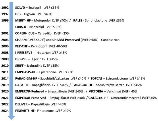
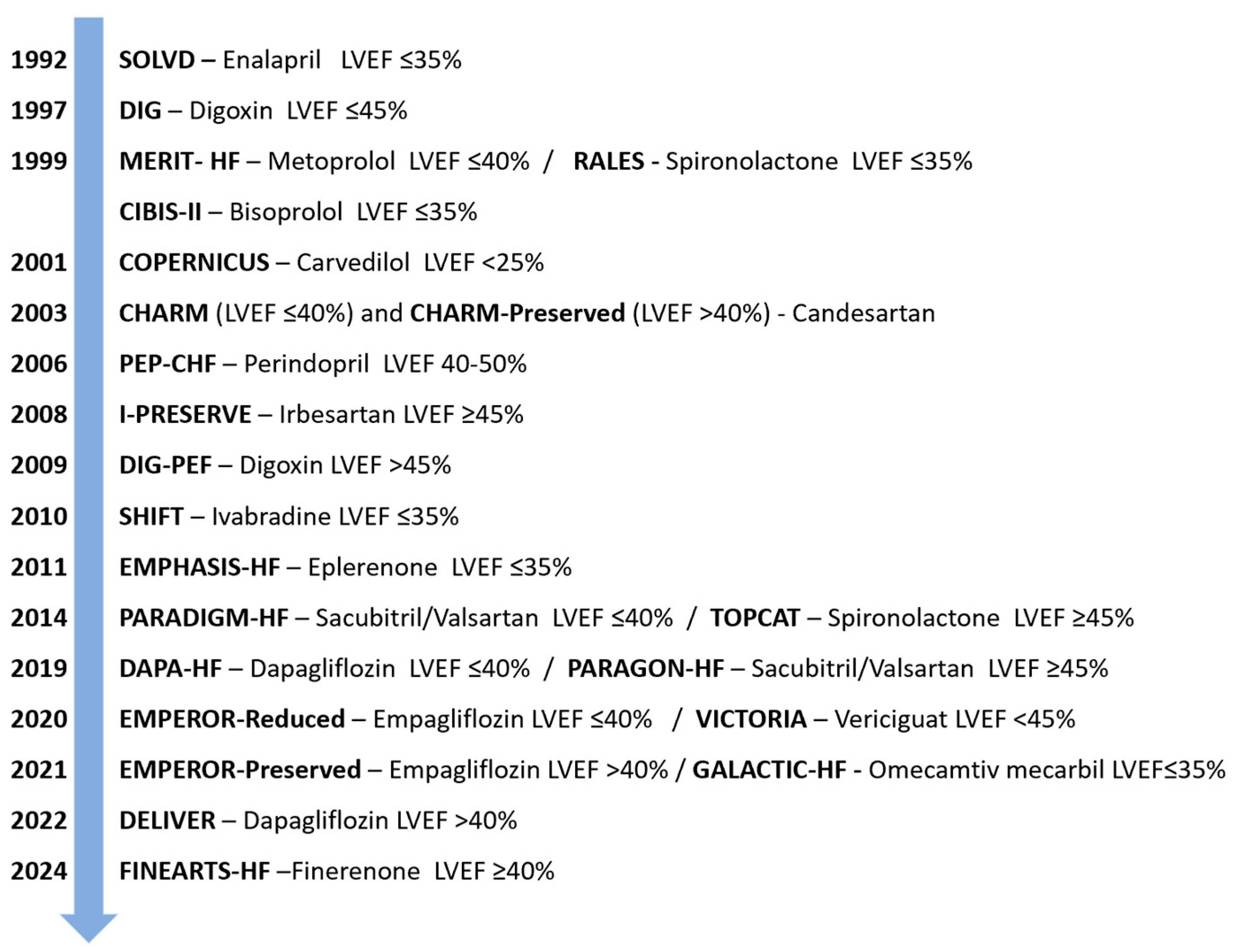

Disclaimer/Publisher’s Note: The statements, opinions and data contained in all publications are solely those of the individual author(s) and contributor(s) and not of MDPI and/or the editor(s). MDPI and/or the editor(s) disclaim responsibility for any injury to people or property resulting from any ideas, methods, instructions or products referred to in the content. |
© 2026 by the authors. Licensee MDPI, Basel, Switzerland. This article is an open access article distributed under the terms and conditions of the Creative Commons Attribution (CC BY) license.