


Abstract
Background/Objectives: Lupus nephritis (LN) is one of the most severe organ manifestations of systemic lupus erythematosus (SLE). Chronic kidney disease (CKD) and its progression into end-stage renal disease (ESRD) are serious complications in LN and the main cause of death in SLE. We aimed to investigate the prognostic factors of the progression of CKD and the development of ESRD in SLE patients. Methods: In our retrospective cohort study, we assessed the clinical and laboratory data of 127 patients who were diagnosed with LN between 1990 and 2022 and received regular follow-up care at our autoimmune centre. We compared class IV (diffuse) LN patients with non-class IV LN patients and assessed the differences in clinical and laboratory data of the patients, subdivided into complete, partial, and non-responders to therapy. Results: The prevalence of class IV LN is significantly higher in patients with CKD stage 3–5. Age above 42, class IV LN, Coombs positivity, and high chronicity index are prognostic factors for the development of CKD stage 3–5. On the other hand, anti-RNP and anti-SS-B antibody positivity and a high chronicity index are prognostic factors for the development of ESRD. The chronicity index, as well as the SLICC/ACR Damage Index (SDI) score, was significantly higher in non-responders compared to patients with complete remission. Conclusions: Based on our results, the progression of CKD into stage 3–5 or the development of ESRD should be expected at a chronicity index above 3.5 points. An early diagnosis, as well as aggressive, timely, and adequate treatment, is fundamental to prevent unfavourable outcomes of LN.
1. Introduction
Lupus nephritis (LN) is one of the most severe organ manifestations of systemic lupus erythematosus (SLE). Almost all patients with SLE have renal involvement during the course of the disease, but manifest lupus nephritis develops in 40–70% of patients [1,2]. Based on a recent review of the literature, LN may be the first manifestation of SLE in 7–31% of cases, and it develops in 31–48% of patients during the course of the disease [3]. In most cases, LN appears in the first five years of the disease, after which its development is rare [4]. As lupus nephritis implies persistent alterations to kidney structures and functions, all patients with lupus nephritis have chronic kidney disease (CKD) [5]. There has been a rapid advancement in our understanding of the pathogenesis of LN. In genetically susceptible individuals, an immune-complex-mediated glomerulonephritis develops with the contribution of certain epigenetic factors. Among the clinical signs, the appearance of proteinuria, active urine sediment, decreased renal function, nephrosis, and nephritis syndrome indicate glomerular damage. At the same time, not only can the glomeruli be involved, but sometimes, interstitial and tubular damages occur, or thrombotic microangiopathy develops, which can also contribute to renal impairment. The degree of proteinuria is influenced by the destruction of podocytes [4,6]. The gold standard for the diagnosis of LN is a renal biopsy. In all cases, if lupus nephritis is suspected, a renal biopsy is recommended, which helps in the differential diagnosis of other glomerular diseases, as well as with determining the patient’s treatment. It also has a prognostic value [7,8]. LN is classified into six histological categories, which were classified by the WHO and then revised twice by the ISN/RPS, leaving the basic histological types intact [9,10,11]. The class IV LN, namely diffuse proliferative glomerulonephritis, is the most common histological type and is considered a poor prognostic factor. The progression of CKD and the development of end-stage renal disease (ESRD) are the most prevalent characteristics in this category [12,13]. The introduction of cyclophosphamide was a milestone in the therapy of LN and significantly improved the survival of patients [14]. In the last two decades, significant advances have been made in the therapy of LN [15]. The use of mycophenolate mofetil has been introduced in both induction and maintenance treatment. A new calcineurin inhibitor, voclosporin, has been registered in combination with MMF in the treatment of LN. Belimumab treatment in lupus nephritis is also based on an accepted indication in combination with cyclophosphamide, azathioprine, and MMF. Rituximab may also be administered in therapy-refractory cases [16,17,18,19,20]. To follow the course of the disease and measure the effectiveness of the therapy, one option is a repeated renal biopsy, but this cannot be a routine procedure. An alternative method in everyday practice is to measure certain serum and urine biomarkers. The quantity of proteinuria, examination of urine sediment, ratio of urine total protein/creatinine, estimated glomerular filtration rate (eGFR), and anti-double-stranded DNA (anti-dsDNA) antibodies are the most commonly monitored parameters. However, these parameters are not specific and sensitive enough for calculating the renal activity and damage [3]. A number of new urine and serum biomarkers have been investigated that may help in the monitoring of renal disease, but they are not yet available in daily practice [21,22]. Therefore, clinical observations and currently available laboratory methods can be used for this purpose. Nevertheless, working groups reported different results on their usage [23,24,25]. Recently, Mahajan et al. highlighted hypocomplementemia, class III or IV LN, high chronicity index, hypertension, older age, male sex, and black race as the factors that are most commonly associated with ESRD [3]. Of note, data on the white population of Central Europe is limited, and Hungarian data on the clinical characteristics of lupus nephritis are not available from the past 10 years. It is not known whether there is a difference in the course of lupus nephritis between the histological classes. It is also unknown which further factors may affect the progression of CKD. Furthermore, the differences in clinical course and laboratory parameters between patients with complete remission (CR), partial remission (PR) and no remission (NR) are not fully known. In view of all this, we found it worthwhile to investigate the parameters mentioned above and to assess prognostic factors of an unfavourable renal outcome.
2. Materials and Methods
2.1. Study Pulation
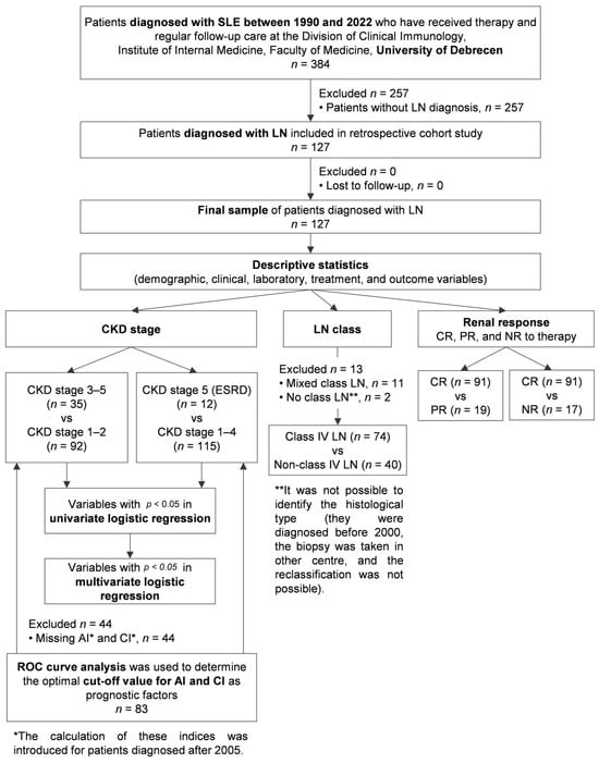
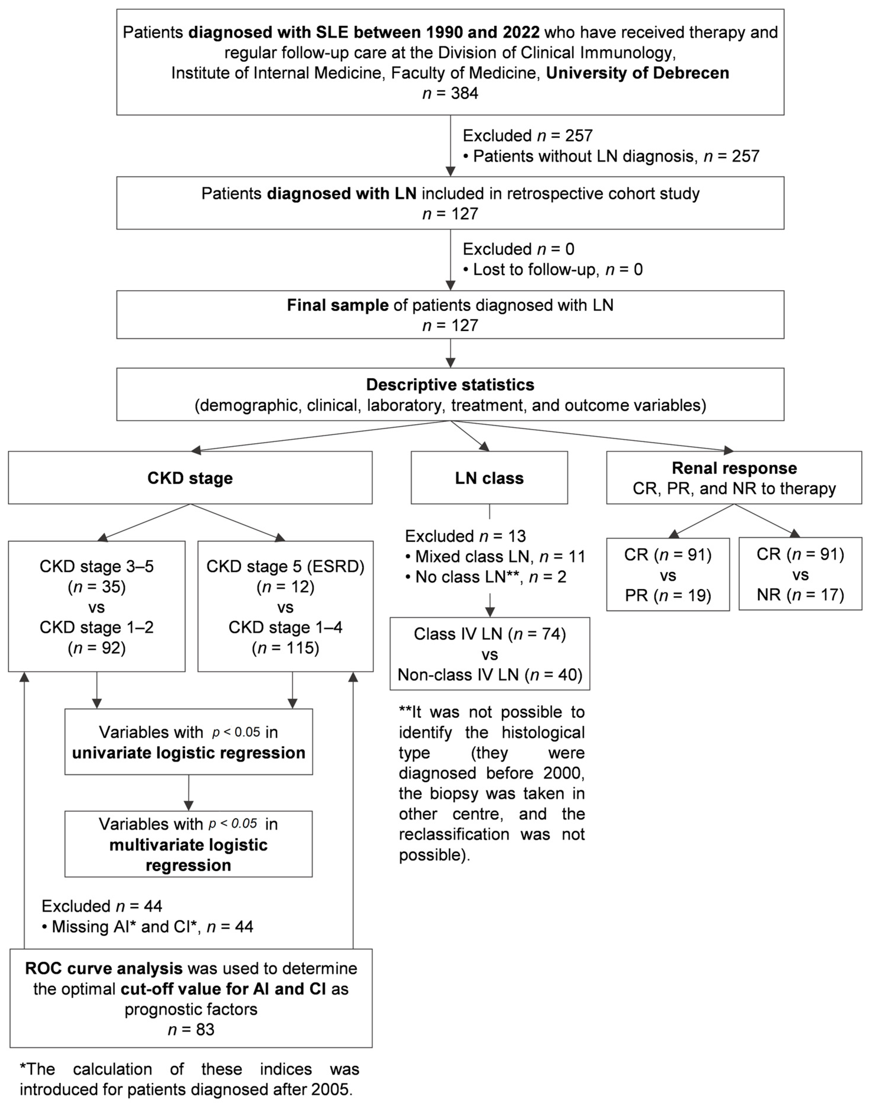
In our retrospective cohort study, we gathered the data of 384 Hungarian SLE patients, all of whom were white Europeans from the Central European region. They were diagnosed with SLE between 1990 and 2022 and have received therapy and regular follow-up care at the Division of Clinical Immunology, Institute of Internal Medicine, Faculty of Medicine, University of Debrecen. Patients who were diagnosed with SLE before 2012 were revised according to the SLICC criteria for SLE [26]; additionally, all SLE patients fulfilled the EULAR/ACR 2019 classification criteria for lupus [27]. SLE patients with secondary antiphospholipid syndrome (APS) fulfilled the 2006 Sydney criteria; of note, all of them met the criteria of the 2023 APS ACR/EULAR classification criteria, as well [28,29]. Lupus nephritis was diagnosed in 127 of them; therefore, only these patients were included in the further examinations of the present study. The clinical and laboratory data of the patients were extracted from medical documentations and records for statistical analyses. The date of data collection was January, 2024. Figure 1 shows the flow chart of this study.
Figure 1.
Study design flow diagram. Abbreviations: SLE, systemic lupus erythematosus; n, number of patients; LN, lupus nephritis; CKD, chronic kidney disease; ESRD, end-stage renal disease; CR, complete remission; PR, partial remission; NRs, no responders; ROC, receiver operating characteristic; AI, activity index; CI, chronicity index.
2.2. Clinical and Laboratory Evaluation
All patients were routinely followed up throughout the studied period, and their medical records contained detailed information on medical history, treatments, clinical symptoms, physical conditions, and laboratory and other findings of each visit. The following demographic and clinical data were analysed: sex, age, age at diagnosis, disease duration, histological class of lupus nephritis, clinical symptoms and laboratory results, immunoserological abnormalities, applied treatments, complete and partial remission, and non-responsive status. We defined CKD stages according the KDIGO guidelines [30]. We combined CKD stage 1 and stage 2 into a single category. CKD stage 3 was described as eGFR 30 to <60 mL/min per 1.73 m2, and CKD stage 4 was described as eGFR 15 to <30 mL/min per 1.73 m2 for at least 3 consecutive months. CKD stage 5 (ESRD) was defined as eGFR ≤ 15 mL/min per 1.73 m2 for at least 3 consecutive months or ongoing dialysis. Histological sampling was carried out at the Department of Nephrology, Institute of Internal Medicine, Faculty of Medicine, University of Debrecen, and the evaluation of the kidney biopsy samples was performed at the Department of Pathology, Faculty of Medicine, University of Debrecen. We used the relevant WHO, ISN/RPS2003 and ISN/RPS2018 classification systems for the histological evaluation [9,10,11]. The calculation of activity and chronicity indices was introduced for patients who were diagnosed after 2005, meaning that 83 patients (20 with CKD stage 3–5 and 63 with CKD stage 1–2) had data about the LN Activity and Chronicity Index. Complete or partial remission of LN was defined according to the KDIGO guidelines [16]. In the case of a CR, the degree of proteinuria decreased below 0.5 g in 6 months, and the creatinine level returned to the initial value. In case of PR, the degree of proteinuria was halved in 6 months, and the serum creatinine level stabilized or improved but did not return to baseline. Chronic organ damage in SLE was determined using the SLICC damage index [31]. Immune serological parameters were determined from serum samples. The presence of antinuclear antibodies (ANAs) was detected by an indirect immunofluorescence method on the HEp2 cell line. Enzyme-linked immunosorbent assay (ELISA) was used for the detection of the following antibodies: anti-dsDNA (Orgentec, Mainz, Germany), anti-SS-A, anti-SS-B, anti-RNP, and anti-Sm (Hycor, Biomedical, Garden Grove, CA, USA), as well as antiphospholipid antibodies including anti-cardiolipin (aCL) IgG/IgA/IgM and anti-ß2GPI IgG/IgA/IgM antibodies (Orgentec, Mainz, Germany). All laboratory tests were performed under standardized conditions, according to the manufacturer’s instructions, at the Department of Laboratory Medicine, Faculty of Medicine, University of Debrecen.
2.3. Statistical Analysis
Statistical analysis was performed using SPSS Statistics for Windows, Version 28.0 (IBM Corporation, Armonk, NY, USA), and GraphPad Prism version 9.5 for Windows (GraphPad Software, San Diego, CA, USA). A Shapiro–Wilk test of normality was conducted to determine whether the continuous data were normally distributed. Values are expressed as median with an interquartile range (IQR) for continuous variables and frequency with percentage for categorical variables. Continuous variables were compared with a nonparametric Mann–Whitney U test. Categorical variables were compared with Pearson’s chi-squared test or Fisher’s exact test. Univariate and multivariate binary logistic regression analyses were performed to identify predictors of the progression of CKD. First, we performed a univariate analysis using variables with p < 0.05 in a Mann–Whitney U test or Pearson’s chi-squared test and Fisher’s exact test to avoid over-fitting; only variables with p < 0.05 in the univariate binary logistic regression analysis were included in the multivariate analysis. A receiver operating characteristic (ROC) curve analysis was used to determine the optimal cut-off value for AI and CI as prognostic factors of the progression of CKD into stage 3–5 and the development of ESRD. All statistical tests were two-sided, and differences were considered statistically significant at <0.05 level and reported using p-values and/or 95% confidence intervals (95% CIs).
3. Results
3.1. Main Analyses
The population of our retrospective study consisted of 127 LN patients, 114 (89.8%) women and 13 (10.2%) men. Their age at the time of SLE diagnosis was 28.4 ± 9.9 years, and they were followed up regularly at our autoimmune centre for 16.9 ± 8.1 years. All patients who were included in the study underwent a renal biopsy. The most common histological type was class IV LN, followed by class V and then class III. Mixed histological types were found in 11 patients, while for two patients, it was not possible to identify the histological type (they were diagnosed before 2000, the biopsy was taken in another centre, and reclassification was not possible). We did not detect class VI LN in our patient group. CKD stage 3–5 developed in 35 patients; among them, 12 patients had CKD stage 5 (ESRD). At the time of the data collection, 71.6% of patients were in CR, 15% were in PR, while 13.4% were non-responders to the therapy (Table 1).
Table 1.
Characteristics of LN patients (n = 127).
3.2. Comparison of Patients Based on the Stage of CKD
We compared the demographics, clinical characteristics, laboratory parameters, therapies, outcome, and mortality of CKD stage 3–5 patients with the ones with CKD stage 1–2, as well as CKD stage 5 (ESRD) patients with the ones with CKD stage 1–4 (non-ESRD). Our findings are shown in Table 2 and Table 3.
Table 2.
Comparison of demographics, clinical features, and positive laboratory findings between the groups of LN patients with different CKD stages (n = 127).
Table 3.
Comparison of renal pathological findings, treatment, renal response, and outcomes between the groups of LN patients with different CKD stages (n = 127).
CKD stage 3–5 patients were significantly older at the time of data collection than CKD stage 1–2 patients, while there was no difference between the ages of ESRD and non-ESRD patients. We found no differences between CKD stage 3–5 and CKD stage 1–2, nor between the ESRD and non-ESRD groups in terms of SLE clinical symptoms, autoantibodies, and cardiovascular complications. The association with Sjögren’s syndrome was significantly more common in the CKD stage 3–5 group compared to CKD stage 1–2 patients. Coombs positivity was significantly more common in CKD stage 3–5 patients, while anti-DNA positivity was significantly less common. Anti-RNP and anti-SS-B autoantibody positivity were detected significantly more frequently in ESRD patients compared to non-ESRD patients. The occurrence of class IV LN was significantly more frequent in the CKD stage 3–5 group; on the other hand, class III LN did not occur in patients with CKD stage 3–5 at all. The chronicity index, as well as SDI score, was significantly higher in both the CKD and ESRD groups compared to CKD stage 1–2 and non-ESDR patients. The administration of immunosuppressive therapy did not differ between CKD stage 3–5 and CKD stage 1–2 or between the ESRD and non-ESRD groups. The cumulative steroid dose was significantly higher in the CKD stage 3–5 group compared to patients with CKD stage 1–2. A CR developed significantly less frequently in CKD stage 3–5, as well as in the ESRD group. None of the ESRD patients reached CR. Consequently, the ratio of non-responder patients was significantly higher in both the CKD and ESRD groups. The total mortality rate was significantly higher in patients with CKD stage 3–5 than in patients with CKD stage 1–2; however, there was no difference regarding the cause of death.
3.3. Prognostic Factors for the Progression of CKD and the Development of ESRD
Univariate and multivariate logistic regression analyses revealed that age above 42 years, class IV lupus nephritis, Coombs positivity, and a higher chronicity index are important prognostic factors of the development of CKD stage 3–5 (Table 4).
Table 4.
Univariate and multivariate logistic regression analyses of the prognostic factors of the development of CKD stage 3–5 (n = 35) in patients with LN (n = 127).
Univariate logistic regression analysis revealed that anti-RNP and anti-SS-B positivity, as well as a high chronicity index, are important prognostic factors of the development of ESRD. Of note, the multivariate logistic regression analysis only confirmed the high chronicity index among these (Table 5).
Table 5.
Univariate and multivariate logistic regression analyses of the prognostic factors of the development of CKD stage 5 (ESRD) (n = 12) in patients with LN (n = 127).
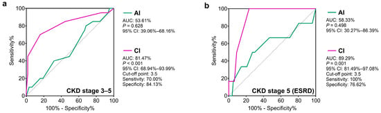
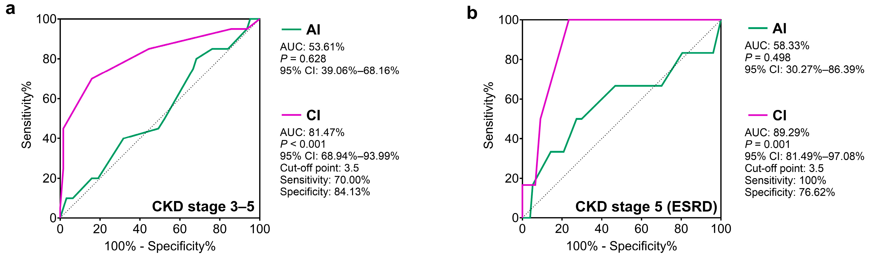
We also evaluated the possible associations between activity and chronicity index scores and the development of CKD stage 3–5 and ESRD. Figure 2 shows the results of the ROC analysis. The activity score did not show any correlation with the development of CKD stage 3–5 or ESRD. However, a chronicity index score above 3.5 points showed a significant correlation with the development of both CKD stage 3–5 and ESRD.
Figure 2.
ROC curve and analysis of cut-off values of AI and CI in relation to CKD stage 3–5 (a) and ESRD (b). Total number of LN patients is 127, of which 83 patients (20 CKD stage 3–5 and 63 CKD stage 1–2; 6 ESRD and 77 non-ESRD) had data about LN Activity and Chronicity Index. Abbreviations: AI, activity index; CI, chronicity index; CKD, chronic kidney disease; ESRD, end-stage renal disease; ROC, receiver operating characteristic; AUC, area under the ROC curve; 95% CI, 95% confidence interval.
3.4. Differences Between Class IV LN and Non-Class IV LN Patients
Given that class IV LN was the most common histological type and a prognostic factor of the development of CKD stage 3–5, we compared it with other non-class IV LN patients. The results are summarized in Table 6 and Table 7.
Table 6.
Comparison of demographics, extrarenal clinical features, and positive laboratory findings of ISN/RPS Class IV LN patients with other classes of LN patients.
Table 7.
Comparison of renal pathological and clinical features, treatment, renal responses, and outcomes of ISN/RPS Class IV LN patients with other classes of LN patients.
Class IV LN patients were younger than the others at the time of data collection; however, their age at diagnosis did not differ. Regarding the clinical features, deep vein thrombosis, as well as rheumatoid arthritis, was significantly less common in class IV LN patients. During the assessment of kidney pathology, we found that both the activity and chronicity indexes were significantly higher, and the development of CKD stage 3–5 was more prevalent in the class IV lupus nephritis patients. We did not find any difference in immunoserological results. Azathioprine was significantly more frequently used as a maintenance treatment in the class IV LN group, while cyclosporine A was used significantly less often. Total mortality was not more frequent in the class IV lupus nephritis group, but among the causes of death, tumour mortality was significantly less frequent.
3.5. Differences Between Complete, Partial, and Non-Responders
We also assessed the clinical and laboratory parameters of the patients with CR, PR, and NR. The results are summarized in Table 8.
Table 8.
Comparison of disease characteristics between renal responses in patients with LN (n = 127).
The age at data collection was significantly higher, and the duration of disease was significantly longer, in the patients in CR than the patients in PR. Regarding autoantibodies, no differences were found between the three groups. The occurrence of CKD stage 3–5 and ESRD was significantly more frequent among non-responders compared to patients in CR, and their chronicity index was also significantly higher. Steroid use was significantly more frequent among patients in PR compared to patients in CR. MMF induction and maintenance treatments were used significantly more often in patients in PR. MMF induction therapy was significantly more common among non-responders compared to patients in CR. Both tacrolimus and belimumab were used significantly more often in patients in PR than in CR. Patients in CR achieved lupus low-disease-activity state (LLDAS) more often than patients in PR. The SDI score was significantly higher among non-responders compared to those in CR.
4. Discussion
Lupus nephritis is one of the most serious organ manifestations of SLE, in addition to neuropsychiatric manifestations [32]. LN usually manifests in the first five years of the disease and may be the only clinical manifestation of SLE. This latter fact is taken into account by the latest EULAR/ACR classification criteria system, according to which, in addition to the entry criteria, the presence of histologically confirmed class IV lupus nephritis is sufficient to establish a diagnosis of SLE [27]. The most common histological type is class IV LN [3,33,34], and we found it in nearly 60% of our patients. A similar frequency was reported by Vajgel et al., although, in a non-white population [35]. CKD stage 3–5 developed in 27.6% of our patients. However, when comparing our observations with the results of other working groups, it is important to highlight the fact that LN implies persistent alterations to kidney structures and functions; therefore, all LN patients develop CKD, and the CKD stage of these patients determines their health outcomes [5]. In the publications that have been published so far in the literature, the definition used by each working group for the diagnosis of CKD in their LN patients corresponded to our definition of CKD stages 3–5 (defined by eGFR < 60 mL/min per 1.73 m2 for at least 3 consecutive months). Consequently, their definition for non-CKD LN patients corresponds to the definition of LN patients with CDK stage 1–2 in our study. An Egyptian working group observed a CKD rate of 32.7% in a 5-year study of more than 900 patients with lupus nephritis; however, these data come from a non-white population [36]. The frequency of ESRD in our patient population was 9.4%, which is similar to the data in the RELESSER register (10.35%) [37]. Platinga et al. found the 5-year cumulative incidence of ESRD to be 2.5% in white and 6.4% in black individuals [38]. Similar results were obtained by Hanly et al., who found that for all patients with LN, the 5-year cumulative incidence of ESRD was 3.3%, while the 10-year cumulative incidence was 4.3% [24]. When we evaluated the demographic, clinical, and laboratory data and renal outcomes of CKD stage 3–5 patients, we found that they were older, and an association with Sjögren’s syndrome was more common, compared to the patients with CKD stage 1–2. Park et al. found CKD patients to be similarly older in a Korean population. Similarly to these results, the clinical manifestations of SLE did not differ between our patients in the CKD stage 3–5 and CKD stage 1–2 groups. At the same time, in the Korean cohort, hypertension, elevated serum creatinine levels, reduced eGFR, and nephrotic proteinuria were more common in the CKD group at the time of LN diagnosis [39]. We could not examine these parameters, as the initial laboratory parameters were not available for all patients due to the long follow-up period. We found that class IV lupus nephritis and a higher chronicity index are significantly more common in patients with CKD stage 3–5. It is similar to the results of other studies; however, we found no correlation with the activity scores [40,41]. Direct Coombs positivity was also more common in the CKD stage 3–5 group, but anti-DNA positivity was significantly less frequent, which contradicts the literature data. Based on a Spanish register’s data, ESRD showed a significant association with anti-DNA positivity; however, it was also described that there is a significant correlation between hemolytic anaemia and ESRD, and they did not comment on the isolated Coombs positivity [37]. Regarding therapy, we found no difference between the CKD stage 3–5 and CKD stage 1–2 groups, except for the higher cumulative steroid dose. The reason for this may be that CKD patients had a more severe form of LN, and a higher dose of steroid was needed. Not surprisingly, among patients with CKD stage 3–5, there were fewer patients in CR and more non-responders. We observed higher SDI scores and mortality rates in the CKD stage 3–5 group, but there was no difference in causes of death. Based on our results, age above 42 years, class IV LN, Coombs positivity, and a higher chronicity index proved to be important prognostic factors of the development of CKD stage 3–5. Formerly, several working groups identified hypertension, elevated serum creatinine levels, and a lack of remission as prognostic factors [3,35]. A higher chronicity index, on the other hand, is described by numerous working groups as a prognostic factor for CKD and ESRD [3,39]. We found a score of 3.5 points to be the value above which the development of CKD stage 3–5 and ESRD must be more anticipated.
We compared the data of ESRD and non-ESRD patients as well. We found no differences in the clinical symptoms of SLE, but anti-RNP and anti-SS-B positivity were significantly more frequent in the ESRD group. Several working groups have described anti-SS-A as an independent predictor of ESRD, without mentioning anti-SS-B [42,43]. Anti-RNP and anti-SS-B positivity, as well as a higher chronicity index, were found to be prognostic factors for the development of ESRD; however, our multivariate analysis only confirmed the prognostic value of a higher chronicity index. Other study groups found hypocomplementemia, class III, IV, VI lupus nephritis, hypertension, older age, male sex, and black race to be the main prognostic factors for ESRD [35]. We did not find class III LN to be more frequent; moreover, this class did not even occur in neither the CKD stage 3–5, nor the ESRD group.
Given that class IV LN was the most common histological type, and CKD stage 3–5 and ESRD did not occur in class III LN patient group, as a next step, we compared class IV and non-class IV LN patients. Duran et al. examined the clinical characteristics and disease outcome of proliferative and non-proliferative LN patients. They found no difference in SLE clinical symptoms between the two groups, just as we found no difference between the class IV and non-class IV LN patients. They found significant differences, however, in several parameters indicating kidney activity, such as the serum creatinine level, eGFR, proteinuria, hypocomplementemia, active urine sedimentation, and renal SLE DAI [44]. We did not examine these parameters in our study. We found a significantly higher activity and chronicity index in the class IV LN patients. A Columbian working group obtained similar results when comparing patients with proliferative and non-proliferative LN [45]. In terms of therapy, we found a difference only in the administration of cyclosporine A. We found that this was used significantly less often in class IV LN, similarly to Duran et al. [44]. We found no difference in mortality between patients with class IV and non-class IV LN; however, the tumour mortality was significantly lower among class IV LN patients.
Finally, we also compared patients who were in CR or PR and non-responders. Patients in CR were older and had a longer disease duration compared to patients in PR. The damage index and the prevalence of CKD stage 3–5 and ESRD were significantly higher among the non-responders. We found no differences between the individual groups in terms of clinical symptoms and laboratory abnormalities, but there was a difference in their treatment. Patients in PR received newer drugs, such as MMF, tacrolimus, and belimumab significantly more frequently. In the therapeutic recommendations for lupus nephritis, these drugs have been introduced in recent years, and the latest KDIGO guidelines and ACR/EULAR recommendations also include them [15,16]. Presumably, the newer and registered drugs will be used more and more widely, not only in patients in PR, but also in the entire lupus nephritis patient population. Among immunosuppressive treatments, steroid therapy was also more common in patients in PR. Patients in CR took steroids less often, which is also in line with the current therapeutic guidelines and our therapeutic efforts. Nevertheless, even more attention should be paid to the use of antimalarial drugs, because half of our patients only received antimalarial drugs, even though it is well known that HCQ has a beneficial effect on the outcome of the disease in many respects. Park et al. also reported that the use of hydroxychloroquine before the onset of lupus nephritis protects patients from developing CKD [39]. When examining the disease outcome, we found that mortality did not differ between the groups, but LLDA was significantly more prevalent in the CR group, which may improve the morbidity and mortality indicators of patients in the long term. At the same time, we found a higher damage index among non-responders, which may have a negative effect on the long-term outcome.
This study has some limitations. It was a retrospective study, and some risk factors could not be included in the analysis as covariates or potential confounders. Regarding the treatment adherence of the included patients, despite knowing the medications, the doses used, and the high level of accuracy of the administrative staff based on the documentation of drug prescriptions, we cannot exclude the possibility of erratic patient adherence. Despite these limitations, our study had two major strengths. One of them is the length of the period examined, which was more than three decades in some cases (mean follow-up time was 16.9 ± 8.1 years). Another strength is that we investigated and analysed multiple clinical parameters and disease outcomes, providing more real-world data about the natural course of LN. Nevertheless, our observations have to be confirmed in a larger patient population as well; moreover, further studies are needed to elucidate the pathophysiological role of Coombs positivity and anti-RNP and anti-SS-B antibodies in the progression of LN.
In conclusion, class IV lupus nephritis is the most common form of LN in our centre, in which the renal outcome is clearly unfavourable. Age above 42 and Coombs positivity are also prognostic for the development of CKD stage 3–5, while a high chronicity index is prognostic for both CKD stage 3–5 and ESRD. Based on our results, the development of CKD stage 3–5 or ESRD should be expected at a chronicity index above 3.5 points. The chronicity index, as well as SLICC/ACR Damage Index (SDI) score, was significantly higher in non-responders compared to patients with complete remission. Among patients in CR, we achieved LLDA more often, and the average damage score and steroid use were significantly lower. Hopefully, the newly registered drugs and the ones that are still under development will enable a higher rate of CR in lupus nephritis patients, which will have a favourable effect on the long-term outcome.
Author Contributions
Conceptualization, T.T.; methodology, B.B.; investigation, B.P., G.P., B.B. and T.T.; data curation, C.M., L.B. and N.F.; writing—original draft preparation, B.P. and B.B.; writing—review and editing, G.P. and T.T.; visualization, B.B.; supervision, T.T. and J.B.; project administration, T.T. and G.P. All authors have read and agreed to the published version of the manuscript.
Funding
This research received no external funding.
Institutional Review Board Statement
The study was conducted in accordance with the Declaration of Helsinki and approved by the Regional and Institutional Ethics Committee of the University of Debrecen (protocol number: 4879-2017, approval date: 25 September 2017) and the Policy Administration Services of Public Health of the Government Office (registration number: 1660-2/2018/EÜIG, approval date: 26 February 2018).
Informed Consent Statement
Informed consent was obtained from all subjects involved in the study.
Data Availability Statement
The original contributions presented in the study are included in the article. Further inquiries can be directed to the corresponding author.
Conflicts of Interest
The authors declare no conflicts of interest.
References
- Cojocaru, M.; Cojocaru, I.M.; Silosi, I.; Vrabie, C.D. Manifestations of systemic lupus erythematosus. Maedica 2011, 6, 330–336. [Google Scholar] [PubMed]
- Beckwith, H.; Lightstone, L. Rituximab in systemic lupus erythematosus and lupus nephritis. Nephron Clin. Pract. 2014, 128, 250–254. [Google Scholar] [CrossRef] [Scilit]
- Mahajan, A.; Amelio, J.; Gairy, K.; Kaur, G.; Levy, R.A.; Roth, D.; Bass, D. Systemic lupus erythematosus, lupus nephritis and end-stage renal disease: A pragmatic review mapping disease severity and progression. Lupus 2020, 29, 1011–1020. [Google Scholar] [CrossRef] [Scilit]
- Anders, H.J.; Saxena, R.; Zhao, M.H.; Parodis, I.; Salmon, J.E.; Mohan, C. Lupus nephritis. Nat. Rev. Dis. Primers 2020, 6, 7. [Google Scholar] [CrossRef] [Scilit] [PubMed]
- Lichtnekert, J.; Anders, H.J. Lupus nephritis-related chronic kidney disease. Nat. Rev. Rheumatol. 2024, 20, 699–711. [Google Scholar] [CrossRef] [Scilit] [PubMed]
- Yu, F.; Haas, M.; Glassock, R.; Zhao, M.H. Redefining lupus nephritis: Clinical implications of pathophysiologic subtypes. Nat. Rev. Nephrol. 2017, 13, 483–495. [Google Scholar] [CrossRef] [Scilit] [PubMed]
- Gordon, C.; Jayne, D.; Pusey, C.; Adu, D.; Amoura, Z.; Aringer, M.; Ballerin, J.; Cervera, R.; Calvo-Alén, J.; Chizzolini, C.; et al. European consensus statement on the terminology used in the management of lupus glomerulonephritis. Lupus 2009, 18, 257–263. [Google Scholar] [CrossRef] [Scilit] [PubMed]
- Bertsias, G.K.; Tektonidou, M.; Amoura, Z.; Aringer, M.; Bajema, I.; Berden, J.H.; Boletis, J.; Cervera, R.; Dörner, T.; Doria, A.; et al. Joint European League Against Rheumatism and European Renal Association-European Dialysis and Transplant Association (EULAR/ERA-EDTA) recommendations for the management of adult and paediatric lupus nephritis. Ann. Rheum. Dis. 2012, 71, 1771–1782. [Google Scholar] [CrossRef] [Scilit] [PubMed]
- Churg, J.; Sobin, L. Renal Disease: Classification and Atlas of Glomerular Disease; Igaku-Shoin Ltd.: Tokyo, Japan, 1982. [Google Scholar]
- Weening, J.J.; D'Agati, V.D.; Schwartz, M.M.; Seshan, S.V.; Alpers, C.E.; Appel, G.B.; Balow, J.E.; Bruijn, J.A.; Cook, T.; Ferrario, F.; et al. The classification of glomerulonephritis in systemic lupus erythematosus revisited. Kidney Int. 2004, 65, 521–530. [Google Scholar] [CrossRef] [Scilit]
- Bajema, I.M.; Wilhelmus, S.; Alpers, C.E.; Bruijn, J.A.; Colvin, R.B.; Cook, H.T.; D’Agati, V.D.; Ferrario, F.; Haas, M.; Jennette, J.C.; et al. Revision of the International Society of Nephrology/Renal Pathology Society classification for lupus nephritis: Clarification of definitions, and modified National Institutes of Health activity and chronicity indices. Kidney Int. 2018, 93, 789–796. [Google Scholar] [CrossRef] [Scilit] [PubMed]
- Inderjeeth, C.A.; Habib, P.; Sharma, C.; Nossent, J. Systemic lupus erythematosus: Reducing life-threatening progression. Med. Today 2018, 19, 31–41. [Google Scholar]
- Lau, W.L.; Appel, G.B. Lupus nephritis in 2017: An update. ASN Kidney News 2017, 9, 17–18. [Google Scholar]
- Mok, C.C.; Wong, R.W.; Lai, K.N. Treatment of severe proliferative lupus nephritis: The current state. Ann. Rheum. Dis. 2003, 62, 799–804. [Google Scholar] [CrossRef] [Scilit] [PubMed]
- Mok, C.C.; Teng, Y.K.O.; Saxena, R.; Tanaka, Y. Treatment of lupus nephritis: Consensus, evidence and perspectives. Nat. Rev. Rheumatol. 2023, 19, 227–238. [Google Scholar] [CrossRef] [Scilit]
- Rovin, B.H.; Ayoub, I.M.; Chan, T.M.; Liu, Z.H.; Mejía-Vilet, J.M.; Floege, J.; Kidney Disease: Improving Global Outcomes (KDIGO) Lupus Nephritis Work Group Collaborators. KDIGO 2024 Clinical Practice Guideline for the management of Lupus Nephritis. Kidney Int. 2024, 105, S1–S69. [Google Scholar] [CrossRef] [Scilit] [PubMed]
- Fanouriakis, A.; Kostopoulou, M.; Andersen, J.; Aringer, M.; Arnaud, L.; Bae, S.C.; Boletis, J.; Bruce, I.N.; Cervera, R.; Doria, A.; et al. EULAR recommendations for the management of systemic lupus erythematosus: 2023 update. Ann. Rheum. Dis. 2024, 83, 15–29. [Google Scholar] [CrossRef] [Scilit]
- Furie, R.; Rovin, B.H.; Houssiau, F.; Malvar, A.; Teng, Y.K.O.; Contreras, G.; Amoura, Z.; Yu, X.; Mok, C.-C.; Santiago, M.B.; et al. Two-Year, Randomized, Controlled Trial of Belimumab in Lupus Nephritis. N. Engl. J. Med. 2020, 383, 1117–1128. [Google Scholar] [CrossRef] [Scilit] [PubMed]
- Tanaka, Y.; Nakayamada, S.; Yamaoka, K.; Ohmura, K.; Yasuda, S. Rituximab in the real-world treatment of lupus nephritis: A retrospective cohort study in Japan. Mod. Rheumatol. 2023, 33, 145–153. [Google Scholar] [CrossRef] [Scilit] [PubMed]
- Rovin, B.H.; Teng, Y.K.O.; Ginzler, E.M.; Arriens, C.; Caster, D.J.; Romero-Diaz, J.; Gibson, K.; Kaplan, J.; Lisk, L.; Navarra, S.; et al. Efficacy and safety of voclosporin versus placebo for lupus nephritis (AURORA 1): A double-blind, randomised, multicentre, placebo-controlled, phase 3 trial. Lancet 2021, 397, 2070–2080. [Google Scholar] [CrossRef] [Scilit] [PubMed]
- Mok, C.C.; Mohan, C. Urinary Biomarkers in Lupus Nephritis: Are We There Yet? Arthritis Rheumatol. 2021, 73, 194–196. [Google Scholar] [CrossRef] [Scilit]
- Palazzo, L.; Lindblom, J.; Mohan, C.; Parodis, I. Current Insights on Biomarkers in Lupus Nephritis: A Systematic Review of the Literature. J. Clin. Med. 2022, 11, 5759. [Google Scholar] [CrossRef] [Scilit]
- Tektonidou, M.G.; Dasgupta, A.; Ward, M.M. Risk of End-Stage Renal Disease in Patients with Lupus Nephritis, 1971–2015: A Systematic Review and Bayesian Meta-Analysis. Arthritis Rheumatol. 2016, 68, 1432–1441. [Google Scholar] [CrossRef] [Scilit] [PubMed]
- Hanly, J.G.; O’Keeffe, A.G.; Su, L.; Urowitz, M.B.; Romero-Diaz, J.; Gordon, C.; Bae, S.-H.; Bernatsky, S.; Clarke, A.E.; Wallace, D.J.; et al. The frequency and outcome of lupus nephritis: Results from an international inception cohort study. Rheumatology 2016, 55, 252–262. [Google Scholar] [CrossRef] [Scilit] [PubMed]
- Siso, A.; Ramos-Casals, M.; Bove, A.; Brito-Zerón, P.; Soria, N.; Nardi, N.; Testi, A.; Perez-de-Lis, M.; Díaz-Lagares, C.; Darnell, A.; et al. Outcomes in biopsy-proven lupus nephritis: Evaluation of 190 white patients from a single center. Medicine 2010, 89, 300–307. [Google Scholar] [CrossRef] [Scilit] [PubMed]
- Petri, M.; Orbai, A.M.; Alarcón, G.S.; Gordon, C.; Merrill, J.T.; Fortin, P.R.; Bruce, I.N.; Isenberg, D.; Wallace, D.J.; Nived, O.; et al. Derivation and validation of the Systemic Lupus International Collaborating Clinics classification criteria for systemic lupus erythematosus. Arthritis Rheum. 2012, 64, 2677–2686. [Google Scholar] [CrossRef] [Scilit] [PubMed]
- Aringer, M.; Costenbader, K.; Daikh, D.; Brinks, R.; Mosca, M.; Ramsey-Goldman, R.; Smolen, J.S.; Wofsy, D.; Boumpas, D.T.; Kamen, D.L.; et al. 2019 European League Against Rheumatism/American College of Rheumatology Classification Criteria for Systemic Lupus Erythematosus. Arthritis Rheumatol. 2019, 71, 1400–1412. [Google Scholar] [CrossRef] [Scilit] [PubMed]
- Miyakis, S.; Lockshin, M.D.; Atsumi, T.; Branch, D.W.; Brey, R.L.; Cervera, R.; Derksen, R.H.W.M.; Groot, P.G.D.E.; Koike, T.; Meroni, P.L.; et al. International consensus statement on an update of the classification criteria for definite antiphospholipid syndrome (APS). J. Thromb. Haemost. 2006, 4, 295–306. [Google Scholar] [CrossRef] [Scilit] [PubMed]
- Barbhaiya, M.; Zuily, S.; Naden, R.; Hendry, A.; Manneville, F.; Amigo, M.C.; Amoura, Z.; Andrade, D.; Andreoli, L.; Artim-Esen, B.; et al. The 2023 ACR/EULAR Antiphospholipid Syndrome Classification Criteria. Arthritis Rheumatol. 2023, 75, 1687–1702. [Google Scholar] [CrossRef] [Scilit]
- International Society of Nephrology. Kidney Disease: Improving Global Outcomes (KDIGO). Chapter 1: Definition and classification of CKD. Kidney Int. Suppl. 2013, 3, 19–62. [Google Scholar] [CrossRef] [Scilit]
- Gladman, D.; Ginzler, E.; Goldsmith, C.; Fortin, P.; Liang, M.; Urowitz, M.; Bacon, P.; Bombardieri, S.; Hanly, J.; Hay, E.; et al. The development and initial validation of the Systemic Lupus International Collaborating Clinics/American College of Rheumatology damage index for systemic lupus erythematosus. Arthritis Rheum. 1996, 39, 363–369. [Google Scholar] [CrossRef] [Scilit]
- Tarr, T.; Dérfalvi, B.; Győri, N.; Szántó, A.; Siminszky, Z.; Malik, A.; Szabó, A.J.; Szegedi, G.; Zeher, M. Similarities and differences between pediatric and adult patients with systemic lupus erythematosus. Lupus 2015, 24, 796–803. [Google Scholar] [CrossRef] [Scilit] [PubMed]
- Wang, H.; Ren, Y.L.; Chang, J.; Gu, L.; Sun, L.Y. A Systematic Review and Meta-analysis of Prevalence of Biopsy-Proven Lupus Nephritis. Arch. Rheumatol. 2017, 33, 17–25. [Google Scholar] [CrossRef] [Scilit]
- Gasparotto, M.; Gatto, M.; Binda, V.; Doria, A.; Moroni, G. Lupus nephritis: Clinical presentations and outcomes in the 21st century. Rheumatology 2020, 59, v39–v51. [Google Scholar] [CrossRef] [Scilit] [PubMed]
- Vajgel, G.; Oliveira, C.B.L.; Costa, D.M.N.; Cavalcante, M.A.G.M.; Valente, L.M.; Sesso, R.; Crovella, S.; Kirsztajn, G.M.; Sandrin-Garcia, P. Initial renal histology and early response predict outcomes of Brazilian lupus nephritis patients. Lupus 2020, 29, 83–91. [Google Scholar] [CrossRef] [Scilit]
- Momtaz, M.; Fayed, A.; Wadie, M.; Gamal, S.M.; Ghoniem, S.A.; Sobhy, N.; Kamal Elden, N.M.; Hamza, W.M. Retrospective analysis of nephritis response and renal outcome in a cohort of 928 Egyptian lupus nephritis patients: A university hospital experience. Lupus 2017, 26, 1564–1570. [Google Scholar] [CrossRef] [Scilit]
- Galindo-Izquierdo, M.; Rodriguez-Almaraz, E.; Pego-Reigosa, J.M.; López-Longo, F.J.; Calvo-Alén, J.; Olivé, A.; Fernández-Nebro, A.; Martinez-Taboada, V.; Vela-Casasempere, P.; Freire, M.; et al. Characterization of Patients With Lupus Nephritis Included in a Large Cohort From the Spanish Society of Rheumatology Registry of Patients With Systemic Lupus Erythematosus (RELESSER). Medicine 2016, 95, e2891. [Google Scholar] [CrossRef] [Scilit]
- Plantinga, L.; Lim, S.S.; Patzer, R.; McClellan, W.; Kramer, M.; Klein, M.; Pastan, S.; Gordon, C.; Helmick, C.; Drenkard, C. Incidence of endstage renal disease among newly diagnosed systemic lupus erythematosus patients: The Georgia Lupus Registry. Arthritis Care Res. 2016, 68, 357–365. [Google Scholar] [CrossRef] [Scilit]
- Park, D.J.; Kang, J.H.; Lee, J.W.; Lee, K.E.; Kim, T.J.; Park, Y.W.; Lee, J.S.; Choi, Y.D.; Lee, S.S. Risk factors to predict the development of chronic kidney disease in patients with lupus nephritis. Lupus 2017, 26, 1139–1148. [Google Scholar] [CrossRef] [Scilit] [PubMed]
- Moroni, G.; Quaglini, S.; Gallelli, B.; Banfi, G.; Messa, P.; Ponticelli, C. Progressive improvement of patient and renal survival and reduction of morbidity over time in patients with lupus nephritis (LN) followed for 20 years. Lupus 2013, 22, 810–818. [Google Scholar] [CrossRef] [Scilit] [PubMed]
- Tang, Y.; Zhang, X.; Ji, L.; Mi, X.; Liu, F.; Yang, L.; Qin, W. Clinicopathological and outcome analysis of adult lupus nephritis patients in China. Int. Urol. Nephrol. 2015, 47, 513–520. [Google Scholar] [CrossRef] [Scilit]
- Korbet, S.M.; Lewis, E.J.; Schwartz, M.M.; Reichlin, M.; Evans, J.; Rohde, R.D. Factors predictive of outcome in severe lupus nephritis. Lupus Nephritis Collaborative Study Group. Am. J. Kidney Dis. 2000, 35, 904–914. [Google Scholar] [CrossRef] [Scilit] [PubMed]
- Park, D.J.; Joo, Y.B.; Bang, S.Y.; Lee, J.; Lee, H.S.; Bae, S.C. Predictive Factors for Renal Response in Lupus Nephritis: A Single-center Prospective Cohort Study. J. Rheum. Dis. 2022, 29, 223–231. [Google Scholar] [CrossRef] [Scilit] [PubMed]
- Duran, E.; Yıldırım, T.; Taghiyeva, A.; Bilgin, E.; Arıcı, M.; Sağlam, E.A.; Özen, S.; Üner, M.; Erdem, Y.; Kalyoncu, U.; et al. Differences and similarities of proliferative and non-proliferative forms of biopsy-proven lupus nephritis: Single centre, cross-disciplinary experience. Lupus 2022, 31, 1147–1156. [Google Scholar] [CrossRef] [Scilit]
- Velásquez-Franco, C.J.; Colina Vargas, Y.A.; Pérez, S.C.; Ospina, F.O.; Correa, M.T.; Vargas, J.M.; Zapata-Castellanos, A.L.; Rodríguez Padilla, L.M.; Pinto Pennaranda, L.F.; Mesa Navas, M.A. Clinicopathological relationship in Colombian patients with lupusnephritis. Rev. Colum. Rheumatol. 2017, 24, 211–218. [Google Scholar] [CrossRef] [Scilit]

Disclaimer/Publisher’s Note: The statements, opinions and data contained in all publications are solely those of the individual author(s) and contributor(s) and not of MDPI and/or the editor(s). MDPI and/or the editor(s) disclaim responsibility for any injury to people or property resulting from any ideas, methods, instructions or products referred to in the content. |
© 2025 by the authors. Licensee MDPI, Basel, Switzerland. This article is an open access article distributed under the terms and conditions of the Creative Commons Attribution (CC BY) license (https://creativecommons.org/licenses/by/4.0/).