


Abstract
Background/Objectives: Aortic valve stenosis (AS) is a prevalent cardiovascular condition among elderly patients frequently treated with Transcatheter Aortic Valve Implantation (TAVI). Traditional hemodynamic monitoring during TAVI relies on invasive methods. The ClearSight® Finger Cuff system offers a non-invasive alternative for continuous hemodynamic monitoring. To compare the reliability and feasibility of non-invasive hemodynamic monitoring with traditional invasive hemodynamic monitoring during TAVI procedures. Methods: In this prospective observational study, patients undergoing elective TAVI were recruited from two tertiary hospitals between March and August 2023. Invasive hemodynamic measurements were obtained using arterial and pigtail catheters, with a subset undergoing right heart catheterization. Non-invasive measurements were captured using the ClearSight® system. Data on baseline characteristics, procedural details, and 30-day follow-up outcomes were collected. Results: The study cohort comprised 50 patients (median age 82 years (IQR 78.0, 85.8), 50% female). Non-invasive measurements of cardiac output (CO), cardiac index (CI), and stroke volume (SV) were consistently lower than invasive measurements (CO: 4.1 vs. 4.8 L/min, p = 0.03; CI: 2.2 vs. 2.7 L/min/m2, p = 0.01, SV: 66 vs. 77 mL, p = 0.25). Non-invasive blood pressure readings were lower than invasive radial and aortic measurements before and after TAVI. Correlation of non- and invasive measurements was low but similar before and after TAVI (Mean percentage error of 52%). Conclusions: The ClearSight® system provided lower absolute values for all evaluated hemodynamic parameters as well as low correlation compared to traditional methods pre- as well as post-interventional.
1. Introduction
Aortic valve stenosis (AS) is one of the most common valvular diseases and is the third most common cardiovascular disease in developed countries [1,2]. Transcatheter Aortic Valve Implantation (TAVI) has emerged as an efficient and safe minimally invasive therapeutic alternative to surgical aortic valve replacement (SAVR) [3,4]. Due to clinical advancements in the peri-procedural management of TAVI, current strategies predominantly rely on invasive techniques. Right heart catheterization to provide additional hemodynamic information, such as cardiac output, cardiac index (CI), or stroke volume (SV) for procedural guidance, is rarely performed during TAVI given its invasive nature. Although it enables precise hemodynamic measurements, right heart catheterization results in prolonged preparation time and is not without risks, including procedural complications, such as vascular trauma, thrombus formation, hemorrhage, and infection, especially in an already susceptible patient cohort, underscoring the imperative for non-invasive monitoring alternatives [4,5,6,7].
The ClearSight® Finger Cuff system represents a non-invasive modality for the continuous assessment of several hemodynamic parameters using an inflatable cuff [8,9,10]. Empirical comparisons have demonstrated significant concordance between hemodynamic data acquired via the ClearSight® system and those obtained through established invasive methodologies, indicating its potential to offer a reliable and continuous non-invasive alternative to traditional invasive monitoring approaches in patients undergoing TAVI without the risk of infection and thrombus formation [11,12,13,14,15,16].
This study aims to directly compare the reliability and feasibility of a novel non-invasive hemodynamic monitoring tool with invasive measurements for monitoring during TAVI procedures.
2. Materials and Methods
2.1. Study Design and Patient Cohort
In this prospective observational study, patients with severe aortic stenosis undergoing elective TAVI were recruited at the University Hospital Basel in Switzerland and at the Vancouver General Hospital in Canada. Unstable patients were excluded if they did not undergo elective TAVI or if they had severe tricuspid or mitral regurgitation or severe mitral valve stenosis. Informed consent was obtained from all patients. The TAVI procedure itself, including choice of access, device, and sedation management was at the discretion of the treating physician. Follow-up was performed 30 days after the procedure.
2.2. Invasive Hemodynamic Measurements
All patients received arterial measurements using a left radial artery catheter and aortal measurements using a pigtail catheter inserted from the contralateral femoral side or right radial side into the ascending aorta. In a subset of patients, a right heart catheterization (RHC) was also performed to determine hemodynamic parameters, such as cardiac output (CO) and cardiac index (CI), using Fick’s principle [17]. These measurements were taken a few minutes before and after valve deployment.
2.3. Non-Invasive Hemodynamic Measurements
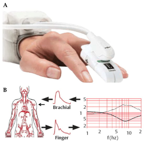
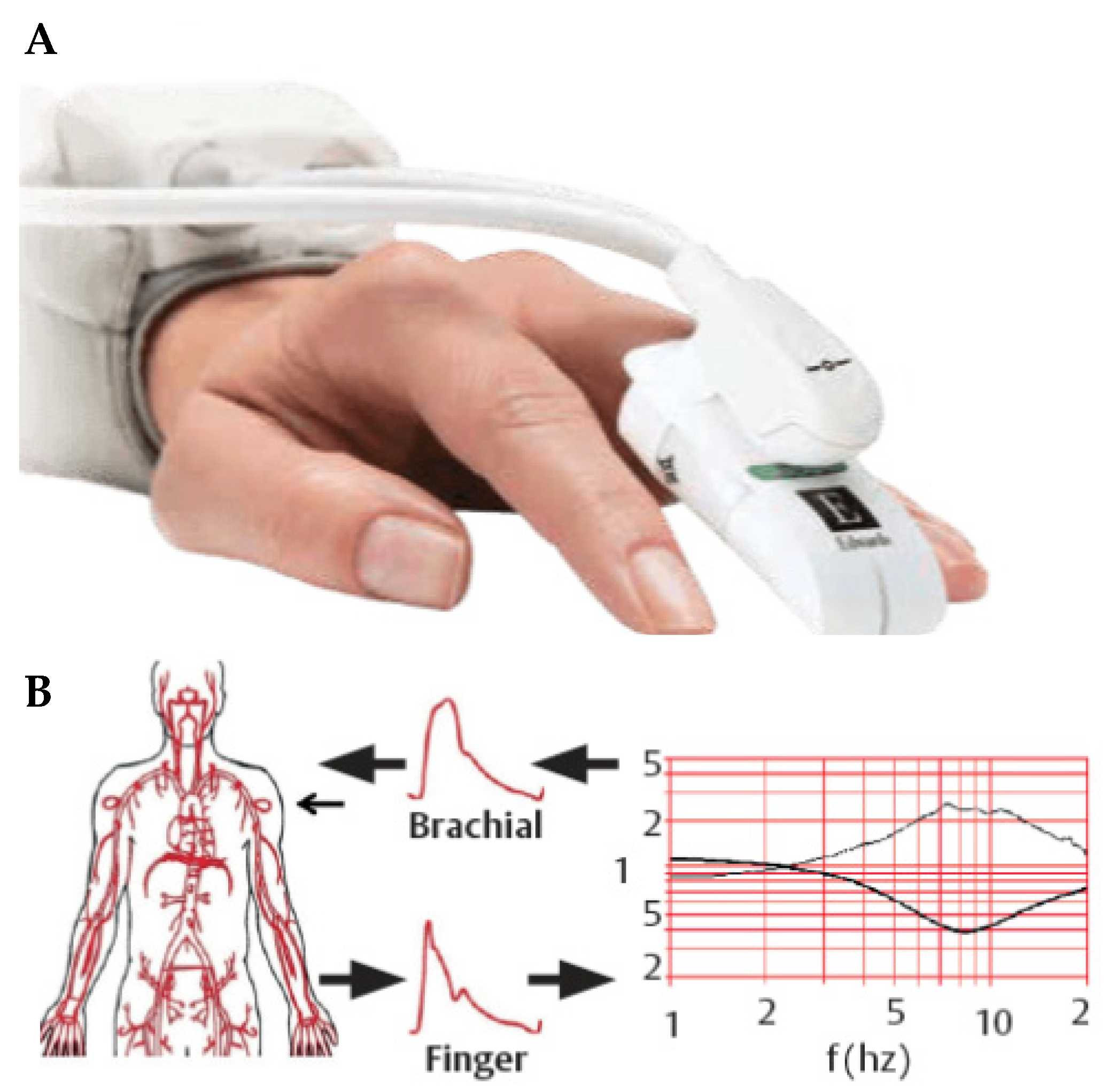
The ClearSight® system (Edwards Lifesciences, Irvine, CA, USA) works non-invasively by utilizing an inflatable cuff which adjusts automatically to measure the arterial blood pressure waveform over the full pulse, based on the Peňáz principle (Figure 1). In this cuff, there is an infrared photodiode and light detector that measures the diameter of the finger artery [8]. It is the absorption of the infrared light through the finger that is used to measure the arterial volume [16]. Cuff pressure is constantly adjusted during the cardiac cycle to keep the volume in the finger constant and these adjustments over time are used to create the blood pressure waveform and enable analysis of the pulse wave [15]. Different pathophysiological changes affect the arterial compliance and resistance, but the ClearSight® system can adjust to these changes. ClearSight® is able to provide information regarding blood pressure, heart rate, SV, CO, cardiac index (CI), indexed stroke volume (SVI), stroke volume variation (SVV), systemic vascular resistance (SVR) and SVR index [8].
Figure 1.
(A) ClearSight® finger cuff and (B) schematic of the reconstruction of brachial arterial pressure waveform from finger arterial pressure waveform [18].
The ClearSight® cuff was applied to the patient’s finger according to the instructions for use at the beginning of the TAVI procedure. Based on the standard setup of the operation room, the left index finger was used for all patients. The cuff was then connected to the monitor. The monitor unit recorded the measurements continuously during the whole procedure and saved all measurements. After the procedure, all measurements were assigned to the study-specific study ID and transferred to the database.
2.4. Data Collection and Follow-Up
Baseline characteristic, imaging data from transthoracic echocardiography before and after the procedure, laboratory results, procedural data, outcome data as well as follow-up information within 30 days after the procedure have been collected. Adverse events were systematically collected during the 30 days of follow-up and adjudicated by a dedicated clinical event committee based on detailed documentation of reported endpoints using the VARC-3 definitions.
2.5. Study Objectives
The first aim of this study was to test the correlation of non-invasive measurements (ClearSight®) compared with invasive measurements (radial and aortal measurements). Systolic and diastolic blood pressure, heart rate, CO, CI, and SV measured with ClearSight® and RHC were compared at a certain timepoint a few minutes before and after valve implantation.
The second aim was to show trends of hemodynamic parameters (systolic and diastolic BP, CO, CI, HR, and SV) during the whole procedure and especially during rapid pacing and valve implantation.
2.6. Statistical Analysis
Categorical variables are shown as count (percentage), and continuous variables as median (interquartile range [IQR]). Comparisons between continuous variables were performed using the Mann–Whitney U test. Categorical variables were compared using Pearson’s Chi-squared test or Fisher’s exact test, as appropriate. Absolute and relative differences in patients’ hemodynamic parameters measured invasively and non-invasively immediately during the TAVI procedure were calculated and illustrated with Bland–Altman plots. The absolute bias was defined as the differences between invasive and non-invasive measurements and the precision as standard deviation (SD). The lower and upper limits of agreement (LoA) were defined as 1.96 × SD. Coefficients of error (CE) were calculated by dividing the SD of the differences by the mean of the measurements, while the percentages of error (PE) were defined as 2 × CE × 100. Acceptable levels of correlation were defined as an absolute bias of 20% and a PE of 30%, as previously used by Di Cori et al. [19]. A p value < 0.05 was considered statistically significant. All statistical analyses were performed using R 4.2.3 (R Foundation for Statistical Computing, Vienna, Austria).
3. Results
3.1. Baseline Patient and Procedural Characteristics
Patients were recruited from March to August 2023. Median age was 82.0 [78, 86] years old and 50% were female. Median Euro SCORE II was 1.90 [1.40, 4.30], while the STS calculated risk of mortality was 2.40 [1.65, 4.02].
Median left ventricular ejection fraction (LVEF) was 60% and the majority of patients (94%) had a NYHA-Score of II or more. No patients had undergone previous aortic valve surgery or TAVI (Table 1). 36% of patients had coronary artery disease and 14% underwent revascularization. Atrial fibrillation or flutter was present in 18% of patients.
Table 1.
Characteristics of patients.
All patients underwent femoral route access. A total of 32 patients (64%) received a SAPIEN S3 or SAPIEN S3 Ultra valve, 10 (20%) an Evolut pro+, and 8 (16%) a Acurate NEO 2 device. The most common prosthesis sizes were 23 and 26 mm (8 and 11 patients).
3.2. Continuous Non-Invasive Measurements
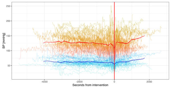
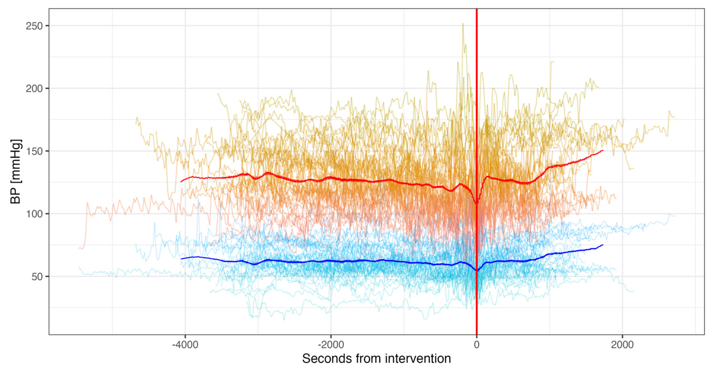
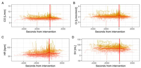
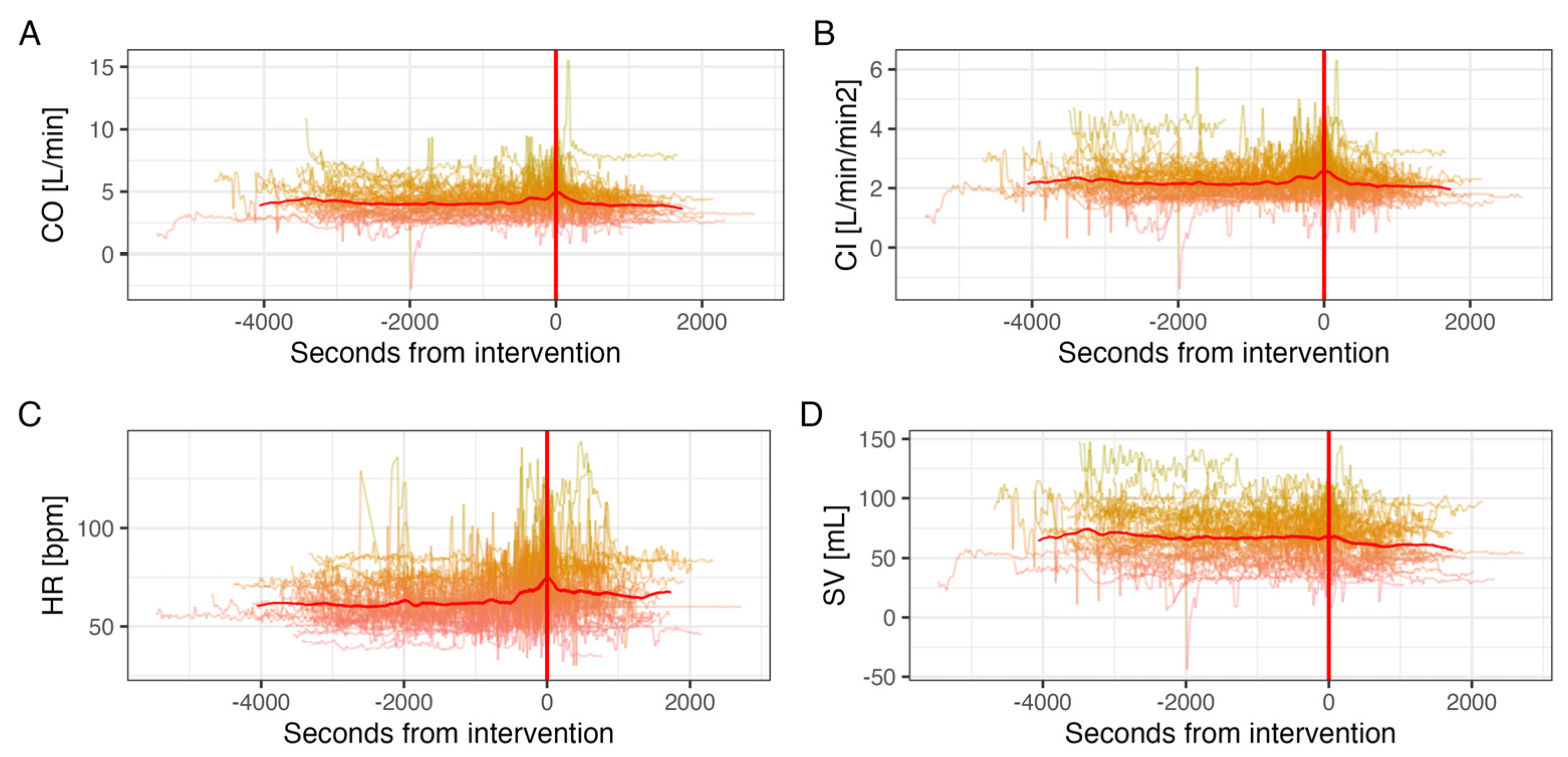
Continuous non-invasive measurements of systolic and diastolic BP, CO, CI, HR and SV were collected for all 50 participants. Patients had a mean BP of 125/61 ± 26/13 mmHg before valve implantation. After implantation, patients had a mean BP of 130/64 ± 28/15 mmHg (Figure 2). CI and CO remained similar between the pre- and post-implantation period (Figure 3A,B). HR was significantly higher pre-implantation than post-implantation (65 vs. 60 bpm, p < 0.001) (Figure 3C). SV decreased continuously from pre- to post-implantation (67 vs. 60 mL, p < 0.001) (Figure 3D).
Figure 2.
Mean continuous non-invasive (ClearSight®) blood pressure measurements from pre- to post-interventional. (Valve deployment marked with red line, systolic blood pressure in red, diastolic in blue).
Figure 3.
Mean continuous non-invasive (ClearSight®) (A) cardiac output, (B) cardiac index, (C) heart rate, and (D) stroke volume measurements from pre-to post interventional. (Valve deployment marked with red line).
3.3. Agreement Between Non-Invasive, Aortal, and Radial Measurements
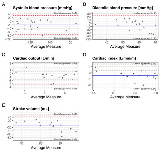
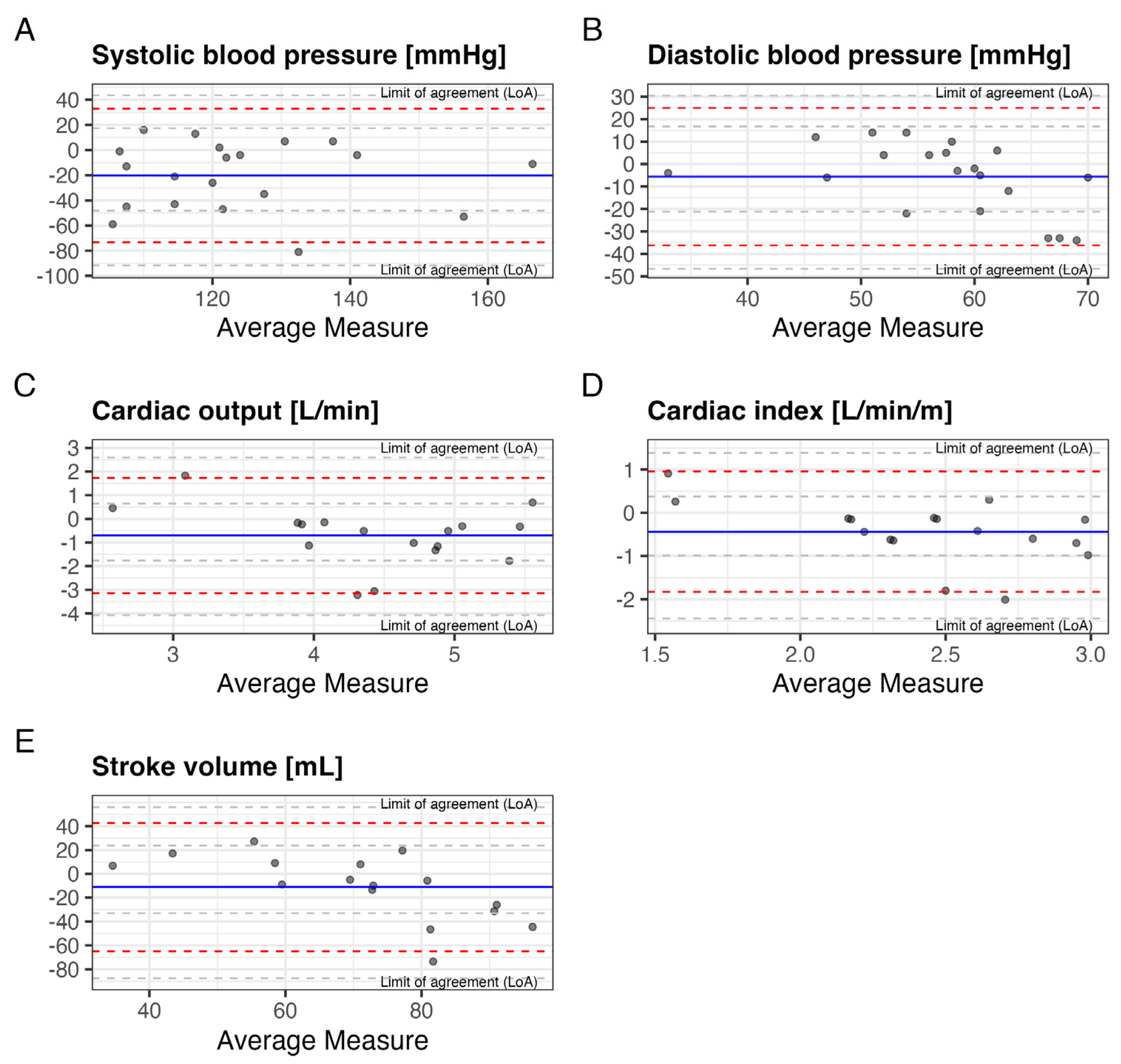
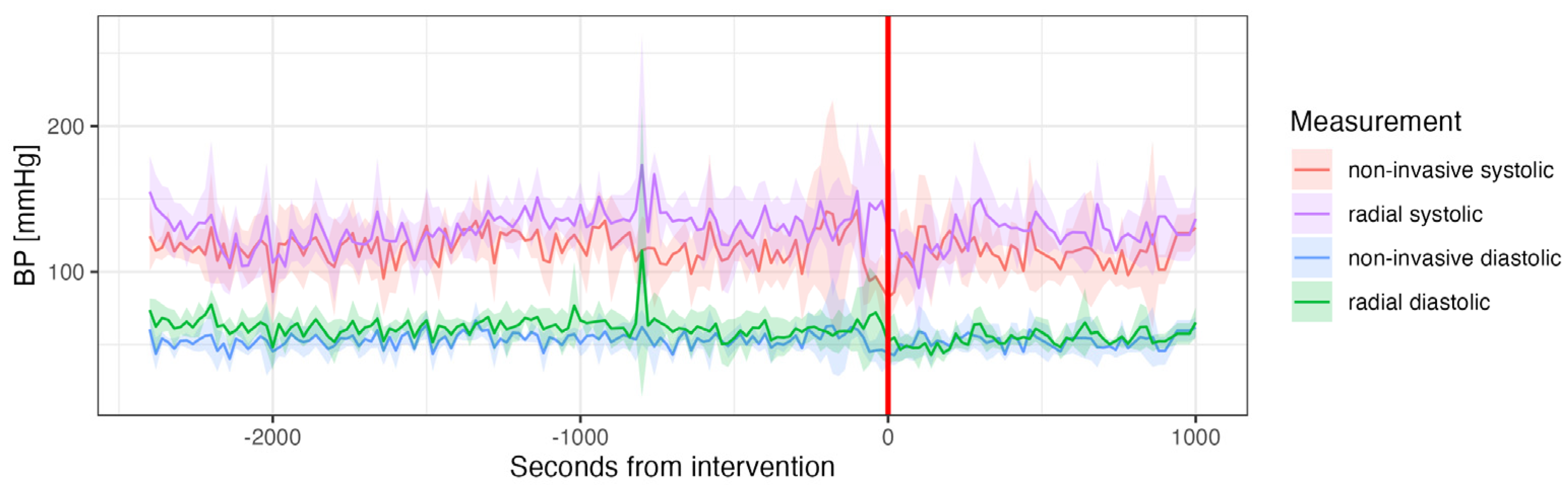
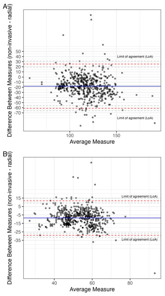
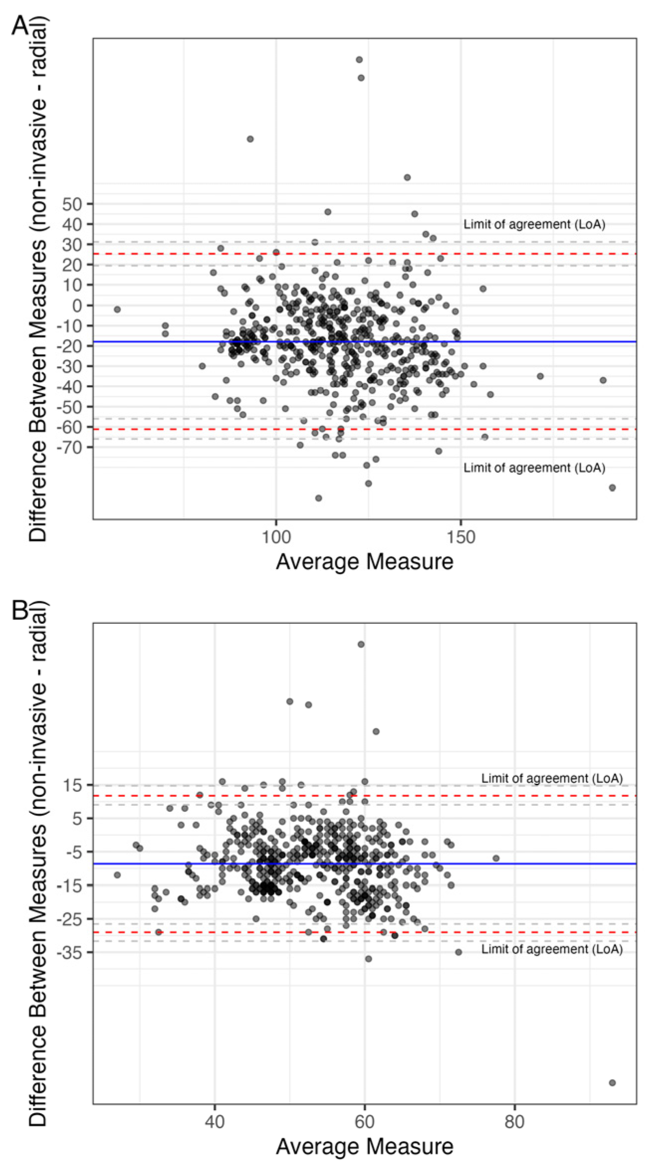
A comparison between non-invasive and radial measurements was performed in 25 patients. Invasive measurements of systolic BP, CO and CI were significantly higher than non-invasive ones (132 vs. 113 mmHg, p = 0.004; 4.8 vs. 4.1 L/min, p = 0.03, and 2.7 vs. 2.2 L/min/m2, p = 0.01). Invasive measurements of diastolic BP and SV were similar to non-invasive ones (59.5 vs. 57 mmHg, p = 0.24, and 77 vs. 66 mL, p = 0.25). (Figure 4A–E) Comparisons of continuous radial to non-invasive measurements of systolic and diastolic blood pressure revealed consistently higher values for radial measurements in both systolic, as well as diastolic measurements (127/60 vs. 113/52 mmHg, p < 0.001 for both), with no difference from before to after valve deployment. There was no significant difference between aortal and radial BP measurements (Figure 5 and Figure 6A,B). None of the observed values showed correlation within the pre-defined acceptable levels of absolute bias (20%) and PE (30%). (Mean PE 52%) A summary table of all results from performed Bland–Altman analyses is given in Table 2.
Figure 4.
Bland–Altman plots comparing aortal to non-invasive (ClearSight®) measurements of (A) Systolic, (B) diastolic blood pressure, (C) cardiac output, (D) cardiac index and (E) stroke volume.
Figure 5.
Comparison of mean measurements of radial and non-invasive (ClearSight®) blood pressure measurements and standard deviation over time. (Valve deployment marked with red line).
Figure 6.
Bland–Altman plots comparing radial to non-invasive (ClearSight®) measurements of (A) systolic and (B) diastolic blood pressure.
Table 2.
Summary statistics of the BA analyses comparing non-invasive ones (ClearSight®) measurements (aortal for SV, CO, and CI and radial for BP) and invasive ones.
3.4. Outcomes
Device success was achieved in all patients. Follow-up information at 30 days was available in all patients. Patients were discharged on average after 3 days [1, 5]. Mean gradient at discharge was 10 [8, 15] mmHg. During the 30-day follow-up, no patient died or suffered any major bleeding, myocardial infarction, stroke, or other adverse events.
4. Discussion
In this study, we aimed to investigate the utility of non-invasive compared to invasive hemodynamic measurements among 50 patients undergoing TAVI at two large, tertiary university hospitals. We report five major findings:
First, median systolic and diastolic radial, as well as aortic BP measurements were consistently and significantly higher compared to non-invasive measurements (127/60 and 132/60 vs. 113/52 mmHg, p < 0.001; absolute SD: 22.07/10.41 mmHg and 27.15/15.61 mmHg, PE: 38/39% and 44/55%). Di Cori et al. found similar results in patients both with and without atrial fibrillation [19], while Yahagi and Sasaki, and Vos et al. reported comparable percentage errors for systolic and diastolic BP (24.9% and 31.9%) [15,20]. Radial (systolic) BP tends to be higher than central aortal BP due to pulse pressure amplification, which occurs when the arterial pulse wave travels from the central aorta to the peripheral arteries. This results in higher systolic and lower diastolic pressures at peripheral sites like the radial artery. This consistent finding may therefore be attributed to the anatomical and physiological differences in pressure waveforms between the radial artery and central circulatory measurements. Recent publications, however, have shown a much narrower difference between systolic aortic and radial BP measurements, and no differences in mean and diastolic BP, highlighting the need for further research on the topic [21]. Furthermore, differences in arterial stiffness could contribute to discrepancies in measurements [22].
Second, CO, CI and SV measurements were slightly higher when obtained through invasive methods compared to non-invasive measurements (mean bias of −62% for non-invasive measurements and mean PE of 51%). Prior research has demonstrated ClearSight® to be both accurate and safe when compared to Pulse Contour Cardiac Output (PiCCO) and RHC, the current gold standard in continuous hemodynamic monitoring [11,12,13,14,15]. Gellert et al. analyzed ten previous studies that compared invasive and non-invasive CO measurements and found similar standard deviations (SD) and percentage errors (PE), as well as generally lower non-invasive measurements, consistent with our findings (mean SD: 0.4, mean PE: 43%) [11]. Overall, our findings in patients with severe AS were consistent to prior studies in different clinical settings.
Third, all evaluated parameters showed significant discrepancies between invasive and non-invasive measurements; however, none of the analyzed parameters fell below the threshold of acceptable bias of 20% and percentage error of 30%, as previously used by Di Cori et al. [19].
Fourth, continuous measurements reveal a drop in BP during valve deployment, as well as a spike in CO, CI and SV. This might be explained by a temporary obstruction of the LVOT during valve deployment and a subsequent increase in output after the obstruction is released. There is limited data on the effect of valve deployment on hemodynamic parameters. However, Alperi et al. have shown a significant drop in valvular gradients periprocedurally, which fits our findings [23].
Fifth, our study did not allow for an evaluation of the impact on clinical endpoints due to a lack of events. This is unlikely due to an unusually healthy patient cohort, as our baseline characteristics were comparable to those in other studies [24,25]. Despite representing one of the largest populations investigating the peri-interventional application of non-invasive monitoring in patients undergoing TAVI, our findings suggest that a larger sample size and longer follow-up period may be necessary to validate these observations further. The absence of clinical events highlights the need for extended monitoring to capture more comprehensive data on potential clinical outcomes. Future studies should investigate possible benefits of ClearSight® in earlier diagnosis of peri- and immediately post-TAVI complications.
ClearSight® has shown a level concordance of 84–100% with PAC and PiCCO, indicating that it generally follows the same trend as these gold-standard measurements, though the absolute values may differ [16]. This discrepancy might be due to inherent differences in measurement techniques, with invasive methods providing more direct and possibly more precise assessments of hemodynamic parameters. Additionally, ClearSight® has shown good correlation with other methods such as FloTrac and echocardiography, but large differences in absolute values have been noted [12,26].
5. Conclusions
In conclusion, while non-invasive measurements offer a valuable alternative for continuous hemodynamic monitoring and can effectively indicate general trends observed with invasive techniques, they currently lack the precision and accuracy of invasive methods. The discrepancies in BP, CO, CI, SV measurements between the two modalities highlight the inherent limitations of non-invasive techniques. Despite these limitations, non-invasive systems like ClearSight® have demonstrated concordance with invasive methods and provide significant clinical benefits, such as earlier detection of hypotensive episodes and a lower risk of complications. However, for critical care settings where precise hemodynamic monitoring is essential, invasive techniques remain the gold standard. Further research with larger sample sizes and extended follow-up periods is warranted to enhance the accuracy and reliability of non-invasive monitoring systems and to fully evaluate their impact on clinical outcomes.
Author Contributions
Conceptualization, T.N. and T.S.; methodology, T.N.; software, T.N. and T.S.; formal analysis, T.N. and T.S.; investigation, T.N. and T.S.; data curation, T.N. and T.S.; writing—original draft preparation, T.N. and T.S.; writing—review and editing, all authors; visualization, T.N. and T.S.; supervision, T.N.; project administration, T.N.; funding acquisition, T.N. All authors have read and agreed to the published version of the manuscript.
Funding
Thomas Nestelberger has received research support from the Swiss National Science Foundation (P400PM_191037/1), the Prof. Dr. Max Cloëtta Foundation, the Margarete und Walter Lichtenstein-Stiftung (3MS1038), and the University Hospital Basel as well as speaker honoraria/consulting honoraria from Edwards Lifesciences, Siemens, Beckman Coulter, Bayer, Ortho Clinical Diagnostics and Orion Pharma, outside the submitted work. Jasper Boeddinghaus is supported by an Edinburgh Doctoral College Scholarship and research grants from the University of Basel, the University Hospital of Basel, the Division of Internal Medicine, the Swiss Academy of Medical Sciences, the Gottfried and Julia Bangerter-Rhyner Foundation, the Swiss National Science Foundation, and the “Freie Akademische Gesellschaft” and has received honoraria from Siemens, Roche Diagnostics, Ortho Clinical Diagnostics, Quidel Corporation, and Beckman Coulter, and travel support from Medtronic and Cordis, all outside the submitted work. Felix Mahfoud is supported by Deutsche Gesellschaft für Kardiologie (DGK), Deutsche Forschungsgemeinschaft (SFB TRR219, Project-ID 322900939), and Deutsche Herzstiftung. Saarland University has received scientific support from Ablative Solutions, Medtronic and ReCor Medical. Until May 2024, Felix Mahfoud has received speaker honoraria/consulting fees from Ablative Solutions, Amgen, Astra-Zeneca, Bayer, Boehringer Ingelheim, Inari, Medtronic, Merck, ReCor Medical, Servier, and Terumo.
Institutional Review Board Statement
The study was conducted according to the guidelines of the Declaration of Helsinki, and approved by the local Ethics Committee (Ethikkommission Nordwest- und Zentralschweiz EKNZ, protocol code SNCTP000005347, 03.02.2023).
Informed Consent Statement
Informed consent was obtained from all subjects involved in the study.
Data Availability Statement
The data that support the findings of this study are available from the corresponding author, TN, upon reasonable request.
Acknowledgments
We are indebted to the patients who participated in the study and to the study nurse team, for their most valuable efforts.
Conflicts of Interest
All other authors declare that they have no conflict of interest with this study and that they have critically reviewed the manuscript and approved the final version for submission. The manuscript and its contents have not been published previously and are not being considered for publications elsewhere in whole or in part in any language, including publicly accessible web sites or e-print servers.
Abbreviations
The following abbreviations are used in this manuscript:
| AS | Aortic valve stenosis |
| TAVI | Transcatheter Aortic Valve Implantation |
| SAVR | Surgical Aortic Valve Replacement |
| RHC | Right heart catheterization |
| CO | Cardiac output |
| CI | Cardiac index |
| SV | Stroke volume |
| SVI | Stroke volume index |
| SVV | Stroke volume variation |
| SVR | Systemic vascular resistance |
| PiCCO | Pulse Contour Cardiac Output |
| PAC | Pulmonary artery catheterization |
| BP | Blood pressure |
| HR | Heart rate |
| IQR | Interquartile range |
| SD | Standard deviation |
| LoA | Limits of agreement |
| CE | Coefficients of error |
| PE | Percentages of error |
| LVEF | Left ventricular ejection fraction |
| NYHA | New York Heart Association |
| VARC3 | Valve Academic Research Consortium3 |
| EuroSCORE II | European System for Cardiac Operative Risk Evaluation II |
| STS | Society of Thoracic Surgeons |
References
- Nishimura, R.A.; Otto, C.M.; Bonow, R.O.; Carabello, B.A.; Erwin, J.P.; Guyton, R.A.; O’Gara, P.T.; Ruiz, C.E.; Skubas, N.J.; Sorajja, P.; et al. 2014 AHA/ACC Guideline for the Management of Patients With Valvular Heart Disease: A report of the American College of Cardiology/American Heart Association Task Force on Practice Guidelines. Circulation 2014, 129, e521–e643. [Google Scholar] [CrossRef] [Scilit] [PubMed]
- Baumgartner, H.; Falk, V.; Bax, J.J.; De Bonis, M.; Hamm, C.; Holm, P.J.; Iung, B.; Lancellotti, P.; Lansac, E.; Rodriguez Muñoz, D.; et al. 2017 ESC/EACTS Guidelines for the management of valvular heart disease. Eur. Heart J. 2017, 38, 2739–2791. [Google Scholar] [CrossRef] [Scilit] [PubMed]
- Otto, C.M.; Nishimura, R.A.; Bonow, R.O.; Carabello, B.A.; Erwin, J.P.; Gentile, F.; Jneid, H.; Krieger, E.V.; Mack, M.; McLeod, C.; et al. 2020 ACC/AHA Guideline for the Management of Patients With Valvular Heart Disease: A Report of the American College of Cardiology/American Heart Association Joint Committee on Clinical Practice Guidelines. Circulation 2021, 143. [Google Scholar] [CrossRef] [Scilit]
- Mack, M.J.; Leon, M.B.; Thourani, V.H.; Makkar, R.; Kodali, S.K.; Russo, M.; Kapadia, S.R.; Malaisrie, S.C.; Cohen, D.J.; Pibarot, P.; et al. Transcatheter Aortic-Valve Replacement with a Balloon-Expandable Valve in Low-Risk Patients. N. Engl. J. Med. 2019, 380, 1695–1705. [Google Scholar] [CrossRef] [Scilit]
- Engoren, M.; Barbee, D. Comparison of cardiac output determined by bioimpedance, thermodilution, and the Fick method. Am. J. Crit. Care 2005, 14, 40–45. [Google Scholar] [CrossRef] [Scilit]
- Baumgartner, H.; Hung, J.; Bermejo, J.; Chambers, J.B.; Evangelista, A.; Griffin, B.P.; Iung, B.; Otto, C.M.; Pellikka, P.A.; Quiñones, M.; et al. Echocardiographic assessment of valve stenosis: EAE/ASE recommendations for clinical practice. J. Am. Soc. Echocardiogr. 2009, 22, 1–23; quiz 101–102. [Google Scholar] [CrossRef] [Scilit]
- Gorlin, R.; Gorlin, S.G. Hydraulic formula for calculation of the area of the stenotic mitral valve, other cardiac valves, and central circulatory shunts. I. Am. Heart J. 1951, 41, 1–29. [Google Scholar] [CrossRef] [Scilit]
- Pour-Ghaz, I.; Manolukas, T.; Foray, N.; Raja, J.; Rawal, A.; Ibebuogu, U.N.; Khouzam, R.N. Accuracy of non-invasive and minimally invasive hemodynamic monitoring: Where do we stand? Ann. Transl. Med. 2019, 7, 421. [Google Scholar] [CrossRef] [Scilit]
- Teboul, J.-L.; Saugel, B.; Cecconi, M.; De Backer, D.; Hofer, C.K.; Monnet, X.; Perel, A.; Pinsky, M.R.; Reuter, D.A.; Rhodes, A.; et al. Less invasive hemodynamic monitoring in critically ill patients. Intensive Care Med. 2016, 42, 1350–1359. [Google Scholar] [CrossRef] [Scilit]
- Kuck, K.; Baker, P.D. Perioperative Noninvasive Blood Pressure Monitoring. Anesth. Analg. 2018, 127, 408–411. [Google Scholar] [CrossRef] [Scilit]
- Gellert, G.; Bramlage, P. Use of the ClearSight® System for Continuous Noninvasive Hemodynamic Monitoring during Heart Valve Interventions: Review of the Literature and Single-Site Experience. Heart Surg. Forum 2018, 21, E476–E483. [Google Scholar] [CrossRef] [Scilit] [PubMed]
- van der Spoel, A.G.E.; Voogel, A.J.; Folkers, A.; Boer, C.; Bouwman, R.A. Comparison of noninvasive continuous arterial waveform analysis (Nexfin) with transthoracic Doppler echocardiography for monitoring of cardiac output. J. Clin. Anesth. 2012, 24, 304–309. [Google Scholar] [CrossRef] [Scilit] [PubMed]
- Eeftinck Schattenkerk, D.W.; van Lieshout, J.J.; van den Meiracker, A.H.; Wesseling, K.R.; Blanc, S.; Wieling, W.; van Montfrans, G.A.; Settels, J.J.; Wesseling, K.H.; Westerhof, B.E. Nexfin noninvasive continuous blood pressure validated against Riva-Rocci/Korotkoff. Am. J. Hypertens. 2009, 22, 378–383. [Google Scholar] [CrossRef] [Scilit]
- Martina, J.R.; Westerhof, B.E.; van Goudoever, J.; de Beaumont, E.M.F.H.; Truijen, J.; Kim, Y.-S.; Immink, R.V.; Jöbsis, D.A.; Hollmann, M.W.; Lahpor, J.R.; et al. Noninvasive continuous arterial blood pressure monitoring with Nexfin®. Anesthesiology 2012, 116, 1092–1103. [Google Scholar] [CrossRef] [Scilit]
- Vos, J.J.; Poterman, M.; Mooyaart, E.a.Q.; Weening, M.; Struys, M.M.R.F.; Scheeren, T.W.L.; Kalmar, A.F. Comparison of continuous non-invasive finger arterial pressure monitoring with conventional intermittent automated arm arterial pressure measurement in patients under general anaesthesia. Br. J. Anaesth. 2014, 113, 67–74. [Google Scholar] [CrossRef] [Scilit]
- Ameloot, K.; Palmers, P.-J.; Malbrain, M.L.N.G. The accuracy of noninvasive cardiac output and pressure measurements with finger cuff: A concise review. Curr. Opin. Crit. Care 2015, 21, 232–239. [Google Scholar] [CrossRef] [Scilit]
- Geerts, B.F.; Aarts, L.P.; Jansen, J.R. Methods in pharmacology: Measurement of cardiac output. Br. J. Clin. Pharmacol. 2011, 71, 316–330. [Google Scholar] [CrossRef] [Scilit]
- Edwards Lifesciences Corporation ClearSight System. Noninvasive Continuous Hemodynamic Monitoring. Available online: https://www.edwards.com/healthcare-professionals/products-services/hemodynamic-monitoring/clearsight-system (accessed on 12 April 2024).
- Di Cori, A.; Parollo, M.; Fiorentini, F.; Della Volpe, S.; Mazzocchetti, L.; Barletta, V.; Segreti, L.; Viani, S.; De Lucia, R.; Paperini, L.; et al. Feasibility and Accuracy of Noninvasive Continuous Arterial Pressure Monitoring during Transcatheter Atrial Fibrillation Ablation. JCM 2023, 12, 2388. [Google Scholar] [CrossRef] [Scilit]
- Yahagi, M.; Sasaki, M. Comparison of direct intra-arterial pressure and ClearSight finger cuff arterial pressure measurements in elderly patients undergoing transcatheter aortic valve replacement. Blood Pressure Monitoring 2023, 28, 309–315. [Google Scholar] [CrossRef] [Scilit]
- Chemla, D.; Millasseau, S. A systematic review of invasive, high-fidelity pressure studies documenting the amplification of blood pressure from the aorta to the brachial and radial arteries. J. Clin. Monit. Comput. 2021, 35, 1245–1252. [Google Scholar] [CrossRef] [Scilit]
- Finnegan, T.P.; Spence, J.D.; Wong, D.G.; Wells, G.A. Blood pressure measurement in the elderly: Correlation of arterial stiffness with difference between intra-arterial and cuff pressures. J. Hypertens. 1985, 3, 231–235. [Google Scholar] [CrossRef] [Scilit] [PubMed]
- Alperi, A.; Moris, C.; Del Valle, R.; Pascual, I.; Antuna, P.; Almendárez, M.; Hernández-Vaquero, D.; Betanzos, J.L.; Rodés-Cabau, J.; Avanzas, P. Hemodynamic Performance of Self-expandable Transcatheter Aortic Valve Replacement Systems During Valve Deployment. J. Invasive Cardiol. 2024. [Google Scholar] [CrossRef] [Scilit] [PubMed]
- Stolte, T.; Boeddinghaus, J.; Allegra, G.; Leibundgut, G.; Reuthebuch, O.; Kaiser, C.; Müller, C.; Nestelberger, T. Incidence and Outcomes of Valve-in-Valve Transcatheter Aortic Valve Implantation in Failed Bioprosthetic Valves. J. Clin. Med. 2023, 12, 5868. [Google Scholar] [CrossRef] [Scilit]
- Reichl, J.J.; Stolte, T.; Tang, S.; Boeddinghaus, J.; Wagener, M.; Leibundgut, G.; Kaiser, C.A.; Nestelberger, T. Prognostic Impact of Left Ventricular Ejection Fraction Improvement after Transcatheter Aortic Valve Replacement. J. Clin. Med. 2024, 13, 3639. [Google Scholar] [CrossRef] [Scilit]
- Sakai, Y.; Yasuo, M.T.; Oyama, T.; Murakami, C.; Kakuta, N.; Tanaka, K. Noninvasive continuous blood pressure monitoring by the ClearSight system during robot-assisted laparoscopic radical prostatectomy. J. Med. Investig. 2018, 65, 69–73. [Google Scholar] [CrossRef] [Scilit]

Disclaimer/Publisher’s Note: The statements, opinions and data contained in all publications are solely those of the individual author(s) and contributor(s) and not of MDPI and/or the editor(s). MDPI and/or the editor(s) disclaim responsibility for any injury to people or property resulting from any ideas, methods, instructions or products referred to in the content. |
© 2025 by the authors. Licensee MDPI, Basel, Switzerland. This article is an open access article distributed under the terms and conditions of the Creative Commons Attribution (CC BY) license (https://creativecommons.org/licenses/by/4.0/).