Measures of Corticalization


Abstract
:1. Introduction
2. Materials and Methods
3. Results
4. Discussion
5. Conclusions
Funding
Institutional Review Board Statement
Informed Consent Statement
Data Availability Statement
Acknowledgments
Conflicts of Interest
References
- Carneiro, L.S.; da Cunha, H.A.; Leles, C.R.; Mendonça, E.F. Digital subtraction radiography evaluation of longitudinal bone density changes around immediate loading implants: A pilot study. Dentomaxillofac. Radiol. 2012, 41, 241–247. [Google Scholar] [CrossRef] [PubMed]
- Hartman, G.A.; Cochran, D.L. Initial implant position determines the magnitude of crestal bone remodeling. J. Periodontol. 2004, 75, 572–577. [Google Scholar] [CrossRef] [PubMed]
- Kozakiewicz, M.; Skorupska, M.; Wach, T. What Does Bone Corticalization around Dental Implants Mean in Light of Ten Years of Follow-Up? J. Clin. Med. 2022, 11, 3545. [Google Scholar] [CrossRef] [PubMed]
- Kozakiewicz, M.; Szymor, P.; Wach, T. Influence of General Mineral Condition on Collagen-Guided Alveolar Crest Augmentation. Materials 2020, 13, 3649. [Google Scholar] [CrossRef]
- Kozakiewicz, M.; Wach, T. New oral surgery materials for bone reconstruction—A comparison of five bone substitute materials for dentoalveolar augmentation. Materials 2020, 13, 2935. [Google Scholar] [CrossRef]
- Kozakiewicz, M.; Gabryelczak, I. The Osteosynthesis of the Mandibular Head, Does the Way the Screws Are Positioned Matter? J. Clin. Med. 2022, 11, 2031. [Google Scholar] [CrossRef]
- Kozakiewicz, M.; Wilamski, M. Technika standaryzacji wewnątrzustnych zdjęć rentgenowskich [Standardization technique for intraoral radiographs]. Czas. Stomat. 1999, 52, 673–677. [Google Scholar]
- Szczypiński, P.M.; Strzelecki, M.; Materka, A.; Klepaczko, A. MaZda-A software package for image texture analysis. Comput. Methods Programs Biomed. 2009, 94, 66–76. [Google Scholar] [CrossRef]
- Kozakiewicz, M.; Bogusiak, K.; Hanclik, M.; Denkowski, M.; Arkuszewski, P. Noise in subtraction images made from pairs of intraoral radiographs: A comparison between four methods of geometric alignment. Dentomaxillofac. Radiol 2008, 37, 40–46. [Google Scholar] [CrossRef]
- Kołaciński, M.; Kozakiewicz, M.; Materka, A. Textural entropy as a potential feature for quantitative assessment of jaw bone healing process. Arch. Med. Sci. 2015, 11, 78–84. [Google Scholar] [CrossRef]
- Wach, T.; Kozakiewicz, M. Are recent available blended collagen-calcium phosphate better than collagen alone or crystalline calcium phosphate? Radiotextural analysis of a 1-year clinical trial. Clin. Oral Investig. 2021, 25, 3711–3718. [Google Scholar] [CrossRef] [PubMed]
- Wach, T.; Kozakiewicz, M. Fast-Versus Slow-Resorbable Calcium Phosphate Bone Substitute Materials—Texture Analysis after 12 Months of Observation. Materials 2020, 13, 3854. [Google Scholar] [CrossRef] [PubMed]
- Haralick, R. Statistical and Structural Approaches to Texture. Proc. IEEE 1979, 67, 786–804. [Google Scholar] [CrossRef]
- Materka, A.; Strzelecki, M. Texture Analysis Methods—A Review, COST B11 Report (Presented and Distributed at MC Meeting and Workshop in Brussels, June 1998); Technical University of Lodz: Lodz, Poland, 1998. [Google Scholar]
- Appleton, R.S.; Nummikoski, P.V.; Pigno, M.A.; Cronin, R.J.; Chung, K.-H. A radiographic assessment of progressive loading on bone around single osseointegrated implants in the posterior maxilla. Clin. Oral Implants Res. 2005, 16, 161–167. [Google Scholar] [CrossRef]
- Brägger, U. Digital imaging in periodontal radiography. A review. J. Clin. Periodontol. 1988, 15, 551–557. [Google Scholar] [CrossRef] [PubMed]
- Brägger, U.; Bürgin, W.; Marconi, M.; Häsler, R.U.; Lang, N.P. Influence of contrast enhancement and pseudocolor transformation on the diagnosis with digital subtraction images (DSI). J. Periodontal. Res. 1994, 29, 95–102. [Google Scholar] [CrossRef]
- Brägger, U.; Pasquali, L. Color conversion of alveolar bone density changes in digital subtraction images. J. Clin. Periodontol. 1989, 16, 209–214. [Google Scholar] [CrossRef]
- Dudek, D.; Kozakiewicz, M. Szerokość beleczek kostnych w szczęce i żuchwie człowieka na podstawie cyfrowych radiologicznych zdjęć wewnąrzustnych [Bone trabecula width in the human maxilla and mandible based on digital intraoral radiographs]. Mag. Stomat. 2012, 236, 77–80. [Google Scholar]
- Duinkerke, A.S.; van de Poel, A.C.; Doesburg, W.H.; Lemmens, W.A. Densitometric analysis of experimentally produced periapical radiolucencies. Oral Surg. Oral Med. Oral Pathol. 1977, 43, 782–797. [Google Scholar] [CrossRef]
- Gröndahl, H.G.; Gröndahl, K.; Webber, R.L. A digital subtraction technique for dental radiography. Oral Surg. Oral Med. Oral Pathol. 1983, 55, 96–102. [Google Scholar] [CrossRef]
- de Molon, R.S.; Batitucci, R.G.; Spin-Neto, R.; Paquier, G.M.; Sakakura, C.E.; Tosoni, G.M.; Scaf, G. Comparison of changes in dental and bone radiographic densities in the presence of different soft-tissue simulators using pixel intensity and digital subtraction analyses. Dentomaxillofac. Radiol. 2013, 42, 20130235. [Google Scholar] [CrossRef] [PubMed]
- da Silva, R.; Duailibi Neto, E.F.; Todescan, F.F.; Ruiz, G.M.; Pannuti, C.M.; Chilvarquer, I. Evaluation of cervical peri-implant optical density in longitudinal control of immediate implants in the anterior maxilla region. Dentomaxillofac. Radiol. 2020, 49, 20190396. [Google Scholar] [CrossRef] [PubMed]
- Webber, R.L.; Ruttimann, U.E.; Heaven, T.J. Calibration errors in digital subtraction radiography. J. Periodontal. Res. 1990, 25, 268–275. [Google Scholar] [CrossRef]
- Bogowicz, M.; Vuong, D.; Huellner, M.W.; Pavic, M.; Andratschke, N.; Gabrys, H.S.; Guckenberger, M.; Tanadini-Lang, S. CT radiomics and PET radiomics: Ready for clinical implementation? Q. J. Nucl. Med. Mol. Imaging 2019, 63, 355–370. [Google Scholar] [CrossRef] [PubMed]
- Mayerhoefer, M.E.; Materka, A.; Langs, G.; Häggström, I.; Szczypiński, P.; Gibbs, P.; Cook, G. Introduction to Radiomics. J. Nucl. Med. 2020, 61, 488–495. [Google Scholar] [CrossRef] [PubMed]
- Noortman, W.A.; Vriens, D.; Grootjans, W.; Tao, Q.; de Geus-Oei, L.F.; Van Velden, F.H. Nuclear medicine radiomics in precision medicine: Why we can’t do without artificial intelligence. Q. J. Nucl. Med. Mol. Imaging 2020, 64, 278–290. [Google Scholar] [CrossRef]
- Reuzé, S.; Schernberg, A.; Orlhac, F.; Sun, R.; Chargari, C.; Dercle, L.; Deutsch, E.; Buvat, I.; Robert, C. Radiomics in Nuclear Medicine Applied to Radiation Therapy: Methods, Pitfalls, and Challenges. Int. J. Radiat. Oncol. Biol. Phys. 2018, 102, 1117–1142. [Google Scholar] [CrossRef]
- Pociask, E.; Nurzynska, K.; Obuchowicz, R.; Bałon, P.; Uryga, D.; Strzelecki, M.; Izworski, A.; Piórkowski, A. Differential Diagnosis of Cysts and Granulomas Supported by Texture Analysis of Intraoral Radiographs. Sensors 2021, 21, 7481. [Google Scholar] [CrossRef]
- Kozakiewicz, M.; Marciniak-Hoffman, A.; Denkowski, M. Long term comparison of application of two betatricalcium phosphates in oral surgery. Dent. Med. Probl. 2009, 46, 284–388. [Google Scholar]
- Kozakiewicz, M.; Marciniak-Hoffman, A.; Olszycki, M. Comparative Analysis of Three Bone Substitute Materials Based on Co-Occurrence Matrix. Dent. Med. Probl. 2010, 47, 23–29. [Google Scholar]
- Linkevicius, T.; Linkevicius, R.; Gineviciute, E.; Alkimavicius, J.; Mazeikiene, A.; Linkeviciene, L. The influence of new immediate tissue level abutment on crestal bone stability of subcrestally placed implants: A 1-year randomized controlled clinical trial. Clin. Implant Dent. Relat. Res. 2021, 23, 259–269. [Google Scholar] [CrossRef] [PubMed]
- Dudek, D. Ocena Przebudowy Kości Obciążonej Wszczepami Zębowymi z Zastosowaniem Cyfrowej Analizy Tekstur Obrazu Radiologicznego [Evaluation of Dental Implants Loaded Bone Remodeling Using Digital Texture Analysis in Radiographic Images]. Doctorate Thesis, Medical University of Lodz, Lodz, Poland, 2012. [Google Scholar]
- Baer, R.A.; Nölken, R.; Colic, S.; Heydecke, G.; Mirzakhanian, C.; Behneke, A.; Behneke, N.; Gottesman, E.; Ottria, L.; Pozzi, A.; et al. Immediately provisionalized tapered conical connection implants for single-tooth restorations in the maxillary esthetic zone: A 5-year prospective single-cohort multicenter analysis. Clin. Oral Investig. 2022, 26, 3593–3604. [Google Scholar] [CrossRef]
- Dowgierd, K.; Pokrowiecki, R.; Borowiec, M.; Kozakiewicz, M.; Smyczek, D.; Krakowczyk, Ł. A Protocol for the Use of a Combined Microvascular Free Flap with Custom-Made 3D-Printed Total Temporomandibular Joint (TMJ) Prosthesis for Mandible Reconstruction in Children. Appl. Sci. 2021, 11, 2176. [Google Scholar] [CrossRef]
- Kinaia, B.M.; Shah, M.; Neely, A.L.; Goodis, H.E. Crestal bone level changes around immediately placed implants: A systematic review and meta-analyses with at least 12 months’ follow-up after functional loading. J. Periodontol. 2014, 85, 1537–1548. [Google Scholar] [CrossRef] [PubMed]
- Kozakiewicz, M.; Gabryelczak, I. Bone Union Quality after Fracture Fixation of Mandibular Head with Compression Magnesium Screws. Materials 2022, 15, 2230. [Google Scholar] [CrossRef] [PubMed]
- Dowgierd, K.; Pokrowiecki, R.; Borowiec, M.; Sokolowska, Z.; Dowgierd, M.; Wos, J.; Kozakiewicz, M.; Krakowczyk, Ł. Protocol and Evaluation of 3D-Planned Microsurgical and Dental Implant Reconstruction of Maxillary Cleft Critical Size Defects in Adolescents and Young Adults. J. Clin. Med. 2021, 10, 2267. [Google Scholar] [CrossRef]
- Kütan, E.; Bolukbasi, N.; Yildirim-Ondur, E.; Ozdemir, T. Clinical and Radiographic Evaluation of Marginal Bone Changes around Platform-Switching Implants Placed in Crestal or Subcrestal Positions: A Randomized Controlled Clinical Trial. Clin. Implant Dent. Rel. Res. 2014, 17, e364–e375. [Google Scholar] [CrossRef]
- Moraschini, V.; Poubel, L.A.D.C.; Ferreira, V.F.; Barboza, E.D.S.P. Evaluation of survival and success rates of dental implants reported in longitudinal studies with a follow-up period of at least 10 years: A systematic review. Int. J. Oral Maxillofac. Surg. 2015, 44, 377–388. [Google Scholar] [CrossRef]
- Pellicer-Chover, H.; Díaz-Sanchez, M.; Soto-Peñaloza, D.; Peñarrocha-Diago, M.A.; Canullo, L.; Peñarrocha-Oltra, D. Impact of crestal and subcrestal implant placement upon changes in marginal peri-implant bone level. A systematic review. Med. Oral Patol. Oral Cir. Bucal 2019, 24, e673–e683. [Google Scholar] [CrossRef]
- Pokrowiecki, R.; Szałaj, U.; Fudala, D.; Zaręba, T.; Wojnarowicz, J.; Łojkowski, W.; Tyski, S.; Dowgierd, K.; Mielczarek, A. Dental Implant Healing Screws as Temporary Oral Drug Delivery Systems for Decrease of Infections in the Area of the Head and Neck. Int. J. Nanomed. 2022, 17, 1679–1693. [Google Scholar] [CrossRef] [PubMed]
- von Wilmowsky, C.; Moest, T.; Nkenke, E.; Stelzle, F.; Schlegel, K.A. Implants in bone: Part II. Research on implant osseointegration: Material testing, mechanical testing, imaging and histoanalytical methods. Oral Maxillofac. Surg. 2014, 18, 355–372. [Google Scholar] [CrossRef]
- Kowalski, J.; Łapińska, B.; Nissan, J.; Łukomska-Szymanska, M. Factors Influencing Marginal Bone Loss around Dental Implants: A Narrative Review. Coatings 2021, 11, 865. [Google Scholar] [CrossRef]
- Sargolzaie, N.; Zarch, H.H.; Arab, H.; Koohestani, T.; Ramandi, M.F. Marginal bone loss around crestal or subcrestal dental implants: Prospective clinical study. J. Korean Assoc. Oral Maxillofac. Surg. 2022, 48, 159–166. [Google Scholar] [CrossRef] [PubMed]
- Chrcanovic, B.R.; Albrektsson, T.; Wennerberg, A. Reasons for failures of oral implants. J. Oral Rehabil. 2014, 41, 443–476. [Google Scholar] [CrossRef]
- Wiesner, A.; Szuta, M.; Galanty, A.; Paśko, P. Optimal Dosing Regimen of Osteoporosis Drugs in Relation to Food Intake as the Key for the Enhancement of the Treatment Effectiveness-A Concise Literature Review. Foods 2021, 29, 720. [Google Scholar] [CrossRef] [PubMed]
- Tam, C.S.; Harrison, J.E.; Reed, R.; Cruickshank, B. Bone apposition rate as an index of bone metabolism. Metabolism 1978, 27, 143–150. [Google Scholar] [CrossRef]
- Pazzaglia, U.E.; Congiu, T.; Marchese, M.; Spagnuolo, F.; Quacci, D. Morphometry and Patterns of Lamellar Bone in Human Haversian Systems. Anat. Rec. Adv. Integr. Anat. Evol. Biol. 2012, 295, 1421–1429. [Google Scholar] [CrossRef]
- Nyssen-Behets, C.; Arnould, V.; Dhem, A. Hypermineralized lamellae below the bone surface: A quantitative microradiographic study. Bone 1994, 15, 685–689. [Google Scholar] [CrossRef]
- Jabłoński, S.; Brocki, M.; Kordiak, J.; Misiak, P.; Terlecki, A.; Kozakiewicz, M. Acute mediastinitis: Evaluation of clinical risk factors for death in surgically treated patients. ANZ J. Surg. 2013, 83, 657–663. [Google Scholar] [CrossRef]
- Jabłoński, S.; Brocki, M.; Krzysztof, K.; Wawrzycki, M.; Santorek-Strumiłło, E.; Łobos, M.; Kozakiewicz, M. Evaluation of prognostic value of selected biochemical markers in surgically treated patients with acute mediastinitis. Med. Sci. Monit. 2012, 18, CR308–CR315. [Google Scholar] [CrossRef]
- Hadzik, J.; Kubasiewicz-Ross, P.; Simka, W.; Gębarowski, T.; Barg, E.; Cieśla-Niechwiadowicz, A.; Trzcionka Szajna, A.; Szajna, E.; Gedrange, T.; Kozakiewicz, M.; et al. Fractal Dimension and Texture Analysis in the Assessment of Experimental Laser-Induced Periodic Surface Structures (LIPSS) Dental Implant Surface-In Vitro Study Preliminary Report. Materials 2022, 15, 2713. [Google Scholar] [CrossRef] [PubMed]
- Jaźwiecka-Koscielniak, E.; Kozakiewicz, M. A new modification of the individually designed polymer implant visible in X-ray for orbital reconstruction. J. Cranio-Maxillofac. Surg. 2014, 42, 1520–1529. [Google Scholar] [CrossRef] [PubMed]
- Wach, T.; Kozakiewicz, M. Comparison of Two Clinical Procedures in Patient Affected with Bone Deficit in Posterior Mandible. Dent. Med. Probl. 2016, 53, 22–28. [Google Scholar] [CrossRef]
- Kozakiewicz, M. Change in Pull-Out Force during Resorption of Magnesium Compression Screws for Osteosynthesis of Mandibular Condylar Fractures. Materials 2021, 14, 237. [Google Scholar] [CrossRef]
- Kozakiewicz, M. Small-diameter compression screws completely embedded in bone for rigid internal fixation of the condylar head of the mandible. Br. J. Oral Maxillofac. Surg. 2018, 56, 74–76. [Google Scholar] [CrossRef]
- Alhammadi, S.H.; Burnside, G.; Milosevic, A. Clinical outcomes of single implant supported crowns versus 3-unit implant-supported fixed dental prostheses in Dubai Health Authority: A retrospective study. BMC Oral Health 2021, 21, 171. [Google Scholar] [CrossRef]
- Cheng, X.; Zhou, X.; Liu, C.; Xu, X. Oral Osteomicrobiology: The Role of Oral Microbiota in Alveolar Bone Homeostasis. Front. Cell Infect. Microbiol. 2021, 11, 751503. [Google Scholar] [CrossRef]
- Lee, J.H.; Kim, G.H.; Park, M.J. Clinical outcomes of open-wedge corrective osteotomy using autogenous or allogenic bone grafts for malunited distal radius: A novel parameter for measuring the rate of bone union. Acta Orthop. Traumatol. Turc. 2022, 56, 199–204. [Google Scholar] [CrossRef]
- Fox, J.; Enriquez, B.; Bompadre, V.; Carlin, K.; Dales, M. Observation Versus Cast Treatment of Toddler’s Fractures. J. Pediatr. Orthop. 2022, 42, e480–e485. [Google Scholar] [CrossRef]
- Reindl, S.; Jawny, P.; Girdauskas, E.; Raab, S. Is it Necessary to Stabilize Every Fracture in Patients with Serial Rib Fractures in Blunt Force Trauma? Front Surg. 2022, 9, 845494. [Google Scholar] [CrossRef]
- Dowgierd, K.; Borowiec, M.; Kozakiewicz, M. Bone changes on lateral cephalograms and CBCT during treatment of maxillary narrowing using palatal osteodistraction with bone-anchored appliances. J. Craniomaxillofac. Surg. 2018, 46, 2069–2081. [Google Scholar] [CrossRef] [PubMed]
- Cai, Y.; Khanpara, S.; Timaran, D.; Spence, S.; McCarty, J.; Aein, A.; Nunez, L.; Arevalo, O.; Riascos, R. Traumatic spondylolisthesis of axis: Clinical and imaging experience at a level one trauma center. Emerg. Radiol. 2022, 29, 715–722. [Google Scholar] [CrossRef] [PubMed]
- Egol, K.A.; Walden, T.; Gabor, J.; Leucht, P.; Konda, S.R. Hip-preserving surgery for nonunion about the hip. Arch. Orthop. Trauma Surg. 2022, 142, 1451–1457. [Google Scholar] [CrossRef]
- Dowgierd, K.; Lipowicz, A.; Kulesa-Mrowiecka, M.; Wolański, W.; Linek, P.; Myśliwiec, A. Efficacy of immediate physiotherapy after surgical release of zygomatico-coronoid ankylosis in a young child: A case report. Physiother Theory Pract. 2021, 15, 1–7. [Google Scholar] [CrossRef]
- Stowers, J.M.; Black, A.T.; Kavanagh, A.M.; Mata, K.; Bohm, A.; Katchis, S.D.; Weiner, L.S.; Spielfogel, W.; Rahnama, A. Predicting Nonunions in Ankle Fractures Using Quantitative Tibial Hounsfield Samples From Preoperative Computed Tomography: A Multicenter Matched Case Control Study. J. Foot Ankle Surg. 2022, 61, 562–566. [Google Scholar] [CrossRef] [PubMed]
- Borowska, M.; Bębas, E.; Szarmach, J.; Oczeretko, E. Multifractal characterization of healing process after bone loss. Biomed. Signal Process. Control 2019, 52, 179–186. [Google Scholar] [CrossRef]
- Borowska, M.; Szarmach, J.; Oczeretko, E. Fractal texture analysis of the healing process after bone loss. Comput. Med. Imaging Graph. 2015, 46, 191–196. [Google Scholar] [CrossRef]
- Kozakiewicz, M.; Chaberek, S.; Bogusiak, K. Using fractal dimension to evaluate alveolar bone defects treated with various bone substitute materials. Open Med. 2014, 8, 776–789. [Google Scholar] [CrossRef]


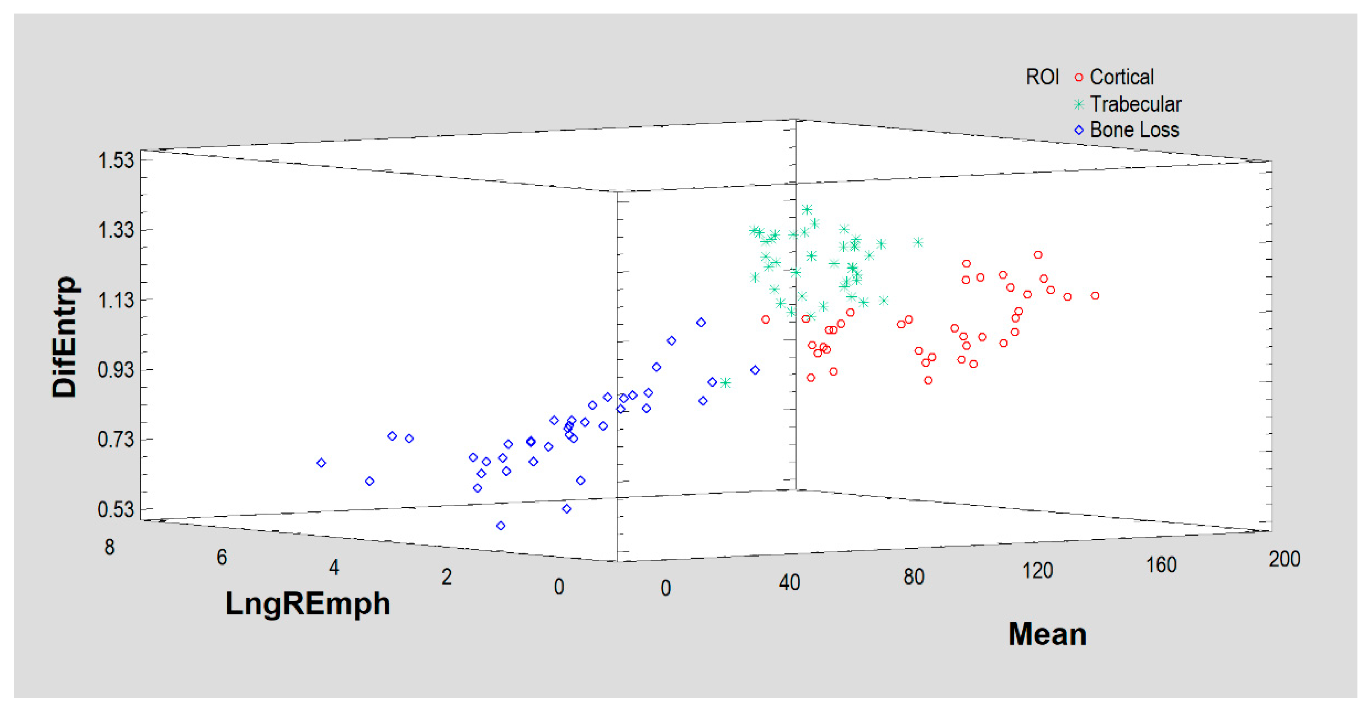

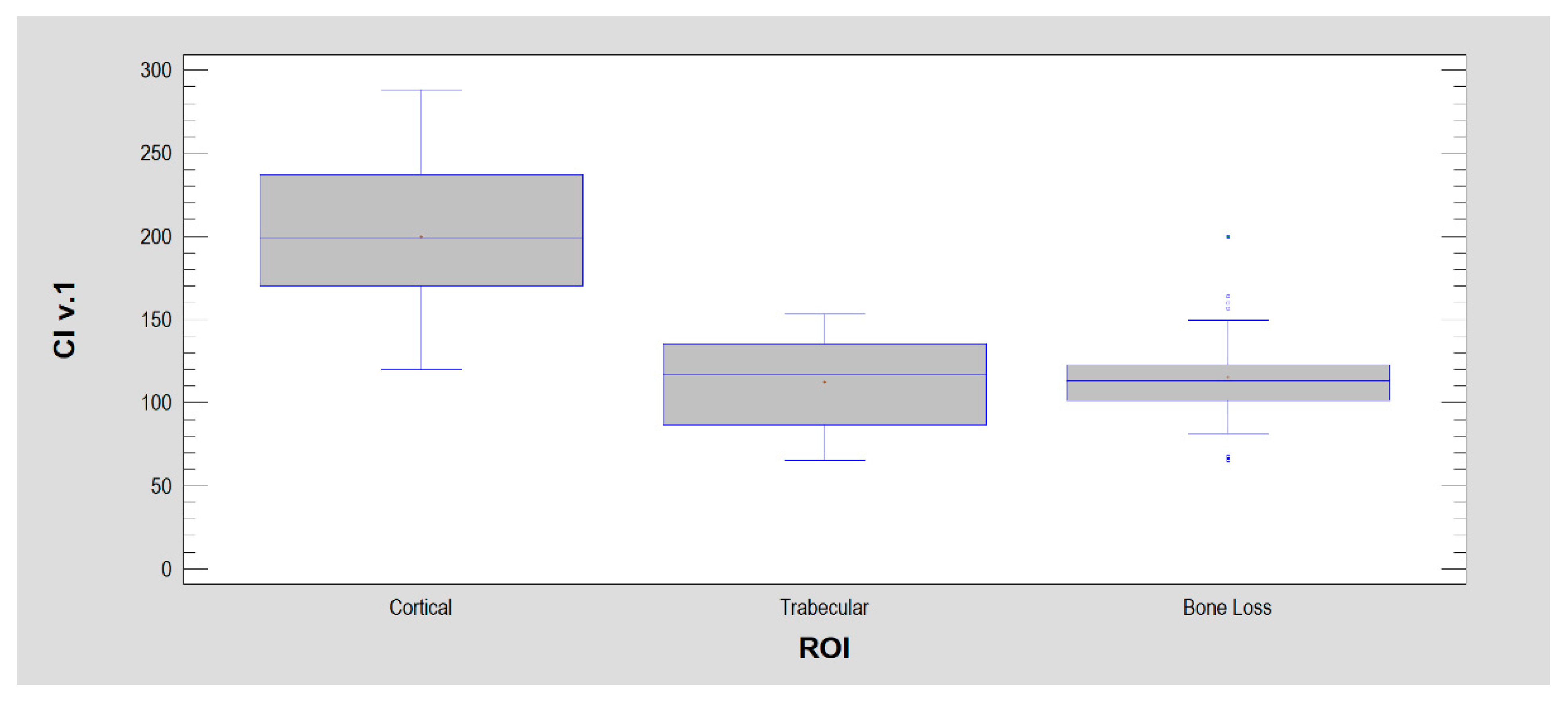
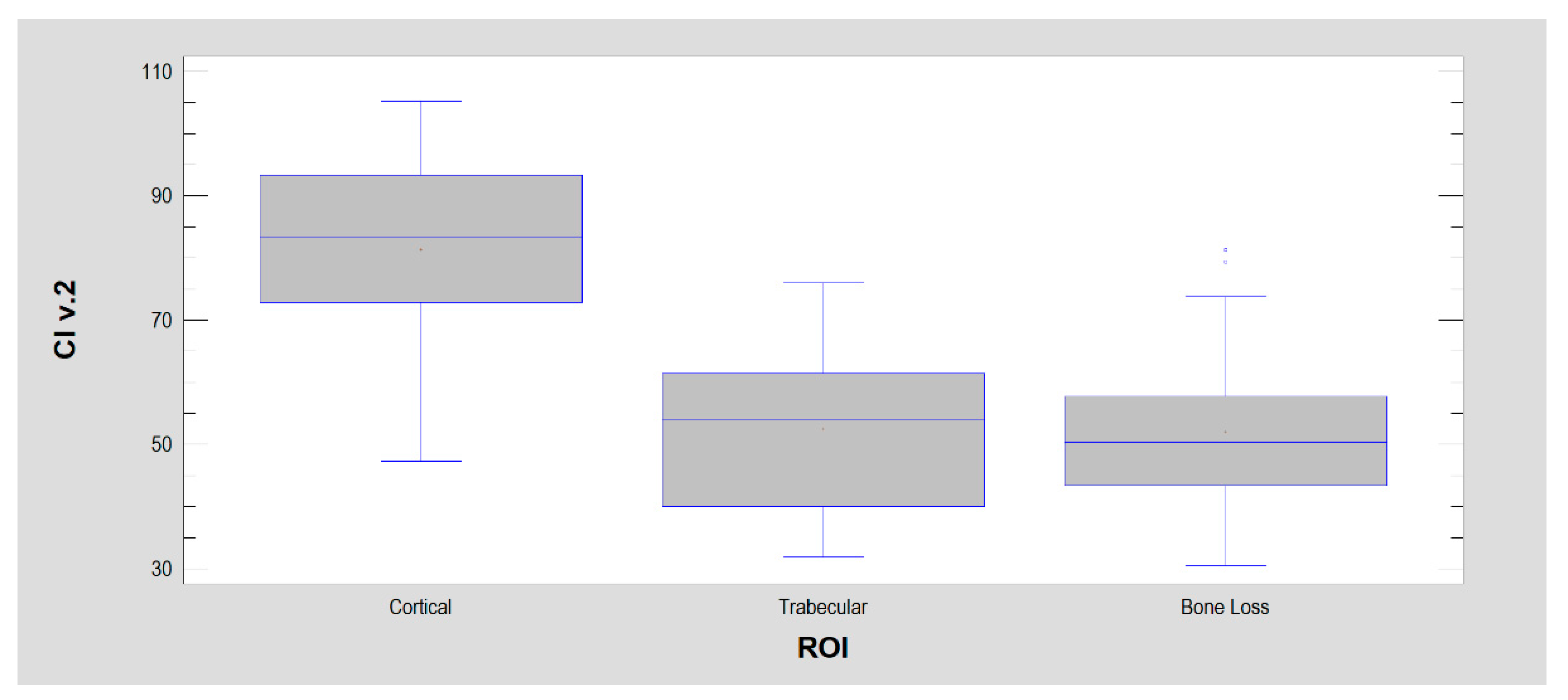

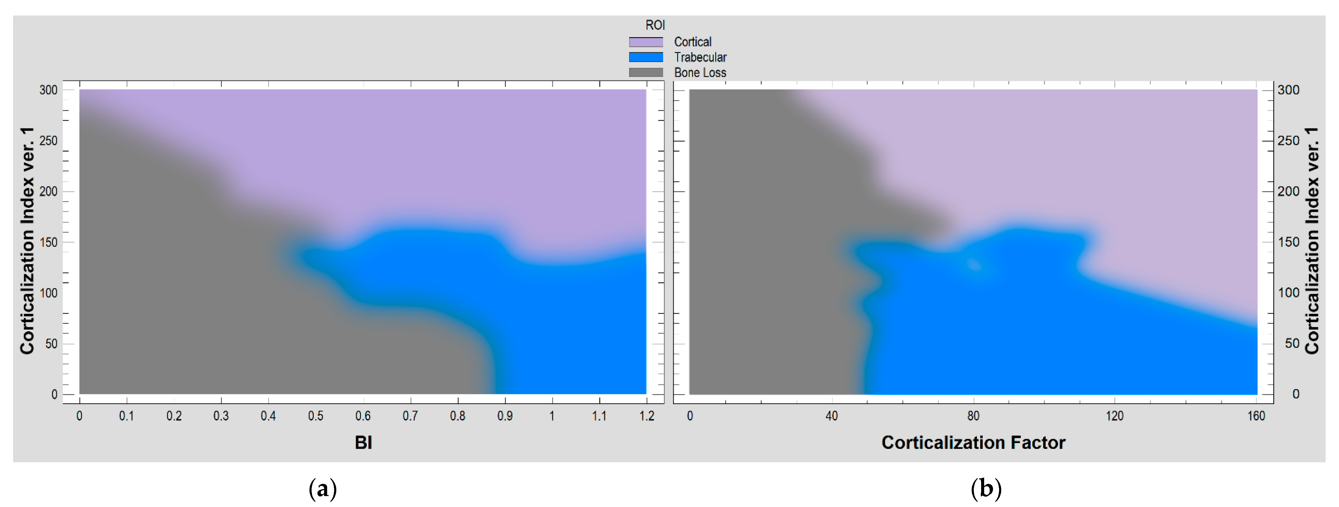

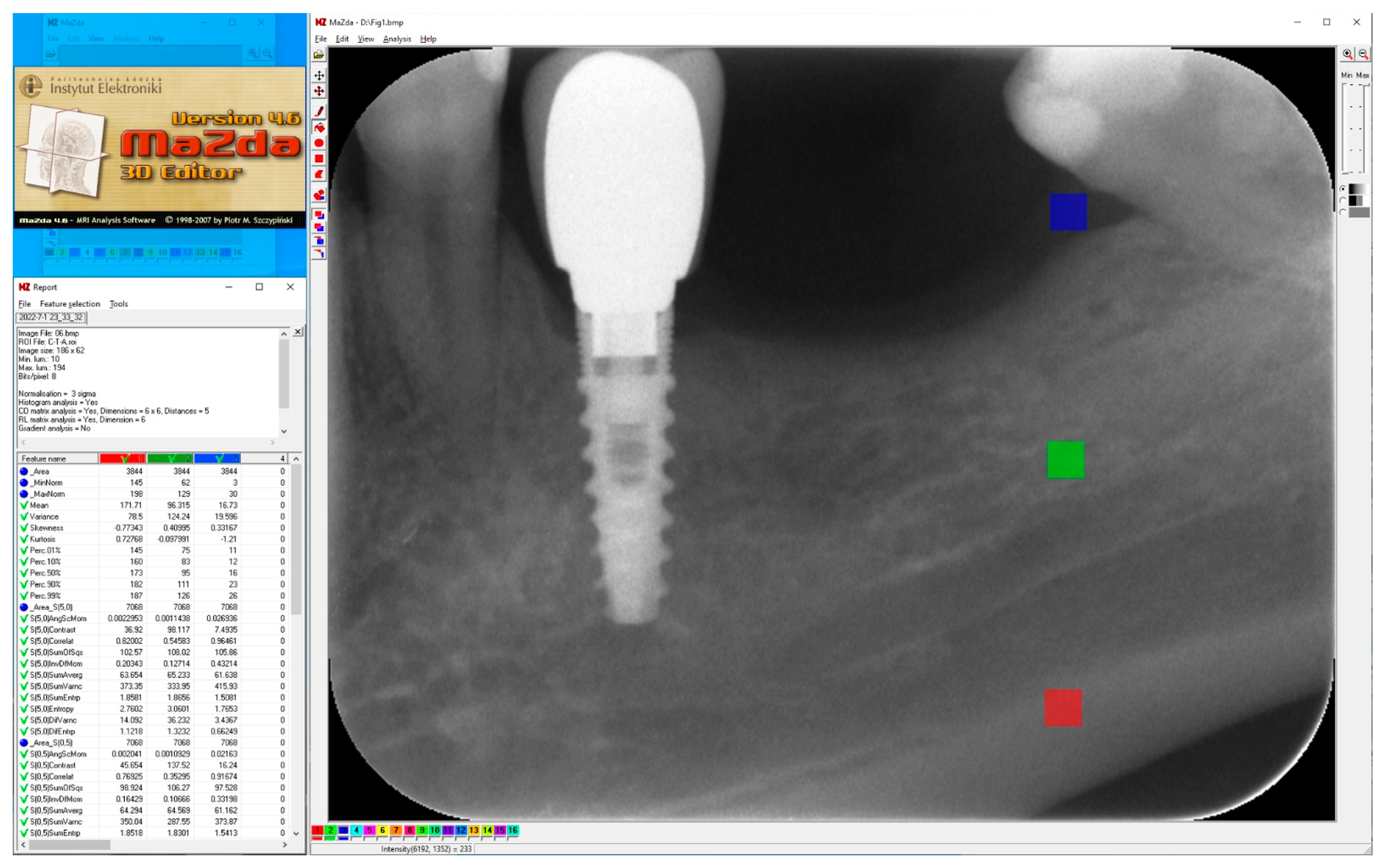
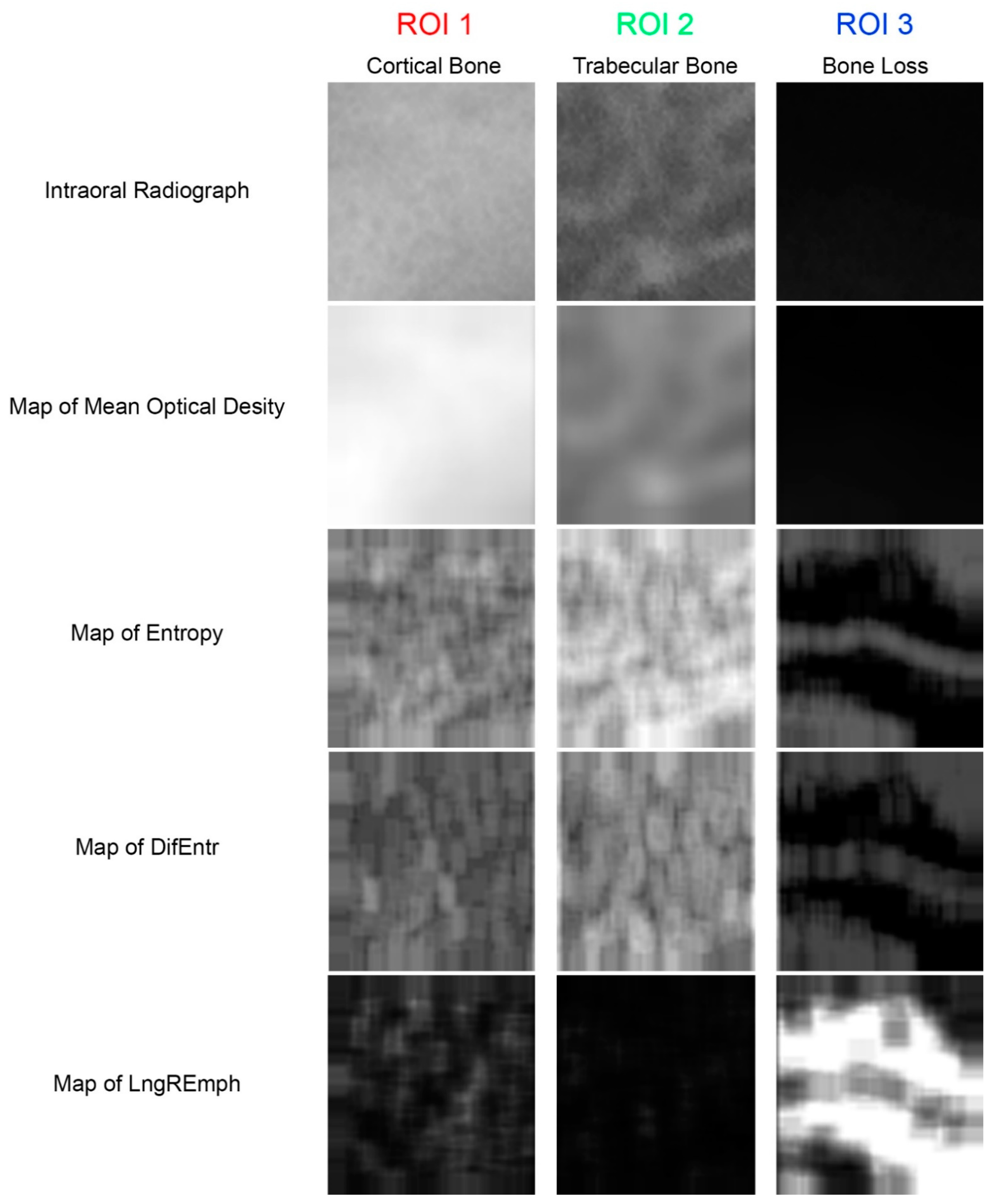
| Measure of Corticalization | ROI 1 Cortical Bone | ROI 2 Trabecular Bone | ROI 3 Bone Loss | Note |
|---|---|---|---|---|
| Mean Optical Density | 132 ± 27 | 91 ± 15 | 34 ± 15 | p < 0.001 1 |
| Entropy | 2.68 ± 0.15 | 2.74 ± 0.19 | 1.79 ± 0.27 | p < 0.001 2 |
| Differential Entropy | 1.10 ± 0.09 | 1.28 ± 0.10 | 0.81 ± 0.15 | p < 0.001 1 |
| LngREmph | 1.66 ± 0.21 | 1.55 ± 0.18 | 3.01 0.97 | p < 0.001 3 |
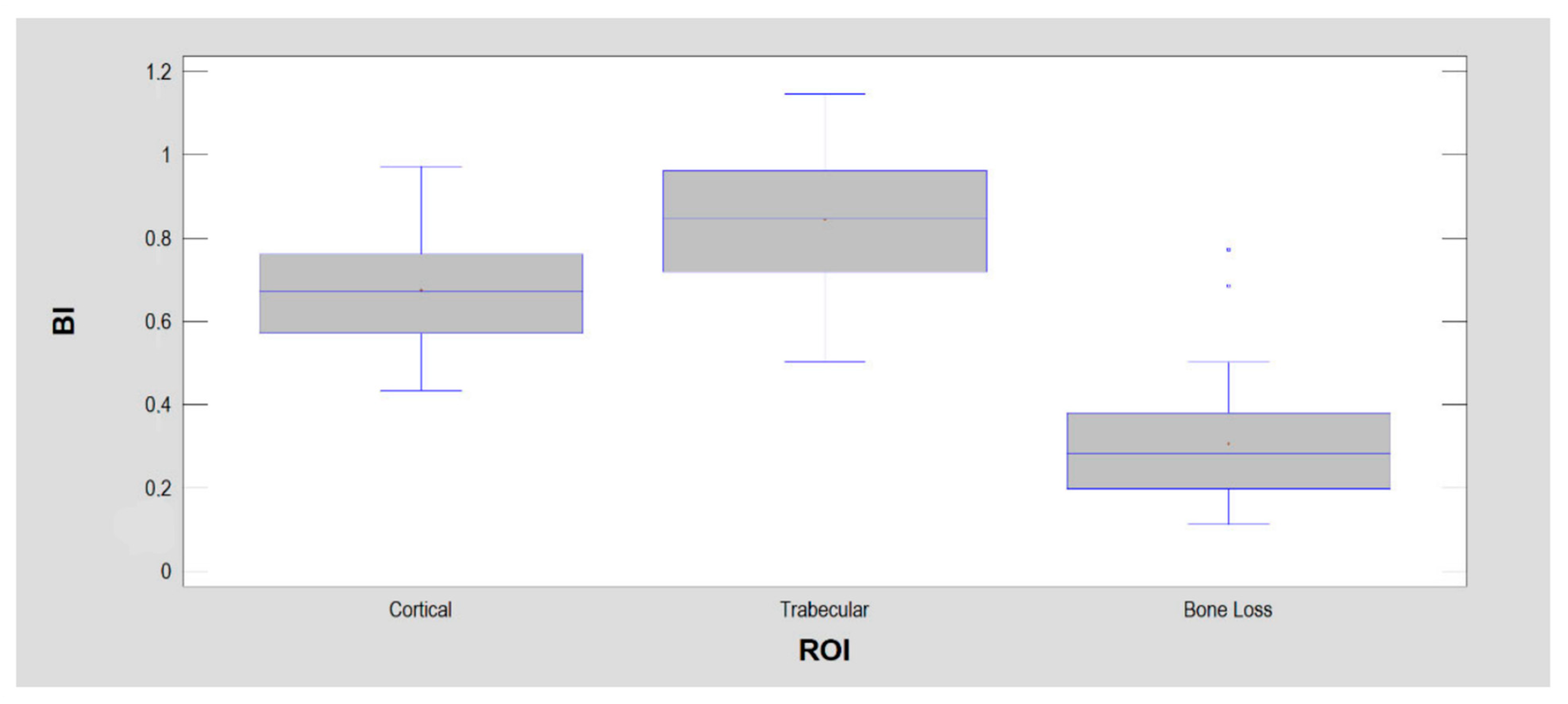
| Bone Index | 0.67 ± 0.13 | 0.84 ± 0.15 | 0.31 ± 0.14 | p < 0.001 1 |
| Corticalization Index ver.1 | 200 ± 42 | 112 ± 28 | 115 ± 26 | p < 0.001 4 |
| Corticalization Index ver.2 | 81 ± 15 | 53 ± 13 | 52 ± 12 | p < 0.001 4 |
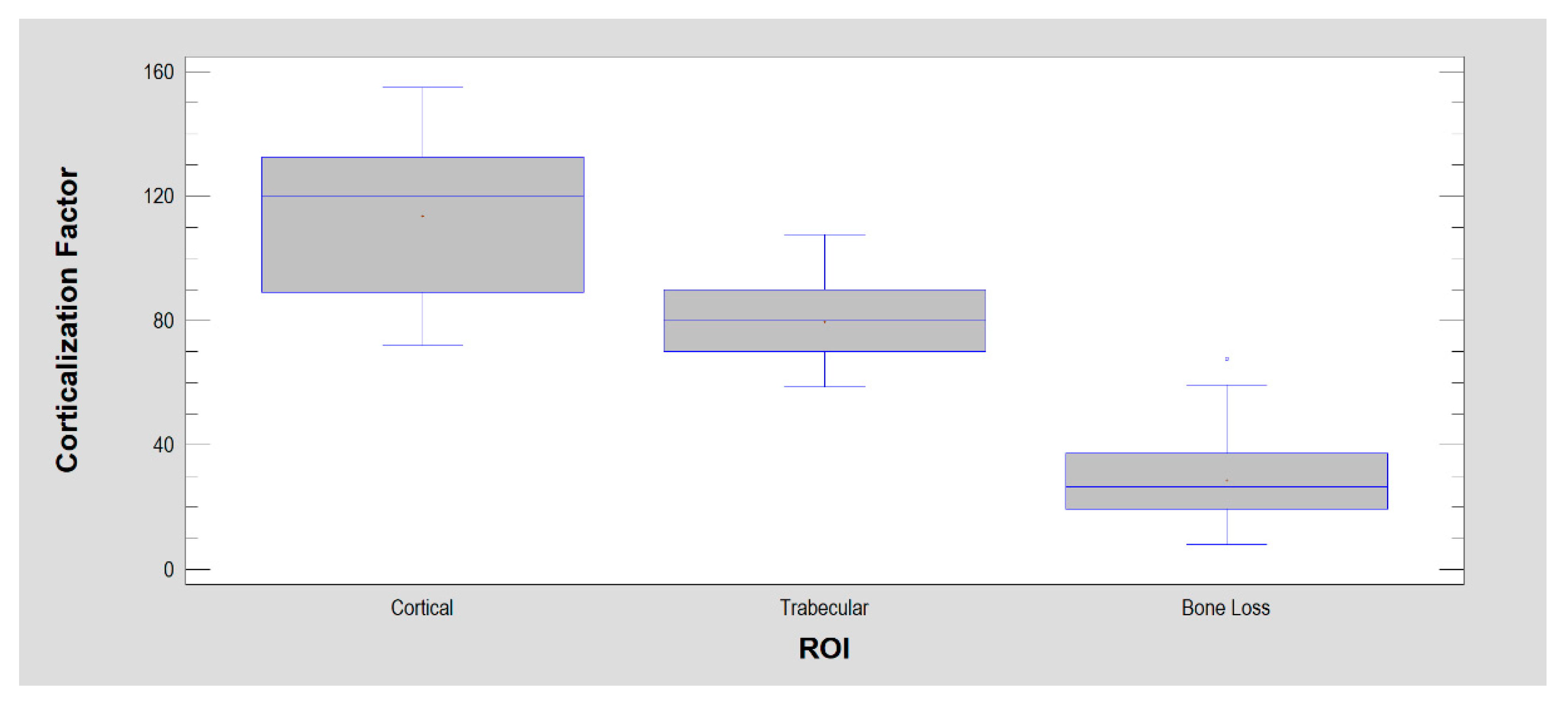
| Corticalization Factor | 114 ± 23 | 80 ± 12 | 29 ± 14 | p < 0.001 1 |
Publisher’s Note: MDPI stays neutral with regard to jurisdictional claims in published maps and institutional affiliations. |
© 2022 by the author. Licensee MDPI, Basel, Switzerland. This article is an open access article distributed under the terms and conditions of the Creative Commons Attribution (CC BY) license (https://creativecommons.org/licenses/by/4.0/).
Share and Cite
Kozakiewicz, M. Measures of Corticalization. J. Clin. Med. 2022, 11, 5463. https://doi.org/10.3390/jcm11185463
Kozakiewicz M. Measures of Corticalization. Journal of Clinical Medicine. 2022; 11(18):5463. https://doi.org/10.3390/jcm11185463
Chicago/Turabian StyleKozakiewicz, Marcin. 2022. "Measures of Corticalization" Journal of Clinical Medicine 11, no. 18: 5463. https://doi.org/10.3390/jcm11185463
APA StyleKozakiewicz, M. (2022). Measures of Corticalization. Journal of Clinical Medicine, 11(18), 5463. https://doi.org/10.3390/jcm11185463