


Abstract
Background/Objectives: Acinetobacter baumannii in the intensive care setting has become increasingly carbapenem resistant in recent years. In addition to developing new antibiotics, alternative intervention strategies, such as monoclonal antibodies, bacteriophages, and vaccines, have also been explored. Capsular polysaccharides (CPS), virulence factors prominently displayed on the bacterial surface, are excellent vaccine candidates, as demonstrated by their use in preventing infections caused by Streptococcus pneumoniae, Neisseria meningitidis, and Haemophilus influenzae. The objective of this study is to develop and evaluate a glycoconjugate vaccine against the A. baumannii LAC-4, a K locus 49 (KL49) human clinical isolate that is hypervirulent in mice. Methods: The LAC-4 CPS was isolated and purified using a simplified base treatment method that immediately releases CPS from the anchor lipids, which also degrades DNA/RNA. Crude CPS was obtained by removal of small molecules through dialysis and proteins through phenol–water extraction. Pure K49 was obtained by SEC-HPLC on a Superose-6 column. To facilitate the separation of glycoconjugate from free polysaccharide by SEC, the CPS was depolymerized by sonication to an estimated molecular weight of 80 kDa. Because K49 lacks diol groups required for periodate oxidation, an alternative conjugation method relying on thioether bond formation rather than the commonly used reductive amination for linking K49 CPS to CRM197 was applied. Results: Immunization of mice with the conjugate vaccine elicits capsule-type-specific but strain-transcending antibody (IgM and IgG) responses. It protects immunized mice against lethal respiratory and intraperitoneal infections with both LAC-4 and other KL49 A. baumannii clinical isolates (such as BEI-24). Conclusions: To the best of our knowledge, this represents the first evaluation of a KL49 CPS glycoconjugate A. baumannii vaccine. Our results support further development of capsule-based vaccines targeting clinically important A. baumannii lineages.
1. Introduction
Acinetobacter baumannii has emerged as one of the most important causes of healthcare-associated infections worldwide, particularly among critically ill patients requiring intensive care [1]. The bacterium is responsible for ventilator-associated pneumonia, wound infections, bloodstream infections, and sepsis. The remarkable capacity of A. baumannii to acquire resistance determinants has resulted in the global dissemination of multidrug-resistant, extensively drug-resistant, and carbapenem-resistant strains [2]. In recognition of the urgent need for new interventions, the World Health Organization has designated carbapenem-resistant A. baumannii as a critical-priority pathogen for antibiotic research and development [3]. The limited therapeutic options available have stimulated increasing interest in developing alternative intervention approaches, such as monoclonal antibodies [4], bacteriophages [5], and vaccines [6,7]. These strategies target virulence factors exposed on the surface of bacterial cells, particularly capsular polysaccharides (CPS) and outer membrane proteins [8].
The capsular polysaccharide is a major virulence determinant of many Gram-negative bacterial pathogens, including A. baumannii, and represents one of the most attractive targets for vaccine development. The capsule contributes to bacterial survival by promoting resistance to complement-mediated killing, inhibiting phagocytosis, facilitating persistence in the host, and protecting against environmental stresses [9]. Importantly, as the outermost surface structure of bacterial cells, CPS is readily accessible to antibodies and can serve as a target for opsonophagocytic and complement-dependent immune mechanisms. Several experimental studies have demonstrated that capsule-specific antibodies and vaccines protect against A. baumannii infection [10,11] and confirmed the feasibility of CPS-directed immunization strategies.
A major challenge in developing capsule-based vaccines against A. baumannii is the remarkable diversity of CPS structures. Over 100 distinct capsule types have been identified among clinical isolates, reflecting extensive antigenic diversity across circulating clinical strains [9]. Similar to serotype-specific vaccines developed for other encapsulated bacterial pathogens, this diversity may necessitate multivalent formulations to include clinically important capsule types. Consequently, evaluation of individual CPS antigens is an essential step toward establishing the protective potential and breadth of capsule-targeted vaccine approaches.
Although multiple experimental A. baumannii vaccine candidates have been investigated, including outer membrane proteins, outer membrane vesicles, inactivated whole cells, and CPS, no licensed vaccine is currently available [7]. Furthermore, only a limited number of capsule types have been evaluated as glycoconjugate vaccine antigens, and the immunogenicity and protective potential of many clinically relevant CPS structures remain to be investigated [12].
The objective of the present study was to develop and evaluate a glycoconjugate vaccine based on the CPS extracted from A. baumannii LAC-4, a human clinical isolate with hypervirulence in mice [13]. The CPS of LAC-4 belongs to the KL49 type and has a negatively charged trisaccharide repeating unit of -8)-α-8eLegp5Ac7Ac-(2-3)-α-L-FucpNAc-(1-3)-α-D-GlcpNAc-(1- [14]. In this report, we first describe a simplified method for CPS isolation under alkaline conditions. Second, because KL49, like several other KL-type CPS, lacks diol groups required for periodate oxidation, we developed an alternative conjugation strategy for linking KL49 CPS to carrier protein CRM197 (a non-toxic, genetically altered mutant of diphtheria toxin) via a thioether bond rather than through reductive amination. Finally, we demonstrate that the resulting conjugate vaccine elicited KL49 CPS-specific antibody responses and provided protection against lethal challenge with multiple KL49 A. baumannii strains in mice.
2. Materials and Methods
Bacterial strains: The BEI panel of A. baumannii strains were obtained through the BEI Resource Repository, NIAID, NIH. The panel is part of the Acinetobacter baumannii Diversity Panel provided by the Multidrug-Resistant Organism Repository and Surveillance Network (MRSN) at the Walter Reed Army Institute of Research (Silver Spring, MD, USA) [15]. LAC-4 is a hypervirulent clinical isolate originally obtained from a hospital outbreak in Los Angeles, CA [16]. In addition to LAC-4, other KL49 bacterial strains included in this study are BEI-3, BEI-24 and BEI-54.
Reagents and chemicals: The carrier protein EcoCRM® (CRM197) is a product from Fina Biosolutions (Rockville, MD, USA). Aluminum hydroxide gel (Alhydrogel® adjuvant 2%) was purchased from InvivoGen (San Diego, CA, USA). All other chemicals were purchased from MilliporeSigma Canada Ltd. (Oakville, ON, Canada) and used without further purification.
Analysis: SEC-HPLC was performed on an Agilent 1260 Infinity system with a Superose-6 or Superose-12 column (10/300 GL) and PBS as eluent. NMR spectra were recorded on a JEOL 400 MHz (JNM-ECZL400S) spectrometer (JEOL Ltd., Tokyo, Japan). MALDI data were collected on a 4800 TOF-TOF (SCIEX, Framingham, MA, USA) in reflection mode, and α-Cyano-4-hydroxy-cinnamic acid and 2,5-dihydroxybenzoic acid were used as the matrix.
Culture and phenol-killing of A. baumannii: The LAC-4 cells were collected from frozen stock, placed into 10 mL Tryptic Soy Broth (TSB) and incubated at 37 °C with 200 rpm shaking for 8 h. Cells were then subcultured 1:400 into 4 L TSB and incubated at 37 °C with 200 rpm shaking for 18 h. Phenol was added at 2% v/v, and cells were incubated at 37 °C with shaking at 200 rpm for 4 h. Cells were centrifuged at 11,000 rpm for 10 min, then washed twice with an equal volume of PBS.
CPS extraction and purification: The above phenol-killed bacterial pellet was suspended in 0.1–0.4 N NaOH, and the mixture was stirred at 40 °C for 24 to 48 h and acidified to c.a. pH 5.0 by the addition of 30% acetic acid with stirring. The precipitates were removed by centrifugation at 8000 rpm for 30 min. The aqueous phase was collected and dialyzed (10K MWCO) against distilled water overnight and lyophilized. The white fluffy material obtained was dissolved in water, and an equal volume of 90% phenol was added. The mixture was shaken vigorously and centrifuged at 4500 rpm for 30 min. The upper aqueous layer was collected, dialyzed (10K MWCO) against distilled water overnight, and lyophilized to afford crude polysaccharides. Final purification was performed by SEC-HPLC on a Superose-6 column with PBS as eluent. The fractions were collected, dialyzed (or desalted with a centrifugal filter), and lyophilized to give pure polysaccharides with 1–2% (w/w) yields (Table 1). Hereafter, the CPS purified from the LAC-4 strain (KL49) is referred to as K49.
Depolymerization of polysaccharide: K49 in water (1–2 mg/mL) was cooled to 0 °C and kept in an ice bath during sonication. Sonication was performed using a Branson Digital Sonifier 450 (Branson Ultrasonics Corp., Brookfield, CT, USA) as follows: a 5 s pulse with a 20 s rest at 70% amplitude and a 5 min total sonication time per run. The extent of depolymerization was monitored by the shift in elution time of the CPS peak by SEC-HPLC on a Superose-12 column with PBS as eluent at 0.4 mL/min. Two sonication runs were required to yield the desired depolymerization. The CPS solution was transferred to a 50 mL tube and centrifuged at 8000 rpm for 30 min. The solution was lyophilized to a white solid, which was further sized by SEC-HPLC on a Superose-12 column. The desired fractions were desalted and lyophilized to yield a white solid (recovery yield: 70–85%). The SEC-HPLC on Superose-12 indicates that the sized KL49 CPS had a similar retention time to that of 80K Dextran (24.2 min).
Partial de-N-acetylation of sized KL49 CPS: Sized K49 (7 mg) in 1 N NaOH (4 mL) was kept at 110 °C for 1 h and then was neutralized by addition of 30% acetic acid. The solution was dialyzed (10K MWCO) overnight and lyophilized to give c.a. 5.0 mg of a white, fluffy product. Batch-to-batch variation in the degree of de-N-acetylation occurred, and 1H-NMR indicated about 15–25% de-N-acetylation based on the integration of downshifted Leg H-3e.
S-Acetylthioacetamide of sized K49 (K49-SAc): Partially de-N-acetylated K49 in PBS (3 mg/0.5 mL) was treated with SATA (N-succinimidyl S-acetylthioacetate) (3 mg in 0.3 mL DMSO) (Scheme 1). The mixture was kept at rt for 1 h, dialyzed (10K MWCO) and lyophilized to give c.a. 3 mg product. 1H-NMR showed the S-Ac resonance at 2.45 ppm (Figure 1), and the integration indicates c.a. 15–25% modification.
Thioacetamide of sized K49 (K49-SH): K49-SAc in PBS (3 mg/0.5 mL) was mixed with a solution of 0.1 N hydroxylamine (0.5 mL, pH 6.0–7.0). The mixture was kept at room temperature for 40 min, and the reagent was removed by a centrifugal filter (10K MWCO) and washed with 10 mM phosphate buffer (pH 6.0, 3 mL × 5). K49-SH in the phosphate buffer was used for coupling with the activated proteins without further characterization.
Maleimide derivatization of CRM197 (CRM-Mal): Mal-Peg4-NHS ester (0.4 mg) in 15 μL of DMSO was added to a solution of CRM197 in 10 × PBS (4 mg/0.5 mL) with stirring. The mixture was kept at room temperature for 20 min, and HPLC indicated a small shift in retention time to 34.64 min from 34.96 on a Superose-12. The reagent was removed by a centrifugal filter (10K MWCO) (0.5 mL × 5). The final product was kept in PBS (0.5 mL). MALDI analysis showed CRM-Mal with Mw at 61,275, suggesting an average of 7.8 maleimide groups per CRM molecule by comparing it to CRM Mw at 58,713.
Bromoacetamide of human serum albumin (HSA) (HSA-Br): A solution of HSA in PBS (6 mg/1.2 mL) was mixed with 0.6 mg of bromoacetic acid N-hydroxysuccinimide ester in DMSO (10 mg/mL). The mixture was kept at room temperature for 1 h, and the reagent was removed by a centrifugal filter (10K MWCO) and washed with PBS (2 mL × 5) to give the product (HSA-Br) in PBS (0.8 mL). HPLC on a Superose-12 column showed a shift of retention time from 30.827 for HSA to 30.376 for HSA-Br. MALDI analysis indicated that HSA was modified by 26.4 BrAc on average (HSA-Br Mw at 70,168 vs. HSA Mw at 66,942).
Conjugation: K49-SH derived from 3 mg of K49-SAc in PBS (0.15 mL) was mixed with CRM-Mal (c.a. 4 mg in 0.5 mL PBS), or HSA-Br (c.a. 3 mg in 0.4 mL PBS) and the mixtures were incubated at 37 °C for 40–48 h, purified by SEC-HPLC on a Superose-6 with PBS and concentrated with a centrifugal filter (10K MWCO) to about 0.5 mL PBS solution and the conjugates were stored at 4 °C. HPLC analysis (Figure 2 and Scheme 2) showed that the K49-CRM conjugate had a concentration of c.a. 10.0 mg/mL with c.a. 50% (w/w) of KL49, while the K49-HSA in PBS was at 4.7 mg/mL and KL49 in conjugate at c.a. 43% (w/w).
Mice. Eight- to twelve-week-old, specific-pathogen-free female BALB/c mice were purchased from Charles River Laboratories (St. Constant, QC, Canada). The animals were maintained and used in accordance with the recommendations of the Canadian Council on Animal Care Guide to the Care and Use of Experimental Animals. All animal experimental procedures were approved by the institutional animal care committee (Human Health Therapeutics Research Center, National Research Council Canada, Ottawa) under the Animal User Protocol AUP2021.01.
Immunization, bacterial challenge, and sample collection. Figure 3 shows a schematic summary of the key experimental design. For immunization, the K49-CRM vaccine was prepared with a 2X working concentration of vaccine solution and an equal volume of Alhydrogel®. The mixture was shaken at 60 rpm for 60 min at room temperature. For each immunization, mice were subcutaneously administered 0.1 mL of the vaccine containing 2 µg CPS on days 0, 14, and 28. Nonterminal blood samples were collected from individual mice at 3 days before (−3) and 11, 25, 42, and 109 days after the 1st immunization. Terminal bleeding was performed in assigned groups of 5 mice at day 42 or 8 days after the boosting immunization (day 120).
Groups of immunized and unimmunized mice (n = 5/group) were challenged intranasally (i.n., 4 × 10⁷ colony-forming units, CFU) or intraperitoneally (i.p., 9.6 × 10⁵ CFU) with freshly grown LAC-4 three weeks after the last immunization. Some mice were challenged i.p. with 6.8 × 10⁷ CFU of A. baumannii KL49 strain BEI-24 (MRSN7124, a multidrug-resistant strain isolated from a wound) [15]. The actual inoculum in each experiment was determined by quantitative bacteriology. Body weight, clinical scores, and survival of the mice were monitored and recorded as we described previously [13]. The overall clinical assessment for each mouse was scored as 0 (normal, active, healthy), 1.0 (slightly sick, slightly ruffled fur, otherwise normal), 2.0 (sick, ruffled fur, slow movement, hunching), 3.0 (very sick, ruffled fur, very slow movement, hunched, eyes shut), 4.0 (moribund), or 5.0 (dead).
In some experiments, groups of five mice were sacrificed at 6 or 24 h post-challenge. Blood, spleen, and lung tissues were aseptically removed and used for quantitative bacteriology [13,17].
Quantitative bacteriology. The lungs and spleens were homogenized in sterile saline. The number of viable A. baumannii in the respective organs was determined by plating 100 µL aliquots of 10-fold serial dilutions of the homogenates on brain–heart infusion agar plates [13]. Blood samples were similarly cultured to assess bacteremia.
ELISA for A. baumannii CPS-specific serum IgG and IgM or whole cell-specific IgG antibodies. Serum levels of A. baumannii K49- or whole cell-specific antibodies were assessed by ELISA. Briefly, 96-well microplates were pre-coated with 0.2 µg K49-HSA per well in 100 µL of sodium bicarbonate buffer (pH 9.6) overnight at 4 °C or 2.5 × 10⁷ per well of formalin- or heat-killed LAC-4 or BEI strain cells in 50 µL of Milli-Q water overnight at 37 °C. Single prediluted or serial dilutions of serum samples were used for the assays. Serum dilutions (1:500) from individual mice collected at different time points after immunization were tested for K49-specific IgG or IgM. A series of diluted sera collected on day 42 was tested for the level of specific IgG against the inactivated LAC-4 or BEL strain cells.
Western blotting of bacterial lysates. A. baumannii LAC-4, BEI-3 (MRSN918), BEI-24 (MRSN7124), BEI-54 (MRSN16880), BEI-1 (MRSN334), and ATCC17978 were streaked on LB agar plates and incubated overnight. A single colony from each strain was picked, inoculated into 10 mL LB medium, and cultured overnight. The bacterial cells were then subcultured in 10 mL of fresh LB medium with an initial OD600 of 0.1. The bacterial cells were harvested by centrifugation when OD600 reached 1.0. The harvested cells were resuspended in 0.8 mL of solution containing 62.5 mM Tris-HCl pH 7.4, 1% SDS, and 1 mg/mL Proteinase K, incubated at 37 °C for 2 h, then 0.4 mL of 3× electrophoresis loading buffer was added, and kept at −20 °C in aliquots. The cell lysates (40 µL/well) were loaded and separated on a 10-well, 4–15% Bio-Rad Mini-PROTEAN TGX precast gel (Bio-Rad Laboratories Canada Ltd., Mississauga, ON, Canada). Electrophoresis was performed at 100 V for 15 min, followed by 150 V for 35 min. Separated CPS were then tank-transferred onto a 0.45 μm Immobilon-E PVDF membrane (Millipore Sigma Canada Ltd., Oakville, ON, Canada). The membrane was blocked with 1% Casein/PBS at room temperature for 2 h with shaking and incubated with 1:2000 immunized mouse sera at 4 °C overnight with shaking. After several washes, the membrane was incubated with horseradish peroxidase-conjugated goat anti-mouse IgG (L + H) (1:5000) at room temperature for 2 h, and then with SuperSignal West Femto Maximum Sensitivity Substrate (Thermo Fisher Scientific, Nepean, ON, Canada) after washing. Images were taken by a ChemiDoc Imaging System (Bio-Rad) with the onboard Image Lab Touch Software (version 3.0.1.14).
Statistical analysis. Data are presented either as individual samples or as means ± standard deviation (SD) for each group. Differences were assessed by Student’s t-test, one-way or two-way analysis of variance (ANOVA) followed by Bonferroni’s post hoc multiple comparison tests, when appropriate. Survival rates were analyzed by the log-rank test followed by Bonferroni’s method for multiple comparisons of survival curves. Differences were considered significant when p < 0.05.
3. Results
3.1. CPS Isolation and Glycoconjugates
CPS preparation generally follows a multi-step process that often includes cell lysis, phenol–water extraction, selective precipitation, enzymatic treatment to digest residual nucleic acids and proteins, ultrafiltration/diafiltration to remove low-molecular-weight contaminants, and finally ion-exchange or size-exclusion chromatography.
A. baumannii CPS isolation mostly uses a procedure modified by Knirel et al. [18]. Typically, it includes (1) extraction of cells with 45% aqueous phenol; (2) protein precipitation with aqueous 50% trichloroacetic acid; (3) lipid removal by acid hydrolysis of the crude CPS; and (4) final purification by gel permeation chromatography [19]. The CPS isolation yield varies with strains and growth conditions, typically about 1–5% from dry cell weight. By this procedure, CPS with a lipid anchor is extracted with hot water–phenol, and the lipids are later removed when treated with 2% acetic acid at 100 °C. Since those A. baumannii CPS are not base-sensitive except for the O-acetyl groups in some K-types, we revised the procedure by releasing CPS from their anchor lipids first under basic conditions. After the base treatment, the mixture was acidified with 30% acetic acid to precipitate the majority of fatty acids, which were then removed by centrifugation. Other small molecules, such as hydrolyzed DNA/RNA, remained in the aqueous solution and were removed by dialysis (10K MWCO). The CPS crudes still contained significant amounts of proteins, which were then extracted with phenol–water (45%, v/v) to separate them from CPS. Finally, the pure K49 was obtained by SEC-HPLC on a Superose-6 column. This process is straightforward and avoids trichloroacetic acid precipitation and acid hydrolysis. By this method, we have isolated K1, K1v*, K2, K3, K9, K19, K25, K49, and K58 from A. baumannii AYE, BEI-73, BEI-16, ATCC17978, BEI-61, BEI-71, BEI-25, LAC-4, and BEI-75, respectively, with variable yields (1–6% w/w).
In addition to specific strains and growth conditions, the CPS recovery yields can be affected by the effective release of CPS from the anchor lipids. To examine the impact of base concentration, for example, we extracted K49 from A. baumannii strain LAC-4 under conditions of aqueous sodium hydroxide from 0.1 N to 0.4 N for 24 h or 48 h at 40 °C (Table 1). We observed only slight differences in CPS yields under these conditions, indicating that 0.1 N NaOH is likely strong enough to free CPS from the anchor lipids. The K49 yields (1–2%) from this experiment are somewhat lower than expected, as we once extracted K49 from LAC-4, K1 from AYE and K9 from BEI-61 at c.a. 6% (w/w) yield once obtained from other isolations, e.g., suggesting the growth conditions and isolation procedures need to be optimized.
Table 1.
KL49 CPS isolation from A. baumannii strain LAC-4 under basic conditions.
Although K49 is not O-acetylated, this isolation method concurrently removes O-acetyl groups from polysaccharides. It has been known that the glycoconjugates derived from de-O-acetylated CPS of N. meningitis serogroups C, W, Y, and Group B Streptococcus provided satisfactory immunity and protection. Whereas O-acetylation in CPS of N. meningitis serogroup A and Salmonella typhi Vi seems to be important, it has been suggested that the position and the role of the O-acetyl group are more critical to CPS repeating units comprised of single or few saccharides, and the O-acetyl group out of the sugar ring seems less relevant to the immunological response [20]. A. baumannii CPS repeating units commonly contain multiple saccharides, and some contain O-acetylation on the sugar ring (e.g., K3, K26, K35, K53 and K83). As it remains unknown if the glycoconjugates from de-O-acetylated A. baumannii CPS would impact their functional immunogenicity, this deserves further evaluation as part of vaccine development.
K49 has a trisaccharide repeating unit, i.e., -8)-α-8eLeg5Ac7Ac-(2-3)-α-L-FucNAc-(1-3)-β-D-GlcNAc-(1-. The native polysaccharide extracted has a size of over 300 KD, as it elutes in the void volume on a Superose-6 column. To facilitate the separation of glycoconjugate from free polysaccharide by SEC and induce the desired immune response, it is necessary to reduce the CPS to a proper size. Therefore, we used sonication to depolymerize the CPS [21] to an estimated molecular weight of around 80K, as referenced to the Dextran standard (Figure S1), although the actual molecular weight could be smaller since K49 has a negatively charged legionaminic acid. The depolymerized CPS was fractionated by SEC-HPLC on a Superose-12 column and analyzed by 1H-NMR, which confirms its structural integrity by comparison to the native K49 polysaccharide (Figure 1A and Figure S2).
Figure 1.
1H-NMR spectra of depolymerized K49 (A) partially de-N-acetylated CPS (B) and K49-SAc (C).
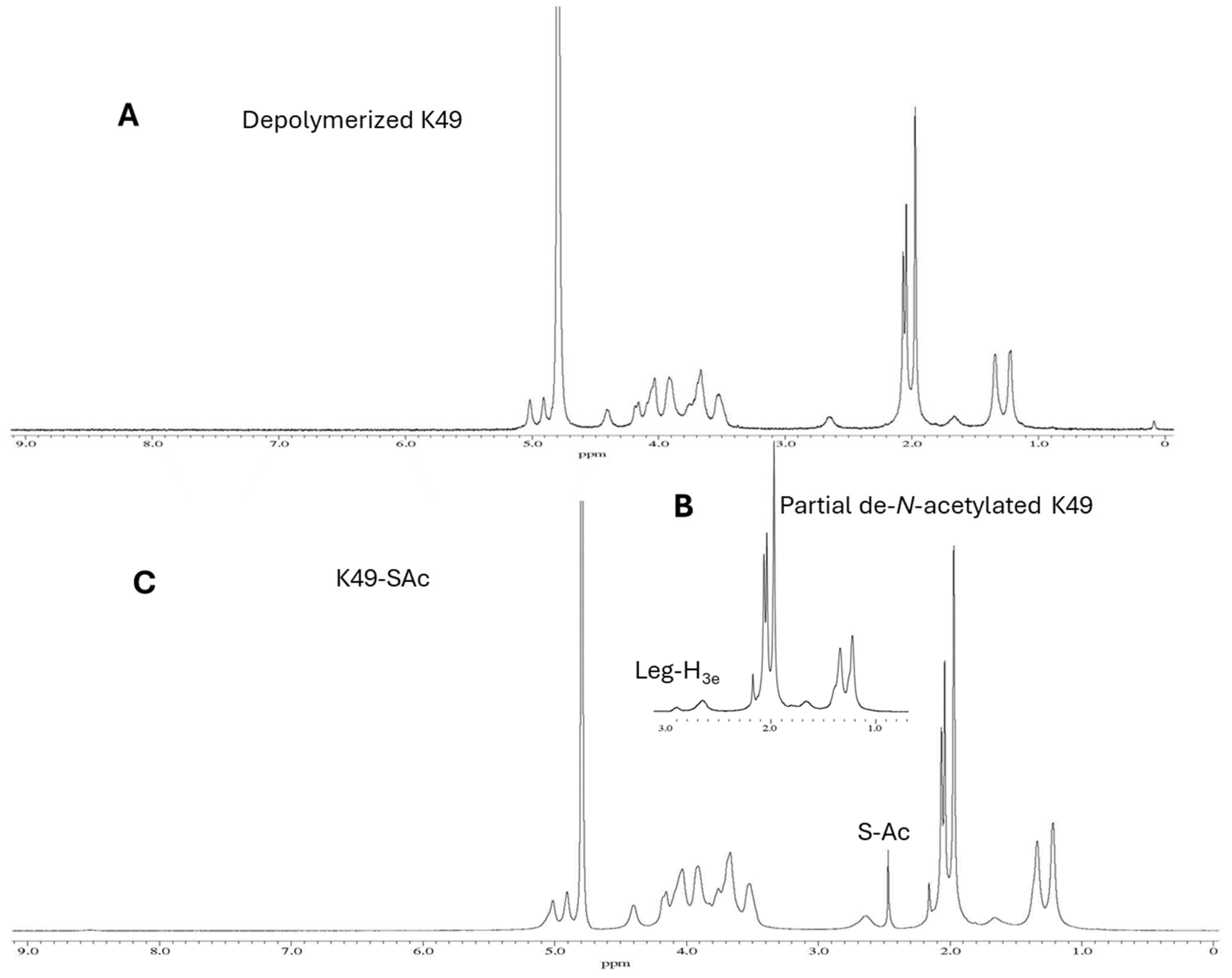
Although the immunogenicity of a glycoconjugate vaccine largely depends on the individual polysaccharide and the carrier protein, the method of conjugation can also be an important factor. The commercial glycoconjugate vaccines are manufactured using two major methods. One is based on a polysaccharide activated by cyanate ester [22], and the other conjugates periodate-oxidized polysaccharide with protein by reductive amination [23], which our laboratory has contributed to its development and used for the synthesis of various glycoconjugates [24]. Periodate oxidation converts diols in polysaccharides to active aldehydes. However, a few of A. baumannii polysaccharides, including K49, lack sugars with vicinal diols; therefore, the reductive amination approach is inapplicable. Alternatively, we applied another way of conjugation here in which the K49 polysaccharide and protein are joined through a thioether linkage. The method requires the removal of some N-acetyl groups and the use of the resultant amino groups for further derivatization (Scheme 1). Thus, the sized K49 was treated with 1 N NaOH at 110 °C for 1 h, which resulted in 15–25% de-N-acetylation as indicated by the 1H-NMR analysis based on the appearance of a new N-Ac resonance at 2.2 ppm and the down-field shift of the H-3e of legionaminic acid from 2.65 ppm to 2.90 ppm due to the de-N-acetylation (Figure 1B). The resonance signal at 2.90 ppm indicates that the Legionaminic acid was sensitively affected, suggesting one of its N-acetyl groups was most likely removed. The resultant amino groups were reacted with SATA to introduce a latent thiol (K49-SAc) for coupling with protein. This amide formation restored the H-3e resonance at 2.90 ppm back to 2.65 ppm, and a comparison of their 1H-NMR spectra (Figure 1A,C) showed that K49-SAc and K49 are almost identical, suggesting that the chemical modification did not alter the polysaccharide conformation and its immune epitopes. The 1H-NMR of K49-SAc also provided a more accurate estimation of the degree of de-N-acetylation by comparing the integration of the S-Ac resonance at 2.48 ppm to that of N-Ac groups.
Scheme 1.
Glycoconjugate synthesis includes (1) KL49 CPS (K49) depolymerization; (2) partial de-N-acetylation; (3) the introduction of a latent thiol group; and (4) coupling of the CPS-thiol with activated CRM197 and HSA.
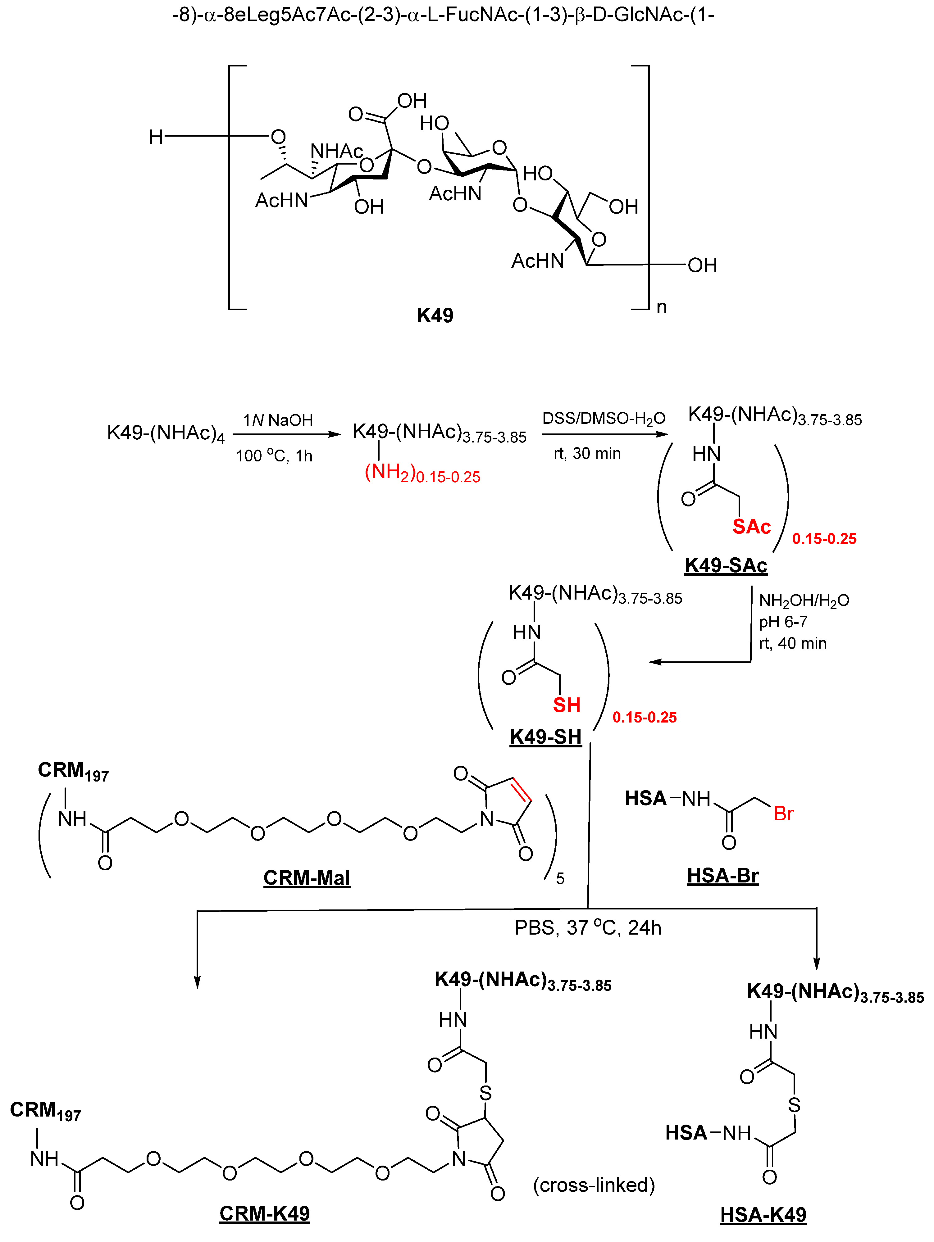
Meanwhile, thiol-reactive maleimide and bromoacetyl groups were introduced to CRM197 and HSA by reaction with respective activated esters (Scheme 1). The MALDI analysis showed CRM-Mal at Mw 61,275, an increase of 2562 Da, which corresponds to an average of 7.8 maleimide groups per CRM (see Figure S3); while HSA-Br has about 26.4 bromoacetyl groups attached to HSA (from Mw 66,942 to 70,168) (see Figure S4).
De-S-acetylation by hydroxylamine converted K49-SAc to reactive K49-SH, which was mixed with CRM-Mal and HSA-Br in PBS to form a thioether bond. Both conjugations proceeded smoothly, yet residual amounts of proteins were still left in both cases after two days (Figure S5). Both conjugates (CRM-K49 and HSA-K49) were purified on a Superose-6 column with PBS as eluent (Figure S6). The former was used for vaccination, and the latter as a coating antigen for ELISA; the different linkers were intentionally introduced to detect CPS-specific antibodies and minimize the interference from irrelevant linker-specific antibodies. As both polysaccharide and protein are activated on multiple sites, the conjugates are extensively cross-linked, including disulfide formation, as evidenced by the SEC-HPLC analysis. Cross-linked glycoconjugates are desired, which could lead to T cell receptors interacting with both the peptide-MHC and glycopeptide-MHC after antigen processing, therefore enhancing the immune response [25,26].
3.2. Glycoconjugate Analysis
The polysaccharide size and its ratio to protein are important factors in glycoconjugates to achieve optimal immune response [27]. Protein and carbohydrate contents are traditionally analyzed by colorimetric assay, but high-performance anion exchange chromatography coupled with pulsed amperometric detection (HPAEC-PAD) has recently been used for saccharide analysis [28,29]. Colorimetric assays often show variable sensitivities, resulting in less accurate outcomes even with calibration using the same polysaccharide and protein. We found that the BCE protein assay underestimates the CRM197 content in the K49 conjugate as compared to other conjugates, and the phenol–sulfuric acid-based colorimetric assay for carbohydrate was insensitive to K49 due to its lack of hexose. As an alternative, we attempted to estimate the protein and polysaccharide content by a simple SEC-HPLC analysis on the conjugates. HPLC has been routinely used for quantification by comparing the peak area or height of the chromatogram recorded by refractive index detection (RID) or UV detection to a calibration curve generated with known standards. However, it is technically difficult in a preliminary investigation to have glycoconjugate standards with a defined CPS/protein ratio. On the other hand, the average specific refractive index increment (dn/dc) for polysaccharides and proteins is 0.15 mL/g and 0.185 mL/g, respectively [30], which means that the proteins, in general, have only slightly more sensitive RID responses to their concentration in comparison to the polysaccharides. Specifically, we evaluated relative RID responses of five polysaccharides (K49, K1, K9, Pn1, and Dextra500) by SEC-HPLC analysis and compared their RID responses to that of HSA. The results showed that RID areas of polysaccharide and HSA (Table 2 and Figure S7) are comparable within ±20% deviation. Although we do not have the dn/dc for the conjugates, the value would likely fall between 0.15 and 0.185 mL/g. Therefore, the error from the estimation of the glycoconjugate concentration referenced to the protein carrier is likely smaller than 20%. Meanwhile, since the polysaccharides have minimal UV absorption at 280 nm and provided that the conjugation has no impact on the chromophores of the protein, we can estimate both the glycoconjugate concentration and the polysaccharide/protein ratio through a simple SEC-HPLC analysis using the formulas illustrated in Scheme 2. For example, a mixture of purified K49-HSA-K49 and standard HSA (5 mg/mL) was analyzed by SEC-HPLC and recorded by RID and UV at 280 nm (Figure 2). Those peak areas were used for the estimation of both conjugate concentration and polysaccharide/protein ratio, which gave K49-HSA in PBS at 4.7 mg/mL and K49 in conjugate at 42.6%. Similarly, the polysaccharide/protein ratio and the concentration of K49-CRM were also estimated and summarized in Table 3.
Scheme 2.
An estimation of the glycoconjugate concentration and the CPS/protein ratio in glycoconjugate. Without consideration of differences in specific refractive index increment (dn/dc), the peak areas of the chromatogram recorded by RID were used for calculation of glycoconjugate concentration (Formula (1)), whereas the protein content in glycoconjugate was calculated using Formula (2) based on the areas recorded by UV at 280 nm. Carbohydrates in glycoconjugate can be derived using Formulas (3) and (4).
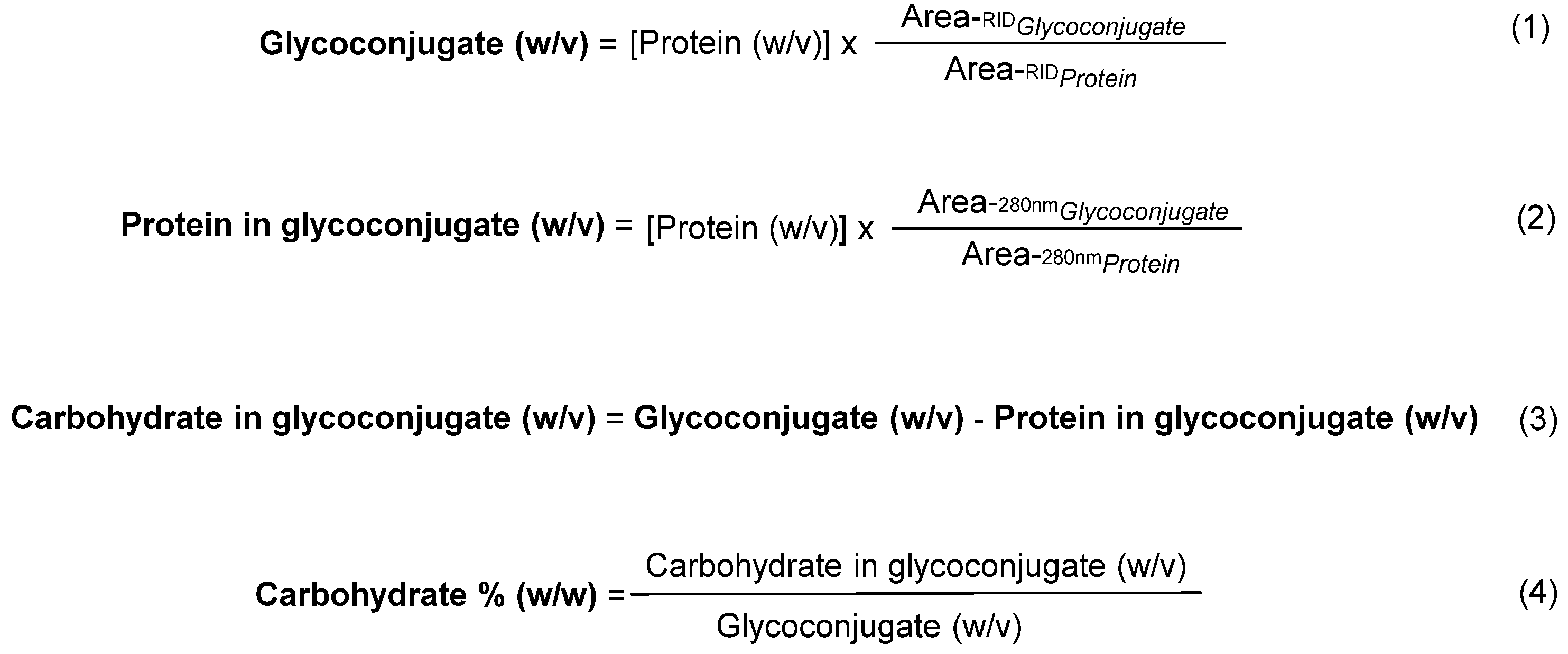
Figure 2.
Purified K49-HSA conjugate in PBS was mixed with HSA/PBS (5 mg/mL) in a ratio of 1:1 (v/v). The mixture was analyzed by HPLC on a Superose-12 column (10/300 GL, GE Health) with PBS as eluent at a rate of 0.4 mL/min.
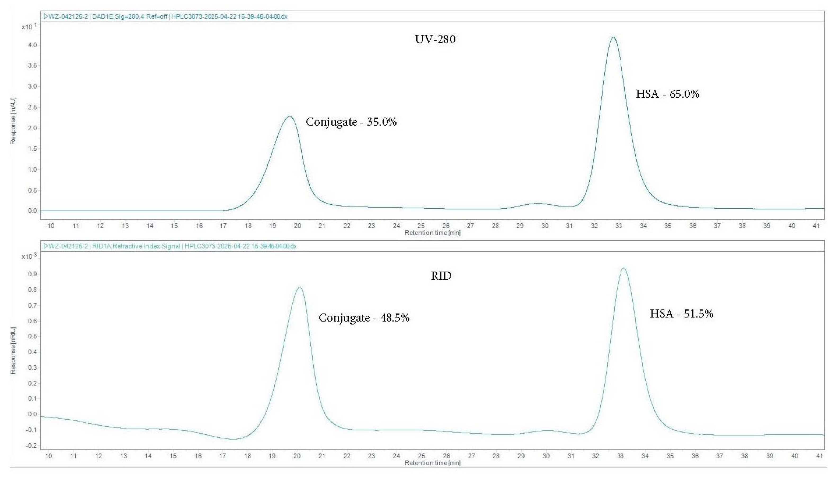
Table 2.
Relative areas recorded by RID at the same concentration (5 mg/mL in PBS).
Table 3.
Glycoconjugate and CPS/protein ratio estimated by HPLC analysis (protein standard: 5 mg/mL).
The above analysis of glycoconjugates by SEC-HPLC is only suitable for preliminary evaluation of a glycoconjugate vaccine, as we do not think that the margin of error in the analysis would fundamentally affect the outcome of the immune response in mice. The method could be further refined when the differences in individual dn/dc are taken into consideration.
3.3. Immunogenicity of the LAC-4 K49-CRM Conjugate Vaccine
We followed a procedure illustrated in Figure 3 To determine the immunogenicity of the LAC-4 CPS conjugate vaccine, the serum samples were collected before and at various time points after immunization. As shown in Figure 4a, the vaccine induced moderate levels of K49-specific IgM and detectable levels of specific IgG responses as early as day 11 after the 1st immunization. The IgM levels were substantially reduced on day 25 but remained at detectable levels throughout the experiment duration (day 109). The K49-specific IgG level increased after the 2nd and 3rd immunizations, peaked on day 42, and waned to a low but detectable level by day 109 (Figure 4a). The analysis of the isotypes of the K49-specific IgG responses showed that the vaccine induced almost exclusively IgG1 with little to no detectable IgG2a (Figure S9). Those results show that the K49-CRM conjugate vaccine is capable of inducing a rapid, early IgM response and a boostable Th2-biased specific IgG response.
Figure 3.
The schematic diagram of the major experimental design.
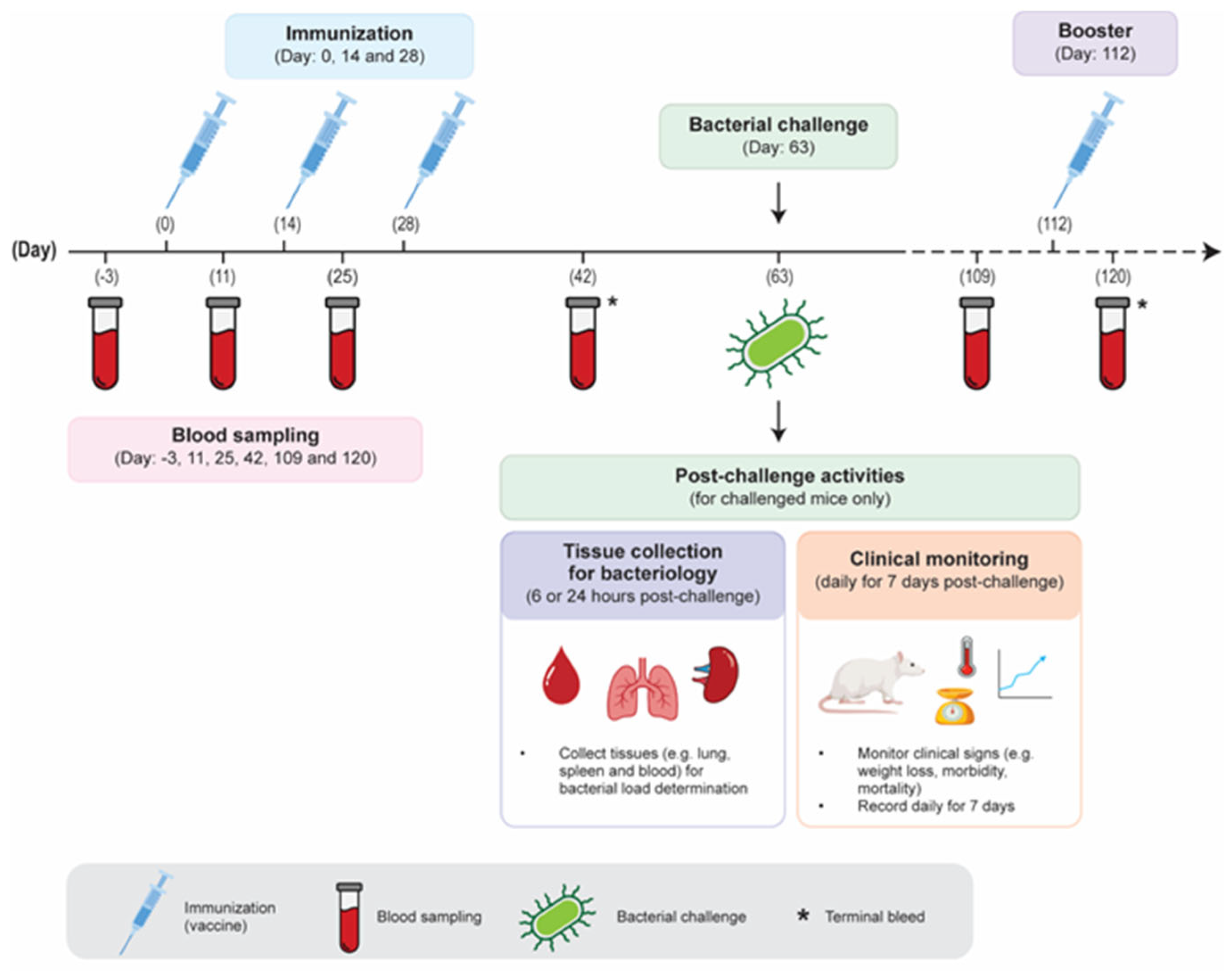
Figure 4.
Specific antibody responses in mice immunized with K49-CRM conjugate vaccine. (a) Serum LAC-4 K49-specific IgM and IgG responses before and at various times after immunization. (b) Serum IgG responses to whole killed LAC-4 bacterial cells at day 42. Groups of mice (n = 5/group) were immunized with the LAC-4 K49-CRM conjugate vaccine on days 0, 14, and 28 or were unimmunized. Nonterminal bleeding was collected on days –3, 11, 25, 42 and 109 for serum sample preparation. Additional groups of 5 immunized and unimmunized mice were sacrificed on day 42 for terminal blood collection. The serum IgM and IgG specific to LAC-4 CPS- or inactivated LAC-4 cells were measured by an indirect ELISA (1:500 dilution). The dots represent individual mouse data, and the bars show the means of experimental groups. HK: heat-killed; FK: formalin-killed.
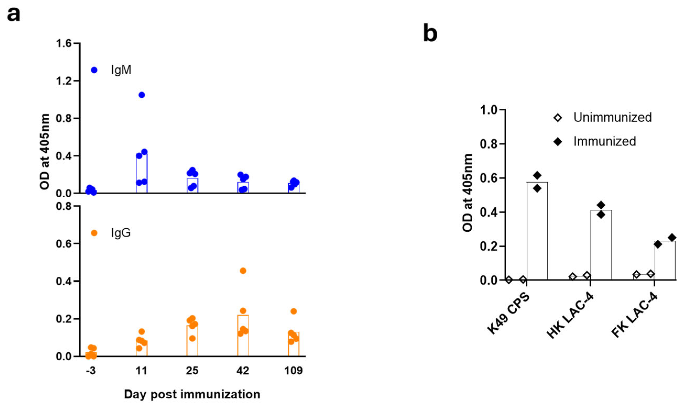
We next assessed if the K49-specific IgG induced by the vaccine recognizes the CPS on the surface of whole LAC-4 cells. Formalin- and heat-killed LAC-4 cells were prepared and used for ELISA. Serum samples collected from immunized mice at day 42 showed strong reactions to both formalin- and heat-killed LAC-4 cells, although the responses to the former were lower than those to the killed LAC-4 cells (Figure 4b).
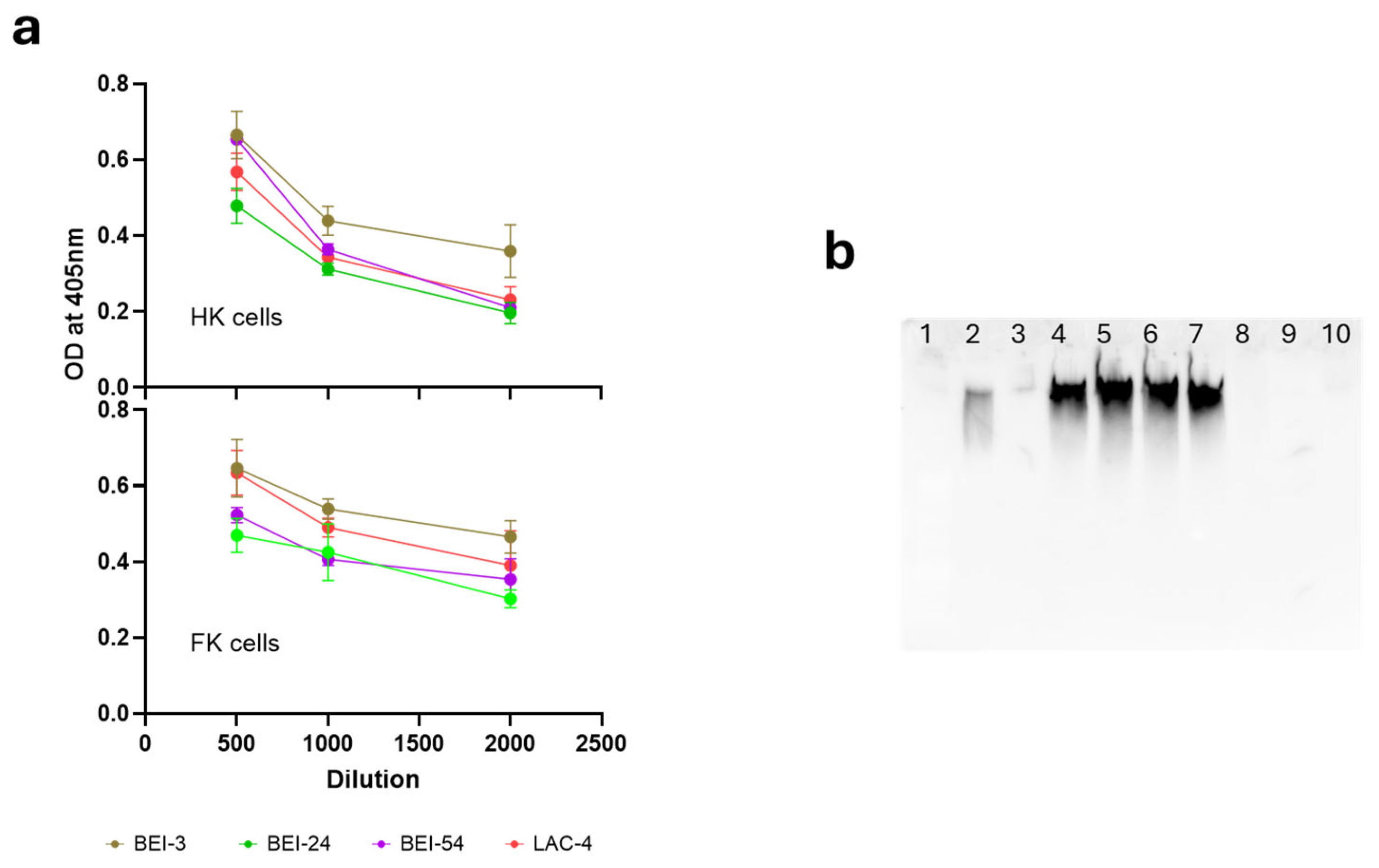
To further characterize the immunogenicity of the LAC-4 KL49 CPS conjugate vaccine candidate, we assessed if the induced specific IgG response cross-reacts against other KL49 A. baumannii isolates available in our laboratory (BEI-3, BEI-24, and BEI-54). We first used an indirect ELISA to measure the specific IgG level to the individual inactivated bacterial cells of these KL49 isolates. We found that the sera collected from the immunized animals showed substantial IgG responses to both heat- and formalin-killed other KL49 bacterial cells, and the magnitude of the responses was comparable to that of inactivated LAC-4 cells (Figure 5a).
Figure 5.
Cross-reactivities of the sera from mice immunized with the LAC-4 K49 conjugate vaccine to other KL49 A. baumannii strains. (a) Cross-reactivities of serum IgG responses to whole killed other KL49 bacterial cells at day 42. (b) The Western blotting analysis of the immune serum responses to the CPS of other KL49 bacterial cell lysates. Groups of mice (n = 5/group) were immunized with the LAC-4 K49-CRM conjugate vaccine on days 0, 14, and 28 or unimmunized. Mice were sacrificed on day 42 for terminal blood collection. The serum IgG responses to inactivated LAC-4 cells or other KL49 bacterial cells (BEI-3, BEI-24 and BEI-54) were assessed by ELISA. Serial dilutions of pooled serum samples were used in the ELISA assays, whereas a single dilution of 1:2000 was used in the Western blotting analysis. HK: heat-killed; FK: formalin-killed. Lane 1: Pre-stained protein marker; Lane 2: K49-HSA conjugate; Lane 3: Empty well; Lane 4: LAC-4; Lane 5: BEI-3; Lane 6: BEI-24; Lane 7: BEI-54; Lane 8: ATCC17978 (KL3, negative control): Lane 9: BEI-1 (KL3, negative control); Lane 10: Empty well.
We then used Western blotting to detect the cross-reactivity of the immune sera to crude CPS preparations from LAC-4 and other KL49 bacterial isolates. The immune sera showed strong positive reactions against the lysates of all KL49 isolates tested (including LAC-4, BEI-3, BEI-24, and BEI-54) but not to the lysate of BEI-1 or ATCC17978 (KL3 isolates included as a negative control) (Figure 5b and Figure S8). These results indicate that this conjugate vaccine induces KL49-specific antibody responses cross-reactive to multiple KL49 strains and can serve as a promising component in A. baumannii vaccines.
3.4. LAC-4 K49-CRM Conjugate Vaccine Induces Strong Protection Against Intranasal and Intraperitoneal Challenges with A. baumannii LAC-4 and Other KL49 Strains
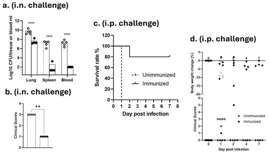
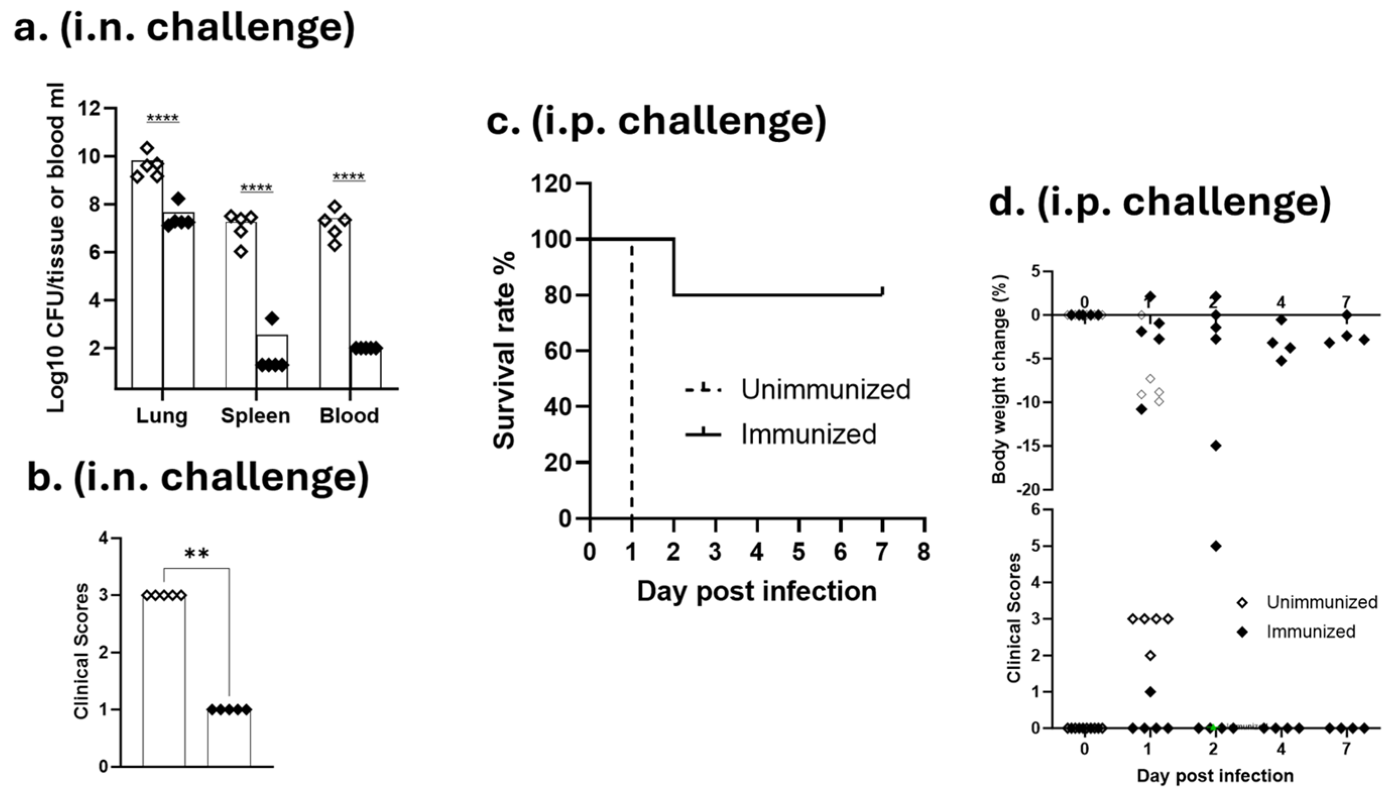
The protective efficacy of the conjugate vaccine was first evaluated in a mouse model of i.n. infection with A. baumannii LAC-4 [13]. Groups of immunized and unimmunized mice were challenged with a sublethal dose (4 × 10⁷ CFU) of LAC-4, and their clinical signs and body weight were observed. The mice were euthanized at 24 h post-challenge, and their blood, lung, and spleen samples were collected for quantitative bacteriology. The immunized mice showed minimal clinical signs and a significant reduction in both tissue (lung and spleen) and blood bacterial burdens (p < 0.001, Figure 6a,b). As in our previous studies with this model [13,17], the magnitude of the bacterial reduction was much more obvious in the blood and spleen and more moderate in the lung. We next assessed the vaccine-induced protection in mice against a lethal dose (9.6 × 10⁵ CFU) of intraperitoneal LAC-4 challenge [17]. The i.p. challenge has been used by many laboratories as a model of sepsis with a rapid clinical course of infection and high mortality [31,32]. Eighty percent of the immunized mice (4 out of 5 mice) survived the challenge, whereas all the naive mice succumbed to this dose of i.p. challenge within 24 h as anticipated (Figure 6c). More importantly, the immunized mice showed no overt clinical signs and minimal body weight loss throughout the experimental period (Figure 6d).
Figure 6.
The protective efficacy of the LAC-4 K49-CRM conjugate vaccine against i.n. and i.p. LAC-4 challenges in mice. The tissue and blood bacterial burdens (a) and clinical signs (b) in the immunized and unimmunized mice after i.n. LAC-4 challenge. The survival rate (c) and clinical signs and body weights (d) in the immunized and unimmunized mice after i.p. challenge with a lethal dose of LAC. Groups of mice (n = 5/group) were immunized with the LAC-4 K49-CRM conjugate vaccine on days 0, 14, and 28 or were unimmunized. The mice were either i.n. challenged with LAC-4 (a,b) or i.p. challenged with a lethal dose of LAC-4 (c,d). The clinical signs, body weight, and survival rate were monitored and recorded. In the i.n. challenge experiment, mice were sacrificed at 24 h after challenge. The lungs, spleens and blood were collected for quantitative bacteriology. The dots and symbols in (a,b,d) represent individual mouse data, and the bars show the means of each experimental group. ** p < 0.01, and **** p < 0.001 vs. unimmunized mice.
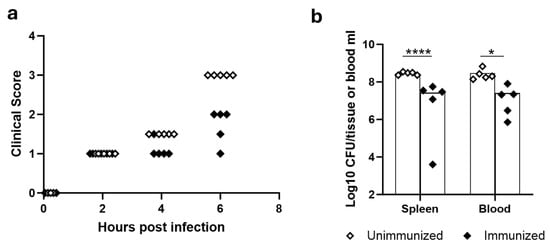
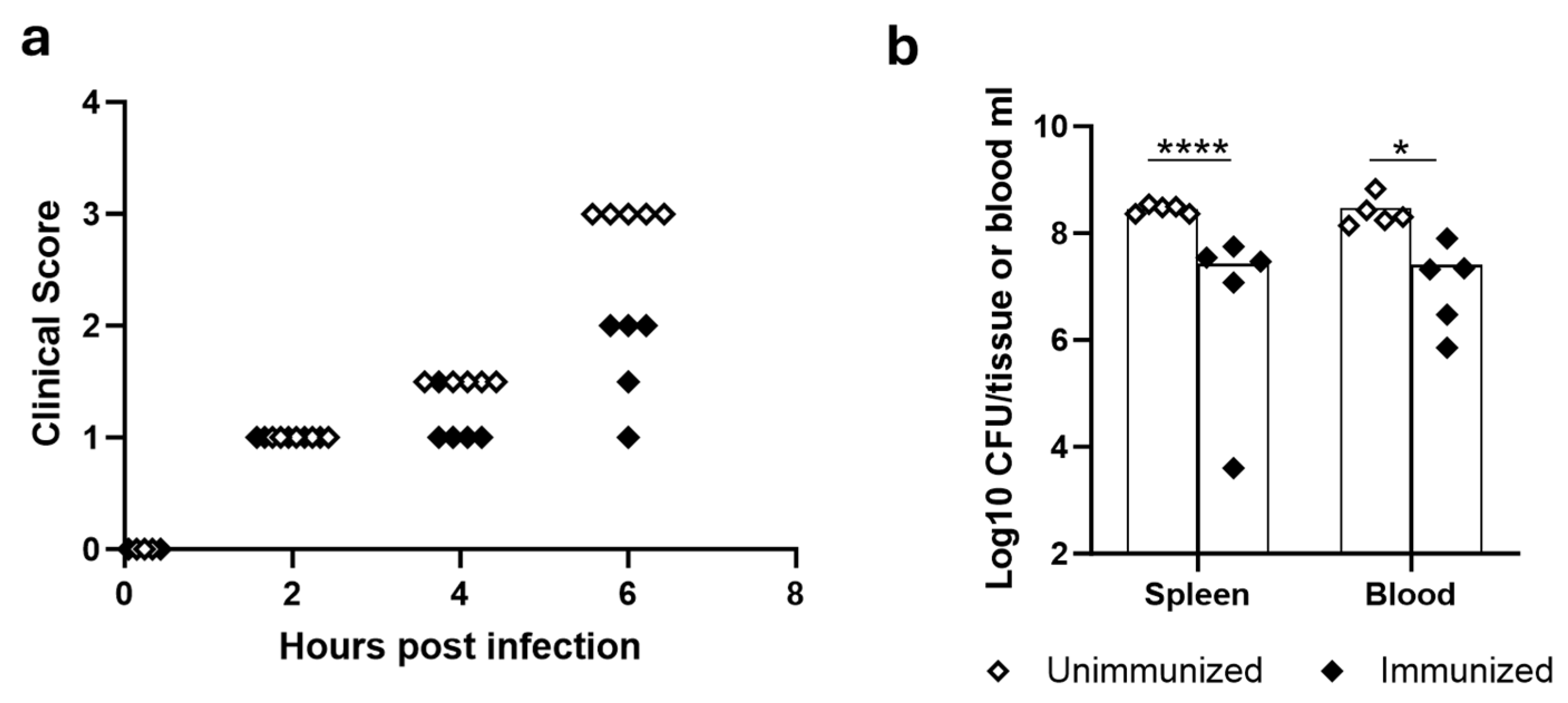
The above results (Figure 6) have convincingly demonstrated that our LAC-4 K49-CRM conjugate vaccine induces strong protective immunity against i.n. and lethal i.p. challenges with A. baumannii LAC-4 isolate, the homologous strain. Previous studies by us and others have shown that LAC-4 is highly virulent in mice [13,17,33,34]. We next assessed if this vaccine also protects against i.p. challenge with other KL49 A. baumannii strains. When i.p. challenged with 6.8 × 10⁷ CFU of BEI-24 [15], all unimmunized mice succumbed to the infection and reached the humane endpoints by 6 h, whereas the immunized mice showed only minimal to moderate clinical signs (Figure 7a). Quantitative bacterial cultures showed that immunized mice harbored significantly lower bacterial burdens in their blood (p < 0.001) and spleen (p < 0.05) than the unimmunized mice (Figure 7b). This result, together with the cross-reactivity immunogenicity data presented above (Figure 5), indicates that the LAC-4 K49-CRM conjugate vaccine induces a broad immune response and protection against multiple KL49 isolates of A. baumannii.
Figure 7.
Protective efficacy of LAC-4 K49-CRM conjugate vaccine against i.p. challenges with other KL49 A. baumannii strains in mice. The clinical signs (a) and spleen and blood bacterial burdens (b) in the immunized and unimmunized mice after i.p challenge with the other KL49 strain (BEI-24). Groups of mice (n = 5/group) were immunized with the LAC-4 K49-CRM conjugate vaccine on days 0, 14, and 28 or were unimmunized. The mice were i.p. challenged with 5.8 × 10⁷ CFU of BEI-24. The clinical signs were monitored and recorded, and the mice were sacrificed 6 h after challenge for humane reasons. The spleens and blood were collected for quantitative bacteriology. The dots represent individual mouse data, and the bars show the means of each experimental group. * p < 0.05, and **** p < 0.001 vs. unimmunized mice.
4. Discussion
Having been proven over three decades, CPS-based conjugate vaccines are very effective against several bacterial infections, and those against N. meningococcus, H. influenzae type b, S. pneumoniae, S. Typhi, etc., have been commercialized [35]. Due to their surface exposure and role in virulence, CPS are highly promising antigen targets for the development of A. baumannii vaccine candidates [12]. CPS-based conjugates (e.g., KL1 and KL9) can produce protective antibodies and induce protection against lethal challenge [36].
The major challenge to developing A. baumannii conjugate vaccines is the CPS structural diversity. So far, over 240 distinct K locus (KL) types have been identified, and more than 75 CPS structures have been determined [37,38]. The structural diversity is further complicated due to additional O-acetylation and occasional isomerization of sugar configurations [39]. Although the number of A. baumannii strains isolated keeps increasing, certain KL types are more frequently isolated in clinical settings. For example, KL2, KL3, KL7, and KL49 have been overwhelmingly associated with severe hospital-acquired infections like bacteremia, wound infections, and ventilator-associated pneumonia [40]. Therefore, it remains feasible to combat A. baumannii with multivalent CPS conjugate vaccines, a similar strategy effectively used against S. pneumoniae.
One surface polysaccharide, poly-N-acetyl-β-(1-6)-glucosamine (PNAG), often associated with biofilm formation and found on many bacterial pathogens, including A. baumannii, can be an ideal vaccine candidate. Indeed, PNAG-conjugate vaccines induced protective antibodies that enhanced opsonophagocytosis with reduced bacterial burdens in animal models of pneumonia and bacteremia [41]. The conjugates with decreased N-acetylation induced a better immune response and engagement of the complement system [42]. The degree of N-acetylation was found crucial for the formation of protective immunity against Staphylococcus aureus in mice [43]. However, immunization strategies targeting PNAG failed to protect yearling horses from infection following contact with S. equi-infected horses [44]. A human trial using PNAG monoclonal antibody in preventing an experimental urethral infection with N. gonorrhoeae was terminated due to an inadequate method to detect a therapeutic effect (ClinicalTrials.gov ID NCT03222401, 2019).
In this proof-of-concept study, we demonstrated that both the CPS and the CPS-CRM conjugates can be produced efficiently and that the glycoconjugate vaccine elicited robust CPS-specific antibody responses and conferred protection against lethal challenge with the homologous LAC-4 strain as well as other KL49 strains. However, our study has several limitations and weaknesses that need to be addressed in future studies. We did not include an unconjugated CPS control in the mouse studies because many similar studies omitted it. It is generally recognized that CPS is a T-cell-independent antigen and induces IgM predominantly with minimal immunological memory. For example, CPS alone yielded only 20% to 30% survival in lethal challenge models with poor reduction in systemic bacterial loads [45]. The inclusion of an unconjugated CPS group will increase the robustness of this study.
The main objective of this study was to assess the potential of using a simplified CPS isolation and alternative conjugation method to develop a protective glycoconjugate vaccine against A. baumannii. Therefore, the investigation of the potential immune protective mechanism of the vaccine is out of the scope of the current study. Future studies on this aspect, such as antibody-mediated killing, the role of complement and phagocytes, and the activation of T cells and cytokine responses, will be important.
Since the conjugates are very large and heterogeneous in molecular size due to cross-linking, MALDI is not suitable for this characterization. The proteins (CRM or HSA) are much smaller than the conjugates and were easily removed by size-exclusion chromatography (Figures S5 and S6, which represent before and after purification). Although the CPS was sonicated to a small size, traces of unconjugated CPS may be present in the purified conjugates. Since we did not use the colorimetric or HPAEC-PAD assays, the residue-free CPS does not affect the estimation of conjugate concentration and protein/CPS ratio by the method we described.
We also did not conduct any stability tests in this proof-of-concept study. One weakness of the thiol-based conjugation method is that the bond is vulnerable to slow reversibility. The best conjugation is through reductive amination if it is feasible.
5. Conclusions
In summary, using the clinically isolated hypervirulent A. baumannii strain LAC-4 (KL49), we investigated (1) a simplified method for CPS isolation and purification; (2) the conjugation of CPS to CRM via a thioether linkage as an alternative to the commonly used reductive amination and alkylisourea chemistries; and (3) the immunization of mice with the resulting conjugate vaccine for immunogenicity and protective efficacy evaluation. We demonstrated that both the CPS and the CPS-CRM conjugates can be produced efficiently and that the glycoconjugate vaccine elicited robust CPS-specific antibody responses and conferred protection against lethal challenge with the homologous LAC-4 strain as well as other KL49 strains. These findings further support the potential of CPS-based conjugate vaccines as a promising strategy for preventing A. baumannii infections.
Supplementary Materials
The following supporting information can be downloaded at https://www.mdpi.com/article/10.3390/vaccines14110921/s1, Figure S1: SEC-HPLC analysis of depolymerized K49; Figure S2: 1H-NMR spectra of K49 and depolymerized K49; Figure S3: MALDI of CRM and CRM-Mal; Figure S4: MALDI of HSA and HSA-Br; Figure S5: SEC-HPLC analysis of conjugation; Figure S6: SEC-HPLC of purified glycoconjugates; Figure S7: RID comparison between polysaccharides and HSA by SEC-HPLC; Figure S8: Original images and densitometric analysis of the Western blot in Figure 5b; Figure S9: Serum K49-specific IgG1 and IgG2a in immunized vs. unimmunized mice.
Author Contributions
Conceptualization, W.Z. and W.C.; methodology, W.Z., D.W., H.Z. and A.L.; validation, D.W., H.Z. and W.Z.; formal analysis, W.Z., H.Z. and W.C.; investigation, W.Z., D.W. and H.Z.; resources, H.H.X. and A.L.; writing—original draft preparation, W.Z. and W.C.; writing—review and editing, W.Z. and W.C.; funding acquisition, W.C. All authors have read and agreed to the published version of the manuscript.
Funding
This work was supported by the National Program Office, National Research Council Canada, under the Ideation Small Team Project funding (ST-01-0900).
Institutional Review Board Statement
Not applicable.
Informed Consent Statement
Not applicable.
Data Availability Statement
The original contributions presented in this study are included in the article/Supplementary Material. Further inquiries can be directed to the corresponding author.
Acknowledgments
We are grateful to Greg Beaudoin for growing A. baumannii, Frank St-Michael for help with the CPS extraction and analysis, Ken Chan for MALDI analysis, and Kanchi.Baijal for help analyzing the densitometric data of Figure S8D and for preparation of Figure 1. We also thank BEI Resources, NIAID, NIH for providing A. baumannii strains.
Conflicts of Interest
A.L. is the CEO and Chief Scientific Officer of Fina Biosolutions LLC (Rockville, MD). The author declares that the research was conducted independently and that this relationship did not influence the study design, data collection, analysis, interpretation, or the conclusions presented in this manuscript. All other authors declare no conflicts of interest.
References
- GBD 2021 Antimicrobial Resistance Collaborators. Global burden of bacterial antimicrobial resistance 1990–2021: A systematic analysis with forecasts to 2050. Lancet 2024, 404, 1199–1226. [Google Scholar] [CrossRef] [Scilit] [PubMed]
- Ayoub Moubareck, C.; Hammoudi Halat, D. Insights into Acinetobacter baumannii: A Review of Microbiological, Virulence, and Resistance Traits in a Threatening Nosocomial Pathogen. Antibiotics 2020, 9, 119. [Google Scholar] [CrossRef] [Scilit] [PubMed]
- Sati, H.; Carrara, E.; Savoldi, A.; Hansen, P.; Garlasco, J.; Campagnaro, E.; Boccia, S.; Castillo-Polo, J.A.; Magrini, E.; Garcia-Vello, P.; et al. The WHO Bacterial Priority Pathogens List 2024: A prioritisation study to guide research, development, and public health strategies against antimicrobial resistance. Lancet Infect. Dis. 2025, 25, 1033–1043. [Google Scholar] [CrossRef] [Scilit] [PubMed]
- Russo, T.A.; Beanan, J.M.; Olson, R.; MacDonald, U.; Cox, A.D.; St Michael, F.; Vinogradov, E.V.; Spellberg, B.; Luke-Marshall, N.R.; Campagnari, A.A. The K1 capsular polysaccharide from Acinetobacter baumannii is a potential therapeutic target via passive immunization. Infect. Immun. 2013, 81, 915–922. [Google Scholar] [CrossRef] [Scilit] [PubMed]
- Cheng, S.; Zhou, S.; Wang, X.; Yu, Z.; Liu, L.; Liu, J.; Qi, L. Advances in phage therapy for Acinetobacter baumannii: Mechanisms and applications. Front. Microbiol. 2026, 17, 1839739. [Google Scholar] [CrossRef] [Scilit] [PubMed]
- Singh, R.; Capalash, N.; Sharma, P. Vaccine development to control the rising scourge of antibiotic-resistant Acinetobacter baumannii: A systematic review. 3 Biotech 2022, 12, 85. [Google Scholar] [CrossRef] [Scilit] [PubMed]
- Ma, C.; Chen, W. Where are we and how far is there to go in the development of an Acinetobacter vaccine? Expert Rev. Vaccines 2021, 20, 281–295. [Google Scholar] [CrossRef] [Scilit] [PubMed]
- Yang, X.; Wei, R.; Liu, H.; Wei, T.; Zeng, P.; Cheung, Y.C.; Heng, H.; Chan, E.W.; Li, X.; Chen, S. Discovery of a Monoclonal Antibody That Targets Cell-Surface Pseudaminic Acid of Acinetobacter baumannii with Direct Bactericidal Effect. ACS Cent. Sci. 2024, 10, 439–446. [Google Scholar] [CrossRef] [Scilit] [PubMed]
- Singh, J.K.; Adams, F.G.; Brown, M.H. Diversity and Function of Capsular Polysaccharide in Acinetobacter baumannii. Front. Microbiol. 2018, 9, 3301. [Google Scholar] [CrossRef] [Scilit] [PubMed]
- Nielsen, T.B.; Pantapalangkoor, P.; Luna, B.M.; Bruhn, K.W.; Yan, J.; Dekitani, K.; Hsieh, S.; Yeshoua, B.; Pascual, B.; Vinogradov, E.; et al. Monoclonal Antibody Protects Against Acinetobacter baumannii Infection by Enhancing Bacterial Clearance and Evading Sepsis. J. Infect. Dis. 2017, 216, 489–501. [Google Scholar] [CrossRef] [Scilit] [PubMed]
- Karatovskaya, A.; Rudenko, N.; Zamyatina, A.; Zvonarev, A.; Oleinikov, V.; Shpirt, A.; Perepelov, A.; Knirel, Y.; Brovko, F. Protective Capacity of Monoclonal Antibodies against Acinetobacter baumannii K9 Capsular Polysaccharide. Microbiol. Spectr. 2023, 11, e0414122. [Google Scholar] [CrossRef] [Scilit] [PubMed]
- Zhang, Y.; Liu, C.; Zhang, Q. Recent Advances of Carbohydrate-Based Vaccines Against Acinetobacter baumannii. Eur. J. Org. Chem. 2025, 28, e202500102. [Google Scholar] [CrossRef] [Scilit]
- Harris, G.; Kuo Lee, R.; Lam, C.K.; Kanzaki, G.; Patel, G.B.; Xu, H.H.; Chen, W. A mouse model of Acinetobacter baumannii-associated pneumonia using a clinically isolated hypervirulent strain. Antimicrob. Agents Chemother. 2013, 57, 3601–3613. [Google Scholar] [CrossRef] [Scilit] [PubMed]
- Vinogradov, E.; Maclean, L.; Xu, H.H.; Chen, W. The structure of the polysaccharide isolated from Acinetobacter baumannii strain LAC-4. Carbohydr. Res. 2014, 390, 42–45. [Google Scholar] [CrossRef] [Scilit] [PubMed]
- Galac, M.R.; Snesrud, E.; Lebreton, F.; Stam, J.; Julius, M.; Ong, A.C.; Maybank, R.; Jones, A.R.; Kwak, Y.I.; Hinkle, K.; et al. A Diverse Panel of Clinical Acinetobacter baumannii for Research and Development. Antimicrob. Agents Chemother. 2020, 64, e00840-20. [Google Scholar] [CrossRef] [Scilit] [PubMed]
- Valentine, S.C.; Contreras, D.; Tan, S.; Real, L.J.; Chu, S.; Xu, H.H. Phenotypic and molecular characterization of Acinetobacter baumannii clinical isolates from nosocomial outbreaks in Los Angeles County, California. J. Clin. Microbiol. 2008, 46, 2499–2507. [Google Scholar] [CrossRef] [Scilit] [PubMed]
- Harris, G.; KuoLee, R.; Xu, H.H.; Chen, W. Mouse Models of Acinetobacter baumannii Infection. Curr. Protoc. Microbiol. 2017, 46, 6G.3.1–6G.3.23. [Google Scholar] [CrossRef] [Scilit] [PubMed]
- Kenyon, J.J.; Kasimova, A.A.; Shneider, M.M.; Shashkov, A.S.; Arbatsky, N.P.; Popova, A.V.; Miroshnikov, K.A.; Hall, R.M.; Knirel, Y.A. The KL24 gene cluster and a genomic island encoding a Wzy polymerase contribute genes needed for synthesis of the K24 capsular polysaccharide by the multiply antibiotic resistant Acinetobacter baumannii isolate RCH51. Microbiology 2017, 163, 355–363. [Google Scholar] [CrossRef] [Scilit] [PubMed]
- Popova, A.V.; Shneider, M.M.; Arbatsky, N.P.; Kasimova, A.A.; Senchenkova, S.N.; Shashkov, A.S.; Dmitrenok, A.S.; Chizhov, A.O.; Mikhailova, Y.V.; Shagin, D.A.; et al. Specific Interaction of Novel Friunavirus Phages Encoding Tailspike Depolymerases with Corresponding Acinetobacter baumannii Capsular Types. J. Virol. 2021, 95, e01714-20. [Google Scholar] [CrossRef] [Scilit] [PubMed]
- Berti, F.; De Ricco, R.; Rappuoli, R. Role of O-Acetylation in the Immunogenicity of Bacterial Polysaccharide Vaccines. Molecules 2018, 23, 1340. [Google Scholar] [CrossRef] [Scilit] [PubMed]
- MacNair, J.E.; Desai, T.; Teyral, J.; Abeygunawardana, C.; Hennessey, J.P., Jr. Alignment of absolute and relative molecular size specifications for a polyvalent pneumococcal polysaccharide vaccine (PNEUMOVAX 23). Biologicals 2005, 33, 49–58. [Google Scholar] [CrossRef] [Scilit] [PubMed]
- Lees, A.; Nelson, B.L.; Mond, J.J. Activation of soluble polysaccharides with 1-cyano-4-dimethylaminopyridinium tetrafluoroborate for use in protein-polysaccharide conjugate vaccines and immunological reagents. Vaccine 1996, 14, 190–198. [Google Scholar] [CrossRef] [Scilit] [PubMed]
- Jennings, H.J.; Lugowski, C. Immunochemistry of groups A, B, and C meningococcal polysaccharide-tetanus toxoid conjugates. J. Immunol. 1981, 127, 1011–1018. [Google Scholar] [CrossRef] [Scilit]
- Zou, W.; Williams, D.; Cox, A. Mitigating base-catalysed degradation of periodate-oxidized capsular polysaccharides: Conjugation by reductive amination in acidic media. Vaccine 2019, 37, 1087–1093. [Google Scholar] [CrossRef] [Scilit] [PubMed]
- Rappuoli, R.; De Gregorio, E.; Costantino, P. On the mechanisms of conjugate vaccines. Proc. Natl. Acad. Sci. USA 2019, 116, 14–16. [Google Scholar] [CrossRef] [Scilit] [PubMed]
- Sun, X.; Stefanetti, G.; Berti, F.; Kasper, D.L. Polysaccharide structure dictates mechanism of adaptive immune response to glycoconjugate vaccines. Proc. Natl. Acad. Sci. USA 2019, 116, 193–198. [Google Scholar] [CrossRef] [Scilit] [PubMed]
- Stefanetti, G.; MacLennan, C.A.; Micoli, F. Impact and Control of Sugar Size in Glycoconjugate Vaccines. Molecules 2022, 27, 6432. [Google Scholar] [CrossRef] [Scilit] [PubMed]
- Santostefano, G.; Corrado, A.; Malzone, C.; Di Pietro, S.; Di Bussolo, V.; De Ricco, R. Glycoconjugate Vaccine Quantification: An Overview on Present and Future Trends in Analytical Development. Anal. Chem. 2025, 97, 9541–9553. [Google Scholar] [CrossRef] [Scilit] [PubMed]
- Townsend, R.R. Chapter 5 Analysis of Glycoconjugates Using High-pH Anion-Exchange Chromatography. In Carbohydrate Analysis; El Rassi, Z., Ed.; Journal of Chromatography Library; Elsevier: Amsterdam, The Netherlands, 1995; Volume 58, pp. 181–209. [Google Scholar]
- Nobbmann, U. Refractive Index Increment dn/dc Values. Available online: https://www.malvernpanalytical.com/en/learn/knowledge-center/insights/refractive-index-increment-dndc-values/?lang=de (accessed on 7 August 2026).
- Luna, B.M.; Yan, J.; Reyna, Z.; Moon, E.; Nielsen, T.B.; Reza, H.; Lu, P.; Bonomo, R.; Louie, A.; Drusano, G.; et al. Natural history of Acinetobacter baumannii infection in mice. PLoS ONE 2019, 14, e0219824. [Google Scholar] [CrossRef] [Scilit] [PubMed]
- Xie, C.; Wang, P.; Wu, H.; Hu, X.; Nie, T.; Li, X.; Pang, P.; Li, G.; Lu, Y.; Yang, X.; et al. Protective effect of the novel cyclic peptide ASK0912 on mice with sepsis induced by Acinetobacter baumannii. Biomed. Pharmacother. 2023, 164, 114965. [Google Scholar] [CrossRef] [Scilit] [PubMed]
- Bjånes, E.; Koh, T.; Qayum, T.; Zurich, R.; McCabe, S.; Hampel, K.; Cartwright, L.; Nizet, V. Exploring Roles of the Polysaccharide Capsule in Pathogenesis of Hypervirulent Acinetobacter baumannii Clinical Isolate Lac-4. Antibiotics 2024, 13, 10. [Google Scholar] [CrossRef] [Scilit] [PubMed]
- Rouma, T.; Barbieux, E.; Bossi, L.; Poncin, K.; Denanglaire, S.; Tafesse, Y.; Vermijlen, D.; Delbauve, S.; Flamand, V.; Van Buylaere, J.; et al. Humoral response induced after intranasal vaccination with heat inactivated Acinetobacter baumannii protects immunodeficient mice against hypervirulent LAC-4 strain. Front. Immunol. 2025, 16, 1641997. [Google Scholar] [CrossRef] [Scilit] [PubMed]
- Berti, F.; Adamo, R. Antimicrobial glycoconjugate vaccines: An overview of classic and modern approaches for protein modification. Chem. Soc. Rev. 2018, 47, 9015–9025. [Google Scholar] [CrossRef] [Scilit] [PubMed]
- Rudenko, N.; Karatovskaya, A.; Zamyatina, A.; Shepelyakovskaya, A.; Semushina, S.; Brovko, F.; Shpirt, A.; Torgov, V.; Kolotyrkina, N.; Zinin, A.; et al. Immune Response to Conjugates of Fragments of the Type K9 Capsular Polysaccharide of Acinetobacter baumannii with Carrier Proteins. Microbiol. Spectr. 2022, 10, e0167422. [Google Scholar] [CrossRef] [Scilit] [PubMed]
- Cahill, S.M.; Hall, R.M.; Kenyon, J.J. An update to the database for Acinetobacter baumannii capsular polysaccharide locus typing extends the extensive and diverse repertoire of genes found at and outside the K locus. Microb. Genom. 2022, 8, mgen000878. [Google Scholar] [CrossRef] [Scilit] [PubMed]
- Roshini, J.; Patro, L.P.P.; Sundaresan, S.; Rathinavelan, T. Structural diversity among Acinetobacter baumannii K-antigens and its implication in the in silico serotyping. Front. Microbiol. 2023, 14, 1191542. [Google Scholar] [CrossRef] [Scilit] [PubMed]
- Sharar, N.S.; Iovine, A.; De Castro, C.; Hall, R.M.; Kenyon, J.J. Phage-encoded enzymes found in Acinetobacter baumannii convert pseudaminic acid to 8-epipseudaminic acid. Commun. Biol. 2025, 8, 700. [Google Scholar] [CrossRef] [Scilit] [PubMed]
- Deng, Q.; Zhang, J.; Zhang, M.; Liu, Z.; Zhong, Y.; Liu, S.; Cui, R.; Shi, Y.; Zeng, H.; Yang, X.; et al. Rapid Identification of KL49 Acinetobacter baumannii Associated with Clinical Mortality. Infect. Drug Resist. 2020, 13, 4125–4132. [Google Scholar] [CrossRef] [Scilit] [PubMed]
- Bentancor, L.V.; O’Malley, J.M.; Bozkurt-Guzel, C.; Pier, G.B.; Maira-Litran, T. Poly-N-acetyl-beta-(1-6)-glucosamine is a target for protective immunity against Acinetobacter baumannii infections. Infect. Immun. 2012, 80, 651–656. [Google Scholar] [CrossRef] [Scilit] [PubMed]
- Gening, M.L.; Pier, G.B.; Nifantiev, N.E. Broadly protective semi-synthetic glycoconjugate vaccine against pathogens capable of producing poly-beta-(1-->6)-N-acetyl-d-glucosamine exopolysaccharide. Drug Discov. Today Technol. 2020, 35–36, 13–21. [Google Scholar] [CrossRef] [Scilit] [PubMed]
- Liao, K.S.; Kao, M.R.; Ma, T.H.; Hsu, M.H.; Chen, T.Y.; Imre, B.; Harris, P.J.; Shie, J.J.; Chiu, C.H.; Wu, C.Y.; et al. Poly-beta-D-(1,6)-N-acetyl-glucosamine (PNAG) glycan vaccines with broad spectrum neutralizing activities. Nat. Commun. 2025, 16, 6179. [Google Scholar] [CrossRef] [Scilit] [PubMed]
- Cohen, N.D.; Cywes-Bentley, C.; Kahn, S.M.; Bordin, A.I.; Bray, J.M.; Wehmeyer, S.G.; Pier, G.B. Vaccination of yearling horses against poly-N-acetyl glucosamine fails to protect against infection with Streptococcus equi subspecies equi. PLoS ONE 2020, 15, e0240479. [Google Scholar] [CrossRef] [Scilit] [PubMed]
- Li, X.; Pan, C.; Liu, Z.; Sun, P.; Hua, X.; Feng, E.; Yu, Y.; Wu, J.; Zhu, L.; Wang, H. Safety and immunogenicity of a new glycoengineered vaccine against Acinetobacter baumannii in mice. Microb. Biotechnol. 2022, 15, 703–716. [Google Scholar] [CrossRef] [Scilit] [PubMed]
Disclaimer/Publisher’s Note: The statements, opinions and data contained in all publications are solely those of the individual author(s) and contributor(s) and not of MDPI and/or the editor(s). MDPI and/or the editor(s) disclaim responsibility for any injury to people or property resulting from any ideas, methods, instructions or products referred to in the content. |
© 2026 by the authors. Licensee MDPI, Basel, Switzerland. This article is an open access article distributed under the terms and conditions of the Creative Commons Attribution (CC BY) license.