


Abstract
Background: Largemouth bass ranavirus (LMBV) triggers heavy mortality in farmed Largemouth bass (Micropterus salmoides). Oral subunit vaccines are suitable for mass fish immunization yet often yield unsatisfactory immunity. Intestinal microbiota strongly modulates mucosal immune competence. This study assessed whether PLGA/selenium nanoparticle (P/Se) pretreatment enhances protection afforded by a PLGA-based oral subunit vaccine (P/V) against LMBV. Methods: A 2 × 2 factorial design was applied. Fish received 28-day oral P/Se pretreatment followed by three oral doses of P/V. Material characterization, 16S rRNA high-throughput sequencing combined with microbial assembly analysis (diversity, NST, Sloan neutral model), qRT-PCR for immune related genes, serum specific antibody ELISA, LMBV immersion challenge and histopathological examination were conducted. Results: P/Se and P/V microspheres were successfully fabricated with acceptable particle size, and loading efficiency. P/Se pretreatment significantly elevated gut microbial α-diversity and enriched Actinobacteriota, in addition to increasing the relative contribution of stochastic processes to gut community assembly. The combined pretreatment–vaccine group markedly up-regulated intestinal mucosal immune genes (IL-1β, IL-8, IFN-γ, TNF-α, IgT) and systemic IgM. Consistently, higher serum-specific IgM antibody levels, improved survival rate, and alleviated virus-induced pathological injuries in trunk kidney and liver were observed in this combined group. Vaccination alone only conferred limited protection, and pretreatment alone produced no obvious antiviral protection. Conclusions: P/Se sequential intervention remodels intestinal microbiota and enhances the protective potency of oral subunit vaccine. This sequential feeding strategy offers a feasible way to optimize oral-vaccine performance for cultured largemouth bass.
1. Introduction
The largemouth bass (Micropterus salmoides) is one of the most important freshwater aquaculture species in China, and its culture scale has expanded steadily in recent years [1]. Largemouth bass ranavirus (LMBV) belongs to the Ranavirus genus of the Iridoviridae family and can infect various species of Percidae. It was initially isolated in the United States and is now regarded as a prevalent pathogen in both farmed and wild largemouth bass populations [2]. The outbreaks of LMBV have caused mass mortality and severely constrained the healthy and sustainable development of the largemouth bass industry [1,2]. Vaccination is the mainstream, environmentally friendly strategy for control of aquatic diseases [3,4]. Compared with injectable vaccines, oral vaccines offer practical advantages such as ease of administration, reduced handling stress, and suitability for large-scale mass delivery [5]. Nevertheless, current oral vaccines in aquaculture generally elicit weak or inconsistent immune responses, which greatly limits their practical uptake and application.
The fish intestine is the largest mucosal immune organ, and intestinal microbial homeostasis is tightly linked to host immune responsiveness [6,7]. Previous studies have shown that reduced gut microbial diversity impairs intestinal immune function, alters the expression of mucosal immune cytokines and immunoglobulins, and thereby diminishes the host’s capacity to recognize and respond to antigens [8,9]. In modern aquafeeds, the narrow diversity of dietary ingredients and long-term feeding of similar diets drive convergence of the gut microbiota and reduce its functional diversity [10]. Therefore, preconditioning the intestinal microbiota and immune status with environmentally friendly bioproducts represents a promising strategy to improve the efficacy of oral vaccination.
Selenium nanoparticles (SeNps) are a low-toxicity, highly active functional delivery material with antioxidant, anti-inflammatory, and immunomodulatory properties [11]. In aquaculture, dietary supplementation with SeNps has been reported to enhance transcription of immune-related and antioxidant genes—such as IL-1β, IL-8, TNF-α and SOD—in species including Penaeus vannamei [12], Oreochromis niloticus [13], Dicentrarchus Labrax [14], Oncorhynchus mykiss [15], and Ctenopharyngodon idella [16]. SeNp feeding has also been shown to remodel fish gut microbiota, increasing the richness and diversity of specific taxa [15]. However, free SeNps are susceptible to degradation by gastric acid and digestive enzymes within the fish gastrointestinal tract.
Poly (lactic-co-glycolic acid) (PLGA) is a biocompatible, biodegradable polymeric carrier capable of effectively encapsulating bioactive agents and protecting them from gastrointestinal degradation [17]. Accordingly, PLGA microspheres encapsulating SeNps can enhance Se0 stability in the digestive tract and thus enable more effective modulation of host immune status and gut microbial ecology. In addition, PLGA microspheres are attractive carriers for oral vaccines because they can protect protein antigens from enzymatic degradation and markedly improve oral antigen delivery efficiency [18].
Here, we tested whether 28-day oral pretreatment with PLGA-encapsulated SeNPs could remodel the gut microbiota and modulate the intestinal immune microenvironment, and whether this pretreatment would be associated with enhanced protective immune responses triggered by subsequent PLGA-encapsulated oral subunit vaccine against LMBV. The present study was designed to test this hypothesis. Using 16S rRNA high-throughput sequencing, mucosal and systemic immune gene expression profiling, challenge protection assays, and histopathological analysis, we comprehensively assessed the effects of PLGA/SeNp pretreatment on intestinal microbiota structure, host immune status, and PLGA/vaccine protective efficacy.
2. Materials and Methods
2.1. Fish Husbandry and Virus
Healthy largemouth bass (15.33 ± 0.45 g) were acclimated in an indoor recirculating aquaculture system. Each experimental group (n = 50 fish) was divided into three replicate 300 L tanks (16–17 fish per tank). During the experiment, dissolved oxygen was maintained >6.0 mg/L, water temperature at 26 ± 1 °C, and pH at 7.2–7.4. Fish were randomly assigned to tanks after the 2-week acclimation period. All tanks within a group received identical treatment and shared the same recirculating water system. Individual fish served as the experimental unit for sampling and challenge observations. The basal diet was obtained from Guangdong Haid Group Co., Ltd. (Guangzhou, China). Individuals showing external injuries or poor condition were removed prior to the formal experiment. The LMBV (1.0 ± 107 copies/mL) stock used in challenge tests was stored at −80 °C in our laboratory.
2.2. Preparation of P/Se and P/V Microspheres
P/Se was prepared by an emulsion-solvent evaporation method [19]. Briefly, Na2SeO3 (CAS: 10102-18-8, Aladdin, Shanghai, China) was dissolved in water to a final concentration of 10 mg/mL and reduced by slow addition of an ascorbic acid (Aladdin, Shanghai, China) solution (40 mg/mL) under stirring to generate free SeNPs. The SeNPs were collected by centrifugation and washed to remove unreacted reagents and free ions. PLGA (0.5 g, P134565, Aladdin, Shanghai, China) was dissolved in 10 mL dichloromethane (Aladdin, Shanghai, China) and the SeNP aqueous phase (10 mg/mL) was added to form a water-in-oil (w/o) emulsion. The w/o emulsion was sonicated in an ice-water bath (200 W, 3 s on/5 s off, total sonication time 3 min) to produce fine dispersed droplets. The primary w/o emulsion was then added dropwise into an external aqueous phase containing 10 mL polyvinyl alcohol (1.5%, P139537, Aladdin, Shanghai, China) to form a w/o/w double emulsion. The organic solvent was evaporated under stirring, allowing PLGA to solidify and encapsulate SeNPs. The resulting microspheres were collected by centrifugation, washed and lyophilized for storage.
The vaccine antigen was based on the LMBV major capsid protein gene (MCP, GenBank: FR682503.1). A truncated MCP fragment (tMCP, 343 bp–1056 bp) with strong immunogenicity was selected as the antigen [20,21]. Aeromonas veronii flagellin C (FlaC) was used as a protein adjuvant. The tMCP-FlaC fusion gene was cloned into the pET-28a(+) vector to generate the recombinant plasmid pctMCP-FlaC, which was synthesized by Qingke Biotechnology Co., Ltd. (Beijing, China) The recombinant plasmid was transformed into Escherichia coli BL21 (DE3) competent cells. Protein expression was induced with IPTG (0.5 mmol/L, Solarbio, Beijing, China) at 28 °C for 6 h. Cells were collected by centrifugation (4 °C, 6000× g, 10 min) and disrupted by sonication (3 s on, 5 s off, total sonication time 15 min). The tMCP-FlaC fusion protein in the soluble fraction was purified using a His-tag Protein Purification Kit (P2226, Beyotime, Beijing, China). Purified protein was analyzed by SDS–PAGE and Western blotting using an anti-His antibody (1:1000; Abclonal, Wuhan, China). P/V were prepared following the same emulsion–solvent evaporation procedure described above.
2.3. Characterization of Nanoparticles and Microspheres
2.3.1. Scanning Electron Microscopy (SEM)
Surface morphology of PLGA/SeNPs, PLGA/vaccine microspheres and SeNPs was observed by SEM. Samples were frozen at −80 °C and lyophilized, then mounted on conductive adhesive, excess powder removed, sputter-coated with gold, and imaged using a Hitachi S4800 (Hitachi High-Tech Corporation, Tokyo, Japan) field-emission scanning electron microscope. Particle diameters of microspheres were measured from SEM images with the particle size analysis tool of ImageJ software (version 1.53t; National Institutes of Health, Bethesda, MD, USA). More than 100 microspheres were randomly selected per group for particle size statistics.
2.3.2. Selenium Loading Quantification of PLGA/SeNPs
The selenium loading of PLGA/SeNPs was quantified by inductively coupled plasma (ICP). Microsphere samples were prepared at 10 mg/mL and digested with concentrated nitric acid. Selenium concentration (Sec) was measured by ICPPRO (Thermo Scientific, Waltham, MA, USA) and loading efficiency (LE) was calculated as follows:
LE (%) = (Sec/10 mg/mL) × 100%
2.3.3. Antigen Loading Quantification of PLGA/Vaccine Microspheres
Protein loading and encapsulation efficiency of PLGA/vaccine microspheres were determined by the BCA protein assay. Microspheres were dissolved in 1 M NaOH to a final concentration of 10 mg/mL, and the protein concentration (Pc) in the solution was measured following the BCA Protein Assay Kit (Solarbio, Beijing, China). Antigen loading efficiency was calculated as follows:
LE (%) = (Pc/10 mg/mL) × 100%
2.4. Feeding Regimen and Experimental Groups
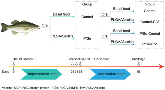
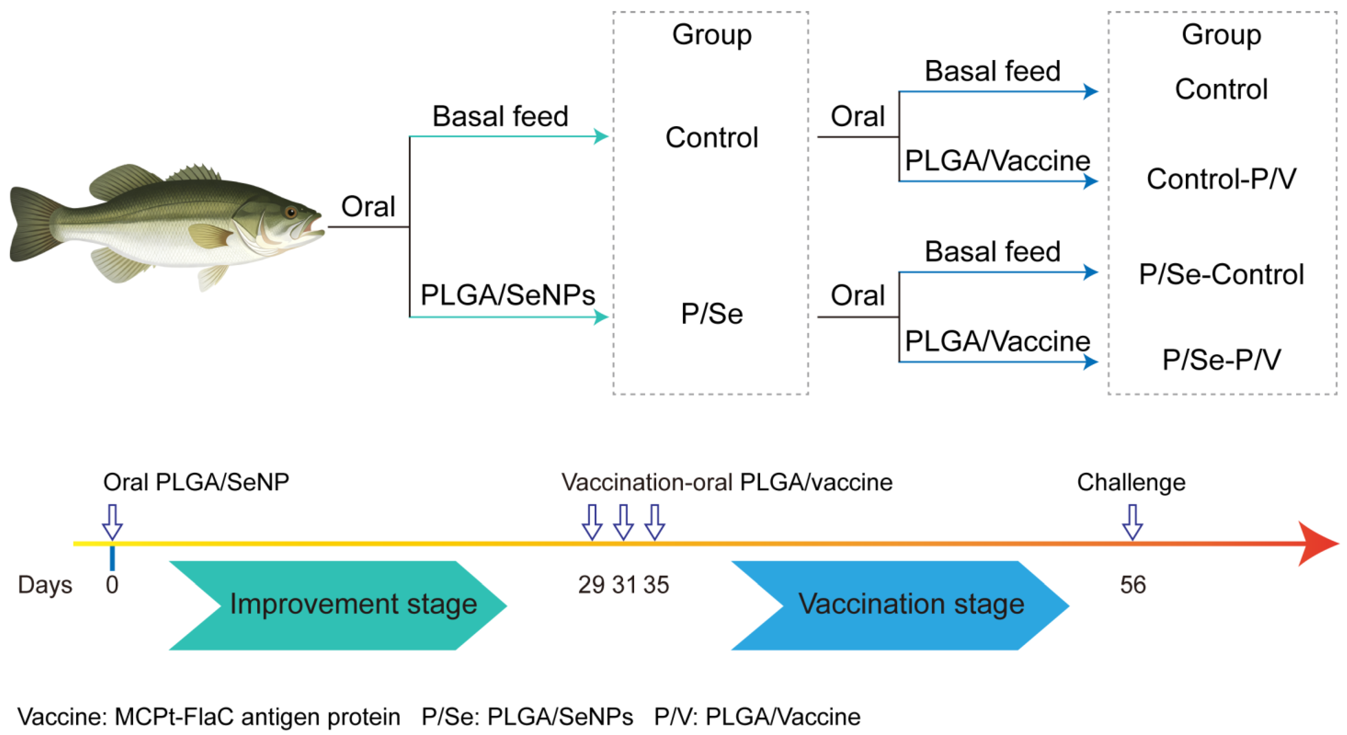
Two pretreatment regimens were set: basal diet (control group, n = 100) and oral pretreatment with P/Se (P/Se group, n = 100). From day 0, fish in the P/Se treatment groups were fed experimental feed containing P/Se (selenium content is 1 mg/Kg) once daily for 28 consecutive days; control fish received an equal amount of basal feed.
Following pretreatment, two immunization treatments were applied: oral basal feed or oral immunization with PLGA-encapsulated subunit vaccine (tMCP-FlaC content is 100 mg/Kg). This resulted in four experimental groups (50 fish per group): Control, Control-P/V, P/Se-Control, and P/Se-P/V. Oral vaccination was performed three times on days 29, 31 and 35; Control-P/V and P/Se-P/V groups received PLGA/vaccine during these events, while the non-vaccinated groups continued on basal feed. Fish were maintained until day 56 after the start of the experiment.
Fish were fed a daily ration of 2% body weight. This corresponds to an approximate selenium intake of 20 µg/kg body weight/day during the 28-day P/Se pretreatment, and an approximate tMCP-FlaC antigen intake of 2 mg/kg body weight on each of the three oral vaccination days. Experimental diets were prepared by thoroughly mixing raw ingredients, pelleted, and stored at −20 °C until use.
2.5. 16S rRNA Gene Sequencing and Bioinformatics
Bioinformatic analyses were performed by Shanghai Personal Biotechnology Co., Ltd. (Shanghai, China). Data visualization was conducted on the Personalbio GeneCloud platform. At the end of the 28-day P/Se pretreatment (before the first vaccination), six fish from each of the two pretreatment groups (Control and P/Se) were randomly selected and euthanized for intestinal content collection (n = 6 fish per group, 12 samples in total). Total DNA was extracted from gut contents and the V3–V4 region of the bacterial 16S rRNA gene was sequenced on an Illumina MiSeq platform. Amplicons were checked by gel electrophoresis, quantified, purified according to the library construction protocol, and sequenced on an Illumina MiSeq platform (2 × 250/300 bp, Illumina, San Diego, CA, USA). Raw reads were quality controlled using QIIME2 or equivalent pipelines, and denoised with DADA2 to obtain ASV/OTU tables. Taxonomic assignment was performed against the SILVA database. Community diversity analyses included alpha-diversity indices (Chao1, Good’s coverage, Simpson, Faith PD). Beta diversity was computed using the Jaccard distance and visualized by UMAP. Statistical significance of the separation between P/Se and control groups was assessed by permutational multivariate analysis of variance (PERMANOVA) based on Jaccard distances, with 999 permutations. The P/Se and control groups differed significantly in gut microbial community composition (PERMANOVA: pseudo-F = 18.6, R2 = 0.37, p = 0.0038). LEfSe analysis was performed with an LDA score threshold of >2.0 and significance set at p < 0.05 (Wilcoxon rank-sum test).
Normalized Stochasticity Ratio (NST) was calculated using the R NST package with Ružička distance to quantify the relative contribution of stochastic processes to gut microbial community assembly in largemouth bass; inter-group differences in NST were tested by the Wilcoxon rank-sum test. A Neutral Community Model (NCM) was fitted to relate ASV occurrence frequency to mean relative abundance in the metacommunity, estimating the migration rate (m) and model fit (R2). ASVs were classified as Generalist, Neutral, or Specialist according to their deviation from the neutral model fit, and the proportions of these categories were compared among groups.
2.6. Immune Protection Test
Largemouth bass used for infection were treated as described above, and the challenge test was performed according to the previously reported method [22]. On day 56, fish from the four treatment groups (50 fish per group) were immersed in water containing LMBV at 1.0 × 10⁵ copies/mL for 2 h. After exposure, fish were rinsed and returned to the recirculating system at 26 ± 1 °C. Fish behavior and mortality were recorded for 21 days until day 77, and survival rates were calculated and subjected to statistical analysis. Relative percent survival (RPS) was calculated according to the following formula:
RPS = [1 − (% mortality in vaccinated group/% mortality in control group)] × 100
2.7. Serum-Specific Antibody Level by ELISA Assay
Serum samples were collected from six fish per group on day 55 (n = 6 per group) and tested at a single 1:200 dilution. Following the previous method [3], whole-blood samples were clotted at 37 °C for 1 h and centrifuged at 1500× g for 5 min to collect serum, which was stored at −80 °C. Serum-specific antibody levels were determined by ELISA. Briefly, 96-well plates were coated with purified recombinant tMCP-FlaC protein and incubated overnight at 4 °C. After washing with PBST (PBS containing 0.1% Tween-20), plates were blocked with buffer containing 1% skimmed milk powder and 0.5% BSA at 37 °C for 2 h. Fish serum was added and incubated at 37 °C for 1 h. Following washing, HRP-conjugated mouse anti-largemouth bass IgM antibody (Biolab Reagents, Wuhan, China, 1:1000) was added and incubated at 37 °C for 0.5 h. Color development was performed with TMB substrate (Solarbio, Beijing, China) for 15 min, and the reaction was stopped using 2 M sulfuric acid. The OD450 values were measured with Varioskan LUX multimode microplate reader (Thermo Scientific, Waltham, MA, USA) after blank calibration. All samples were run in technical duplicates. Six negative control wells were included, receiving dilution buffer without serum; blank wells received PBS only. OD450 values were blank-corrected (n = 6).
2.8. Quantitative RT-PCR
Intestine and head kidney of six fish were collected on D36 for analysis of IL-1β, IL-8, IFN-γ TNF-α, IgT, and IgM. Total RNA was extracted and reverse-transcribed to cDNA. qPCR was performed with the following cycling program: initial denaturation at 95 °C for 30 s, followed by 40 cycles of 95 °C for 10 s and 60 °C for 30 s. β-actin was used as the internal reference gene and relative expression levels were calculated using the ΔΔCt method. Primers for all genes were listed in Table 1.
Table 1.
Primer sequences and their designated application in this study.
2.9. Histology and Pathology
For each group, three fish were randomly selected at day 63 (7 days post challenge), and liver and trunk kidney tissues were collected for H&E staining. The tissues were collected and immediately fixed in 4% paraformaldehyde for 24–48 h. Samples were dehydrated, paraffin-embedded, and sectioned at 4–5 µm. Sections were stained with hematoxylin and eosin (H&E, Solarbio, Beijing, China) and examined for histopathological changes.
2.10. Statistical Analysis
Statistical analyses were performed using GraphPad Prism software (version 11.0; San Diego, CA, USA). Data are presented as mean ± standard deviation (SD). Survival curves were compared by the log-rank test (ns, p > 0.05; * p < 0.05). Inter-group differences were assessed using the Kruskal–Wallis test, followed by Dunn’s multiple comparisons test. Differences were considered ns (not significant, p > 0.05), ** (p < 0.05), ** (p < 0.01), *** (p < 0.001). Different lowercase letters denote significant differences among groups.
3. Results
3.1. Preparation and Characterization of PLGA-Encapsulated SeNPs and Oral Vaccine Microspheres
The grouping and experimental workflow in this study are illustrated in Figure 1. Largemouth bass were orally pretreated with P/Se microspheres for 28 days, followed by three-time oral immunization with P/V. LMBV challenge was performed on day 56. A 2 × 2 factorial design was applied to establish four treatment groups: Control, Control-P/V, P/Se-Control and P/Se-P/V.
Figure 1.
Schematic diagram of experimental design and grouping.
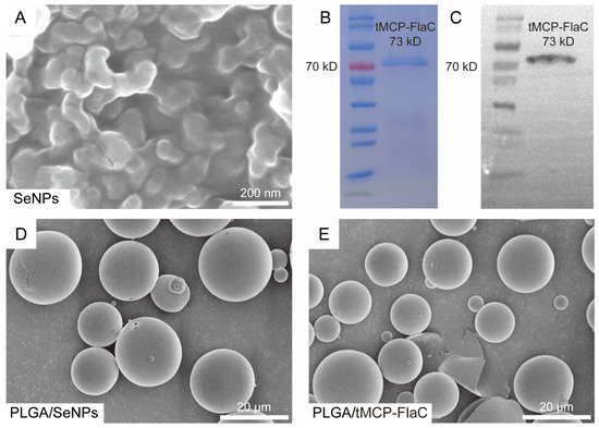
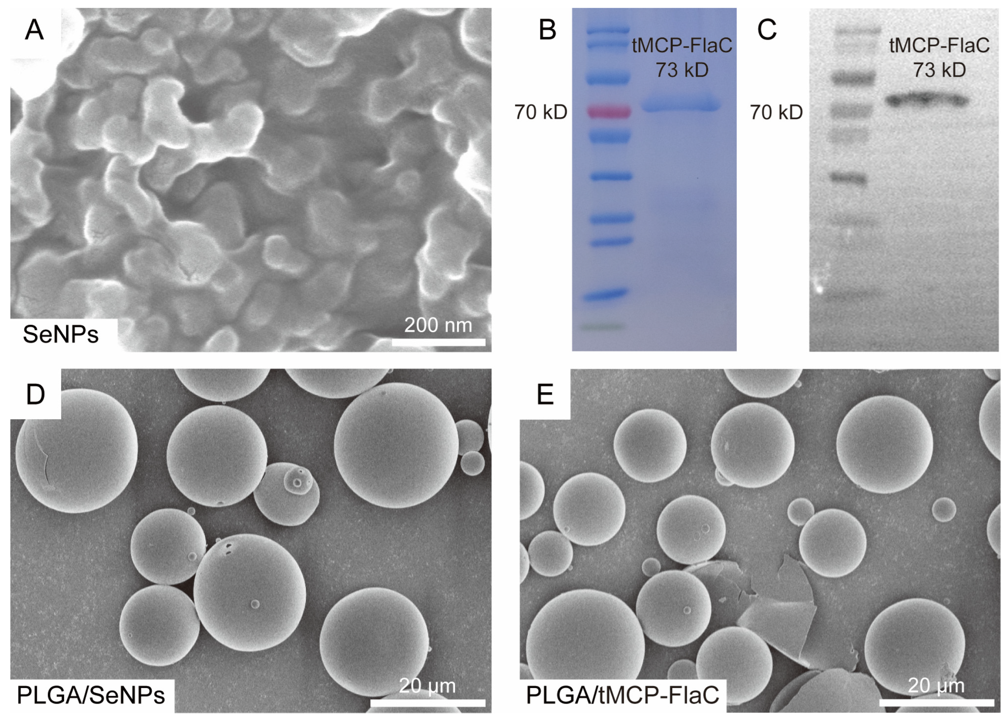
The preparation of SeNPs, P/Se microspheres, and P/V microspheres was evaluated. The unencapsulated SeNPs were nearly spherical, with particle sizes of about 150 nm (Figure 2A). SDS–PAGE and Western blotting revealed a distinct specific band for the purified tMCP-FlaC fusion protein at approximately 73 kDa, which was consistent with its theoretical molecular mass (Figure 2B,C). Both the P/Se and P/V microspheres exhibited a generally regular spherical morphology, with relatively smooth surfaces (Figure 2D,E). The average particle sizes of P/Se and P/V microspheres were 20.78 ± 7.13 μm and 19.62 ± 5.96 μm, respectively, indicating relatively uniform particle size distribution (Table 2). The selenium loading efficiency of P/Se was 1.03 ± 0.03%, and the protein loading efficiency of P/V microspheres reached 2.06 ± 0.02% (Table 2).
Figure 2.
Characterization of nanomaterials and recombinant antigen. (A) SEM image of SeNPs; (B,C) SDS-PAGE and Western blot analysis of recombinant tMCP-FlaC protein; (D) SEM image of P/Se composite microspheres; (E) SEM image of PLGA/tMCP-FlaC microspheres.
Table 2.
The characterization analysis of P/Se and P/V.
3.2. Effects of P/Se Pretreatment on the Intestinal Microbiota of Largemouth Bass
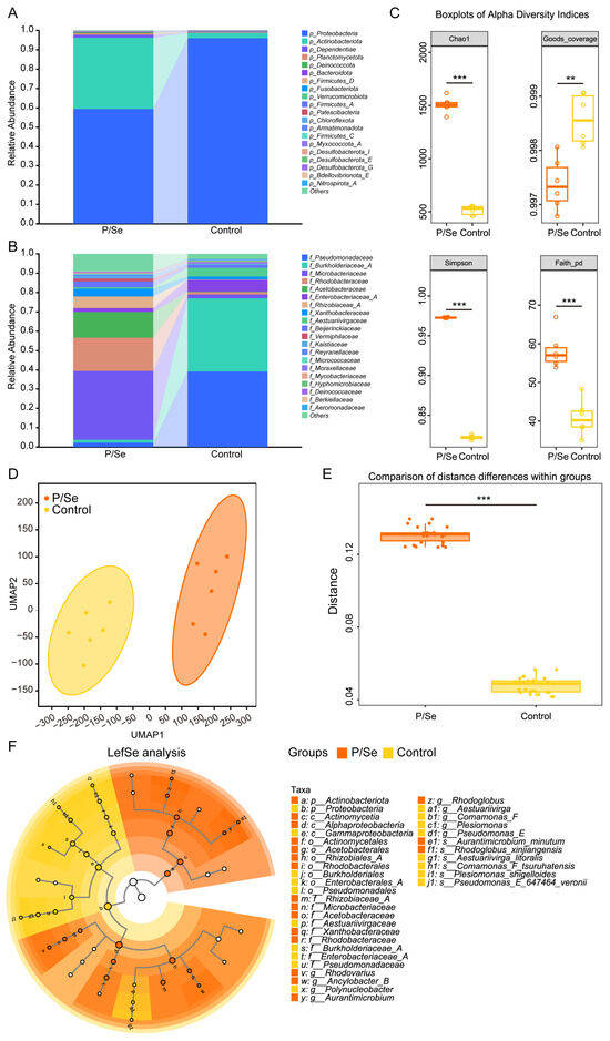
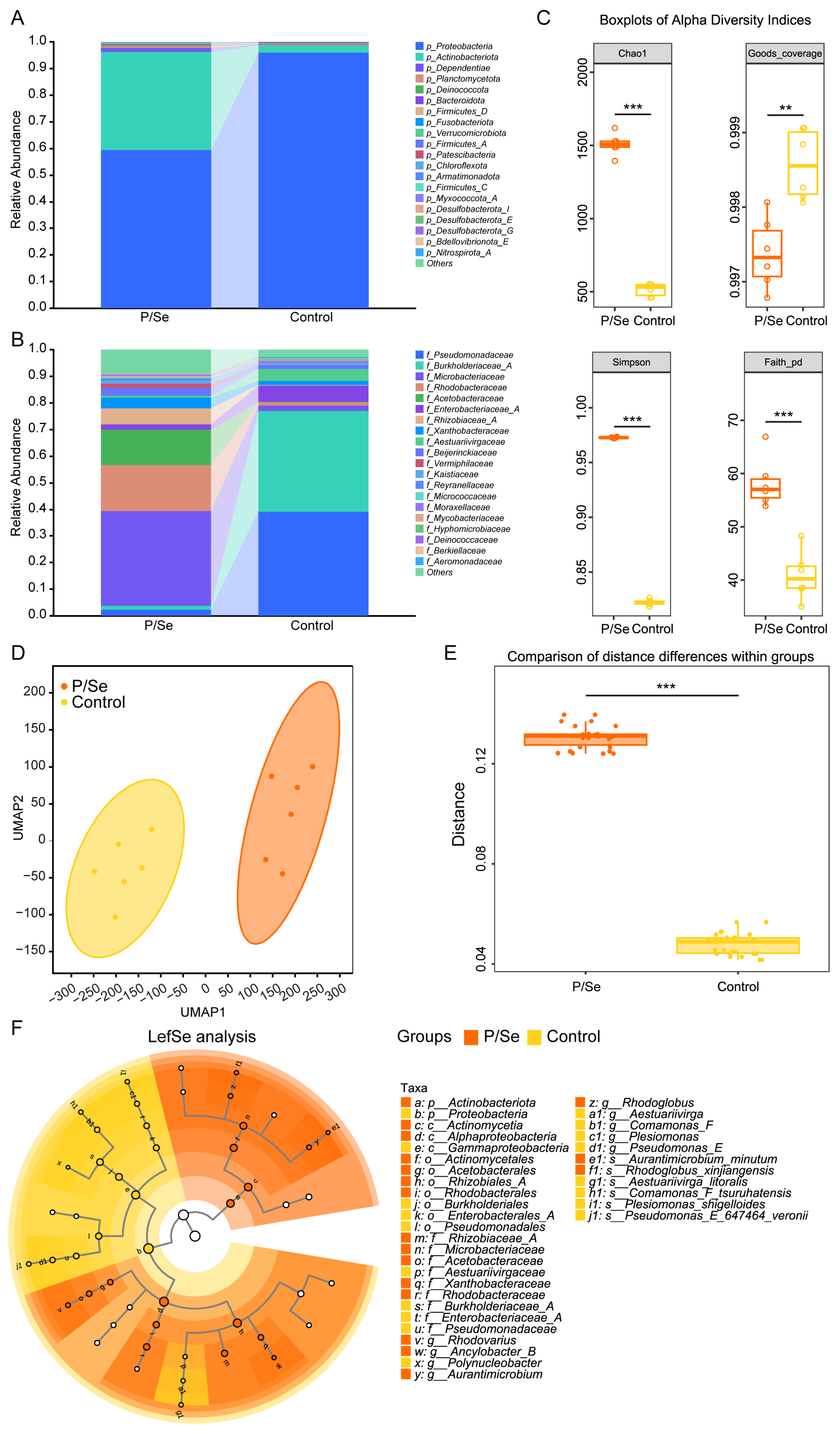
To investigate the regulatory effects of P/Se pretreatment on the intestinal microbiota, 16S rRNA gene sequencing was performed on intestinal microbial samples from all experimental groups. At the phylum level, P/Se pretreatment markedly increased the relative abundance of Actinobacteriota and substantially altered the overall composition of the intestinal bacterial community (Figure 3A). At the family level, the distribution patterns of bacterial abundance in the P/Se group differed markedly from those in the control group (Figure 3B). Alpha-diversity analysis showed that the Chao1, Faith’s phylogenetic diversity, and Simpson indices were significantly higher in the P/Se group than in the control group (Figure 3C). The sequencing depth was sufficient, as indicated by Good’s coverage values exceeding 0.997 in all samples (Figure 3C). Beta-diversity analysis showed clear separation between the P/Se and control groups in the ordination space (Figure 3D), and the overall microbial community structures differed significantly between the two groups (Figure 3E). LEfSe analysis further demonstrated that taxa associated with Actinobacteriota were predominantly enriched in the P/Se group. The representative differentially abundant genera included Aurantimicrobium, Rhodoglobus, and Aestuariivirga (Figure 3F). Overall, P/Se pretreatment reshaped the intestinal microbial community structure and significantly increased bacterial richness and diversity.
Figure 3.
Effects of P/Se pretreatment on intestinal microbiota structure in largemouth bass. (A) Stacked bar plot of intestinal microbial community composition at the phylum level; (B) stacked bar plot of intestinal microbial community composition at the family level; (C) box plots of alpha-diversity indices (Chao1, Goods-coverage, Simpson, Faith pd); (D) beta-diversity analysis based on UMAP ordination; (E) comparison of intra-group distance; (F) LEfSe cladogram showing differential microbial taxa. Data are presented as mean ± SD. ** p < 0.01, *** p < 0.001.
3.3. Effects of P/Se Pretreatment on the Ecological Assembly of the Intestinal Microbiota
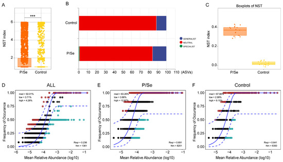
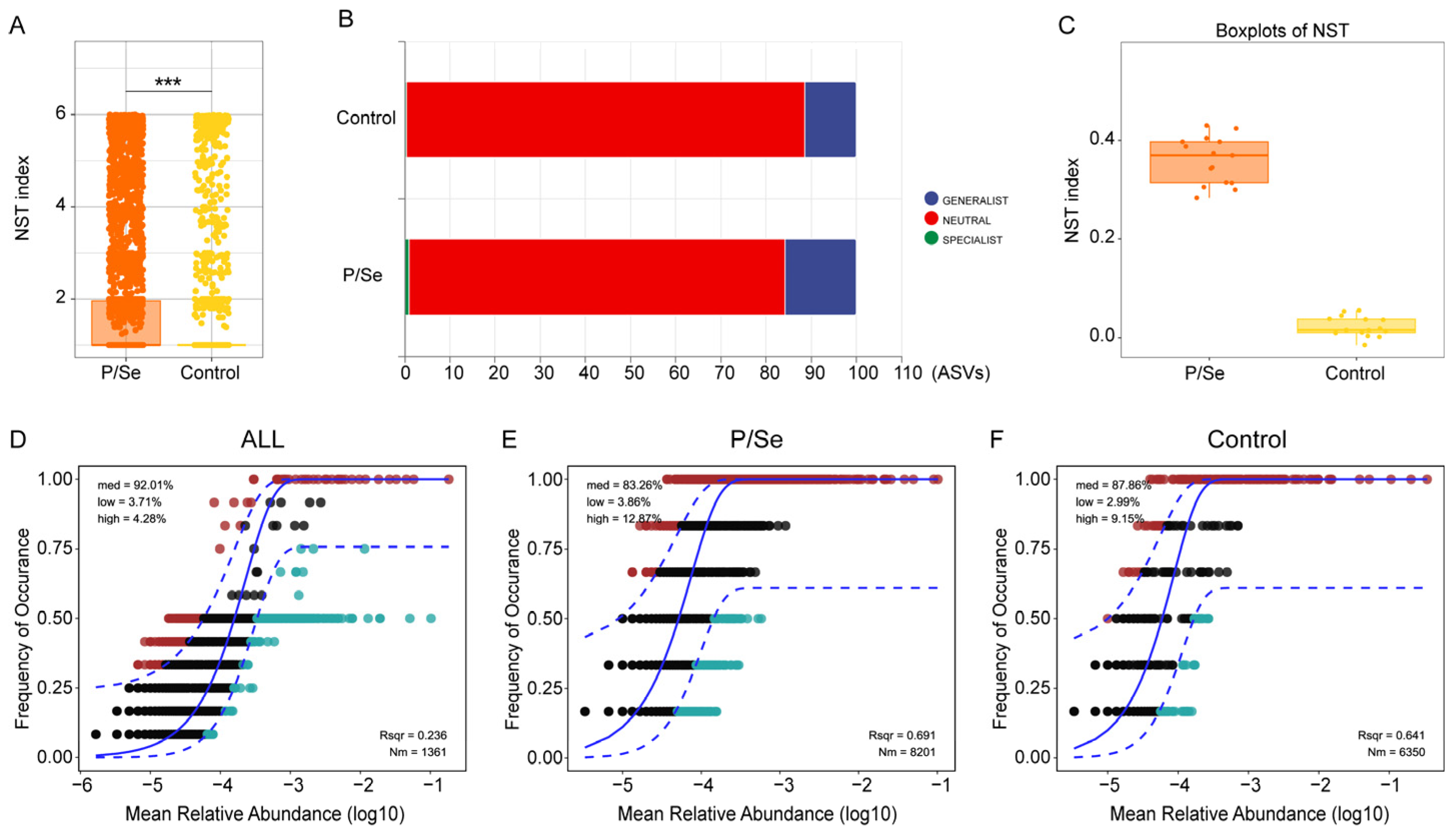
To clarify the ecological processes underlying P/Se-induced restructuring of the intestinal microbiota in largemouth bass, ecological niche classification, the normalized stochasticity ratio (NST), and the Sloan neutral community model were employed. The results of intergroup niche width analysis showed that the microbial niche widths differed extremely significantly between the P/Se and control groups (Figure 4A). Niche classification showed that neutral-process-dominated taxa accounted for the largest proportion of the intestinal microbiota in both groups, followed by generalists, whereas specialists represented only a very small proportion (Figure 4B). The NST analysis showed that the NST values were significantly higher in the P/Se group than in the control group, indicating that P/Se pretreatment increased the relative contribution of stochastic processes to intestinal microbial community assembly (Figure 4C). The Sloan neutral community model was subsequently used to fit the observed patterns of community composition (Figure 4D–F). The coefficient of determination (R2) for all samples was 0.236, whereas the R2 values for the P/Se and control groups reached 0.691 and 0.641, respectively. Taken together, P/Se treatment altered the proportional composition of taxa belonging to different ecological niche categories and enhanced the contribution of neutral ecological processes to intestinal microbial community assembly.
Figure 4.
Impacts of P/Se pretreatment on the assembly processes of intestinal microbial communities in largemouth bass. (A) Comparison of microbial niche width among groups. Data are presented as mean ± SD. *** p < 0.001; (B) proportion statistics of taxa assigned to different niche categories. GENERALIST: generalist taxa; NEUTRAL: neutral taxa; SPECIALIST: specialist taxa; (C) box plot of normalized stochasticity ratio (NST) index; (D–F) fitting results of Sloan neutral community model for all samples, P/Se group and control group, respectively. The solid blue line represents the neutral community model prediction, and the dashed blue lines indicate the 95% confidence intervals around the prediction. Colored points denote ASVs classified as generalists (green), neutral (gray), and specialists (red) based on their deviation from the neutral model fit. The coefficient of determination (R2) and migration rate (m) are indicated in each panel.
3.4. Effects of P/Se Pretreatment Combined with P/V on Immune-Related Gene Expression
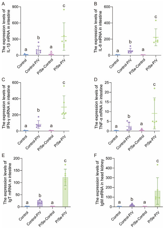
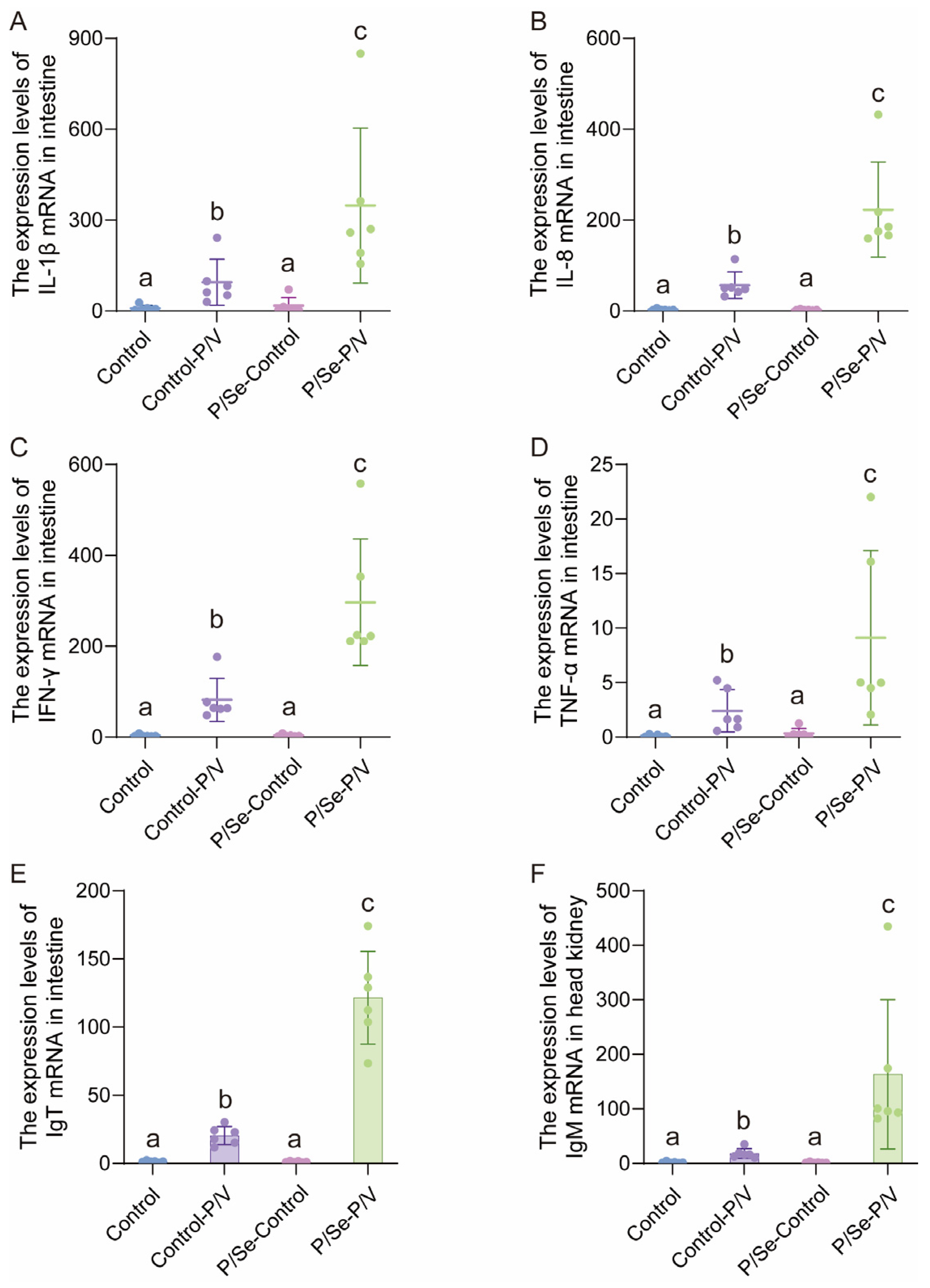
The mRNA expression levels of immune-related genes in the intestine and head kidney were determined by qRT-PCR. In the intestinal tissue, the P/Se-P/V group exhibited significantly higher transcript levels of the pro-inflammatory genes IL-1β, IL-8, IFN-γ, and TNF-α, and the immunoglobulin gene IgT than the Control, P/Se-Control, and Control-P/V groups (Figure 5A–E). These results indicated that the combined treatment induced a moderate but significant activation of intestinal inflammatory and mucosal immune responses. The expression level of IgM was also significantly higher in the P/Se-P/V group than in all other groups (Figure 5F). No evident up-regulation of the examined immune-related genes was observed in the groups that did not receive vaccine stimulation (Figure 5A–F). These findings suggest that P/Se pretreatment enhanced the immune activation induced by the orally administered P/V.
Figure 5.
Relative mRNA expression levels of immune-related genes in the intestine and head-kidney of largemouth bass from different treatment groups. (A) Intestinal IL-1β; (B) intestinal IL-8; (C) intestinal IFN-γ; (D) intestinal TNF-α; (E) intestinal IgT; (F) head-kidney IgM. Different lowercase letters (a, b, c) indicate significant differences among groups (p < 0.05, Kruskal–Wallis test followed by Dunn’s multiple comparisons test). Groups sharing a common letter are not significantly different.
3.5. Evaluation of Protective Efficacy Against LMBV Infection
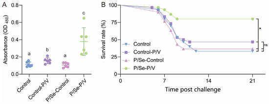
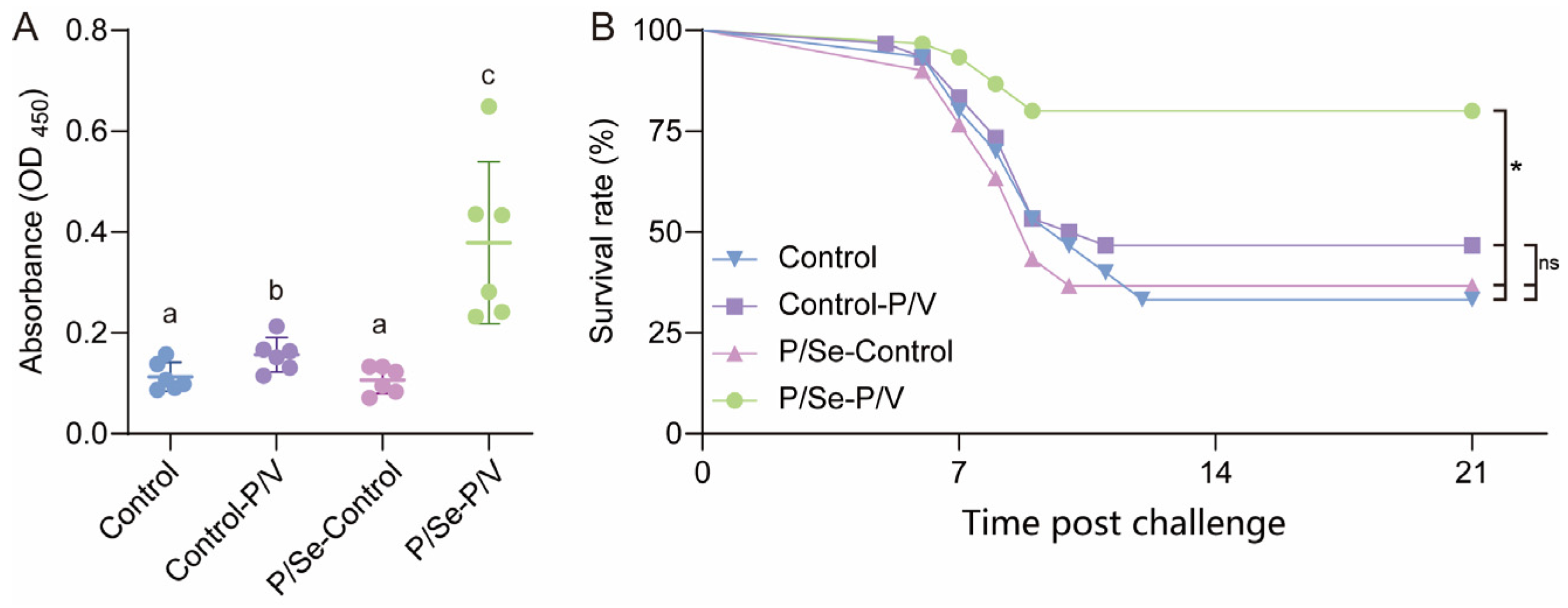
Serum-specific antibody levels in largemouth bass were determined by ELISA (Figure 6A). No significant difference was observed between the Control and P/Se-control groups, which were assigned the same statistical category. The serum antibody level in the Control-P/V group was significantly higher than those in the Control and PS groups. Notably, the P/Se-P/V group exhibited the highest serum antibody absorbance among all treatment groups, which was significantly higher than that of the other three groups.
Figure 6.
Serum-specific antibody levels and survival performance of largemouth bass after LMBV challenge. (A) The OD450 absorbance values of serum-specific antibodies measured by ELISA. Different lowercase letters (a, b, c) indicate significant differences among groups (p < 0.05, Kruskal–Wallis test followed by Dunn’s multiple comparisons test). Groups sharing a common letter are not significantly different. (B) Cumulative survival curves of largemouth bass post LMBV challenge. Data are presented as mean ± SD. * p < 0.05, ns = not significant.
The final cumulative survival rates at 21 days post challenge were as follows: Control, 33.3%; Control-P/V, 46.7%; P/Se-Control, 36.7%; and P/Se-P/V, 80.0% (Figure 6B). The corresponding RPS values were 20.1% for Control-P/V, 5.1% for P/Se-Control, and 70.0% for P/Se-P/V. The P/Se-P/V group showed the highest survival and RPS, significantly greater than those of all other groups. The P/Se-Control group showed negligible RPS (5%), confirming that P/Se pretreatment alone did not confer direct antiviral protection. In contrast, the P/Se-P/V group exhibited a significantly higher survival rate than all other groups.
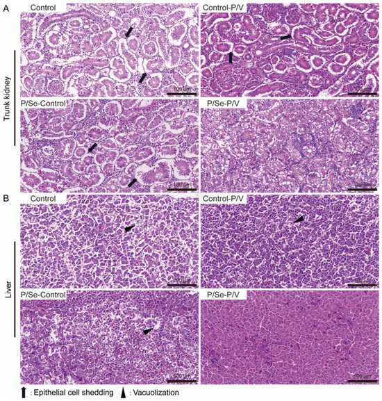
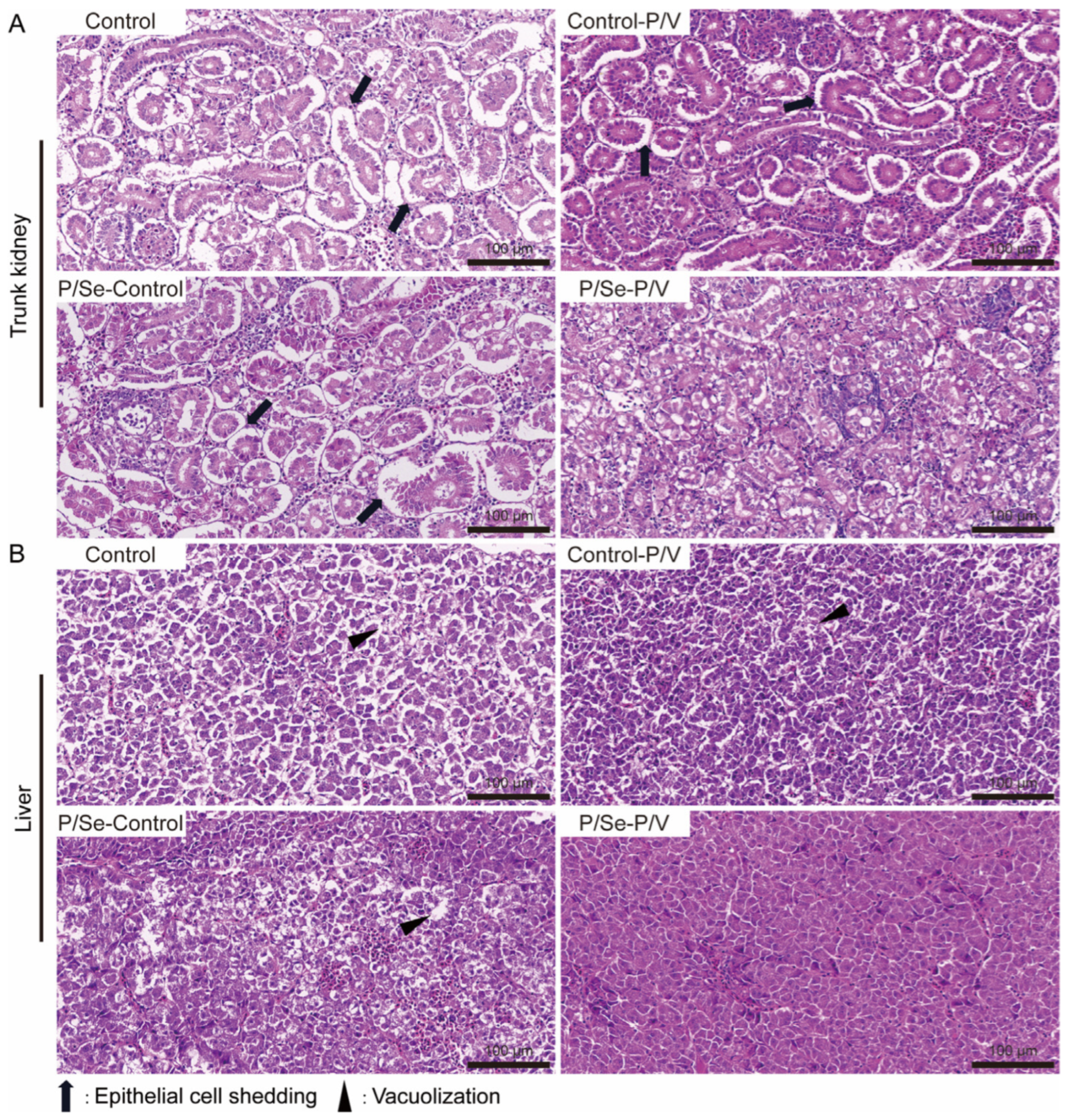
Histopathological examination further supported the protective effect of the combined treatment (Figure 7). In the P/Se-P/V group, the renal tubules in the trunk kidney remained intact, and the hepatocytes were regularly arranged, with no obvious pathological lesions. By contrast, epithelial cell shedding in the renal tubules and marked vacuolar degeneration in the liver were observed in the Control, Control-P/V, and P/Se-control groups, consistent with typical LMBV-associated tissue damage. Histopathological examination of representative sections further supported the protective effect of the combined treatment.
Figure 7.
Histopathological observation of trunk kidney and liver tissues in largemouth bass after LMBV challenge. (A) Trunk kidney tissue; (B) liver tissue. Scale bar = 100 μm. High-resolution versions of these sections are provided in Supplementary Figures S1–S8.
4. Discussion
LMBV infection causes mass mortality and severely threatens the sustainable development of largemouth bass aquaculture [2,23]. Oral vaccines possess prominent advantages, including convenient administration, low-stress response and suitability for mass immunization of farmed fish, exhibiting great application potential for aquatic disease prevention and control [24,25]. Nevertheless, oral delivery systems generally suffer from insufficient immunogenicity and unstable protective efficacy, which limit their practical application [26]. In the present study, a time-sequential intervention strategy was adopted. Largemouth bass were subjected to 28-day feeding pretreatment with P/Se microspheres to modulate host immune status and intestinal microenvironment, aiming to establish a favorable in vivo condition for oral antigen activation. Afterwards, PLGA-encapsulated recombinant tMCP-FlaC protein-based subunit oral vaccine was administrated, and the enhancement effect of this pretreatment strategy on the protective immunity against LMBV elicited by oral vaccine was systematically evaluated.
The present characterization of P/Se and P/V microspheres is limited to surface morphology, particle size, and loading efficiency. Additional parameters valuable for oral delivery development—including in vitro antigen release kinetics, stability under simulated gastrointestinal pH and enzyme conditions, and long-term storage stability—were not assessed in this study. Nevertheless, approximately 20 µm particle size is within the range reported for effective oral PLGA microsphere vaccines in fish [19], and the successful in vivo protection data confirm that the formulations were functionally delivered under the present feeding regimen. A more comprehensive physicochemical evaluation, including release profiling and gastrointestinal stability tests, will be included in our follow-up work.
Previous studies on O. niloticus [13], D. Labrax [14] and Acanthopagrus arabicus [27] have demonstrated that dietary nanoselenium at 1 mg/kg can effectively improve innate immune capacity in fish. Accordingly, the dietary final concentration of nanoselenium was set at 1 mg/kg in this study to achieve expected immunomodulatory effects. In terms of vaccine antigen design, the MCP of LMBV exhibits favorable immunogenicity and has been widely applied as a candidate antigen for DNA vaccines, subunit vaccines and surface-displayed vaccines [20,28,29,30]. FlaC serves as an effective vaccine molecular adjuvant, which triggers host immune responses via the TLR5 signaling pathway [31,32]. In addition, A. veronii, the source of FlaC in this work, is also a prevalent pathogen in largemouth bass aquaculture [33]. Based on previously reported epitope mapping of MCP [20,21], the dominant immunogenic fragment of MCP was truncated and fused with FlaC derived from A. veronii, generating the tMCP-FlaC fusion antigen used in our oral vaccine.
Long-term feeding with formulated artificial diets tends to induce homogenization of intestinal microbiota and reduce the alpha-diversity of gut microbial communities in fish [34]. Decreased intestinal microbial alpha-diversity is frequently accompanied by compromised intestinal mucosal immune responses and impaired host recognition towards mucosal antigens, which represents one potential cause for unstable performance of oral vaccines in aquaculture [35]. Existing aquaculture studies have reported that dietary nanoselenium supplementation elevates intestinal microbial alpha-diversity and enriches beneficial taxa such as Actinobacteria in O. niloticus, thereby regulating host growth and immunity [13]. Dietary nanoselenium supplementation can also reshape gut microbiota, improve intestinal antioxidant and immune capacity, and alleviate stress-triggered intestinal injury in O. mykiss [15]. Consistent with those findings, our results revealed that P/Se pretreatment significantly increased intestinal alpha-diversity indices including Chao1, Simpson and Faith-pd values, enriched Actinobacteria-related taxa, altered the niche width of intestinal microbiota, and enhanced the contribution of stochastic processes to community assembly. These observations indicated that P/Se pretreatment could mitigate the decline in gut microbial diversity induced by single-diet feeding and remodel intestinal microbial communities in largemouth bass.
While higher microbial diversity is generally associated with functional redundancy, the specific roles of the enriched taxa (e.g., Aurantimicrobium, Rhodoglobus, Aestuariivirga) in fish gut immunity are not well established. Increased stochasticity reflects a shift toward neutral community assembly, which does not necessarily indicate a healthier or more beneficial community. The functional implications of these compositional shifts warrant metagenomic or metabolomic investigation in future studies.
Significant up-regulation of pro-inflammatory genes (IL-1β, IL-8, IFN-γ, TNF-α), mucosal immunoglobulin IgT in the intestine and systemic IgM in head-kidney were observed in the P/Se-P/V group receiving combined P/Se pretreatment and oral vaccination. As a key intestinal mucosal antibody in fish, IgT plays a critical role in local intestinal antiviral immunity, whereas IgM reflects the level of systemic humoral immunity [36,37]. The up-regulation of pro-inflammatory cytokines indicated effective activation of local intestinal immunity [38], demonstrating obvious immune-stimulatory effects derived from this sequential pretreatment regime.
Higher gut microbial alpha-diversity represents more abundant microbial functional repertoires [39,40]. Symbiotic commensals can participate in intestinal mucosal immune regulation via metabolites and host–microbe crosstalk, potentially constructing a favorable intestinal microenvironment for subsequent oral-vaccine-triggered immune activation [41,42]. In our study, the elevation in gut microbial alpha-diversity induced by P/Se pretreatment occurred synchronously with enhanced mucosal and systemic immune-related gene expression triggered by P/V vaccination. We speculate that increased intestinal microbial alpha-diversity may serve as one potential pathway whereby P/Se pretreatment amplifies oral-vaccine-mediated immune responses.
However, because no microbiota depletion, FMT, or gnotobiotic experiments were performed, the causal contribution of the altered microbiota versus direct immunomodulation by selenium/PLGA cannot be disentangled. The observed changes in microbial diversity and composition should therefore be interpreted as correlative. Meanwhile, the direct immunomodulatory effects exerted by PLGA–nanoselenium on the host itself cannot be excluded, and further experiments are required to distinguish their respective contributions.
Protective efficacy against pathogenic challenge is the gold standard for evaluating biological agents [43,44]. Combined analysis of specific antibody levels and post-challenge survival demonstrated that the combination of P/Se pretreatment and P/V enhanced specific humoral immunity and improved the survival rate of largemouth bass upon LMBV infection. Fish from the combined treatment group exhibited maximally preserved histological integrity of trunk kidney and liver with minimal pathological lesions. The improved tissue anti-damage capacity, together with elevated specific immune responses, effectively mitigated virus-triggered pathological damages in target organs and strengthened host resistance against LMBV infection. Future studies should quantify viral loads in target tissues (e.g., trunk kidney, spleen) at multiple time points post challenge to directly confirm whether vaccination reduces viral replication. The present survival and histopathological data provide indirect evidence of protection, but direct viral quantification would strengthen the mechanistic interpretation.
From an aquaculture application perspective, the 28-day P/Se pretreatment followed by three oral vaccinations is compatible with standard feeding protocols. The PLGA microspheres can be mixed into feed during pelleting or top-coated with oil binder. However, several practical considerations remain: (1) the 28-day pretreatment can be integrated into the fingerling production phase when fish are on a stable diet; (2) PLGA microsphere production cost and scalability need optimization for commercial-scale use; (3) the stability of antigen-containing feed under farm storage conditions requires validation; and (4) the optimal timing relative to typical LMBV outbreak seasons should be determined in farm-based field trials. Future on-farm trials are essential to bridge laboratory efficacy and practical application.
5. Conclusions
Sequential oral pretreatment with P/Se effectively remodeled the intestinal microbiota of largemouth bass, increasing gut microbial alpha-diversity and the stochastic contribution to community assembly. When combined with P/V oral vaccine, this pretreatment significantly promoted mucosal and systemic immune activation, elevated specific serum antibody levels, improved post-challenge survival, and reduced LMBV-triggered histological lesions in trunk kidney and liver. P/Se pretreatment alone produced no detectable antiviral protection, and vaccination alone yielded only limited protective outcomes. However, the present data do not disentangle whether the enhanced vaccine protection is mediated by the observed microbiota remodeling or by direct immunomodulation of selenium itself. This sequential feeding strategy represents a promising practical approach for enhancing the performance of oral subunit vaccines against LMBV in largemouth bass aquaculture.
Supplementary Materials
The following supporting information can be downloaded at: https://www.mdpi.com/article/10.3390/vaccines14110920/s1, Figure S1: High-resolution H&E-stained trunk kidney section from the Control group; Figure S2: High-resolution H&E-stained trunk kidney section from the Control-P/V group; Figure S3: High-resolution H&E-stained trunk kidney section from the P/Se-Control group; Figure S4: High-resolution H&E-stained trunk kidney section from the P/Se-P/V group; Figure S5: High-resolution H&E-stained liver section from the Control group; Figure S6: High-resolution H&E-stained liver section from the Control-P/V group; Figure S7: High-resolution H&E-stained liver section from the P/Se-Control group; Figure S8: High-resolution H&E-stained liver section from the P/Se-P/V group.
Author Contributions
Writing—original draft preparation, conceptualization, methodology, formal analysis, X.H. and Y.C.; Methodology, software, Z.M. and Y.Z.; Methodology, L.L.; Supervision, H.Y.; Data curation, H.Y.; Project administration, formal analysis, writing—review and editing, N.J. All authors have read and agreed to the published version of the manuscript.
Funding
This research was funded by the BAAFS Exploratory Research Project (TSXM202603), the National Engineering Research Center for Freshwaters (Grant No. PT2026-19), and the Scientific and technological innovation capacity building project of Beijing Academy of Agricultural and Forestry Sciences (KJCX20230215).
Institutional Review Board Statement
All the animal experiments were approved by the Science and Technology Ethics (Review) Committee of Beijing Academy of Agriculture and Forestry Sciences (BAAFS-KJLL-20250911, approval date: 5 September 2025).
Data Availability Statement
All data supporting the findings of this study are included in the published article and its Supporting Information Files. Additional datasets generated and analyzed during the current study are available from the corresponding author upon reasonable request.
Acknowledgments
The authors would like to thank Zhensheng Wang and Yanqi Zhang for academic discussion and guidance. We also thank Kui He and Congcong Bai from Shanghai personalbio Company for their contributions to the analysis of high-throughput sequencing results.
Conflicts of Interest
The authors declare no conflicts of interest.
References
- Yang, S.; Zhao, J.; An, N.; Li, D.; Huang, M.; Fei, H. Updates on Infectious Diseases of Largemouth Bass: A Major Review. Fish Shellfish Immunol. 2024, 154, 109976. [Google Scholar] [CrossRef] [Scilit] [PubMed]
- Zhao, L.; Zhong, Y.; Luo, M.; Zheng, G.; Huang, J.; Wang, G.; Geng, Y.; Qian, X. Largemouth Bass Ranavirus: Current Status and Research Progression. Aquac. Rep. 2023, 32, 101706. [Google Scholar] [CrossRef] [Scilit]
- Huo, X.; Fan, C.; Ai, T.; Su, J. The Combination of Molecular Adjuvant CCL35.2 and DNA Vaccine Significantly Enhances the Immune Protection of Carassius auratus gibelio against CyHV-2 Infection. Vaccines 2020, 8, 567. [Google Scholar] [CrossRef] [Scilit] [PubMed]
- Jones, E.; Cain, K. An Introduction to Relevant Immunology Principles with Respect to Oral Vaccines in Aquaculture. Microorganisms 2023, 11, 2917. [Google Scholar] [CrossRef] [Scilit] [PubMed]
- Wang, W.; Zhu, Q.; Zhi, Y.; Huang, M.; Fei, H.; Yang, S. Updates on the Research on Nanocarriers in Fish Vaccine Oral, Immersion and Injection Delivery: A Major Review. Vaccine 2026, 88, 128873. [Google Scholar] [CrossRef] [Scilit] [PubMed]
- Sheikhzadeh, N.; Mousavi, S.; Hamidian, G.; Firouzamandi, M.; Khani Oushani, A.; Mardani, K. Role of Dietary Spirulina platensis in Improving Mucosal Immune Responses and Disease Resistance of Rainbow Trout (Oncorhynchus mykiss). Aquaculture 2019, 510, 1–8. [Google Scholar] [CrossRef] [Scilit]
- Gómez, D.; Bartholomew, J.; Sunyer, J.O. Biology and Mucosal Immunity to Myxozoans. Dev. Comp. Immunol. 2014, 43, 243–256. [Google Scholar] [CrossRef] [Scilit] [PubMed]
- Wang, J.; He, M.; Yang, M.; Ai, X. Gut Microbiota as a Key Regulator of Intestinal Mucosal Immunity. Life Sci. 2024, 345, 122612. [Google Scholar] [CrossRef] [Scilit] [PubMed]
- Hissen, K.; He, W.; Wu, G.; Criscitiello, M.F. Immunonutrition: Facilitating Mucosal Immune Response in Teleost Intestine with Amino Acids through Oxidant-Antioxidant Balance. Front. Immunol. 2023, 14, 1241615. [Google Scholar] [CrossRef] [Scilit] [PubMed]
- Hasan, I.; Rimoldi, S.; Saroglia, G.; Terova, G. Sustainable Fish Feeds with Insects and Probiotics Positively Affect Freshwater and Marine Fish Gut Microbiota. Animals 2023, 13, 1633. [Google Scholar] [CrossRef] [Scilit] [PubMed]
- Nie, X.; Yang, X.; He, J.; Liu, P.; Shi, H.; Wang, T.; Zhang, D. Bioconversion of Inorganic Selenium to Less Toxic Selenium Forms by Microbes: A Review. Front. Bioeng. Biotechnol. 2023, 11, 1167123. [Google Scholar] [CrossRef] [Scilit] [PubMed]
- Ghaffarizadeh, A.; Sotoudeh, E.; Mozanzadeh, M.T.; Sanati, A.M.; Ghasemi, A. Supplementing Dietary Selenium Nano-Particles Increased Growth, Antioxidant Capacity and Immune-Related Genes Transcription in Pacific Whiteleg Shrimp (Penaeus vannamei) Juveniles. Aquac. Rep. 2022, 25, 101215. [Google Scholar] [CrossRef] [Scilit]
- Dawood, M.A.O.; Zommara, M.; Eweedah, N.M.; Helal, A.I. The Evaluation of Growth Performance, Blood Health, Oxidative Status and Immune-Related Gene Expression in Nile Tilapia (Oreochromis niloticus) Fed Dietary Nanoselenium Spheres Produced by Lactic Acid Bacteria. Aquaculture 2020, 515, 734571. [Google Scholar] [CrossRef] [Scilit]
- Abd El-Kader, M.F.; Fath El-Bab, A.F.; Shoukry, M.; Abdel-Warith, A.-W.A.; Younis, E.M.; Moustafa, E.M.; El-Sawy, H.B.; Ahmed, H.A.; Van Doan, H.; Dawood, M.A.O. Evaluating the Possible Feeding Strategies of Selenium Nanoparticles on the Growth Rate and Wellbeing of European Seabass (Dicentrarchus labrax). Aquac. Rep. 2020, 18, 100539. [Google Scholar] [CrossRef] [Scilit]
- Li, L.; Liu, Z.; Quan, J.; Sun, J.; Lu, J.; Zhao, G. Dietary Nano-Selenium Alleviates Heat Stress-Induced Intestinal Damage through Affecting Intestinal Antioxidant Capacity and Microbiota in Rainbow Trout (Oncorhynchus mykiss). Fish Shellfish Immunol. 2023, 133, 108537. [Google Scholar] [CrossRef] [Scilit] [PubMed]
- Ye, L.; Liu, S.; Zhang, X.; Wang, C.; Li, P.; Zhang, C.; Ji, H.; Yu, H. Dietary Nano-Selenium Improves Health of Liver and Intestine of Grass Carp Ctenopharyngodon idella after Overwintering. Anim. Feed. Sci. Technol. 2023, 306, 115817. [Google Scholar] [CrossRef] [Scilit]
- Vlachopoulos, A.; Karlioti, G.; Balla, E.; Daniilidis, V.; Kalamas, T.; Stefanidou, M.; Bikiaris, N.D.; Christodoulou, E.; Koumentakou, I.; Karavas, E.; et al. Poly(Lactic Acid)-Based Microparticles for Drug Delivery Applications: An Overview of Recent Advances. Pharmaceutics 2022, 14, 359. [Google Scholar] [CrossRef] [Scilit] [PubMed]
- Pavot, V.; Berthet, M.; Rességuier, J.; Legaz, S.; Handké, N.; Gilbert, S.C.; Paul, S.; Verrier, B. Poly(Lactic Acid) and Poly(Lactic-Co-Glycolic Acid) Particles as Versatile Carrier Platforms for Vaccine Delivery. Nanomedicine 2014, 9, 2703–2718. [Google Scholar] [CrossRef] [Scilit] [PubMed]
- Jiang, C.; Huo, X.; Tang, L.; Hu, M.; Yang, C.; Luo, D.; Su, J. Oral PLGA-Based DNA Vaccines Using Interferons as Adjuvants Remarkably Promote the Immune Protection of Grass Carp (Ctenopharyngodon idella) against GCRV Infection. Water Biol. Secur. 2023, 2, 100143. [Google Scholar] [CrossRef] [Scilit]
- Jia, Y.; Xia, J.; Jiang, F.; Li, Y.; Chen, G.; Zhu, B. Antigenic Epitope Screening and Functional Modification of Mannose Enhance the Efficacy of Largemouth Bass Virus Subunit Vaccines. J. Fish Dis. 2022, 45, 1635–1643. [Google Scholar] [CrossRef] [Scilit] [PubMed]
- Liu, Q.; Huo, X.; Wang, P.; Zhao, F.; Yuan, G.; Yang, C.; Su, J. Lactobacillus casei Displaying MCP2α and FlaC Delivered by PLA Microspheres Effectively Enhances the Immune Protection of Largemouth Bass (Micropterus salmoides) against LMBV Infection. Fish Shellfish Immunol. 2024, 153, 109870. [Google Scholar] [CrossRef] [Scilit] [PubMed]
- Huo, X.; Zhao, F.; Yang, C.; Su, J. Antioxidant Anthocyanin Synergistic Immune Enhancer Nanopeptide C-I20 Remarkably Enhances the Protective Effect of Largemouth Bass against Largemouth Bass Ranavirus. Fish Shellfish Immunol. 2024, 154, 109952. [Google Scholar] [CrossRef] [Scilit] [PubMed]
- Cao, W.; Huang, B.; Xu, Q.; Xie, H.; Gao, J.; Mai, X.; Lin, X.; Tian, C.; Huang, X.; Zhang, H. Multiplex qPCR Development for the Simultaneous and Rapid Detection of Largemouth Bass Virus and Infectious Spleen and Kidney Necrosis Virus in Aquaculture. J. Virol. Methods 2024, 330, 115012. [Google Scholar] [CrossRef] [Scilit] [PubMed]
- Huang, X.; Ma, Y.; Wang, Y.; Niu, C.; Liu, Z.; Yao, X.; Jiang, X.; Pan, R.; Jia, S.; Li, D.; et al. Oral Probiotic Vaccine Expressing Koi Herpesvirus (KHV) ORF81 Protein Delivered by Chitosan-Alginate Capsules Is a Promising Strategy for Mass Oral Vaccination of Carps against KHV Infection. J. Virol. 2021, 95, e00415-21. [Google Scholar] [CrossRef] [Scilit] [PubMed]
- Dong, Z.; Mu, Q.; Kong, W.; Qin, D.; Zhou, Y.; Wang, X.; Cheng, G.; Luo, Y.; Ai, T.; Xu, Z. Gut Mucosal Immune Responses and Protective Efficacy of Oral Yeast Cyprinid Herpesvirus 2 (CyHV-2) Vaccine in Carassius auratus gibelio. Front. Immunol. 2022, 13, 932722. [Google Scholar] [CrossRef] [Scilit] [PubMed]
- Embregts, C.W.E.; Forlenza, M. Oral Vaccination of Fish: Lessons from Humans and Veterinary Species. Dev. Comp. Immunol. 2016, 64, 118–137. [Google Scholar] [CrossRef] [Scilit] [PubMed]
- Abdollahi-Mousavi, S.E.; Keyvanshokooh, S.; Mozanzadeh, M.T.; Ghasemi, A. Exploring the Effects and Possible Mechanisms of Nutritional Selenium Nanoparticles on Production Performance and Stress Recovery in the Arabian Yellowfin Seabream (Acanthopagrus arabicus) Model. Aquac. Rep. 2024, 36, 102051. [Google Scholar] [CrossRef] [Scilit]
- Yao, J.; Zhang, C.; Yuan, X.; Huang, L.; Hu, D.; Yu, Z.; Yin, W.; Lin, L.; Pan, X.; Yang, G.; et al. Oral Vaccination With Recombinant Pichia Pastoris Expressing Iridovirus Major Capsid Protein Elicits Protective Immunity in Largemouth Bass (Micropterus salmoides). Front. Immunol. 2022, 13, 852300. [Google Scholar] [CrossRef] [Scilit] [PubMed]
- Jia, Y.; Guo, Z.; Ma, R.; Qiu, D.; Wang, G.; Zhu, B. Protective Immunity of Largemouth Bass Immunized with Immersed DNA Vaccine against Largemouth Bass Ulcerative Syndrome Virus. Fish Shellfish Immunol. 2020, 107, 269–276. [Google Scholar] [CrossRef] [Scilit] [PubMed]
- Zhang, M.; Chen, X.; Xue, M.; Jiang, N.; Li, Y.; Fan, Y.; Zhang, P.; Liu, N.; Xiao, Z.; Zhang, Q.; et al. Oral Vaccination of Largemouth Bass (Micropterus salmoides) against Largemouth Bass Ranavirus (LMBV) Using Yeast Surface Display Technology. Animals 2023, 13, 1183. [Google Scholar] [CrossRef] [Scilit] [PubMed]
- Rhee, J.H.; Khim, K.; Puth, S.; Choi, Y.; Lee, S.E. Deimmunization of Flagellin Adjuvant for Clinical Application. Curr. Opin. Virol. 2023, 60, 101330. [Google Scholar] [CrossRef] [Scilit] [PubMed]
- Chang, J.; Zhu, W.; Huo, X.; Qiao, M.; Yang, C.; Zhang, Y.; Su, J. Oral Lactobacillus casei Expressing VP56310–500 and Adjuvant Flagellin C Delivered by Alginate-Chitosan Microcapsules Remarkably Enhances the Immune Protection against GCRV Infection in Grass Carp. Aquaculture 2023, 567, 739301. [Google Scholar] [CrossRef] [Scilit]
- Yang, S.; Liang, Q.; Liu, D.; Zhang, Z.; Yang, Y.; Yao, X.; Fei, H. The Immunoadjuvant Effects of Interleukin-8 on Aeromonas veronii Inactivated Vaccine in Largemouth Bass (Micropterus salmoides). Aquac. Int. 2024, 32, 6017–6029. [Google Scholar] [CrossRef] [Scilit]
- Yang, G.; Xiang, Y.; Wang, S.; Tao, Y.; Xie, L.; Bao, L.; Shen, K.; Li, J.; Hu, B.; Wen, C.; et al. Response of Intestinal Microbiota to the Variation in Diets in Grass Carp (Ctenopharyngodon idella). Metabolites 2022, 12, 1115. [Google Scholar] [CrossRef] [Scilit] [PubMed]
- Xiong, J.; Nie, L.; Chen, J. Current Understanding on the Roles of Gut Microbiota in Fish Disease and Immunity. Zool. Res. 2019, 40, 70–76. [Google Scholar] [CrossRef] [Scilit] [PubMed]
- Piazzon, M.C.; Galindo-Villegas, J.; Pereiro, P.; Estensoro, I.; Calduch-Giner, J.A.; Gómez-Casado, E.; Novoa, B.; Mulero, V.; Sitjà-Bobadilla, A.; Pérez-Sánchez, J. Differential Modulation of IgT and IgM upon Parasitic, Bacterial, Viral, and Dietary Challenges in a Perciform Fish. Front. Immunol. 2016, 7, 637. [Google Scholar] [CrossRef] [Scilit] [PubMed]
- Arrogante, A.; Morel, E.; Abós, B.; Vicente-Gil, S.; Jiménez-Barrios, P.; Díaz-Rosales, P.; Perdiguero, P.; Herranz-Jusdado, J.G.; Tafalla, C. Transcriptomic Profiling and IgM and IgT Repertoire Dynamics in Rainbow Trout Gills Following Primary and Secondary Challenge with Lactococcus petauri. Front. Immunol. 2026, 17, 1863531. [Google Scholar] [CrossRef] [Scilit]
- Sakai, M.; Hikima, J.; Kono, T. Fish Cytokines: Current Research and Applications. Fish. Sci. 2021, 87, 1–9. [Google Scholar] [CrossRef] [Scilit]
- Chen, J.; Fan, W.; Chen, X.; Zhang, H.; Feng, M.; He, S.; Song, C.; Wang, J. Domestication Shapes the Gut Microbial Structure and Metabolic Function in Felids: A Metagenomic Study of Wild and Domestic Cats. Front. Microbiol. 2026, 17, 1828152. [Google Scholar] [CrossRef] [Scilit] [PubMed]
- Heintz-Buschart, A.; Wilmes, P. Human Gut Microbiome: Function Matters. Trends Microbiol. 2018, 26, 563–574. [Google Scholar] [CrossRef] [Scilit] [PubMed]
- Tezuka, H.; Ohteki, T. Regulation of IgA Production by Intestinal Dendritic Cells and Related Cells. Front. Immunol. 2019, 10, 1891. [Google Scholar] [CrossRef] [Scilit] [PubMed]
- Kim, S.-H.; Jang, Y.-S. Antigen Targeting to M Cells for Enhancing the Efficacy of Mucosal Vaccines. Exp. Mol. Med. 2014, 46, e85. [Google Scholar] [CrossRef] [Scilit] [PubMed]
- Wang, K.; Jiao, X.; Chu, J.; Liu, P.; Han, S.; Hu, Z.; Qin, S.; Cui, Y. Bait Microalga Harboring Antimicrobial Peptide for Controlling Vibrio Infection in Argopecten irradians Aquaculture. Aquaculture 2023, 565, 739128. [Google Scholar] [CrossRef] [Scilit]
- Munang’andu, H.; Paul, J.; Evensen, Ø. An Overview of Vaccination Strategies and Antigen Delivery Systems for Streptococcus Agalactiae Vaccines in Nile Tilapia (Oreochromis niloticus). Vaccines 2016, 4, 48. [Google Scholar] [CrossRef] [Scilit]

Disclaimer/Publisher’s Note: The statements, opinions and data contained in all publications are solely those of the individual author(s) and contributor(s) and not of MDPI and/or the editor(s). MDPI and/or the editor(s) disclaim responsibility for any injury to people or property resulting from any ideas, methods, instructions or products referred to in the content. |
© 2026 by the authors. Licensee MDPI, Basel, Switzerland. This article is an open access article distributed under the terms and conditions of the Creative Commons Attribution (CC BY) license.