


Abstract
Minicitrus (Fortunella hindsii) is a wild kumquat with potential nutritional and medicinal value, but its tissue-specific bioactive metabolites, antioxidant activity, and endophytes remain poorly characterized. Here, we integrated LC-MS profiling, HPLC quantification, chemical and cellular antioxidant assays, 16S rRNA gene sequencing, bacterial isolation, and treatment experiments across five tissues. Among all the metabolites determined by HPLC, 13 were quantified using authentic standards and 24 were semi-quantified as linarin equivalents. Leaves, stems, and fruits accumulated abundant flavone glycosides, with phloretin-3′,5′-di-C-glucoside reaching 1823.19 ± 278.71 μg/g FW in leaves and 847.11 ± 27.28 μg/g FW in fruits, while roots and seeds showed distinct coumarin- and furanocoumarin-rich profiles, respectively. Leaf extracts showed the strongest chemical and cellular antioxidant activities, followed by fruit extracts. Isoorientin 2″-O-rhamnoside and diosmin exhibited strong chemical antioxidant capacity, and several flavone glycosides showed protective effects in cellular assays. Endophytic bacterial communities differed markedly among tissues, with fruit harboring a distinct community dominated by Weissella and Pantoea, which accounted for 65.64% and 19.13% of the relative abundance, respectively. Among two culturable fruit-associated isolates, Weissella sp. SJG-1 treatment was associated with 32.92–104.45% increases in six HPLC-quantified metabolites and higher antioxidant activity of fruit extracts. These findings highlight minicitrus as a source of bioactive metabolites and suggest a potential association between fruit-associated bacteria and fruit functional properties.
1. Introduction
Citrus fruits (family Rutaceae) are widely recognized as rich sources of specialized metabolites with nutritional, sensory, and health-promoting relevance. Among these compounds, flavonoids, phenolic acids, coumarins, and related polyphenols contribute substantially to the antioxidant and other bioactive properties of citrus-derived foods and medicinal materials [1,2]. Kumquats (Fortunella spp.), in particular, are eaten whole (peel and pulp) and have been used in traditional medicine, reflecting their rich phytochemical content [2]. Minicitrus (Fortunella hindsii), a wild kumquat variety, is widely distributed in the low-altitude mountainous areas of South China. Its fruit is small, intensely sour and pungent, and has traditionally been preserved or processed into pickles, herbal teas, and seasonings in some southern regions of China [3]. Beyond its local food uses, recent phytochemical and pharmacological evidence has suggested that the fruit contains multiple bioactive constituents and exhibits potential medicinal activities, supporting its value as an edible-medicinal citrus resource within the broader concept of food–medicine homology [4]. Among these constituents, polyphenols such as flavonoids, phenolic acids, and coumarins are particularly important because they contribute substantially to the antioxidant capacity of citrus and have been associated with anti-inflammatory, cardioprotective, and anticancer effects [5]. Although the roots and leaves of minicitrus have also been used in folk medicine, the fruit is of particular interest because it represents the most directly consumable and application-oriented organ. However, compared with cultivated citrus fruits, the tissue-specific distribution of bioactive metabolites in minicitrus remains insufficiently characterized. In particular, it is still unclear which organs accumulate the major bioactive constituents and which compounds are most closely associated with the functional activity of different tissues.
In addition to host genotype and tissue identity, plant-associated microorganisms can influence plant physiology and metabolism through complex biochemical and metabolic interactions with their hosts [6,7]. Experimental studies further indicate that individual endophytic bacteria can alter plant phenotypes and specialized metabolite accumulation. However, microbiome research in citrus has focused predominantly on microbial community structure, nutrient acquisition, plant growth, and disease-related functions [8], whereas the relationship between fruit-associated endophytes and fruit phytochemical and functional quality remains poorly understood. Moreover, sequencing-based microbiome–metabolite associations alone cannot establish the functional contribution of individual bacterial taxa. Studies combining community profiling with the isolation and experimental evaluation of candidate strains have therefore become important for moving from microbiome description toward functional validation [9]. In this context, integrating metabolite and antioxidant profiling across minicitrus tissues with fruit microbiome characterization and subsequent evaluation of culturable fruit-associated bacteria provides an opportunity to identify candidate microbial factors associated with changes in fruit bioactive metabolites and functional activity.
In this study, we integrated metabolite profiling, antioxidant evaluation, and endophytic bacterial community analysis across five minicitrus tissues to characterize their phytochemical and functional diversity. We further isolated representative bacteria from fruit and evaluated whether bacterial treatment was associated with changes in fruit peel metabolites and antioxidant activity. Through this approach, we aimed to identify bioactive tissues and compounds in minicitrus, characterize its tissue-associated endophytic microbiota, and obtain preliminary strain-level evidence for the potential contribution of fruit-associated bacteria to phytochemical and functional quality.
2. Materials and Methods
The overall study design and experimental workflow are summarized in the schematic overview (Figure 1). The study consisted of tissue-level metabolite, antioxidant, and endophytic bacterial community analyses, followed by the isolation and functional evaluation of representative fruit-associated bacterial strains.
Figure 1.
Schematic summary of the study design and main findings.
2.1. Plant Materials and Sample Preparation
On 11 January 2025, twelve minicitrus plants were collected from a germplasm orchard in Longyan, Fujian Province, China. The selected plants were healthy, free of visible pests and diseases, uniform in height (35–50 cm), and each plant bore fully mature fruits. All plant materials were transported to the laboratory within 24 h after collection. The twelve plants were randomly assigned to four independent biological replicates, with each replicate consisting of three plants. For each tissue type, samples collected from the three plants within the same biological replicate were pooled, resulting in four biological replicates for subsequent analyses. The plants were washed thoroughly with tap water to remove soil and dust and then rinsed twice with sterile distilled water. Each plant was subsequently dissected into five tissues: root, stem, leaf, fruit (without seed), and seed. To eliminate microorganisms present on tissue surfaces, the samples were surface-sterilized by immersion in 1% sodium hypochlorite solution for 5 min, followed by 70% ethanol for 2 min, and finally rinsed twice with sterile distilled water [10]. To verify the effectiveness of surface sterilization, an aliquot of the final rinse water from each biological replicate was spread onto TSA agar plates and incubated under the same conditions used for bacterial cultivation. The absence of bacterial colony growth was considered confirmation of successful surface sterilization. After sterilization, tissues intended for metabolite and microbiome analyses were immediately frozen in liquid nitrogen and stored at −80 °C until further analysis.
2.2. Chemicals and Standards
Analytical standards used for HPLC quantification, including gentiopicrin (Gen), 7-amino-4-methylcoumarin (AMC), rhoifolin (Rho), isopimpinellin (Iso-Pim), vicenin-2 (Vic-2), xanthotoxol (Xan), auraptene (Aur), linarin (Lin), vitexin-2″-O-rhamnoside (Vit-Rha), diosmin (Dio), methoxsalen (MeO-xal), Isoorientin 2″-O-rhamnoside (Iso-Ori-2″Rha), and bergaptol (Ber), were purchased from Shanghai Yuanye Bio-Technology Co., Ltd. (Shanghai, China). Commercial assay kits for catalase (CAT), superoxide dismutase (SOD), glutathione peroxidase (GSH-Px), reduced glutathione (GSH), and malondialdehyde (MDA) were obtained from Beyotime Biotechnology (Shanghai, China). All other chemicals and solvents were of analytical or HPLC grade unless otherwise specified.
2.3. Extraction of Minicitrus
For extraction, the method was adapted from previously published protocols with minor modifications [11]. The five tissue types (root, stem, leaf, fruit, and seed) were first ground into a fine powder using a tissue grinder. A fresh sample of 0.1 g was weighed and extracted with 1 mL of 70% (v/v) methanol under ultrasonic-assisted conditions at 25 °C for 30 min. After centrifugation at 12,000 rpm for 20 min, the supernatant was collected. The pellet was re-extracted with another 1 mL of 80% ethanol using the same procedure. Both supernatants were combined, resulting in a final material-to-solvent ratio of 1:20 (w/v), and stored at 4 °C for further analysis.
2.4. Qualitative Analysis by UPLC-TripleTOF-MS/MS
The qualitative chemical profiling of minicitrus extracts was performed using a UPLC-TripleTOF® 5600 mass spectrometry system (AB Sciex, Marlborough, MA, USA). Chromatographic separation was carried out on a Waters ACQUITY HSS T3 column (1.8 μm, 2.1 mm × 150 mm) with a flow rate of 0.3 mL/min. The mobile phase consisted of solvent A (0.1% formic acid with 2 mM ammonium formate in water) and solvent B (0.1% formic acid in acetonitrile). The gradient elution was programmed as follows: 0 min, 5% B; 25 min, 40% B; 35 min, 95% B; 40 min, 95% B; 41 min, 5% B. The column temperature was maintained at 30 °C, and the injection volume was 3 μL. Detection was monitored at wavelengths of 254 nm and 320 nm.
Mass spectrometric detection was performed in positive electrospray ionization (ESI+) and negative electrospray ionization (ESI-) mode with a full scan range of m/z 100–1000. The ion source parameters were set as follows: nebulizer gas (GS1) and auxiliary gas (GS2) at 55 psi, curtain gas (CUR) at 35 psi, and ion spray voltage (IS) at 5.5 kV. The declustering potential (DP) and collision energy (CE) were set at 80 V and 40 ± 20 eV, respectively. Data acquisition and compound identification were performed using PeakView software 1.2 (AB Sciex, Marlborough, MA, USA).
2.5. Quantitative Analysis by HPLC
The chemical constituents of minicitrus extracts were quantified using a Waters 2695 high-performance liquid chromatography system equipped with a Waters 2998 photodiode array (PDA) detector (Waters, Milford, MA, USA). Chromatographic separation was performed on a SunFire C18 column (4.6 mm × 250 mm, 5 μm; Waters, USA). The mobile phase consisted of solvent A (0.1% formic acid and 2 mM ammonium formate in water) and solvent B (0.1% formic acid in acetonitrile). The gradient program was as follows: 0 min, 5% B; 25 min, 40% B; 35 min, 95% B; 40 min, 95% B; and 41 min, 5% B. The flow rate was maintained at 1.0 mL/min, the column temperature was set at 30 °C, and the injection volume was 3 μL. Chromatograms were monitored at 254 and 320 nm.
For compounds with commercially available authentic standards, quantification was performed using compound-specific external calibration curves. Eight concentration levels were prepared for each standard, and the corresponding chromatographic peak areas were recorded. Calibration curves were constructed by linear regression using standard concentration as the independent variable and peak area as the dependent variable. The contents of individual compounds were subsequently calculated from their respective calibration equations. For compounds for which authentic standards were unavailable, semi-quantitative analysis was performed using the calibration curve of linarin, and their contents were expressed as linarin equivalents. Four independent biological replicates were analyzed for each tissue, and the quantitative results are expressed as mean ± standard deviation.
2.6. 16S rRNA Gene Sequencing of Bacterial Endophytes
To investigate the endophytic bacterial community composition in the five tissues of minicitrus, 16S rRNA gene amplicon sequencing was performed. Total genomic DNA was extracted from each sample using the E.Z.N.A.® Soil DNA Kit (Omega Bio-Tek, Norcross, GA, USA) following the manufacturer’s instructions. The V5-V7 hypervariable regions of the 16S rRNA gene were amplified using the extracted DNA as template with the forward primer 799F (5′-AACMGGATTAGATACCCKG-3′) and reverse primer 1193R (5′-ACGTCATCCCCACCTTCC-3′) [12], both carrying unique barcode sequences. PCR amplification was conducted in a 20 μL reaction mixture containing 10 μL of 2× Pro Taq buffer, 0.8 μL each of forward and reverse primers (5 μM), approximately 10 ng of template DNA, and nuclease-free water to volume. The thermal cycling conditions were as follows: initial denaturation at 95 °C for 3 min; 27 cycles of denaturation at 95 °C for 30 s, annealing at 55 °C for 30 s, and extension at 72 °C for 45 s; followed by a final extension at 72 °C for 10 min, and holding at 4 °C. Amplification was carried out on an ABI GeneAmp® 9700 thermal cycler (Applied Biosystems, Waltham, MA, USA). The PCR products from the same sample were pooled and purified from a 2% agarose gel using the AxyPrep DNA Gel Extraction Kit (Axygen Biosciences, Union City, CA, USA). The purified products were checked on 2% agarose gel and quantified using a Quantus™ Fluorometer (Promega, Madison, WI, USA).
Purified amplicons were pooled in equimolar amounts and subjected to paired-end sequencing on an Illumina MiSeq platform (Illumina, San Diego, CA, USA) by Shanghai Majorbio Bio-Pharm Technology Co., Ltd. (Shanghai, China). Raw paired-end reads were demultiplexed and quality-filtered using fastp v0.19.6. Low-quality bases were trimmed using a 50-bp sliding window when the average quality score fell below 20, and reads shorter than 50 bp after trimming or containing ambiguous bases were discarded. Paired-end reads were merged using FLASH v1.2.11, requiring a minimum overlap of 10 bp and allowing a maximum mismatch ratio of 0.2 within the overlapping region. Samples were distinguished according to their barcode and primer sequences, with no barcode mismatches and up to two primer mismatches permitted.
The quality-filtered sequences were subsequently processed using the DADA2 plugin implemented in QIIME 2 for sequence denoising, dereplication, chimera removal, and inference of amplicon sequence variants (ASVs). Taxonomic classification of representative ASV sequences was performed using the naïve Bayes classifier implemented in QIIME 2 against the SILVA 16S rRNA gene reference database (version 138). Sequences assigned to chloroplasts or mitochondria were removed before downstream analyses.
2.7. Chemical Antioxidant Activity Assays
The in vitro antioxidant capacities of the extracts and commercially available reference compounds were evaluated using DPPH, ABTS, and FRAP assays.
For the DPPH assay, 2 μL of diluted sample solution was mixed with 198 μL of freshly prepared DPPH solution (60 μM). The mixture was incubated in the dark at 25 °C for 2 h, after which the absorbance was measured at 517 nm using a microplate reader (Synergy H1, BioTek, Winooski, VT, USA). For the ABTS assay, the ABTS radical cation solution was prepared by mixing equal volumes of ABTS solution (7 mM) and potassium persulfate solution (2.6 mM), followed by incubation in the dark at room temperature for 12 h. Before use, the resulting solution was diluted approximately 28-fold to obtain an absorbance of approximately 0.63 at 734 nm. Subsequently, 10 μL of diluted sample solution was mixed with 200 μL of ABTS working solution and incubated in the dark at room temperature for 5 min. The absorbance was then measured at 734 nm using a microplate reader. For the FRAP assay, the FRAP working solution was freshly prepared by mixing 100 mL of sodium acetate buffer (300 mM, pH 3.6), 10 mL of TPTZ solution (10 mM), and 10 mL of FeCl3 solution (20 mM). Then, 20 μL of diluted sample solution was mixed with 180 μL of FRAP working solution and incubated in the dark at room temperature for 5 min. The absorbance was measured at 593 nm using a microplate reader.
2.8. Assessment of Cell Viability Using the CCK-8 Assay
The human normal liver cell line L02 was obtained from COBIOER Bioscience Co., Ltd. (Nanjing, China). Cells were cultured in RPMI-1640 medium supplemented with 10% fetal bovine serum and maintained at 37 °C in a humidified atmosphere containing 5% CO2. Cells in the logarithmic growth phase were used for subsequent experiments.
For the CCK-8 assay, L02 cells were seeded into 96-well plates at 5 × 10⁴ cells per well and cultured for 24 h. The cells were then treated with tissue extracts or individual compounds at 12.5, 25, 50, 100, and 200 μg/mL for 48 h. The final DMSO concentration was maintained below 0.5%. After treatment, the medium was replaced with 100 μL of serum-free RPMI-1640 medium containing 10% CCK-8 reagent. Following incubation for 1 h at 37 °C, absorbance was measured at 450 and 620 nm using a microplate reader. Cell viability was calculated relative to the untreated control, and concentrations maintaining > 90% cell viability were considered non-cytotoxic and selected for subsequent cellular antioxidant assays.
For establishment of the oxidative stress model, L02 cells were seeded into 6-well plates at 1 × 10⁶ cells per well and cultured for 24 h. Cells were pretreated with the selected non-cytotoxic concentrations of tissue extracts or individual compounds for 24 h, after which lipopolysaccharide (LPS, from Escherichia coli) was added at a final concentration of 0.1 μg/mL for an additional 12 h. The final DMSO concentration during treatment was maintained below 0.1%. For the tissue extract and standard-compound experiments, the groups included an untreated control group (CK), an LPS-treated model group (Model), five tissue extract groups, and 13 individual standard-compound groups. For the bacteria-treated fruit experiment, the groups included an untreated control, an LPS-treated model group, a group pretreated with extract from sterile-water-treated fruit peel, and a group pretreated with extract from Weissella sp. SJG-1-treated fruit peel. Each treatment was performed in triplicate.
2.9. Antioxidant Enzyme Activity Assay
Based on the CCK-8 results, 25 μg/mL was selected for subsequent cellular antioxidant assays. After treatment, cells were collected by scraping, washed twice with ice-cold PBS, and lysed in NP40 lysis buffer supplemented with protease and phosphatase inhibitors on ice. The cell lysates were centrifuged at 12,000 rpm for 15 min at 4 °C, and the supernatants were collected for subsequent biochemical analyses. Catalase (CAT) activity was determined using a Catalase Assay Kit (Beyotime, Shanghai, China, S0051), superoxide dismutase (SOD) activity using a Total Superoxide Dismutase Assay Kit (Beyotime, Shanghai, China, S0101), glutathione peroxidase (GSH-Px) activity using a Glutathione Peroxidase Assay Kit (Beyotime, Shanghai, China, S0056), reduced glutathione (GSH) content using a Reduced Glutathione Assay Kit (Beyotime, Shanghai, China, S0053), and malondialdehyde (MDA) content using a Lipid Peroxidation MDA Assay Kit (Beyotime, Shanghai, China, S0131S). All assays were performed according to the manufacturer’s instructions.
The total protein concentration of each cell lysate was determined using the bicinchoninic acid (BCA) protein assay with bovine serum albumin as the standard. All antioxidant enzyme activities and metabolite contents were normalized to the protein concentration of the corresponding sample. Each treatment was analyzed in triplicate.
2.10. Isolation and Identification of Minicitrus Fruit Endophytic Bacteria
Mature minicitrus fruits were used for the isolation of culturable endophytic bacteria. The biological replicate and sample-pooling design followed that described in Section 2.1. After surface sterilization and confirmation of sterilization efficiency using the final-rinse plating method described above, fruit tissues were aseptically excised, homogenized in sterile distilled water at a ratio of 1:20 (w/v), and filtered to remove large tissue debris. Aliquots of 100 μL of each filtrate were spread onto TSA, 10% TSA, NA, and LB agar plates, with three replicate plates prepared for each medium. The plates were incubated at 28 °C and 70% relative humidity for 24–48 h. Colonies with distinct morphological characteristics, including colony shape, size, color, margin, and opacity, were selected and repeatedly streaked on the corresponding agar medium until pure cultures were obtained. Purified isolates were inoculated into the corresponding liquid media and cultured at 28 °C with shaking at 200 rpm. For long-term storage, bacterial cultures were mixed with sterile 50% glycerol at a 1:1 ratio to obtain a final glycerol concentration of 25% and stored at −80 °C until further use. Before molecular identification, glycerol stocks were revived on the corresponding solid media, and single colonies were transferred into liquid media and cultured with shaking until visible turbidity developed. Bacterial cells were then collected in sterile 1.5 mL centrifuge tubes for subsequent analysis. The 16S rRNA gene of each purified isolate was amplified and sequenced by Tsingke Biotechnology Co., Ltd., Beijing, China. The obtained sequences were queried against the NCBI nucleotide database using BLAST (https://www.ncbi.nlm.nih.gov/), and taxonomic identities were assigned based on sequence similarity to the best-matching reference sequences.
2.11. Bacterial Inoculation Treatment of Fruit
The selected endophytic bacterial isolates were cultured in the appropriate liquid media under suitable growth conditions. Bacterial cells were collected, resuspended in sterile distilled water, and adjusted to an OD600 of 0.8 before treatment. Surface-sterilized mature minicitrus fruits with seeds removed were randomly assigned to the treatment groups. Fifteen fruits were treated together and pooled as one biological replicate, and four independent biological replicates were prepared for each treatment, corresponding to a total of 60 fruits per treatment. The bacterial suspension was uniformly sprayed onto the fruit surface under aseptic conditions, while sterile distilled water was applied to the control fruits using the same procedure. After natural air-drying, all fruits were incubated under sterile conditions at 25 °C for 24 h. At the end of the treatment, fruits within each biological replicate were pooled, immediately frozen in liquid nitrogen, and used for subsequent extraction, HPLC analysis, and antioxidant assays as described above.
2.12. Statistical Analysis
Statistical analyses and part of the data visualization were performed using GraphPad Prism 10.2.3 (GraphPad Software, Boston, MA, USA). Data are presented as mean ± standard deviation (SD). Comparisons between two independent groups were performed using two-sided unpaired Student’s t-tests. Comparisons among multiple groups were performed using one-way analysis of variance (ANOVA) followed by Tukey’s multiple comparisons test. For microbiome analyses, differences in alpha-diversity indices among tissue groups were assessed using the Kruskal–Wallis rank-sum test, and differences in bacterial community structure were evaluated using analysis of similarities (ANOSIM). The number of independent biological replicates (n) is indicated in the corresponding figure legends. A two-sided p value < 0.05 was considered statistically significant.
3. Results
3.1. Metabolite Distribution Among Different Tissues of Minicitrus
LC-MS metabolite profiling of minicitrus revealed a broad spectrum of secondary metabolite classes, including flavonoids, coumarins, and various glycosidic derivatives (Figure 2, Table 1). From the LC-MS profiles, the root contained relatively few compounds, with only four metabolites detected. The leaf, stem, and fruit exhibited more similar chemical compositions, with the leaf and stem sharing an almost identical metabolite profile. In contrast, the seed displayed a distinct chemical profile compared to the other tissues. The majority were flavone derivatives, such as C-glycosyl flavones (e.g., vicenin-2, orientin and their O-rhamnosides) and O-glycosyl flavones (e.g., diosmin and rhoifolin), which are among the most common phenolics in Citrus species [13]. These flavone derivatives were mainly detected in the leaf, stem, and fruit, whereas the root was characterized by coumarin-related metabolites and lacked most flavone glycosides. The seed contained several metabolites that were not detected in the other tissues, including bitter monoterpene glycosides and furanocoumarins. In addition, a limonoid-type triterpenoid, putatively annotated as a defuranated salannin analog, was detected mainly in the fruit and seed extracts.
Figure 2.
LC-MS chromatograms of root, fruit, stem, leaf, and seed tissues of minicitrus. (Root) 1. Xanthyletin; 2. Auraptene; 3. Linolenic acid; 4. 4α-formyl-4β-methyl-5α-8-cholesten-3β-ol. (Fruit) 1. Vicenin-2; 2. Isoorientin 7-O-rhamnoside; 3. Isoorientin 2″-O-rhamnoside; 4. Hedyotoside; 5. Vitexin-2″-O-rhamnoside; 6. Isovitexin-2″-O-rhamnoside; 7. Phloretin-3′,5′-di-C-glucoside; 8. Diosmin; 9. Limocitrin; 10. Deoxynivalenol-3-glucuronide; 11. Swertisin-2″-O-rhamnoside; 12. Linariifolioside; 13. Rhoifolin; 14. Isowertin-2″-O-rhamnoside; 15. Linarin; 16. Auraptene; 17. 1-O-benzoyl-17-defurano-17-(2-buten-4-olide-2-yl)salannic acid methyl ester; 18. Pyrophaeophorbide A; 19. 4α-formyl-4β-methyl-5α-8-cholesten-3β-ol. (Stem) 1. Vicenin-2; 2. Isoorientin 7-O-rhamnoside; 3. Isoorientin 2″-O-rhamnoside; 4. Hedyotoside; 5. Vitexin-2″-O-rhamnoside; 6. Isovitexin-2″-O-rhamnoside; 7. Phloretin-3′,5′-di-C-glucoside; 8. Diosmin; 9. Limocitrin; 10. Deoxynivalenol-3-glucuronide; 11. Swertisin-2″-O-rhamnoside; 12. Linariifolioside; 13. Rhoifolin; 14. Isowertin-2″-O-rhamnoside; 15. Linarin; 16. 1-O-benzoyl-17-defurano-17-(2-buten-4-olide-2-yl)salannic acid methyl ester; 17. Pyrophaeophorbide A; 18. 4α-formyl-4β-methyl-5α-8-cholesten-3β-ol. (Leaf) 1. Vicenin-2; 2. Isoorientin 7-O-rhamnoside; 3. Isoorientin 2″-O-rhamnoside; 4. Hedyotoside; 5. Vitexin-2″-O-rhamnoside; 6. Isovitexin-2″-O-rhamnoside; 7. Phloretin-3′,5′-di-C-glucoside; 8. Diosmin; 9. Limocitrin; 10. Deoxynivalenol-3-glucuronide; 11. Swertisin-2″-O-rhamnoside; 12. Linariifolioside; 13. Rhoifolin; 14. Isowertin-2″-O-rhamnoside; 15. Linarin; 16. 1-O-benzoyl-17-defurano-17-(2-buten-4-olide-2-yl)salannic acid methyl ester; 17. Pyrophaeophorbide A; 18. 4α-formyl-4β-methyl-5α-8-cholesten-3β-ol. (Seed) 1. Gentiopicrin; 2. Vicenin-2; 3. 6‴-(3-hydroxy-3-methylglutaroyl)-2″-O-β-D-galactopyranosyl orientin; 4. Xanthotoxol; 5. Isovitexin-2″-O-rhamnoside; 6. Phloretin-3′,5′-di-C-glucoside; 7. Picraquassioside A; 8. Tetrahydroswertianolin; 9. Haploperoside B; 10. Swertisin-2″-O-rhamnoside; 11. Apigenin 7-[rhamnosyl-(1->2)-galacturonide]; 12. Isowertin-2″-O-rhamnoside; 13. Linarin; 14. Methoxsalen; 15. Isopimpinellin; 16. Osthenol; 17. 7-Amino-4-methylcoumarin; 18. Bergaptol; 19. Phellopterin; 20. Hexylphenazolone; 21. Homopisatin; 22. N-Methylanthranilic acid; 23. Auraptene; 24. 1-O-benzoyl-17-defurano-17-(2-buten-4-olide-2-yl)salannic acid methyl ester; 25. Pyrophaeophorbide A; 26. 4α-formyl-4β-methyl-5α-8-cholesten-3β-ol.
Table 1.
Identification and quantification of phytochemicals in five tissues of minicitrus by LC-MS and HPLC.
HPLC analysis quantified 13 metabolites using authentic reference standards and semi-quantified the remaining 24 metabolites as linarin equivalents. The resulting profiles showed clear tissue-dependent differences, with the total abundance of quantified metabolites following the order leaf > stem > fruit > seed > root. Phloretin-3′,5′-di-C-glucoside was the predominant metabolite in the leaf, stem, and fruit, reaching 1823.19 ± 278.71, 1576.25 ± 189.18, and 847.11 ± 27.28 μg/g FW, respectively. In contrast, the root showed a highly concentrated profile dominated by xanthyletin, which reached 1760.44 ± 270.14 μg/g FW and accounted for approximately 91.5% of the total quantified metabolites in this tissue. The seed displayed a distinct quantitative profile, with 4α-formyl-4β-methyl-5α-8-cholesten-3β-ol as the most abundant metabolite and several furanocoumarins, including methoxsalen, isopimpinellin, and bergaptol, preferentially accumulated in this tissue.
3.2. Chemical and Cellular Antioxidant Activities of Minicitrus Extracts and Representative Compounds
The chemical antioxidant capacities of extracts from different minicitrus tissues were evaluated using DPPH, ABTS, and FRAP assays (Figure 3A). Leaf extracts exhibited the strongest antioxidant activity across all three assays, with DPPH, ABTS, and FRAP values of 240.56 ± 25.23, 266.12 ± 39.12, and 137.17 ± 2.41 mg TE/g, respectively. These values were 43.56%, 53.71%, and 64.43% higher than those of fruit extracts, which showed the second-highest DPPH and ABTS activities. Stem extracts displayed intermediate activity, whereas seed and root extracts were substantially weaker, with root extracts showing the lowest values across the three assays. Among the 13 representative standard compounds tested (Figure 3B), Isoorientin 2″-O-rhamnoside exhibited the highest antioxidant capacity, reaching 265.74 ± 24.81, 379.70 ± 19.05, and 209.73 ± 15.06 mg TE/g in the DPPH, ABTS, and FRAP assays, respectively. Diosmin also showed consistently high activity, with corresponding values of 200.72 ± 9.42, 228.37 ± 20.80, and 123.25 ± 32.92 mg TE/g. Several other flavonoid-related compounds, including vicenin-2, vitexin-2″-O-rhamnoside, rhoifolin, and linarin, showed intermediate activities, whereas auraptene, gentiopicrin, isopimpinellin, and methoxsalen generally exhibited much lower antioxidant capacities.
Figure 3.
Chemical and cell-based antioxidant activities of minicitrus tissue extracts and representative standard compounds. (A) Chemical antioxidant capacities of extracts from five minicitrus tissues, including leaf, fruit, root, seed, and stem, as determined by DPPH, ABTS, and FRAP assays. (B) Chemical antioxidant capacities of 13 standard compounds determined by FRAP, DPPH, and ABTS assays. (C) Effects of extracts from five minicitrus tissues on cellular antioxidant-related indicators, including CAT, SOD, GSH, GSH-Px, and MDA, in the cell-based oxidative stress model. (D) Effects of 13 standard compounds on cellular antioxidant-related indicators, including CAT, SOD, GSH, GSH-Px, and MDA. Data are presented as mean ± SD. Statistical significance was determined by one-way ANOVA with multiple comparisons. Different lowercase letters indicate significant differences among groups in the chemical antioxidant assays (p < 0.05). Hash symbols indicate significant differences between the CK and model groups, whereas asterisks indicate significant differences between treatment groups and the model group. # or * p < 0.05, ## or ** p < 0.01, ### or *** p < 0.001, and #### or **** p < 0.0001; ns, not significant.
Before evaluating cellular antioxidant activity, non-cytotoxic treatment concentrations were selected using the CCK-8 assay, with 90% cell viability used as the reference threshold (Figure A1). Oxidative stress markedly reduced CAT, SOD, GSH, and GSH-Px from 40.82 ± 0.75 U/mg protein, 327.50 ± 11.41 U/mg protein, 1.153 ± 0.036 μmol/g protein, and 5210.03 ± 90.47 mU/mg protein in the CK group to 20.59 ± 1.25, 174.60 ± 11.40, 0.593 ± 0.026, and 2124.27 ± 263.26, respectively, in the model group, while MDA increased from 367.73 ± 15.77 to 843.27 ± 41.67 nmol/g protein (Figure 3C). Among the tissue extracts, leaf and fruit showed the strongest overall protective effects. Relative to the model group, leaf treatment increased CAT, SOD, GSH, and GSH-Px by 71.4%, 56.4%, 67.6%, and 125.5%, respectively, and reduced MDA by 48.4%; fruit treatment produced comparable changes of 72.8%, 53.0%, 64.3%, 121.4%, and −46.8%, respectively. In contrast, the seed extract showed a weaker response, particularly for SOD, which was not significantly different from the model group. Among the 13 representative compounds tested (Figure 3D), gentiopicrin and vicenin-2 showed strong recovery across multiple cellular antioxidant indicators. Gentiopicrin increased GSH-Px activity to 4883.57 ± 140.53 mU/mg protein and reduced MDA to 408.80 ± 26.63 nmol/g protein, while vicenin-2 increased GSH-Px to 4830.07 ± 88.87 mU/mg protein and reduced MDA to 400.30 ± 37.66 nmol/g protein. Rhoifolin, linarin, vitexin-2″-O-rhamnoside, diosmin, and Isoorientin 2″-O-rhamnoside also significantly improved multiple antioxidant-related indicators, whereas isopimpinellin, auraptene, methoxsalen, and bergaptol showed weaker or non-significant effects on several endpoints.
3.3. Endophytic Bacterial Community Composition in Minicitrus
To explore potentially valuable endophytic bacteria in minicitrus, we profiled the endophytic bacterial communities in five tissues, including fruit, seed, stem, leaf, and root. After initial quality filtering and paired-end read merging, a total of 1,089,958 optimized sequences were retained, ranging from 40,460 to 68,459 sequences per sample. Following DADA2 denoising, dereplication, and chimera removal, 709,294 high-quality sequences remained, with 24,418–46,898 sequences per sample and an average of approximately 35,465 sequences per sample. The number of ASVs detected in individual samples ranged from 13 to 552. The raw 16S rRNA gene amplicon sequencing data generated in this study have been deposited in the NCBI under BioProject accession number PRJNA1489318. We first evaluated the alpha diversity of endophytic bacterial communities in the five minicitrus tissues. The Chao index was used to estimate bacterial richness at the ASV level (Figure 4A). Root samples exhibited the highest Chao index, whereas fruit samples showed the lowest values, indicating that roots harbored richer endophytic bacterial communities than fruit. Significant differences were observed between root and fruit, as well as between root and seed. The Shannon index was further used to evaluate bacterial alpha diversity by considering both ASV richness and community evenness (Figure 4B). Fruit samples exhibited the lowest Shannon index and were significantly different from all other tissues, indicating that the fruit-associated endophytic bacterial community had lower overall diversity. We further performed PCoA to evaluate differences in bacterial community structure among the five minicitrus tissues at the ASV level (Figure 4C). The first two principal coordinates explained 40.78% and 18.91% of the total variation, respectively. Stem, leaf, and seed samples were positioned closely, indicating relatively similar endophytic bacterial community structures among these tissues. In contrast, fruit and root samples were clearly separated from the other tissues and from each other, suggesting that they harbored more distinct endophytic bacterial communities. We next compared the taxonomic composition of endophytic bacterial communities at the genus level (Figure 4D). The top 20 most abundant genera were used to visualize the dominant bacterial groups across tissues. Consistent with the PCoA results, root and fruit samples showed distinct genus-level community profiles compared with the other tissues. The root samples were mainly dominated by the Burkholderia-Caballeronia-Paraburkholderia group, whereas fruit samples were characterized by high relative abundances of Weissella and Pantoea. In seed, stem, and leaf samples, Pseudomonas and Sphingomonas were among the dominant genera. LEfSe analysis was further performed to identify tissue-associated bacterial taxa at different taxonomic levels (Figure 4E). At the phylum level, fruit samples were mainly associated with Bacillota, root samples were characterized by Acidobacteriota, stem samples were associated with Actinomycetota, and leaf samples were enriched in Pseudomonadota. At the genus level, root samples were characterized by the Burkholderia-Caballeronia-Paraburkholderia group, Acidibacter, and Mycobacterium, while leaf samples were associated with genera such as Pseudomonas, Sphingomonas, Sphingobium, and Rhodopseudomonas. Seed samples showed enrichment of several genera, including Acinetobacter, Acidovorax, Bradyrhizobium, Brevundimonas, and Tardiphaga, whereas stem samples were mainly associated with Methylobacterium. In fruit samples, the LEfSe-enriched taxa were more concentrated and were mainly related to Bacillota and its downstream lineage. Together with the genus-level composition shown in Figure 4D, these results indicate that fruit harbored a distinct and relatively focused endophytic bacterial community dominated by Weissella and Pantoea, providing candidate taxa for subsequent isolation and validation.
Figure 4.
Diversity and taxonomic composition of endophytic bacterial communities in different minicitrus tissues. (A) Chao index of endophytic bacterial communities at the ASV level. (B) Shannon index of endophytic bacterial communities at the ASV level. (C) Principal coordinate analysis showing differences in bacterial community structure among different minicitrus tissues at the ASV level. (D) Relative abundance of dominant bacterial genera in different minicitrus tissues. (E) LEfSe cladogram showing differentially abundant bacterial taxa among minicitrus tissues from the phylum to genus levels. For panels (A,B), statistical significance was determined using the Kruskal–Wallis rank-sum test. * p < 0.05. For panel (C), group differences in bacterial community structure were tested using ANOSIM.
3.4. Isolation and Identification of Culturable Endophytic Bacteria from Minicitrus Fruit
To obtain culturable fruit-associated endophytic bacteria for further validation, we isolated endophytic bacteria from minicitrus fruit using different culture media. Five culturable isolates with distinct colony colors and morphologies were obtained and subjected to 16S rRNA gene amplification and sequencing. The 16S rRNA gene sequences of the two representative isolates were deposited in the NCBI GenBank database under accession numbers PZ623460 for Weissella sp. SJG-1 and PZ623461 for Pantoea sp. SJG-3. BLAST analysis against the NCBI database was used for preliminary taxonomic identification. Based on the fruit-associated bacterial community profile described above, isolates assigned to the genera Weissella and Pantoea were selected for subsequent experiments.
Phylogenetic analysis based on 16S rRNA gene sequences further supported the taxonomic placement of the two selected isolates. SJG-1 clustered closest to Weissella cibaria, whereas SJG-3 clustered within the Pantoea ananatis clade. Given the limited species-level resolution of 16S rRNA gene analysis, the isolates were conservatively designated Weissella sp. SJG-1 and Pantoea sp. SJG-3. On agar medium, SJG-1 formed white to milky-white colonies with a round shape, smooth surface, and relatively regular margins, which was consistent with the colony morphology commonly observed for Weissella strains (Figure 5A). Pantoea sp. SJG-3 clustered within the P. ananatis clade, indicating a close phylogenetic relationship with P. ananatis. This isolate produced yellow, round, smooth colonies on agar medium, consistent with the typical yellow-pigmented colony morphology reported for Pantoea species, especially P. ananatis (Figure 5B). Based on the 16S rRNA gene phylogeny and colony characteristics, Weissella sp. SJG-1 and Pantoea sp. SJG-3 were selected as representative culturable fruit-associated endophytic isolates for downstream functional assessment.
Figure 5.
Phylogenetic identification and colony morphology of two representative culturable endophytic bacterial isolates from minicitrus fruit. (A) Phylogenetic tree based on 16S rRNA gene sequences showing the taxonomic placement of Weissella sp. SJG-1, together with representative colony morphology on agar medium. (B) Phylogenetic tree based on 16S rRNA gene sequences showing the taxonomic placement of Pantoea sp. SJG-3, together with representative colony morphology on agar medium.
3.5. Weissella sp. SJG-1 Treatment Enhances Metabolites and Antioxidant Activity in Minicitrus Fruit
To evaluate whether the selected fruit-associated endophytic bacteria could modulate bioactive metabolites in minicitrus fruit, mature fruits were inoculated with Weissella sp. SJG-1 or Pantoea sp. SJG-3, and samples were collected after one day for HPLC analysis. Pantoea sp. SJG-3 treatment did not cause marked changes in the detected metabolites (Table A1), whereas Weissella sp. SJG-1 significantly increased six flavonoid-related metabolites (Figure 6A). The largest increases were observed for vicenin-2, vitexin-2″-O-rhamnoside, and phloretin-3′,5′-di-C-glucoside, which increased by 104.45%, 77.79%, and 54.06%, respectively, compared with the sterile control. Rhoifolin, linariifolioside, and limocitrin were also significantly increased following SJG-1 treatment.
Figure 6.
Effects of Weissella sp. SJG-1 treatment on metabolites and antioxidant activity of minicitrus fruit peel extracts. (A) HPLC-quantified metabolites significantly increased after Weissella sp. SJG-1 treatment (n = 4). (B) Chemical antioxidant activities of sterile and Weissella sp. SJG-1-treated fruit peel extracts measured by DPPH, ABTS, and FRAP assays (n = 4). (C) Cellular antioxidant-related indicators of sterile and Weissella sp. SJG-1-treated fruit peel extracts, including CAT, SOD, GSH, GSH-Px, and MDA (n = 3). Data are presented as mean ± SD. Statistical significance was determined using Student’s t-test for panels (A,B) and one-way ANOVA for panel C. * p < 0.05, ** p < 0.01; ns, not significant.
These metabolite changes were accompanied by enhanced antioxidant activity of the fruit peel extracts. Compared with the sterile control, SJG-1 treatment increased DPPH and ABTS antioxidant capacities by 29.83% and 65.59%, respectively, whereas FRAP increased by 22.92% but the difference was not significant (Figure 6B). In the cellular assays, SJG-1-treated extracts increased SOD, GSH, and GSH-Px by 21.23%, 15.91%, and 15.94%, respectively, and reduced MDA accumulation by 22.51% relative to the sterile control (Figure 6C). The resulting SOD, GSH-Px, and MDA values were restored to levels comparable to those of the CK group, whereas GSH remained significantly lower than the CK level. CAT activity was 19.42% higher after SJG-1 treatment, but this difference was not statistically significant. Together, these results show that Weissella sp. SJG-1 treatment was associated with increased levels of selected flavonoid-related metabolites and improved chemical and cellular antioxidant properties of minicitrus fruit peel extracts.
4. Discussion
Citrus fruits and related small citrus species are rich sources of specialized metabolites, including flavonoids, coumarins, limonoids, and other phenolic derivatives, many of which contribute to plant defense and human health-related bioactivities [19]. In the present study, minicitrus exhibited a clear tissue-dependent distribution of phytochemicals. Leaves, stems, and fruits shared relatively similar metabolite profiles and accumulated abundant flavone glycosides, particularly C-glycosyl flavones and related derivatives. Among these metabolites, phloretin-3′,5′-di-C-glucoside was particularly abundant, reaching 1823.19 ± 278.71 μg/g FW in leaves, 1576.25 ± 189.18 μg/g FW in stems, and 847.11 ± 27.28 μg/g FW in fruits. Vicenin-2, Isoorientin 2″-O-rhamnoside derivatives, and vitexin or isovitexin derivatives were also prominent components of these tissues. Similar C-glycosyl flavones have been reported in kumquat-related germplasm, whereas common cultivated citrus fruits, such as sweet orange and mandarin, are more typically characterized by abundant flavanone glycosides, including hesperidin, narirutin, and naringin [20,21,22]. The relatively high accumulation of phloretin-3′,5′-di-C-glucoside in minicitrus therefore further supports a phytochemical pattern distinct from that commonly described for cultivated citrus. This profile may reflect both the genetic background of minicitrus and its close relationship with kumquat-type citrus resources. Collectively, the prominent accumulation of phloretin derivatives and several less commonly reported flavone glycosides suggests that minicitrus may represent a valuable source of structurally distinctive phenolic compounds. Biologically, this organ-specific allocation may reflect different functional requirements among vegetative, belowground, and reproductive tissues rather than simply differences in total metabolic activity. However, tissue identity is not the only factor that can shape specialized metabolite accumulation; developmental stage, genotype, environmental conditions, and source–sink relationships may also contribute to the observed profiles. Because the present study examined a single minicitrus material under one sampling condition, these tissue-associated patterns should not yet be interpreted as fixed chemotaxonomic characteristics of the species.
The strong enrichment of flavonoid glycosides in leaves and stems may be related to the physiological requirements of photosynthetic tissues. Flavonoids can contribute to ultraviolet screening, reactive oxygen species scavenging, and adaptation to environmental stress, which may explain their preferential accumulation in aerial organs exposed to light and oxidative pressure [23,24]. In contrast, the root displayed a much simpler but highly distinct profile, dominated by xanthyletin and other coumarin-related metabolites. Root-enriched coumarins have been implicated in belowground defense, interactions with soil microorganisms, and responses to biotic or abiotic stress [25]. Thus, the root profile of minicitrus may reflect a specialized chemical strategy distinct from that of aboveground tissues. The seed also showed a clearly differentiated metabolite composition, with the accumulation of furanocoumarins, bitter glycosidic compounds, and a limonoid-like triterpenoid. Citrus seeds are known to contain limonoids and other bitter or defensive compounds, which may contribute to protection against herbivory and microbial attack [26,27]. The detection of methoxsalen, isopimpinellin, bergaptol, and related compounds mainly in seeds further supports the idea that reproductive tissues of minicitrus maintain a chemically specialized defense system [28,29]. Together, these tissue-related patterns indicate that minicitrus allocates different classes of specialized metabolites to different organs, with aerial tissues mainly accumulating flavone glycosides, roots enriched in coumarin-related metabolites, and seeds maintaining a chemically specialized defense profile.
At the extract level, the leaf extract exhibited the strongest chemical and cellular antioxidant activities, followed by the fruit extract. Compared with the fruit extract, the leaf extract showed 43.56%, 53.71%, and 64.43% higher DPPH, ABTS, and FRAP antioxidant capacities, respectively, highlighting the particularly strong antioxidant potential of minicitrus leaves. This pattern is consistent with the distribution of antioxidant phytochemicals in citrus tissues, where metabolically active organs can accumulate high levels of flavonoids, phenolics, pigments, and other redox-active metabolites. In Phlegrean mandarin, peel and seed fractions contained higher levels of total flavonoids, polyphenols, ascorbic acid, and antioxidant capacity than pulp, and citrus peel has also been reported as a phenolic-rich fraction with stronger antioxidant potential than edible pulp in commercial cultivars [5,30]. The strong antioxidant activity of minicitrus leaves is consistent with their high accumulation of quantified metabolites, particularly C-glycosyl flavones and related derivatives. Notably, fruit extracts still displayed relatively strong antioxidant activity despite their lower total quantified metabolite abundance compared with leaves and stems, suggesting that antioxidant performance cannot be explained solely by total metabolite content. Fruits contained abundant phloretin-3′,5′-di-C-glucoside, linarin, isovitexin-2″-O-rhamnoside, swertisin-2″-O-rhamnoside, and other flavone glycosides, several of which also exhibited antioxidant activity in the compound-level assays. Differences in molecular abundance, hydroxylation pattern, glycosylation, and polarity can substantially influence radical-scavenging and reducing activities, making total quantified metabolite content an incomplete predictor of extract-level antioxidant capacity [31].
At the compound level, Isoorientin 2″-O-rhamnoside showed the strongest chemical antioxidant capacity across DPPH, ABTS, and FRAP assays, followed by diosmin. Vicenin-2, vitexin-2″-O-rhamnoside, rhoifolin, linarin, xanthotoxol, and bergaptol showed moderate chemical antioxidant activities, whereas gentiopicrin, auraptene, isopimpinellin, 7-amino-4-methylcoumarin, and methoxsalen showed weaker activities. The cellular assays identified a broader group of active compounds, including gentiopicrin, rhoifolin, vicenin-2, linarin, vitexin-2″-O-rhamnoside, diosmin, and Isoorientin 2″-O-rhamnoside. The difference between chemical and cellular antioxidant performance is expected for phenolic and glycosidic compounds. Chemical assays mainly measure electron transfer or hydrogen atom transfer reactions in simplified reaction systems, while cellular assays integrate membrane transport, intracellular retention, metabolic stability, enzyme regulation, glutathione homeostasis, lipid peroxidation, and the oxidative status of living cells [32,33]. Single-compound results also help interpret the antioxidant differences among tissues. The seed extract showed weaker antioxidant activity than leaf and fruit extracts, and several seed-enriched compounds, including methoxsalen, isopimpinellin, auraptene, and 7-amino-4-methylcoumarin, showed relatively weak chemical antioxidant activity in this study. A similar compound-class effect has been observed in citrus fruit fractions, where the antioxidant profile of peel, pulp, and seed changes according to the dominant phenolic classes and ripening stage [5]. Single standards provide functional markers for interpreting tissue-level antioxidant differences, while whole-extract activity also includes additive and interactive effects among coexisting metabolites.
The endophytic bacterial community of minicitrus showed clear tissue compartmentalization. Root samples harbored the highest bacterial richness, while fruit samples showed the lowest Chao and Shannon indices. This pattern agrees with the general organization of plant-associated microbiomes, in which roots act as a major interface for recruiting soil-derived microorganisms and often contain richer bacterial communities than aboveground or reproductive organs [34]. Studies in Arabidopsis and rice have shown that roots assemble distinct bacterial communities from surrounding soil and that the rhizosphere and endophytic compartments are shaped by both environmental sources and host selection [35,36]. Similar compartment effects have also been reported in perennial and horticultural plants, including grapevine and citrus, where root-associated communities differ strongly from leaf- or fruit-associated microbiota [37,38]. In minicitrus, PCoA further separated root and fruit samples from the other tissues, indicating that tissue type influenced both bacterial richness and community structure. Roots may serve as a major reservoir of endophytic bacterial ASVs, whereas fruit maintained a less diverse but clearly differentiated bacterial community.
The fruit endosphere appeared to be a selective microbial niche. Although fruit samples had the lowest alpha diversity, their genus-level composition was highly concentrated, with Weissella and Pantoea accounting for 65.64% and 19.13% of the relative bacterial abundance, respectively, and together representing 84.77% of the fruit-associated bacterial community. This highly concentrated community structure was more pronounced than that reported for several other citrus tissues. Aboveground plant organs, including leaves, flowers, and fruits, are exposed to fluctuating moisture, radiation, nutrients, and host-derived antimicrobial metabolites, which can impose strong selective pressure on microbial colonization [39,40]. Fruit tissues also provide a chemically distinct environment shaped by sugars, organic acids, flavonoids, coumarins, and other specialized metabolites. These factors may restrict the number of compatible endophytes while allowing adapted taxa to become dominant. The enrichment of Bacillota-related lineages in fruit was consistent with the dominance of Weissella, a lactic acid bacterium commonly associated with plant-derived and fermented substrates [41]. The presence of Pantoea also fits its broad ecological distribution as a plant-associated genus with diverse lifestyles in leaves, fruits, seeds, and other plant tissues [42]. Thus, the low-diversity fruit microbiome should not be interpreted as biologically unimportant. Instead, the concentrated community structure suggests that minicitrus fruit may selectively harbor a limited set of bacterial taxa adapted to its internal chemical environment.
Microbiome profiling is useful for identifying candidate taxa, but amplicon data alone cannot determine whether a taxon can directly influence host metabolites or extract bioactivity. Recent plant microbiome studies have increasingly emphasized the need to move from community description to culturable isolates, synthetic communities, and inoculation-based validation [43]. Seed and endophytic bacteria can influence plant phenotypes through specific metabolites or host interactions, as shown in rice, where a seed-endophytic Sphingomonas strain contributed to disease resistance through anthranilic acid production [9]. In the present study, the combination of fruit relevance, low-diversity community structure, and high relative abundance of Weissella and Pantoea supported the selection of these genera for isolation and subsequent inoculation experiments. These two genera occupy different ecological positions in plant-associated microbial communities. Weissella belongs to lactic acid bacteria and is frequently associated with plant-derived substrates and fermentation environments, where it can adapt to carbohydrate-rich niches and contribute to acidification, polysaccharide production, and microbial interactions [41]. Its dominance in minicitrus fruit may reflect the sugar-rich and chemically selective fruit microenvironment. Pantoea is a highly versatile plant-associated genus with members reported from leaves, seeds, fruits, and other plant tissues, including strains with epiphytic, endophytic, pathogenic, or plant-beneficial lifestyles [42]. The coexistence of these two genera in minicitrus fruit suggests that the fruit endosphere may support both fermentative bacteria adapted to nutrient-rich fruit tissues and broadly plant-associated bacteria with flexible ecological strategies. We retained the names Weissella sp. SJG-1 and Pantoea sp. SJG-3 because 16S rRNA-based identification provides preliminary taxonomic resolution. The recovery of these isolates allowed us to examine whether treatment with representative fruit-associated bacteria was associated with changes in minicitrus peel metabolites and antioxidant activity.
The contrasting responses to the two fruit-associated isolates suggest that the metabolic effects of bacterial treatment may be strain-dependent rather than a general consequence of bacterial exposure. Although both Weissella sp. SJG-1 and Pantoea sp. SJG-3 were isolated from minicitrus fruit, only SJG-1 treatment produced a pronounced response, characterized by coordinated increases in several flavone-related metabolites together with enhanced chemical and cellular antioxidant properties. This divergence is biologically relevant because endophytic bacteria occupying the same host tissue can differ substantially in their metabolic capabilities and interactions with the host. The response associated with SJG-1 therefore points to strain-specific traits that may influence fruit phytochemistry, rather than simply reflecting the presence of an abundant fruit-associated bacterium. However, because only representative isolates of the two dominant genera were examined, these findings should not be generalized to Weissella or Pantoea at the genus level. Further comparison of multiple strains within each genus will be required to determine whether the observed phenotype is specific to SJG-1 or represents a more broadly conserved functional trait.
The present data demonstrate that Weissella sp. SJG-1 treatment was associated with increased levels of several flavone-related metabolites and enhanced antioxidant activity, but they do not identify the biochemical mechanism underlying these changes. Among the possible explanations, microbial biotransformation is supported by previous studies showing that lactic acid bacteria possess glycosidases, esterases, and other enzymes capable of modifying phenolic compounds and flavonoid glycosides in plant matrices [44]. This mechanism is therefore biologically plausible for SJG-1, which belongs to a lactic acid bacterial lineage, but it was not directly tested here because microbial enzyme activities, transformation intermediates, and metabolic fluxes were not measured [45]. Host metabolic elicitation represents another literature-supported hypothesis, as endophytic bacteria can activate plant defense-related pathways, including phenylpropanoid and flavonoid metabolism [43]. However, this explanation remains unverified in the present study because host gene expression and biosynthetic enzyme activities were not examined. Other possibilities, including direct microbial production of these metabolites or indirect effects caused by changes in the local physicochemical environment, are more speculative because no direct evidence for these processes was obtained. Thus, the current experimental design does not allow us to discriminate among microbial biotransformation, host metabolic elicitation, direct microbial metabolite production, or other indirect effects, and more than one process may contribute to the observed response. At the cellular level, the recovery of SOD, GSH-Px, and MDA toward CK levels further indicated an improvement in cellular antioxidant status following Weissella sp. SJG-1 treatment. These findings provide preliminary strain-level evidence of an association between Weissella sp. SJG-1 treatment and changes in the phytochemical and functional properties of minicitrus fruit. However, the persistence and internal colonization of Weissella sp. SJG-1 in fruit tissues were not directly verified after inoculation. Therefore, the observed changes should be interpreted as treatment-associated effects rather than unequivocal evidence that stable endophytic colonization by SJG-1 was responsible for the altered phytochemical and antioxidant properties.
The relationship between endophytic microorganisms and plant specialized metabolites is likely to be reciprocal rather than unidirectional. Endophytes are metabolically active components of plant tissues and can themselves produce diverse bioactive secondary metabolites, while some endophytic microorganisms have also been reported to produce compounds resembling those of their host plants [46]. For example, an endophytic Rhodococcus sp. isolated from Ginkgo biloba was recently shown to produce flavonoid-related metabolites in axenic fermentation, with several constituents resembling those detected in the host leaves [47]. Such direct microbial production represents one potential route by which endophytes may contribute to the chemical environment of plant tissues, although the present study does not demonstrate de novo production of the flavone glycosides detected after Weissella sp. SJG-1 treatment. Conversely, plant specialized metabolites can influence microbial recruitment, persistence, and community assembly [48]. In Arabidopsis, root-secreted coumarins selectively altered bacterial community composition and inhibited the proliferation of specific Pseudomonas strains [49]. This observation is relevant to the tissue-specific patterns observed in minicitrus, particularly the distinct coumarin-rich profile of roots and the abundant flavone glycosides in fruit, suggesting that phytochemical composition may itself contribute to the selection of tissue-associated microbial communities. Microorganisms, in turn, can modify the abundance and activity of plant bioactive compounds. In strawberry, treatment with Bacillus and Paraburkholderia increased fruit phenolics, flavonoids, anthocyanins, and antioxidant activity [50]. Similarly, Lactobacillus plantarum fermentation altered the phenolic profile of kiwifruit while increasing phenolic and flavonoid contents together with DPPH and ABTS radical-scavenging activities [51]. More recently, inoculation with the endophytic Bradyrhizobium sp. MA1 increased catechin and epicatechin accumulation in Amana edulis, providing direct evidence that a selected endophytic bacterium can modify host flavonoid accumulation [52]. Although fermentation systems are not equivalent to endophytic interactions, these studies collectively support the biological feasibility of microbe-associated enhancement of plant phytochemicals and antioxidant properties. In this context, the increases in several flavone glycosides and antioxidant activities following Weissella sp. SJG-1 treatment are consistent with a broader pattern of reciprocal interactions between plant metabolites and associated microorganisms.
Several study-wide limitations should also be considered when interpreting these findings. First, the tissue comparisons were conducted using minicitrus material collected at a single developmental stage and under a defined growth condition; therefore, the extent to which the observed metabolite and microbiome patterns are stable across genotypes, developmental stages, seasons, or environments remains unknown. Second, although HPLC provided targeted quantitative information for representative metabolites, only 13 compounds were quantified using authentic standards, while the remaining metabolites were semi-quantified, limiting direct comparisons of absolute abundance across structurally diverse compounds. Third, 16S rRNA gene sequencing resolves community composition but does not provide direct information on absolute bacterial abundance, metabolic activity, or functional genes. Finally, the antioxidant assays and short-term bacterial treatment experiments provide functional evidence under controlled experimental conditions but do not establish in vivo nutritional efficacy, long-term microbial persistence, or the precise biochemical pathway responsible for the observed changes. These limitations define the current scope of the study and provide clear priorities for future work integrating broader biological replication, absolute microbiome quantification, strain genomics, time-course metabolomics, and host molecular responses.
Overall, this study indicates that the biological value of minicitrus cannot be understood from metabolite abundance, antioxidant activity, or microbial community structure in isolation. Instead, the data support a tissue-dependent framework in which specialized metabolite composition, functional antioxidant properties, and associated endophytic bacteria form interconnected but not necessarily causal components of fruit and organ biology. The strong antioxidant potential of leaves highlights a largely underutilized tissue as a source of bioactive compounds, whereas the fruit-associated Weissella sp. SJG-1 provides a candidate microbial resource for further investigation of microbiome-mediated modulation of fruit phytochemistry. Importantly, the present study establishes these relationships primarily at the level of association and short-term functional validation rather than definitive mechanism. Further studies will be required to determine the generality and mechanistic basis of these interactions.
5. Conclusions
This study provides an integrated characterization of bioactive metabolites, antioxidant activity, and endophytic bacterial communities across different tissues of minicitrus. LC-MS and HPLC analyses revealed clear tissue-dependent phytochemical patterns, with leaves, stems, and fruits enriched in flavone glycosides, roots characterized mainly by coumarin-related metabolites, and seeds containing abundant furanocoumarins and related compounds. Leaf extracts showed the strongest antioxidant activity, while fruit extracts also exhibited considerable chemical and cellular antioxidant potential. The endophytic bacterial communities differed markedly among tissues, and the fruit community was dominated by Weissella and Pantoea. Among the two representative culturable fruit-associated isolates, treatment with Weissella sp. SJG-1 was associated with significant increases in several HPLC-quantified metabolites and enhanced chemical and cellular antioxidant activities of fruit peel extracts, whereas Pantoea sp. SJG-3 did not significantly alter the quantified metabolites. These findings highlight minicitrus as a source of bioactive metabolites and provide preliminary evidence that fruit-associated bacteria may contribute to variation in fruit phytochemical and functional properties.
Author Contributions
Conceptualization, H.Y. and M.W.; methodology, H.Y. and M.W.; software, H.Y. and M.W.; validation, J.C.; formal analysis, H.Y., M.W., X.H. and H.C.; investigation, H.Y. and M.W.; resources, C.S.; data curation, H.Y. and M.W.; writing—original draft preparation, H.Y. and M.W.; writing—review and editing, H.Y., M.W. and Y.W.; visualization, H.Y. and M.W.; supervision, C.S. and Y.W.; project administration, H.Y., M.W. and Y.W.; funding acquisition, J.C., C.S. and Y.W. All authors have read and agreed to the published version of the manuscript.
Funding
This research was supported by the Agricultural Major Technology Collaborative Promotion Program of Zhejiang Province (2026ZDXT02), the Scientific and Technological Cooperation Project of the Agriculture and Rural Affairs Department of Zhejiang Province (Grant No. 2026SNJF079), the National Natural Science Foundation of China (Grant No. U25A20688), the Science and Technology Program of Sichuan Province (Grant No. 2026YFHZ0239), the Hangzhou Joint Fund of the Zhejiang Provincial Natural Science Foundation of China (Grant No. LHZSD24C150001), the National Key R&D Program of China (Grant No. 2023YFD2300604) and the Fundamental Research Funds for the Central Universities (226-2026-00062).
Institutional Review Board Statement
Not applicable.
Informed Consent Statement
Not applicable.
Data Availability Statement
The raw 16S rRNA gene amplicon sequencing data generated in this study have been deposited in NCBI under BioProject accession number PRJNA1489318. The data supporting the findings of this study are included in the article.
Conflicts of Interest
Author Yue Wang was employed by the company Architectural Design & Research Institute of Zhejiang University. The remaining authors declare that the research was conducted in the absence of any commercial or financial relationships that could be construed as a potential conflict of interest.
Appendix A
Figure A1.
Effects of minicitrus tissue extracts and representative standard compounds on cell viability. (A) Effects of extracts from five minicitrus tissues, including root, stem, seed, fruit, and leaf, on cell survival rate. (B) Effects of 13 representative standard compounds on cell survival rate. Data are presented as mean ± SD.
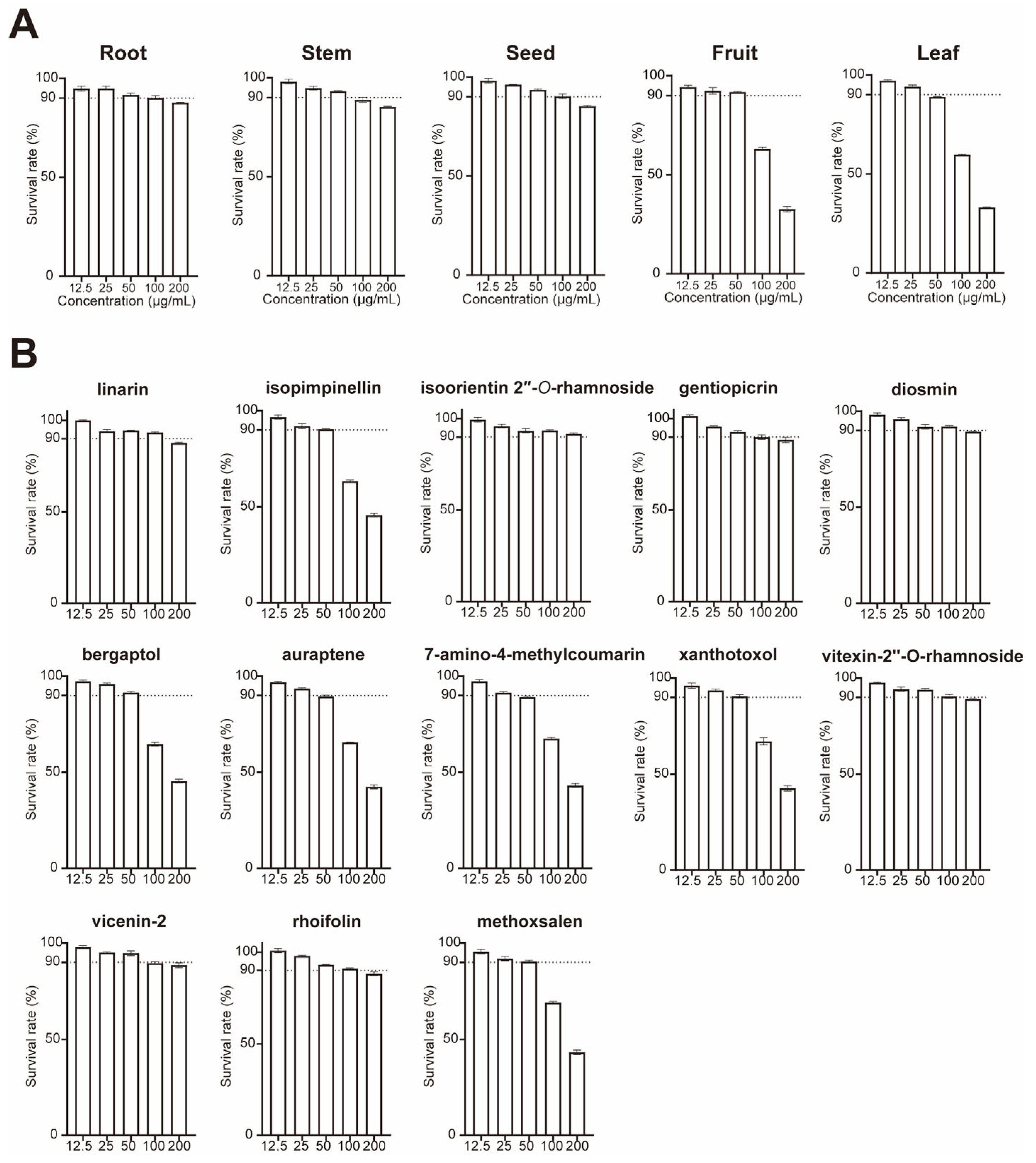
Table A1.
Content of individual substances in the fruits of minicitrus after treatment with the two strains.
References
- Saini, R.K.; Ranjit, A.; Sharma, K.; Prasad, P.; Shang, X.; Gowda, K.G.M.; Keum, Y.-S. Bioactive compounds of citrus fruits: A review of composition and health benefits of carotenoids, flavonoids, limonoids, and terpenes. Antioxidants 2022, 11, 239. [Google Scholar] [CrossRef] [Scilit] [PubMed]
- Lou, S.-N.; Ho, C.-T. Phenolic compounds and biological activities of small-size citrus: Kumquat and calamondin. J. Food Drug Anal. 2017, 25, 162–175. [Google Scholar] [CrossRef] [Scilit] [PubMed]
- Zhu, C.; Zheng, X.; Huang, Y.; Ye, J.; Chen, P.; Zhang, C.; Zhao, F.; Xie, Z.; Zhang, S.; Wang, N. Genome sequencing and CRISPR/Cas9 gene editing of an early flowering Mini-Citrus (Fortunella hindsii). Plant Biotechnol. J. 2019, 17, 2199–2210. [Google Scholar] [CrossRef] [Scilit] [PubMed]
- Sanshita; Devi, N.; Bhattacharya, B.; Sharma, A.; Singh, I.; Kumar, P.; Huanbutta, K.; Sangnim, T. From Citrus to Clinic: Limonene’s Journey Through Preclinical Research, Clinical Trials, and Formulation Innovations. Int. J. Nanomed. 2025, 2025, 4433–4460. [Google Scholar] [CrossRef] [Scilit] [PubMed]
- Costanzo, G.; Vitale, E.; Iesce, M.R.; Naviglio, D.; Amoresano, A.; Fontanarosa, C.; Spinelli, M.; Ciaravolo, M.; Arena, C. Antioxidant properties of pulp, peel and seeds of phlegrean mandarin (Citrus reticulata Blanco) at different stages of fruit ripening. Antioxidants 2022, 11, 187. [Google Scholar] [CrossRef] [Scilit] [PubMed]
- Ju, M.; Zhang, Q.; Wang, R.; Yan, S.; Li, Z.; Li, P.; Gu, P. Correlation in endophytic fungi community diversity and bioactive compounds of Sophora alopecuroides. Front. Microbiol. 2022, 13, 955647. [Google Scholar] [CrossRef] [Scilit] [PubMed]
- Yang, M.-Z.; Ma, M.-D.; Yuan, M.-Q.; Huang, Z.-Y.; Yang, W.-X.; Zhang, H.-B.; Huang, L.-H.; Ren, A.-Y.; Shan, H. Fungal endophytes as a metabolic fine-tuning regulator for wine grape. PLoS ONE 2016, 11, e0163186. [Google Scholar] [CrossRef] [Scilit] [PubMed]
- Xu, J.; Zhang, Y.; Zhang, P.; Trivedi, P.; Riera, N.; Wang, Y.; Liu, X.; Fan, G.; Tang, J.; Coletta-Filho, H.D. The structure and function of the global citrus rhizosphere microbiome. Nat. Commun. 2018, 9, 4894. [Google Scholar] [CrossRef] [Scilit] [PubMed]
- Matsumoto, H.; Fan, X.; Wang, Y.; Kusstatscher, P.; Duan, J.; Wu, S.; Chen, S.; Qiao, K.; Wang, Y.; Ma, B. Bacterial seed endophyte shapes disease resistance in rice. Nat. Plants 2021, 7, 60–72. [Google Scholar] [CrossRef] [Scilit] [PubMed]
- Sahu, P.K.; Tilgam, J.; Mishra, S.; Hamid, S.; Gupta, A.; K, J.; Verma, S.K.; Kharwar, R.N. Surface sterilization for isolation of endophytes: Ensuring what (not) to grow. J. Basic Microbiol. 2022, 62, 647–668. [Google Scholar] [CrossRef] [Scilit] [PubMed]
- Chen, Y.; Ma, H.; Liang, J.; Sun, C.; Wang, D.; Chen, K.; Zhao, J.; Ji, S.; Ma, C.; Ye, X. Hepatoprotective potential of four fruit extracts rich in different structural flavonoids against alcohol-induced liver injury via gut microbiota-liver axis. Food Chem. 2024, 460, 140460. [Google Scholar] [CrossRef] [Scilit] [PubMed]
- Liu, C.; Zhao, D.; Ma, W.; Guo, Y.; Wang, A.; Wang, Q.; Lee, D.-J. Denitrifying sulfide removal process on high-salinity wastewaters in the presence of Halomonas sp. Appl. Microbiol. Biotechnol. 2016, 100, 1421–1426. [Google Scholar] [CrossRef] [Scilit] [PubMed]
- Barreca, D.; Gattuso, G.; Laganà, G.; Leuzzi, U.; Bellocco, E. C-and O-glycosyl flavonoids in Sanguinello and Tarocco blood orange (Citrus sinensis (L.) Osbeck) juice: Identification and influence on antioxidant properties and acetylcholinesterase activity. Food Chem. 2016, 196, 619–627. [Google Scholar] [CrossRef] [Scilit] [PubMed]
- Boruah, J.L.H.; Gogoi, M.; Famhawite, V.; Barman, D.; Das, D.J.; Puro, K.N.; Biswas, A.; Mridha, P.; Gogoi, P.; Gajbhiye, R. Phytochemical Characterization, Prooxidant, Antiproliferative and Anti-Inflammatory Potential of Meyna spinosa Roxb. Ex Link Ripe Fruit. Chem. Biodivers. 2025, 22, e202402342. [Google Scholar] [CrossRef] [Scilit] [PubMed]
- Pei, H.; Su, W.; Gui, M.; Dou, M.; Zhang, Y.; Wang, C.; Lu, D. Comparative analysis of chemical constituents in different parts of lotus by UPLC and QToF-MS. Molecules 2021, 26, 1855. [Google Scholar] [CrossRef] [Scilit] [PubMed]
- Akihisa, T.; Pan, X.; Nakamura, Y.; Kikuchi, T.; Takahashi, N.; Matsumoto, M.; Ogihara, E.; Fukatsu, M.; Koike, K.; Tokuda, H. Limonoids from the fruits of Melia azedarach and their cytotoxic activities. Phytochemistry 2013, 89, 59–70. [Google Scholar] [CrossRef] [Scilit] [PubMed]
- Kamran, S.H.; Ahmad, M.; Ishtiaq, S.; Ajaib, M.; Razashah, S.H.; Shahwar, D.-e. Metabolite profiling and biochemical investigation of the antidiabetic potential of Loranthus pulverulentus Wall n-butanol fraction in diabetic animal models. J. Ethnopharmacol. 2024, 318, 116963. [Google Scholar] [CrossRef] [Scilit] [PubMed]
- Liu, J.-Y.; Li, S.-Y.; Feng, J.-Y.; Sun, Y.; Cai, J.-N.; Sun, X.-F.; Yang, S.-L. Flavone C-glycosides from the flowers of Trollius chinensis and their anti-complementary activity. J. Asian Nat. Prod. Res. 2013, 15, 325–331. [Google Scholar] [CrossRef] [Scilit] [PubMed]
- Yi, L.; Ma, S.; Ren, D. Phytochemistry and bioactivity of Citrus flavonoids: A focus on antioxidant, anti-inflammatory, anticancer and cardiovascular protection activities. Phytochem. Rev. 2017, 16, 479–511. [Google Scholar] [CrossRef] [Scilit]
- Khan, M.K.; Dangles, O. A comprehensive review on flavanones, the major citrus polyphenols. J. Food Compos. Anal. 2014, 33, 85–104. [Google Scholar] [CrossRef] [Scilit]
- Dugrand-Judek, A.; Olry, A.; Hehn, A.; Costantino, G.; Ollitrault, P.; Froelicher, Y.; Bourgaud, F. The distribution of coumarins and furanocoumarins in Citrus species closely matches Citrus phylogeny and reflects the organization of biosynthetic pathways. PLoS ONE 2015, 10, e0142757. [Google Scholar] [CrossRef] [Scilit] [PubMed]
- Wang, X.; Li, S.; Wei, C.-C.; Huang, J.; Pan, M.-H.; Shahidi, F.; Ho, C.-T. Anti-inflammatory effects of polymethoxyflavones from citrus peels: A review. J. Food Bioact. 2018, 3, 76–86. [Google Scholar] [CrossRef] [Scilit]
- Roussos, P.; Flessoura, I.; Petropoulos, F.; Massas, I.; Tsafouros, A.; Ntanos, E.; Denaxa, N. Soil physicochemical properties, tree nutrient status, physical, organoleptic and phytochemical characteristics and antioxidant capacity of clementine mandarin (Citrus clementine cv. SRA63) juice under integrated and organic farming. Sci. Hortic. 2019, 250, 414–420. [Google Scholar] [CrossRef] [Scilit]
- M’hiri, N.; Ioannou, I.; Ghoul, M.; Mihoubi Boudhrioua, N. Phytochemical characteristics of citrus peel and effect of conventional and nonconventional processing on phenolic compounds: A review. Food Rev. Int. 2017, 33, 587–619. [Google Scholar] [CrossRef] [Scilit]
- Gualdani, R.; Cavalluzzi, M.M.; Lentini, G.; Habtemariam, S. The chemistry and pharmacology of citrus limonoids. Molecules 2016, 21, 1530. [Google Scholar] [CrossRef] [Scilit] [PubMed]
- Saewan, N.; Jimtaisong, A. Photoprotection of natural flavonoids. J. Appl. Pharm. Sci. 2013, 3, 129–141. [Google Scholar] [CrossRef] [Scilit]
- Tlak Gajger, I.; Dar, S.A. Plant allelochemicals as sources of insecticides. Insects 2021, 12, 189. [Google Scholar] [CrossRef] [Scilit] [PubMed]
- Roy, A.; Saraf, S. Limonoids: Overview of significant bioactive triterpenes distributed in plants kingdom. Biol. Pharm. Bull. 2006, 29, 191–201. [Google Scholar] [CrossRef] [Scilit] [PubMed]
- Jamiołkowska, A. Natural compounds as elicitors of plant resistance against diseases and new biocontrol strategies. Agronomy 2020, 10, 173. [Google Scholar] [CrossRef] [Scilit]
- de Moraes Barros, H.R.; de Castro Ferreira, T.A.P.; Genovese, M.I. Antioxidant capacity and mineral content of pulp and peel from commercial cultivars of citrus from Brazil. Food Chem. 2012, 134, 1892–1898. [Google Scholar] [CrossRef] [Scilit] [PubMed]
- Huang, D.; Ou, B.; Prior, R.L. The chemistry behind antioxidant capacity assays. J. Agric. Food Chem. 2005, 53, 1841–1856. [Google Scholar] [CrossRef] [Scilit] [PubMed]
- Wolfe, K.L.; Liu, R.H. Cellular antioxidant activity (CAA) assay for assessing antioxidants, foods, and dietary supplements. J. Agric. Food Chem. 2007, 55, 8896–8907. [Google Scholar] [CrossRef] [Scilit] [PubMed]
- Williams, R.J.; Spencer, J.P.; Rice-Evans, C. Flavonoids: Antioxidants or signalling molecules? Free Radic. Biol. Med. 2004, 36, 838–849. [Google Scholar] [CrossRef] [Scilit] [PubMed]
- Edwards, J.; Johnson, C.; Santos-Medellín, C.; Lurie, E.; Podishetty, N.K.; Bhatnagar, S.; Eisen, J.A.; Sundaresan, V. Structure, variation, and assembly of the root-associated microbiomes of rice. Proc. Natl. Acad. Sci. USA 2015, 112, E911–E920. [Google Scholar] [CrossRef] [Scilit] [PubMed]
- Bulgarelli, D.; Rott, M.; Schlaeppi, K.; Ver Loren van Themaat, E.; Ahmadinejad, N.; Assenza, F.; Rauf, P.; Huettel, B.; Reinhardt, R.; Schmelzer, E. Revealing structure and assembly cues for Arabidopsis root-inhabiting bacterial microbiota. Nature 2012, 488, 91–95. [Google Scholar] [CrossRef] [Scilit] [PubMed]
- Lundberg, D.S.; Lebeis, S.L.; Paredes, S.H.; Yourstone, S.; Gehring, J.; Malfatti, S.; Tremblay, J.; Engelbrektson, A.; Kunin, V.; Rio, T.G.d. Defining the core Arabidopsis thaliana root microbiome. Nature 2012, 488, 86–90. [Google Scholar] [CrossRef] [Scilit] [PubMed]
- Coleman-Derr, D.; Desgarennes, D.; Fonseca-Garcia, C.; Gross, S.; Clingenpeel, S.; Woyke, T.; North, G.; Visel, A.; Partida-Martinez, L.P.; Tringe, S.G. Plant compartment and biogeography affect microbiome composition in cultivated and native Agave species. New Phytol. 2016, 209, 798–811. [Google Scholar] [CrossRef] [Scilit] [PubMed]
- Blaustein, R.A.; Lorca, G.L.; Meyer, J.L.; Gonzalez, C.F.; Teplitski, M. Defining the core citrus leaf-and root-associated microbiota: Factors associated with community structure and implications for managing huanglongbing (citrus greening) disease. Appl. Environ. Microbiol. 2017, 83, e00210–00217. [Google Scholar] [CrossRef] [Scilit] [PubMed]
- Vorholt, J.A. Microbial life in the phyllosphere. Nat. Rev. Microbiol. 2012, 10, 828–840. [Google Scholar] [CrossRef] [Scilit] [PubMed]
- Müller, D.B.; Vogel, C.; Bai, Y.; Vorholt, J.A. The plant microbiota: Systems-level insights and perspectives. Annu. Rev. Genet. 2016, 50, 211–234. [Google Scholar] [CrossRef] [Scilit] [PubMed]
- Fusco, V.; Quero, G.M.; Cho, G.-S.; Kabisch, J.; Meske, D.; Neve, H.; Bockelmann, W.; Franz, C.M. The genus Weissella: Taxonomy, ecology and biotechnological potential. Front. Microbiol. 2015, 6, 155. [Google Scholar] [CrossRef] [Scilit] [PubMed]
- Walterson, A.M.; Stavrinides, J. Pantoea: Insights into a highly versatile and diverse genus within the Enterobacteriaceae. FEMS Microbiol. Rev. 2015, 39, 968–984. [Google Scholar] [CrossRef] [Scilit] [PubMed]
- Compant, S.; Samad, A.; Faist, H.; Sessitsch, A. A review on the plant microbiome: Ecology, functions, and emerging trends in microbial application. J. Adv. Res. 2019, 19, 29–37. [Google Scholar] [CrossRef] [Scilit] [PubMed]
- Hur, S.J.; Lee, S.Y.; Kim, Y.-C.; Choi, I.; Kim, G.-B. Effect of fermentation on the antioxidant activity in plant-based foods. Food Chem. 2014, 160, 346–356. [Google Scholar] [CrossRef] [Scilit] [PubMed]
- Rodríguez, H.; Curiel, J.A.; Landete, J.M.; De Las Rivas, B.; de Felipe, F.L.; Gómez-Cordovés, C.; Mancheño, J.M.; Muñoz, R. Food phenolics and lactic acid bacteria. Int. J. Food Microbiol. 2009, 132, 79–90. [Google Scholar] [CrossRef] [Scilit] [PubMed]
- Gupta, S.; Chaturvedi, P.; Kulkarni, M.G.; Van Staden, J. A critical review on exploiting the pharmaceutical potential of plant endophytic fungi. Biotechnol. Adv. 2020, 39, 107462. [Google Scholar] [CrossRef] [Scilit] [PubMed]
- Zhou, M.; Zhang, M.; Ding, Y.; Zhang, D.; Wang, F.; Feng, D.; Ye, S. Biosynthesis and characterization of flavonoids by Ginkgo biloba endophytic Rhodococcus sp. FY-5. Microchem. J. 2024, 204, 111115. [Google Scholar] [CrossRef] [Scilit]
- Singh, G.; Agrawal, H.; Bednarek, P. Specialized metabolites as versatile tools in shaping plant–microbe associations. Mol. Plant 2023, 16, 122–144. [Google Scholar] [CrossRef] [Scilit] [PubMed]
- Voges, M.J.; Bai, Y.; Schulze-Lefert, P.; Sattely, E.S. Plant-derived coumarins shape the composition of an Arabidopsis synthetic root microbiome. Proc. Natl. Acad. Sci. USA 2019, 116, 12558–12565. [Google Scholar] [CrossRef] [Scilit] [PubMed]
- Rahman, M.; Sabir, A.A.; Mukta, J.A.; Khan, M.M.A.; Mohi-Ud-Din, M.; Miah, M.G.; Rahman, M.; Islam, M.T. Plant probiotic bacteria Bacillus and Paraburkholderia improve growth, yield and content of antioxidants in strawberry fruit. Sci. Rep. 2018, 8, 2504. [Google Scholar] [CrossRef] [Scilit] [PubMed]
- Zhou, Y.; Wang, R.; Zhang, Y.; Yang, Y.; Sun, X.; Zhang, Q.; Yang, N. Biotransformation of phenolics and metabolites and the change in antioxidant activity in kiwifruit induced by Lactobacillus plantarum fermentation. J. Sci. Food Agric. 2020, 100, 3283–3290. [Google Scholar] [CrossRef] [Scilit] [PubMed]
- Zhang, S.-Y.; Wu, X.-H.; Gu, Q.-Y.; Xu, M.-H.; Li, X.-Y.; Sun, K.; Zhang, W.; Dai, C.-C.; Cai, Y.-P. The Endophytic Microbiota Drives Plant Growth and Flavonoid Dynamics in Amana edulis under Geographical Variation. J. Agric. Food Chem. 2026, 74, 16952–16963. [Google Scholar] [CrossRef] [Scilit] [PubMed]
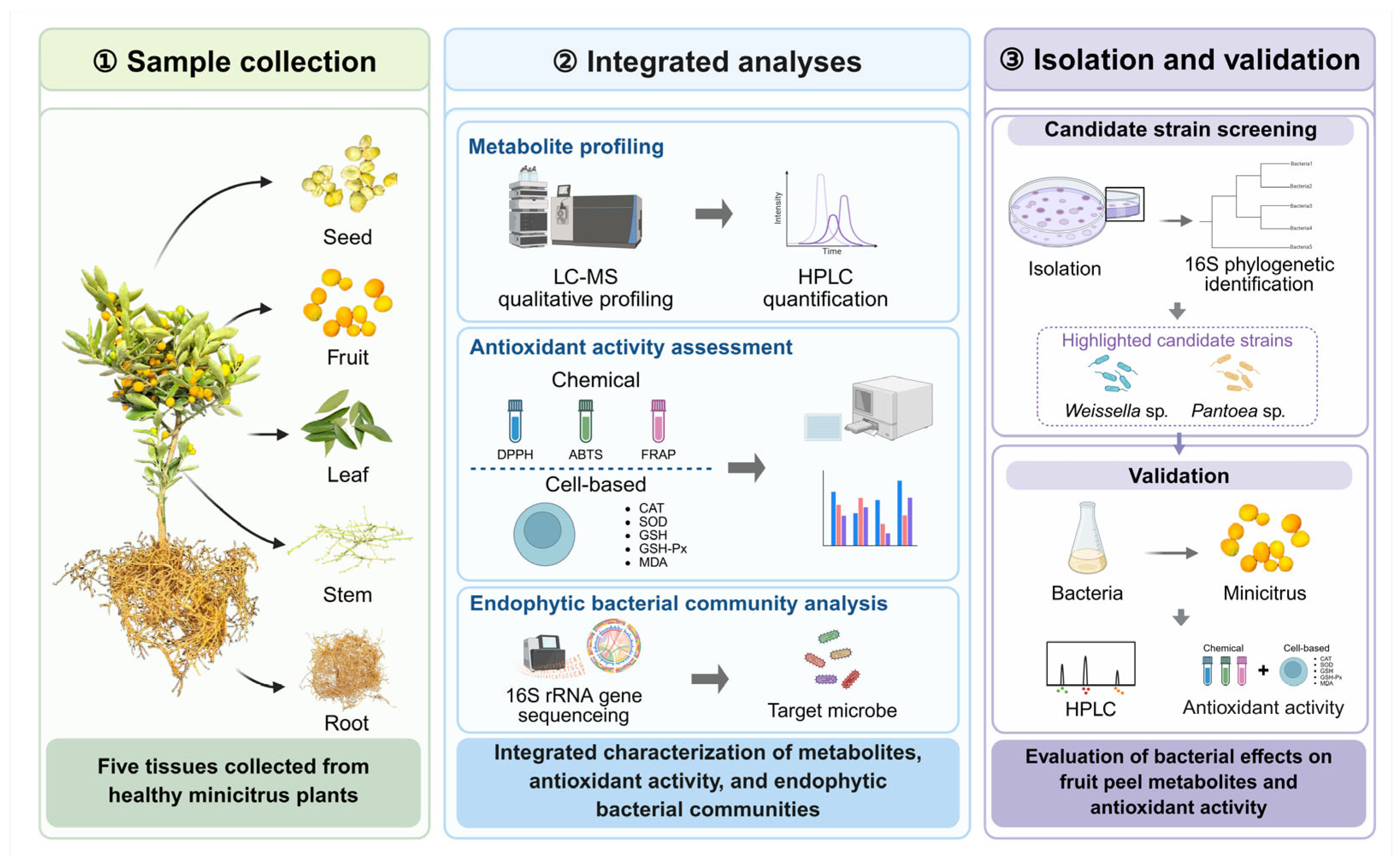
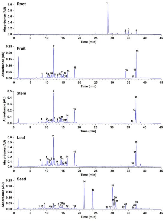
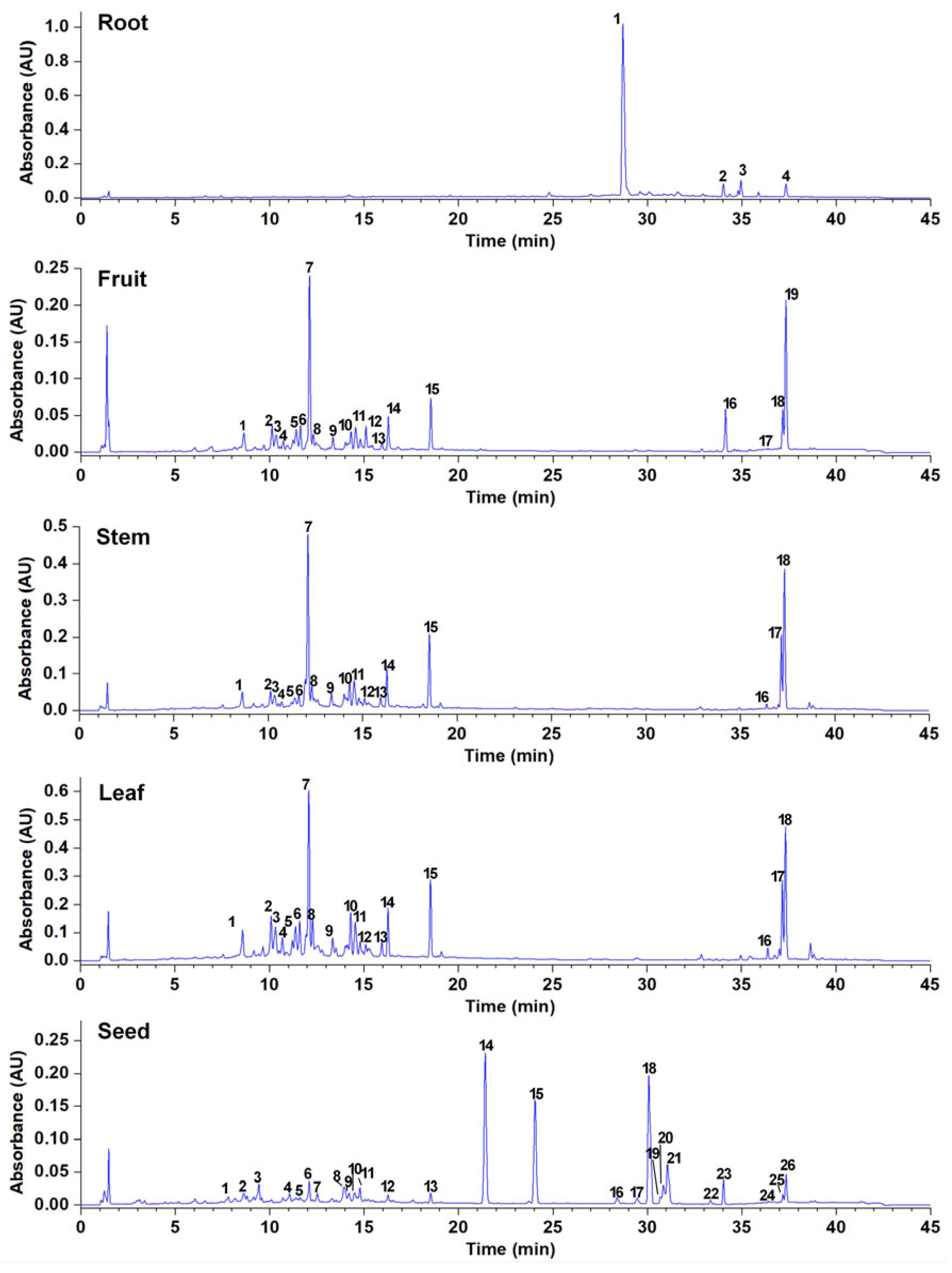
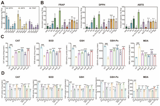
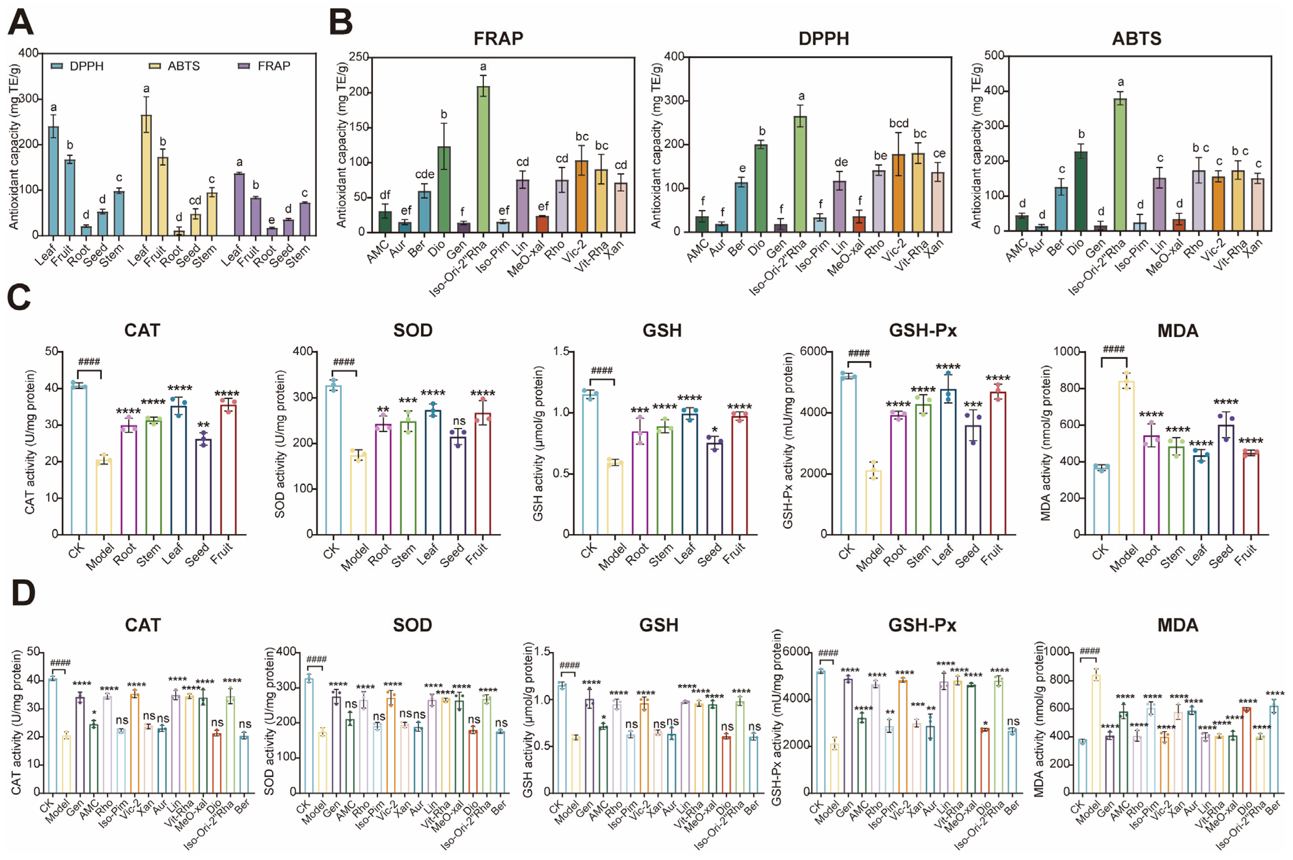
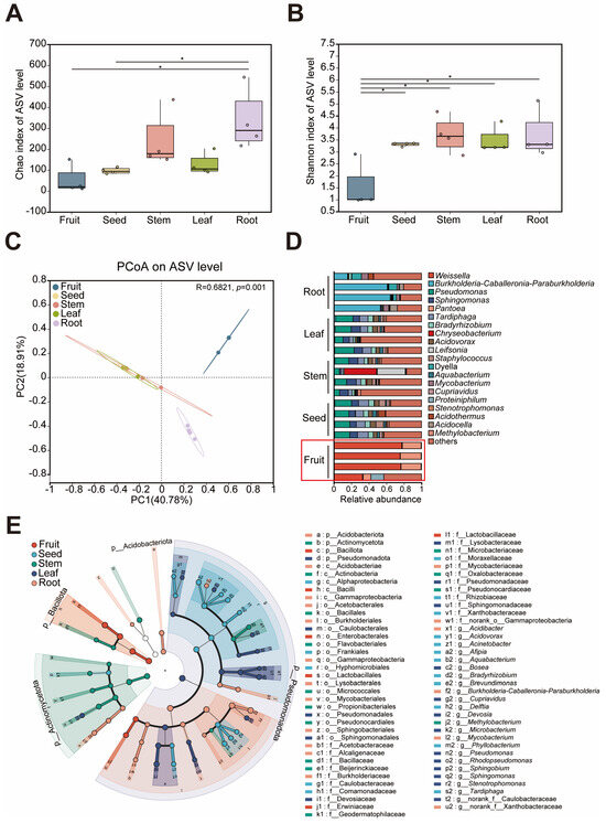
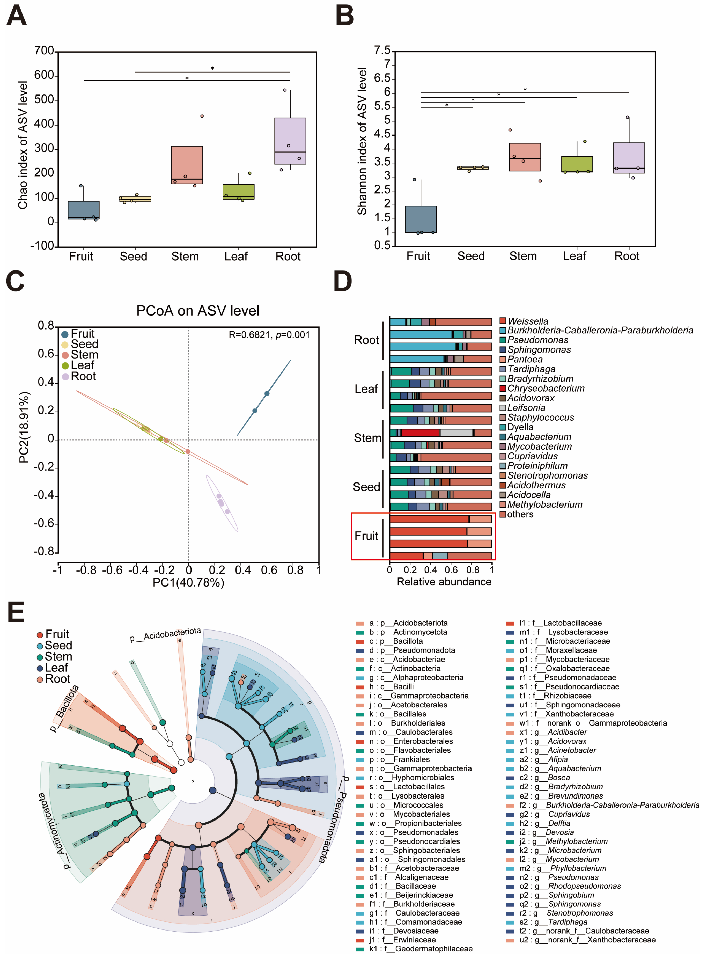
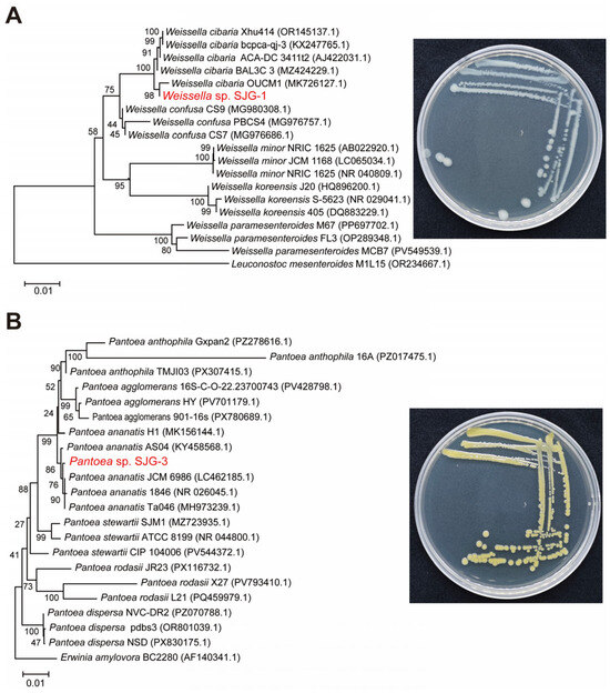
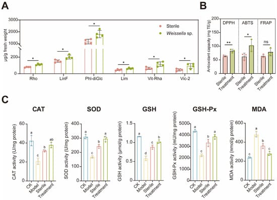
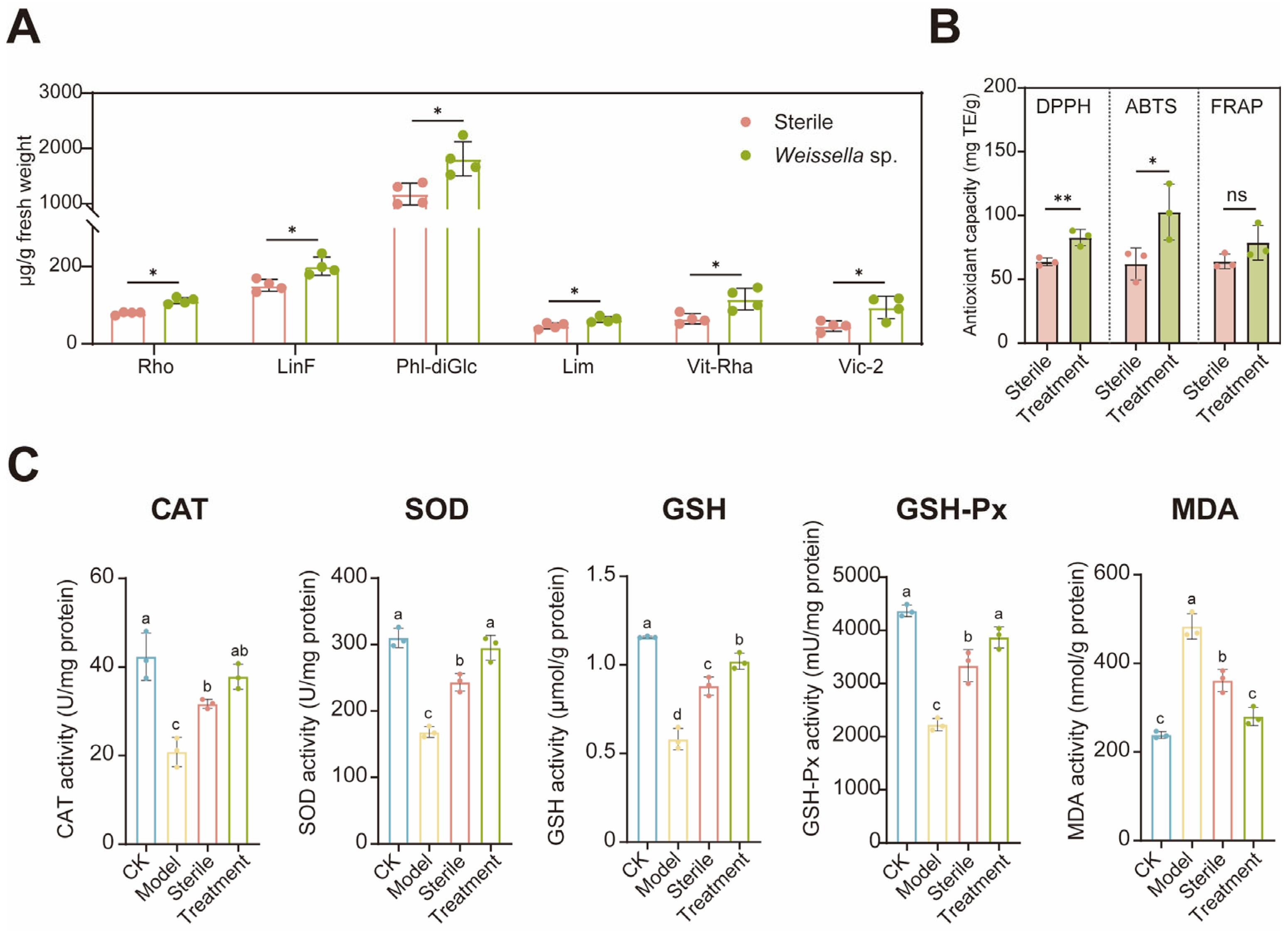

Disclaimer/Publisher’s Note: The statements, opinions and data contained in all publications are solely those of the individual author(s) and contributor(s) and not of MDPI and/or the editor(s). MDPI and/or the editor(s) disclaim responsibility for any injury to people or property resulting from any ideas, methods, instructions or products referred to in the content. |
© 2026 by the authors. Licensee MDPI, Basel, Switzerland. This article is an open access article distributed under the terms and conditions of the Creative Commons Attribution (CC BY) license.