


Abstract
Micro- and nanoplastics (MPs/NPs) are ubiquitous environmental contaminants increasingly detected in air, food, drinking water, and human tissues, raising concerns about their potential long-term health effects. Accumulating evidence indicates that these particles can enter the human body, cross biological barriers, and elicit cellular and molecular responses relevant to disease development. This review synthesizes current mechanistic evidence linking MP/NP exposure to cardiovascular disease (CVD) and cancer, two leading global causes of morbidity and mortality that share interconnected pathogenic pathways. Key mechanisms include chronic inflammation, oxidative stress, gut microbiota dysbiosis, genotoxicity, and epigenetic alterations, all of which are widely implicated in both conditions. However, the available evidence is still largely derived from in vitro and animal studies, with limited human epidemiological data. Important uncertainties remain regarding real-world exposure characterization, dose–response relationships, and long-term clinical outcomes, underscoring the need for standardized analytical approaches, validated exposure and effect biomarkers, and large-scale longitudinal studies to clarify causal associations for both cancer and CVD. Taken together, current evidence suggests that MPs/NPs may represent emerging environmental contributors to shared pathogenic pathways linking CVD and cancer; however, establishing causality in humans will require well-designed longitudinal studies that integrate exposure assessment and clinical outcomes.
1. Introduction
Global plastic production has increased exponentially over recent decades, rising from 234 million tons (Mt) in 2000 to 460 Mt in 2019, with no indication of a future decline [1,2]. Owing to their favorable physicochemical properties—including versatility, durability, hydrophobicity, low density, and low production costs—plastics are used across nearly all industrial and consumer sectors, with demand further amplified during the COVID-19 pandemic [3,4]. Consequently, plastics have become ubiquitous in all environmental compartments, and their waste now represents a persistent and pervasive pollutant [5]. Global plastic use is projected to reach 884 Mt by 2050, with waste generation expected to double over the same period, even if packaging recycling rates exceed 75% [1].
Microplastics (MPs) and nanoplastics (NPs), generated through the degradation of plastic waste, have emerged as major environmental and public health concerns due to their widespread distribution across ecosystems and their potential to infiltrate human tissues and barriers [5,6]. Although MPs and NPs are often considered relatively inert materials, they can absorb substances such as additives, heavy metals, proteins, and microorganisms onto their surfaces, potentially increasing their toxicity [7]. While the environmental impact of MPs and NPs is well documented, their effects on human health remain incompletely understood due to uncertainties related to exposure routes, detection methods, and toxicity evaluation [6,8]. However, recent evidence suggests that plastic particles are increasingly detected in various human organs and may trigger adverse biological responses, including inflammation, oxidative stress, and cytotoxicity, with possible serious health consequences [8,9]. Such biological alterations are recognized contributors to the development of chronic non-communicable diseases, including cancer and cardiovascular disease (CVD), two of the leading causes of global morbidity and mortality [10]. Shared mechanisms include chronic inflammation, oxidative stress, immune dysregulation, clonal hematopoiesis, gut dysbiosis, and genetic and epigenetic alterations [10,11].
Given the growing body of evidence—largely derived from preclinical studies—showing that plastic particles may adversely affect the cardiovascular system and influence biological pathways involved in tumor initiation and progression, this review aims to provide an integrated overview of the current mechanistic evidence. In particular, it explores how MPs and NPs may trigger or exacerbate biological pathways that are commonly implicated in both CVD and cancer.
To this end, a comprehensive literature search was conducted in the PubMed database to identify studies published in English between January 2000 and April 2026 that investigated the potential role of MPs and NPs in CVD and cancer. The search strategy combined terms related to plastic particles (“microplastics”, “nanoplastics”) with terms associated with cardiovascular and oncological outcomes (“cardiovascular disease”, “atherosclerosis”, “thrombosis”, “cancer”, “carcinogenesis”) and shared pathogenic mechanisms (“inflammation”, “oxidative stress”, “gut dysbiosis”, “genotoxicity”, “epigenetic modifications”, and “cellular senescence”). In addition, the reference lists of relevant articles were screened to identify further eligible studies.
Beyond summarizing the mechanisms underlying MP/NP-associated cardiovascular and cancer risk, with particular attention to the role of antioxidant defenses in modulating plastic-induced adverse effects, this review also seeks to highlight current research gaps and propose future directions for risk assessment and translational investigations.
2. Microplastics and Nanoplastics: Sources, Characteristics, and Routes of Human Exposure
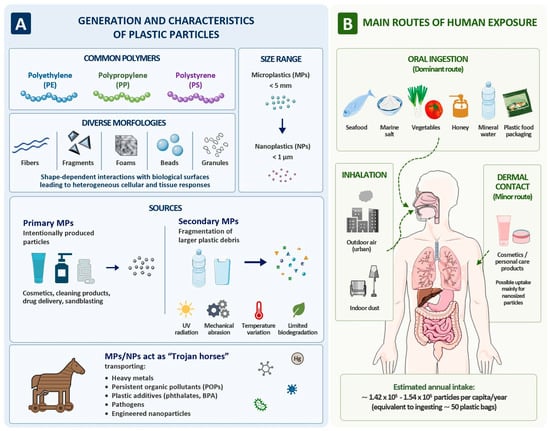
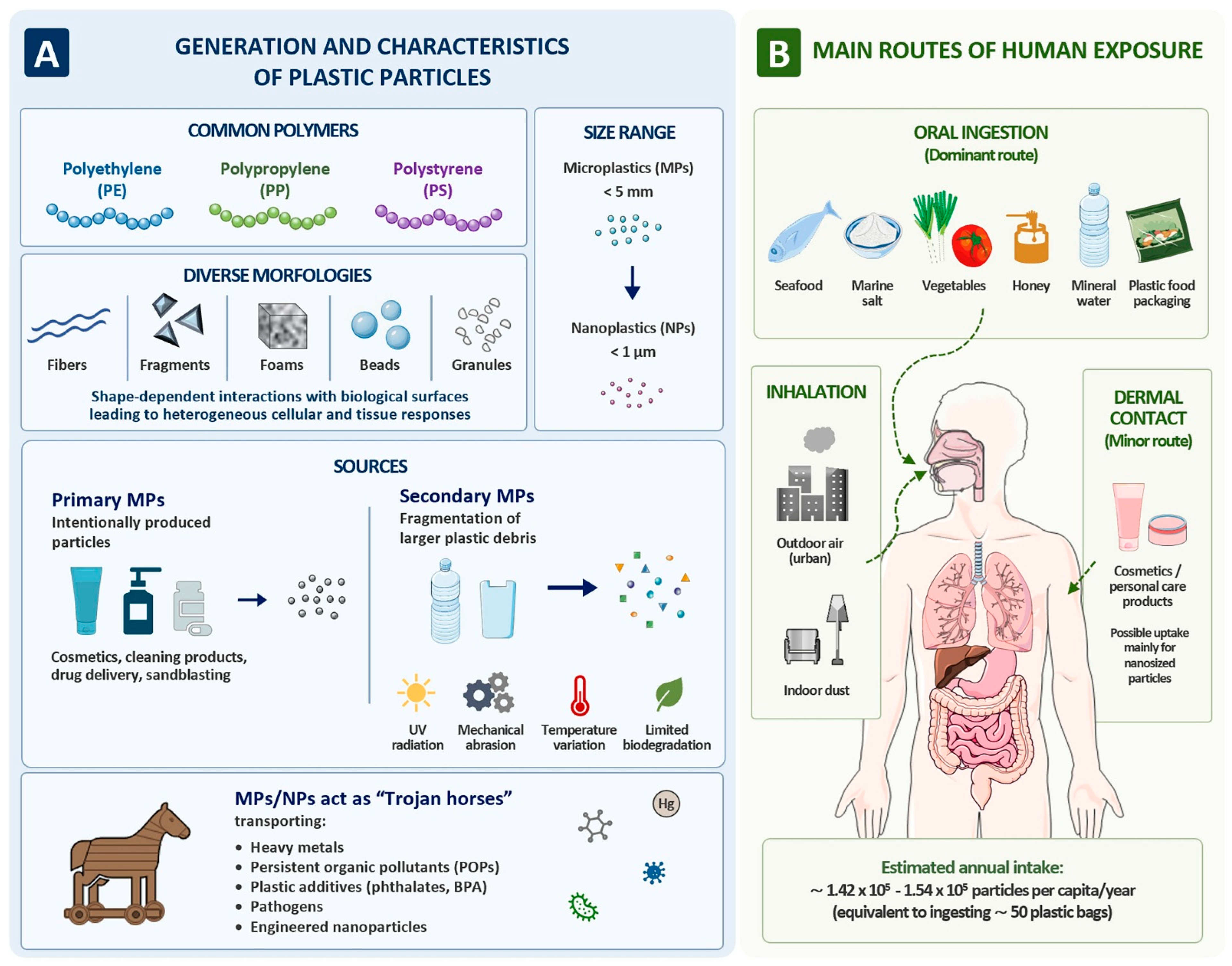
MPs are commonly defined as plastic particles with a diameter smaller than 5 mm and originate from the progressive degradation of larger plastic materials [12,13]. This process can occur over hundreds or even thousands of years, depending on the physicochemical properties of the plastics and the surrounding environmental conditions, and involves a complex interplay of mechanisms including ultraviolet radiation, mechanical abrasion, temperature fluctuations, and, to a lesser extent, biodegradation [7,14]. Besides resulting from the degradation of larger plastic waste (secondary MPs), primary MPs can also be deliberately produced for consumer and commercial uses, such as cleaning products, cosmetics, drug delivery systems, and sandblasting applications [7]. Polyethylene (PE), polypropylene (PP), and polystyrene (PS) are among the most prevalent polymers detected in environmental microplastic contamination [7]. The small size of MPs, which confers a high surface-to-volume ratio and enhanced mobility, enables them to act as “Trojan horses,” facilitating the transport of heavy metals, persistent organic pollutants, plastic additives (e.g., phthalates and bisphenol A—BPA), harmful pathogens, and engineered nanoparticles [7,15,16]. Furthermore, plastic particles exhibit a wide range of morphologies, and differences in shape (e.g., fibers, foams, cylindrical or spherical beads, and granules) critically modulate their interactions with biological surfaces, leading to heterogeneous cellular and tissue responses [17].
Notably, despite being widely distributed in the environment, MPs are subject to bioaccumulation and potentially biomagnification along aquatic food webs [18]. Accordingly, human exposure to MPs occurs primarily through the consumption of seafood, marine salt, vegetables, honey, and mineral water, as well as through ingestion from plastic food packaging [5,18]. In addition, exposure occurs through the inhalation of outdoor air, particularly in urban environments, and indoor dust containing MPs, while dermal contact via cosmetics and personal care products appears to represent a comparatively minor exposure route [4,5,6]. Annual human intake of MPs from food is estimated to reach approximately 1.42 × 10⁵–1.54 × 10⁵ particles per capita, an amount considered equivalent to the ingestion of about 50 plastic bags [19]. Plastic microparticles can further fragment into NPs, typically defined as particles smaller than 1 µm [7]. However, unlike their microplastic counterparts, current research on NPs has mainly focused on aquatic systems, and knowledge of their potential impacts on human health—although exposure may occur through the food chain—remains limited [7,14].
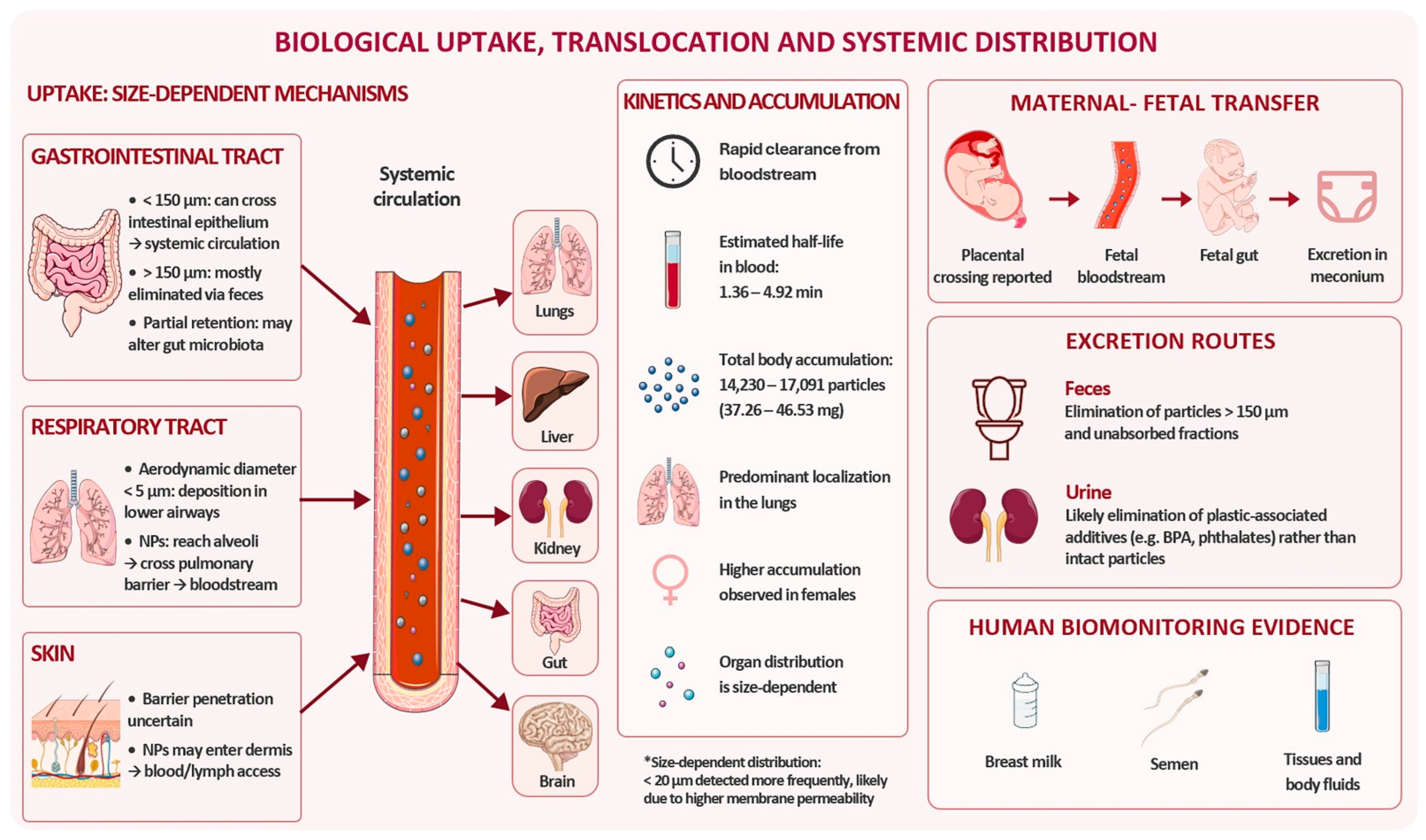
Once ingested, the absorption and translocation of MPs/NPs across the gastrointestinal tract largely depend on particle size [16]. Particles smaller than 150 µm have the potential to penetrate the intestinal epithelium and enter the systemic circulation, whereas most particles larger than 150 µm are eliminated via feces [16]. A small fraction of these larger particles may be retained in the gut, where they can potentially alter the composition of the colonic microbial community [16,20]. Similarly, inhalation represents an additional exposure route, with the entry of MPs/NPs into the respiratory system being largely restricted to the lower airways for particles with aerodynamic diameters smaller than 5 µm [21]. Notably, particles in the submicron range can penetrate the alveolar region, traverse the pulmonary epithelial barrier, and subsequently enter the systemic circulation [16]. In parallel, although the ability of plastic particles to cross the skin barrier remains a matter of debate, the direct application of cosmetics containing MPs/NPs may allow nanosized particles to be absorbed via percutaneous uptake and, upon dermal entry, reach the dermis and gain access to the bloodstream or lymphatic system, thereby potentially enabling systemic distribution [22,23] (Figure 1).
Figure 1.
Generation, characteristics, and human exposure pathways of microplastics and nanoplastics. (A) Overview of the origin and physicochemical characteristics of plastic particles. Common polymers include polyethylene (PE), polypropylene (PP), and polystyrene (PS). Plastic debris occurs in different morphologies (fibers, fragments, foams, beads, and granules) and spans a broad size range, from microplastics (MPs; <5 mm) to nanoplastics (NPs; <1 μm). MPs may originate as primary particles intentionally manufactured for industrial or commercial applications or as secondary particles generated through the fragmentation of larger plastic items by environmental weathering processes, including UV radiation, mechanical abrasion, temperature fluctuations, and limited biodegradation. Owing to their large surface area and physicochemical properties, MPs and NPs can also act as “Trojan horses,” adsorbing and transporting co-contaminants such as heavy metals, persistent organic pollutants (POPs), plastic additives, pathogens, and engineered nanoparticles. (B) Main routes of human exposure to MPs and NPs. Oral ingestion represents the predominant exposure pathway through contaminated food and beverages, including seafood, marine salt, vegetables, honey, and drinking water, as well as through migration from plastic food packaging. Additional exposure occurs through inhalation of airborne particles from outdoor and indoor environments, whereas dermal contact is considered a minor route, mainly relevant for nanosized particles present in cosmetics and personal care products. Once internalized, plastic particles may interact with multiple organ systems and contribute to biological responses associated with human health effects. Image adapted from Servier Medical Art (https://smart.servier.com (accessed on 10 May 2026)), licensed under CC BY 4.0 (https://creativecommons.org/licenses/by/4.0/ (accessed on 10 May 2026)).
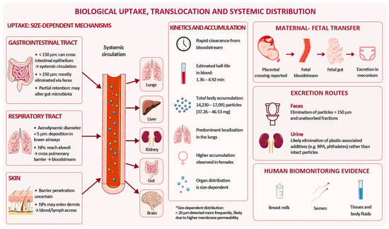
Of note, beyond being detected in a variety of human tissues and body fluids, including breast milk and semen, MPs may also cross the placental barrier, enter the fetal bloodstream, and ultimately reach the fetal gut, where they can be excreted in meconium [17,24]. In addition, their detection in the olfactory bulb suggests the existence of a potential translocation pathway to the brain [17,25]. MPs and NPs exhibit size-dependent clearance and biodistribution, with overall in vivo elimination occurring on the order of hours, as evidenced by experimental tracking studies in animal models [26]. Recent estimates indicate that total MP accumulation may range from 14,230 to 17,091 particles, corresponding to a mass of 37.26–46.53 mg, with predominant localization in the lungs and higher accumulation observed in females compared with males [27]. Organ-specific distribution appears to be size-dependent, with particles smaller than 20 µm detected in greater abundance, likely reflecting differences in the permeability of biological membranes [28,29]. Furthermore, in addition to fecal elimination, MPs/NPs or their associated additives may also be excreted via urine, a process hypothesized to primarily reflect the renal clearance of plastic-associated contaminants, such as BPA and phthalates, commonly used in food packaging and personal care products, rather than intact particles [8] (Figure 2).
Figure 2.
Biodistribution and potential elimination pathways of microplastics and nanoplastics in the human body. This schematic representation illustrates the potential systemic distribution of MPs/NPs following human exposure, including their translocation across biological barriers and possible presence in different organs. Plastic particles have been reported in multiple tissues and body fluids, reflecting exposure and biodistribution processes; however, such findings should not be interpreted as evidence of fully established or generalized pathways of translocation and accumulation. Potential routes of elimination include fecal excretion and urinary clearance, mainly related to associated compounds rather than intact particles. Current evidence on biodistribution and elimination is heterogeneous and largely derived from experimental models and limited human studies and may vary depending on particle size, physicochemical properties, and analytical detection methods. Therefore, the depicted processes should be interpreted as indicative of possible distribution patterns and kinetic behavior rather than definitive evidence of biological effects or causal relationships with disease. Image adapted from Servier Medical Art (https://smart.servier.com (accessed on 10 May 2026)), licensed under CC BY 4.0 (https://creativecommons.org/licenses/by/4.0/ (accessed on 10 May 2026)).
Overall, the ubiquitous environmental presence of MPs/NPs, combined with multiple exposure routes and evidence of systemic distribution and organ accumulation, highlights their potential to interfere with biological processes relevant to human health. However, despite increasing evidence of widespread exposure and systemic distribution, current estimates of human MP/NP exposure remain highly uncertain due to methodological heterogeneity and the lack of standardized analytical approaches.
Therefore, to ensure scientific rigor in the interpretation of the available evidence, it is important to distinguish between different levels of evidence. In this review, we therefore differentiate between: (i) detection of particles in human tissues, which reflects exposure; (ii) associations with clinical or biological markers, which are observational and do not imply causality; (iii) mechanistic evidence derived from in vitro and in vivo experimental models; and (iv) evidence of causality in humans, which is currently lacking. Based on this framework, the following sections will focus on mechanistic insights derived from experimental models, while human evidence is discussed in terms of exposure and association rather than causation.
4. Exposure Relevance and Methodological Considerations
The interpretation of experimental findings on MP/NP toxicity requires careful consideration of exposure relevance and methodological constraints.
Despite the growing number of studies reporting the presence of MPs/NPs in human cardiovascular and tumor tissues, these findings should be interpreted with caution due to several methodological limitations. The analysis of MPs/NPs in biological samples is highly susceptible to contamination throughout the analytical workflow, including sample collection, processing, and laboratory handling [140]. Airborne microfibers represent a major source of background contamination, while plastic laboratory consumables, reagents, and infrastructure can introduce additional particles that compromise data reliability and reproducibility [140,141]. Consequently, the implementation of procedural blanks, field blanks, and contamination-control measures is essential to ensure data quality [142]. However, the use and reporting of these controls remain heterogeneous across studies [142]. In addition, the analytical techniques employed differ substantially in their ability to identify plastic particles. While some studies rely primarily on morphological characterization, more robust approaches combine microscopic observation with spectroscopic confirmation, such as Fourier-transform infrared spectroscopy, Raman spectroscopy, or pyrolysis–gas chromatography–mass spectrometry, allowing chemical identification of polymers and reducing the risk of particle misclassification [143,144].
Another important source of variability is represented by detection limits and particle-size thresholds. Most currently available methods reliably detect particles in the micrometer range but have limited sensitivity for NPs, which may result in significant underestimation of the total particle burden [144]. Furthermore, studies report MP abundance using different metrics, including particle counts, particle size distributions, mass concentrations, or polymer-specific measurements, making direct comparisons challenging [143]. The biological interpretation of detected particles also remains uncertain. The presence of MPs in tissues may reflect chronic environmental exposure and progressive bioaccumulation [8]. However, given the widespread use of plastic-containing materials in clinical settings and the persistent methodological challenges associated with MP detection, perioperative contamination, exposure to medical devices, and analytical artifacts cannot be completely excluded and should be carefully considered when interpreting tissue findings [8,145]. Finally, disease-associated alterations in tissue architecture, vascular permeability, inflammatory cell infiltration, extracellular matrix composition, or impaired clearance mechanisms may favor particle retention within pathological tissues [146]. Consequently, the higher MP concentrations reported in atherosclerotic plaques, thrombi, and tumor tissues compared with control tissues do not necessarily demonstrate a causal role in disease pathogenesis and may partially reflect differential particle trapping within diseased microenvironments.
While the existing evidence provides important mechanistic insights, its interpretation is limited by methodological heterogeneity, the lack of standardized dose metrics, and a predominant reliance on in vitro and in vivo models [147]. Many experimental studies employ MP/NP concentrations that exceed currently estimated human environmental exposure levels, often by several orders of magnitude. Although such high-dose models are valuable for identifying potential hazard mechanisms, they may not accurately reflect real-world chronic low-dose exposure conditions [148]. Furthermore, the lack of standardized approaches for estimating internal human MP/NP burdens makes direct quantitative comparisons between experimental doses and human exposures particularly challenging. It should also be noted that many experimental studies rely on exposure methods such as direct gavage or injection, which may fail to accurately capture real-world human exposure pathways (e.g., ingestion, inhalation, or dermal contact). This limitation further complicates the interpretation of organ-specific effects and their relevance to different exposure routes.
Most experimental studies are based on acute or short-term exposure paradigms, whereas human exposure is expected to occur predominantly at low doses over prolonged periods [147]. Differences in exposure kinetics may substantially influence biological responses and limit the direct translational extrapolation of experimental findings to human health outcomes [147]. Furthermore, systemic effects described in experimental models may depend on the ability of MPs/NPs to translocate from primary sites of exposure, such as the lung or gastrointestinal tract, into the circulation. However, the extent and efficiency of such translocation in humans remain incompletely characterized and may vary depending on particle size, physicochemical properties, and exposure conditions.
Additional uncertainty arises from the frequent use of uniform, spherical polystyrene particles in laboratory studies [149,150]. These particles differ considerably from environmentally weathered MPs and NPs, which display substantial heterogeneity in size, shape, polymer composition, surface properties, and pollutant-binding capacity [151]. Weathering processes can further modify particle physicochemical characteristics and biological interactions [152].
NPs may exhibit higher cellular uptake and systemic distribution compared with larger MPs, further complicating direct comparisons across studies that do not clearly distinguish between these particle classes [153,154]. Moreover, the toxicological effects observed in experimental models may not be attributable solely to the polymeric core of MPs/NPs. Plastic additives, adsorbed environmental pollutants, heavy metals, and biofilm formation may all contribute to the observed biological responses, potentially acting in a synergistic manner [155]. When ingested, plastic particles may expose organisms to higher concentrations of these co-occurring contaminants or enhance their bioavailability and toxicity [155].
Finally, the absence of harmonized protocols for MP/NP detection, quantification, and characterization, together with the risk of contamination during sample collection and processing, continues to hamper inter-study comparability and exposure assessment [156,157]. Collectively, these limitations highlight the need for standardized methodologies and caution when extrapolating experimental findings to real-world human exposures and disease risk. At the same time, a more integrated framework linking exposure route, toxicokinetics, and tissue-specific biological responses is essential to improve the physiological relevance and interpretation of current evidence.
5. Conclusions and Future Challenges
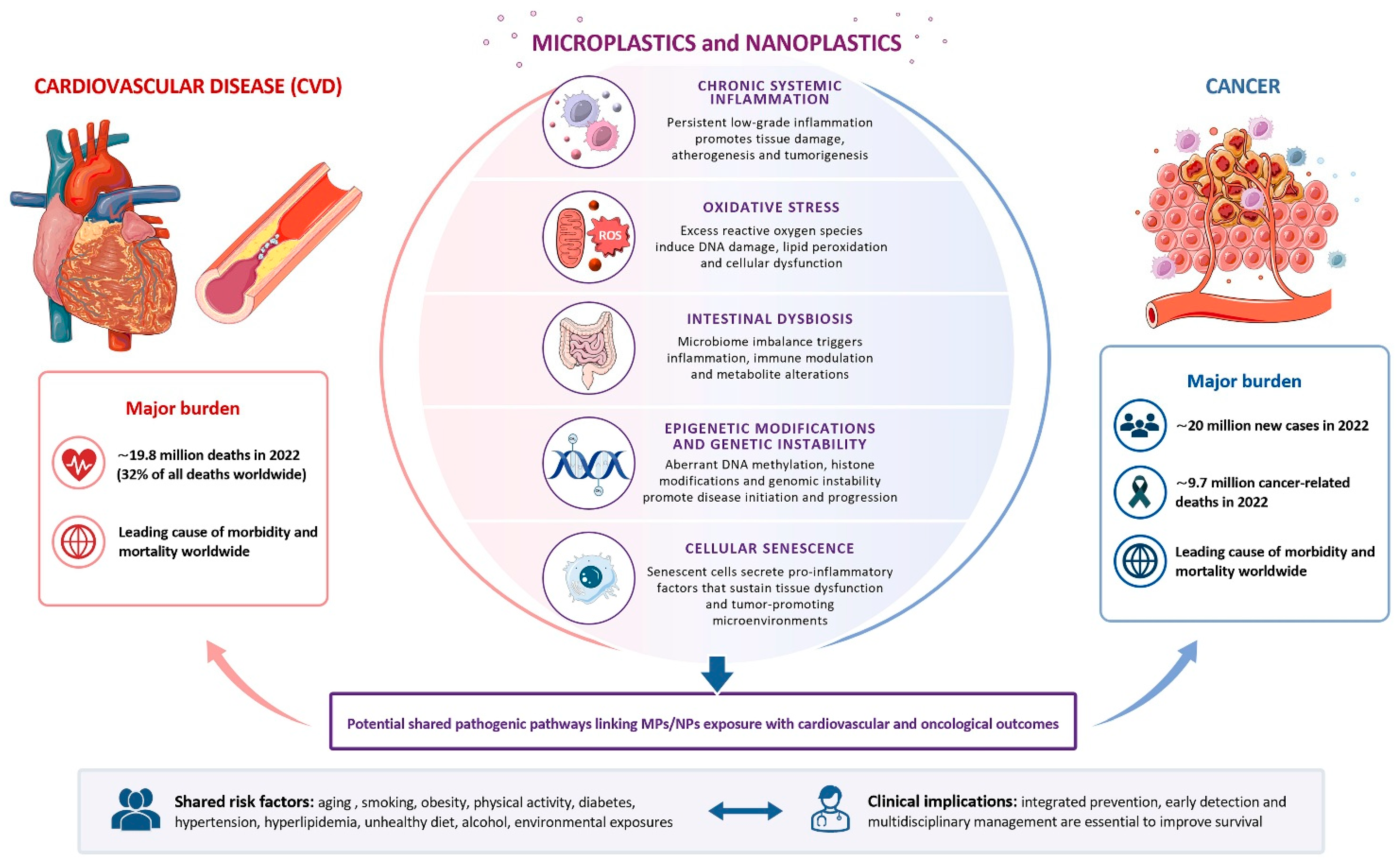
The evidence summarized in this review supports the emerging concept that MPs/NPs may represent previously underestimated contributors to biological processes relevant to the pathogenesis of both CVD and cancer, which together account for a substantial proportion of global morbidity and mortality. This is of particular importance, since CVD alone is responsible for approximately 32% of all global deaths, while cancer accounts for about 16%, meaning that together they contribute to nearly half of worldwide mortality [31,32].
These particles have been shown, particularly in experimental models, to activate several interconnected mechanisms, including chronic inflammation, oxidative stress, gut microbiota alterations, and genotoxic and epigenetic changes, that are already recognized as central drivers of both carcinogenesis and cardiovascular injury. Among these, oxidative stress emerges as a key and converging mechanism, potentially representing one of the earliest and most consistently observed biological responses to MP/NP exposure and a central hub linking environmental exposure to downstream pathological effects.
Inflammation plays a prominent role in mediating the toxic effects of MPs/NPs, particularly in the cardiovascular system, where human evidence supports an association between MP accumulation and inflammatory biomarkers. In contrast, evidence linking MP/NP-induced inflammation to cancer remains largely based on experimental studies, with limited direct translation to human disease. Similarly, increasing evidence suggests that alterations in gut microbiota composition may contribute to systemic inflammation and cardiometabolic dysfunction, although the role of microbiota-mediated pathways in cancer remains less clearly defined.
Emerging mechanistic insights also indicate that MPs/NPs may contribute to biological processes linked to cancer and CVD through genotoxic and epigenetic mechanisms, including DNA damage, altered DNA methylation patterns, and miRNA dysregulation. However, current data remain largely experimental and require further validation in human populations. In this context, an additional and still underexplored area of interest is clonal hematopoiesis of indeterminate potential (CHIP), a condition characterized by the accumulation of somatic mutations in hematopoietic stem cells and associated with both hematological malignancies and increased cardiovascular risk, partly through enhanced inflammatory signaling [11]. Given that cancer and CVD are key outcomes potentially associated with MP/NP exposure, it is plausible that chronic exposure may influence the emergence or expansion of CHIP clones. However, this possibility remains purely hypothetical, with no direct experimental or epidemiological evidence currently available linking MP/NP exposure to CHIP development or clonal expansion. CHIP should be considered as a future hypothesis that warrants investigation in appropriately designed studies, rather than as a mechanism currently supported by evidence.
Despite the growing body of mechanistic evidence, important uncertainties remain, particularly regarding exposure assessment and standardization. Addressing these methodological limitations will be essential for improving exposure characterization and strengthening causal inference in future studies [158].
Further research should prioritize large-scale epidemiological studies integrating accurate exposure assessment, validated biomarkers, and long-term clinical outcomes. In this context, further investigation into antioxidant defense systems and redox-regulating pathways is warranted, particularly to determine whether antioxidant-based strategies may mitigate MP/NP-induced biological damage and reduce disease risk.
Finally, although definitive causal evidence is still lacking, current data suggest that MPs/NPs should no longer be considered biologically inert environmental contaminants. Rather, MPs/NPs may represent emerging components of the exposome with biological plausibility to influence complex disease-related pathways shared by CVD and cancer. However, the magnitude of their contribution to human disease risk and their clinical relevance remain uncertain.
Importantly, the evidence reviewed in this manuscript derives from heterogeneous experimental systems, including in vitro cellular models, animal studies, and human investigations. These models provide complementary insights into molecular mechanisms and biological plausibility; however, their ability to support clinical inference differs substantially. While cellular and animal studies are particularly valuable for identifying mechanistic pathways and establishing biological plausibility, evidence directly linking MP/NP exposure to clinical outcomes in humans remains limited and requires confirmation through well-designed epidemiological and longitudinal studies.
These findings should therefore be interpreted with caution, as causal relationships in humans have not yet been established. Such evidence highlights the need for coordinated public health and regulatory strategies targeting plastic production, waste management, food packaging, and environmental contamination. Bridging current knowledge gaps through interdisciplinary research will be critical to define the true impact of MPs/NPs on human health and to support evidence-based preventive strategies.
Author Contributions
Conceptualization, A.B.; methodology, A.B. and F.G.; writing—original draft preparation, A.B., M.P. and F.G.; writing—review and editing, A.B., M-P., A.T., F.M., H.W. and F.G. All authors have read and agreed to the published version of the manuscript.
Funding
This research received no external funding.
Institutional Review Board Statement
Not applicable.
Informed Consent Statement
Not applicable.
Data Availability Statement
No new data were created.
Acknowledgments
During the preparation of this manuscript, the authors used Microsoft Copilot 365 GPT-5, to improve language quality and clarity. The authors have reviewed and edited the output and take full responsibility for the content of this publication.
Conflicts of Interest
The authors declare no conflicts of interest.
Abbreviations
The following abbreviations are used in this manuscript:
| ACS | Acute coronary syndrome |
| BPA | Bisphenol A |
| CAT | Catalase |
| CVD | Cardiovascular disease |
| CHIP | clonal hematopoiesis of indeterminate potential |
| DNMT | DNA methyltransferase |
| GPx | Glutathione peroxidase |
| IL | Interleukin |
| LPS | Lipopolysaccharide |
| MDA | Malondialdehyde |
| MI | Myocardial infarction |
| MP | Microplastic |
| NP | Nanoplastic |
| NO | Nitric oxide |
| PA66 | Polyamide 66 |
| PE | Polyethylene |
| PET | Polyethylene terephthalate |
| PP | Polypropylene |
| PS | Polystyrene |
| PVC | Polyvinyl chlorine |
| ROS | Reactive oxygen species |
| SA-β-gal | Senescence-associated β-galactosidase |
| SASP | Senescence-associated secretory phenotype |
| SOD | Superoxide dismutase |
| TNF-α | Tumor necrosis factor alpha |
References
- Dokl, M.; Copot, A.; Krajnc, D.; Van Fan, Y.; Vujanović, A.; Aviso, K.B.; Tan, R.R.; Kravanja, Z.; Čuček, L. Global projections of plastic use, end-of-life fate and potential changes in consumption, reduction, recycling and replacement with bioplastics to 2050. Sustain. Prod. Consum. 2024, 51, 498–518. [Google Scholar] [CrossRef] [Scilit]
- OECD. Global Plastics Outlook. Economic Drivers, Environmental Impacts and Policy Options. 2022. Available online: https://www.oecd.org/en/publications/global-plastics-outlook_de747aef-en.html (accessed on 29 April 2026).
- Belmaker, I.; Anca, E.D.; Rubin, L.P.; Magen-Molho, H.; Miodovnik, A.; van der Hal, N. Adverse health effects of exposure to plastic, microplastics and their additives: Environmental, legal and policy implications for Israel. Isr. J. Health Policy Res. 2024, 13, 44. [Google Scholar] [CrossRef] [Scilit] [PubMed]
- Ziani, K.; Ioniță-Mîndrican, C.B.; Mititelu, M.; Neacșu, S.M.; Negrei, C.; Moroșan, E.; Drăgănescu, D.; Preda, O.T. Microplastics: A Real Global Threat for Environment and Food Safety: A State of the Art Review. Nutrients 2023, 15, 617. [Google Scholar] [CrossRef] [Scilit] [PubMed]
- Abbas, G.; Ahmed, U.; Ahmad, M.A. Impact of Microplastics on Human Health: Risks, Diseases, and Affected Body Systems. Microplastics 2025, 4, 23. [Google Scholar] [CrossRef] [Scilit]
- Rodrigues, A.C.B.; de Jesus, G.P.; Waked, D.; Gomes, G.L.; Silva, T.M.; Yariwake, V.Y.; da Silva, M.P.; Magaldi, A.J.; Veras, M.M. Scientific Evidence about the Risks of Micro and Nanoplastics (MNPLs) to Human Health and Their Exposure Routes through the Environment. Toxics 2022, 10, 308. [Google Scholar] [CrossRef] [Scilit] [PubMed]
- Snehamayee, N.; Somya, S.; Kumar, S.C.; Niranjan, M.; Ranjan, S.B.; Kumar, M.N. Microplastics and Human Health: A Comprehensive Review on Exposure Pathways, Toxicity, and Emerging Risks. Microplastics 2026, 5, 8. [Google Scholar] [CrossRef] [Scilit]
- Roslan, N.S.; Lee, Y.Y.; Ibrahim, Y.S.; Tuan Anuar, S.; Yusof, K.M.K.K.; Lai, L.A.; Brentnall, T. Detection of microplastics in human tissues and organs: A scoping review. J. Glob. Health 2024, 14, 04179. [Google Scholar] [CrossRef] [Scilit] [PubMed]
- Pan, W.; Hao, J.; Zhang, M.; Liu, H.; Tian, F.; Zhang, X.; Jiang, Z.; Chen, C.; Gao, M.; Zhang, H. Identification and analysis of microplastics in peritumoral and tumor tissues of colorectal cancer. Sci. Rep. 2025, 15, 16130. [Google Scholar] [CrossRef] [Scilit] [PubMed]
- Wilcox, N.S.; Amit, U.; Reibel, J.B.; Berlin, E.; Howell, K.; Ky, B. Cardiovascular disease and cancer: Shared risk factors and mechanisms. Nat. Rev. Cardiol. 2024, 21, 617–631. [Google Scholar] [CrossRef] [Scilit] [PubMed]
- Alhuneafat, L.; Guha, A.; Blaes, A.; Konety, S.H. Cancer and Cardiovascular Disease: Shared Risk Factors, Mechanisms, and Clinical Implications: JACC: CardioOncology State-of-the-Art Review. JACC Cardio Oncol. 2025, 7, 453–469. [Google Scholar] [CrossRef] [Scilit]
- Liu, J.; Zheng, L. Microplastic migration and transformation pathways and exposure health risks. Environ. Pollut. 2025, 368, 125700. [Google Scholar] [CrossRef] [Scilit] [PubMed]
- Zhang, K.; Hamidian, A.H.; Tubić, A.; Zhang, Y.; Fang, J.K.H.; Wu, C.; Lam, P.K.S. Understanding plastic degradation and microplastic formation in the environment: A review. Environ. Pollut. 2021, 274, 116554. [Google Scholar] [CrossRef] [Scilit] [PubMed]
- Lehner, R.; Weder, C.; Petri-Fink, A.; Rothen-Rutishauser, B. Emergence of Nanoplastic in the Environment and Possible Impact on Human Health. Environ. Sci. Technol. 2019, 53, 1748–1765. [Google Scholar] [CrossRef] [Scilit] [PubMed]
- Saeed, G.; Afzal, A.; Nimra, A.; Ahmad, Z.; Rehman, A.; Maqsood, K.; Jan, T.; Ahmad, M.I.; Karim, N.; Jeon, B.H.; et al. Microplastics as Trojan horses: Vectors of pathogens, pollutants, and antimicrobial resistance genes. Environ. Res. 2026, 300, 124365. [Google Scholar] [CrossRef] [Scilit] [PubMed]
- Cirillo, F.; Gianì, F.; Sergi, V.; Scordamaglia, D.; Giallongo, S.; Tibullo, D.; Volti, G.L.; Maggiolini, M.; Malaguarnera, R.; Lappano, R.; et al. The hidden threat: Microplastics and cancer biology. J. Hazard. Mater. 2026, 505, 141332. [Google Scholar] [CrossRef] [Scilit] [PubMed]
- Aimo, A.; Panichella, G.; Tommasi, E.; Revuelta-López, E.; Berastegui, E.; Bayes-Genis, A. The effects of microplastics and nanoplastics on cardiovascular disease: Mechanisms and perspectives. Nat. Rev. Cardiol. 2026. [Google Scholar] [CrossRef] [Scilit] [PubMed]
- Lu, K.; Que, Y.; Wang, L.; Wang, Y.; Qiu, J.; Jia, Y.; Ding, C.; Wang, D.; Cheng, W.; Zhang, Y. Environmental exposure pathways of microplastics and their toxic effects on ecosystems and the nervous system. Front. Toxicol. 2025, 7, 1649282. [Google Scholar] [CrossRef] [Scilit] [PubMed]
- Bai, C.L.; Liu, L.Y.; Hu, Y.B.; Zeng, E.Y.; Guo, Y. Microplastics: A review of analytical methods, occurrence and characteristics in food, and potential toxicities to biota. Sci. Total Environ. 2022, 806, 150263. [Google Scholar] [CrossRef] [Scilit] [PubMed]
- Tamargo, A.; Molinero, N.; Reinosa, J.J.; Alcolea-Rodriguez, V.; Portela, R.; Bañares, M.A.; Fernández, J.F.; Moreno-Arribas, M.V. PET microplastics affect human gut microbiota communities during simulated gastrointestinal digestion, first evidence of plausible polymer biodegradation during human digestion. Sci. Rep. 2022, 12, 528. [Google Scholar] [CrossRef] [Scilit] [PubMed]
- Woo, J.H.; Seo, H.J.; Lee, J.Y.; Lee, I.; Jeon, K.; Kim, B.; Lee, K. Polypropylene nanoplastic exposure leads to lung inflammation through p38-mediated NF-κB pathway due to mitochondrial damage. Part. Fibre Toxicol. 2023, 20, 2. [Google Scholar] [CrossRef] [Scilit]
- Döge, N.; Hadam, S.; Volz, P.; Wolf, A.; Schönborn, K.H.; Blume-Peytavi, U.; Alexiev, U.; Vogt, A. Identification of polystyrene nanoparticle penetration across intact skin barrier as rare event at sites of focal particle aggregations. J. Biophotonics 2018, 11, e201700169. [Google Scholar] [CrossRef] [Scilit] [PubMed]
- Gopinath, P.M.; Twayana, K.S.; Ravanan, P.; Thomas, J.; Mukherjee, A.; Jenkins, D.F.; Chandrasekaran, N. Prospects on the nano-plastic particles internalization and induction of cellular response in human keratinocytes. Part. Fibre Toxicol. 2021, 18, 35. [Google Scholar] [CrossRef] [Scilit]
- Liu, S.; Guo, J.; Liu, X.; Yang, R.; Wang, H.; Sun, Y.; Chen, B.; Dong, R. Detection of various microplastics in placentas, meconium, infant feces, breastmilk and infant formula: A pilot prospective study. Sci. Total Environ. 2023, 854, 158699. [Google Scholar] [CrossRef] [Scilit] [PubMed]
- Amato-Lourenço, L.F.; Dantas, K.C.; Júnior, G.R.; Paes, V.R.; Ando, R.A.; de Oliveira Freitas, R.; da Costa, O.M.M.M.; Rabelo, R.S.; Soares Bispo, K.C.; Carvalho-Oliveira, R.; et al. Microplastics in the Olfactory Bulb of the Human Brain. JAMA Netw. Open 2024, 7, e2440018. [Google Scholar] [CrossRef] [Scilit] [PubMed]
- Im, C.; Kim, H.; Zaheer, J.; Kim, J.Y.; Lee, Y.J.; Kang, C.M.; Kim, J.S. PET Tracing of Biodistribution for Orally Administered 64Cu-Labeled Polystyrene in Mice. J. Nucl. Med. 2022, 63, 461–467. [Google Scholar] [PubMed]
- Silva, G.C.A.; Fioretto, M.N.; Lima, M.C.; Scarano, W.R.; Delella, F.K. Micro and nanoplastics in human carcinogenesis: Insights from in vitro studies. Toxicology 2026, 519, 154331. [Google Scholar] [CrossRef] [Scilit] [PubMed]
- Gao, H.; Li, J.; Xu, Z.; Duan, Y.; Su, B.; Li, M.; Zheng, B.; Ren, M.; Chang, H.; Wang, H.; et al. A new insight of size-dependent plastics particles kinetics with regarding of metabolomics effects in liver and kidney. Environ. Pollut. 2025, 386, 127287. [Google Scholar] [CrossRef] [Scilit] [PubMed]
- Li, Y.; Chen, L.; Zhou, N.; Chen, Y.; Ling, Z.; Xiang, P. Microplastics in the human body: A comprehensive review of exposure, distribution, migration mechanisms, and toxicity. Sci. Total Environ. 2024, 946, 174215. [Google Scholar] [CrossRef] [Scilit] [PubMed]
- Zmaili, M.; Alzubi, J.; Alkhayyat, M.; Albakri, A.; Alkhalaileh, F.; Longinow, J.; Moudgil, R. Cancer and Cardiovascular Disease: The Conjoined Twins. Cancers 2024, 16, 1450. [Google Scholar] [CrossRef] [Scilit] [PubMed]
- WHO, World Health Organization. Cardiovascular Diseases (CVDs). 2025. Available online: https://www.who.int/news-room/fact-sheets/detail/cardiovascular-diseases-(cvds) (accessed on 2 May 2026).
- WHO, World Health Organization. Global Cancer Burden Growing, Amidst Mounting Need for Services. 2024. Available online: https://www.who.int/news/item/01-02-2024-global-cancer-burden-growing--amidst-mounting-need-for-services (accessed on 2 May 2026).
- Gao, H.; Chen, Z.; Yao, Y.; He, Y.; Hu, X. Common biological processes and mutual crosstalk mechanisms between cardiovascular disease and cancer. Front. Oncol. 2024, 14, 1453090. [Google Scholar] [CrossRef] [Scilit] [PubMed]
- Kumar, N.; Lamba, M.; Pachar, A.K.; Yadav, S.; Acharya, A. Microplastics—A Growing Concern as Carcinogens in Cancer Etiology: Emphasis on Biochemical and Molecular Mechanisms. Cell Biochem. Biophys. 2024, 82, 3109–3121. [Google Scholar] [CrossRef] [Scilit] [PubMed]
- Yang, Y.; Xie, E.; Du, Z.; Peng, Z.; Han, Z.; Li, L.; Zhao, R.; Qin, Y.; Xue, M.; Li, F.; et al. Detection of Various Microplastics in Patients Undergoing Cardiac Surgery. Environ. Sci. Technol. 2023, 57, 10911–10918. [Google Scholar] [CrossRef] [Scilit] [PubMed]
- Wang, T.; Yi, Z.; Liu, X.; Cai, Y.; Huang, X.; Fang, J.; Shen, R.; Lu, W.; Xiao, Y.; Zhuang, W.; et al. Multimodal detection and analysis of microplastics in human thrombi from multiple anatomically distinct sites. eBioMedicine 2024, 103, 105118. [Google Scholar] [CrossRef] [Scilit] [PubMed]
- Liu, S.; Wang, C.; Yang, Y.; Du, Z.; Li, L.; Zhang, M.; Ni, S.; Yue, Z.; Yang, K.; Wang, Y.; et al. Microplastics in three types of human arteries detected by pyrolysis-gas chromatography/mass spectrometry (Py-GC/MS). J. Hazard. Mater. 2024, 469, 33855. [Google Scholar] [CrossRef] [Scilit]
- Marfella, R.; Prattichizzo, F.; Sardu, C.; Fulgenzi, G.; Graciotti, L.; Spadoni, T.; D’Onofrio, N.; Scisciola, L.; La Grotta, R.; Frigé, C.; et al. Microplastics and Nanoplastics in Atheromas and Cardiovascular Events. N. Engl. J. Med. 2024, 390, 900–910. [Google Scholar] [CrossRef] [Scilit] [PubMed]
- Zhang, Y.; Gao, Q.; Gao, Q.; Xu, M.; Fang, N.; Mu, L.; Han, X.; Yu, H.; Zhang, S.; Li, Y.; et al. Microplastics and nanoplastics increase major adverse cardiac events in patients with myocardial infarction. J. Hazard. Mater. 2025, 489, 137624. [Google Scholar] [CrossRef] [Scilit] [PubMed]
- Yang, Y.; Zhang, F.; Jiang, Z.; Du, Z.; Liu, S.; Zhang, M.; Jin, Y.; Qin, Y.; Yang, X.; Wang, C.; et al. Microplastics are associated with elevated atherosclerotic risk and increased vascular complexity in acute coronary syndrome patients. Part. Fibre Toxicol. 2024, 21, 34. [Google Scholar]
- Zhao, J.; Zhang, H.; Shi, L.; Jia, Y.; Sheng, H. Detection and quantification of microplastics in various types of human tumor tissues. Ecotoxicol. Environ. Saf. 2024, 283, 116818. [Google Scholar] [CrossRef] [Scilit] [PubMed]
- Wang, M.; Liu, Q.; Zhang, X.; Jiang, H.; Zhang, X. Identification and analysis of microplastics in human penile cancer tissues. Sci. Total Environ. 2025, 969, 178815. [Google Scholar] [CrossRef] [Scilit] [PubMed]
- Deng, C.; Zhu, J.; Fang, Z.; Yang, Y.; Zhao, Q.; Zhang, Z.; Jin, Z.; Jiang, H. Identification and analysis of microplastics in para-tumor and tumor of human prostate. eBioMedicine 2024, 108, 105360. [Google Scholar] [CrossRef] [Scilit] [PubMed]
- Xu, H.; Dong, C.; Yu, Z.; Ozaki, Y.; Hu, Z.; Zhang, B.; Yao, W.; Yu, J.; Xie, Y. Detection and analysis of microplastics in tissues and blood of human cervical cancer patients. Environ. Res. 2024, 259, 119498. [Google Scholar] [CrossRef] [Scilit] [PubMed]
- Xu, J.; Qu, J.; Jin, H.; Mao, W. Associations between microplastics in human feces and colorectal cancer risk. J. Hazard. Mater. 2025, 495, 139099. [Google Scholar] [CrossRef] [Scilit] [PubMed]
- Pulvirenti, E.; Ferrante, M.; Barbera, N.; Favara, C.; Aquilia, E.; Palella, M.; Cristaldi, A.; Conti, G.O.; Fiore, M. Effects of Nano and Microplastics on the Inflammatory Process: In Vitro and In Vivo Studies Systematic Review. Front. Biosci. (Landmark Ed.) 2022, 27, 287. [Google Scholar] [CrossRef] [Scilit] [PubMed]
- Mahmud, F.; Sarker, D.B.; Jocelyn, J.A.; Sang, Q.A. Molecular and Cellular Effects of Microplastics and Nanoplastics: Focus on Inflammation and Senescence. Cells 2024, 13, 1788. [Google Scholar] [CrossRef] [Scilit] [PubMed]
- Bishop, B.; Webber, W.S.; Atif, S.M.; Ley, A.; Pankratz, K.A.; Kostelecky, R.; Colgan, S.P.; Dinarello, C.A.; Zhang, W.; Li, S. Micro- and nano-plastics induce inflammation and cell death in human cells. Front. Immunol. 2025, 16, 1528502. [Google Scholar] [CrossRef] [Scilit] [PubMed]
- Qin, Z. The use of THP-1 cells as a model for mimicking the function and regulation of monocytes and macrophages in the vasculature. Atherosclerosis 2012, 221, 2–11. [Google Scholar] [CrossRef] [Scilit] [PubMed]
- Busch, M.; Bredeck, G.; Waag, F.; Rahimi, K.; Ramachandran, H.; Bessel, T.; Barcikowski, S.; Herrmann, A.; Rossi, A.; Schins, R.P.F. Assessing the NLRP3 Inflammasome Activating Potential of a Large Panel of Micro- and Nanoplastics in THP-1 Cells. Biomolecules 2022, 12, 1095. [Google Scholar] [CrossRef] [Scilit] [PubMed]
- Delfos, L.; Depuydt, M.A.C.; Chemaly, M.; Coyle, S.; Schaftenaar, F.H.; van Santbrink, P.J.; Lindenbergh, P.P.; Bernabé Kleijn, M.N.A.; Costello, C.; Power, C.A.; et al. NLRP3 Inflammasome Inhibition by the Novel Bispecific Antibody InflamAb Attenuates Atherosclerosis in Apolipoprotein E-Deficient Mice. JACC Basic Transl. Sci. 2025, 10, 826–840. [Google Scholar] [CrossRef] [Scilit] [PubMed]
- Vlacil, A.K.; Bänfer, S.; Jacob, R.; Trippel, N.; Kuzu, I.; Schieffer, B.; Grote, K. Polystyrene microplastic particles induce endothelial activation. PLoS ONE 2021, 16, e0260181. [Google Scholar] [CrossRef] [Scilit] [PubMed]
- Libby, P. Inflammation in atherosclerosis. Nature 2002, 420, 868–874. [Google Scholar] [CrossRef] [Scilit] [PubMed]
- Tabas, I.; Lichtman, A.H. Monocyte-macrophages and T cells in atherosclerosis. Immunity 2017, 47, 621–634. [Google Scholar] [CrossRef] [Scilit] [PubMed]
- Yan, J.; Pan, Y.; He, J.; Pang, X.; Shao, W.; Wang, C.; Wang, R.; He, Y.; Zhang, M.; Ye, J.; et al. Toxic vascular effects of polystyrene microplastic exposure. Sci. Total Environ. 2023, 905, 167215. [Google Scholar] [CrossRef] [Scilit] [PubMed]
- Song, Z.; Wu, H.; Fang, X.; Feng, X.; Zhou, L. The cardiovascular toxicity of polystyrene microplastics in rats: Based on untargeted metabolomics analysis. Front. Pharmacol. 2024, 15, 1336369. [Google Scholar] [CrossRef] [Scilit] [PubMed]
- Wei, J.; Wang, X.; Liu, Q.; Zhou, N.; Zhu, S.; Li, Z.; Li, X.; Yao, J.; Zhang, L. The impact of polystyrene microplastics on cardiomyocytes pyroptosis through NLRP3/Caspase-1 signaling pathway and oxidative stress in Wistar rats. Environ. Toxicol. 2021, 36, 935–944. [Google Scholar] [CrossRef] [Scilit] [PubMed]
- Zhang, Y.; Zhao, D.; Wang, T.; Li, P.; Yu, D.; Gao, H.; Zhao, M.; Qin, L.; Zhang, K. Pyroptosis, a double-edged sword during pathogen infection: A review. Cell Death Discov. 2025, 11, 289. [Google Scholar] [CrossRef] [Scilit] [PubMed]
- Grebe, A.; Hoss, F.; Latz, E. NLRP3 inflammasome and the IL-1 pathway in atherosclerosis. Circ. Res. 2018, 122, 1722–1740. [Google Scholar] [CrossRef] [Scilit] [PubMed]
- Liu, X.; Luo, P.; Zhang, W.; Zhang, S.; Yang, S.; Hong, F. Roles of pyroptosis in atherosclerosis pathogenesis. Biomed. Pharmacother. 2023, 166, 115369. [Google Scholar] [CrossRef] [Scilit] [PubMed]
- Förstermann, U.; Sessa, W.C. Nitric oxide synthases: Regulation and function. Eur. Heart J. 2012, 33, 829–837. [Google Scholar] [PubMed]
- Li, H.; Horke, S.; Förstermann, U. Vascular oxidative stress, nitric oxide and atherosclerosis. Atherosclerosis 2014, 237, 208–219. [Google Scholar] [CrossRef] [Scilit] [PubMed]
- Wang, Y.; Xu, X.; Jiang, G. Microplastics exposure promotes the proliferation of skin cancer cells but inhibits the growth of normal skin cells by regulating the inflammatory process. Ecotoxicol. Environ. Saf. 2023, 267, 115636. [Google Scholar] [CrossRef] [Scilit] [PubMed]
- Milillo, C.; Aruffo, E.; Di Carlo, P.; Patruno, A.; Gatta, M.; Bruno, A.; Dovizio, M.; Marinelli, L.; Dimmito, M.P.; Di Giacomo, V.; et al. Polystyrene nanoplastics mediate oxidative stress, senescence, and apoptosis in a human alveolar epithelial cell line. Front. Public Health 2024, 12, 1385387. [Google Scholar] [CrossRef] [Scilit] [PubMed]
- Xiao, S.; Qin, D.; Hou, X.; Tian, L.; Yu, Y.; Zhang, R.; Lyu, H.; Guo, D.; Chen, X.-Z.; Zhou, C.; et al. Cellular senescence: A double-edged sword in cancer therapy. Front. Oncol. 2023, 13, 1189015. [Google Scholar] [CrossRef] [Scilit] [PubMed]
- Tian, S.; Li, R.; Li, J.; Zou, J. Polystyrene nanoplastics promote colitis-associated cancer by disrupting lipid metabolism and inducing DNA damage. Environ. Int. 2025, 195, 109258. [Google Scholar] [CrossRef] [Scilit] [PubMed]
- Yang, Q.; Dai, H.; Wang, B.; Xu, J.; Zhang, Y.; Chen, Y.; Ma, Q.; Xu, F.; Cheng, H.; Sun, D.; et al. Nanoplastics Shape Adaptive Anticancer Immunity in the Colon in Mice. Nano Lett. 2023, 23, 3516–3523. [Google Scholar] [CrossRef] [Scilit] [PubMed]
- Kadac-Czapska, K.; Ośko, J.; Knez, E.; Grembecka, M. Microplastics and Oxidative Stress-Current Problems and Prospects. Antioxidants 2024, 13, 579. [Google Scholar] [CrossRef] [Scilit] [PubMed]
- Forrester, S.J.; Kikuchi, D.S.; Hernandes, M.S.; Xu, Q.; Griendling, K.K. Reactive oxygen species in metabolic and inflammatory signaling. Circ. Res. 2018, 122, 877–902. [Google Scholar] [CrossRef] [Scilit] [PubMed]
- Sies, H. Oxidative stress: Concept and some practical aspects. Antioxidants 2020, 9, 852. [Google Scholar] [CrossRef] [Scilit] [PubMed]
- Wu, G.; Fang, Y.Z.; Yang, S.; Lupton, J.R.; Turner, N.D.; New Collective Author. Glutathione metabolism and its implications for health. J. Nutr. 2004, 134, 489–492. [Google Scholar] [CrossRef] [Scilit] [PubMed]
- Cuadrado, A.; Rojo, A.I.; Wells, G.; Hayes, J.D.; Cousin, S.P.; Rumsey, W.L.; Attucks, O.C.; Franklin, S.; Levonen, A.-L.; Kensler, T.W.; et al. Therapeutic targeting of the NRF2 and KEAP1 partnership in chronic diseases. Nat. Rev. Drug Discov. 2019, 18, 295–317. [Google Scholar] [CrossRef] [Scilit] [PubMed]
- Yamamoto, M.; Kensler, T.W.; Motohashi, H. The KEAP1–NRF2 system: A thiol-based sensor-effector apparatus for maintaining redox homeostasis. Physiol. Rev. 2018, 98, 1169–1203. [Google Scholar] [CrossRef] [Scilit] [PubMed]
- Ayala, A.; Muñoz, M.F.; Argüelles, S. Lipid peroxidation: Production, metabolism, and signaling mechanisms of malondialdehyde and 4-hydroxy-2-nonenal. Oxidative Med. Cell. Longev. 2014, 2014, 360438. [Google Scholar] [CrossRef] [Scilit]
- Valavanidis, A.; Vlachogianni, T.; Fiotakis, C. 8-hydroxy-2′-deoxyguanosine (8-OHdG): A critical biomarker of oxidative stress and carcinogenesis. J. Environ. Sci. Health C Environ. Carcinog. Ecotoxicol. Rev. 2009, 27, 120–139. [Google Scholar] [CrossRef] [Scilit] [PubMed]
- Mah, L.J.; El-Osta, A.; Karagiannis, T.C. γH2AX: A sensitive molecular marker of DNA damage and repair. Leukemia 2010, 24, 679–686. [Google Scholar] [PubMed]
- Schieber, M.; Chandel, N.S. ROS function in redox signaling and oxidative stress. Curr. Biol. 2014, 24, R453–R462. [Google Scholar] [CrossRef] [Scilit] [PubMed]
- Münzel, T.; Gori, T.; Bruno, R.M.; Taddei, S. Is oxidative stress a therapeutic target in cardiovascular disease? Eur. Heart J. 2010, 31, 2741–2748. [Google Scholar] [CrossRef] [Scilit] [PubMed]
- Touyz, R.M.; Briones, A.M. Reactive oxygen species and vascular biology: Implications in human hypertension. Hypertens. Res. 2011, 34, 5–14. [Google Scholar] [PubMed]
- Kovacs, K.; Bodis, J.; Vass, R.A. Microplastics, Endocrine Disruptors, and Oxidative Stress: Mechanisms and Health Implications. Int. J. Mol. Sci. 2026, 27, 399. [Google Scholar]
- Shiwakoti, S.; Ko, J.Y.; Gong, D.; Dhakal, B.; Lee, J.H.; Adhikari, R.; Gwak, Y.; Park, S.H.; Jun Choi, I.; Schini-Kerth, V.B.; et al. Effects of polystyrene nanoplastics on endothelium senescence and its underlying mechanism. Environ. Int. 2022, 164, 107248. [Google Scholar] [CrossRef] [Scilit] [PubMed]
- Campagna, R.; Mazzanti, L.; Pompei, V.; Alia, S.; Vignini, A.; Emanuelli, M. The Multifaceted Role of Endothelial Sirt1 in Vascular Aging: An Update. Cells 2024, 13, 1469. [Google Scholar] [CrossRef] [Scilit] [PubMed]
- Zarzuelo, M.J.; López-Sepúlveda, R.; Sánchez, M.; Romero, M.; Gómez-Guzmán, M.; Ungvary, Z.; Pérez-Vizcaíno, F.; Jiménez, R.; Duarte, J. SIRT1 inhibits NADPH oxidase activation and protects endothelial function in the rat aorta: Implications for vascular aging. Biochem. Pharmacol. 2013, 85, 1288–1296. [Google Scholar] [CrossRef] [Scilit] [PubMed]
- Salazar, G. NADPH Oxidases and Mitochondria in Vascular Senescence. Int. J. Mol. Sci. 2018, 19, 1327. [Google Scholar] [CrossRef] [Scilit] [PubMed]
- Chia, S.P.S.; Pang, J.K.S.; Winanto, W.; Soh, B.S. Nanoplastics induces arrhythmia in human stem-cells derived cardiomyocytes. Ecotoxicol. Environ. Saf. 2025, 289, 117657. [Google Scholar] [CrossRef] [Scilit] [PubMed]
- Alhumaydhi, F.A.; Younus, H.; Khan, M.A. Catalase Functions and Glycation: Their Central Roles in Oxidative Stress, Metabolic Disorders, and Neurodegeneration. Catalysts 2025, 15, 817. [Google Scholar] [CrossRef] [Scilit]
- Oakes, S.A.; Papa, F.R. The role of endoplasmic reticulum stress in human pathology. Annu. Rev. Pathol. 2015, 10, 173–194. [Google Scholar] [CrossRef] [Scilit] [PubMed]
- Sun, M.; Ding, R.; Ma, Y.; Sun, Q.; Ren, X.; Sun, Z.; Duan, J. Cardiovascular toxicity assessment of polyethylene nanoplastics on developing zebrafish embryos. Chemosphere 2021, 282, 131124. [Google Scholar] [CrossRef] [Scilit] [PubMed]
- Duan, Z.; Wang, J.; Zhang, H.; Wang, Y.; Chen, Y.; Cong, J.; Gong, Z.; Sun, H.; Wang, L. Elevated temperature decreases cardiovascular toxicity of nanoplastics but adds to their lethality: A case study during zebrafish (Danio rerio) development. J. Hazard. Mater. 2023, 458, 131679. [Google Scholar] [CrossRef] [Scilit] [PubMed]
- Li, Z.; Zhu, S.; Liu, Q.; Wei, J.; Jin, Y.; Wang, X.; Zhang, L. Polystyrene microplastics cause cardiac fibrosis by activating Wnt/β-catenin signaling pathway and promoting cardiomyocyte apoptosis in rats. Environ. Pollut. 2020, 265, 115025. [Google Scholar] [CrossRef] [Scilit] [PubMed]
- Balatskyi, V.V.; Sowka, A.; Dobrzyn, P.; Piven, O.O. WNT/β-catenin pathway is a key regulator of cardiac function and energetic metabolism. Acta Physiol. 2023, 237, e13912. [Google Scholar] [CrossRef] [Scilit]
- Shi, J.; Deng, H.; Zhang, M. Whole transcriptome sequencing analysis revealed key RNA profiles and toxicity in mice after chronic exposure to microplastics. Chemosphere 2022, 304, 135321. [Google Scholar] [CrossRef] [Scilit] [PubMed]
- Wu, H.; Guo, J.; Yao, Y.; Xu, S. Polystyrene nanoplastics induced cardiomyocyte apoptosis and myocardial inflammation in carp by promoting ROS production. Fish. Shellfish Immunol. 2022, 125, 1–8. [Google Scholar] [CrossRef] [Scilit] [PubMed]
- Bonanomi, M.; Salmistraro, N.; Porro, D.; Pinsino, A.; Colangelo, A.M.; Gaglio, D. Polystyrene micro and nano-particles induce metabolic rewiring in normal human colon cells: A risk factor for human health. Chemosphere 2022, 303, 134947. [Google Scholar] [CrossRef] [Scilit] [PubMed]
- Wu, B.; Wu, X.; Liu, S.; Wang, Z.; Chen, L. Size-dependent effects of polystyrene microplastics on cytotoxicity and efflux pump inhibition in human Caco-2 cells. Chemosphere 2019, 221, 333–341. [Google Scholar] [CrossRef] [Scilit] [PubMed]
- Herrala, M.; Huovinen, M.; Järvelä, E.; Hellman, J.; Tolonen, P.; Lahtela-Kakkonen, M.; Rysä, J. Micro-sized polyethylene particles affect cell viability and oxidative stress responses in human colorectal adenocarcinoma Caco-2 and HT-29 cells. Sci. Total Environ. 2023, 867, 161512. [Google Scholar] [CrossRef] [Scilit] [PubMed]
- Han, M.; Liang, J.; Wang, K.; Si, Q.; Zhu, C.; Zhao, Y.; Khan, N.A.K.; Abdullah, A.L.B.; Shau-Hwai, A.T.; Li, Y.M.; et al. Integrin A5B1-mediated endocytosis of polystyrene nanoplastics: Implications for human lung disease and therapeutic targets. Sci. Total Environ. 2024, 953, 176017. [Google Scholar] [CrossRef] [Scilit] [PubMed]
- Ding, R.; Chen, Y.; Shi, X.; Li, Y.; Yu, Y.; Sun, Z.; Duan, J. Size-dependent toxicity of polystyrene microplastics on the gastrointestinal tract: Oxidative stress related-DNA damage and potential carcinogenicity. Sci. Total Environ. 2024, 912, 169514. [Google Scholar] [CrossRef] [Scilit] [PubMed]
- Tang, X.; Fan, X.; Xu, T.; He, Y.; Chi, Q.; Li, Z.; Li, S. Polystyrene nanoplastics exacerbated lipopolysaccharide-induced necroptosis and inflammation via the ROS/MAPK pathway in mice spleen. Environ. Toxicol. 2022, 37, 2552–2565. [Google Scholar] [CrossRef] [Scilit] [PubMed]
- Santus, P.; Signorello, J.C.; Danzo, F.; Lazzaroni, G.; Saad, M.; Radovanovic, D. Anti-inflammatory and anti-oxidant properties of N-acetylcysteine: A fresh perspective. J. Clin. Med. 2024, 13, 4127. [Google Scholar] [CrossRef] [Scilit] [PubMed]
- Wu, X.Y.; Luo, A.Y.; Zhou, Y.R.; Ren, J.H. N-acetylcysteine reduces oxidative stress, nuclear factor-κB activity and cardiomyocyte apoptosis in heart failure. Mol. Med. Rep. 2014, 10, 615–624. [Google Scholar] [CrossRef] [Scilit] [PubMed]
- Jeong, C.B.; Kang, H.M.; Lee, M.C.; Kim, D.H.; Han, J.; Hwang, D.S.; Souissi, S.; Lee, S.J.; Shin, K.H.; Park, H.G.; et al. Adverse effects of microplastics and oxidative stress-induced MAPK/Nrf2 pathway-mediated defense mechanisms in the marine copepod Paracyclopina nana. Sci. Rep. 2017, 7, 41323. [Google Scholar] [CrossRef] [Scilit] [PubMed]
- Hou, K.; Wu, Z.X.; Chen, X.Y.; Wang, J.Q.; Zhang, D.; Xiao, C.; Zhu, D.; Koya, J.B.; Wei, L.; Li, J.; et al. Microbiota in health and diseases. Signal Transduct. Target. Ther. 2022, 7, 135. [Google Scholar] [CrossRef] [Scilit] [PubMed]
- Winter, S.E.; Bäumler, A.J. Gut dysbiosis: Ecological causes and causative effects on human disease. Proc. Natl. Acad. Sci. USA 2023, 120, e2316579120. [Google Scholar] [CrossRef] [Scilit] [PubMed]
- Shen, Y.; Fan, N.; Ma, S.X.; Cheng, X.; Yang, X.; Wang, G. Gut Microbiota Dysbiosis: Pathogenesis, Diseases, Prevention, and Therapy. MedComm 2025, 6, e70168. [Google Scholar] [CrossRef] [Scilit] [PubMed]
- Nesci, A.; Carnuccio, C.; Ruggieri, V.; D’Alessandro, A.; Di Giorgio, A.; Santoro, L.; Gasbarrini, A.; Santoliquido, A.; Ponziani, F.R. Gut Microbiota and Cardiovascular Disease: Evidence on the Metabolic and Inflammatory Background of a Complex Relationship. Int. J. Mol. Sci. 2023, 24, 9087. [Google Scholar] [CrossRef] [Scilit] [PubMed]
- Yu, J.; Yang, Y.N.; Chen, W.; Hu, J.; Jin, Z.; Wu, C.; Li, Y. Role of gut microbiota and derived metabolites in cardiovascular diseases. iScience 2025, 28, 113247. [Google Scholar] [CrossRef] [Scilit] [PubMed]
- Parada Venegas, D.; De la Fuente, M.K.; Landskron, G.; González, M.J.; Quera, R.; Dijkstra, G.; Harmsen, H.J.M.; Faber, K.N.; Hermoso, M.A. Short chain fatty acids (SCFAs)-mediated gut epithelial and immune regulation and its relevance for inflammatory bowel diseases. Front. Immunol. 2019, 10, 1486. [Google Scholar] [CrossRef] [Scilit] [PubMed]
- Di Vincenzo, F.; Del Gaudio, A.; Petito, V.; Lopetuso, L.R.; Scaldaferri, F. Gut microbiota, intestinal permeability, and systemic inflammation: A narrative review. Intern. Emerg. Med. 2024, 19, 275–293. [Google Scholar] [CrossRef] [Scilit] [PubMed]
- Zhao, L.Y.; Mei, J.X.; Yu, G.; Lei, L.; Zhang, W.H.; Liu, K.; Chen, X.L.; Kołat, D.; Yang, K.; Hu, J.K. Role of the gut microbiota in anticancer therapy: From molecular mechanisms to clinical applications. Signal Transduct. Target. Ther. 2023, 8, 201. [Google Scholar] [CrossRef] [Scilit] [PubMed]
- Yao, Y.; Zhu, Y.; Chen, K.; Chen, J.; Li, Y.; Li, D.; Wei, P. Microbiota in cancer: Current understandings and future perspectives. Signal Transduct. Target. Ther. 2026, 11, 39. [Google Scholar] [CrossRef] [Scilit] [PubMed]
- Garrett, W.S. Cancer and the microbiota. Science 2015, 348, 80–86. [Google Scholar] [CrossRef] [Scilit] [PubMed]
- Herrera-Quintana, L.; Vázquez-Lorente, H.; Lopez-Garzon, M.; Cortés-Martín, A.; Plaza-Díaz, J. Cancer and the microbiome of the human body. Nutrients 2024, 16, 2790. [Google Scholar] [CrossRef] [Scilit]
- Thin, Z.S.; Chew, J.; Ong, T.Y.Y.; Raja Ali, R.A.; Gew, L.T. Impact of microplastics on the human gut microbiome: A systematic review of microbial composition, diversity, and metabolic disruptions. BMC Gastroenterol. 2025, 25, 583. [Google Scholar] [CrossRef] [Scilit] [PubMed]
- Sofield, C.E.; Anderton, R.S.; Gorecki, A.M. Mind over microplastics: Exploring microplastic-induced gut disruption and gut-brain-axis consequences. Curr. Issues Mol. Biol. 2024, 46, 4186–4202. [Google Scholar] [PubMed]
- Adhikari, M.; Biswas, C.; Bera, R.; Pramanick, K. Polystyrene microplastics-induced gut microbiota dysbiosis with altered short chain fatty acid is associated with reproductive physiology and endocrine disorder in female zebrafish. J. Hazard. Mater. 2025, 500, 140468. [Google Scholar] [CrossRef] [Scilit] [PubMed]
- Chen, J.; Cheng, Y.; Fu, R.; Chen, X.; Zhang, P.; Lu, Y.; Liu, B.; Chen, P.; Wang, J.; Cao, H.; et al. Multiomics reveals nonphagocytosable microplastics induce colon inflammatory injury via bile acid–gut microbiota interactions and barrier dysfunction. ACS Appl. Mater. Interfaces 2025, 17, 44138–44159. [Google Scholar] [PubMed]
- Jarmakiewicz-Czaja, S.; Ferenc, K.; Sokal-Dembowska, A.; Wojdyła, D.; Filip, R. Effects of microplastics on the intestines. BMC Gastroenterol. 2026, 26, 289. [Google Scholar] [CrossRef] [Scilit] [PubMed]
- Wang, S.; Yan, K.; Dong, Y.; Chen, Y.; Song, J.; Chen, Y.; Liu, X.; Qi, R.; Zhou, X.; Zhong, J.; et al. The influence of microplastics on hypertension-associated cardiovascular injury via the modulation of gut microbiota. Environ. Pollut. 2025, 368, 125760. [Google Scholar] [CrossRef] [Scilit] [PubMed]
- Nikfarjam, S.; Singh, K.K. DNA damage response signaling: A common link between cancer and cardiovascular diseases. Cancer Med. 2023, 12, 4380–4404. [Google Scholar] [CrossRef] [Scilit] [PubMed]
- Alhmoud, J.F.; Woolley, J.F.; Al Moustafa, A.E.; Malki, M.I. DNA Damage/Repair Management in Cancers. Cancers 2020, 12, 1050. [Google Scholar] [CrossRef] [Scilit] [PubMed]
- Borghini, A.; Cervelli, T.; Galli, A.; Andreassi, M.G. DNA modifications in atherosclerosis: From the past to the future. Atherosclerosis 2013, 230, 202–209. [Google Scholar] [CrossRef] [Scilit] [PubMed]
- Tang, K.H.D. Genotoxicity of Microplastics on Living Organisms: Effects on Chromosomes, DNA and Gene Expression. Environments 2025, 12, 10. [Google Scholar] [CrossRef] [Scilit]
- Barguilla, I.; Domenech, J.; Rubio, L.; Marcos, R.; Hernández, A. Nanoplastics and arsenic co-exposures exacerbate oncogenic biomarkers under a long-term in vitro exposure scenario. Int. J. Mol. Sci. 2022, 23, 2958. [Google Scholar] [CrossRef] [Scilit] [PubMed]
- Geissler, F.; Nesic, K.; Kondrashova, O.; Dobrovic, A.; Swisher, E.M.; Scott, C.L.J.; Wakefield, M. The role of aberrant DNA methylation in cancer initiation and clinical impacts. Ther. Adv. Med. Oncol. 2024, 16, 17588359231220511. [Google Scholar] [CrossRef] [Scilit] [PubMed]
- Krolevets, M.; Cate, V.T.; Prochaska, J.H.; Schulz, A.; Rapp, S.; Tenzer, S.; Andrade-Navarro, M.A.; Horvath, S.; Niehrs, C.; Wild, P.S. DNA methylation and cardiovascular disease in humans: A systematic review and database of known CpG methylation sites. Clin. Epigenet. 2023, 15, 56. [Google Scholar] [CrossRef] [Scilit]
- Marei, H.E. Epigenetic regulators in cancer therapy and progression. npj Precis. Oncol. 2025, 9, 206. [Google Scholar] [CrossRef] [Scilit] [PubMed]
- Wu, L.; Li, W.; Ye, W. Role of histone post-translational modifications in atherosclerosis and the therapeutic potential of targeting epigenetic modifiers. Front. Cell Dev. Biol. 2025, 13, 1705966. [Google Scholar] [CrossRef] [Scilit] [PubMed]
- Shi, Y.; Zhang, H.; Huang, S.; Yin, L.; Wang, F.; Luo, P.; Huang, H. Epigenetic regulation in cardiovascular disease: Mechanisms and advances in clinical trials. Signal Transduct. Target. Ther. 2022, 7, 200. [Google Scholar] [CrossRef] [Scilit] [PubMed]
- Rayner, K.J.; Esau, C.C.; Hussain, F.N.; McDaniel, A.L.; Marshall, S.M.; van Gils, J.M.; Ray, T.D.; Sheedy, F.J.; Goedeke, L.; Liu, X.; et al. Inhibition of miR-33a/b in non-human primates raises plasma HDL and lowers VLDL triglycerides. Nature 2011, 478, 404–407. [Google Scholar] [CrossRef] [Scilit] [PubMed]
- Zhu, Y.; Pan, W.; Yang, T.; Meng, X.; Jiang, Z.; Tao, L.; Wang, L. Upregulation of Circular RNA CircNFIB Attenuates Cardiac Fibrosis by Sponging miR-433. Front. Genet. 2019, 10, 564. [Google Scholar] [CrossRef] [Scilit] [PubMed]
- Li, M.; Ding, W.; Tariq, M.A.; Chang, W.; Zhang, X.; Xu, W.; Hou, L.; Wang, Y.; Wang, J. A circular transcript of ncx1 gene mediates ischemic myocardial injury by targeting miR-133a-3p. Theranostics 2018, 8, 5855–5869. [Google Scholar] [CrossRef] [Scilit] [PubMed]
- Stojkovic, M.; Ortuño Guzmán, F.M.; Han, D.; Stojkovic, P.; Dopazo, J.; Stankovic, K.M. Polystyrene nanoplastics affect transcriptomic and epigenomic signatures of human fibroblasts and derived induced pluripotent stem cells: Implications for human health. Environ. Pollut. 2023, 320, 120849. [Google Scholar] [PubMed]
- Zhang, M.; Shi, J.; Zhou, J.; Song, L.; Ding, J.; Deng, H.P.; Weng, L.; Zhu, Y.; Xu, Z. N6-methyladenosine methylation mediates non-coding RNAs modification in microplastic-induced cardiac injury. Ecotoxicol. Environ. Saf. 2023, 262, 115174. [Google Scholar] [CrossRef] [Scilit] [PubMed]
- Mierzejewski, K.; Kurzyńska, A.; Golubska, M.; Gałęcka, I.; Całka, J.; Bogacka, I. Extracellular vesicles as mediators of metabolomic changes in response to PET microplastics. Environ. Pollut. 2025, 385, 127047. [Google Scholar] [CrossRef] [Scilit] [PubMed]
- Polimeni, A.; Castiello, D.S.; Quarta, R.; Gori, T.; Martino, G.; Romano, L.R.; Meringolo, F.; Lopes, G.; Yasuda, M.; Indolfi, C.; et al. Microplastics and Nanoplastics in Cardiovascular Disease: An Emerging Cardiovascular Risk Factor. Cardiovasc. Toxicol. 2026, 26, 38. [Google Scholar] [CrossRef] [Scilit] [PubMed]
- Blackburn, K.; Green, D. The potential effects of microplastics on human health: What is known and what is unknown. Ambio 2022, 51, 518–530. [Google Scholar] [PubMed]
- Im, J.; Eom, H.-J.; Choi, J. Effect of early-life exposure of polystyrene microplastics on behavior and DNA methylation in later life stage of zebrafish. Arch. Environ. Contam. Toxicol. 2022, 82, 558–568. [Google Scholar] [CrossRef] [Scilit] [PubMed]
- Barguilla, I.; Domenech, J.; Ballesteros, S.; Rubio, L.; Marcos, R.; Hernández, A. Long-term exposure to nanoplastics alters molecular and functional traits related to the carcinogenic process. J. Hazard. Mater. 2022, 438, 129470. [Google Scholar] [CrossRef] [Scilit] [PubMed]
- Hermsen, E.; Mintenig, S.M.; Besseling, E.; Koelmans, A.A. Quality criteria for the analysis of microplastic in biota samples: A critical review. Environ. Sci. Technol. 2018, 52, 10230–10240. [Google Scholar] [CrossRef] [Scilit] [PubMed]
- Aminah, I.S.; Ikejima, K. Potential sources of microplastic contamination in laboratory analysis and a protocol for minimising contamination. Environ. Monit. Assess. 2023, 195, 808. [Google Scholar] [CrossRef] [Scilit] [PubMed]
- Shruti, V.C.; Kutralam-Muniasamy, G. Blanks and bias in microplastic research: Implications for future quality assurance. Trends Environ. Anal. Chem. 2023, 38, e00203. [Google Scholar] [CrossRef] [Scilit]
- Huang, M.; Si, C.; Qiu, C.; Wang, G. Microplastics analysis: From qualitative to quantitative. Environ. Sci. Adv. 2024, 3, 1652–1668. [Google Scholar] [CrossRef] [Scilit]
- Hossain, R.; Sahajwalla, V. A comprehensive toolkit for micro- to nanoplastic analysis. Environ. Sci. Nano 2026, 13, 122–149. [Google Scholar] [CrossRef] [Scilit]
- Zielli, S.B.; Pascali, J.P.; Mazzotti, A.; Fais, P.; Fini, M.; Faldini, C.; Pelotti, S. Microplastics and nanoplastics in human tissues: Systematic review of evidence, analytical protocols, and methodological challenges. Talanta Open 2026, 13, 100615. [Google Scholar] [CrossRef] [Scilit]
- Yong, C.Q.Y.; Valiyaveettil, S.; Tang, B.L. Toxicity of microplastics and nanoplastics in mammalian systems. Int. J. Environ. Res. Public Health 2020, 17, 1509. [Google Scholar] [CrossRef] [Scilit] [PubMed]
- Johari, A.S.; Budihardjo, M.A.; Syafrudin; Puspita, A.S.; Chegenizadeh, A.; Saputro, A.B. Human biomonitoring evidence of microplastics: What do we really know about internal exposure and health risk? Hybrid Adv. 2026, 13, 100669. [Google Scholar] [CrossRef] [Scilit]
- Vethaak, A.D.; Legler, J. Microplastics and human health. Science 2021, 371, 672–674. [Google Scholar] [CrossRef] [Scilit] [PubMed]
- Gouin, T.; Ellis-Hutchings, R.; Pemberton, M.; Wilhelmus, B. Addressing the relevance of polystyrene nano- and microplastic particles used to support exposure, toxicity and risk assessment: Implications and recommendations. Part. Fibre Toxicol. 2024, 21, 39. [Google Scholar] [CrossRef] [Scilit] [PubMed]
- Wieland, S.; Ramsperger, A.F.R.M.; Gross, W.; Lehmann, M.; Witzmann, T.; Caspari, A.; Obst, M.; Gekle, S.; Auernhammer, G.K.; Fery, A.; et al. Nominally identical microplastic models differ greatly in their particle-cell interactions. Nat. Commun. 2024, 15, 922. [Google Scholar] [CrossRef] [Scilit] [PubMed]
- Sarkar, A.K.; Rubin, A.E.; Zucker, I. Engineered polystyrene-based microplastics of high environmental relevance. Environ. Sci. Technol. 2021, 55, 10491–10501. [Google Scholar] [CrossRef] [Scilit] [PubMed]
- Al Harraq, A.; Brahana, P.J.; Arcemont, O.; Zhang, D.; Valsaraj, K.T.; Bharti, B. Effects of weathering on microplastic dispersibility and pollutant uptake capacity. ACS Environ. Au 2022, 2, 549–555. [Google Scholar] [CrossRef] [Scilit] [PubMed]
- Khan, A.; Jia, Z. Recent insights into uptake, toxicity, and molecular targets of microplastics and nanoplastics relevant to human health impacts. iScience 2023, 26, 106061. [Google Scholar] [CrossRef] [Scilit] [PubMed]
- Popa, R.P.; Țăbăran, A.F. A systematic review of the toxicokinetics of micro- and nanoplastics in mammals following digestive exposure. Appl. Sci. 2025, 15, 6135. [Google Scholar] [CrossRef] [Scilit]
- Prata, J.C.; da Costa, J.P.; Lopes, I.; Duarte, A.C.; Rocha-Santos, T. Environmental exposure to microplastics: An overview on possible human health effects. Sci. Total Environ. 2020, 702, 134455. [Google Scholar] [CrossRef] [Scilit] [PubMed]
- Solórzano, L.A.; Gavilanes, D.; Cadena, F.; Irusta, L.; González, A.; Aldas, M. A review of the sampling, analysis, and identification techniques of microplastics in the air: Insights into PM2.5 and PM10. Polymers 2025, 17, 3045. [Google Scholar] [CrossRef] [Scilit] [PubMed]
- Brander, S.M.; Renick, V.C.; Foley, M.M.; Steele, C.; Woo, M.; Lusher, A.; Carr, S.; Helm, P.; Box, C.; Cherniak, S.; et al. Sampling and quality assurance and quality control: A guide for scientists investigating the occurrence of microplastics across matrices. Appl. Spectrosc. 2020, 74, 1099–1125. [Google Scholar] [CrossRef] [Scilit] [PubMed]
- Monikh, F.; Materić, D.; Valsami-Jones, E.; Grossart, H.P.; Altmann, K.; Holzinger, R.; Lynch, I.; Stubenrauch, J.; Peijnenburg, W. Challenges in Studying Microplastics in Human Brain. Nat. Med. 2025, 31, 4034–4035. [Google Scholar] [CrossRef] [Scilit]










Disclaimer/Publisher’s Note: The statements, opinions and data contained in all publications are solely those of the individual author(s) and contributor(s) and not of MDPI and/or the editor(s). MDPI and/or the editor(s) disclaim responsibility for any injury to people or property resulting from any ideas, methods, instructions or products referred to in the content. |
© 2026 by the authors. Licensee MDPI, Basel, Switzerland. This article is an open access article distributed under the terms and conditions of the Creative Commons Attribution (CC BY) license.