


Abstract
Reparative macrophage polarization and macrophage-derived reactive oxygen species (ROS) are required for ischemia-induced revascularization in peripheral artery disease (PAD). Our previous study showed that mitochondrial fission protein dynamin-related protein 1 (DRP1) promotes reparative polarization and metabolic reprogramming in macrophages and post-ischemic neovascularization. However, the redox-dependent mechanism governing DRP1 activation in this context remains elusive. Here, using a mouse hindlimb ischemia (HLI) model of PAD, we identify cysteine sulfenylation (CysOH) of DRP1 as a critical redox modification induced in ischemic bone marrow (BM)-derived cells. BM chimeric mice reconstituted with CRISPR/Cas9-generated “redox-dead” DRP1-C631A knock-in mutant (Drp1C/A) BM exhibited markedly reduced limb perfusion recovery and CD31+ capillary density in ischemic muscles following HLI. These defects were associated with enhanced Ly6G+ neutrophil accumulation, pro-inflammatory F4/80+CD80+ M1-like macrophages and reduced anti-inflammatory F4/80+CD206+ M2-like macrophages in ischemic muscle. Mechanistically, using an in vitro PAD model, hypoxia serum starvation (HSS) rapidly induced NADPH oxidase 2-dependent cytosolic ROS production and DRP1-CysOH formation in wild-type macrophages. In contrast, Drp1C/A macrophages failed to undergo DRP1-CysOH-dependent mitochondrial fission under HSS, resulting in aberrant metabolic reprogramming characterized by enhanced glycolysis and mitochondrial ROS, pro-inflammatory p-NF-κB and M1-genes, and suppressed anti-inflammatory p-AMPK, efferocytosis and M2-genes. Thus, our findings establish DRP1 sulfenylation as a previously unrecognized redox-sensing mechanism that links ischemia-induced ROS to reparative macrophage reprogramming and revascularization, identifying a novel therapeutic target for PAD.
1. Introduction
Peripheral artery disease (PAD) is a common aging-related cardiovascular disorder characterized by impaired ischemia-induced revascularization, chronic inflammation, and progressive tissue damage that can culminate in limb loss [1]. Despite advances in revascularization procedures, there are no effective therapies that restore endogenous vascular regeneration in patients with critical limb ischemia. A major barrier to therapeutic development is the incomplete understanding of how inflammatory and reparative immune responses are regulated within ischemic tissue [1,2,3,4]. Monocyte/macrophage recruitment, efferocytosis, the clearance of apoptotic cells by macrophage, macrophage polarization, and metabolic reprogramming represent critical therapeutic targets for resolving vascular inflammation and enabling revascularization of ischemic muscle [4]. Macrophage phenotypic switching from pro-inflammatory M1-like macrophages that amplify tissue injury and recruit neutrophils to reparative M2-like macrophages that promote angiogenesis is essential for post-ischemic vascular regeneration [4,5]. The balance between these macrophage states determines whether ischemic tissue undergoes enhanced or impaired regeneration. However, the molecular mechanisms that instruct macrophage fate decisions in response to ischemia remain incompletely defined.
Emerging evidence suggests that reactive oxygen species (ROS) derived from NADPH oxidase (NOX) at optimal levels function as signaling molecules to promote ischemia-induced reparative revascularization [6,7,8,9,10]. We reported that NOX2-derived ROS in bone marrow (BM) cells and infiltrating immune cells act as key regulators of regenerative myelopoiesis and reparative neovascularization following hindlimb ischemia (HLI), a preclinical model of PAD [11,12].
However, whether and how ROS-mediated redox signaling regulates macrophage reprogramming during ischemia-driven neovascularization remains unknown. ROS activates redox signaling mainly through oxidative modification of redox-sensitive cysteine residues, generating cysteine sulfenic acid (Cys-OH), a transient intermediate that can drive disulfide bond formation and alter protein conformation, activity, and downstream signaling [13,14,15].
Mitochondria are central regulators of macrophage activation, metabolism, and survival, thereby shaping immune responses. Changes in mitochondrial respiration, mitochondrial ROS (mitoROS), tricarboxylic acid (TCA) cycle metabolites, membrane potential, and morphology influence macrophage polarization toward distinct functional states [16]. Pro-inflammatory M1-like macrophages are characterized by enhanced glycolysis, NF-κB activation, and impaired oxidative phosphorylation, whereas anti-inflammatory and pro-angiogenic M2-like macrophages depend on intact mitochondrial respiration and AMP-activated protein kinase (AMPK) signaling, which counteracts NF-κB-driven inflammatory responses [16,17,18,19]. The mitochondrial fission GTPase dynamin-related protein 1 (DRP1) regulates macrophage reprogramming in a context-dependent manner and its activity is controlled by multiple post-translational modifications such as phosphorylation, sumoylation, ubiquitination, and S-nitrosylation [20,21,22,23]. We previously demonstrated that myeloid-specific DRP1 deficiency impairs reparative macrophage polarization, metabolic reprogramming, and post-ischemic neovascularization [24]. Importantly, ischemia-induced DRP1 activation occurred independently of canonical phosphorylation at Ser616 or Ser637, suggesting a non-canonical regulatory mechanism. Because DRP1 contains a redox-sensitive cysteine residue within its GTPase effector domain (Cys644 in human; Cys631 in mouse), we hypothesized that ischemia activates DRP1 in macrophages through a redox-dependent cysteine oxidation, thereby promoting reparative macrophage polarization and neovascularization.
In this study, using a mouse HLI model of PAD and bone marrow (BM) chimeras reconstituted with CRISPR/Cas9-generated “redox-dead” DRP1C631A knock-in mutant (Drp1C/A) mice [25], we identify NOX2-dependent DRP1 sulfenylation at Cys631 in macrophages as a critical redox switch that links ischemia-induced ROS to drive reparative macrophage reprograming and revascularization. Mechanistically, loss of DRP1 sulfenylation in macrophages failed to undergo proper DRP1-dependent fission and metabolic reprogramming characterized by excessive mitoROS production, glycolytic flux, pro-inflammatory p-NfkB and suppressed anti-inflammatory p-AMPK and efferocytosis, thereby skewing toward a pro-inflammatory M1-like macrophage phenotype, which contributes to impairing reparative neovascularization.
2. Materials and Methods
2.1. Ethics Statement for Animal Study
All animal studies were conducted in accordance with protocols approved by the Augusta University Institutional Animal Care and Use Committee and Institutional Biosafety Committee. Animals were housed in ventilated cages (2–5 mice per cage) under controlled environmental conditions (22.5 °C; 50–60% humidity) with a 12 h light/dark cycle. A CRISPR/Cas9-generated knock-in mouse carrying a cysteine-to-alanine substitution at the conserved DRP1 redox-sensitive residue (mouse Cys631, corresponding to human Cys644) was produced by the Augusta University Transgenic and Genomic Core Facility. Genotype verification was performed by PCR and targeted next-generation sequencing as previously reported [26]. Correct targeting of the DRP1 locus was confirmed by PCR and next-generation amplicon sequencing, as reported [26]. Homozygous Drp1C/A KI mice were viable, fertile, and exhibited body weights comparable to C57BL/6 wild-type (WT) mice. Age- and sex-matched WT and Drp1C/A KI mice (12–18 weeks old) were used for all in vivo and ex vivo BMDM culture experiments.
2.2. Hindlimb Ischemia Model
Experimental hindlimb ischemia was induced in WT and Drp1-C631A (Drp1-C/A) mice under ketamine/xylazine anesthesia (87/13 mg/kg, intraperitoneal). Following exposure of the femoral artery, proximal and distal ligations were placed using 6–0 silk sutures, and the intervening arterial segment was removed while preserving the adjacent nerve. The incision was closed with nylon sutures. Limb perfusion was monitored using laser Doppler perfusion imaging (PeriScan PIM 3,Perimed AB Stockholm, Sweden) [6,11]. Mice were anesthetized and placed on a heating plate at 37 °C for 10 min to minimize temperature variation. Before and after surgery, LDBF analysis was performed in the plantar sole. Blood flow recovery was calculated as the ratio of perfusion in the ischemic limb to that of the contralateral control limb.
2.3. BM Transplantation (BMT)
BMT was performed as described previously [11,15,26]. Donor marrow cells were isolated and purified before intravenous delivery into lethally irradiated recipient mice (9.5 Gy). Each recipient received 3 × 10⁶ donor cells 24 h after irradiation. Reconstitution was allowed for 4–6 weeks before induction of hindlimb ischemia.
2.4. Histological and Western Blot Analysis
For tissue analysis, animals were perfused with saline through the left ventricle prior to tissue collection. Gastrocnemius muscles were fixed in 4% paraformaldehyde, cryoprotected in sucrose, embedded in OCT, and sectioned at 7 μm thickness. Endothelial cells, macrophages, and neutrophils were identified using antibodies against CD31, F4/80, and Ly6G, respectively. Immunohistochemical detection was performed using a peroxidase-based staining system followed by DAB development. Images were captured using Keyence flourscecne miscroscope BZ-X710 (Osaka, Japan) or Zeiss 780 inverted confocal microscope (AG, Jena, Germany) and quantified using ImageJ 1.49v or Zeiss analysis software (ZEN Version 3.12.107.0300). Protein expression was evaluated by immunoblotting using standard procedures as previously described [11,15,26].
2.5. Flow Cytometry (FACS) Analysis
Peripheral blood was collected by cardiac puncture into EDTA-coated syringes, while bone marrow cells were obtained from femurs and tibias. Following collection, erythrocytes were removed using RBC lysis buffer (Invitrogen, Thermo Fisher Scientific, Boston, MA, USA), and the remaining leukocytes were washed and maintained in ice-cold DPBS containing 2% FCS. For analysis of ischemic tissues, gastrocnemius muscles were excised, finely minced, and digested for 1 h at 37 °C in an enzyme cocktail containing collagenase I, collagenase XI, hyaluronidase, and DNase I. The resulting cell suspension was sequentially filtered through 70 and 40 μm strainers and enumerated before staining. To minimize nonspecific antibody binding, cells were incubated with Fc receptor-blocking reagent (anti-CD16/CD32) in the presence of 2% FCS. Samples were subsequently labeled with fluorophore-conjugated antibodies against CD45, CD11b, Ly6G, Ly6C, F4/80, CD206, and CD80, together with a viability dye. Antibody information is provided in Supplementary Table S1. Corresponding isotype controls were included to establish gating parameters. After fixation with 4% paraformaldehyde, samples were analyzed using an Attune NxT flow cytometer (Thermo Fisher Scientific, Boston, MA, USA), and data were processed with FlowJo v10 software, as we reported [24].
2.6. Isolation and Primary Culture of BM-Derived Macrophages (BMDMs)
Primary macrophages were generated from bone marrow harvested from age-matched WT and Drp1C/A mice. Bone marrow cells were flushed from femurs and tibias, passed through a 70 μm cell strainer, and cultured in DMEM supplemented with 10% fetal bovine serum, antibiotics, and 20% L929 cell-conditioned medium as a source of macrophage colony-stimulating factors. Cells were maintained for seven days with periodic removal of non-adherent cells. Differentiation into macrophages was confirmed by flow cytometric analysis of CD11b, Ly6C, and F4/80 expression using antibodies listed in Supplementary Table S2.
2.7. In Vitro Hypoxia Serum Starvation (HSS)
To mimic ischemic stress in vitro, differentiated BMDMs were rinsed to remove residual serum and transferred to serum-free starvation medium (Cell Applications Inc., San Diego, CA, USA). Cells were then exposed to a hypoxic environment (2% O2) for the indicated time periods, as described [24].
2.8. Quantitative RT-PCR
Total RNA was isolated from cultured cells or tissue samples using TRI Reagent followed by phenol–chloroform extraction. Complementary DNA was synthesized from 2 μg RNA using a high-capacity reverse transcription kit (Applied Biosystems, Thermo Fisher Scientific, Boston, MA, USA). Quantitative PCR was performed with SYBR Green chemistry (Qiagen, Hilden, Germany) on an ABI Prism 7000 detection platform according to the manufacturers’ instructions. Primer sequences are listed in Supplementary Table S2. Relative transcript abundance was calculated after normalization to HPRT expression.
2.9. Detection of Protein Sulfenylation by DCP-Bio1 Labeling
Protein cysteine sulfenic acid formation was assessed using the sulfenic acid-reactive probe DCP-Bio1. Tissues or cells were lysed under degassed conditions in buffer containing HEPES, EDTA, NaCl, phosphatase inhibitors, iodoacetamide, DTPA, Triton X-100, catalase, protease inhibitors, and DCP-Bio1. Biotinylated proteins generated through DCP-Bio1 labeling were captured with streptavidin-conjugated beads overnight at 4 °C. Following enrichment, sulfenylated proteins were detected by immunoblotting with the indicated antibodies. To evaluate assay specificity, parallel negative-control samples were processed using streptavidin beads incubated with DCP-Bio1-containing lysis buffer in the absence of biological samples. The absence of signal in these controls confirmed minimal nonspecific binding attributable to the beads or assay reagents, as reported [26]. As negative control, streptavidin beads incubated with lysis buffer containing DCP-Bio1 in the absence of cell lysates were used [15,26].
2.10. BMDM Efferocytosis Assay
THP-1 monocytes in RPMI supplemented with 10% FCS were fluorescently labeled by incubating with 5 µM of CellTrackerTM Green (CMFDA) (Invitrogen, C7025) for 20 min. Labeled apoptotic THP-1 cells were generated by exposing (10 million cells per mL in a 10 mm cell culture dish) overnight in 1 µM Staurosporine (81590-1, Cayman chemical, Ann Arbor, MI, USA)) at 37 degrees Celsius. Late apoptotic or necrotic THP-1 cells were generated by incubation at 56 degrees Celsius for 30 min. A mixture of early apoptotic and late apoptotic (10:1) cells were used for the efferocytosis assay. BMDMs with or without hypoxia serum starvation (HSS) for 8 h were incubated with 5:1 CMFDA+ early and late apoptotic monocytes cells for 2 h under normoxia or HSS conditions. BMDMs were washed three times with 1X PBS to remove unbound cells and then harvested in Trizol for RT-PCR analysis of efferocytotic marker genes or immunostained with PE-anti-mouse F4/80 (clone BM8) (BioLegend, San Diego, CA, USA, 23110) and assessed for apoptotic cell uptake by IF imaging as reported [27,28].
2.11. BMDM Mitochondrial Structure, Mitochondrial Potential and mtROS Analysis
BMDMs under normoxia or HSS stimulation were stained as per the manufacturer’s protocol with MitoTracker Red FM (M22425, Invitrogen) at 100 nM, mitoSOX TM Red (M36008, Invitrogen) at 5 µM or Image-iTTM TMRM (I34361, Invitrogen) at 200 nM to visualize mitochondrial structure, mtROS and mitochondrial membrane potential respectively. Images were acquired by confocal microscopy (Zeiss 780 inverted confocal microscope (AG, Jena, Germany) or Nikon AX NSPARC, (Nikon Corporation, Tokyo, Japan) and fluorescence intensity was quantified using ImageJ software.
2.12. BMDM CellROX Measurement
BMDMs under normoxia and HSS stimulation with or without specific small-molecule NOX2 inhibitor (GSK2795039, CAYMAN) pre-treatment at 10 µM for 2 h as reported [29] were stained with CellROXTM Orange Reagent (C10443, Invitrogen) for 30 min at 5 µM using the manufacturer’s protocol as reported [26]. CellROX fluorescence was imaged by Zeiss LSM 710 confocal microscopy. Total cellular ROS intensity was quantified using ImageJ software.
2.13. Statistical Analysis
All experiments were performed a minimum of 3 independent times, and results are expressed as mean ± SEM. Differences between two groups were evaluated using an unpaired two-tailed Student’s t-test. Comparisons involving more than two groups were analyzed by one-way ANOVA followed by Tukey’s post hoc test or Bonferroni correction for multiple comparisons, as appropriate. Values of * p < 0.05, ** p < 0.01, and *** p < 0.001 were considered statistically significant. Statistical analyses were conducted using GraphPad Prism version 10 (GraphPad Software10.6.1San Diego, CA, USA).
3. Results
3.1. Myeloid DRP1 Sulfenylation Is Required for Ischemia-Induced Reparative Angiogenesis in Response to HLI
We previously demonstrated that myeloid DRP1 and BM-derived ROS are essential for post-ischemic reparative angiogenesis in the HLI model of PAD [11,24,30,31]. Notably, ischemia activates DRP1-dependent mitochondrial fission in macrophages without increasing canonical DRP1 phosphorylation, suggesting a non-phosphorylation-based activation mechanism. We therefore investigated whether ischemia induces cysteine oxidation of DRP1 in vivo and whether this modification is functionally required for reparative neovascularization. Using the HLI model, DCP-Bio1 assay revealed that DRP1 sulfenylation (DRP1-CysOH) in BM cells was markedly increased at day 1 post HLI compared with non-ischemic BM cells, and gradually declined by day 3 (Figure 1A). To define the functional significance of endogenous DRP1 cysteine oxidation, we generated a CRISPR/Cas9-engineered “redox-dead” Drp1C631A (Drp1C/A) KI mice (Figure 1B), as described [25]. To specifically assess the role of myeloid DRP1 sulfenylation in post-ischemic angiogenesis, we performed BM transplantation (BMT) to reconstitute lethally irradiated WT recipients with BM from either WT or Drp1C/A KI donors (Figure 1C). Laser speckle contrast imaging revealed that perfusion recovery at days 7 and 14 following HLI was significantly impaired in Drp1C/A-to-WT BM chimeric mice compared with WT-to-WT controls (Figure 1D). Consistent with these findings, immunohistochemical analysis of ischemic muscle demonstrated a marked reduction in CD31-positive capillary density in Drp1C/A -to-WT mice after HLI (Figure 1E). Collectively, these results demonstrate that myeloid DRP1 sulfenylation is required for effective perfusion recovery and reparative neovascularization following ischemic injury, identifying DRP1 cysteine oxidation as a critical redox-dependent mechanism linking ischemia to macrophage-driven angiogenesis.
3.2. Myeloid DRP1 Sulfenylation Promotes Macrophage Efferocytosis and Pro-Angiogenic M2-like Macrophage Polarization in Ischemic Muscle
To determine whether myeloid DRP1 sulfenylation regulates immune cell infiltration, macrophage differentiation, and polarization during ischemic injury, we performed flow cytometry-based immunophenotyping of ischemic GC muscles from WT and Drp1C/A KI BM chimera mice at days 3 and 7 following HLI (Figure 2A). Immunofluorescence analysis revealed a decrease in total number of F4/80+ macrophages in GC muscle of Drp1C/A KI BM chimera compared with WT mice (Figure 2B). Further, flow cytometry analysis confirmed that the number of F4/80+CD64+ mature macrophages was significantly reduced at day 3, but not at day 7, after HLI in Drp1C/A KI BM chimeric mice compared with WT controls (Figure 2C,D). Notably, inhibition of myeloid DRP1 cysteine oxidation resulted in a marked shift in macrophage polarization. The number of F4/80+CD64+CD206+ M2-like pro-angiogenic macrophages significantly decreased, whereas F4/80+CD64+CD80+ M1-like pro-inflammatory macrophages significantly increased at both days 3 and 7 after HLI in ischemic muscle of Drp1C/A KI BM chimera mice relative to WT mice (Figure 2E,F). We next assessed whether myeloid DRP1-CysOH reduction alters monocyte or neutrophil recruitment to ischemic tissue. While Ly6G+ neutrophils were significantly increased at day 3 after HLI in Drp1C/A KI BM chimera mice compared with WT mice; their numbers declined by day 7 (Figure 3A–C). In contrast, Ly6Chi monocyte numbers were comparable between genotypes at both days 3 and 7 following HLI (Figure 3B,C).
Figure 1.
Drp1C/A bone marrow chimera mice exhibit impaired revascularization after HLI. (A) Immunobloting for DRP1Cys-OH in ischemic and non-ischemic bone marrow (BM) of WT mice at indicated days after HLI measured by DCP-Bio1 assay. DCP-Bio1 pulldown experiments were performed with a “beads-only” negative control consisting of streptavidin beads incubated with lysis buffer containing DCP-Bio1 in the absence of cell lysates. n = 3 sets per group pooled from 2 WT bone marrow, 1-way ANOVA followed by Tukey’s multiple comparisons test. (B) Schematic of CRISPR/CAS9-mediated “redox-dead” mDRP1-C631A (corresponding to human DRP1-Cys644A) knock-in mutant mice generation. (C) Schematic of WT and Drp1C/A bone marrow transplantation (BMT) and hindlimb ischemia. (D) Blood flow recovery (ischemic/non-ischemic legs) of WT (black) and Drp1C/A (red) after HLI measured by laser Doppler flow analyzer n= 4 mice per group, 2-way ANOVA followed by Bonferroni’s multiple comparisons test. (E) CD31+ angiogenesis in GC muscle at day 21 after HLI. Scale bar: 20 µM. n = 3 mice per group, unpaired Student’s t-test. Sex- and age-matched 12–18-week-old WT and Drp1C/A mice were used. Data are mean ± SEM (n = 3–4). * p < 0.05, ** p < 0.01, *** p < 0.001, **** p < 0.0001.
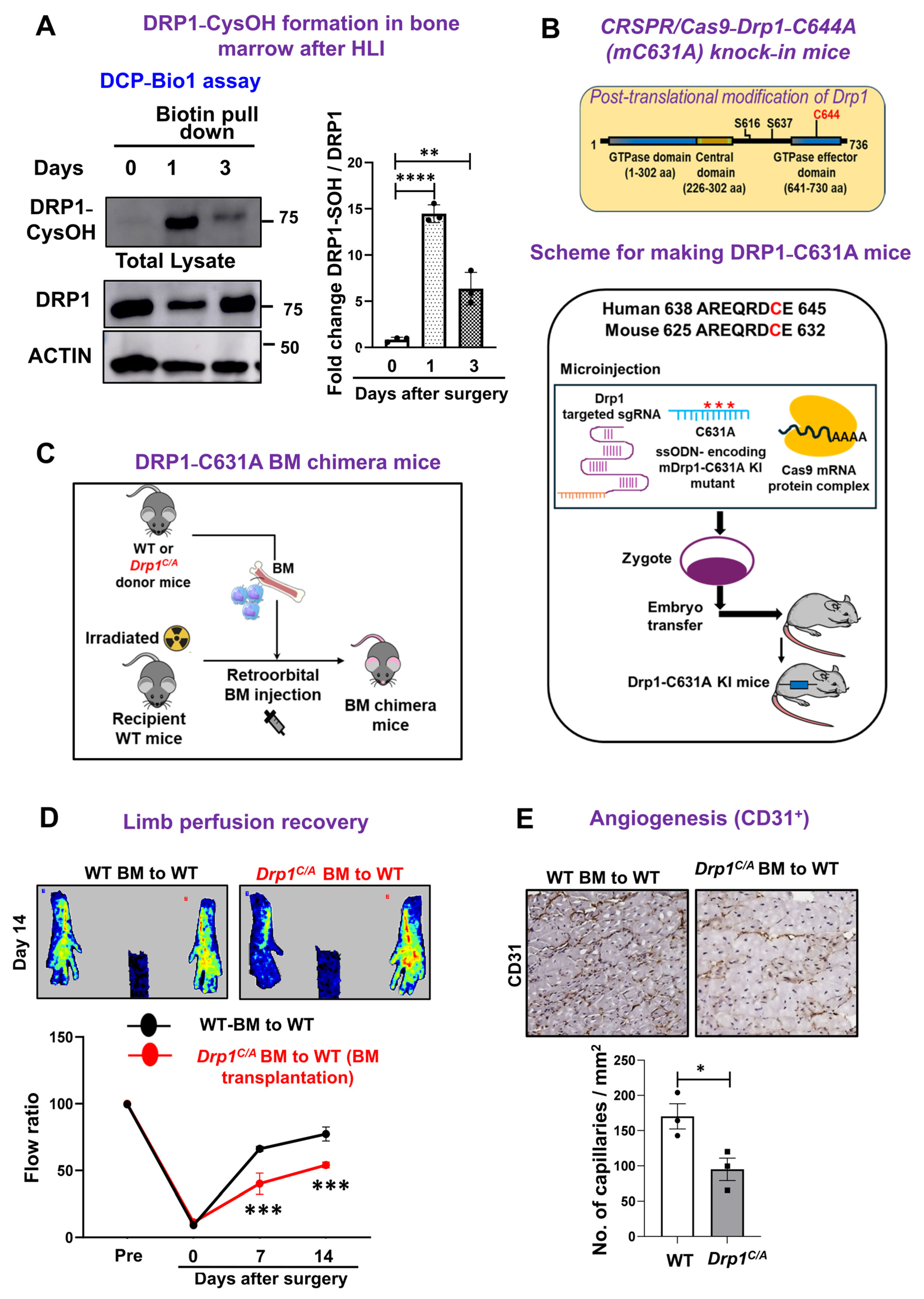
Figure 2.
Drp1 C/A bone marrow chimera mice show increased pro-inflammatory M1-MΦ polarization and decreased anti-inflammatory M2-MΦ polarization in ischemic muscles after HLI. (A) Schematic of flow cytometry-based immunophenotyping of ischemic gastrocnemius (GC) muscle after BMT and HLI surgery. (B) Representative immunofluorescence images of F4/80+ (green) macrophages and quantification in GC muscle at day 3 after HLI. Scale bar: 20 µM. n = 4 mice per group, unpaired Student’s t-test. (C,D) Representative FACS contour plot of total F4/80+ CD64+ macrophages and quantification. n = 4 mice per group, 1-way ANOVA followed by Bonferroni’s multiple comparisons test. (E,F) CD80+M1- and CD206+M2-MΦ and quantification in ischemic GC muscle at day 3 and day 7 post HLI. n = 4 mice per group, 1-way ANOVA followed by Bonferroni’s multiple comparisons test. Sex- and age-matched 12–18-week-old WT and Drp1C/A mice were used. Data are mean ± SEM (n = 4 mice). * p < 0.05, ** p < 0.01.
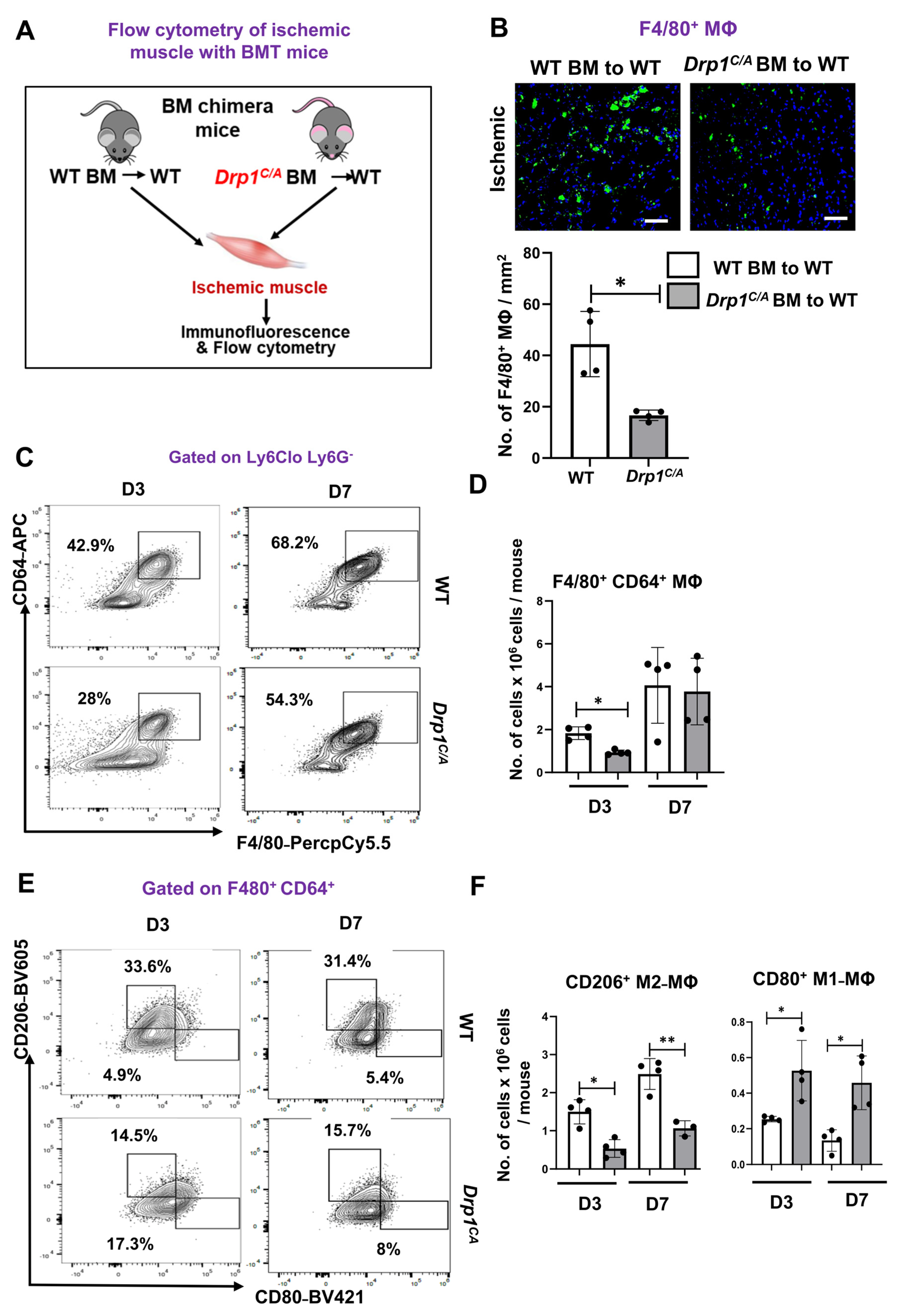
Figure 3.
Increased neutrophils in ischemic muscle and impaired efferocytosis in Drp1C/A BMDMs during in vitro ischemia. (A) Representative immunofluorescence images of Ly6G+ neutrophils (green) and quantification in GC muscle at day 3 after HLI. Scale bar: 20 µM. n = 4 mice per group, unpaired Student’s t-test. (B,C) Representative flow cytometry contour plots and quantification of numbers of Ly6Ghi neutrophils and Ly6Chi monocytes in ischemic GC muscle of WT and Drp1C/A mice after day 3 of HLI. n = 4 mice per group, 1-way ANOVA followed by Bonferroni’s multiple comparisons test. (D) Schematic representation of in vitro BMDM efferocytosis assay. (E) IF imaging and quantification of labeled apoptotic cell uptake in WT and Drp1C/A BMDMs during normoxia and 8 h of HSS stimulation (indicated by arrow head)Scale bar: 20 µM. n = 3 mice per group, 1-way ANOVA followed by Bonferroni’s multiple comparisons test. (F) qRT-PCR analysis of Mertk and Gpnb gene normalized to Hprt in WT and Drp1C/A BMDMs after 8 h of HSS and treatment with apoptotic cells for 2 h. n = 4 sets of BMDMs per group. BMDMs from sex- and age-matched 12–18-week-old WT and Drp1C/A mice were used. Data are mean ± SEM. * p < 0.05, ** p < 0.01 (Schematic hypothesis model, created in BioRender. Ushio-Fukai, M. (2026), https://BioRender.com/96r8gxt).
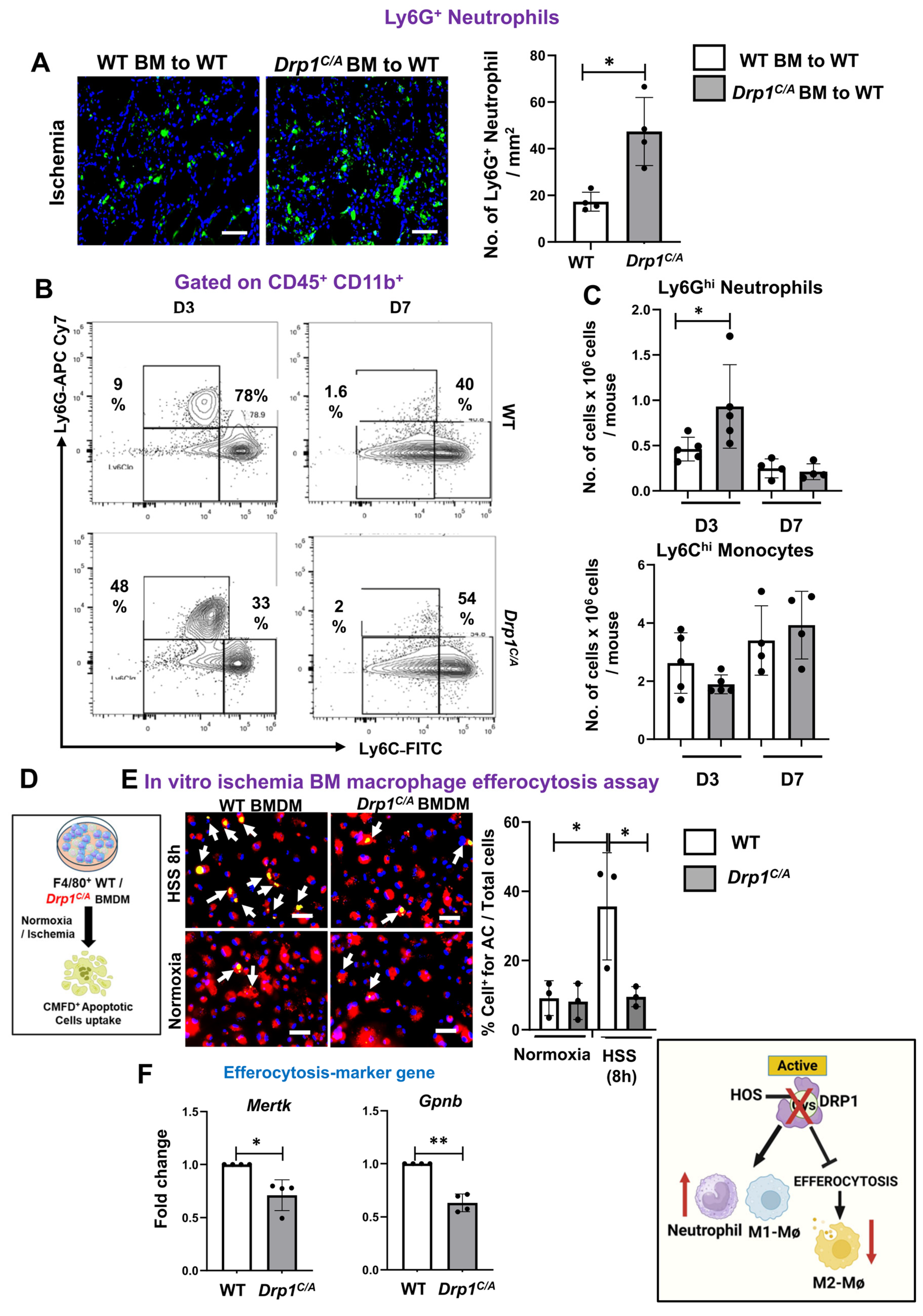
Given the critical role of efferocytosis in resolving inflammation and promoting reparative M2 macrophage polarization, we performed in vitro efferocytosis assays [28,32] using WT and Drp1C/A BMDMs under basal and HSS conditions. HSS stimulation for 8 h significantly enhanced the uptake of apoptotic cells by WT BMDMs compared with normoxic controls. In contrast, Drp1C/A BMDMs exhibited significantly impaired efferocytosis following HSS stimulation compared with WT BMDMs, whereas no difference was observed under normoxic conditions (Figure 3D,E). Consistent with these findings, the HSS-induced expression of efferocytosis-associated genes (Mertk and Gpnb) was markedly reduced in Drp1C/A BMDMs relative to WT BMDMs (Figure 3F). Together, these findings demonstrate that myeloid DRP1 sulfenylation is not required for monocyte recruitment but is essential for macrophage reprogramming toward a reparative, pro-angiogenic M2-like phenotype in ischemic muscle. Loss of DRP1-CysOH shifts macrophages toward a pro-inflammatory M1-like state, accompanied by impaired efferocytosis and increased neutrophil accumulation. These alterations likely contribute to unresolved inflammation and establish a mechanistic link between defective macrophage reprogramming and impaired ischemia-induced neovascularization following HLI.
3.3. Inhibition of Ischemia-Induced DRP1 Sulfenylation Promotes Mitochondrial Hyperfusion, Reduced Membrane Potential, and Excessive mitoROS Production in Macrophages in Vitro
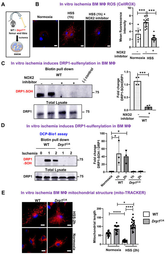
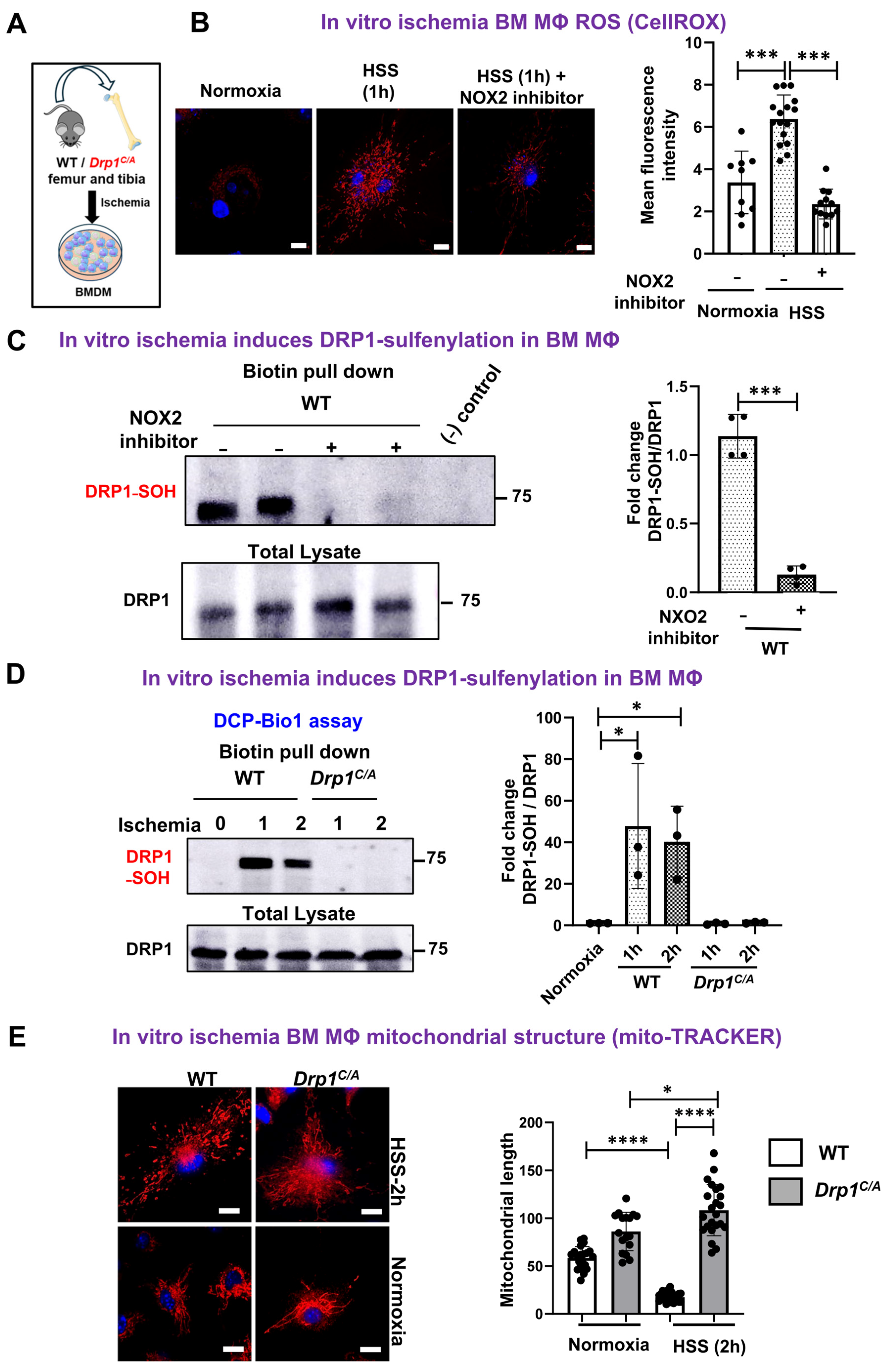
Since NOX2-derived ROS in myeloid cells play a critical role in post-ischemic reparative neovascularization [11], we investigated whether NOX2 mediates ischemia-induced DRP1 sulfenylation in macrophages using BMDMs exposed to hypoxia serum starvation (HSS), an established in vitro model of PAD [24] (Figure 4A). HSS stimulation for 1 h significantly increased ROS production in WT BMDMs, as measured by the CellROX fluorescent probe, and this response was abolished by pre-treatment with the selective NOX2 inhibitor GSK2795039 (Figure 4B). Similar results were obtained using the DCF-DA probe, where HSS-induced ROS production was suppressed by PEG-catalase (Figure S1A,B). DCP-Bio1 assay revealed that DRP1 sulfenylation (Cys-OH) was robustly induced in WT BMDMs within 1–2 h of HSS stimulation. This response was completely abolished by NOX2 inhibitor (Figure 4C) and was absent in Drp1C/A BMDMs (Figure 4D), indicating that NOX2-derived ROS are required for ischemia-induced DRP1 sulfenylation in macrophages. Previous study shows that HSS induced a transient increase in mitochondrial fission in BMDMs, peaking at 1–2 h and returning to baseline by 8 h, without changes in canonical DRP1 phosphorylation at Ser616 or Ser637, total DRP1 expression, or levels of other mitochondrial fission and fusion proteins (MFF, MFN1/2, OPA1) [24]. In contrast, Drp1C/A BMDMs displayed elongated, hyperfused mitochondria at 2 h under HSS (Figure 4E), closely resembling the phenotype observed in DRP11-deficient BMDMs [24].
Figure 4.
Preventing DRP1-CysOH formation in Drp1C/A KI BMDMs during ischemia-induced mito-fusion. (A) Schematic of in vitro BMDM hypoxia serum starvation (HSS) model. (B) IF imaging and quantification of CellROX-stained (red) WT BMDMs with or without 10 µM NOX2 inhibitor. Scale bar: 5 µM. Each dot represents number of cells quantified from n = 3 sets of BMDMs, 1-way ANOVA followed by Bonferroni’s multiple comparisons test. Immunoblotting and quantification for DRP1 sulfenylation using DCP-Bio1. (C) WT BMDMs with or without NOX2 inhibitor after 1 h of HSS stimulation. (D) WT and Drp1C/A BMDMs at indicated time post hypoxia serum starvation (HSS). DCP-Bio1 pulldown experiments were performed with a “beads-only” negative control consisting of streptavidin beads incubated with lysis buffer containing DCP-Bio1 in the absence of cell lysates. n = 3–4 sets of BMDMs, 1-way ANOVA followed by Bonferroni’s multiple comparisons test. (E) IF imaging of mitoTracker (red) staining and its quantification. Scale bar: 5 µM. Each dot represents number of cells quantified from n = 3 sets of BMDMs, 1-way ANOVA followed by Bonferroni’s multiple comparisons test. BMDMs from sex- and age-matched 12–18-week-old WT and Drp1C/A mice were used. Data are mean ± SEM. * p < 0.05, *** p < 0.001, **** p < 0.0001.
We next assessed whether loss of DRP1-CysOH affects mitochondrial function by measuring oxygen consumption rate (OCR) using Seahorse analysis. Under HSS conditions, Drp1C/A BMDMs exhibited no significant differences in basal respiration, maximal respiration, or ATP production compared with WT BMDMs (Figure S2A). Consistent with these findings, no significant difference in expression of immune-responsive gene 1 (Irg1), which encodes the enzyme responsible for itaconate production from the TCA cycle intermediate cis-aconitate [33], was observed between WT and Drp1C/A BMDM after 8 h of HSS stimulation (Figure S2B). Despite preserved mitochondrial respiratory capacity, Drp1C/A BMDMs displayed elevated mitochondrial ROS (mitoROS) production after 2 h of HSS stimulation, as measured by MitoSOX fluorescence (Figure 5A,B). This increase in mitoROS was accompanied by a significant reduction in mitochondrial membrane potential, assessed by TMRM fluorescence, compared with WT BMDMs (Figure 5C,D). Collectively, these findings indicate that ischemia-induced DRP1 sulfenylation is dispensable for maintaining mitochondrial respiration but is required for preserving mitochondrial integrity under ischemic stress. Loss of DRP1-CysOH results in mitochondrial hyperfusion, membrane depolarization, and excessive mitoROS generation, providing a mechanistic link between redox-dependent regulation of DRP1, mitochondrial dynamics, and macrophage dysfunction in PAD.
3.4. Inhibition of Ischemia-Induced DRP1 Sulfenylation Suppresses AMPK Activation, Enhances Glycolysis and Skews Macrophage Polarization to Pro-Inflammatory Phenotype
To directly determine the role of DRP1-CysOH in macrophage metabolic reprogramming and polarization under ischemic conditions, BMDMs were subjected to HSS in vitro. Because ischemia-driven pro-inflammatory macrophage polarization is associated with a metabolic shift from oxidative phosphorylation to aerobic glycolysis [34,35], glycolytic activity was assessed by measuring extracellular acidification rate (ECAR) using a Seahorse assay. As shown in Figure 6A, Drp1C/A BMDMs exhibited significantly increased basal glycolysis and glycolytic capacity compared with WT BMDMs under HSS. We found that there were no significant differences in the protein expression of key glycolytic enzymes including PFKFB3, GAPDH, and PKM2 between WT and Drp1C/A BMDMs following 2 h of HSS stimulation (Figure S3A). To elucidate the mechanism underlying enhanced glycolysis in Drp1C/A KI macrophages, we next examined AMPK signaling, which promotes anti-inflammatory, pro-angiogenic M2 polarization in part by suppressing glycolysis, as previously reported in Drp1−/−BMDMs [24]. Consistent with this pathway, Drp1C/A BMDMs displayed markedly reduced p-AMPK and a modest increase in NF-κB phosphorylation compared with WT controls under HSS (Figure 6B). Since we previously reported that DRP1 deficiency in BMDMs with ischemia reduces p-AMPK in a mitoROS-independent manner [24], we next examined if elevated mitoROS contributes to inflammatory signaling such as p-NFkB in Drp1C/A BMDMs under ischemia. We found that mitochondrial-targeted ROS scavenger mitoTEMPO significantly reduced p-NF-κB/NF-κB expression in Drp1C/A BMDMs with HSS stimulation (Figure S3B). These results suggest that mitoROS acts upstream of NF-κB activation in Drp1C/A BMDMs under ischemic stress conditions.
Figure 5.
Drp1C/A BMDMs exhibited excess mitoROS production and reduced mito-membrane potential. (A,B) mitoSOX staining of mitoROS (red), each dot represents number of cell quantified from n = 3 sets of BMDMs. Scale bar: 5 µM. 1-way ANOVA followed by Bonferroni’s multiple comparisons test. (C,D) TMRM (red) and mtTRACKER (green) staining and qunatification of mitochondrial membrane potentialin WT and Drp1C/A BMDMs post 2 h of HSS stimulation. Scale bar: 5 µM. n = 4 sets of BMDMs, 1-way ANOVA followed by Bonferroni’s multiple comparisons test. Data are mean ± SEM. * p < 0.05, ** p < 0.01, **** p < 0.0001.
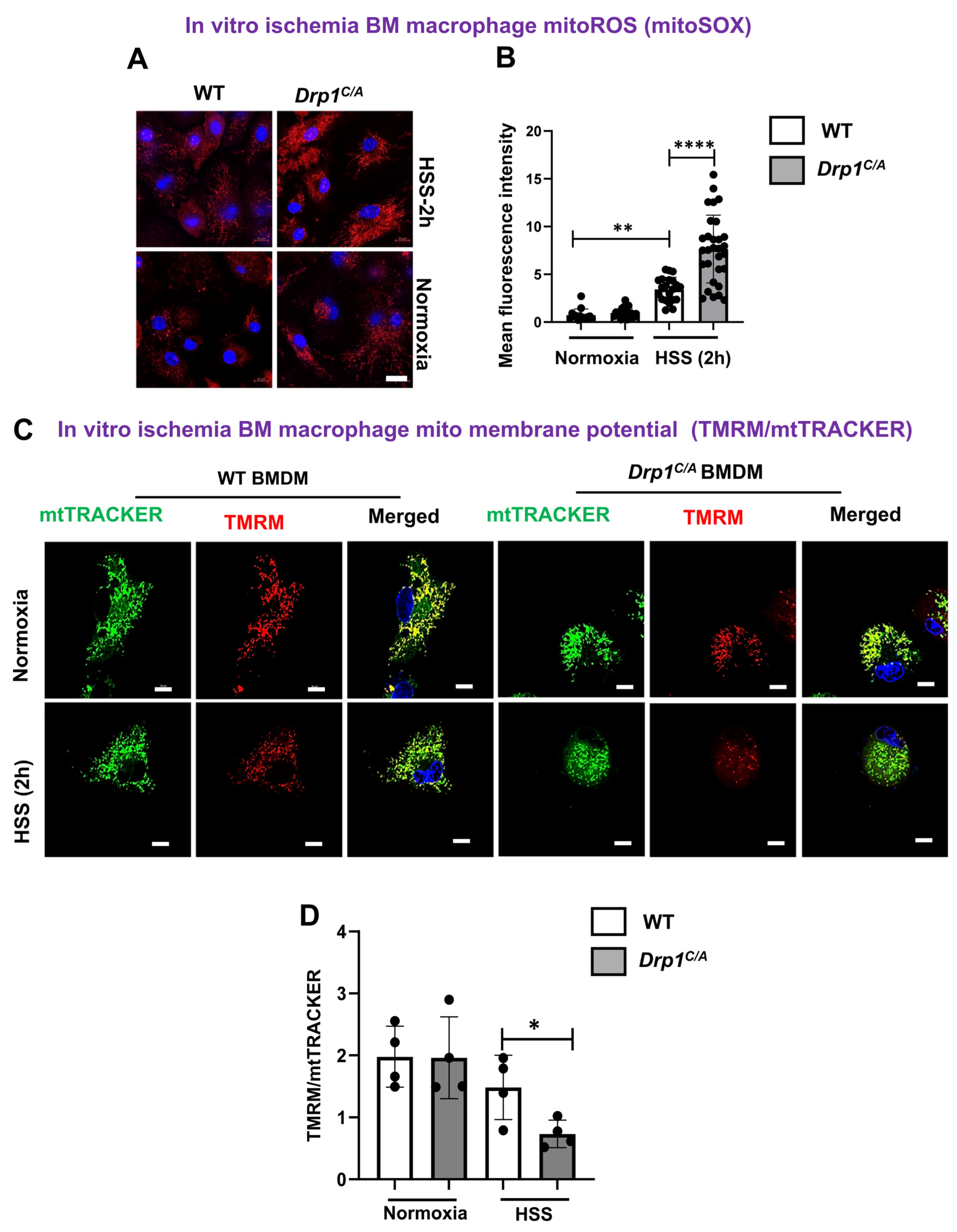
Figure 6.
Drp1 C/A BMDMs suppressed ischemia-induced p-AMPK, increased glycolysis and pro-inflammatory M1-genes, and decreased anti-inflammatory M2-genes after HSS. WT (red) and Drp1C/A (blue) BMDMs after 2 h of HSS were used to measure (A) glycolysis using Seahorse analyzer, (B) p-AMPK, p-NFkB, total AMPK and NFkB using immunoblotting. n = 3 sets of BMDMs per group, unpaired Student’s t-test. (C) M1-and M2-marker genes normalized to Hprt after 8 h of HSS (D) with or without 100 µM AICAR pre-treatment by qRT-PCR analysis. n = 4 sets of BMDMs per group, 1-way ANOVA followed by Bonferroni’s multiple comparisons test. BMDMs from sex- and age-matched 12–18-week-old WT and Drp1C/A mice were used. Data are mean ± SEM. * p < 0.05, ** p < 0.01, *** p < 0.001. (Schematic hypothesis model, created in BioRender. Ushio-Fukai, M. (2026), https://BioRender.com/4zlx47i).
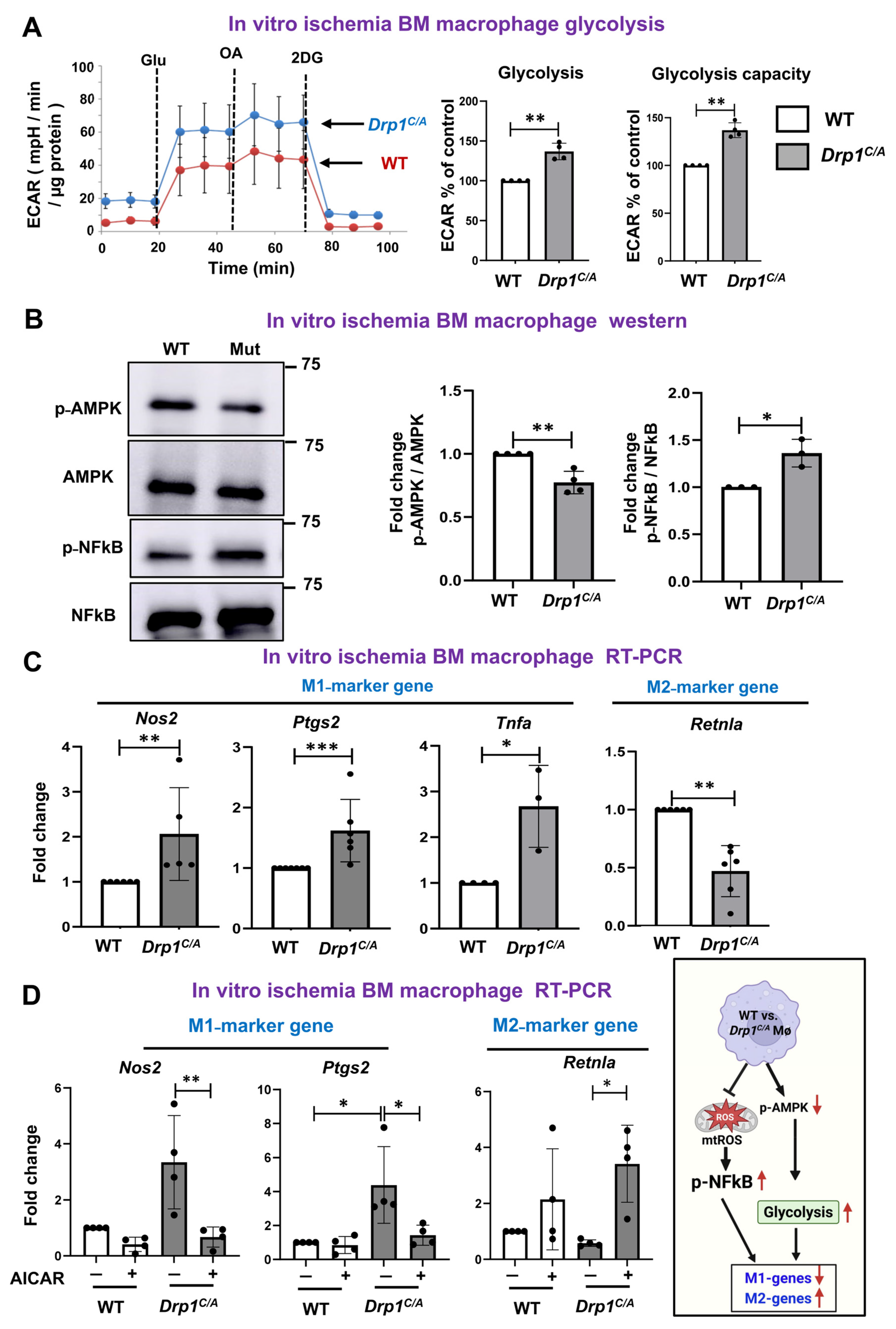
In coherence with our in vivo hindlimb ischemia findings, Drp1C/A BMDMs exposed to HSS showed significantly elevated expression of M1-associated genes (Nos2, Ptgs2, and Tnfα) and reduced expression of the M2-marker gene Retnla relative to WT BMDMs (Figure 6C). To determine whether decreased AMPK activity contributes to enhanced glycolysis and pro-inflammatory macrophage polarization in Drp1C/A BMDMs under HSS as we observed in Drp1−/−BMDMs with HSS [24], we performed rescue experiments using the AMPK activator AICAR [36,37]. Activating AMPK by AICAR at a concentration that did not affect glycolysis in WT BMDMs under HSS [24] rescued the elevated expression of pro-inflammatory M1-genes Nos2 and Ptgs2 in Drp1C/A BMDMs under HSS as comparable to those observed in HSS-exposed WT BMDMs (Figure 6D). Notably, AICAR also rescued the reduced expression of genes associated with the anti-inflammatory M2-marker gene in Drp1C/A BMDM under HSS (Figure 6D). These findings indicate that loss of DRP1-CysOH enhances glycolytic reprogramming and NF-κB activation while attenuating AMPK signaling, thereby skewing macrophages toward a pro-inflammatory M1-like phenotype and impairing reparative M2-like polarization under ischemic conditions in vitro.
4. Discussion
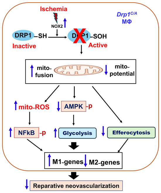
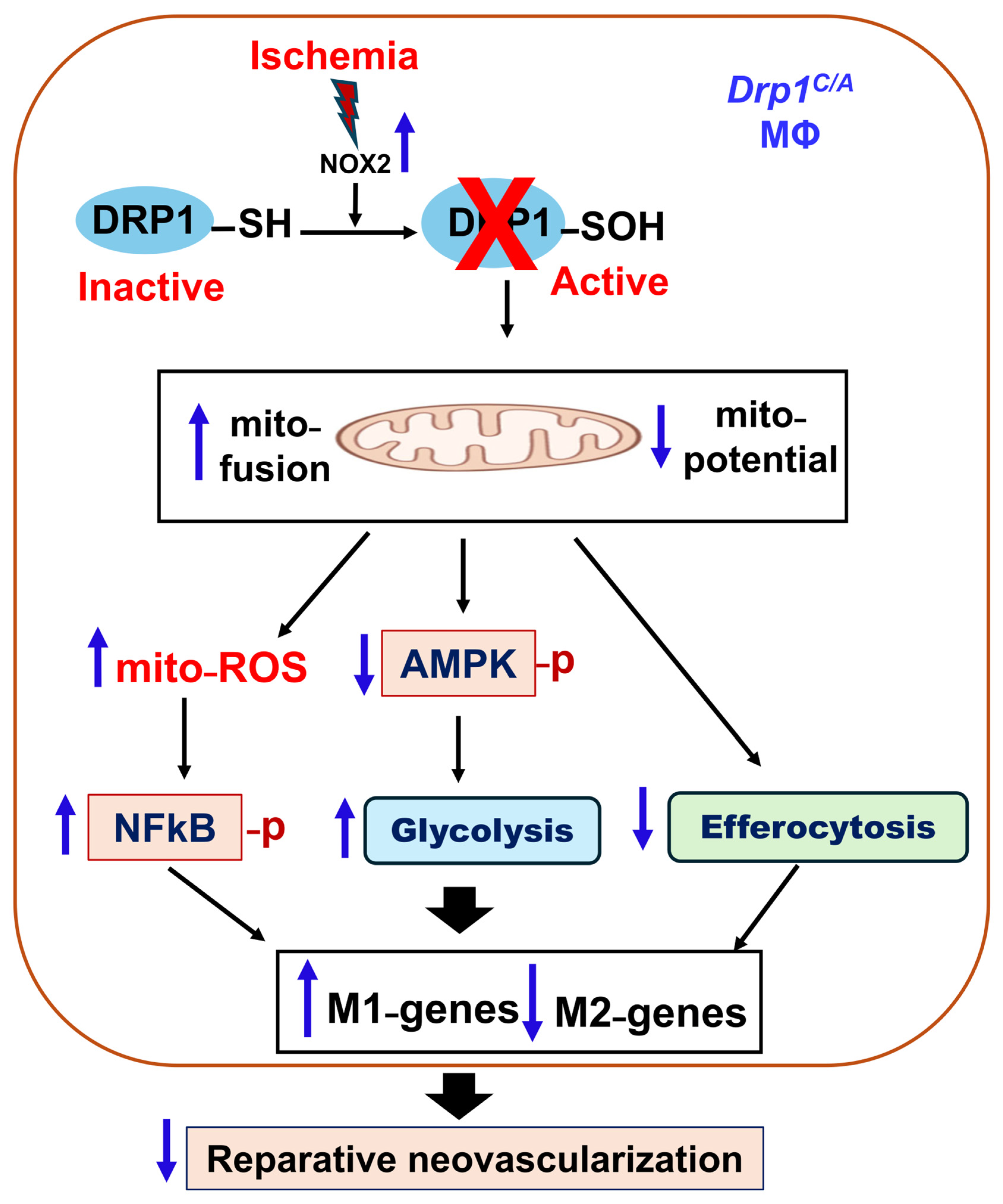
ROS generated by BM cells in addition to endothelial cells are key signaling mediators required for ischemia-induced neovascularization in PAD [11,31,38,39]. We previously showed that ischemia activates the mitochondrial fission protein DRP1 in macrophages independently of canonical Ser616/Ser637 phosphorylation; however, the upstream regulatory mechanism remained unclear. Here, we identify ischemia-induced cysteine sulfenylation of DRP1 as a critical redox-dependent modification in BM cells, especially macrophages that are required for effective perfusion recovery and angiogenesis after HLI. BM chimeric mice expressing a “redox-dead” Drp1C/A KI mutant displayed impaired post-ischemic revascularization, accompanied by excessive neutrophil accumulation, persistent pro-inflammatory M1 macrophage polarization, and reduced M2 macrophage responses in ischemic muscle. Mechanistically, ischemic stress induced NOX2-ROS-dependent DRP1 sulfenylation and mitochondrial fission independently of changes in DRP1 phosphorylation. In contrast, disruption of DRP1 cysteine oxidation impaired mitochondrial dynamics, resulting in metabolic and inflammatory reprogramming characterized by enhanced glycolysis, increased mitoROS–NF-κB signaling, reduced AMPK activation and efferocytosis, and a shift toward a pro-inflammatory macrophage phenotype (Figure 7).
Figure 7.
Schematic of proposed hypothesis model. Myeloid DRP1 sulfenylation at Cys644 is required for reparative neovascularization after in vivo ischemic injury. Preventing MΦ DRP1-Cys oxidation under in vitro ischemia increases glycolysis via reduced p-AMPK and efferocytosis as well as excess mitoROS-NFkB axis, driving pro-inflammatory M1-like MΦ polarization, which limits revascularization in experimental PAD. Upward arrow indicates increase and downward arrow indicates decrease.
DRP1 is a central mediator of mitochondrial fission, and its activity is tightly controlled by multiple post-translational modifications (PTMs), including phosphorylation, SUMOylation, ubiquitination, S-nitrosylation, and O-GlcNAcylation, which collectively fine-tune mitochondrial and cellular dynamics [40]. We previously reported that ischemia activates DRP1 in macrophages without phosphorylation, which is required for reparative neovascularization [24]. In the present study, we demonstrate for the first time that DRP1-CysOH formation is dramatically increased in BM cells following HLI. Importantly, BM chimera mice harboring the redox-dead DRP1-C631A KI mutation exhibit impaired limb perfusion recovery and angiogenesis after HLI, establishing a functional requirement of DRP1 sulfenylation in BM cells for driving ischemic revascularization. Furthermore, in vitro ischemia increased mitochondrial fission, H2O2 levels and DRP1-CysOH formation in BMDMs, independently of phosphorylation at Ser616 or Ser637 [24], confirming that macrophage DRP1 is activated primarily through Cys631 sulfenylation rather than canonical phosphorylation pathways.
Our previous studies identified NOX2 as a major source of ROS in BM-derived cells that promotes reparative neovascularization in ischemic muscle [11,12]. Consistent with reports showing that NOX2-derived ROS facilitate monocyte-to-M2-like macrophage polarization and inflammation resolution [41,42], pharmacological inhibition of NOX2 markedly attenuated both HSS-induced ROS production and DRP1-CysOH formation in WT BMDMs. These findings suggest that NOX2-derived ROS act upstream of DRP1 cysteine oxidation in ischemic macrophages. Although potential off-target effects of the inhibitor cannot be excluded, future studies using BMDMs isolated from Nox2 deficient mice will further validate this mechanism. Collectively, our data supports a model in which NOX2-derived ROS generated during ischemia are sensed by DRP1 through Cys631 sulfenylation, thereby promoting reparative macrophage reprogramming and post-ischemic neovascularization.
Ischemia-induced revascularization requires coordinated pro-angiogenic and metabolic reprogramming of macrophages, in which the balance between oxidative metabolism and glycolysis critically influences inflammatory status and reparative function [16]. Our findings identify myeloid DRP1 sulfenylation as a previously unrecognized redox-dependent mechanism that promotes reparative macrophage reprogramming and post-ischemic neovascularization through regulation of mitochondrial dynamics. Consistent with our previous findings in MΦDrp1−/− mice [24], Drp1C/A KI BM chimera mice exhibited increased neutrophil accumulation and reduced macrophage infiltration at day 3 after HLI, despite normal monocyte recruitment. These changes were associated with a sustained increase in pro-inflammatory CD80+ M1-like macrophages and a reduction in reparative CD206+ M2-like macrophages at both days 3 and 7 post-HLI. Although macrophage numbers recovered by day 7, the polarization defect persisted, suggesting that DRP1 sulfenylation primarily regulates macrophage functional programming rather than recruitment. Notably, the early accumulation of neutrophils in Drp1C/A KI BM chimera mice also suggests a role for DRP1 sulfenylation in promoting efferocytotic clearance of apoptotic neutrophils, leading to M2-like polarization and inflammation resolution in ischemic tissues [43,44,45]. Supporting this possibility, Drp1C/A BMDM exhibited impaired efferocytosis following in vitro ischemic stimulation. While the underlying mechanisms remain to be fully defined, previous studies have shown that DRP1-mediated mitochondrial fission is required for hypoxia-induced efferocytosis and tissue repair [45,46], and that mitochondrial signaling through AMPK axis enhances macrophage uptake of apoptotic cells and the resolution of inflammation [47]. Together, these findings suggest that impaired efferocytosis may contribute, at least in part, to the excessive neutrophil accumulation and defective reparative macrophage polarization in Drp1C/A macrophages under ischemic conditions.
Mechanistically, inhibition of DRP1 sulfenylation under ischemia impaired DRP1-dependent mitochondrial fission, resulting in mitochondrial hyperfusion and dysregulated mitochondrial homeostasis. This was associated with reduced AMPK activation, enhanced glycolysis, increased mitochondrial dysfunction and mitoROS production, augmented NF-κB signaling, and impaired efferocytic capacity, ultimately driving M1-like polarization and suppressing reparative M2-like polarization. These alterations likely contribute to the impaired neovascularization in experimental PAD. Our findings are consistent with our previous studies using myeloid-specific Drp1−/− mice and macrophages, which demonstrated that loss of DRP1 promotes glycolytic reprogramming through suppression of p-AMPK as well as inducing mitochondrial dysfunction and excessive mitoROS generation, leading to enhanced pro-inflammatory M1-genes and reduced anti-inflammatory M2-genes [24]. In the present study, pharmacological activation of AMPK with AICAR partially rescued the exaggerated pro-inflammatory phenotype and restored anti-inflammatory marker expression in ischemic Drp1C/A macrophages. These results support a key role for impaired AMPK signaling in mediating the effects of defective DRP1 sulfenylation. In parallel, scavenging mitoROS with mito-TEMPO attenuated p-NF-κB in Drp1C/A BMDMs, indicating that excessive mitoROS contributes to inflammatory NF-κB signaling downstream of impaired DRP1 redox regulation. However, further studies examining the contribution of NF-κB signaling, including the use of specific NF-κB inhibitors to determine whether M1 polarization can be reversed, would provide additional mechanistic insight. Notably, TMRM analysis revealed a significant reduction in mitochondrial membrane potential in Drp1C/A BMDMs despite preserved oxygen consumption, suggesting that defective DRP1 sulfenylation compromises mitochondrial quality and bioenergetic integrity before overt respiratory failure. This in turn may promote electron leakage and mitoROS production, thereby activating NF-κB-dependent inflammatory pathways. Thus, these findings support a model in which DRP1 sulfenylation functions as a redox-sensitive regulator of mitochondrial dynamics that preserves mitochondrial fitness, sustains AMPK signaling, and restrains mitoROS-NF-κB activation, thereby promoting reparative macrophage polarization during ischemic tissue repair (Figure 7).
These are several limitations of the present study. First, while our data using the NOX2 chemical inhibitor supports a role of NOX2-derived ROS for DRP1 sulfenylation in macrophage reprogramming during ischemia, the specific role of NOX2 in this response should be investigated using myeloid-specific NOX2-deficient mice. Second, the present study used CellROX and DCF-DA fluorescence probes to measure ROS which was inhibited by the NOX2 inhibitor or PEG-catalase. Future studies using genetic approaches and advanced H2O2 biosensors such as HyPer7 or roGFP2-Orp1 will help define the spatial and temporal regulation of this pathway. However, implementation of these approaches in primary BMDMs, which are difficult to transfect using plasmids and adenoviruses, would require substantial optimization. Third, while loss of DRP1 sulfenylation enhanced glycolysis, more comprehensive metabolic analyses, including isotope tracing and assessment of TCA cycle immunometabolites such as succinate and itaconate [33,48,49], are needed to further define the underlying metabolic mechanisms. Fourth, whether impaired DRP1 sulfenylation activates additional inflammatory pathways, such as the mitochondrial DNA-cGAS-STING axis, remains to be determined. Fifth, because BM transplantation reconstitutes multiple hematopoietic cell populations, contributions from immune cells other than macrophages cannot be excluded. However, the consistent defects observed in Drp1C/A BMDMs support a macrophage-intrinsic role for DRP1 sulfenylation. Sixth, although rescue experiments such as re-expression of WT DRP1 in Drp1C/A macrophages could further strengthen causality, technical challenges in transfection in primary macrophages and the lack of faithful sulfenylation-mimetic, cysteine-to-aspartate substitutions (e.g., C631D) mutants limited these approaches. Finally, whether DRP1 sulfenylation similarly regulates macrophage function in other disease settings, such as diabetic PAD or vascular aging, warrants future investigation. Together, these studies will further define the therapeutic potential of targeting DRP1 redox signaling to enhance tissue repair and revascularization.
5. Conclusions
In conclusion, these findings uncover DRP1 sulfenylation as a redox-sensitive molecular switch that couples ischemia-induced ROS signaling to macrophage metabolic reprogramming and reparative neovascularization. By uncoupling DRP1 activation from canonical phosphorylation and identifying sulfenylation as the dominant regulatory mode in ischemic macrophages, these findings redefine how macrophage mitochondrial dynamics are controlled during tissue repair. Targeting DRP1 redox signaling may therefore represent a novel therapeutic strategy to enhance macrophage-mediated revascularization while limiting chronic inflammation in PAD.
Supplementary Materials
The following supporting information can be downloaded at https://www.mdpi.com/article/10.3390/antiox15060768/s1. Figure S1: A. In vitro ischemia induced H2O2 in WT BMDM. DCF-DA (H2O2production) fluorescence in WT BMDMs during normoxia vs 1h of HSS stimulation with or without PEG-catalase. Figure S2: Mitochondrial respiration in Drp1C/A BMDMs with ischemia is unaffected. (A). WT and Drp1C/A BMDMs after 2 h of HSS were used to measure mitochondrial respiration rate (OCR) using Seahorse. n = 4 sets of BMDM per group. (B) RT-PCR of Irg1 gene normalized to Hprt after 8 h of HSS. n = 4 sets of BMDM per group. BMDMs from Sex- and age-matched 12-18 weeks WT and Drp1C/A mice were used. Data are mean ± SEM. Figure S3: mitoTEMPO inhibits excess NFΚB activation in Drp1C/A BMDM upon HSS. WT and Drp1C/A BMDM after 2 h of HSS (A) glycolytic enzymes PFKB3, GAPDH and PKM2 (B) p-NFkB, and NFkB with or without 50µM mitoTMEPO pre-treatment using immunoblotting. n = 4–5 sets of BMDM per group, 1-way ANOVA followed by Bonferroni’s multiple comparisons test. BMDMs from Sex- and age-matched 12–18 weeks WT and Drp1C/A mice were used. Data are mean ± SEM. * p < 0.05, ** p < 0.01, *** p < 0.001. Table S1: Antibodies used for flow cytometry based immunophenotyping. Table S2: Sequences of primers used for RT-PCR.
Author Contributions
M.U.-F., T.F. and S.Y. designed the study; S.Y. performed research; V.S., R.K., S.K.S., S.N. and A.D. assisted research; M.U.-F., T.F. and S.Y. analyzed data; R.V.K. and S.K.S. performed mouse genotyping; M.U.-F., T.F. and S.Y. wrote the manuscript. All authors have read and agreed to the published version of the manuscript.
Funding
This work was supported by NIH R01HL160014 (to M.U.-F.), R01HL174014 (to T.F., M.U-F., V.S.), R01HL147550 (to M.U.-F., T.F.), P01HL160557 (to T.F., M.U-F.), American Heart Association (AHA) 22TPA971863 (to T.F.), 23POST1022971 (to S.N.), 24CDA1273746. (to A.D.), and Veterans Administration (VA) Merit Review Award 2I01BX001232 (to T.F.).
Institutional Review Board Statement
Animal studies were carried out in compliance with the recommendations of the Guide for the Care and Use of Laboratory Animals, and the protocol was approved by the Animal Model Experimental Ethics Committee of Augusta University (IACUC Approval Protocol 2017-0852).
Informed Consent Statement
Not applicable.
Data Availability Statement
The data presented in this study are available in the article and Supplementary Materials.
Acknowledgments
We acknowledge Lin Gan at Transgenic & Genomic Editing Core at Augusta University Medical College of Georgia for generating Drp1-C631A KI mutant mice. Graphical Abstract and some figures were created in BioRender. Ushio-Fukai, M. (2026) https://BioRender.com/7jgug2k.
Conflicts of Interest
The authors declare no conflicts of interest.
References
- Song, P.; Rudan, D.; Zhu, Y.; Fowkes, F.J.I.; Rahimi, K.; Fowkes, F.G.R.; Rudan, I. Global, regional, and national prevalence and risk factors for peripheral artery disease in 2015: An updated systematic review and analysis. Lancet Glob. Health 2019, 7, e1020–e1030. [Google Scholar] [CrossRef] [Scilit] [PubMed]
- Jetten, N.; Donners, M.M.; Wagenaar, A.; Cleutjens, J.P.; van Rooijen, N.; de Winther, M.P.; Post, M.J. Local delivery of polarized macrophages improves reperfusion recovery in a mouse hind limb ischemia model. PLoS ONE 2013, 8, e68811. [Google Scholar] [CrossRef] [Scilit] [PubMed]
- Krishnasamy, K.; Limbourg, A.; Kapanadze, T.; Gamrekelashvili, J.; Beger, C.; Hager, C.; Lozanovski, V.J.; Falk, C.S.; Napp, L.C.; Bauersachs, J.; et al. Blood vessel control of macrophage maturation promotes arteriogenesis in ischemia. Nat. Commun. 2017, 8, 952. [Google Scholar] [CrossRef] [Scilit] [PubMed]
- Tan, R.P.; Ryder, I.; Yang, N.; Lam, Y.T.; Santos, M.; Michael, P.L.; Robinson, D.A.; Ng, M.K.; Wise, S.G. Macrophage Polarization as a Novel Therapeutic Target for Endovascular Intervention in Peripheral Artery Disease. JACC Basic Transl. Sci. 2021, 6, 693–704. [Google Scholar] [CrossRef] [Scilit] [PubMed]
- Watanabe, S.; Alexander, M.; Misharin, A.V.; Budinger, G.R.S. The role of macrophages in the resolution of inflammation. J. Clin. Investig. 2019, 129, 2619–2628. [Google Scholar] [CrossRef] [Scilit] [PubMed]
- Tojo, T.; Ushio-Fukai, M.; Yamaoka-Tojo, M.; Ikeda, S.; Patrushev, N.; Alexander, R.W. Role of gp91phox (Nox2)-containing NAD(P)H oxidase in angiogenesis in response to hindlimb ischemia. Circulation 2005, 111, 2347–2355. [Google Scholar] [CrossRef] [Scilit] [PubMed]
- Craige, S.M.; Chen, K.; Pei, Y.; Li, C.; Huang, X.; Chen, C.; Shibata, R.; Sato, K.; Walsh, K.; Keaney, J.F., Jr. NADPH oxidase 4 promotes endothelial angiogenesis through endothelial nitric oxide synthase activation. Circulation 2011, 124, 731–740. [Google Scholar] [CrossRef] [Scilit] [PubMed]
- Manuneedhi Cholan, P.; Cartland, S.P.; Kavurma, M.M. NADPH Oxidases, Angiogenesis, and Peripheral Artery Disease. Antioxidants 2017, 6, 56. [Google Scholar] [CrossRef] [Scilit] [PubMed]
- Prieto-Bermejo, R.; Hernandez-Hernandez, A. The Importance of NADPH Oxidases and Redox Signaling in Angiogenesis. Antioxidants 2017, 6, 32. [Google Scholar] [CrossRef] [Scilit] [PubMed]
- Ushio-Fukai, M. Redox signaling in angiogenesis: Role of NADPH oxidase. Cardiovasc. Res. 2006, 71, 226–235. [Google Scholar] [CrossRef] [Scilit] [PubMed]
- Urao, N.; Inomata, H.; Razvi, M.; Kim, H.W.; Wary, K.; McKinney, R.; Fukai, T.; Ushio-Fukai, M. Role of nox2-based NADPH oxidase in bone marrow and progenitor cell function involved in neovascularization induced by hindlimb ischemia. Circ. Res. 2008, 103, 212–220. [Google Scholar] [CrossRef] [Scilit] [PubMed]
- Fang, M.M.; Barman, P.K.; Thiruppathi, M.; Mirza, R.E.; McKinney, R.D.; Deng, J.; Christman, J.W.; Du, X.; Fukai, T.; Ennis, W.J.; et al. Oxidant Signaling Mediated by Nox2 in Neutrophils Promotes Regenerative Myelopoiesis and Tissue Recovery following Ischemic Damage. J. Immunol. 2018, 201, 2414–2426. [Google Scholar] [CrossRef] [Scilit] [PubMed]
- Poole, L.B.; Karplus, P.A.; Claiborne, A. Protein sulfenic acids in redox signaling. Annu. Rev. Pharmacol. Toxicol. 2004, 44, 325–347. [Google Scholar] [CrossRef] [Scilit] [PubMed]
- Poole, L.B.; Nelson, K.J. Discovering mechanisms of signaling-mediated cysteine oxidation. Curr. Opin. Chem. Biol. 2008, 12, 18–24. [Google Scholar] [CrossRef] [Scilit] [PubMed]
- Das, A.; Ash, D.; Fouda, A.Y.; Sudhahar, V.; Kim, Y.M.; Hou, Y.; Hudson, F.Z.; Stansfield, B.K.; Caldwell, R.B.; McMenamin, M.; et al. Cysteine oxidation of copper transporter CTR1 drives VEGFR2 signalling and angiogenesis. Nat. Cell Biol. 2022, 24, 35–50. [Google Scholar] [CrossRef] [Scilit] [PubMed]
- Wang, Y.; Li, N.; Zhang, X.; Horng, T. Mitochondrial metabolism regulates macrophage biology. J. Biol. Chem. 2021, 297, 100904. [Google Scholar] [CrossRef] [Scilit] [PubMed]
- Benmoussa, K.; Garaude, J.; Acin-Perez, R. How Mitochondrial Metabolism Contributes to Macrophage Phenotype and Functions. J. Mol. Biol. 2018, 430, 3906–3921. [Google Scholar] [CrossRef] [Scilit] [PubMed]
- Guo, Q.; Jin, Y.; Chen, X.; Ye, X.; Shen, X.; Lin, M.; Zeng, C.; Zhou, T.; Zhang, J. NF-κB in biology and targeted therapy: New insights and translational implications. Signal Transduct. Target. Ther. 2024, 9, 53. [Google Scholar] [CrossRef] [Scilit] [PubMed]
- Cui, Y.; Chen, J.; Zhang, Z.; Shi, H.; Sun, W.; Yi, Q. The role of AMPK in macrophage metabolism, function and polarisation. J. Transl. Med. 2023, 21, 892. [Google Scholar] [CrossRef] [Scilit] [PubMed]
- Umezu, R.; Koga, J.I.; Matoba, T.; Katsuki, S.; Wang, L.; Hasuzawa, N.; Nomura, M.; Tsutsui, H.; Egashira, K. Macrophage (Drp1) Dynamin-Related Protein 1 Accelerates Intimal Thickening After Vascular Injury. Arterioscler. Thromb. Vasc. Biol. 2020, 40, e214–e226. [Google Scholar] [CrossRef] [Scilit] [PubMed]
- Wang, L.; Li, X.; Hanada, Y.; Hasuzawa, N.; Moriyama, Y.; Nomura, M.; Yamamoto, K. Dynamin-related protein 1 deficiency accelerates lipopolysaccharide-induced acute liver injury and inflammation in mice. Commun. Biol. 2021, 4, 894. [Google Scholar] [CrossRef] [Scilit] [PubMed]
- Yu, W.; Zhao, J.; Liu, R.; Liu, J.; Wang, Z.; Peng, J.; Zhang, X.; Long, Z.; Hao, W.; Kong, D.; et al. Corrigendum to ‘Stat2-Drp1 mediated mitochondrial mass increase is necessary for pro-inflammatory differentiation of macrophages’[Redox Biology 37 (2020) 101761]. Redox Biol. 2021, 39, 101786. [Google Scholar] [CrossRef] [Scilit] [PubMed]
- Park, S.; Won, J.H.; Hwang, I.; Hong, S.; Lee, H.K.; Yu, J.W. Defective mitochondrial fission augments NLRP3 inflammasome activation. Sci. Rep. 2015, 5, 15489. [Google Scholar] [CrossRef] [Scilit] [PubMed]
- Yadav, S.; Ganta, V.C.; Varadarajan, S.; Ong, V.; Shi, Y.; Das, A.; Ash, D.; Nagarkoti, S.; McMenamin, M.; Kelley, S.; et al. Myeloid DRP1 deficiency limits revascularization in ischemic muscles via inflammatory macrophage polarization and metabolic reprogramming. JCI Insight 2025, 10, e177334. [Google Scholar] [CrossRef] [Scilit] [PubMed]
- Nagarkoti, S.; Kim, Y.M.; Das, A.; Ash, D.; Vitriol, E.A.; Read, T.A.; Sudhahar, V.; Hossain, M.S.; Yadav, S.; McMenamin, M.; et al. Endothelial Drp1 Couples VEGF-induced Redox Signaling with Glycolysis Through Cysteine Oxidation to Drive Angiogenesis. bioRxiv 2024. [Google Scholar] [CrossRef] [Scilit] [PubMed]
- Nagarkoti, S.; Kim, Y.M.; Das, A.; Ash, D.; Vitriol, E.A.; Read, T.A.; Sudhahar, V.; Hossain, M.S.; Yadav, S.; Kelley Spears, S.; et al. Endothelial Drp1 integrates VEGF-induced redox signaling with glycolysis through cysteine oxidation to drive angiogenesis. Nat. Commun. 2026. [Google Scholar] [CrossRef] [Scilit] [PubMed]
- Piollet, M.; Porsch, F.; Rizzo, G.; Kapser, F.; Schulz, D.J.J.; Kiss, M.G.; Schlepckow, K.; Morenas-Rodriguez, E.; Sen, M.O.; Gropper, J.; et al. TREM2 protects from atherosclerosis by limiting necrotic core formation. Nat. Cardiovasc. Res. 2024, 3, 269–282. [Google Scholar] [CrossRef] [Scilit] [PubMed]
- Takahashi, K.; Drolet, J.; Liu, J.; Jackson, J.R.; Babcock, M.; Thiruppathi, M.; Fang, M.; Paul, T.; Ganesh, G.; Fukasawa, Y.; et al. Obesity Disrupts H3K4me3-Mediated Lactate Accumulation and Efferocytosis in Hypoxic Macrophages. FASEB J. 2026, 40, e71591. [Google Scholar] [CrossRef] [Scilit] [PubMed]
- Hirano, K.; Chen, W.S.; Chueng, A.L.; Dunne, A.A.; Seredenina, T.; Filippova, A.; Ramachandran, S.; Bridges, A.; Chaudry, L.; Pettman, G.; et al. Discovery of GSK2795039, a Novel Small Molecule NADPH Oxidase 2 Inhibitor. Antioxid. Redox Signal. 2015, 23, 358–374. [Google Scholar] [CrossRef] [Scilit] [PubMed]
- Kaplan, N.; Urao, N.; Furuta, E.; Kim, S.J.; Razvi, M.; Nakamura, Y.; McKinney, R.D.; Poole, L.B.; Fukai, T.; Ushio-Fukai, M. Localized cysteine sulfenic acid formation by vascular endothelial growth factor: Role in endothelial cell migration and angiogenesis. Free Radic. Res. 2011, 45, 1124–1135. [Google Scholar] [CrossRef] [Scilit] [PubMed]
- Urao, N.; McKinney, R.D.; Fukai, T.; Ushio-Fukai, M. NADPH oxidase 2 regulates bone marrow microenvironment following hindlimb ischemia: Role in reparative mobilization of progenitor cells. Stem Cells 2012, 30, 923–934. [Google Scholar] [CrossRef] [Scilit] [PubMed]
- Kumar Jha, P.; Aikawa, M.; Aikawa, E. Macrophage Heterogeneity and Efferocytosis: Beyond the M1/M2 Dichotomy. Circ. Res. 2024, 134, 186–188. [Google Scholar] [CrossRef] [Scilit] [PubMed]
- Michelucci, A.; Cordes, T.; Ghelfi, J.; Pailot, A.; Reiling, N.; Goldmann, O.; Binz, T.; Wegner, A.; Tallam, A.; Rausell, A.; et al. Immune-responsive gene 1 protein links metabolism to immunity by catalyzing itaconic acid production. Proc. Natl. Acad. Sci. USA 2013, 110, 7820–7825. [Google Scholar] [CrossRef] [Scilit] [PubMed]
- Koo, S.J.; Garg, N.J. Metabolic programming of macrophage functions and pathogens control. Redox Biol. 2019, 24, 101198. [Google Scholar] [CrossRef] [Scilit] [PubMed]
- Soto-Heredero, G.; Gomez de Las Heras, M.M.; Gabande-Rodriguez, E.; Oller, J.; Mittelbrunn, M. Glycolysis—A key player in the inflammatory response. FEBS J. 2020, 287, 3350–3369. [Google Scholar] [CrossRef] [Scilit] [PubMed]
- Giri, S.; Nath, N.; Smith, B.; Viollet, B.; Singh, A.K.; Singh, I. 5-aminoimidazole-4-carboxamide-1-β-4-ribofuranoside inhibits proinflammatory response in glial cells: A possible role of AMP-activated protein kinase. J. Neurosci. 2004, 24, 479–487. [Google Scholar] [CrossRef] [Scilit] [PubMed]
- Mounier, R.; Théret, M.; Arnold, L.; Cuvellier, S.; Bultot, L.; Göransson, O.; Sanz, N.; Ferry, A.; Sakamoto, K.; Foretz, M.; et al. AMPKα1 regulates macrophage skewing at the time of resolution of inflammation during skeletal muscle regeneration. Cell Metab. 2013, 18, 251–264. [Google Scholar] [CrossRef] [Scilit] [PubMed]
- Urao, N.; Sudhahar, V.; Kim, S.J.; Chen, G.F.; McKinney, R.D.; Kojda, G.; Fukai, T.; Ushio-Fukai, M. Critical role of endothelial hydrogen peroxide in post-ischemic neovascularization. PLoS ONE 2013, 8, e57618. [Google Scholar] [CrossRef] [Scilit] [PubMed]
- Urao, N.; Razvi, M.; Oshikawa, J.; McKinney, R.D.; Chavda, R.; Bahou, W.F.; Fukai, T.; Ushio-Fukai, M. IQGAP1 is involved in post-ischemic neovascularization by regulating angiogenesis and macrophage infiltration. PLoS ONE 2010, 5, e13440. [Google Scholar] [CrossRef] [Scilit] [PubMed]
- Jin, J.Y.; Wei, X.X.; Zhi, X.L.; Wang, X.H.; Meng, D. Drp1-dependent mitochondrial fission in cardiovascular disease. Acta Pharmacol. Sin. 2021, 42, 655–664. [Google Scholar] [CrossRef] [Scilit] [PubMed]
- Xu, Q.; Choksi, S.; Qu, J.; Jang, J.; Choe, M.; Banfi, B.; Engelhardt, J.F.; Liu, Z.G. NADPH Oxidases Are Essential for Macrophage Differentiation. J. Biol. Chem. 2016, 291, 20030–20041. [Google Scholar] [CrossRef] [Scilit] [PubMed]
- Bode, K.; Hauri-Hohl, M.; Jaquet, V.; Weyd, H. Unlocking the power of NOX2: A comprehensive review on its role in immune regulation. Redox Biol. 2023, 64, 102795. [Google Scholar] [CrossRef] [Scilit] [PubMed]
- Soehnlein, O.; Zernecke, A.; Eriksson, E.E.; Rothfuchs, A.G.; Pham, C.T.; Herwald, H.; Bidzhekov, K.; Rottenberg, M.E.; Weber, C.; Lindbom, L. Neutrophil secretion products pave the way for inflammatory monocytes. Blood 2008, 112, 1461–1471. [Google Scholar] [CrossRef] [Scilit] [PubMed]
- Greenlee-Wacker, M.C. Clearance of apoptotic neutrophils and resolution of inflammation. Immunol. Rev. 2016, 273, 357–370. [Google Scholar] [CrossRef] [Scilit] [PubMed]
- Wang, Y.; Subramanian, M.; Yurdagul, A., Jr.; Barbosa-Lorenzi, V.C.; Cai, B.; de Juan-Sanz, J.; Ryan, T.A.; Nomura, M.; Maxfield, F.R.; Tabas, I. Mitochondrial Fission Promotes the Continued Clearance of Apoptotic Cells by Macrophages. Cell 2017, 171, 331–345 e322. [Google Scholar] [CrossRef] [Scilit] [PubMed]
- Gerlach, B.D.; Ampomah, P.B.; Yurdagul, A., Jr.; Liu, C.; Lauring, M.C.; Wang, X.; Kasikara, C.; Kong, N.; Shi, J.; Tao, W.; et al. Efferocytosis induces macrophage proliferation to help resolve tissue injury. Cell Metab. 2021, 33, 2445–2463.e8. [Google Scholar] [CrossRef] [Scilit] [PubMed]
- Jiang, S.; Park, D.W.; Stigler, W.S.; Creighton, J.; Ravi, S.; Darley-Usmar, V.; Zmijewski, J.W. Mitochondria and AMP-activated protein kinase-dependent mechanism of efferocytosis. J. Biol. Chem. 2013, 288, 26013–26026. [Google Scholar] [CrossRef] [Scilit] [PubMed]
- Peace, C.G.; O’Neill, L.A. The role of itaconate in host defense and inflammation. J. Clin. Investig. 2022, 132, e148548. [Google Scholar] [CrossRef] [Scilit] [PubMed]
- Wu, G.; Fan, X.; Cheng, L.; Chen, Z.; Yi, Y.; Liang, J.; Huang, X.; Yang, N.; Yin, J.; Guo, W.; et al. Metabolism-driven posttranslational modifications and immune regulation: Emerging targets for immunotherapy. Sci. Adv. 2025, 11, eadx6489. [Google Scholar] [CrossRef] [Scilit] [PubMed]
Disclaimer/Publisher’s Note: The statements, opinions and data contained in all publications are solely those of the individual author(s) and contributor(s) and not of MDPI and/or the editor(s). MDPI and/or the editor(s) disclaim responsibility for any injury to people or property resulting from any ideas, methods, instructions or products referred to in the content. |
© 2026 by the authors. Licensee MDPI, Basel, Switzerland. This article is an open access article distributed under the terms and conditions of the Creative Commons Attribution (CC BY) license.